Unusual Presentation of Congenital Stenosis of Sigmoid Colon: … · transverse colon. Patients...
2
Remedy Publications LLC. Journal of Surgical Techniques and Procedures 2017 | Volume 1 | Issue 1 | Article 1002 1 Unusual Presentation of Congenital Stenosis of Sigmoid Colon: Case Report OPEN ACCESS *Correspondence: Basant Kumar, Department of Pediatric Surgery, Sanjay Gandhi Post Graduate Institute of Medical Sciences, Lucknow, India, Tel: +91-8004904839; E-mail: [email protected] Received Date: 14 Mar 2017 Accepted Date: 19 Jun 2017 Published Date: 29 Jun 2017 Citation: Bharti LK, Kumar B, Upadhyaya VD, Rao RN, Kumar S. Unusual Presentation of Congenital Stenosis of Sigmoid Colon: Case Report. J Surg Tech Proced. 2017; 1(1): 1002. Copyright © 2017 Basant Kumar. This is an open access article distributed under the Creative Commons Attribution License, which permits unrestricted use, distribution, and reproduction in any medium, provided the original work is properly cited. Case Report Published: 29 Jun, 2017 Abstr act Colonic stenosis/atresias account only 5% to 15% of all atresias and mostly occur in ascending and transverse colon. Patients usually present in neonatal period with symptoms and signs of distal bowel obstruction. We present a 5-month-old male child with congenital stenosis of sigmoid colon having recurrent malena and abdominal distension, borborygmi, anemia and failure to thrive. Pediatric Gastroenterologist diagnosed the condition and tried to treat it by endoscopic balloon dilatation. e child was referred to us with colonic perforation. is case highlights the superior role of surgery over endoscopic dilatation in congenital colonic stenosis. Keywords: Congenital colonic stenosis; Malena; Anemia; Perforation Introduction Colon is the least common site of congenital bowel stenosis/atresias and accounts only 5% to 15% [1,2]. Reported incidence is approximately 1 of 40,000 of live birth [3]. Colonic stenosis is rarer than atresia and mostly occurs in ascending and transverse colon [4]. Sigmoid colonic stenosis is extremely rare. e length of the stenotic segment may range from 3 cm to 15 cm [5]. Majority of the patients usually present in neonatal period with features of large bowel obstruction. Late presentation of colonic stenosis is reported in literature but the usual mode of presentation is bowel obstruction. We managed a 5-month-old male child having congenital stenosis of sigmoid colon with perforation peritonitis aſter unsuccessful endoscopic dilatation. Initially, child was presented to a Pediatric Gastroenterologist with complaints of recurrent malena, abdominal distension, anemia, failure to thrive and borborygmi. Case Presentation A 5-month-old male child, weighing 3.2 kg was referred with perforation peritonitis aſter an attempt to endoscopic dilatation of colonic stricture. Child was under the treatment of a Pediatric Gastroenterologist for last 2 months. Child presented with complaint of recurrent abdominal distension, borborygmi and on and off constipation followed by diarrhea since birth. He was full term, born with normal vaginal delivery and perinatal period was uneventful. Birth weight was 3 kg. Initially he gained weight up to 3 rd month of life but rapid weight loss occurred in last 2 month when he develop increase frequency of abdominal distension, excessive borborygmi and constipation followed by diarrhea with passage of black/dark brown loose stools. Pediatric Gastroenterologist evaluated the child. UGI endoscopy was normal while colonoscopy showed a luminal narrowing in sigmoid colon about 12 cm to 15 cm proximal to anal verge (Figure 1). Barium enema showed a stenosis in the sigmoid colon which was about 18 mm long (Figure 2A). Endoscopic dilatation was tried but it was unsuccessful. e child developed features of perforation peritonitis aſter 3 rd session of endoscopic balloon dilatation. Finally child was referred to us with a plain X-ray abdomen having gas under diaphragm. Child was anemic (Hb: 6 g%), sick looking, hypothermic and had tense abdomen and electrolyte imbalance. On deep palpation, there was a vague lump in leſt lower abdomen. He was resuscitated and optimize for emergency surgery. Operative findings showed ~2 cm long sigmoid stenosis having perforation distal to stenosis. Small bowel was adherent at perforated site. Bowel proximal to stricture was dilated. We noticed black/dark brown stool in the peritoneal cavity. Aſter adhesiolysis, stenosed colonic segment was resected (Figure 2B) and end to end colo-colic anastomosis was Laxmi Kant Bharti 1 , Basant Kumar 2 *, Vijai D Upadhyaya 2 , Ram Nawal Rao 3 and Sheo Kumar 4 1 Department of Pediatric Gastroenterology, Sanjay Gandhi Post Graduate Institute of Medical Sciences, India 2 Department of Pediatric Surgery, Sanjay Gandhi Post Graduate Institute of Medical Sciences, India 3 Department of Pathology, Sanjay Gandhi Post Graduate Institute of Medical Sciences, India 4 Department of Radio Diagnosis, Sanjay Gandhi Post Graduate Institute of Medical Sciences, India
Transcript of Unusual Presentation of Congenital Stenosis of Sigmoid Colon: … · transverse colon. Patients...
Remedy Publications LLC.
Journal of Surgical Techniques and Procedures
2017 | Volume 1 | Issue 1 | Article 10021
Unusual Presentation of Congenital Stenosis of Sigmoid Colon: Case Report
OPEN ACCESS
*Correspondence:Basant Kumar, Department of Pediatric Surgery, Sanjay Gandhi Post Graduate Institute of Medical Sciences, Lucknow,
India, Tel: +91-8004904839;E-mail: [email protected] Date: 14 Mar 2017Accepted Date: 19 Jun 2017Published Date: 29 Jun 2017
Citation: Bharti LK, Kumar B, Upadhyaya VD, Rao RN, Kumar S. Unusual
Presentation of Congenital Stenosis of Sigmoid Colon: Case Report. J Surg
Tech Proced. 2017; 1(1): 1002.
Copyright © 2017 Basant Kumar. This is an open access article distributed
under the Creative Commons Attribution License, which permits unrestricted
use, distribution, and reproduction in any medium, provided the original work
is properly cited.
Case ReportPublished: 29 Jun, 2017
AbstractColonic stenosis/atresias account only 5% to 15% of all atresias and mostly occur in ascending and transverse colon. Patients usually present in neonatal period with symptoms and signs of distal bowel obstruction. We present a 5-month-old male child with congenital stenosis of sigmoid colon having recurrent malena and abdominal distension, borborygmi, anemia and failure to thrive. Pediatric Gastroenterologist diagnosed the condition and tried to treat it by endoscopic balloon dilatation. The child was referred to us with colonic perforation. This case highlights the superior role of surgery over endoscopic dilatation in congenital colonic stenosis.
Keywords: Congenital colonic stenosis; Malena; Anemia; Perforation
IntroductionColon is the least common site of congenital bowel stenosis/atresias and accounts only 5% to
15% [1,2]. Reported incidence is approximately 1 of 40,000 of live birth [3]. Colonic stenosis is rarer than atresia and mostly occurs in ascending and transverse colon [4]. Sigmoid colonic stenosis is extremely rare. The length of the stenotic segment may range from 3 cm to 15 cm [5]. Majority of the patients usually present in neonatal period with features of large bowel obstruction. Late presentation of colonic stenosis is reported in literature but the usual mode of presentation is bowel obstruction. We managed a 5-month-old male child having congenital stenosis of sigmoid colon with perforation peritonitis after unsuccessful endoscopic dilatation. Initially, child was presented to a Pediatric Gastroenterologist with complaints of recurrent malena, abdominal distension, anemia, failure to thrive and borborygmi.
Case PresentationA 5-month-old male child, weighing 3.2 kg was referred with perforation peritonitis after an
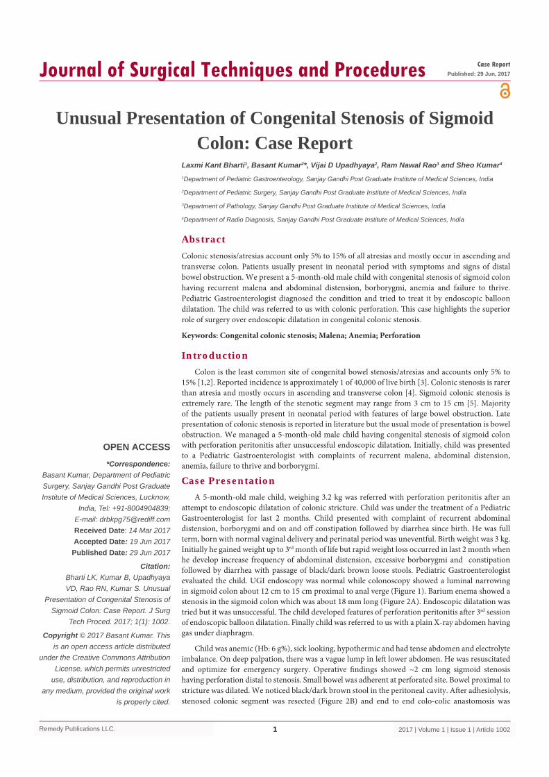
attempt to endoscopic dilatation of colonic stricture. Child was under the treatment of a Pediatric Gastroenterologist for last 2 months. Child presented with complaint of recurrent abdominal distension, borborygmi and on and off constipation followed by diarrhea since birth. He was full term, born with normal vaginal delivery and perinatal period was uneventful. Birth weight was 3 kg. Initially he gained weight up to 3rd month of life but rapid weight loss occurred in last 2 month when he develop increase frequency of abdominal distension, excessive borborygmi and constipation followed by diarrhea with passage of black/dark brown loose stools. Pediatric Gastroenterologist evaluated the child. UGI endoscopy was normal while colonoscopy showed a luminal narrowing in sigmoid colon about 12 cm to 15 cm proximal to anal verge (Figure 1). Barium enema showed a stenosis in the sigmoid colon which was about 18 mm long (Figure 2A). Endoscopic dilatation was tried but it was unsuccessful. The child developed features of perforation peritonitis after 3rd session of endoscopic balloon dilatation. Finally child was referred to us with a plain X-ray abdomen having gas under diaphragm.
Child was anemic (Hb: 6 g%), sick looking, hypothermic and had tense abdomen and electrolyte imbalance. On deep palpation, there was a vague lump in left lower abdomen. He was resuscitated and optimize for emergency surgery. Operative findings showed ~2 cm long sigmoid stenosis having perforation distal to stenosis. Small bowel was adherent at perforated site. Bowel proximal to stricture was dilated. We noticed black/dark brown stool in the peritoneal cavity. After adhesiolysis, stenosed colonic segment was resected (Figure 2B) and end to end colo-colic anastomosis was
Laxmi Kant Bharti1, Basant Kumar2*, Vijai D Upadhyaya2, Ram Nawal Rao3 and Sheo Kumar4
1Department of Pediatric Gastroenterology, Sanjay Gandhi Post Graduate Institute of Medical Sciences, India
2Department of Pediatric Surgery, Sanjay Gandhi Post Graduate Institute of Medical Sciences, India
3Department of Pathology, Sanjay Gandhi Post Graduate Institute of Medical Sciences, India
4Department of Radio Diagnosis, Sanjay Gandhi Post Graduate Institute of Medical Sciences, India

Basant Kumar, et al., Journal of Surgical Techniques and Procedures
Remedy Publications LLC. 2017 | Volume 1 | Issue 1 | Article 10022
performed. In view of low general condition of patient, a protective loop ileostomy was done. Histology confirmed the diagnosis (Figure 3). Post-operative period was uneventful and child was allowed to oral feed on 2nd post-operative day. Child is under follow-up and in last 4 months, his weight was almost doubled.
DiscussionColonic atresias/stenosis may contribute about 5% to 15% of all
gut atresias. Isolated colonic stenosis is even rarer than atresias [6]. Left sided colonic stenosis is uncommon than right-sided colonic stenosis. It is thought that it may be due to vascular insult in utero [7]. Fetal herniation, kinks, intussusceptions, or primary vascular accidents can cause interruption of the blood supply [8]. Depending on the severity of vascular insult, stenosis or atresia may occur. Patient with colonic stenosis may present early or late in life depending on the severity of stenosis and other concomitant factors. The treatment option used in most of the cases of colonic atresia and stenosis is colo-colic anastomosis with or without enterostomy.
Usual clinical features include vomiting, abdominal distension and small amount meconium or failure to pass meconium in early presenting cases. Patient may remain asymptomatic for varying period of time but almost all reported cases were presented with signs and symptoms of large bowel obstruction. In our case, child had very unusual presentations not described earlier like recurrent episodes of malena substantiated by drop in hemoglobin and frequent constipation followed by diarrhea and growth failure. Frequent malena and drop in hematocrit drawled the attention of gastroenterologist and he performed upper and lower GI endoscopy. He found luminal narrowing in sigmoid colon and finally diagnosed a stenosis in the sigmoid colon which was measured about 18 mm long on barium enema. In the literature, usual lengths of reported
stenosis in colon may vary from 3 cm to 15 cm [7]. Small stricture length encouraged Pediatric Gastroenterologist for trial of Phase wise, endoscopic, pressure controlled balloon dilatation but he failed after initial two dilatations and accidentally perforated the bowel.
In the literature, one case of sigmoid stenosis is described by Santulli and Blanc [8] while Pai and Pai [9] had reported a case of recto-sigmoid stenosis in 4-month-old infant. In our case, there was unusual clinical presentation. We thought that partial obstruction of sigmoid colon causes fecal stasis and constipation which might progress to enterocolitis and diarrhea. Repeated episodes of enterocolitis leads to mucosal injury presented as frequent malena, drop in hematocrit and growth failure of child. The stricture length is probably the smallest reported length just about 2 cm in our case.
ConclusionThis case indicates that in congenital nature of colonic strictures;
endoscopic dilatation procedures should be avoided and surgery should be the preferred treatment of choice (by open or minimally invasive techniques depending on condition of patients and expertise of surgeons). Whether there is any role of colonoscopy balloon dilatation; we could not found any peer reviewed evidence in literature. It should be studied and till then this procedure as management should be avoided.
References1. Sax EJ. Pediatric case of the day. Congenital colonic stenosis. AJR Am J
Roentgenol. 1991;156(6):1315-17.
2. Powell RW, Raffensperger JG. Congenital colonic atresia. J Pediatr Surg. 1982;17(2):163-4.
3. Franken EA. Gastrointestinal imaging in pediatrics. 2nd ed. New York: Harper & Row, USA; 1982. p. 286-7.
4. Ruggeri G, Libri M, Gargano T, Pavia S, Pasini L, Tani G, et al. Congenital colonic stenosis: A case of late onset. Pediatr Med Chir. 2009;31(3):130-3.
5. Galván-Montaño A, Suárez-Roa Mde L, Carmona-Moreno E. Congenital stenosis of the colon with foreign bodies. Case report. Cir Cir. 2010;78(3):259-61.
6. Louw JH. Congenital Intestinal atresia and stenosis in the newborn: observation on its pathogenesis and treatment. Ann R Coll Surg Engl. 1959;25:209-34.
7. Hall TR, Zaninović A, Lewin D, Barrett C, Boechat MI. Neonatal intestinal ischemia with bowel perforation: an in utero complication of maternal cocaine abuse. AJR Am J Roentgenol. 1992;158(6):1303-4.
8. Santulli TV, Blanc WA. Congenital atresia of the intestine: pathogenesis and treatment. Ann Surg. 1961;154:939-48.
9. Pai GK, Pai PK. A case of congenital colonic stenosis presenting as rectal prolapse. J Pediatr Surg.1990;25(6):699-700.
Figure 1: Endoscopic view of colonic stenosis during balloon dilatation.
Figure 2: A) Barium enema showing length of stenosis and dilated proximal colon. B) Excised stenosed segment of colon.
Figure 3: Histology section shows ulceration, perforation and granulation tissue, capillaries and mixed inflammatory cells comprising of neutrophils, lymphocytes and histiocytes.