University of Zurich fileTowards Tailored Communication Networks in Assemblies of Artificial Cells...
11
University of Zurich Zurich Open Repository and Archive Winterthurerstr. 190 CH-8057 Zurich http://www.zora.uzh.ch Year: 2009 Towards tailored communication networks in assemblies of artificial cells Hadorn, M; Burla, B; Eggenberger Hotz, P Hadorn, M; Burla, B; Eggenberger Hotz, P (2009). Towards tailored communication networks in assemblies of artificial cells. In: Korb, KB; Randall, M; Hendtlass, T. Artificial Life: Borrowing from Biology. Berlin und Heidelberg, 126-135. Postprint available at: http://www.zora.uzh.ch Posted at the Zurich Open Repository and Archive, University of Zurich. http://www.zora.uzh.ch Originally published at: Korb, KB; Randall, M; Hendtlass, T 2009. Artificial Life: Borrowing from Biology. Berlin und Heidelberg, 126-135.
Transcript of University of Zurich fileTowards Tailored Communication Networks in Assemblies of Artificial Cells...
University of ZurichZurich Open Repository and Archive
Winterthurerstr. 190
CH-8057 Zurich
http://www.zora.uzh.ch
Year: 2009
Towards tailored communication networks in assemblies ofartificial cells
Hadorn, M; Burla, B; Eggenberger Hotz, P
Hadorn, M; Burla, B; Eggenberger Hotz, P (2009). Towards tailored communication networks in assemblies ofartificial cells. In: Korb, KB; Randall, M; Hendtlass, T. Artificial Life: Borrowing from Biology. Berlin undHeidelberg, 126-135.Postprint available at:http://www.zora.uzh.ch
Posted at the Zurich Open Repository and Archive, University of Zurich.http://www.zora.uzh.ch
Originally published at:Korb, KB; Randall, M; Hendtlass, T 2009. Artificial Life: Borrowing from Biology. Berlin und Heidelberg,126-135.

Towards Tailored Communication Networks in Assemblies of Artificial Cells
Maik Hadorn1, Bo Burla2,3 and Peter Eggenberger Hotz1,4
1 University of Zurich, Department of Informatics, Artificial Intelligence Laboratory, 8050 Zurich, Switzerland
2 University of Zurich, Institute of Plant Biology, 8008 Zurich, Switzerland 3 Global Research Laboratory, 790784 Pohang, South Korea
4 University of Southern Denmark, The Mærsk Mc-Kinney Møller Institute, Campusvej 55, 5230 Odense M, Denmark
[email protected], [email protected], [email protected]
Abstract. Living Technology is researching novel IT making strong use of pro-grammable chemical systems. These chemical systems shall finally converge to artificial cells resulting in evolvable complex information systems. We focus on procedural manageability and information processing capabilities of such in-formation systems. Here, we present a novel resource-saving formation, processing, and examination procedure to generate and handle single compart-ments representing preliminary stages of artificial cells. Its potential is exempli-fied by testing the influence of different glycerophospholipids on the stability of the compartments. We discuss how the procedure could be used both in evolu-tionary optimization of self-assembling amphiphilic systems and in engineering tailored communication networks enabling life-like information processing in multicompartment aggregates of programmable composition and spatial confi-guration.
Keywords: Living Technology, self-assembly, programmability, glycerophos-pholipids, vesicles, multivesicular aggregates, adhesion plaque, phase transition
1 Introduction
Engineering Living Technology from non-living materials has attracted particular attention in minimal life and complex information systems. As part of the complex systems Future Emerging Technologies initiative, PACE (Programmable Artificial Cell Evolution) was researching novel Information Technology (IT) that makes strong use of life-like properties such as robustness, homeostasis, self-repair, self-assembly, modularity, self-organization, self-reproduction, genetic programmability, evolvabili-ty, complex systems design, and bootstrapping complexity. In this context, PACE has created the foundation for a new generation of embedded IT to build evolvable com-plex information systems using programmable chemical systems that converge to artificial cells [1]. Because experiments were realized both in the laboratory and in

2 Maik Hadorn, Bo Burla and Peter Eggenberger Hotz
simulation, findings of in vitro and in silico experiments interacted and lead to essen-tial additions to the evolutionary approach in design of laboratory experimentation [2].
According to the guidelines of PACE, the creation of simple forms of life from scratch in the laboratory should have pursued a bottom-up strategy choosing simple organic compounds of low molecular weight over highly evolved polypeptides. Com-plexity of an artificial cell featuring all aspects of a living system should have been achieved in an evolutionary process. It is hypothesized that a membrane partitioning internal constituents off the environment might have been one of the minimal re-quirements for living systems to arise [3, 4]. A lipid membrane represents a formida-ble barrier to the passage of polar molecules. It organizes biological processes by compartmentalizing them. Compartmentalization enables segregation of specific
Fig. 1. Structure of glycerophospholipids and bilayer formation. The structure of 1-palmitoyl-2-oleoyl-sn-glycero-3-phosphatidylcholine (PC(16:0/18:1(∆9-Cis)) is represented (A) as a struc-tural formula, (B) as a skeletal formula, and (C) as a schematic representation used throughout this publication. Glycerophospholipids are amphiphilic molecules with lipophilic hydrocarbon “tails” and hydrophilic “heads”. In PC(16:0/18:1(∆9-Cis) the headgroup is phosphatidylcho-line; a saturated C16 palmitic acid (16:0) hydrocarbon chain and a monounsaturated C18 oleic acid (18:1) hydrocarbon chain occur at the C1 and C2 position of the glycerophospholipid and constitute the tail. The double bond (monounsaturation) occurs between the C9 and C10 atoms (∆9) of the oleic acid, has cis configuration, and puts a rigid 30° bend in the hydrocarbon chain. (C.1) Phospholipids can form lipid bilayers (membranes) that partition an aqueous compart-ment off the surrounding medium. (C.2) In vesicles an intravesicular fluid (light blue) is sepa-rated from the intervesicular medium (white).
Towards Tailored Communication Networks in Assemblies of Artificial Cells 3
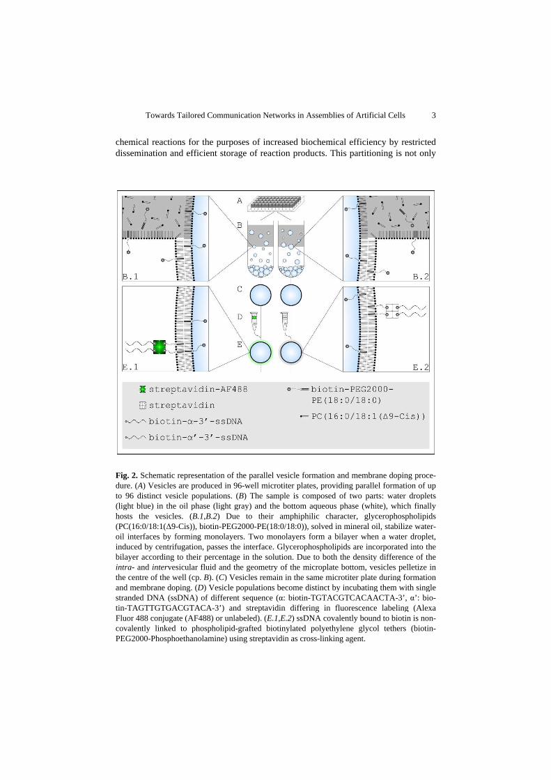
chemical reactions for the purposes of increased biochemical efficiency by restricted dissemination and efficient storage of reaction products. This partitioning is not only
Fig. 2. Schematic representation of the parallel vesicle formation and membrane doping proce-dure. (A) Vesicles are produced in 96-well microtiter plates, providing parallel formation of up to 96 distinct vesicle populations. (B) The sample is composed of two parts: water droplets (light blue) in the oil phase (light gray) and the bottom aqueous phase (white), which finally hosts the vesicles. (B.1,B.2) Due to their amphiphilic character, glycerophospholipids (PC(16:0/18:1(∆9-Cis)), biotin-PEG2000-PE(18:0/18:0)), solved in mineral oil, stabilize water-oil interfaces by forming monolayers. Two monolayers form a bilayer when a water droplet, induced by centrifugation, passes the interface. Glycerophospholipids are incorporated into the bilayer according to their percentage in the solution. Due to both the density difference of the intra- and intervesicular fluid and the geometry of the microplate bottom, vesicles pelletize in the centre of the well (cp. B). (C) Vesicles remain in the same microtiter plate during formation and membrane doping. (D) Vesicle populations become distinct by incubating them with single stranded DNA (ssDNA) of different sequence (α: biotin-TGTACGTCACAACTA-3’, α’: bio-tin-TAGTTGTGACGTACA-3’) and streptavidin differing in fluorescence labeling (Alexa Fluor 488 conjugate (AF488) or unlabeled). (E.1,E.2) ssDNA covalently bound to biotin is non-covalently linked to phospholipid-grafted biotinylated polyethylene glycol tethers (biotin-PEG2000-Phosphoethanolamine) using streptavidin as cross-linking agent.

4 Maik Hadorn, Bo Burla and Peter Eggenberger Hotz
realized between the cell and its environment, but it is even recapitulated within the cell. Vesicles, as an instance of minimality, feature an aqueous compartment parti-tioned off the surrounding by an impermeable lipid membrane. Like cellular mem-branes, vesicular membranes consist of amphiphilic phospholipids that link a hydro-philic “head” and a lipophilic “tail” (Fig. 1). Suspended phospholipids can self-assemble to form closed, self-sealing solvent-filled vesicles that are bounded by a two-layered sheet (a bilayer) of 6 nm in width, with all of their tails pointing toward the center of the bilayer. This molecular arrangement excludes water from the center of the sheet and thereby eliminates unfavorable contacts between water and the lipo-philic (= hydrophobic) tails. The lipid bilayer provides inherent self-repair characte-ristics due to lateral mobility of its phospholipids. Wide usage of artificial vesicles is found in analytics [5-9] and synthetics, where their applications include bioreactors [10-12], and drug delivery systems [13-17].
In the laboratory work, we focused on intrinsic information processing capabilities of vesicles and multivesicular assemblies. To achieve compartmentalization we de-veloped methods to self-assemble multivesicular aggregates of programmable compo-sition and spatial configuration composed of distinct vesicle populations that differ in membrane and intravesicular fluid composition. The assembly process of multivesi-cular aggregates was based on the hybridization of biotinylated single-stranded DNA (ssDNA) with which the vesicles were doped. Doping was realized by anchoring biotinylated ssDNA to biotinylated phospholipids via streptavidin as a cross-linking agent (Fig. 2). The potential of a programmable self-assembly of superstructures with high degrees of complexity [18] has attracted significant attention to nanotechnologi-cal applications in the last decade. So far, cross-linkage based on DNA hybridization was proposed to induce self-assembly of complementary monohomophilic vesicles [5, 19] or hard sphere colloids [20-23], to induce programmable fusion of vesicles [5, 24], or to specifically link vesicles to surface supported membranes [5, 25-27]. By introducing a surface doping of distinct populations of ssDNA, as realized in the self-assembly of hard sphere colloids [28, 29], we provide n-arity to the assembly process. As a result, linkage of more than two distinct vesicle populations, as proposed by Chiruvolu et al. [30] and already realized for hard sphere colloids [31], becomes feas-ible.
Concerning procedural manageability in laboratory experimentation, we estab-lished a new protocol for in vitro vesicle formation and modification. It increases the versatility of the underlying vesicle formation method [11, 32, 33] by introducing microtiter plates and vesicle pelletization (Fig. 2). The potential of the vesicle forma-tion method that provides independent composition control of the intra- and interve-sicular fluid as well as of the inner and outer bilayer leaflet was exemplified by the production of asymmetric vesicles combining biocompatibility and mechanical en-durance in asymmetric vesicles [32]. Here, we exemplify the advantages of the novel protocol by carrying out a high-throughput analysis of constituents affecting vesicle formation and stability. Thereby the effect of nine different glycerophospholipids on vesicle formation was tested. We discuss how this procedure could be used both in evolutionary optimization of self-assembling amphiphilic systems and to realize tai-

Towards Tailored Communication Networks in Assemblies of Artificial Cells 5
lored communication networks in assemblies of artificial cells by programmable loca-lization of glycerophospholipids within the vesicular membrane.
2 Material & Methods
Major technical modifications of the vesicle formation protocol reported in Ref. [32] were: the introduction of (i) 96-well microtiter plates U96 to provide a high-throughput analysis and (ii) a density difference between intra- and intervesicular solution to induce vesicle pelletization. For a description of the modified vesicle pro-tocol see Fig. 2. To analyze the effect of different glycerophospholipids on vesicle formation, data on vesicle yield of four times nine equimolar mixtures of 100%-m (m � {100, 50, 10, 1}) PC(16:0/18:1(∆9-Cis)) and m% PC(x:0/x:0) (x � {12, 14, 16, 18, 24}), PC(y:1(∆9-Cis)/y:1(∆9-Cis)) (y � {14, 16, 18}), or PC(24:1(∆15-Cis)/24:1(∆15-Cis)) were collected and compared to 100%-m PC(16:0/18:1(∆9-Cis)) and m% mineral oil (solvent for all glycerophospholipids) as control. Vesicle forma-tion was performed in duplication. Length of circumference of the vesicle pellet is used as a measure of vesicle yield (Fig. 3). Light-microscopy was performed using a Wild M40 inverted microscope equipped with a MikoOkular microscope camera. All camera settings were identical for the recordings. Confocal laser scanning microscopy was performed using an inverted Leica Confocal DMR IRE2 SP2 confocal laser scanning microscope.
Fig. 3. Measurement of vesicle yield. (A) No vesicle pellet emerged for the mixture of 50% PC(24:1(∆15-Cis)/24:1(∆15-Cis)) and 50% PC(16:0/18:1(∆9-Cis)). (B) The vesicle yield is expressed by length of circumference of the vesicle pellet (circle) emerged for the mixture 10% PC(24:1(∆15-Cis)/24:1(∆15-Cis)) and 90% PC(16:0/18:1(∆9-Cis)). Fibers represent pollutants not affecting vesicular yield or handling.

6 Maik Hadorn, Bo Burla and Peter Eggenberger Hotz
3 Results and Discussion
In the literature many examples of artificially produced vesicles are reported, whose membranes are composed of PC(16:0/18:1(∆9-Cis)) (POPC) exclusively.1 To vary the intrinsic material properties of membranes we have to be able to alter their phospholi-pid content. In this study, we realized a high-throughput analysis of glycerophospho-lipids affecting vesicle formation. All glycerophospholipids differed in length and saturation of their hydrocarbon chains only. In the control experiment, vesicle yield was constant when POPC was present (Fig. 4). Influence of the phospholipids tested at an admixture of one percent was marginal and will not be discussed. Intergroup comparisons (cp. Fig. 4) of saturated and unsaturated glycerophospholipids of equal chain length revealed remarkable differences in vesicle yield. Whereas unsaturated glycerophospholipids of chain length up to 18 carbon atoms both could form vesicles by themselves and did not or just slightly affect the yield at 50 and 10 percent, satu-rated lipids seem to disturb vesicle formation down to 10 percent of admixture. For a chain length of 24 carbon atoms the situation is diametrical. A small amount of ve-sicles is found for solutions containing 100 percent saturated PC(24:0/24:0). Whereas vesicle yield is halved at 50 percent for this lipid, vesicle formation is inhibited totally by the unsaturated PC(24:1(∆15-Cis)/24:1(∆15-Cis)) up to an admixture of 10 percent (cp. Fig. 3). Intragroup comparison within saturated or unsaturated glycerophospholi-pids reveals a decrease in vesicle yield depending on chain length (except for PC(24:0/24:0) at 100 percent). Only a limited number of glycerophospholipid is able
1 In 2008 more than 150 articles were published concerning POPC vesicles or liposomes.
Fig. 4. Effect of different glycerophospholipids on vesicle formation. Vesicle yield of 100%-m (m � {100, 50, 10, 1}) POPC = PC(16:0/18:1(∆9-Cis)) and m% mineral oil serves as standard (XY (scatter) chart). Deviations from control for the glycerophospholipids tested are presented in absolute values as bar charts. Error bars indicate the standard deviation. The glycerophos-pholipids tested can be summarized according to the level of saturation of their hydrocarbon chains in two groups (saturated, PC(x:0/x:0) (x � {12, 14, 16, 18, 24}); unsaturated, PC(y:1(∆9-Cis)/y:1(∆9-Cis)) (y � {14, 16, 18}), PC(24:1(∆15-Cis)/24:1(∆15-Cis))).
Towards Tailored Communication Networks in Assemblies of Artificial Cells 7
to form vesicles on their own or in cooperation with POPC. By providing parallelism in vesicle formation, processing and examination (cp.
Fig. 2), we not only increased the experimental throughput and the procedural mana-geability, but we lowered the costs and reduced the amount of contributory factors. This reduction of dimensionality and the parallelism in vesicle formation provided by the novel vesicle formation method presented herein may prove to be useful in evolu-tionary design of experiments by shortening the search toward the optimality region of the search space [2]. The application of microtiter plates may further enable the automatization in vesicle formation.
By self-assembling multivesicular aggregates of programmable composition and spatial configuration, composed of vesicles differing in membrane and intravesicular fluid composition, an inhomogeneous distribution of information carriers is achieved
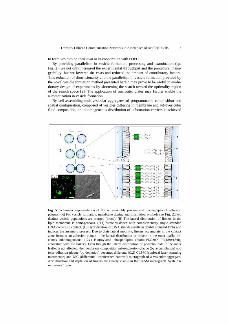
Fig. 5. Schematic representation of the self-assembly process and micrographs of adhesion plaques. (A) For vesicle formation, membrane doping and illustration symbols see Fig. 2.Two distinct vesicle populations are merged (brace). (B) The lateral distribution of linkers in the lipid membrane is homogeneous. (B.1) Vesicles doped with complementary single stranded DNA come into contact. (C) Hybridization of DNA strands results in double stranded DNA and induces the assembly process. Due to their lateral mobility, linkers accumulate in the contact zone forming an adhesion plaque – the lateral distribution of linkers in the outer leaflet be-comes inhomogeneous. (C.1) Biotinylated phospholipids (biotin-PEG2000-PE(18:0/18:0)) colocalize with the linkers. Even though the lateral distribution of phospholipids in the inner leaflet is not affected, the membrane composition intra-adhesion-plaque (by accumulation) and inter-adhesion-plaque (by depletion) becomes different. (C.2) CLSM (confocal laser scanning microscope) and DIC (differential interference contrast) micrograph of a vesicular aggregate. Accumulation and depletion of linkers are clearly visible in the CLSM micrograph. Scale bar represents 10µm.
8 Maik Hadorn, Bo Burla and Peter Eggenberger Hotz
(Fig. 5). To build complex information systems, the exchange of information carriers between the compartments themselves and/or between the environment and the com-partments has to be realized. Biological membranes contain membrane proteins that were evolutionary optimized for catalyzing numerous chemical reactions, mediating the flow of nutrients and wastes, as well as participating in communication across the membrane. The use of such membrane proteins in information exchange was ex-cluded by the guidelines of PACE. Thus, information processing capabilities of mul-tivesicular aggregates had to be realized by exploiting intrinsic material properties of the phospholipid membrane. A key property of the lipid membrane is its phase [34]. Lipid bilayers can undergo phase transitions in which they become a gel-like solid and therefore lose their fluidity. The state depends on temperature; the transition tem-perature of a bilayer increases with the chain length and the degree of saturation of its fatty acid residues. The phase transitions are triggered externally by changing temper-ature; a fact exploited in exchanging reactants between the surrounding medium and a vesicular compartment setting up consecutive enzymatic reactions in a single contain-er [35]. A selective exchange of information between compartments of a multivesicu-lar aggregate relying on inherent material properties of phospholipid membranes has not been demonstrated so far. We recognized that the formation of adhesion plaques [36, 37] triggered by the aggregation process results in an inhomogeneous distribution of phospholipids in the lateral dimension of the membrane (Fig. 5). This inhomogene-ity in phospholipid distribution causes a difference in phase transition characteristics of membrane portions intra- and inter-adhesion-plaque. Selective opening of commu-nication channels between the compartments, whereas the membrane portions inter-adhesion-plaques are still impermeable, becomes conceivable. By using membrane anchors of different phospholipid composition the adhesion plaques would differ in phase transition characteristics among each other enabling programmable and trigger-able communication networks in multivesicular aggregates of programmable compo-sition and spatial configuration.
Realization of such tailored communication networks is hindered by restrictions in commercial availability of lipid anchors to dope vesicular surfaces with ssDNA. The high-throughput analysis presented herein allows for identification of lipid candidates not affecting vesicle formation and differing in phase transition characteristics. In a next step the head group of these candidates could be covalently linked to biotinylated PEG tethers therefore providing anchors for biotinylated ssDNA (via (strept-)avidin).
Acknowledgements. This work was conducted as part of the European Union integrated project PACE (EU-IST-FP6-FET-002035). Maik Hadorn was supported by the Swiss National Foundation Project 200020-118127 Embryogenic Evolution: From Simulations to Robotic Applications. Peter Eggenberger Hotz was partly supported by PACE. Wet laboratory experiments were performed at the Molecular Physiology Laboratory of Professor Enrico Martinoia (Institute of Plant Biology, University of Zurich, Switzerland).
Towards Tailored Communication Networks in Assemblies of Artificial Cells 9
References
1. Rasmussen, S.: Protocells: bridging nonliving and living matter. MIT Press, Cambridge, Massachusetts (2009)
2. Forlin, M., Poli, I., De March, D., Packard, N., Gazzola, G., Serra, R.: Evolutionary expe-riments for self-assembling amphiphilic systems. Chemometrics Intell. Lab. Syst. 90, 153--160 (2008)
3. Griffiths, G.: Cell evolution and the problem of membrane topology. Nat. Rev. Mol. Cell Biol. 8, 1018--1024 (2007)
4. Israelachvili, J.N., Mitchell, D.J., Ninham, B.W.: Theory of self-assembly of lipid bilayers and vesicles. Biochimica Et Biophysica Acta 470, 185--201 (1977)
5. Chan, Y.H.M., van Lengerich, B., Boxer, S.G.: Effects of linker sequences on vesicle fu-sion mediated by lipid-anchored DNA oligonucleotides. Proc. Natl. Acad. Sci. U. S. A. 106, 979--984 (2009)
6. Hase, M., Yoshikawa, K.: Structural transition of actin filament in a cell-sized water droplet with a phospholipid membrane. J. Chem. Phys. 124, 104903 (2006)
7. Hotani, H., Nomura, F., Suzuki, Y.: Giant liposomes: from membrane dynamics to cell morphogenesis. Curr. Opin. Colloid Interface Sci. 4, 358--368 (1999)
8. Limozin, L., Roth, A., Sackmann, E.: Microviscoelastic moduli of biomimetic cell enve-lopes. Phys. Rev. Lett. 95, 178101 (2005)
9. Luisi, P., Walde, P.: Giant vesicles. John Wiley & Sons, Ltd, Chichester (2000) 10. Michel, M., Winterhalter, M., Darbois, L., Hemmerle, J., Voegel, J.C., Schaaf, P., Ball, V.:
Giant liposome microreactors for controlled production of calcium phosphate crystals. Langmuir 20, 6127--6133 (2004)
11. Noireaux, V., Libchaber, A.: A vesicle bioreactor as a step toward an artificial cell assem-bly. Proc. Natl. Acad. Sci. U. S. A. 101, 17669--17674 (2004)
12. Nomura, S., Tsumoto, K., Hamada, T., Akiyoshi, K., Nakatani, Y., Yoshikawa, K.: Gene expression within cell-sized lipid vesicles. Chembiochem 4, 1172--1175 (2003)
13. Abraham, S.A., Waterhouse, D.N., Mayer, L.D., Cullis, P.R., Madden, T.D., Bally, M.B.: The liposomal formulation of doxorubicin. In: Liposomes, Pt E. Elsevier Academic Press Inc, San Diego (2005)
14. Allen, T.M., Cullis, P.R.: Drug delivery systems: Entering the mainstream. Science 303, 1818--1822 (2004)
15. Marjan, J.M.J., Allen, T.M.: Long circulating liposomes: Past, present and future. Biotech-nology Advances 14, 151--175 (1996)
16. Tardi, P.G., Boman, N.L., Cullis, P.R.: Liposomal doxorubicin. J. Drug Target. 4, 129--140 (1996)
17. Sengupta, S., Eavarone, D., Capila, I., Zhao, G.L., Watson, N., Kiziltepe, T., Sasisekharan, R.: Temporal targeting of tumour cells and neovasculature with a nanoscale delivery sys-tem. Nature 436, 568--572 (2005)
18. Licata, N.A., Tkachenko, A.V.: Errorproof programmable self-assembly of DNA-nanoparticle clusters. Physical Review E (Statistical, Nonlinear, and Soft Matter Physics) 74, 041406 (2006)
19. Beales, P.A., Vanderlick, T.K.: Specific binding of different vesicle populations by the hybridization of membrane-anchored DNA. J. Phys. Chem. A 111, 12372--12380 (2007)
10 Maik Hadorn, Bo Burla and Peter Eggenberger Hotz
20. Biancaniello, P.L., Crocker, J.C., Hammer, D.A., Milam, V.T.: DNA-mediated phase beha-vior of microsphere suspensions. Langmuir 23, 2688--2693 (2007)
21. Mirkin, C.A., Letsinger, R.L., Mucic, R.C., Storhoff, J.J.: A DNA-based method for ration-ally assembling nanoparticles into macroscopic materials. Nature 382, 607--609 (1996)
22. Valignat, M.P., Theodoly, O., Crocker, J.C., Russel, W.B., Chaikin, P.M.: Reversible self-assembly and directed assembly of DNA-linked micrometer-sized colloids. Proc. Natl. Acad. Sci. U. S. A. 102, 4225--4229 (2005)
23. Biancaniello, P., Kim, A., Crocker, J.: Colloidal interactions and self-assembly using DNA hybridization. Phys. Rev. Lett. 94, 058302 (2005)
24. Stengel, G., Zahn, R., Hook, F.: DNA-induced programmable fusion of phospholipid ve-sicles. J. Am. Chem. Soc. 129, 9584--9585 (2007)
25. Benkoski, J.J., Hook, F.: Lateral mobility of tethered vesicle - DNA assemblies. J. Phys. Chem. B 109, 9773--9779 (2005)
26. Yoshina-Ishii, C., Boxer, S.G.: Arrays of mobile tethered vesicles on supported lipid bilay-ers. J. Am. Chem. Soc. 125, 3696--3697 (2003)
27. Li, F., Pincet, F., Perez, E., Eng, W.S., Melia, T.J., Rothman, J.E., Tareste, D.: Energetics and dynamics of SNAREpin folding across lipid bilayers. Nat. Struct. Mol. Biol. 14, 890--896 (2007)
28. Maye, M.M., Nykypanchuk, D., Cuisinier, M., van der Lelie, D., Gang, O.: Stepwise sur-face encoding for high-throughput assembly of nanoclusters. Nat. Mater. 8, 388--391 (2009)
29. Prabhu, V.M., Hudson, S.D.: Nanoparticle assembly: DNA provides control. Nat. Mater. 8, 365--366 (2009)
30. Chiruvolu, S., Walker, S., Israelachvili, J., Schmitt, F.J., Leckband, D., Zasadzinski, J.A.: Higher-order self-assembly of vesicles by site-specific binding. Science 264, 1753--1756 (1994)
31. Xu, X.Y., Rosi, N.L., Wang, Y.H., Huo, F.W., Mirkin, C.A.: Asymmetric functionalization of gold nanoparticles with oligonucleotides. J. Am. Chem. Soc. 128, 9286--9287 (2006)
32. Pautot, S., Frisken, B.J., Weitz, D.A.: Engineering asymmetric vesicles. Proc. Natl. Acad. Sci. U. S. A. 100, 10718--10721 (2003)
33. Träuble, H., Grell, E.: Carriers and specificity in membranes. IV. Model vesicles and mem-branes. The formation of asymmetrical spherical lecithin vesicles. Neurosciences Research Program bulletin 9, 373--380 (1971)
34. Feigenson, G.W.: Phase diagrams and lipid domains in multicomponent lipid bilayer mix-tures. Biochim. Biophys. Acta-Biomembr. 1788, 47--52 (2009)
35. Bolinger, P.Y., Stamou, D., Vogel, H.: An integrated self-assembled nanofluidic system for controlled biological chemistries. Angew. Chem.-Int. Edit. 47, 5544--5549 (2008)
36. NopplSimson, D.A., Needham, D.: Avidin-biotin interactions at vesicle surfaces: Adsorp-tion and binding, cross-bridge formation, and lateral interactions. Biophys. J. 70, 1391--1401 (1996)
37. Farbman-Yogev, I., Bohbot-Raviv, Y., Ben-Shaul, A.: A statistical thermodynamic model for cross-bridge mediated condensation of vesicles. J. Phys. Chem. A 102, 9586--9592 (1998)