University of Veterinary Medicine Hanover
132
University of Veterinary Medicine Hanover Center for Systems Neuroscience Department of Neuroanatomy, Hanover Medical School The co-layer method as an efficient way for neurotrophic factor release by transplanted genetically modified neuronal progenitor cells in a rat model of Parkinson’s disease Analysis of morphological and functional integration THESIS Submitted in partial fulfillment of the requirements for the degree of DOCTOR OF PHYLOSOPHY (PhD) awarded by the University of Veterinary Medicine Hanover by Ieva Kalve born in Riga, Latvia Hanover 2013
Transcript of University of Veterinary Medicine Hanover

University of Veterinary Medicine Hanover
Center for Systems Neuroscience
Department of Neuroanatomy, Hanover Medical School
The co-layer method as an efficient way for
neurotrophic factor release by transplanted
genetically modified neuronal progenitor cells in a
rat model of Parkinson’s disease
Analysis of morphological and functional integration
THESIS
Submitted in partial fulfillment of the requirements for the degree of
DOCTOR OF PHYLOSOPHY (PhD)
awarded by the University of Veterinary Medicine Hanover
by
Ieva Kalve
born in Riga, Latvia
Hanover 2013

II
Supervisor: Prof. Dr. Claudia Grothe Supervision group: Prof. Dr. Claudia Grothe
Prof. Dr. med. Joachim K. Krauss
Prof. Dr. Gerd Bicker
1st evaluation: Prof. Dr. Claudia Grothe
Institute of Neuroanatomy
Hanover Medical School
Carl-Neuberg-Straße 1
30625 Hanover
Prof. Dr. med. Joachim K. Krauss
Department of Neurosurgery
Hanover Medical School
Carl-Neuberg-Straße 1
30625 Hanover
Prof. Dr. Gerd Bicker
Division of Cell Biology
University of Veterinary Medicine
Bischofsholer Damm 15
30173 Hanover
2nd evaluation: Prof. Hans R. Widmer, PhD
Scientific laboratory for restorative Neurosciences (OP Ost E218)
Department of Neurosurgery
University Hospital Bern
Freiburgstrasse
3010 Bern
Switzerland
Date of final exam: 25.10.2013
This work was supported by the Georg-Christoph-Lichtenberg Fellowship by the State of Lower Saxony, Germany.

III
The research was carried out between November 2007 and December 2010 at the
Institute for Neuroanatomy at the Hanover Medical School, Germany.
Preliminary results from the PhD research have been presented at the “NECTAR
(Network of European CNS Transplantation & Restoration)’ 09” 19th Annual Meeting,
November 26th – 28th, 2009 in Cardiff, Wales and “NECTAR’10” 20th Annual Meeting,
November 25th-27th, 2010 in Freiburg, Germany.
Publication list:
RATZKA, A.*, KALVE, I.*, OZER, M., NOBRE, A., WESEMANN, M., JUNGNICKEL, J., KOSTER-PATZLAFF, C., BARON, O. & GROTHE, C. (2012) The co-layer method as an efficient way to genetically modify mesencephalic progenitor cells transplanted into 6-OHDA rat model of Parkinson's disease. Cell Transplant, 21, 749-762., *authors contributed equally
JUNGNICKEL, J.*, KALVE, I.*, REIMERS, L., NOBRE, A., WESEMANN, M.,
RATZKA, A., HALFER, N., LINDEMANN, C., SCHWABE, K., TOLLNER, K., GERNERT, M. & GROTHE, C. (2011) Topology of intrastriatal dopaminergic grafts determines functional and emotional outcome in neurotoxin-lesioned rats. Behav Brain Res, 216, 129-35., *authors contributed equally
NOBRE, A., KALVE, I., CESNULEVICIUS, K., RAGANCOKOVA, D., RATZKA, A.,
HALFER, N., WESEMANN, M., KRAMPFL, K., CLAUS, P. & GROTHE, C. (2010) Characterization and differentiation potential of rat ventral mesencephalic neuronal progenitor cells immortalized with SV40 large T antigen. Cell Tissue Res, 340, 29-43.

IV
Jūs naktsmājas dosit – es teikšu – paldies
Ja palikšu, kas manu ceļu ies? Es nezinu, cilvēki, kur manas mājas
Vien balti putekļi putinājas
Es ieraudāšos Bet, kad raudāt beigšu
Es iešu pa mājām Un visiem teikšu
Ka nav nekas dārgāks Par ceļiem šiem
Kas aiziet uz rītiem Un vakariem.
/I. Ziedonis; Ceļi/ (in Latvian)
You will give me a dwelling – and I will say – thank you
Who will be going my way in case I stay? People, I do not know where my home is
There is just a white dust filling the air
I will turn on the weeps But when I am done with weeping I will be going from home to home
And I will be telling everyone That there is nothing more precious
Than these roads Leading to East
And West. /The Roads by I. Ziedonis/

V
To my mother and my godfather
Manai māmiņai un manam krusttētiņam (in Latvian)

VII
TABLE OF CONTENTS
SUMMARY ................................................................................................................. 1
ZUSAMMENFASSUNG .............................................................................................. 2
1 INTRODUCTION ..................................................................................................... 4
1.1 Parkinson’s disease (PD) – pathophysiological and clinical aspects ................... 4
1.2 Therapeutic strategies in PD ....................................................................................... 6
1.3 Donor tissue for transplantation in PD ....................................................................... 8
1.3.1 Potential donor tissue ........................................................................................... 8
1.3.2 Cell-replacement related dopaminergic neuron loss ..................................... 10
1.4 State of art in clinical trials on cell replacement in PD .......................................... 11
1.5 Basal ganglia circuitry and the midbrain DA system ............................................. 14
1.5.1 6-hydroxydopamine (6-OHDA) animal model of Parkinson’s disease ........ 15
1.5.2 Drug-induced rotation tests in unilateral rat 6-OHDA model – analysis of
functional deficits and recovery ................................................................................... 17
1.5.3 Neuronal transplantation in rat 6-OHDA model of PD ................................... 19
1.6 Neurotrophic factors (NTFs) for treatment of PD and brain-derived neurotrophic
factor (BNDF) as the factor of interest ........................................................................... 20
2 AIMS OF THIS PROJECT ................................................................................................ 24
3 MATERIALS AND METHODS ......................................................................................... 25
3a MATERIALS used for the in vivo study ........................................................................ 25
3b METHODS ........................................................................................................................ 30
3.1 Animals and breeding................................................................................................. 30
3.2 Stereotactic unilateral medial forebrain bundle lesion with 6-OHDA .................. 30
3.3 Apomorphine- and amphetamine-induced rotation ............................................... 32
3.4 Stereotactic intrastriatal microtransplantation of E12 neuronal progenitor cells
(NPCs) ................................................................................................................................. 34
3.4.1 Obtaining material for intrastriatal engraftment .............................................. 34
i) Time-mating of SPRD rats and preparation of the embryonic tissue…………….34
ii) Cell culture – the old (mono-layer) and the novel (co-layer) protocols…………..35
iii) Nucleofection………………………………………………………………………….36
iiii) Expression vector used……… .................................................................................. 36
3.4.2 Microtransplantation ............................................................................................ 36
3.5 Transcardial perfusion of the experimental animals, postfixation of the brains 38
3.6 Sectioning of the brains on freezing microtome, cryoprotection of the slices ... 38

VIII
3.7 Immunohistochemistry (IHC)..................................................................................... 39
3.8 Imaging of the transplanted ventral mesencephalic neuronal progenitor cells . 40
3.9 Histological evaluation ............................................................................................... 41
3.9.1 Stereological quantification of TH positive neurons in the intrastriatal
transplants ...................................................................................................................... 41
3.9.2 Morphological evaluation of reinnervation – densitometry ............................ 41
3.9.3 Location of transplants ........................................................................................ 42
3.9.4 Histological evaluation of the 6-OHDA lesion efficacy .................................. 43
3.10 Statistical analysis .................................................................................................... 43
3.11 Experimental design ................................................................................................. 44
4 RESULTS ........................................................................................................................ 45
4.1 Qualitative pCAGGS vector validation in vivo and optimization of the co-layer
time-schedule ..................................................................................................................... 45
4.1.1 Vector evaluation in a co-layer layout .............................................................. 46
4.1.2 Optimization of the transplantation time-schedule when using the co-layer
method for culturing the ventral mesencephalic NPCs ........................................... 48
i) Approval of abundance of additional treatment with attachment medium ............ 51
ii) Validation of the optimal differentiation period in vitro with the novel protocol
before transplantation procedure .................................................................................... 51
4.2 Detection and evaluation of transgene delivery 9 and 12 weeks after
transplantation .................................................................................................................... 51
4.3 Intrastriatal graft evaluation 2 weeks after transplantation ................................... 55
4.4 Evaluation of BDNF restorative capacity in a novel co-layer protocol ................ 58
4.4.1 No significant increase in dopaminergic cell numbers after 2 weeks in vivo
when delivering plasmid-based BDNF ....................................................................... 58
4.4.2 Evaluation of morphological and functional integrity of the BDNF-supported
grafts 13 weeks after transplantation ......................................................................... 61
i) Qualitative evaluation of 13 week intrastriatal grafts ................................................ 62
ii) Behavioral analysis of 13 week grafts using rotometry ........................................... 63
iii) No significant differences between experimental groups in morphometric
analyzes .............................................................................................................................. 66
iv) Histological evaluation of 6-OHDA lesion efficacy and correlation analysis ....... 67
v) transplant topology and correlation analysis to rotational results .......................... 67
5 DISCUSSION ...................................................................................................................... 69

IX
5.1. Co-layer format for effective nonviral delivery of neurotrophic factors without
feeder cells ......................................................................................................................... 70
5.2 Transgene delivery up to at least 13 weeks post implantation ............................ 71
5.3 Optimization of the transplantation-schedule ......................................................... 72
5.4 DA numerical dominance and immunohistochemical characteristics of the
ventral mesencephalic transplants treated in a co-layer setup .................................. 74
5.5 BDNF as a candidate factor to be introduced in the expression vector in the co-
layer set-up in vivo – an attempt to prove the principle ............................................... 76
5.6 Sufficient 6-OHDA lesion efficacy and adequate transplant topology ................ 79
5.7 Concluding remarks .................................................................................................... 80
6 REFERENCE LIST ............................................................................................................ 81
7 APPENDIX .......................................................................................................................... 98
7.1 Mediums used for cell culture ................................................................................... 98
7.2 Stereological quantification results of the transplanted DA neurons in Exp IV . 99
7.3 Stereological quantification results of the transplanted DA neurons in Exp V 100
7.4 Results of stereological quantification of the transplanted DA neurons and
morphological parameters in Exp VI............................................................................. 102
7.5 Results in apomorphine-induced rotation test in Exp VI ..................................... 103
7.6 Results in amphetamine-induced rotation test in Exp VI .................................... 105
7.7 Correlation analysis on the morphometric data 13 weeks post implantation in
Exp VI ................................................................................................................................ 107
7.8 Histological evaluation of 6-OHDA lesioning efficacy – Exp VI ......................... 108
7.8.1 The number of rest THir in the SN (Pc, Pl, Pr) on the lesioned (right) side
........................................................................................................................................ 108
7.8.2 6-OHDA lesion efficacy in the scores ............................................................. 109
7.8.3 Correlation between lesion efficacy and amphetamine rotation behavior 110
7.8.4 Correlation between lesion efficacy and apomorphine rotation behavior . 111
7.9 Topology of the transplants – Exp VI ..................................................................... 112
7.9.1 The localization of the central part and the whole transplant volume in the
craniocaudal (AP) plane ............................................................................................. 112
7.9.2 The scores for the localization of each transplant in AP, dorsoventral
(VERT), and mediolateral (LAT) planes ................................................................... 113
7.9.3 Resultsof correlation analysis between the Aphetamine rotation outcome
and graft location ......................................................................................................... 114

X
7.9.4 Results of correlation analysis between the apomorphine rotation outcome
and graft topology ........................................................................................................ 116
ACKNOWLEDGEMENTS .................................................................................................. 118

XI
ABBREVIATIONS
A med or A Attachment medium AB antibody Amphe D-amphetamine Apo apomorphine AP craniocaudal BBB blood-brain-barrier BDNF brain-derived neurotrophic factor BG basal ganglia CNS central nervous system D med or D Differentiation medium DA dopamine mDA mesencephalic dopaminergic neuron DAB 3,3'-Diaminobenzidine DAPI 4',6-diamidino-2-phenylindole d. day DBS deep brain stimulation DMEM Dulbecco’s Modified Eagle Medium DRs DA receptors ECM extracellular matrix EGFP enhanced green fluorescence protein EGFPir enhanced green fluorescence protein immunoreactive EGFP pos enhanced green fluorescence protein positive ELISA enzyme-linked immunosorbent assay Exp experiment EP embryo preparation ESCs embryonic stem cells EtOH Ethyl Alcohol E12 embryonal day 12 FCS fetal calf serum FGF fibroblast growth factor G group GDNF glial cell-derived neurotrophic factor GFAP glial fibrillary acidic protein GIDs graft-induced dyskinesias GIRK2 G-protein regulated inward-rectifier potassium channel second subunit GPe Globus pallidus externus (Pars lateralis) GPi Globus pallidus internus (Pars medialis) HD Huntington’s disease hESCs human embryonic stem cells H2O2 Hydrogen peroxide Hz hertz I.D. inner diameter IHC immunohistochemistry i.p. intraperitoneal K+ potassic LAT mediolateral L-dopa levadopamine MFB medial forebrain bundle μl microlitre ml milliliter mg milligram min minute MPTP 1-methyl-4-phenyl-1,2,3,6-tetahydropyridine NeuN neuronal nuclear antigen NTF neurotrophic factor 6-OHDA 6-hydroxydopamine O.D. outer diameter OD optical density P med or P Proliferation medium

XII
PCD programmed cells death PD Parkinson’s disease PET positron emission tomography PBS phosphate-buffered saline P med or P Proliferation medium proBDNF BNDF precursor rBDNF BDNF recombinant protein R410 pCAGGS-EGFP plasmid R412 pCAGGS-EGFP-FLAG plasmid R413 pCAGGS-BDNF-FLAG- plasmid s.c. subcutaneous SD standard deviation SEM standard error of the mean Ser series of the brain slices (one of six) SN substantia nigra SNc substantia nigra pars compacta SNl Substantia nigra pars lateralis SNr substantia nigra pars reticularis TH tyrosine hydroxylase THir tyrosine hydroxylase immunoreactive TH pos tyrosine hydroxylase positive tpm turns per minute Tub ß-III Tubulin UPDRS unified Parkinson’s disease rating scale VERT dorsoventral VM ventral mesencephalic VMP ventral mesencephalic progenitor vs versus VTA ventral tegmental area 5-HT serotonin Ø diameter

XIII
LIST FO FIGURES
Figure 1. Schematic drawing on stem cell-derived neurons for grafting in PD. ..................................... 9 Figure 2. Cell preparation/grafting conditions that can trigger grafted cell death. ................................ 10 Figure 3. Surviving DA neurons labeled with antibody to TH in a graft transplanted 16 years before the death of the patient. ............................................................................................................................... 12 Figure 4. Transplantation of embryonic mesencephalic tissue or of DA neuroblasts generated from stem cells in a human PD brain. ............................................................................................................ 13 Figure 5. Schematic representation of the major connections of the BG. ............................................ 15 Figure 6. Diagram of the nigrostriatal pathway and rotational responses produced by apomorphine and D-amphetamine. ............................................................................................................................. 18 Figure 7. The modified micro-transplantation instrument. .................................................................... 19 Figure 8. A, The yin and yang of neurotrophin receptors and neurotrophin functions. B, Experimental therapeutic strategies for restoring BDNF function in neurodegenerative diseases. ............................ 22 Figure 9. Stereotactic surgeries. ........................................................................................................... 30 Figure 10. Rotometer bowl. .................................................................................................................. 33 Figure 11. Position of the cannula in the ascending aorta when performing perfusion. ...................... 38 Figure 12. Experimental design of the Experiment (Exp) I ................................................................... 47 Figure 13. Experimental desing of the Exp II........................................................................................ 49 Figure 14. DA cells in the E12 grafts exhibiting a range of sizes in TH-immunostained coronal sections.................................................................................................................................................. 50 Figure 15. Photomicrographs of representative grafts in TH immunostained sections for all three groups where the additional day in attachment medium had been abolished in pretreatment of E12 cells. ....................................................................................................................................................... 52 Figure 16. Photomicrographs of representative grafts in TH immunostained sections for all three groups where the attachment medium had been given in pretreatment of E12 cells. .......................... 53 Figure 17. Presence of dense networks of graft-derived TH-immunoreactive neurites already 2 weeks post implantation. .................................................................................................................................. 54 Figure 18. Time-course in vivo in the Exp III ........................................................................................ 55 Figure 19. Immunohistochemical stainings of grafted cells for EGFP and TH in coronal brain sections 1 week (A), 9 weeks (B) and 12 weeks (C) after transplantation. ......................................................... 55 Figure 20. Micrographs of coronal rat brain sections immunostained for TH showing effect of in vitro pretreatment 2 weeks post engraftment. ............................................................................................... 57 Figure 21. The effect of in vitro pretreatment on the number of THir cells 2 weeks post transplantation in Exp IV (A) and in vivo validation of the co-layer set-up 2 weeks after transplantation using EGFP- and BDNF-expression plasmids in Exp V (B)........................................................................................ 58 Figure 22. Micrographs of coronal rat brain sections immunostained for TH showing effect of in vitro pretreatment 2 weeks post engraftment. ............................................................................................... 60 Figure 23. Development and maturation of intrastriatal DA-rich allografts treated with co-layer method at different survival times post engraftment into adult rats with a right, unilateral 6-OHDA lesion of the nigrostriatal pathway .............................................................................................................................. 64 Figure 24. Rotational behavior 13 weeks after transplantation ............................................................ 65 Figure 25. Schematic drawing demonstrating topology of the lateral and medial grafts in dorso-ventral and anterior-posterior directions in the striatum 13 weeks after transplantation in all experimental groups (coronar section) ........................................................................................................................ 68
XIV
LIST OF TABLES
Table 1. Materials, substances, and equipment used for this study listed in chronological order of usage ..................................................................................................................................................... 25 Table 2. Lesioning parameters: coordinates for complete unilateral MFB lesion with 6-OHDA in a rat model for PD (Paxinos & Watson, 1998). .............................................................................................. 31 Table 3. Experimental groups in the Preliminary Experiment I ............................................................. 32 Table 4. Experimental groups in the Preliminary experiment II - Adjustment of the parameters for rotation behavior .................................................................................................................................... 34 Table 5. Transplantation parameters for intrastriatal grafting given in mm with reference to the Bregma and dura (Paxinos and Watson, 1998). ................................................................................................. 37 Table 6. Scoring system for topological evaluation of the intrastriatal transplants in the Exp VI ........ 42 Table 7. Scoring system for topological evaluation of the 6-OHDA lesion efficacy based on DA neuron presence in the SN ................................................................................................................................ 43 Table 8. Time-schedule of the Exp VI .................................................................................................. 62 Table 9. Morphometric quantification of the intrastriatal grafts after 13 weeks .................................... 66

1
SUMMARY
Ieva Kalve: “The co-layer method as an efficient way for neurotrophic factor
release by transplanted genetically modified neuronal progenitor cells in a rat
model of Parkinson’s disease – Analysis of morphological and functional
integration”
Parkinson’s disease (PD) is a neurodegenerative disorder of the central nervous
system. The cardinal motor symptoms result from the loss of dopaminergic (DA)
neurons in the midbrain. Exogenous cell replacement represents a potent treatment
option for this disease state, still the DA survival rate after transplantation is low.
According to our hypothesis the numerical and subsequent functional outcome would
improve in case the implanted cells received an adequate neurotrophic support. This
study was performed in a rat model of PD and the ventral mesencephalic neuronal
progenitor cells (NPCs) were transiently genetically modified via nucleofection in
order to provoke an overexpression of certain DA survival factors. DA cells were
detected via immunohistochemical staining against the tyrosine hydroxylase (TH).
Nucleofection itself and cell detachment evolved in this procedure decreases the
number of TH immunoreactive (THir) neurons compared to non-transfected sister
cultures. Therefore, the detached and transfected cells were seeded on top of the
adherent non-transfected sister culture in a ratio 1:3. These optimized cell culture
conditions were named the co-layer method. The previously in our lab used protocol
where the detached and transfected NPCs were seeded on empty wells was called
the mono-layer method. Comparison of both cell culture procedures two weeks after
grafting revealed a strongly reduced THir neuron number in the mono-layer group
(271 ± 62) compared to the co-layer group (1723 ± 199). Based on the promising in
vitro data obtained by other doctoral colleagues in our lab, brain-derived neurotrophic
factor (BDNF)-transfected cultures were implanted in the following in vivo
experiments. However no numerical or functional differences were observed
between the BDNF-transfected and non-transfected co-layers as controls.
Nevertheless, the co-layer protocol proved to be an efficient way for transient delivery
of cell-based neurotrophic factors released by transplanted progenitor cells. Thus this
cell culture set-up will be used to study the numerical and functional DA effects in
vitro and in vivo by further factors of interest, with respect to elaborated platform of
more complex behavioral analyses.

2
ZUSAMMENFASSUNG
Ieva Kalve: „Die Colayer-Methode als eine effiziente Strategie zur Produktion
neurotropher Faktoren durch genetisch veränderte neuronale Vorläuferzellen
im Parkinsonmodell der Ratte’’
Die Parkinson’sche Erkrankung ist eine neurodegenerative Erkrankung des zentralen
Nervensystems. Der Verlust der dopaminergen Neurone in der Substantia nigra pars
compacta ist die Hauptursache für die motorischen Kardinalsymptome. Die
intrastriatale Zelltransplantation bietet in diesem Zusammenhang einen relevanten
therapeutischen Ansatz, jedoch stellt das schlechte überleben der transplantierten
Zellen weiterhin ein ungelöstes Problem dar. Dahingehend nehmen wir an, dass das
langfristige Überleben und Funktion der transplantierten Zellen verbessert wird, wenn
die Transplantate mit adequatem neurotrophen Faktoren versorgt werden. Dafür
wurde diese Studie in einem Rattenmodel der Parkinsonschen Erkrankung
durchgeführt, wobei die neuronalen Progenitorzellen aus dem ventralen
Mesencephalon mittels Nukleofektion transient gentisch modifiziert wurden, um für
dopaminegre Neurone spezifische Überlebensfaktoren zu überexpremieren. Die
Detektion der dopaminergen Zellen wurde mittels eines immunzytochemischen
Nachweises der Tyrosinhydroxylase (TH) durchgeführt.
Die Prozedur der Nukleofektion selbst sowie die ablösung des Zellrasens erniedrigte
die Anzahl der TH-positiven Zellen im vergleich zu nicht transfizierten
Schwesterkulturen. Daher wurden die abgelösten und transfizierten Zellen auf die
adhärenten, nicht transfizierten Schwesterkulturen in einem Verhältnis 1:3 aufgesät.
Dieses optimierte Zellkulturprotokoll wurde Colayer-Methode genannt, während das
zuvor in unserem Labor eingesetzte Protokoll, in dem die abgelösten, transfizierten
Zellen in leere Wells eingesät wurden, die Monolayer-Methode genannt wurde.
Der Vergleich der beiden Protokolle ergab zwei Wochen nach der Transplantation
eine signifikant stark erniedrigte Anzahl der TH-positiven Zellen in der Monolyaer-
Gruppe (271 ± 62) im Vergleich zu der Co-Layer-Gruppe (1723 ± 199). Aufgrund der
viel versprechenden in vitro Daten der Kollegen in unserem Labor, wurden die brain-
derived neurotrophic factor (BDNF)-transfizierten Zellen in folgenden in vivo
Experimenten transplantiert. Keine signifikaten numerischen oder funktionalen
Unterscheide wurden zwischen den Kontroll- und BDNF-transfizierten Transplantaten
festgestellt.

3
Insgesammt gesehen, hat jedoch die Colayer-Methode gezeigt, dass sie einen
effizienten Weg zur Versorgung der transplantierten neuronalen Vorläuferzellen mit
neurotrophen Faktoren darstellt. Daher, wird diese Methode weiterhin in vitro und in
vivo eingesetzt, um die Wirkung anderer relevanter Faktoren zu testen, auch im
Zusammenhang mit einer elaborierten Plattform für komplexere Verhaltensanalysen.
4
1 INTRODUCTION
Parkinson’s disease (PD) is a neurodegenerative disorder that is classically
characterized by its main motor features tremor, rigidity, and bradykinesia. However,
also its non-motor symptoms, i.e., cognitive declines, psychiatric disturbances,
autonomic failures and sleep difficulties, serve as a reason of considerable burden in
PD patients. Neuropathologically the motor manifestations of this disease status are
caused by selective degeneration of mainly dopaminergic (DA) neurons, which
originate in the substantia nigra pars compacta (SNc) and project to the striatum.
Currently available PD therapies are symptomatic, do not stop further decease of this
specific neuron population, and even though being able to transiently effectively
counteract the symptoms, are linked to appearance of severe side effects and loss of
effectiveness during the disease process.
The cell replacement strategy is being considered a suitable alternative therapeutic
approach. And dopamine research has been unique within the neurosciences in the
way it has bridged basic science and clinical practice (Bjorklund and Dunnett, 2007,
Lane et al., 2008). Availability of well-established animal models, both rodent and
primate, facilitated the development of reparative interventions for PD. This has
guided the approach from bench to bedside as the transplanted cells can survive,
restore DA levels and partially reverse motor deficits. Still there are shortages to be
solved before replacing lost DA neurons becomes a part of mainstream medicine.
Apart from such issues as tissue handling, transplantation procedure, patient
selection, immunosuppression, and limited cell availability, it is the poor survival rate
of the transplanted DA cells calling for improvements.
In the coming chapter I will concentrate on neural transplantations in parkinsonian
animal models and in clinical trials outlining the 6-hydroxydopamine (6-OHDA)
animal model in the first instance, alternative treatment strategies and strategies for
overcoming the obstacle of low DA survival post implatation, as well as highlight the
prospects of brain-derived neurotrophic factor (BDNF) as the neurotrophic factor of
interest which has in turn been used for the experimental work presented in this
dissertation.
1.1 Parkinson’s disease (PD) – pathophysiological and clinical aspects
PD afflicts about 1 % of the population worldwide above the age of 60 (Samii et al.,
2004). It is classically defined as a disorder of the basal ganglia (BG), which is

5
fundamentally characterized by the loss of DA in the nigrostriatal pathway. The
progressive loss of DA neurons results in DA deafferentation of the BG and
consequent decreased stimulation of the motor cortex. It is one of the most common
neurodegenerative, age-associated diseases. Parkinson’s disease is both chronic
and progressive. The changes in motor function symptomatic of PD are not usually
apparent until DA levels in the striatum have dropped to less than 20% of normal. In
most series, the frequency of PD is the same for both sexes (Lennox et al., 1989).
Apart from genetic predisposition, oxidative stress associated depressed
mitochondrial complex I activity (Gu et al., 1998) eventually leading to apoptosis are
thought to be involved in the pathogenesis of this disease status (Gu et al., 1998, Vila
and Przedborski, 2003). Measurements of oscillatory activity (local field potentials) in
the basal ganglia of patients with PD reveal exaggeration of beta activity (8-30 Hz).
And it is known that oscillatory activity in the beta frequency in general is modulated
during the preparation and execution of voluntary movements at both, cortical and
subcortical levels (Oswal et al., 2012, Anzak et al., 2012).
The cardinal motor symptoms of PD are rigidity (stands for an increase in muscle
tone leading to a resistance to passive movement throughout the range of motion),
bradykinesia (describes a slowness in the execution of movement) or akinesia
(inability to initiate movement), resting “pill-rolling” tremor and postural imbalance.
These signs of motor dysfunction, if lateralized, can be clinically diagnostic of PD
(Jean-Paul Macher, 2004). In addition patients display other motor symptoms, such
as speech and swallowing disturbance, masked face (a mask-like face also known as
hypomimia) with infrequent blinking, micrographia (small, cramped handwriting) and
impaired fine motor dexterity and gross motor coordination. The understanding of PD
as exclusively movement disorder with few if any non-motor features prevailing in the
1980s-1990s has been radically revised in recent years as neuropathologically it is a
multisystem neurodegeneration (Williams-Gray et al., 2009, Klein, 2007). In fact,
affective symptoms can manifestate years before the patient seeks for medical help
concerning the motor symptomatic (Brand et al., 2007). Parkinsonian patients
present with cognitive deficits, insomnia, depression (Ressler and Nemeroff, 2000,
Walsh and Bennett, 2001) as well as constipation, urinary urgency, impotence,
orthostatic hypotension, and excessive sweating (Lees et al., 2009). The diversity of
symptoms is reflected in the rating scale most frequently used to follow the
longitudinal clinical course of PD called Unified Parkinson’s Disease Rating Scale

6
(Unified Parkinson’s Disease Rating Scale). This scale is not only used to follow the
progression of the disease in an objective manner, but also to measure benefits from
a given therapy in a unified and accepted rating system.
A definite diagnosis of PD requires pathological conformation of two invariant
features: distinctive intraneuronal inclusions of α-synuclein positive Lewy bodies and
reduced numbers of DA neurons in the SNc. The Lewy body itself is a rounded
eosinophilic inclusion that is found in the cell soma and neurites of certain neuron
populations, most notably the SN. An important component of Lewy bodies is an
abnormal, post-translationally modified and aggregated form of the presynaptic
protein α-synuclein (Lees et al., 2009, Spillantini et al., 1997). Αlpha-synuclein is
normally highly enriched in presynaptic terminals, where 50% of it is associated with
synaptic membranes and the other 50% are cytosolic. Αlpha-synuclein mutations
abolish and this could result in reduced number of vesicles being available for DA
storage, leading to an accumulation of DA in the cytoplasm and increased levels of
oxidative stress (Lotharius and Brundin, 2002).
1.2 Therapeutic strategies in PD
As the PD symptomatic is clearly diminishing the quality of life and the middle-age of
the population is rising (the prevalence of this disease is increasing with the age),
there is obviously a need for good therapeutic strategies. At present, PD is still an
incurable progressive disease, though there are therapies available providing
substantial relief from the motor symptoms and thus improving quality of life and
functional capacity, mostly by restoring DA transmission in the striatum.
It was in 1817 that James Parkinson gave the first clinical description of the disease
in his An Essay on the Shaking Palsy, but only in early 1960s it was discovered that
the core pathological event in PD is the loss of the DA network within the SN. This
led to the therapeutic approach of replacing the lost neurochemical in the form of DA
precursor levodopa (L-dopa). After passing the blood-brain-barrier (BBB), L-dopa is
converted to DA. This therapy continues to be the mainstay of treatment to the
present day. However, only 1-5% of L-dopa enters the DA neurons and the
amelioration of parkinsonian symptoms offered by L-dopa therapy and other
alternative drug therapies available (dopamine receptor agonists, inhibitors of
dopamine catabolism) is effective in the early stages. Their efficacy is lost during the
course of the disease (Bjorklund and Dunnett, 2007, Behrstock et al., 2006). In case

7
of L-dopa there are unique side-effects developing, such as levodopa-induced
dyskinesias (LIDs) (Bezard et al., 2001).
Therapeutic option available for not only tremor, but also being highly effective for all
cardinal symptoms of PD and L-dopa-induced motor complications is deep brain
stimulation (DBS). It is based on the empirical observation that high-frequency
electrical stimulation of specific brain targets can reset abnormal firing patterns. DBS
is accompanied by permanently implanting an electrode into the target area and
connecting it to an internal pulse generator, which can be programmed telemetrically
(Volkmann, 2007, Volkmann, 2004). The two most common sites targeted in
parkinsonian patients are the subthalamic nucleus (STN) and the globus pallidus
interna (GPi) (Plaha et al., 2006, Rosa et al., 2012). Subthalamic local field potentials
recorded during the DBS in PD patients, within the first month after DBS electrode
implantation and after seven years, show an almost constant decrease in beta
oscillations (Giannicola et al., 2012).
Altogether, treatment strategies for PD can be categorized into drug and surgical
approaches. The standard drug treatment is the oral administration of a combination
of L-dopa and carbidopa, whereas the standard surgical treatment is the DBS. In the
STN, DBS locally excites and concurrently inhibits at single-unit level, synchronizes
low-frequency (2-7 Hz) activity, desynchronizes beta activity and induces various
neurochemical changes (Rosa et al., 2012). Additionally, gene therapy and
transplantation of DA neurons are recognised as promising therapeutic strategies in
PD (Sortwell, 2003).
Evidence suggests that loading the system with DA, without replacing lost neurons,
may elevate the amount of DA in the microenvironment to toxic levels which
contributes to the degeneration of the few remaining nigral neurons (Fahn, 1996,
Alexander et al., 1997). Physical replacement of the in the disease lost DA
population provides the potential for regulated release of DA, buffering the toxic
effects of DA in the microenvironment by replenishing the complement of DA
terminals, and represents a therapy that is theoretically good for the lifetime to the
patient (Sortwell, 2003). During the last 30 years neural transplantation has gained
increasing interest because of its potential to restore impaired brain function,
especially in chronic degenerative diseases like PD.
8
Already in year 1890 W. Gilman Thompson, MD reported in Science on successful
cat brain tissue grafting in dog’s brain (Thompson, 1890). Ever since an enormous
development has taken place in this field also regarding nigrostriatal animal models,
as well as a large number of clinical studies. In 1979, two groups independently used
the nigrostriatal model to provide the first demonstration that neural grafts can
ameliorate the behavioral effects of brain damage in adult mammals (Dunnett and
Bjorklund, 1987, Bjorklund and Stenevi, 1979, Perlow et al., 1979).
In the following paragraphs I will focus more closely on experimental studies, as well
as state of art in clinical trials on cell replacement and neuroprotective strategies in
PD.
1.3 Donor tissue for transplantation in PD
Clinical studies performed during the 1990s, where DA neurons derived from the
human embryonic brain were transplanted into the striatum of patients with PD,
provided proof-of-principle that long-lasting therapeutic benefits can be achieved
(Brundin et al., 2010). Nevertheless, intracerebral transplantation of DA cells is
further on to be explored as a potential restorative therapy for PD as there is a row of
factors affecting the actual usability of this approach in clinics. One of the most
important obstacles is the lack of the donor tissue. Briefly, the challenges, when
using the different stem cell types for grafting in PD are to develop protocols that
efficiently promote the stem cells to differentiate in vitro into DA neurons of the
midbrain phenotype and ensure that the differentiated DA neurons survive
intracerebral grafting and continue to function as DA neurons after surgery. Least but
not last, it has to be ensured that the stem cells-derived cells do not continue to
proliferate excessively and generate tumors after transplantation into the brain
(Brundin et al., 2010).
1.3.1 Potential donor tissue
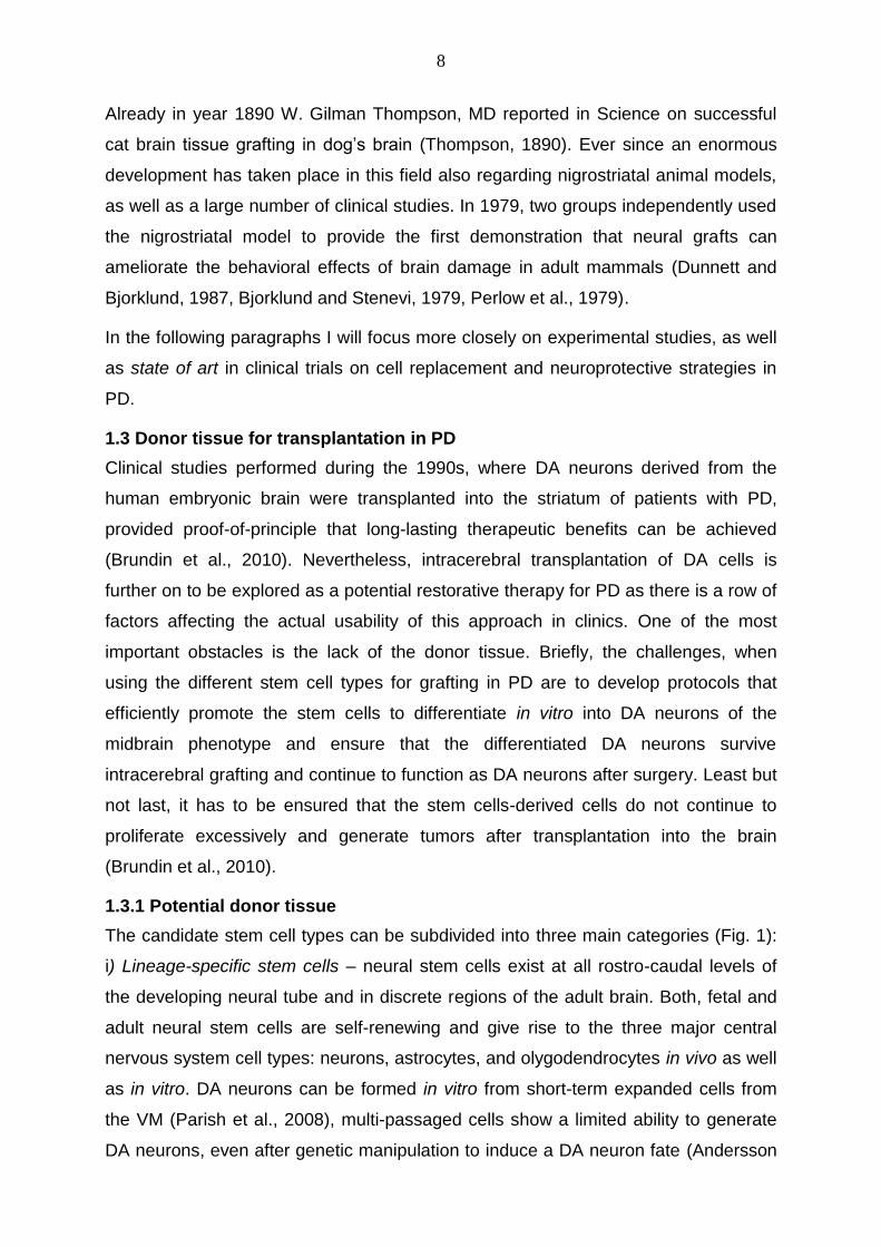
The candidate stem cell types can be subdivided into three main categories (Fig. 1):
i) Lineage-specific stem cells – neural stem cells exist at all rostro-caudal levels of
the developing neural tube and in discrete regions of the adult brain. Both, fetal and
adult neural stem cells are self-renewing and give rise to the three major central
nervous system cell types: neurons, astrocytes, and olygodendrocytes in vivo as well
as in vitro. DA neurons can be formed in vitro from short-term expanded cells from
the VM (Parish et al., 2008), multi-passaged cells show a limited ability to generate
DA neurons, even after genetic manipulation to induce a DA neuron fate (Andersson

9
et al., 2007, Roybon et al., 2008). DA neurons are uniquely derived from floor plate
cells that become neurogenic only in the midbrain region. Floor plate cells may
require different conditions for expansion and thus they are not maintained using
standard culture conditions, resulting in a diminished capacity to generate DA
neurons. ii) Embryonic stem cells (ESCs) – these cells are self-renewing and
pluripotent, derived from the inner cell mass of the pre-implantation blastocyst.
Human ESCs (hESCs) maintain the developmental potential to contribute to cells of
all three germ layers (Amit et al., 2000). ESCs differentiate into neurons with
characteristics of all levels of the central nervous system. However, there are studies
showing that grafted hESC-derived DA neurons failed to ameliorate behavioral
deficits to the same degree as fetal VM-derived neurons (Christophersen and
Brundin, 2007). Additionally, the survival of the DA neuron phenotype is poor when
hESC-derived neurons are grafted into the adult brain. It is surely owing to the
immunogenity of the hESC-derived cells (Li et al., 2008a). A major obstacle to use
Figure 1. Schematic drawing on stem cell-derived neurons for grafting in PD.
The left part of the figure depicts hESCs and fetal tissue sources, which both result in allografts; the right part of the figure illustrate iPS and iN cells that can be sources of syngeneic “personalized” donor tissue originating from the patient. Adapted from (Brundin et al., 2010)
the ESCs in clinical setting is their capacity to form teratomas and neural overgrowth
in the host brain (Pruszak et al., 2009, Placantonakis et al., 2009). iii) Induced
pluripotent stem cells (iPS cells) – re-programmed somatic cells are phenotypically

10
and morphologically very similar to ESCs and are germ-line-competent. The iPS
could enable to generate PD-patient-specific stem cells on demand (Soldner et al.,
2009). Combinatorial expression of neural-specific transcription converts fibroblasts
directly into functional neurons in vitro (induced neuronal cells (iN cells)) (Vierbuchen
et al., 2010). The iPS cells share the same proliferative capacity as ESCs and thus
imply the same risk of incomplete and unsynchronized differentiation. Furthermore,
since the reprogrammed cells do not proliferate, very large numbers of
reprogrammed neurons should be generated for each patient in order to provide
sufficient material for transplantation. Even more, it could be argumented that PD-
patient-derived iN cells or iPS cell-derived neurons might degenerate after grafting
due to PD, in analogy to the patient’s own DA neurons (Brundin et al., 2010).
1.3.2 Cell-replacement related dopaminergic neuron loss
The percentage of grafted embryonic DA neurons that survive transplantation is low,
estimated at 5-20%. Out of two possible patterns of cell death it is apoptosis
occurring at early time following grafting in mesencephalic tissue grafts (Mahalik et
al., 1994, Emgard et al., 1999,
Sortwell et al., 2000, Emgard et
al., 2003). Specific conditions
associated with the
transplantation procedure and
post transplantation interval set a
variety of signals in the cell’s
environment free that can trigger
an intrinsic genetically driven cell
“suicide” program (Raff et al.,
1993). The overwhelming
majority of apoptotic cell death
occurs within the first 7 days after transplantation of mesencephalic graft
suspensions (Sortwell et al., 2000).
The transplantation process can be divided in 3 stages (Fig. 2): (i) Stage 1 – Tissue
dissection and preparation. The dissociation procedure itself is a triggering factor and
a large population of DA neurons is already committed to die prior to implantation.
This is largely explained by anoikis – apoptosis due to detachment from the
extracellular matrix (ECM) and disruption of contacts to the neighboring cell. The
Figure 2. Cell preparation/grafting conditions that can trigger grafted cell death.
Adapted from (Sortwell, 2003)
11
term comes from the ancient Greek word for “homelessness”. Integrins being the
major ECM receptors in vivo play a crucial role in this phenomenon. (ii) Stage 2 –
Graft environment. It is likely analogue to what is experienced in the brain during
ischemic insult, where access to blood-born nutrients is denied. In rats, the peak of
growth of the host and donor vessels occurs at approximately three days post
transplantation. Apoptotic cell death within this stage appears not only due to
hypoxia, but also in response to trophic factor withdrawal (Sortwell, 2003).
Namely, many of the molecules needed for the DA neuron survival and growth are
down-regulated or absent in the mature host striatum (Collier and Sortwell, 1999). It
could also be that failure of the implanted DA cells to access striatal target-derived
trophic factors might lead to apoptosis. (iii) Stage 3 – Immune response. A relatively
small portion of DA neurons is dying in this phase. Intraparenchymal allografts of
fetal mesencephalic cell suspensions can survive well without immunosuppression
for at least 12 weeks in a rat model of PD (Brandis et al., 1998, Sortwell et al., 2000).
Hence adequate immunosuppression is required in clinical setting to avoid graft
rejection (Barker, 2006).
The immediate post implantation interval and established graft-phase evolving DA
graft survival, maturation and differentiation are typically described also as two
phases. The phase1 is lasting 2 weeks post implantation, characterized by loss of
80%-90% of the transplanted DA neurons and the initial axonal outgrowth. In phase2
the final synaptic and functional integration of DA grafts into the host BG circuitry
takes place (Hahn et al., 2009). This arrangement in chronologically relevant phases
gives the basis for time-spans and evaluation criteria in short- and long-term
experimental studies on morphological and functional integrity of DA transplants in
parkinsonian animal models.
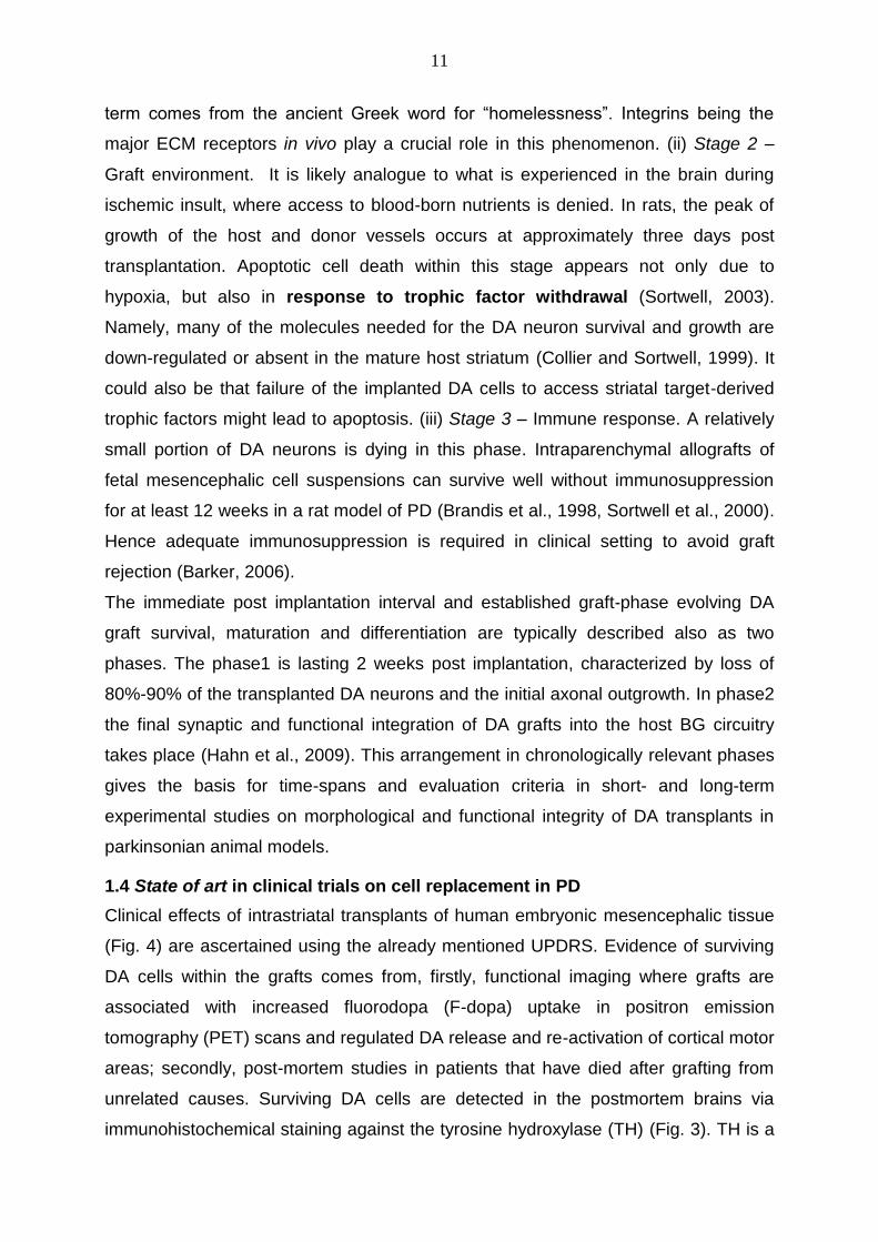
1.4 State of art in clinical trials on cell replacement in PD
Clinical effects of intrastriatal transplants of human embryonic mesencephalic tissue
(Fig. 4) are ascertained using the already mentioned UPDRS. Evidence of surviving
DA cells within the grafts comes from, firstly, functional imaging where grafts are
associated with increased fluorodopa (F-dopa) uptake in positron emission
tomography (PET) scans and regulated DA release and re-activation of cortical motor
areas; secondly, post-mortem studies in patients that have died after grafting from
unrelated causes. Surviving DA cells are detected in the postmortem brains via
immunohistochemical staining against the tyrosine hydroxylase (TH) (Fig. 3). TH is a
12
rate-limiting enzyme of catecholamine (dopamine,
epinephrine and norepinephrine) biosynthesis. It
converts tyrosine to DA (Daubner et al., 2011).
The first clinical trials in the field of neural grafting in
PD began in the late 1980’s in Mexico (Madrazo et al.,
1988) and Sweden (Lindvall et al., 1989). Because of
significant improvements post transplantation several
open-label clinical studies followed in different centers
across Europe (Wenning et al., 1997, Lindvall et al.,
1990, Brundin et al., 2000a, Hauser et al., 1999). The
number of patients in each of the studies was low
ranging from two to six, nevertheless the results were
showing that fetal allografts became functionally
integrated and produced clinical benefits. In the
following double-blind placebo-controlled studies from
Freed et al. (Freed et al., 2001) and Olanow et al.
(Olanow et al., 2003) the number of participants was thirty-two and forty respectively.
These studies intended to show that VM grafting in patients with PD was able to
produce a real effect over placebo-mediated benefits. In the study of Freed et al.
there were significant improvements seen in the UPDRS motor scores in grafted
patients who were less than 60 years old compared to sham surgery group. In case
of the Olanow et al. study transplanted patients showed significant motor
improvement compared to placebo at 6 and 9 months after engraftment, but not
thereafter. This coincided with the discontinuation of the immunosuppressive therapy.
The major side-effect of the transplantation procedure were the graft-induced
dyskinesias (GIDs) characterised as stereotypic, rhythmic movements of one or both
lower extremities (Freed et al., 2001, Olanow et al., 2003). The GIDs could be
effectively treated with DBS in the globus pallidus.
Up to now, it is known that post-mitotic DA neuroblasts of the VM of 6-9 week old
human embryos (3-4 donors per striatum) survive transplantation into the brain of PD
patients (Fig. 3). The grafts restore striatal DA release as PET scans detect
increased F-dopa uptake (Brundin et al., 2000b, Freed et al., 2001, Lindvall et al.,
1990, Olanow et al., 2003). In addition, histopathological studies show extensive
long-term (Fig. 3) synaptic reinnervation in the patient’s striatum (Kordower et al.,
Figure 3. Surviving DA neurons labeled with antibody to TH in a graft transplanted 16 years before the death of the patient.
Surviving cells mainly exist in the periphery of the graft with classical morphology of DA neurons, extending long processes into the host striatum. Adapted from (Li et al., 2008b)

13
2008, Kordower et al., 1996, Li et al., 2008b, Mendez et al., 2008, Olanow et al.,
2003, Freed et al., 2001, Mendez et al., 2005). Clear clinical benefits associated with
graft survival have been shown in both, open-labeled (Brundin et al., 2000b, Hagell et
al., 1999, Hauser et al., 1999) and sham surgery controlled trials (Freed et al., 2001,
Olanow et al., 2003). In the most successful cases antiparkinsonian medication could
be withdrawn and the clinical
improvements were
experienced for several
years. Reports on whether
grafted neurons are affected
by pathogenic factors
intrinsic the PD are still
controversial and depend on
the transplantation
methodology used in the
particular group (Li et al.,
2008b, Mendez et al., 2008,
Kordower et al., 2008).
Altogether, several
fundamental experimental
design issues have been
identified, like: (i) the follow-
up time after grafting, (ii) the
type of the cells, the amount
of the tissue used for single
transplantation and incubation times prior to grafting, (iii) the need for
immunosuppression (Brundin et al., Kuan and Barker, 2005, Barker, 2006, Ma et al.,
Olanow et al., 2001). In order to ensure reproducibility of successful results across all
up-coming patients receiving VM tissue transplants in all centres, leading clinicians,
scientists, industrial partners, ethicists and patients’ representatives have joined in
the TRANSEURO European research consortium. The first transplantations within
clinical trials from this consortium are planned to take place at the end of year 2012.
Figure 4. Transplantation of embryonic mesencephalic tissue or of DA neuroblasts generated from stem cells in a human PD brain.
In the normal brain, DA neurosn located in the substantia nigra send their axons to the striatum (i.e. putamen and caudate nucleus). In the PD brain, degeneration of these neurons causes loss of DA in the striatum. Transplantation of embryonic tissue, which is rich in DA neuroblasts, or of DA neuroblasts generated from stem cells aims to restore striatal DA innervations thereby alleviating PD symptoms. Adapted from (Lindvall and Kokaia, 2009)

14
1.5 Basal ganglia circuitry and the midbrain DA system
The term basal ganglia (Fig. 5) refers to a group of subcortical nuclei that include the
striatum, globus pallidus (GP), substantia nigra (SN) and subthalamic nucleus (STN).
Each of these is clinically profoundly important. Striatum consisting of the caudate
nucleus and putamen receives most of the cortical input on the BG. Degeneration of
neurons in the striatum leads to Huntington’s disease (HD) and related hyperkinetic
disorders. The GP (consisting of internal and external segments) receives most of
the output of the striatum. The pallidum is the site of therapeutic lesion (pallidotomy)
and DBS procedures used to relieve PD. The subthalamic nucleus is a key structure
controlling pallidal function (Krack et al., 1998, Zahm and Heimer, 1988).
Functional connections between these anatomical substrates are known as striato-
pallido-thalamic loop. Interconnected, the BG nuclei process motor, limbic, sensory,
and associative information coming from virtually all areas of the cerebral cortex and
return the processed information to the same cortical regions. The overall function of
the BG is to control the initiation and selection of voluntary movements.
The two output nuclei of the BG, the internal pallidal segment and the SN pars
reticulate (SNr), tonically inhibit their target nuclei in the thalamus and brain stem.
This inhibitory output is thought to be modulated by the two parallel pathways that
run from the striatum to the two output nuclei: one direct and the other indirect. The
two pathways originate from different subsets of striatal neurons and, in the model
(Fig. 5), remain functionally segregated. In the direct pathway, striatal γ-aminobutyric
acidergic (GABAergic) neurons, expressing D1 dopamine receptors, project
monosynaptically to the SNr and medial globus pallidus. In the indirect pathway, the
striatal output reaches the target nuclei via a more complex route. In fact, a different
subset of GABAergic neurons, which expresses D2 receptors, project to the lateral
globus pallidus, which in turn sends GABAergic projections to the subthalamic
nucleus. The subthalamic nucleus sends its glutamatergic efferents to the output
nuclei and to the lateral GP. From the output nuclei, inhibitory, GABAergic projections
reach the ventral lateral and ventral anterior nuclei of the thalamus. Thalamic nuclei
then send glutamatergic projections to the motor cortex, then closing the loop (Albin
et al., 1989, Alexander and Crutcher, 1990, Gerfen, 1992b, Graybiel, 1990, Gerfen,
1992a).

15
Figure 5. Schematic representation of the major connections of the BG.
The main component of the BG, the striatum, receives inputs from the cortex. Two major striatal output pathways target GP and enteropeduncular (EP)-SNr complex. DA neurons in the SN pars compacta (SNc) receive inputs from the striatum (not diagrammed) and provide feedback via the nigrostriatal DA pathway. EP and SNr neurons provide inhibitory inputs to the pedunculopontine nucleus (PPN), superior colliculus, and thalamus. Nigrothalamic inputs target intralaminar nuclei that provide feedback to the striatum (not shown) and ventral tier thalamic nuclei that provide inputs to the frontal cortex. Adapted from (Gerfen, 1992b)
Thus the BG have distinct pathways that compete with each other functionally to
trigger movement (the direct pathway) or to inhibit movement (the indirect pathway).
These competing pathways act like a break and accelerator in a car. In the simplest
view, the poverty of movement in PD results from over-activity of the indirect
pathway, whereas excess movement in disorders such as HD represents over-
activity of the direct pathway.
In rodents, the midbrain DA system is divided in 3 groups with distinct anatomical
localization and topographical projection to the striatum and other brain regions. The
A10 cell group is located in the ventral tegmental area (VTA) and projects to the
limbic forebrain areas, i.e. septal area, prefrontal cortex, olfactory tubercle and the
nucleus accumbens (ventral striatum). The A9 and A8 cell groups are situated in the
SNc and the retrorubral area (RRA), respectively, and provide all the remaining DA
projection to the striatum (Smith and Kieval, 2000). Particularly the A9 subpopulation
plays a crucial role in improvement of motor performance after DA transplantation.
There exists a marker which is expressed in the midbrain exclusively in the THir A9
neurons, namely G-protein-gated inwardly rectifying K+ channel (Girk2). Therefore, it
is possible to detect whether the correct (nigral) DA neuron subtype is present in the
implanted fetal mesencephalic tissue (Grealish et al., 2010, Thompson et al., 2005).
1.5.1 6-hydroxydopamine (6-OHDA) animal model of PD
Animal models of PD can be roughly divided in (i) neurotoxin models, (ii) gene
alteration models, (iii) kindling models (disease induced by electrical means), and (iv)
spontaneous mutation models (in rodents). The most commonly used neurotoxins
are 6-OHDA and 1-Methyl-4-phenyl-1,2,3,6-tetrahydropyridine (MPTP) (Bove et al.,

16
2005). Discovery of familial PD mutations has opened a way to develop genetic
models of PD.
6-OHDA is widely used in rats and mice, to reproduce the loss of DA innervations to
the striatum typical in PD. Because 6-OHDA does not cross the BBB it has to be
administered with intracerebral or intraventricular infusion. 6-OHDA is taken up by
DA and noradrenaline (NA) membrane transporters (DAT and NAT respectively) and
accumulated in the cytosol. DAT and NAT recognize 6-OHDA due to its structural
similarity with endogenous catecholamines (Simola et al., 2007). 6-OHDA has been
shown to inhibit mitochondrial respiratory chain complex (Glinka et al., 1996).
Consequently, reactive oxygen species (ROS) are generated and oxidative stress
leads to cell death. Apoptosis markers including cleaved caspase-3 and condensed
chromatin, are detected in cells treated with 6-OHDA. 6-OHDA injection also induces
gliosis and astrocytic activation (Bove et al., 2005, Marti et al., 1997).
After 6-OHDA is injected into SNc or medial forebrain bundle (MFB), DA neurons
start to die within the first 24 hours (Jeon et al., 1995). Maximum reduction of striatal
DA level is reached within 3-4 days after lesion (Faull and Laverty, 1969).
Interestingly, despite the dramatic loss of DA neurons in the SNc after a MFB
injection of a high dose of 6-OHDA, levels of extracellular DA are still close to normal
(Sarre et al., 2004). This could be explained by somatodendritic release of DA from
the few spared neurons in the SN. When injected into the dorsal striatum, 6-OHDA
produces more protracted retrograde degeneration of the nigrostriatal system which
peaks from 1 to 3 weeks (Przedborski et al., 1995, Sauer and Oertel, 1994).
Even if a drawback common to all the PD models induced by neurotoxins is the
rapidity of DA neurodegeneration compared to that occurring in parkinsonian patients
(lasting for years), it seems that the 6-OHDA lesions can at least in part reproduce its
progression. Rodents with bilateral lesions show a range of motor impairments, such
as postural abnormalities at rest (Wolfarth et al., 1996), reduction of spontaneous
movements (Schallert et al., 1978, Schallert et al., 1979), and increased muscle
resistance to passive stimuli (Schallert et al., 1978, Wolfarth et al., 1996). Muscle
resistance in bilaterally lesioned rats has electromyographic features that are similar
to parkinsonian rigidity (Wolfarth et al., 1996, Berardelli et al., 2001). In addition,
footprint analysis shows that these animals take short steps when they walk forward,
which reproduces the gait abnormalities observed in PD patients (Schallert et al.,

17
1978). However, since bilateral DA depletion causes severe anhedonia (inability to
experience pleasure from activities formerly found enjoyable) for food and water, the
animals require intensive post-operative care. On the contrary, unilateral lesions are
well tolerated by both rats and mice.
Therefore, unilaterally lesioned (referred to also as hemiparkinsonian) rats and mice
are the most widely used animal model of PD (Schwarting and Huston, 1996).
Unilateral administration of 6-OHDA in rodents leads to unilateral degeneration of the
nigrostriatal DA pathway. This induces asymmetries of the body posture and
contralateral sensorimotor deficits. The extent of DA depletion can be assessed by
examining circling behavior in response to certain pharmacological substances -
amphetamine and apomorphine, for instance. And normalization of DA function after
application of restorative treatments is usually determined in this model by decreased
drug-induced rotational behavior (the respective tests will be discussed more detailed
in 1.5.2). Other specific motor disabilities observed in the unilateral 6-OHDA-lesioned
rodent model include impaired ability to use the forelimb contralateral to the side of
the lesion, to adjust posture, explore, walk, and groom (Olsson et al., 1995, Cenci et
al., 2002, Carli et al., 1985, Spirduso et al., 1985, Metz et al., 2001, Montoya et al.,
1991). Similar movement fragmentation and abnormal postural adjustments are seen
in people with Parkinson’s disease.
1.5.2 Drug-induced rotation tests in unilateral rat 6-OHDA model – analysis of functional deficits and recovery
In year 1987 Dunnett and Björklund wrote in their review on mechanisms of function
of neural grafts in the adult mammalian brain that the DA nigrostriatal system is
ideally suited for behavioral studies since there exists the selective neurotoxin 6-
OHDA, and the behavioral sequelae of such lesions is dramatic and simple to assess
(Dunnett and Bjorklund, 1987). And indeed, already in 1970 Ungersted and
Arbuthnott had published on quantitative recording of rotational behavior in rats post
lesion with 6-OHDA carried out using a rotometer (Ungerstedt and Arbuthnott, 1970).
As already mentioned, unilateral administration of 6-OHDA in rodents leads to
degeneration of the ipsilateral nigrostriatal DA pathway. Turning deficits after 6-
OHDA lesions appear due to a loss of ability to exert force to displace the body
(Miklyaeva et al., 1995). The extent of DA depletion can be assessed by examining
circling behavior in response to certain pharmacological substances - amphetamine

18
and apomorphine, for instance. Normalization of DA function is determined in this
model by decrease in drug-induced rotational behavior.
Administration of agonists of DA receptors (DRs), such as apomorphine, or L-DOPA
induces rotations towards the side contralateral to the lesion (Fig. 6), while DA
releasing drugs, such as amphetamine,
elicit turning behavior towards the side
ipsilateral to the lesion. Apomorphine
stimulates DRs directly, preferentially
on the denervated side due to
denervation-induced DR
supersensitivity (Creese et al., 1977).
Amphetamine, on the other hand,
increases synaptic levels of DA by
inducing its release from and inhibiting
its reuptake into intact terminals,
causing ipsilateral rotations due to
increased DA activity on the intact side.
This drug affects DRs indirectly by
increasing extracellular availability of
endogenous DA. As amphetamine requires endogenous DA for its action, it
increases DA activity especially or solely in the intact (contralateral) hemisphere and
thus exaggerates the DA imbalance between the denervated and intact side
(Ungerstedt and Arbuthnott, 1970, Schwarting and Huston, 1996).
Studies in rats having received unilateral 6-OHDA injections into the MFB, have
shown objective differences in how predictive of the extent of nigrostriatal DA
depletion is either apomorphine- or D-amphetamine-induced rotation behavior. The
latter one elicits rotations in most animals with unilateral 6-OHDA lesions that
produce striatal depletions of 75% or more. In general, submaximally lesioned (75-
90% depleted) rats do rotate on D-amphetamine but not on apomorphine.
Apomorphine has been shown to be a better predictor of extensive unilateral lesions
by using behavioral and histochemical measures (Carman et al., 1991, Casas et al.,
1988, Hefti et al., 1980). Virtually no apomorphine-induced rotations are seen unless
striatal depletions are at least 90% (Heikkila et al., 1981, Marshall and Ungerstedt,
1977).
Figure 6. Diagram of the nigrostriatal pathway and rotational responses produced by apomorphine and D-amphetamine.
Shaded areas indicate the loss of DA due to MFB injection of 6-OHDA. L, left, R, right, STR, striatum, SN, substantia nigra. Adapted from (Hudson et al., 1993)

19
No correlation has been seen between the extent of VTA depletion and either
apomorphine- or amphetamine-induced turning behavior (Hudson et al., 1993).
Ipsilateral rotation has been used to monitor the effects of DA-denervating lesions,
neural grafts and neuroprotective treatment, while contralateral rotation has been
used to assess the anti-kinetic potential of candidate antiparkinsonian drugs
(Schwarting and Huston, 1996, Torres and Dunnett, 2007).
1.5.3 Neuronal transplantation in rat 6-OHDA model of PD
To date, neuronal transplantations in a rat unilateral 6-OHDA model of PD are
performed based on the microtransplantation
approach. The standard protocol for the
implantation of fetal nigral cell suspensions to
the host brain was originally described back in
year 1983 (Bjorklund et al., 1983a, Bjorklund et
al., 1983b). In the earlier studies, there were
solid pieces of tissue used.
The implantation of cell suspensions has
various advantages compared to this previous
approach: (i) these can be grafted to the target
sites causing less trauma and with high
stereotactic accuracy; and (ii) the neuronal cell
suspensions can be manipulated prior to
grafting in the rat Parkinson model. It has
become a golden standard technique for
preparation and implantation of not only DA
grafts since then. Nikkhah et al. (1994)
modified this protocol and introduced glass
capillaries (Fig. 7) to optimize the
transplantation technique. The so-called
microtransplantation approach led to better graft survival and functional integration.
The metal canula (Ø 0.5mm) of a 1- or 2-µl Hamilton microsyringe was connected to
a glass capillary (a long-shanked glass micropipette) (Ø 50-70µm) with a cuff of
polyethylene tubing as an adapter (Fig. 7). This resulted in an excellent infusion of
single cell suspensions, minimal mechanic trauma during the injection, and ensured
the possibility to inject multiple deposits of implanted cells, also along the same
Figure 7. The modified micro-transplantation instrument.
A, the Hamilton microsyringe is fitted with a long-shanked glass micropipette using adaptive tubing. B, ratio between extruded drop and tip size of a 500µm glass capillary, both extruding 1µl of ink. Adapted from (Nikkhah et al., 1994b, Jiang, 2008)

20
implantation tracts (Nikkhah et al., 1994a, Nikkhah et al., 1994b, Winkler et al., 1999,
Nikkhah et al., 2009). Microtransplantation offered an efficient, reliable, and precise
technique for quantitative studies on the influence of grafting parameters onto the in
vivo survival, differentiation, and functional capacity of fetal DA neuron-rich grafts in a
rat model of PD (Nikkhah et al., 2009).
This technique achieves a 2.5 to 20.7 fold higher survival of THir cells, a 2 fold graft-
volume and a 2 to 3 fold higher THir fiber density of grafted cells in the
transplantation of embryonic VM cell suspension in PD models than their canula-
transplanted control groups. Moreover, the mechanical trauma leading to formation of
astroglial scar (evaluated by glial fibrillary acidic protein (GFAP) staining) was
significantly lower in the micro-transplanted groups than in the canula-transplanted
controls (Nikkhah et al., 1994a). In addition, the reduced level of trauma and
subsequent reduced major histocompatibility complex and GFAP expression may,
thereby, minimize the risk of graft rejection (Brandis et al., 1998).
1.6 Neurotrophic factors (NTFs) for treatment of PD and brain-derived neurotrophic factor (BNDF) as the factor of interest
Ever since 1987 it is known that only a limited percentage of the grafted DA neurons
survive the procedure (Brundin and Bjorklund, 1987). It is considered that insufficient
neurotrophic support at the transplantation site may be at least in part responsible for
the poor survival rate of grafted DA neurons. Furthermore, although lack of NTFs
has not been shown to directly cause neurodegenerative diseases, the levels of
NTFs are decreased in neurodegeneration (Chao et al., 2006).
NTFs are diffusible peptides secreted from neurons and neuron-supporting cells.
They serve as growth factors for the development, maintenance, repair, and survival
of specific neuronal populations, and they act via retrograde signalling from target
cells via paracrine and autocrine mechanisms (Yuen et al., 1996). In adulthood, they
help to maintain neuronal functions and specific neuronal phenotypes (Levy et al.,
2005). Additionally, as these factors embody a potential of modifying the astrocytic
activation, inflammatory reactions, and neuronal dysfunction under pathologic
conditions, they embody a therapeutic potential for regenerative therapies (Peterson
and Nutt, 2008).
NTFs belong to superfamilies of structurally and functionally related molecules (Levy
et al., 2005). There is a row of NTFs, such as the fibroblast-growth factor-2 (FGF-2)
(Otto and Unsicker, 1990, Timmer et al., 2004, Timmer et al., 2007, Jensen et al.,

21
2008, Grothe and Timmer, 2007) and the brain-derived neurotrophic factor
(BDNF), which have been identified to play a role in midbrain DA neuron
maintenance (Krieglstein, 2004) and to be able to arrest and restore function of DA
neurons in PD models (Peterson and Nutt, 2008).
BDNF belongs to the neurotrophin superfamily and is one the best established NTFs
in experimental settings for parkinsonian research. At the molecular level, the BDNF
gene encodes a BDNF precursor (proBDNF), which is cleaved post translationally to
form the mature BDNF. This mature form has a potential to promote neural survival
and enhance synaptic plasticity. The BDNF acts by binding to distinct classes of
transmembrane receptors: the p75 receptor (p75NTR) and the TrkB receptor. The
actions of the BDNF as well as other neurotrophins are binary. The prosperity of
neurotrophins to produce diametrically opposing effects on cell survival is being
reflected in a so called “yin and yan” model. These effects depend on both, the form
of the neurotrophin, namely pro-isoform versus mature protein, and the class of the
receptor that is activated (Lu et al., 2005) (see Fig. 8A). Thus, for example, proBDNF
induces neuronal apoptosis by activating p75NTR (Teng et al., 2005).
BNF promotes survival of the mesencephalic DA neurons in vitro (Murer et al., 2001,
Hyman et al., 1991) and in vivo (Hagg, 1998). Decreased BDNF mRNA expression
and protein content have been observed in the SN of PD patients (Mogi et al., 1999,
Parain et al., 1999, Howells et al., 2000). Therefore, prolonged supply of BDNF in the
substantia nigra/striatum might be a possible therapy for PD (Pezet and Malcangio,
2004, Zuccato and Cattaneo, 2009). However, the large molecular size of BDNF,
coupled with the BBB, prevents its delivery to DA neurons to promote cell survival in
the PD brains (Lee et al., 2001). One of the approaches research is currently running
on deals with identification and testing of low molecular weight compounds either
mimicking NTFs or increasing synthesis of the respective NTF. As for BDNF, such
substances as salicylic acid, cGMP analogues, dipyridamole and glutamate have
been identified to increase the production of BDNF in the DA neurons of substantia
nigra (Chun et al., 2000) (Fig. 8B). Other therapeutic strategies tending to raise the
concentration of therapeutic trophic factors in the target tissue are invasive. In a rat
6-OHDA model of PD, intrastriatal injection of BDNF reduces the loss of DA cells and
behavioral changes associated with this model (Shults et al., 1995). This is true also
in case of direct nigral or intrathecal infusion of BDNF in MPTP mice or monkeys
where reversing of the decrease in DA concentrations can be achieved (Hung and

22
Lee, 1996, Tsukahara et al., 1995). Striatal grafts of genetically modified fibroblasts
engineered to synthesise BDNF show a beneficial effect after intrastriatal
administration of 6-OHDA (Levivier et al., 1995) or of MPTP (Frim et al., 1994,
Galpern et al., 1996, Isacson et al., 1995).
Implanting cells programmed to secrete the trophic factor of interest allows
combining restorative strategies for PD (replacement of the lost DA population) and
protective strategies, promoting survival of both, the transplanted DA cells as well as
the remaining host DA neurons. These strategies can be combined in three
distinctive ways: i) co-culture of generated DA neurons with feeder cells expressing
the NTF of interest (Timmer et al., 2004); ii) addition of soluble NTFs of interest into
cell culture prior to transplantation (Andereggen et al., 2009); iii) genetic modification
of cells to express the NTF of interest (Behrstock et al., 2006, Parish et al., 2008).
With regard to clinical compatibility, cells to transplant should be free of feeder cells
and the usage of viral vectors should be avoided.
B A
Figure 8. A, The yin and yang of neurotrophin receptors and neurotrophin functions. B, Experimental therapeutic strategies for restoring BDNF function in neurodegenerative diseases.
A The actions of neurotrophins are mediated by two principal transmembrane-receptor signaling systems. Each neurotrophin receptor – TrkA, TrkB, TrkC and the p75 neurotrophin receptor (p75NTR) – is characterized by specific affinities for the neurotrphins nerve growth factor (NGF), brain-derived neurotrophic factor (BDNF), neurotrophin 3 (NT3) and NT4. An emerging concept is that the two distinct receptor classes, Trk (top) and p75NTR (bottom) preferentially bind mature and proneurotrophins (neurotrphin precursors), respectively, to elicit opposing biological responses. LTD, long-term depression, LTP, long-term potentiation. Adapted from (Lu et al., 2005) B Adapted from (Zuccato and Cattaneo, 2009)

23
In this context, our newly established co-layer method combining transient genetic
modification via nucleofection and optimized cell culture conditions evolving no
feeder cells is offering a safe way to deliver NTFs at the site of transplantation.

24
2 AIMS OF THIS PROJECT
Neither the pharmacological therapies nor surgical approaches are able to stop or
reverse the neurodegenerative process underlying the PD. Exogenous cell
replacement therapy provides a novel treatment option for terminal stages of the
disease. In this regard, experimental as well as clinical studies have already shown
promising results. However, only up to 5% of the grafted dopaminergic (DA) cells are
reported to survive the transplantation procedure.
Our group has previously shown that it is possible to non-virally genetically modify
E12 NPCs, even though the numerical outcome of THir cells in vitro and in vivo
remained unsatisfactory (Cesnulevicius et al., 2006). The aim of the present study
was to establish a method allowing to nucleofect the neuronal progenitor cultures
and at the same time ensuring high yield of both - THir cells and genetically modified
cells in the cell suspension engrafted into unilateral 6-hydroxydopamine (6-OHDA)
rat model of PD.
The objectives of the present study were:
1) Determination of the most proficient expression plasmid for optimal transfection
rates of VM NPCs, as well as expression time-spans of reporter sequences and
respective growth factors in vivo.
2) Establishment of a method preventing reduction of DA neurons after their genetic
modification by nucleofection to overexpress NTFs (see 3.4.1 ii).
3) Detection of the grafted mesencephalic NPCs over reasonable time-spans in vivo
(up to 13 weeks).
4) Selection in vitro of the appropriate plasmid-based delivered NTF with the aim to
achieve significant morphological and functional restorative capacity after
transplantation into unilateral parkinsonian animal model (Data obtained by A. Nobré,
PhD and Dr. M. Hohmann).
5) Evaluation of the functional and morphological restorative potential of the
transplanted DA neurons by means of pharmacologic behavioral tests, qualitative
and quantitative evaluation of the grafted DA population, and morphological integrity
of the grafts when implanting genetically modified NPCs treated with the newly-
established method into a rat PD model.

25
3 MATERIALS AND METHODS
3a MATERIALS used for the in vivo study
Table 1. Materials, substances, and equipment used for this study listed in chronological order of usage
Materials Manufacturer
6-OHDA lesion surgery
Chloral hydrate Sigma-Aldrich Chemie GmbH, Steinheim, Germany
5 ml-single-use syringe B. Braun Melsungen AG, Melsungen, Germany
BD Microlance™ 3 canule (21 G, 1½´´, 0,8 x 40 mm, i.p.)
Becton Dickinson GmbH, Heidelberg, Germany
Beard trimmer MT4640 Grundig Intermedia GmbH, Nürnberg, Germany
Lab Standard Stereotaxic - Single w/18 Degree Earbars
Stoelting Co., Illinois, USA
Large Probe Holder Stoelting Co., Illinois, USA
Disposable scalpel (Nr. 15) pfm – Produkte für die Medizin AG, Collogne, Germany
Drill Micromot 50/E Proxxon, Niersbach, Germany
Micro drill steel burs (spherical, Ø 2,1 mm) Hager & Meisinger GmbH, Neuss, Germany
Cellulose pads Zelletten (5 x 4 cm) Lohmann & Rauscher International GmbH & Co. KG, Rengsdorf, Germany
Cotton staves Centramed Medizintechnik HandelsGmbH & Company, Koblenz, Germany
Hydrogen peroxide 30% (in water) Sigma-Aldrich Chemie GmbH, Steinheim, Germany
Saline solution (NaCl 0,9%) B. Braun Melsungen AG, Melsungen, Germany
Ethanol 70% (100% diluted) Mallinckrodt Baker B.V., Deventer, Netherlands
6-Hydroxydopamine hydrochloride Sigma-Aldrich Chemie GmbH, Steinheim, Germany
L-ascorbic-acid Sigma-Aldrich Chemie GmbH, Steinheim, Germany
10 μl Fixed Needle - Plunger Protection Syringe, 10F-5C
SGE GmbH, Griesheim, Germany
Microsyringe 801RN 10 μl SYR (26s/2''/2) Hamilton Bonaduz AG, Bonaduz, Switzerland
Microsyringe 701 RN 10 μl SYR (26s/2''/2) Hamilton Bonaduz AG, Bonaduz, Switzerland
Michel suture clip (7,5 x 1,75 mm) B. Braun Aesculap AG, Tuttlingen, Germany
26
Bepanthen eye- and nose-ointment Bayer Vital, Leverkusen, Germany
Bulldog Serrefines - Straight, 35 mm Fine Science Tools GbmH, Heidelberg, Germany
Raspiratory, 6 mm wide, 90 degrees angled B. Braun Aesculap AG, Tuttlingen, Germany
Xylocain 1% injection solution, 50 ml with Adrenalin 1:200’000
Astra Zenica
Student Surgical Scisors - Sharp/Blunt, 14,5 cm Fine Science Tools GmbH, Heidelberg, Germany
Ethilolon II 4-0, 45 cm monofilament suture thread
Johnson & Johnson MEDICAL GmbH, Nordstedt, Germany
Adson Greafe Aichele Werkzeuge GmbH, Crailsheim, Germany
Pharmacologically-induced rotation test
1 ml- single-use syringe –F (25 G x 5/8´´) B. Braun Melsungen AG, Melsungen, Germany
R-(-)-Apomorphin-hydrochlorid-hemihydrate Sigma-Aldrich Chemie GmbH, Steinheim, Germany
D-amphetamine Sulfate Sigma-Aldrich Chemie GmbH, Steinheim, Germany
Rotometer boxes (Plexiglas) Forschungswerkstätten Medizinische
Hochschule Hannover, Germany
Software
National Instruments LabView 6.1
National Instruments Germany GmbH,
Munich, Germany
Microtransplantation
(additionally to instruments needed for the 6-OHDA lesion surgery)
Microsyringe (2 μl)
7002KH 2.0 μl SYR (25/2.75’’/3)
Hamilton Bonaduz AG, Bonaduz, Switzerland
Student Dumont #5 Forceps - Standart tip, Straight
Fine Science Tools GmbH, Heidelberg, Germany
Glas capillaries (O.D. 1 mm) /Microcaps 50 mikroleters
Drummond Scientific Company, Pennsylvania, USA
Verrical Pipette Puller, Model 700C David Kopf Instruments, Tujunga, California
Heater coils Forschungswerkstätten Medizinische Hochschule Hannover, Germany
Polyethylene tubing, 30,5 m (I.D. 0,58 mm; O.D. 1,09 mm)
Intramedic, Becton Dickinson GmbH, Heidelberg, Germany
Polyethylene tubing, 30,5 m (I.D. 0,38 mm; O.D. 0,965 mm)
Intramedic, Becton Dickinson GmbH, Heidelberg, Germany
DMEM/Ham´s F-12 (without L-Glutamine) medium
PAA Laboratories GmbH, Pasching, Austria

27
Transcardial perfusion
PBS (w/o Ca2+, Mg2+) Biochrom AG, Berlin, Germany
Paraformaldehyde Fluka, Buchs, Switzerland
Student Surgical Scisors - Sharp/Blunt, 14,5 cm Fine Science Tools GmbH, Heidelberg, Germany
Student Iris Scisors - Straight, 11,5 cm Fine Science Tools GmbH, Heidelberg, Germany
Nub-canule (22 G, straight) Fine Science Tools GmbH, Heidelberg, Germany
Student Adson Forceps, Serrated Fine Science Tools GmbH, Heidelberg, Germany
Saccharose Carl Roth GmbH & Co., Karlsruhe, Germany
Friedman Rongeur - 2,5mm, Cup Curved Fine Science Tools GmbH, Heidelberg, Germany
Discofix-3, three-way plug valve B. Braun Melsungen AG, Melsungen, Germany
Cover glass forceps – angled, 10,5 cm Fine Science Tolls GmbH, Heidelberg, Germany
Glycerol ReagentPlus® (≥ 99,0%) Sigma-Aldrich Chemie GmbH,
Steinheim, Germany
Ethylene glycol Riedel-de Haën AG, Seelze, Germany
Histology
Cryostat Leica CM3050 Leica Microsystems GmbH, Wetzlar, Germany
Tissue-Tek Sakura Finetek Europe B.V.,
Zoeterwoude, Netherlands
Live Insect Forceps - Narrow, 10 cm Fine Science Tolls GmbH, Heidelberg, Germany
Cover Glass Forceps - Angled, 10,5 cm Fine Science Tolls GmbH, Heidelberg, Germany
Methanol 10% (100% diluted) Mallinckrodt Baker B.V., Deventer, Netherlands
(Griesheim, Germany)
Triton-X-100 Roche, Mannheim, Germany
Bovine serum albumin Sigma-Aldrich Chemie GmbH,
Steinheim, Germany
Normal goat serum Invitrogen™ (Gibco®), Karlsruhe, Germany
Anti-TH, monoclonal
(Mouse IgG1)
Sigma-Aldrich Chemie GmbH,
Steinheim, Germany
Biotinyl. rabbit anti-mouse, polyclonal Dako Denmark A/S, Glostrup, Denmark
Anti-TH, polyclonal
(Rabbit)
Millipore Corporation, Kalifornia, USA

28
Alexa Fluor 555
Goat anti-rabbit IgG (H+L), polyclonal
Invitrogen™, Karlsruhe, Germany
Anti-GFAP, polyclonal (Rabbit) Dako Denmark A/S, Glostrup, Denmark
Cy2-conjugated AffiniPure
Goat anti-rabbit IgG (H+L), polyclonal
Dianova, Hamburg, Germany
Anti-β-III-Tubulin, monoclonal
(Mouse IgG2a)
Biomol GmbH, Hamburg, Germany
Alexa Fluor 555
Goat anti-mouse IgG (H+L), polyclonal
Invitrogen™, Karlsruhe, Germany
4’,6-Diamidin-2’-phenylindol-dilactate Sigma-Aldrich Chemie GmbH,
Steinheim, Germany
ABC-Kit Vectastain® Elite (Mouse IgG) Linaris Biologische Produkte GmbH,
Wertheim, Germany
3,3‘-Diaminobenzidine Sigma-Aldrich Chemie GmbH,
Steinheim, Germany
Ammoniumnicelsulfate-6-hydrate Riedel-de Haën AG, Seelze, Germany
Object slide
Thermo Scientific (76x26 mm) for TH-DAB
Menzel GmbH & Co KG, Braunschweig, Germany
Object slide
Thermo Scientific Superfrost® Plus
(25x75x1 mm) for fluorescent stainings
Menzel GmbH & Co KG, Braunschweig, Germany
Gelatine Merck KGaA, Darmstadt, Germany
Xylol Mallinckrodt Baker B.V., Deventer, Netherlands
Coverslip (24x60 mm) Menzel GmbH & Co KG, Braunschweig, Germany
Dako Fluorescence Mounting Medium Dako Denmark A/S, Glostrup, Denmark
Corbit-balsam I. Hecht, Kiel-Hassee, Germany
Olympus BX60F5
Olympus Optical Co. (Europa) GmbH,
Hamburg, Germany
Olympus U-TV1X (U-CMAD-2)
Digital camera
Color View, Soft Imaging System GmbH,
Münster, Germany
Software CellP 2.6 Olympus Soft Imaging Solutions GmbH,
Münster, Germany

29
Olympus BX50F4 (for Stereology) Olympus Optical Co. (Europa) GmbH, Hamburg, Germany
Software C.A.S.T.-Grid 2.1.5.8 Olympus Danmark A/S, Albertslund, Denmark
Sony DXC-151AP
Color Video Camera Hyper HAD
CCD-IRIS/RGB
Sony Deutschland GmbH, Berlin, Germany
Camera-Adapter Sony CMA-D2CE Sony Deutschland GmbH, Berlin, Germany
Microcator ND281 Dr. Johannes Heidenhain GmbH,
Traunreut, Germany
Stage Controller Multi Control 2000 iTK Dr. Kassen GmbH, Lahnau, Germany
Immersion oil neoLab Migge Laborbedarf-
VertriebsGmbH, Heidelberg, Germany

30
3b METHODS
3.1 Animals and breeding
One hundred and fifteen adult female Sprague-Dawley (SPRD) rats (Charles River
Laboratories, research Models and Services, Germany GmbH, Sulzfeld, Germany)
weighing between 220 and 260g at the time of the first surgical procedure were
included in this study (transplanted animals included in the final analysis). Rats were
housed four animals per cage (Macrolon cages, type IV) in the Central Animal House
under a 14:10-hour light/dark cycle in a controlled temperature with food (Altromin)
and water ad libitum and allowed to acclimatize for at least 7 days in the animal care
facility prior to surgery. All procedures outlined were performed at the Hanover
Medical School according to the local guidelines for experimental studies and
permission (No 05/944 and 10/0098) of the regional administration (Hanover District
Council, Germany).
3.2 Stereotactic unilateral medial forebrain bundle lesion with 6-OHDA
Figure 9. Stereotactic surgeries.
A, Stereotactic apparatus. Head of the animals was fixed by bilateral ear bars in the external auditory canal. Ten µl Hamilton syringe was fixed on the stereotactic arm. Antero-posterior and lateral coordinates, as well as depth of lesions were adjusted with the aid of the micrometer drivers. Photo from personal archive. B, Schematic picture of the rat skull – injection sites for 6-OHDA administration (the green spots) and transplantation (the red spots). Adapted from (Paxinos, 1998). Bregma is the anatomical point on the skull at which the coronal suture is intersected perpendicularly by the sagittal suture. It is the point where the frontal bone and parietal bone meet. Bregma is used as a reference point for stereotactic surgeries on the brain. Lambda is the point of intersection of the sagittal and lambdoid sutures. C, Schematic representation of the 6-OHDA injection into the MFB. SNpc, substantia nigra pars compacta, SNpr, substantia nigra pars reticulata. Adapted from (Feldman, 1997).

31
In order to generate a hemiparkisonian rat model, animals were anesthetized with
chloral hydrate (370 mg/kg, i.p.) causing low side effects on neurodegenerative and
neuroprotective processes (Ozden and Isenmann, 2004). This was followed by two
stereotaxic injections of 6-OHDA (3.6 µg/µl in 0.2 mg/ml L-ascorbate-saline) targeting
the right medial forebrain bundle (MFB) (Fig. 9C). The neurotoxin was always kept on
ice and in darkness during the surgical procedure. For all of the surgical procedures,
the animals were fixed in the stereotactic apparatus (Fig. 9A). For both targets in the
right MFB one 2 x 2 mm hole was burred in the rat skull (Fig. 9B), injection
coordinates in relation to Bregma were calculated according to the atlas of Paxinos
and Watson et al. (Paxinos, 1998).
In order to adjust the parameters of the lesioning surgery several protocols for
stereotactic coordinates were examined in the Preliminary experiment (Exp) I. The
minimal parameter variances in the surgical protocols used for different animal
groups aiming to achieve a complete right MFB lesion are easy to follow in the Table
2.
Table 2. Lesioning parameters: coordinates for complete unilateral MFB lesion with 6-OHDA in a rat model for PD (Paxinos & Watson, 1998) The stereotactic coordinates are given according to the Bregma for craniocaudal and lateral coordinates, and to the dura mater or the brain surface (marked with *) for the ventral coordinate respectively.
Tooth-bar or incisor bar
Craniocaudal Lateral from the midline
Ventral Volume of injection
For the holes to burr
-2,4 or
-2,3
-4,4
-4,0
-1,2
-0,8
For the injections
-2,4*
+3,4
-4,4
-4,0
-1,2
-0,8
-7,8
-8,0
2,5μl
3,0μl
Firstly, stereological unilateral MFB lesion with 6-OHDA was performed testing
different tooth-bar heights for the first injection. Secondly, 10µl microsyringes
produced by two different companies, Hamilton and SGE respectively, were used.
The success of the lesioning surgery was evaluated in reference to pharmacologic
behavior tests (apomorphine and amphetamine rotation asymmetry) 5 and 7 weeks
after the surgical procedure.

32
Preliminary Exp I was comprised of 4 groups, 6 animals in each group as illustrated
in the Table 3.
Table 3. Experimental groups in the Preliminary Exp I
10μl Hamilton syringe used for lesioning procedure 10μl SGE syringe used for lesioning procedure
-2,4 tooth-bar for the 1st injection
-2,3 tooth-bar for the 1st injection
-2,4 tooth-bar for the 1st injection
-2,3 tooth-bar for the 1st injection
Group 1 Group 2 Group 3 Group 4
All animals received two stereotactic injections of 6-OHDA using a 10μl Hamilton or
SGE syringe into the right mesostriatal pathway (Fig. 9C) at the following coordinates
(in millimeters with reference to the Bregma and dura or the brain surface).
First, 2,5μl of 6-OHDA at: AP: -4,4, LAT: -1,2, VERT: -7,8, tooth-bar: -2,3 (Hahn et
al., 2009, Timmer et al., 2006) or 2,4 (Nikkhah et al., 1994a, Bartlett et al., 2004). In
case of toothbar coordinate -2,4 below the interauricular line, the modification
described by Barlett et al. (Bartlett et al., 2004) was applied, namely, the ventral
coordinate was measured according to the brain surface at the injection site. Second,
3μl of 6-OHDA at: AP: -4, LAT: -0,8, VERT: -8, tooth-bar: +3,4. The injection rate was
1μl/minute, and the canule (Ø 0.47mm) was left in place for additional 3 minutes
before slowly retracting it.
After having analyzed the results from pharmacologically induced rotation behavior of
the lesioned animals, decision was made to apply the stereotactic coordinate
protocol with the tooth-bar coordinate -2,4 for the 1st injection along with the
modification described by Barlett et al. (2004) and to use the 10μl syringe produced
by Hamilton Bonaduz AG for the following experiments.
3.3 Apomorphine- and amphetamine-induced rotation
Behavioral tests used in the present set of experiments were selected to examine the
simple sensorimotor behavioral performance of experimental animals representative
for the status of BG function and dysfunction. These tests had already been used in
previous studies on rat hemiparkinsonian model in our group (Timmer et al., 2006,
Timmer et al., 2004).
Three and 6 weeks after 6-OHDA lesion (in the set-ups of all 6 experiments) and 3,
6, 9 and 12 weeks after (in case of Exp VI) intrastriatal transplantation apomorphine-

33
and amphetamine-induced rotation was monitored in automatic rotometer bowls as
previously described in 1970 (Ungerstedt and Arbuthnott, 1970).
The animals were placed in a hemispherical
bowl (Fig. 10) and attached to a wire by an
elastic strap over the thorax. The steel wire was
linked to a mechanical pivot, 90 cm over the
bowl, which monitored each 90o rotation of
animal’s body and transformed this information
to a computer system recording online the
number of complete turns over a selected time
interval. Complete body turns to the right side,
ipsilateral to stereotactic lesion side, were
recorded as positive values and to the left side,
contralateral to lesion, as negative values.
Turning behavior to both sides (complete body turns) was continuously recorded for
40 minute time period, after s.c. administration of postsynaptic DA receptor agonist
apomorphine in the nape of the neck in a dose of 0.05 mg/kg. This drug was
dissolved in 0.2 mg/ml of ascorbate saline. After a 4-7 day recovery time, drug-
induced turning behavior was evaluated by i.p. injection of the presynaptic DA-
releasing and re-uptake inhibiting drug D-amphetamine (5 mg/kg, dissolved in saline)
for 90 minute time period.
Animals were included into the study when exhibiting rotation asymmetry of <-4 tpm
after injection of apomorphine and >7.0 tpm after injection of amphetamine. The
lesioned animals were divided into equal groups based on their rotation scores.
There was a Preliminary Exp II carried out in order to adjust the parameters of the
pharmacological rotation tests according to the experimental design of the particular
study and the available literature on studies in the field. Preliminary Exp II was
comprised of four groups (n=4) as illustrated in the Table 4.
Unilateral MFB lesion with 6-OHDA was performed as described before. To assess
the extent of the lesion, rotation behavior was tested 3 and 6 weeks after surgery, (i)
the concentration of the D-amphetamine sulfate and (ii) the order the apomorphine
and amphetamine rotation score assessment being the variable parameters. The
parameter combination applied for the Group 2 was further on used for the particular
Figure 10. Rotometer bowl. One of the experimental rats turning ipsilateral to stereotactic lesion side during the amphetamine-induced rotation test. Photo from personal archive.

34
study. All behavioral tests were performed and analyzed by observers blinded to the
groups.
Table 4. Experimental groups in the Preliminary Exp II - adjustment of the parameters for rotation behavior tests
Experimental group
Number of animals
Concentration of D-amphetamine sulfate
injected i/p
The order the animals were given Apo and Amphe injections 3 and 6 weeks after
surgery respectively
Group 1 4 2,5 mg/kg Apo / Amphe
Group 2 4 5 mg/kg Apo / Amphe
Group 3 4 2,5 mg/kg Amphe / Apo
Group 4 4 5 mg/kg Amphe / Apo
3.4 Stereotactic intrastriatal microtransplantation of E12 neuronal progenitor cells (NPCs)
3.4.1 Obtaining material for intrastriatal engraftment
i) Time-mating of SPRD rats and preparation of the embryonic tissue (performed by A. Nobre, PhD, Dr. M. Hohmann, M. Wesemann, Dipl.-Biol., Dr. O. Baron, I. Kalve)
SPRD rat females purchased from Charles River were paired with breeder males just
prior to the end of the light cycle. The following morning, each female was examined
for the presence of vaginal plug composing of coagulated secretions from male
accessory sex glands and indicating that coitus and ejaculation had occurred. The
day the plug was observed was considered day 0 of gestation. These time-mated
SPRD rats were exposed to CO2 for 3 minutes on the calculated embryonic day 12
(E12) and the isolated fetuses collected in cold phosphate-buffered saline (PBS).
Subsequently, the intact brain was removed. The E12 VM tissue was then dissected
as described by Nikkhah et al. (Nikkhah et al., 1994a, Nikkhah et al., 1994b) based
on the cell suspension technique according to Björklund et al. (Bjorklund et al.,
1983a). The protocol previously used in our lab described by Timmer et al. (Timmer
et al., 2006) was modified in abandoning of trypsin.
There were about 100,000 (depending on the age: 70,000–150,000) cells/VM
obtained from the 12-day-old embryos having a crown-rump length of about 6 mm
(Nobre, 2009, Cesnulevicius, 2006).

35
ii) Cell culture – the old (mono-layer) and the novel (co-layer) protocols (performed by A. Nobre, PhD, Dr. M. Hohmann, M. Wesemann, Dipl.-Biol.)
For in vitro differentiation of DA neurons three cell-culture media were used (see in
Appendix 7.1) – adhesion, proliferation, and differentiation mediums respectively.
The proliferation medium resembled the composition of the attachment medium
omitting fetal calf serum and B27 supplement. The differentiation medium contained
no mitogen FGF-2. Cultures were maintained at 37oC in humidified 5% CO2–95% air
incubator under normal oxygen conditions (20%). In order to detach the cells, the
surface was rinsed with phosphate-buffered saline (PBS) once, and the cells were
removed with trypsin / EDTA (PAA) incubation for 3–4 minutes. Trypsinization was
stopped by adding attachment medium, and the cells were mechanically removed
with a cell scraper. The cell aggregates were transferred into falcon tubes and
triturated using a shaped glass pipette (three to five times). The latter three steps
were very critical, especially for transplantation purpose. The cells were centrifuged
for 5 min with 1000 rpm, and the pellet was resuspended in 1 ml of attachment
medium. During these steps (particularly the cell scraping), about 10% of the cells
got lost. The cells were counted, and the viability was controlled using trypan blue
exclusion (viability: more than 98%).
The primary VM progenitor cells were cultured as already described in our lab
(Timmer et al., 2006). The cells were counted using a cell-counting chamber
(hemocytometer), adjusted to defined densities and plated on polyornithine
(0.1mg/ml) / laminin (6 µg/ml) coated multi-well culture plates. So the freshly
prepared cell suspensions were cultured for 1 day in attachment medium, followed by
3 days in proliferation medium.
For grafting experiments, the cells were initially seeded 400,000 cells per 6-well. On
the 4th day in vitro (DIV4) sister-cultures were detached and total cell number
estimated, followed by nucleofection (see 3.5.1. iii)). Afterwards the medium was
switched to differentiation medium for 2 – 6 days (Ratzka et al., 2012). As for
reseeding of DIV4 sister-cultures post-nucleofection, cells were either seeded on
polyornithine / laminin coated plates for the mono-layer method as previously
described by a former doctoral student in our lab (Cesnulevicius et al., 2006) or on
top of the bottom-layer cultures in a certain ratio 1:3, 1:6 or 1:8 for the co-layer
protocol respectively.

36
iii) Nucleofection (performed by A. Nobre, PhD, Dr. M. Hohmann, M. Wesemann, Dipl.-Biol.)
For nucleofection, primary mesencephalic cells were adjusted to a volume of
2,000,000. After centrifugation (1000 rpm for 5 minutes) the medium was removed
and the cells were re-suspended in 100 µl of nucleofection solution and 5 µg of
plasmid DNA. The Basic Nucleofector Kit for primary mammalian neurons (Amaxa)
with the program A-033 of the Nucleofector device (Amaxa) was used as described
by Cesnulevicius et al. (Cesnulevicius et al., 2006). After the pulse, 900 µl of RPMI
1640 medium (Biochrom AG, Berlin) containing 10% FCS was immediately added to
neutralize the nucleofection solution.
iv) Expression vector used (performed by Dr. A. Ratzka)
The following DNA plasmid constructs were used for transfection: pCAGGS-EGFP,
pCAGGS-EGFP-FLAG, and pCAGGS-BDNF-FLAG. pCAGGS-EGFP plasmid was a
gift from Dr. Whiteford Alexopoulou (Alexopoulou et al., 2008). The pCAGGS-EGFP-
FLAG and pCAGGS-BDNF-FLAG plasmids were constructed by Dr. A. Ratzka
(Institute of Neuroanatomy, Hanover Medical School). The CAG-promoter is
composed of a chicken β-actin promoter + cytomegalovirus (CMV) enhancer. In case
of two latter plasmids it is possible to detect EGFP protein and BDNF via c-terminal
3xFLAG tag by immunocytochemistry, immunohistochemistry, and western blotting.
The decision to use the respective plasmid backbone was made based on
preliminary in vitro experiments regarding the time and expression level following
transfection of primary E12 VM neuronal progenitor cultures (Nobre, 2009).
In preliminary in vivo studies in 6-OHDA neurotoxin lesion rats other DNA plasmid
constructs have been used, e.g. p3xFLAG-CDNF-CMV-14 (R403) and p3xFLAG-
RGFP-CMV-14 (R405).
3.4.2 Microtransplantation
Not earlier than 8 weeks after 6-OHDA lesioning surgery and after having allocated
animals in equal experimental groups according to the drug-induced rotational
asymmetry, microtransplantation surgery was performed using a pulsed air pressure
system connected to a glass micropipette (see Introduction 1.5.3.).
On the transplantation day, the cells were detached, resuspended with 0.05% DNase
/ DMEM and adjusted to a final density of cells – 100,000 cells/µl for Exp I – III and
130,000 cells/µl for Exp IV – VI.

37
After i.p. injection of Chloral hydrate 3.6% (3.6 dissolved in 100 ml dH2O), the
animals were fixed in the stereotactic apparatus (Fig. 9A) and the tooth-bar was
adjusted to 0,0 mm. The stereotactic transplantation into the denervated striatum was
performed using a glass capillary attached to a 2µl Hamilton syringe as described by
Nikkhah et al. (Nikkhah et al., 1994b). After rinsing the capillary with cell-free
medium, single cell suspension was placed into the right striatum. The grafts
consisted of two deposits (each 2 x 1µl) over two implantation tracks, one medial and
one lateral, and the injection rate was 1μl/minute. After implantation of each deposit
(the lower graft implanted first along each of the tracks), the capillary was kept in
place for 3 minutes prior to slow retraction and rinsed with medium after each track
respectively. This was followed by suturing the wound.
The slightly modified stereotactic coordinates (+0,5 mm in case of the craniocaudal
coordinate) have been previously described by Timmer et al. (Timmer et al., 2004).
For implantation parameters see Table 5.
Table 5. Transplantation parameters for intrastriatal grafting given in mm with reference to the Bregma and dura (Paxinos and Watson, 1998) Grafts on the right side
Before the in vivo experiments involving stereological quantification of the surviving
DA neurons in the intrastriatal grafts (Exp IV to VI) were started, preliminary tests
simulating the transplantation situation in a toxin-lesioned rat brain were applied to
the pulsed air pressure system connected to a glass micropipette used for the
microtransplantations. The cells were detached, resuspended with 0.05% DNase /
DMEM and adjusted to a final density of 100,000 cells/µl. One μl deposits were then
injected in separate eppendorf tubes and the number of cells counted afterwards. As
a result, the final suspension used for intrastritatal engrafting was adjusted to
Tooth-bar or incisor bar
Craniocaudal
Coordinate
Lateral (from the midline) coordinate
Ventral
coordinate
Injection volume
For the holes to burr
0,0
0,0
+1,0
+1,0
-2,3
-3,3
For the injections
0,0
0,0
0,0
0,0
+1,0
+1,0
+1,0
+1,0
-2,3
-3,3
-2,3
-3,3
-5
-4
-5
-4
1,0μl
1,0μl
1,0μl
1,0μl

38
130,000 cells/μl. Such adjustment was supported by data published by Nikkhah et al.
(2009) depicting that graft size can be tailored effectively by varying the density of the
final cell suspension at least between 11,000 and 320,000 cells/μl, resulting in
comparable survival of the DA neurons in the range of 2-4%, when using the
microtransplantation approach.
The animals were not immunosuppressed as it is not necessary for intraparenchymal
allografts of fetal mesencephalic cell suspensions in this Parkinson model (Brandis et
al., 1998).
3.5 Transcardial perfusion of the experimental animals, postfixation of the brains
For optimal preservation of the rat brains and further morphological analysis, one,
two, nine or thirteen weeks after transplantation, animals were transcardially
perfused under deep (terminal overdose) Chloral hydrate
anaesthesia (i.p.). The abdomen was opened, the
diaphragm was removed and the rib-cage opened to
expose the heart. A 22-gauge cannula, connected to a
perfusion system, was transmyocardially inserted into
the left ventricle and from there into the ascending aorta
(Fig. 11). The right auricle was penetrated with fine
scissors to permit the perfusion liquids to drain off. The
animals were perfused with at least 125 ml ice-cold
saline up to the moment when the liver obtained “nougat”
color, followed by 250 ml ice-cold paraformaldehyde (4%
PFA in PBS). The perfusion rate was set at about 1500
ml/h. Finally, animals were decapitated, the brains were
carefully removed and transferred to a glass-vial
containing 4% PFA. After post-fixation overnight, the brains were immersed in
sucrose (30% in PBS) and stored at 4°C for at least 2 days or until they sank before
tissue sampling.
3.6 Sectioning of the brains on freezing microtome, cryoprotection of the slices
The brains were serially sectioned in the coronal plane at 30µm or 40µm consistent
with experimental conditions on a cryostat throughout the striatum and SN, namely,
between 3,24 mm and -7,80 mm according to Bregma (Paxinos, 1998). Six series of
Figure 11. Position of the cannula in the ascending aorta when performing perfusion.
Adapted from (Jiang, 2008)

39
sections were collected. Cryoprotection was done at -80oC in anti-freeze-medium
(30% glycerin (v/v), 30% ethylene glycol (v/v), 40% PBS).
3.7 Immunohistochemistry (IHC)
IHC was performed free-floating using 2 – 4 ml of reagents per 6-well. With an
exception for incubation with the 1st antibody in case of fluorescent staining (at 4°C),
the well-plates with samples were placed on a platform shaker at room temperature
during incubation periods. The brain-sections were rinsed 3 times with 1xPBS after
every step (omitted for preincubation). For single and double immunofluorescence
labeling, sections were incubated overnight in the primary antibodies followed by one
hour incubation in the corresponding fluorescent-tagged antibodies.
Tyrosine hydroxylase (TH) was used as a marker to identify DA neurons. It is a rate-
limiting enzyme, which converts tyrosine to dopamine. The labeling for TH enabled
stereological quantification of the surviving THir cells, as well as further qualitative
and quantitative analyses of the intrastriatal DA grafts. Every sixth section (Exp I-III)
or every third section (Exp IV-VI) was processed for anti-TH staining. The brain
sections were incubated in 3% H2O2 / 10% Methanol dissolved in 1xPBS for 10 min
at room temperature (RT) for elimination of endogenous peroxidase activity and
favoring of cell permeabilization, then rinsed and incubated overnight with the
primary antibody - anti-TH antibody (T1299, Sigma, 1:2500). Subsequently,
incubation with the secondary biotinylated polyclonal rabbit anti-mouse antibody
(E0432, Dako, 1:200) was performed for 1 hour. After incubation with the avidin-
biotin-complex (ABC)-solution for 1 hour, the probes were incubated with 0.05% 3´,3-
diaminobenzidine (DAB) / 0.01% H2O2. The incubation time was individually
assesses and lasted for 2-8 minutes, until the intensity of DAB staining was optimal.
For the brain slices obtained in Exp I and Exp II, ammonium-nickel sulfate was added
to the DAB-solution (and the concentration of the primary antibody set at 1:5000).
After careful rinsing, the sections were mounted on degreased glass slides and dried
at room temperature overnight. Finally, the tissue was dehydrated by submersion in
the following solutions for 3 min each: 70% EtOH (Ethyl Alcohol) -- 70% EtOH -- 96%
EtOH -- 100% EtOH -- 100% EtOH – Xylole – Xylole. Immediately afterwards, the
slides were covered with Corbit-Balsam.
The remaining sections were processed for immunofluorescence using: Anti-FLAG-
M2 (F1804, Sigma, 1:500), anti-GFAP (IR524, Dako, 1:400), anti-GFP (1814460,
Roche, 1:200), anti-Girk2 (Kir3.2, APC-006, Alomone labs, 1:100) and anti-TH

40
(AB152, Milipore, 1:500 or T1299, Sigma, 1:500) antibodies. The secondary anti-
rabbit/mouse antibodies were conjugated with Alexa488 (A11029, Invitrogen, 1:500),
Alexa555 (A21429, Invitrogen, 1:750) and Cy2 (115-225-003, Jackson
ImmunoResearch, 1:200).
The anti-FALG and anti-EGFP stainings enabled detection of the transfected VM
cells by fluorescence microscopy.
Glial fibrillary acidic protein (GFAP) is a well-established intermediate filament in
mature astrocytes of the central nervous system (CNS). As a member the
cytoskeletal protein family, GFAP is important in modulating astrocyte motility and
shape by providing structural stability to astrocytic processes. In the CNS of higher
vertebrates, following injury, either as a result of trauma, disease, genetic disorders,
or chemical insult, astrocytes become reactive and respond in a typical manner,
termed astrogliosis. Astrogliosis is characterized by rapid synthesis of GFAP and is
demonstrated by increase in protein content or by immunostaining with GFAP
antibody (Eng et al., 2000). As it is known that the astrocytic response post
implantation of VM progenitor cultures has an influence on the risk of graft rejection
(Brandis et al., 1998), the amount of the tissue trauma (formation of the glial scar)
was reflected by the anti-GFAP staining.
As already mentioned, it is crucial for an optimal behavioral recovery that the A9 DA
subpopulation is present in the DA grafts (Grealish et al., 2010, Thompson et al.,
2005). Therefore, a Girk2ir/THir double staining of DA neurons was used to verify
proper differentiation of the grafted cells into A9 subtype DA neurons. This
immunohistochemical labeling was performed by Dr. C. Köster-Patzlaff.
3.8 Imaging of the transplanted ventral mesencephalic neuronal progenitor cells
Single and double immunofluorescence labeled sections were examined using an
Olympus BX60F5 fluorescence microscope. Images of immunofluorescent stainings,
as well as TH staining with DAB intensification were made using the Olympus U-
TV1X Digital camera and Computer-Software CellP 2.6 (Olympus Soft Imaging
Solutions GmbH).
Confocal microscopy was performed by Prof. Dr. P. Claus on a Leica TCS SP2,
equipped with Leica acquisition software, using oil immersion objective HCX PL APO
41
BL (63x, numerical aperture 1.4) and HCX PL APO CS (40x, numerical aperture
1.25).
3.9 Histological evaluation
3.9.1 Stereological quantification of TH positive neurons in the intrastriatal transplants
Graft-derived TH positive (TH pos) neurons in the striatum (medial and lateral grafts
together) were counted on every third section using a Computer Software C.A.S.T.-
Grid 2.1.5.8 (Olympus Denmark A/S) and the Digital camera (Sony DXC-151AP)
attached to light microscope (Olympus BX50F4). For this purpose every third series
of each brain (Exp IV-VI) was sequenced for TH staining with DAB intensification
(see 3.7). DAB stained THir cells were quantified using the model based (2D) cell
counting method as the calculated DA neuron numbers in the intrastriatal grafts of
Exp IV standard EGFP group (G2, see 4.3) contained fewer than 300 cells per brain
(Nair-Roberts et al., 2008). The optical CAST-grid system (Olympus) on an Olympus
BX50 microscope equipped with a motorized stage was used. After the graft was
outlined in every third section (120 µm apart) at 10x magnification, all THir cells
within the section profile were counted at 100x magnification (oil immersion,
numerical aperture 1.25). The approximation of the total graft THir cell number was
calculated according to the formula of Abercrombie et al. (Abercrombie, 1946):
P = (1/f) x A x [M/ (D +M)], where P is the corrected number of TH pos cell profiles in
the grafts; f - the frequency of sections selected for IHCical analysis; A - the raw
count of the cell profiles; M- section thickness in micrometers and D - average cell
profile diameter in micrometers. The correction factor was calculated using cell
diameter and tissue thickness measurements in the z-axis obtained with an electric
microcator (ND 281; Heidenhain, Traunreut, Germany). Profile cell diameters were
determined by picking randomly 30 TH pos cells from each brain, profile section
thickness was calculated as medium values obtained by measuring slice thickness
within counting frames covering one tenths of the total engraftment (medial and
lateral grafts together) area in each brain respectively.
3.9.2 Morphological evaluation of reinnervation – densitometry
The degree of morphological reinnervation was quantified by measuring the density
of TH pos fiber outgrowth into the striatum using ImageJ software at four anterior-
posterior (AP) sections in the central part of medial and lateral grafts separately.
Distance of the THir fiber outgrowth (THir halo thickness) was measured

42
perpendicular to the graft border at 8 positions surrounding the graft, excluding fiber
outgrowth in the region between medial and lateral graft when both halos were fused.
For THir density measurements background value measured in the corpus callosum
was subtracted from the striatal value. The results of the density were given as mean
optical fiber density in percentage of the contralateral intact side.
All morphometric measurements were conducted in a blinded manner using coded
sections. Images were assembled with Adobe Photoshop.
3.9.3 Location of transplants
Further on, topology of the E12-transplants in the right host striatum of
hemiparkinsonian rats was detected in every brain slice processed for TH staining
and containing the engraftment. The localization was monitored by a scoring system
ranging (Table 6) from 1-5 in anterior – posterior (AP), dorsal-ventral (VERT), and
mediolateral (LAT) direction of the right striatum refering to the Atlas of Paxinos and
Watson (Paxinos, 1998). The reference in AP direction was in mm to Bregma, in
VERT direction in mm to Bregma and Lambda, and to the sagittal midline for LAT
direction. In most cases the lateral and medial transplants were discernible, thus the
coordinates were calculated for each of the engraftments separately.
Table 6. Scoring system for topological evaluation of the intrastriatal transplants in the Exp VI Topology of the intrastriatal E12-transplants calculated in the rostrocaudal, dorsoventral, and mediolateral planes according to the Atlas of Paxinos and Watson et al. (Paxinos, 1998)
Score AP
with reference to Bregma, mm
VERT
with reference to Bregma and Lamda, mm
LAT
with reference to the sagittal midline, mm
1 1,80 to 1,44 3,30 to 4,10 1,20 to 1,80
2 1,32 to 0,96 4,20 to 5,00 1,90 to 2,50
3 0,84 to 0,48 5,10 to 5,90 2,60 to 3,20
4 0,36 to 0,00 6,00 to 6,80 3,30 to 3,90
5 -0,12 to -0,48 6,90 to 7,70 4,00 to 4,60
The acquired data were then used in exploration of the correlation between graft
localization and drug-induced rotation behavior in the Exp VI as we had previously

43
already provided evidence for an interaction of locomotor activity/anxiety-like
behavior with graft localization in the host striatum (Jungnickel et al., 2011).
3.9.4 Histological evaluation of the 6-OHDA lesion efficacy
The number of rest THir in the SN was counted in the all three parts of the SN
detectable in the DAB intensified anti-TH staining on the lesioned (right) side, SN
pars compacta (SNc), SN pars lateralis (SNl), SN pars reticularis (SNr) respectively,
and evaluated using a scoring system depicted in Table 7.
Table 7. Scoring system for histological evaluation of the 6-OHDA lesion efficacy based on DA neuron presence in the SN
Score THir cells in the SN (in 2 series (Ser)) THir cells in the SN (in 6 Ser)
5 0 – 10 0 – 30
4 11 – 90 31 – 270
3 91 – 150 271 – 450
2 151 – 200 451 – 600
1 > 200 > 600
Quantification of rest THir was performed under light microscope (Olympus BX60F5)
(10x) in 2 Ser and the result was then multiplied by 3 in order to acquire the value
corresponding to the whole brain. The obtained scores were correlated with the
survival of transplanted THir cell, as well as the drug-induced rotation behavior in the
Exp VI.
3.10 Statistical analysis
Results are expressed as means ± SEM. Statistical evaluation was performed using
GraphPad InStat program (version 3.06). Comparison between two groups was
performed by Mann-Whitney U-test or if indicated separately by paired student t-test.
The two-way analysis of variance (ANOVA) followed by Bonferroni post hoc test
(compared to the pre-grafting scores of the same experimental group), where type of
transplant and time were two factors.
Parametric Pearson’s correlation coefficient was used to analyze the correlation
between rotation behavior and THir cell number within the intrastriatal transplants or
THir cell numbers still detectable in the right SN, whereas nonparametric Spearman’s

44
correlation coefficient was performed for correlation of graft localization and rotation
behavior.
P-values below 0.05 were considered as statistically significant. Definition of the
number of asterisks in the figure legends: * / 0.01 to 0.05 (significant); ** / 0.001 to
0.01 (very significant); *** / <0.001 (extremely significant).
3.11 Experimental design
All animals received E12 ventral midbrain-derived cell suspensions.
Three sets of experiments were performed in the pilot project, followed by two short-
term and one long-term study. Detailed description is outlined in the corresponding
paragraphs of the Chapter 4 (Results).
In addition to the experimental groups shown, I had tested other expression vectors
for EGFP and FLAG reporter genes in vivo prior to setting the design of the
aforementioned pilot project.

45
4 RESULTS
4.1 Qualitative pCAGGS vector validation in vivo and optimization of the co-
layer time-schedule
In the previous study carried out in our group it had been shown that it is possible to
successfully transfect mesencephalic NPCs with different constructs carrying NTFs
or expression reporters, for example, EGFP. Nucleofection was found to be the most
effective non-viral genetical modification method of these cells achieving transfection
rate up to 47% (Cesnulevicius et al., 2006). Still the issues of poor numerical THir
outcome and the need for a new plasmid back-bone were to be addressed. The first
issue was attempted by establishment and elaboration of a novel cell culture method
for pretreatment of genetically modified VM NPCs prior to engraftment; the second
aspect – by usage of the pCAGGS back-bone previously described by Alexopoulu et
al. (2008) and Niwa et al. (1991). (Alexopoulou et al., 2008, Niwa et al., 1991)
In the above mentioned study carried out in our group CMV-promoter driven 18 kDa-
FGF-2 overexpression continuously decreased and reached the level of non-
transfected cells in differentiation phase in the cell culture on the 11th day shown by
the FGF cell-ELISA. Faint CMV-promoter driven EGFP expression was detectable 14
days post transfection in vitro. The VM-derived NPCs were seeded on empty wells
following transfection. This cell culture protocol will be called “mono-layer method”
in the following chapters.
In order to satisfy the challenges set by the goals of these thesis and provide
overexpression of definite NTFs for at least 2 weeks in vivo, a reasonable expression
level should had been required for at least 22 days post transfection in the case
the cells were treated with the mono-layer method.
In preliminary in vitro experiments performed by A. Nobre, PhD, Institute of
Neuroanatomy, MHH, the CAG-promoter based expression plasmid pCAGGS-EGFP
showed its dominance over the CMV-promoter based expression plasmid by insuring
higher numbers of EGFP immunoreactive (EGFPir) cells, brighter fluorescent signal
and a longer-lasting expression for up to at least 28 days. This gave an encouraging
hint regarding the intrastriatal microtransplantation paradigm in unilaterally 6-
OHDA lesioned rats as the next step after the in vitro validation.
Further on, a novel cell culture protocol was established where on the 4th day in vitro
sister-cultures were detached and after nucleofection as previously described

46
(Cesnulevicius et al., 2006) seeded, instead of pre-coated surface, on the top of the
bottom-layer of non-transfected VM NPC cultures from the same embryo preparation
(Ratzka et al., 2012). This resulted then in further culturing in co-layer conditions
instead of culturing on a pre-coated surface and without detachment as previously
described in our group (Timmer et al., 2006). An indeed A. Nobré, PhD showed in his
doctoral work (Institute of Neuroanatomy, MHH) that this protocol called “co-layer
method” ensured increased DA progenitor pools following transfection of primary
E12 VM neuronal progenitor culture. Moreover, faster axonal sprouting of neuron-like
cells was observed (Nobre, 2009).
There were three sets of experiments performed in the in vivo pilot project
(Experiments I to III) applying the combination of the CAGGS-plasmid backbone and
the co-layer set-up for the first time in vivo. Additionally, the optimal transplantation
schedule was validated.
4.1.1 Vector evaluation in a co-layer layout
Transfection of primary E12 VM NPCs via nucleofection has been previously
described in our group (Cesnulevicius et al., 2006) as critical for good THir cell
survival. In fact, the number of THir neurons in vitro was decreasing in the non-
transfected / detached group and the mono-layer EGFP-transfected group to 61%
and 40%, respectively, compared to the non-transfected / non-detached control
(Ratzka et al., 2012). This issue was to be addressed when establishing the co-layer
method.
In the first attempt (Exp I, Fig. 12) eight lesioned rats were distributed equally over
four experimental groups; in each group one rat was sacrificed 1 week and one 2
weeks post implantation. The VM progenitor cells transplanted in co-layer and mono-
layer EGFP-transfected groups undergone transfection with pCAGGS-EGFP vector.
This was the first time the combination of the new CAGGS plasmid backbone and the
innovative co-layer set-up was used in vivo. Moreover, this design enabled direct
qualitative comparison of intrastriatal DA grafts between the co-layer EGFP-
transfected group vs the protocols used in our lab previously: monolayer EGFP-
transfected group (Cesnulevicius et al., 2006) and non-transfected / non-detached
group (Timmer et al., 2006). Non-transfected / detached control group was included
in order to exclude the detrimental effect of detachment on DA survival already
discussed in 1.3.2.
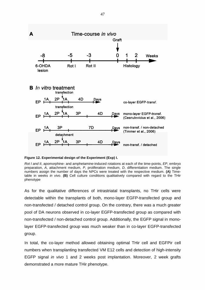
47
Figure 12. Experimental design of the Experiment (Exp) I.
Rot I and II, apomorphine- and amphetamine-induced rotations at each of the time-points, EP, embryo preparation, A, attachment medium, P, proliferation medium, D, differentiation medium. The single numbers assign the number of days the NPCs were treated with the respective medium. (A) Time-table in weeks in vivo. (B) Cell culture conditions qualitatively compared with regard to the THir phenotype
As for the qualitative differences of intrastriatal transplants, no THir cells were
detectable within the transplants of both, mono-layer EGFP-transfected group and
non-transfected / detached control group. On the contrary, there was a much greater
pool of DA neurons observed in co-layer EGFP-transfected group as compared with
non-transfected / non-detached control group. Additionally, the EGFP signal in mono-
layer EGFP-transfected group was much weaker than in co-layer EGFP-transfected
group.
In total, the co-layer method allowed obtaining optimal THir cell and EGFPir cell
numbers when transplanting transfected VM E12 cells and detection of high-intensity
EGFP signal in vivo 1 and 2 weeks post implantation. Moreover, 2 week grafts
demonstrated a more mature THir phenotype.

48
These qualitative results revealed (i) the negative effect of detachment on the THir
cell population in the intrastriatal transplants; (ii) the negative effect of transfection on
the THir pool in vivo; (iii) the in turn DA numerical dominance of the co-layer EGFP
group over the cell culture protocols used in our group previously; (iv) the capability
of the pCAGGS plasmid backbone to ensure high intensity EGFP signal after 2
weeks in vivo, with the dominance of EGFP-transfected co-layer group present
regarding also this aspect.
The pCAGGS-EGFP plasmid had proved to be the potentially right plasmid back-
bone in the perspective of long-time in vivo experimental design and was therefore
used for all subsequent experiments regarding intrastriatal microtransplantation
paradigm in unilaterally 6-OHDA lesioned rats. For image-guided and quantitative
results supporting the qualitative results in this experiment see 4.3.
From this point on evaluation of different methodological variations in the method
combining transient genetic modification of NPCs with an optimized cell culture
protocol prior to intrastriatal transplantation into parkinsonian rats could start.
4.1.2 Optimization of the transplantation time-schedule when using the co-layer method for culturing the ventral mesencephalic NPCs
In the first experiment, the newly generated plasmid construct pCAGGS-EGFP was
successfully tested in vivo and there was a clear hint on the beneficial effect of the
novel pretreatment protocol concerning the yield of transplanted and surviving DA
neurons. Logically, we kept the track of optimizing the transplantation time-schedule
when using the co-layer method.
M. Timmer, Institute of Neuroanatomy, MHH, had previously demonstrated that the
highest percentage of THir cells was found in vitro and in vivo when expanded E12
VM cells were differentiated for 4 days. After longer differentiation, THir neurons
exhibited in vitro very long axons and dendrites and the subsequent DA numerical
and behavioral outcome in vivo was diminished (Timmer et al., 2006). As the studies
on the possibilities to non-virally increase the in vivo survival of the expanded
progenitors in our lab started, the differentiation interval of 4 days was integrated in
the cell culture protocol of the VM NPCs after transfection (the mono-layer method)
(Cesnulevicius et al., 2006). Notably, A. Nobré, PhD, Institute of Neuroanatomy,
MHH, made an important observation. He showed that in the cultures where

49
transfected E12 were treated according to the co-culture format DA neurons
Figure 13. Experimental desing of the Exp II.
Rot I and II, apomorphine- and amphetamine-induced rotations at each of the time-points, EP, embryo preparation, A, attachment medium, P, proliferation medium, D, differentiation medium. The single numbers assign the number of days the NPCs were treated with the respective medium. (A) Time-table in weeks in vivo. (B) Cell culture conditions qualitatively compared with regard to the THir phenotype
displayed a dramatic increase in axonal sprouting after a single day in vitro, if
compared to transfected E12 cells cultured according to the mono-layer method
(Nobre, 2009). This meant that the THir cells treated with the co-layer method
required much shorter time to exhibit a mature morphology. The time the tranfected
co-layers were treated in culture could be reduced when reducing the differentiation
interval and when passing the co-layer cultures directly into differentiation following
the reseeding on the non-transfected bottom-layer cells post transfection. Namely, it
was known from the previous in vitro experiments that the transfected NPCs require
treatment with attachment medium containing fetal calf serum for one day when re-
seeded on pre-coated wells.

50
In this context, two specified questions were addressed in the next in vivo
experiment: Firstly, would considerable and comparable amounts of TH expressing
cells be observed after 2 weeks in vivo if no attachment medium was used after
seeding the transfected cells on the non-transfected E12-cell bottom-monolayer?
Secondly, would the TH expressing cell yield be affected if the differentiation period
in vitro was shorter than 4 days?
In the second in vivo experiment (Fig. 13) twelve unilaterally 6-OHDA lesioned
animals were allocated in 6 experimental groups (n=2) discerned based on the
differentiation-span and medium conditions the VM progenitors were subjected to
post transfection with pCAGGS-EGFP plasmid. All transplanted E12 cells were
cultured for three days (1A2P), followed by detachment and transfection of the top-
layer cells, which were then reseeded on non-detached sister cultures. The cell
suspensions were transplanted after one (1D), two (2D or 1A1D), three (3D or
1A2D), or four (1A3D) days of further in vitro treatment, depending on the
experimental group. Rats were transcardially perfused 2 weeks post implantation and
brains were processed for immunohistochemistry (IHC). Evaluation of the intrastriatal
DA grafts was qualitative.
Figure 14. DA cells in the E12 grafts exhibiting a range of sizes in TH-immunostained coronal sections.
Dense axonal networks present within the grafts in all three transplantation groups where E12 cells had not been treated with attachment medium for additional day (D, E, and F). By contrast, DA cells seem to cohere in a much closer distance in groups 1A2D (B) and 1A3D (C).

51
i) Approval of abundance of additional treatment with attachment medium
Design of the Exp II allowed obtaining qualitative comparison of DA pools within
intrastriatal grafts in unilateral toxin-lesioned rats, when directly comparing the THir
morphology in groups transplanted with cell suspensions differentiated from a single
up to 3 days after transfection with or without additional treatment with attachment
medium, utilizing E12 VM NPCs obtained from the same embryo preparation-session
(Fig. 14). There were qualitatively detectable clear differences in overall THir neuron
population and fiber outgrowth in favor of all groups (Fig. 14-16) where pretreatment
with additional day with attachment medium was abandoned. This resulted in not
only comparable amounts of TH expressing cells; moreover, dense axonal networks
were presented in the grafts (Fig. 15).
Thus, the co-layer cultures could be passed directly into differentiation phase post
nucleofection of the top-layer sister cultures in the following experiments and the in
vitro time-span prior to engraftment could be shortened.
ii) Validation of the optimal differentiation period in vitro with the novel protocol before transplantation procedure
Results in the Exp II were evaluated qualitatively with respect to the DA neuron
morphology and distinctive patterns of THir neuronal outgrowths within the
transplants, as well as the extent of THir processes running along to reinnervate the
host striatum. The three groups, where co-layer cultures had been directly treated
with the differentiation medium post nucleofection, were compared. The
differentiation interval had been lasting for one (1D), two (2D) or three (3D) days.
The 2D pretreatment proved to elicit more pronounced THir connectivity within the
graft, as well as graft-derived THir fiber extensions into the denervated striatum (Fig.
17). Thus the 2D pretreatment was chosen for the further co-layer experiments.
4.2 Detection and evaluation of transgene delivery 9 and 12 weeks after transplantation
Results from the previous experiments showed that non-virally transfected VMP cells
pretreated with co-layer method survived the engraftment procedure as there were
considerable pools of surviving DA- and EGFP-positive cells detectable in the 2 week
grafts. Moreover, the most potent expression vector seemed to have been found.
DA graft survival, maturation, and differentiation occur in two phases and the 2 week
time-point serves as a border-line. The final synaptic and functional integration
appears beginning with the 3rd week post implantation (Hahn et al., 2009). Thus the

52
Figure 15. Photomicrographs of representative grafts in TH immunostained sections for all three groups where the additional day in attachment medium had been abolished in pretreatment of E12 cells.
The boxed areas are shown in higher magnifications numbered from 1 to 3 respectively. This allows clear observation of THir perikarya and the TH fibers. Note the differences in graft morphology, with more graft-derived THir fibers extended into the adjacent denervated striatum and more pronounced THir connectivity within the graft in 2D group (B)

53
Figure 16. Photomicrographs of representative grafts in TH immunostained sections for all three groups where the attachment medium had been given in pretreatment of E12 cells.
The boxed areas are shown in higher magnifications numbered from 1 to 3 respectively. In comparison to the Fig. 4, there is a clear difference in the number of the TH-immunostained cell bodies with almost total absence of fiber outgrowth from the transplant tissue into the adjacent striatum, although all the animals in the Exp II had been transplanted with the same amount of genetically modified E12 VM cell suspension.

54
Figure 17. Presence of dense networks of graft-derived TH-immunoreactive neurites already 2 weeks post implantation.
Fine and coarse THir processes extending within and from the transplants into the host caudate-putamen (A, C). Fiber networks present within the grafts in all three transplantation groups, 1D, 2D, and 3D respectively, more prone in the first 2 groups however (A, B). Long and coarse THir axons extend from the graft reinnervating the surrounding striatum in 2D group (C).
overexpression of a certain NTF in vivo aiming to sustain an adequate neurotrophic
support for the implanted cells should last longer than two weeks. The Exp III (Fig.
14) was designed in order to find out for how long the pCAGGS plasmid backbone
and the novel cell culture method could ensure transgene delivery at the
transplantation site.
In the Exp III four lesioned SPRD rats were transplanted with E12 NPCs treated with
the co-layer method. The pCAGGS-EGFP plasmid was used for genetic modification.
Two animals were sacrificed after 9 weeks and two 12 weeks post implantation.
Evaluation of the intrastriatal DA grafts was qualitative.

55
Figure 18. Time-course in vivo in the Exp III.
Rot I and II, apomorphine- and amphetamine-induced rotations at each of the time-points
And indeed, EGFP expression was detected up to 12 weeks in vivo concomitant with
optimal pool of surviving DA cells (Fig. 19C). This observation was the crucial green-
light signal for further quantitative short- and long-term in vivo experiments in 6-
OHDA unilateral rat model of PD evolving the co-layer method in the given
transplantation paradigm.
Figure 19. Immunohistochemical stainings of grafted cells for EGFP and TH in coronal brain sections 1 week (A), 9 weeks (B) and 12 weeks (C) after transplantation.
The yield of the EGFPir cells in 12 week graft is still considerable (C).
4.3 Intrastriatal graft evaluation 2 weeks after transplantation
After testing the pCAGGS-based construct in vivo and optimizing the transplantation
protocol, the next in vivo experiment (Exp IV) was designed in order to compare
groups were E12 VM NPCs were simply expanded and differentiated (Timmer et al.,
2006), versus groups were the neuronal progenitors were transfected with pCAGGS-
EGFP and cultured according to the mono-layer (Cesnulevicius et al., 2006) or co-
layer format quantitatively.
In the Exp IV eighteen lesioned rats with good apomorphine rotation asymmetry
score [<-4 turns per minute (tpm)] were split equally (n=6) in respect to their rotation
scores in three experimental groups. The VM progenitor cells transplanted in all

56
animals were treated in culture for four days (1A3P), followed by detachment and
reseeding on empty plates (non-transfected / detached control group) or detachment
and nucleofection with the pCAGGS-EGFP plasmid followed by seeding either on
coated empty plates (mono-layer EGFP-transfected group) or on top of an adherent
sister culture at ratio 1:8 (co-layer EGFP-transfected group). Engraftment followed
after three (1A2D) additional days in culture. One has to admit, that the one
additional day of treatment with attachment medium had exceptionally to be included
in the post transfection procedure as my former experience in vitro had shown that
this was ultimatively necessary in order to promote adhesion of the transfected E12
VM cells on the pre-coated wells. Transplantations were performed in 2 grafting
sessions (n=3). Rats were sacrificed 2 weeks post engraftment. For the first time, the
effect of the in vitro pretreatment on the number of surviving transplanted DA
neurons per striatum was evaluated on immunohistochemically processed brain
slices quantitatively (by means of stereological quantification, see 3.9.1) two weeks
after surgery (n = 6 animals per group).
The qualitative impression (Fig. 20) was strongly supported by numbers obtained by
stereological quantification. The lowest numbers of THir cells (271 ± 62) were found
in monolayer EGFP-transfected group, where animals received cells detached,
nucleofected and reseeded on empty coated well. This number was significantly
reduced compared to both, the co-layer 1:8 EGFP-transfected group (1723 ± 199; ***
p < 0.001) and the non-transfected / detached control group (1020 ± 173; * p < 0.05).
THir numbers differed significantly between the non-transfected / detached control
group and the co-layer 1:8 EGFP-transfected group (* p < 0.05) in favour of the latter
one (Fig. 21A). The control group receiving detached, but not transfected cells
reseeded on empty coated wells contained 59% of THir cells compared to the co-
layer group, indicating that the detachment and reseeding procedures accounted at
least partially for the diminished DA outcome. Additionally, the even more dramatic
decrease in THir cell numbers observed in monolayer EGFP-transfected group (16%
compared to the co-layer group) demonstrated the negative numerical effect of the
nucleofection procedure.
Most importantly, the co-layer method proved to assure not only similar, but even
higher THir cell numbers compared to the control group in contrast to the mono-layer
EGFP-transfected controls.

57
Figure 20. Micrographs of coronal rat brain sections immunostained for TH showing effect of in vitro pretreatment 2 weeks post engraftment.
The boxed areas are shown in higher magnifications numbered from 1 to 3 respectively. The transplant representative for the co-layer group (C) seems to contain a higher pool of DA neurons (supported also by the quantitative data – see Fig. 24A). The THir cells and neurites in co-layer EGFP-transf. group are more evenly distributed throughout the graft (C1, C2) and form steadily fine dense fiber networks (C, C3) if compared with non-transfected / detached control group (A) and mono-layer EGFP-transf. group (B)

58
Figure 21. The effect of in vitro pretreatment on the number of THir cells 2 weeks post transplantation in Exp IV (A) and in vivo validation of the co-layer set-up 2 weeks after transplantation using EGFP- and BDNF-expression plasmids in Exp V (B).
Number of surviving THir neurons in all graft groups expressed as mean ± SEM. A: In Exp IV (n = 6), the 1:8 co-layer EGFP-transf. group provided the highest numbers of THir cells compared with both other groups, mono-layer EGFP-transf. group (*** p < 0.001; 16% compared to the co-layer group) and non-transfected / detached control group (* p < 0.05; 59% compared to the co-layer group). B: In Exp V (n = 6-9) neither 1:6 nor 1:3 BDNF-transfected co-layers displayed an increased number of THir neurons in vivo if compared with EGFP-transfected and co-layer non-transfected controls (no significant differences between the groups). Varying the ratio of the transfected top-layer cells to non-transfected bottom layer cells, 1:3 and 1:6 respectively, had no significant effect on the total number of THir neurons neither in EGFP, nor BDNF groups. Two weeks post implantation DA neuron numbers in these groups were similar to the 1:6 co-layer non-transfected controls.
4.4 Evaluation of BDNF restorative capacity in a novel co-layer protocol
My colleagues at the Institute of Neuroanatomy (MHH) A. Nobre, PhD and Dr. M.
Hohmann made thorough studies on selecting the appropriate plasmid-based
delivered NTF which would provide significant increase in the DA neurons generated
from VM cultures. Dr. A. Ratzka supplied the necessary plasmid constructs. It was
the well-established DA-specific brain derived neurotrophic factor (BDNF) showing
the most promising results. Plasmid-based delivery of BDNF increased the number of
DA neurons by 25%, compared to EGFP-transfected controls in vitro. Furthermore,
the overexpressed BDNF-FLAG fusion protein was biologically active as condition
media from pCAGGS-BDNF-FLAG transfected E12 VM cells induced
phosphorylation of BDNF-receptor TrkB in NPCs (Ratzka et al., 2012).
4.4.1 No significant increase in dopaminergic cell numbers after 2 weeks in vivo when delivering plasmid-based BDNF
Based on the above-mentioned in vitro results, the BNDF was used for further
evaluation of the co-layer method in vivo. At first, a short-term experiment (Exp V)

59
was planned expecting the BDNF to produce the positive numerical effect on the DA
neurons seen in vitro also in vivo. Beginning with this experiment, the quantitative
phase in the validation process of the co-layer format started as the THir cells within
the striatal transplants were stereologicaly quantified.
In the Exp V the proportion of nucleofected top-layer cells was increased from initial
in vivo co-layer ratio of 1:8, as it had resulted in relatively low numbers of transfected
cells in vitro and in vivo. In order to detect possible differences in the DA numbers in
vivo, two ratios were used, 1:6 and 1:3 namely. Non-transfected 1:6 co-layer served
as control. Thirty eight unilaterally 6-OHDA lesioned rats were included in the final
analysis (with apomorphine rotation asymmetry score <-4 tpm post lesion) and
distributed into five groups: co-layer 1:6 non-transfeted group (n = 9), co-layer 1:6 (n
= 9) and 1:3 (n = 7) EGFP-transfected groups, and co-layer 1:6 (n = 7) and 1:3 (n =
6) BDNF-transfected groups. The NPCs were first cultured for three days (1A3P),
and then either just detached and reseeded (co-layer 1:6 non-transfeted group) or
nucleofected (co-layer 1:6 and 1:3 EGFP- and BDNF-transfected groups).
Transplantations were performed in 3 grafting sessions. It was proceeded with
sacrification and IHC after two weeks. Finally, stereological quantification of grafted
DA neurons was performed.
Overall, at the 2 weeks time-point the transplanted DA neurons were finished with the
1st or maturation phase (see 1.3.2) and there was a prominent clustering of TH pos
neurons and THir neuritic processes within the graft. It can be clearly seen in
confocal images at high power magnification that there are merely very few graft-
derived DA neurites extending over the graft-host boundary 2 weeks post
implantation (Fig. 23A1, C, F, and G). In contrast, EGFP-FLAG transfected neurons
were uniformly scattered throughout the transplants.
Quantification of the intrastriatal grafts after 2 weeks evidenced a mean of 2341 ±
346 of surviving DA neurons in co-layer 1:6 EGFP-transf. group (n = 9) and 2402 ±
551 in co-layer 1:3 EGFP-transf. group (n = 7) showing no statistical difference
between these groups. Similarly, there was no statistical significance observed
between co-layer 1:6 BDNF-transf. group (n = 7) with a surviving THir neuron mean
of 2206 ± 303 and co-layer 1:3 BDNF-transf. group (n = 6) with its mean of 2257 ±

60
Figure 22. Micrographs of coronal rat brain sections immunostained for TH showing effect of in vitro pretreatment 2 weeks post engraftment.
The boxed areas are shown in higher magnifications numbered from 1 to 3 respectively. No qualitative difference in neither lower nor higher magnification could be observed regarding the morphological DA

61
outcome in grafts of all 3 groups – co-layer 1:6 notransfected (A), co-layer 1:6 EGFP- transfected (B), or co-layer 1:6 BDNF-transfected thus evidencing that the nucleofection with any of the expression plasmids (B, C) had no negative effect on the expectable graft morphology
594. Moreover, no statistical differences were displayed when comparing the
aforementioned BDNF and EGFP overexpresing groups against each other, nor was
this revealed when comparing the already given numbers with a mean of THir
neurons in the co-layer 1:6 non-transfected group (2423 ± 560; n = 9) (Fig. 21B).
Concluding, no significant differences in numbers of surviving THir neurons were
observed neither between BDNF-transfected, EGFP-transfected and control groups,
nor between 1:3 or 1:6 co-layer ratios. Thus BDNF-transfected co-layers did not
display increased DA pools after 2 weeks in vivo. Furthermore, differences in the
THir cell number between the initial 1:8 co-layer experiment (Fig. 21A) and the 1:6
and 1:3 co-layer experiments (Fig. 21B) were statistically not significant.
Even though qualitative evaluation of the intrastriatal DA grafts in all three
experimental groups did not reveal an explicit DA dominance in any group (Fig. 22), it
could most importantly be concluded that nucleofection (Fig. 22B, C) did not
intrude with the morphology of transplanted THir cells and graft-derived THir
fibers.
4.4.2 Evaluation of morphological and functional integrity of the BDNF-supported grafts 13 weeks after transplantation
According to the quantitative results in the last short-term in vivo experiment (Exp V),
BNDF failed to increase the survival of intrastriataly grafted E12 VM DA neurons two
weeks post engraftment, however this just supported the evidence already provided
by other groups where this particular NTF was shown to enhance function rather than
survival of DA grafts in 6-OHDA lesioned rats (Sauer et al., 1993). Therefore, a long-
term transplantation experiment (Exp VI) evolving the co-layer method and the usage
of BNDF-expression plasmid was planned expecting to demonstrate a positive effect
of the plasmid-derived BDNF on the long-term DA survival in vivo and graft
morphology, as well as the functional restorative capacity of the intrastriatal grafts.
In the Exp VI (Tab. 8) thirty six (in the final analysis) lesioned SPRD rats were
allocated in three experimental groups assigned according to apomorphine rotation
asymmetry score <-4 tpm and amphetamine rotation asymmetry >7 tpm post lesion,
as well as the cell culture protocol the NPCs were treated prior to implantation: co-
layer non-transfected group (n = 9); co-layer EGFP-transfected group (n = 14); co-

62
layer BDNF-transfected group (n = 13). The E12 VM cells were cultured for three
days (1A3P), then either just detached (non-transfected group) or nucleofected with
EGFP-FLAG expression plasmid (EGFP-transfected group) or BDNF-FLAG
expression plasmid (BDNF-transfected group) and reseeded as 1:3 co-layer for 2
days (2D) in vitro. In 3-week intervals, drug-induced rotation behavior was assessed.
Finally, animals were sacrificed 13 weeks after transplantation, the brains processed
for IHC. Stereological quantification of grafted DA neurons was performed.
Furthermore, the degree of morphological reinnervation was quantified by (i)
measuring the optical density of THir fiber outgrowth into the striatum (thickness of
THir halo around the graft), determining the (ii) fiber extent, (iii) graft volume, and (iv)
THir neuron density.
Table 8. Time-schedule of the Exp VI
Time Experimental treatments Experimental tests
-8 w* Lesion -
-5w - Rotation tests
-2w - Rotation tests
0 Grafts -
3w - Rotation tests
6w - Rotation tests
9w - Rotation tests
12w - Rotation tests
13w Perfusion -
* w, weeks
i) Qualitative evaluation of 13 week intrastriatal grafts
As previously, survival of DA neurons and development of graft-derived DA
innervations of the host 13 weeks after transplantation of E12 VM cells into the right
striatum of 6-OHDA-lesioned adult rats were assessed by TH IHC.
Abundant TH immunoreactivity was present within the graft at this differential stage
and extended to variable distances into the host striatum forming a halo of TH
immunoreactivity around the grafts (Fig. 23B1, C, H, and I). This halo did not appear
to be more extensive in any of the experimental groups. Additionally, 13 week grafts
were more mature and the striatal volume innervated by the graft appeared

63
consistently greater in 13 week implants vs 2 week grafts. At 13 week time-point THir
cells were preferentially localized in the periphery of the transplant and there were
dense TH positive (pos) fiber networks originating from the transplanted neurons
reinnervating the surrounding striatal neuropil (Fig. 23H, I). Concomitantly, also
EGFP-FGLAGir fiber networks remained within the transplant and EGFP-FLAG
positive neurites were rarely detected in the surrounding host striatum.
Due to the dissection procedure of the embryonic midbrain there are two subtypes of
DA neurons present in the transplants, which are normally localized in either SNc (A9
region) or VTA (A10 region). It is known that the A9 subpopulation plays a critical role
in the improvement of motor performance post transplantation. Therefore, proper
differentiation of the graft DA cells into A9 sub-type was verified by Girk2ir/THir
double staining (Fig. 23E) (data obtained by Dr. C. Köster-Patzlaff). And indeed
semiquantitative analysis of the 13 weeks transplants revealed that the majority (70-
80%) of DA neurons were double-labeled for Girk2ir/THir.
The overall good survival of the transplanted neuronal population was shown by
means of anti-ß-III tubulin staining (Fig. 23J). Immunohistochemical analysis of the
GFAPir cell population in the 13 week transplants revealed a GFAP immunoreactivity
scattered throughout the graft mass. The GFAP pos cells within the graft displayed
small cell bodies and filiform processes vs GFAP pos cells on the graft-host interface
had a broad cytoplasm with elongated, thickened processes (Fig. 23K). These
astrocytes displaced on the border to the surrounding striatum were nestin pos and
thus reactive (Fig. 23D). Semiquantitative evaluation of GFAP reactivity revealed no
differences in the extent of the glial scar between all three experimental groups in the
Exp VI.
ii) Behavioral analysis of 13 week grafts using rotometry
The second phase of DA graft maturation evolving functional integrity begins after the
3rd week post implantation (Hahn et al., 2009). In order to determine time-
dependant functional effects of the BDNF-transfected and control 1:3 co-layers,
qualitative characterization of the implant integration success in all three
experimental groups in Exp VI was assessed by behavioral analysis using rotometry.
After administration of either amphetamine (Fig. 24A) or apomorphine (Fig. 24B)
rotational behavior was monitored pre-grafting and every 3rd week (3, 6, 9, and 12
weeks) after grafting.

64
Figure 23. Development and maturation of intrastriatal DA-rich allografts treated with co-layer method at different survival times post engraftment into adult rats with a right, unilateral 6-OHDA lesion of the nigrostriatal pathway
(A-B1) Micropgraphs of coronal rat brain sections immunohistochemically stained for TH with DAB visualization at the level of caudate-putamen unit. The 6-OHDA-lesioned striatum is dopamine depleted (A1, B1), if to compare to the non-lesioned side exhibiting an extensive THir fiber network (A, B). The medial and lateral implants of embryonic DA neurons are clearly discernible in the lesioned striatum two (A1) and thirteen weeks (B1) after transplantation. Two weeks post engraftment, there is a dense fiber network present within the graft itself but the fiber outgrowths are almost totally absent in the adjacent recipient caudate-putamen unit (A1). An extensive halo of TH immunoreactivity penetrates the host striatum around the graft deposits in 13 week sections (B1). Fluorescently labeled THir (red), as well as EGFP-FLAGir (green) cells and their processes are detectable both, 2 weeks (C, F, and G) and 13 weeks (H, I) post implantation. At the stage of 2 weeks the THir neurons are intermingeled evenly throughout the graft and a few graft-derived THir neurons innervating the host striatum can be seen close to host-graft boundary (B1, C, and F). Thirteen weeks vs 2 weeks after transplantation the striatal reinnervation extended much further into the host tissue and the graft volume increased (A1, B1). As seen in confocal images, in 13 weeks old grafts the majority of the THir neurons resided primarily at the periphery of the implant and extended dense network of THir fibers further from the graft-host border (H, I). In contrast, the EGFP-FLAGir cells and fibers remained in the

65
centre of the graft and rarely extended processes into the host tissue, both 2 weeks (F, G) and 13 weeks (H, I) after engraftment. (J) Note that labeling of the DA grafts for ß-III Tubulin (red) as neuronal marker and EGFP-FLAGir (green) cells (nuclei were visualized by DAPI) affirmed the good overall survival of transplanted neuronal population. Nuclei were visualized by DAPI and the arrowheads point out the nuclei of the stained neurons within the transplant range. (E) Double-labeling for THir (green) and Gritk2ir (red) (yellow in the merged image, arrowheads) revealed the presence of typical A9 (SNc) derived embryonic midbrain DA neurons in the grafts (close to the host border). (K) GFAP (red) immunoreactive cells within the graft tissue displayed a different morphology vs the morphology displayed by the GFAP pos cells at the host-graft border. As confirmed by the presence of nestin (D, arrowheads) immunoreactive cells at this border region, these reactive GFAPir cells stood for reactive astrocytes in the glial scar. No nestin positive cells were observed within the embryonic DA implants already in 2 week brain slices (D).
Amphetamine-induced ipsilateral rotation behavior decreased in all three co-layer
groups (non-transfected, EGFP-transfected and BDNF-transfected) similarly, from
initial pre-grafting values of 13.0, 14.8 and 13.2 rounds per minutes respectively, to
3.1, 3.6 and 1.5 rounds per minutes 12 weeks after transplantation, reaching
statistical significance already 3 weeks after grafting in each group (Fig. 24A).
However, two-way ANOVA analysis of amphetamine-induced rotation data revealed
that the type of transplant did not affect the results (p > 0.05).
Figure 24. Rotational behavior 13 weeks after transplantation.
Pg, pre-grafting, w, week, native, non-transfected, ns, not significant, **, p < 0.01, #, p < 0.001. All three experimental groups, non-transfected (n = 9), EGFP-transfected (n = 14), and BDNF-transfected (n = 13) respectively, received 1:3 co-layer transplants. (A) Amphetamine-induced ipsilateral rotation declined in all three groups similarly, reaching statistical significance already 3 weeks after grafting. (B) Apomorphine-induced rotational behavior reached statistical significance 9 weeks after transplantation in the native-transplant group, whereas in the EGFP- and BDNF-transfected groups the significance level was reached already 3 weeks after grafting (two-way ANOVA followed by Bonferroni post hoc test, compared to the pre-grafting scores of the same experimental group)
Apomorphine-induced contralateral rotation behavior decreased from initial pre-
grafting values of 12.5 rounds per minute to 8.6 (non-transfected), 6.7 (EGFP-
transfected) and 7.7 (BDNF-transfected) rounds per minute (Fig. 24B). Although,
significant improvement of the rotation behavior of the non-transfected group was not
observed until 9th week after transplantation, overall comparison of the

66
apomorphine-induced rotation data using two-way ANOVA analysis did not reveal an
effect of the type of transplant (p > 0.05). In summary, transplantation of VM cells,
which were previously in vitro cultured as 1:3 co-layer, improved drug-induced
rotation behavior independent of the type of transplant. These findings suggested
that neither did transfection procedure using EGFP control plasmid impair rotational
behavior compared to non-transfected co-layer cells, nor did transfection with BDFN
expression plasmid improve rotational behavior.
iii) No significant differences between experimental groups in morphometric analyzes
In vitro, BDNF-FLAG transfected 1:3 co-layers displayed an increased number of
THir neurons by 25%, compared to EGFP-FLAG transfected 1:3 co-layers or non-
transfected 1:3 co-layer controls (Ratzka et al., 2012). Thirteen weeks post
implantation, the DA pool in the transplants was quantified on DAB stained sections.
However, no differences between non-transfected, EGFP-FLAG and BDNF-FLAG
groups were identified in the following parameters: THir neuron number, graft
volume, THir neuron density, THir fiber density, and extend of the THir fiber
outgrowth (thickness of the THir halo around the graft) indicating reinnervation of the
host striatum (Tab. 9). Correlation analysis on these morphological data showed a
clear positive correlation between graft-derived DA pool and the graft volume (see
Appendix 7.7) in all three experimental groups.
Table 9. Morphometric quantification of the intrastriatal grafts after 13 weeks
non-transfected
co-layer
(n=9)
EGFP-transfected co-
layer
(n=9)
BDNF-transfected co-
layer
(n=7)
THir cell number 1981 ± 362 2339 ± 547 2089 ± 403
Graft volume (mm3) 1.80 ± 0.29 2.52 ± 0.55 1.90 ± 0.34
THir cell density (cell/mm3) 1089 ± 81 957 ± 96 1086 ± 130
THir halo thickness (mm) 0.41 ± 0.05 0.52 ± 0.08 0.38 ± 0.05
THir fiber density (% non-
lesioned striatum)
57% ± 9 70% ± 11 63% ± 12

67
Thus i) nucleofection itself did not intrude with the morphology and account of the
graft-derived THir cells in a long-time study when applying the co-layer layout and the
ii) cell-mediated delivered BDNF had no negative effect on neither DA yield, nor other
key parameters of the DA graft survival pattern.
iv) Histological evaluation of 6-OHDA lesion efficacy and correlation analysis
In order to put the lesion efficacy into correlation to morphometric parameters and
induced rotation behavior, the number of remaining THir cells in the SN was counted
and evaluated using a scoring system (see 3.9.4, Tab. 7). The quantification was
done in all three parts of the SN, SNc, SNl and SNr respectively. Five points stood for
maximum of 30 THir neurons in the whole right SN and 1 point for > 600 THir cells
per right SN. Out of thirty six lesioned rats included in the final analyses, nine animals
matched the 5-point category, whereas just 4 animals evidenced an unsatisfactory
lesion efficacy rated with 1 point (see Appendix 7.8.1, 7.8.2).
No correlations could be revealed between the i) THir numbers in the grafts, ii) THir
fiber length, iii) THir fiber density, iv) graft volume (data not shown), as well as iv)
behavioral recovery and the residual THir number in the SN of the 6-OHDA lesion
side (see Appendix 7.8.3, 7.8.4). This also revealed that none of the experimental
groups had been accidentally discriminated against others with regard to the
unilateral DA extent in the lesioned striatum prior to implantation of VM E12
micrografts.
v) transplant topology and correlation analysis to rotational results
The transplants of E12 NPCs were localized within the host striatum according to the
Atlas of Paxinos and Watson (Paxinos, 1998). The localization of grafts was detected
in all three planes – craniocaudal (AP plane), dorsoventral (VERT plane), and
mediolateral (LAT plane). With exception of one brain, there was always a clear
differentiation between the lateral and medial transplant possible.
All in all, the transplants extended over the craniocaudal area from 1.56 mm to -0.96
related to Bregma (see Appendix 7.9.1). The extension in dorsoventral plane took
place from 4.1 mm to 7.5 mm in reference to the Bregma and Lambda. The striatal
transplants extended from -0.4 mm to 4.0 mm in the mediolateral plane referred to
the sagittal midline. Further on, these results were scored (see Appendix 7.9.2)

68
according to the a modified scoring system already described in our previous study
Figure 25. Schematic drawing demonstrating topology of the lateral and medial grafts in dorsoventral and mediolateral directions in the striatum 13 weeks after transplantation in all experimental groups (coronar section).
(Fig. 25) (Jungnickel et al., 2011) and correlated to the behavioral results in both
rotation tests, amphetamine and apomorphine rotation behavior (see Appendix 7.9.3,
7.9.4) respectively. No negative correlation between either amphetamine or
apomorphine rotation behavior or graft location was revealed irrespective the
experimental group. (Dieterlen et al., 2009, Sortwell et al., 2004) (Thompson et al., 2005)
(Georgievska et al., 2004)
(Nikkhah et al., 2009)
(Yurek et al., 1998)
(Yurek et al., 1998)

69
5 DISCUSSION
Both, animal experiments and clinical trials have revealed that transplanted DA cells
survive and display functional efficacy. Still there is a row of problems to circumvent,
before cell replacement becomes a standard therapy option for PD. The lack of donor
tissue and poor survival rate after transplantation are one of the major limiting issues
(Brundin et al., 2000a). To overcome the limited supply of donor tissue, ESCs, iPS
cells or NPCs could be used as alternative cell sources. Since ESCs and iPS cells
display disadvantages like chromosomal abbreviations in mid-term cultures and form
teratomas in the host brain (Draper et al., 2004, Bjorklund et al., 2002, Nishimura et
al., 2003, Pruszak et al., 2009, Brundin et al., 2010), research work in our lab is done
with NPCs. The approach evolves transient delivery of DNA encoding for NTFs in
order to improve the DA survival post implantation. For this purpose, enhanced cell
culture conditions for optimal use of midbrain progenitor cells needed to be
established.
It was known that functionality and survival of DA neurons grafted into rats with
unilateral 6-OHDA lesion was the best after direct grafting (freshly dissociated) of
E14 VM cells (Brundin and Bjorklund, 1987). In order to genetically modify the VM
cells to encode for factors improving the morphological and functional outcome, the
NPCs had to be attached and expanded. M. Timmer, Institute of Neuroanatomy,
MHH showed that TH expression was higher in expanded and differentiated VM E12
cells, compared to E14 cultures. Furthermore, DA neuron survival of expanded and
subsequently for 4 days differentiated E12 VM precursors was equal compared to the
direct E14 donor VM transplants (Timmer et al., 2006). As the next step the most
effective non-viral transfection method for NPCs was detected, nucleofection
respectively. Nucleofected progenitors were viable after transplantation
(Cesnulevicius et al., 2006). Admittedly, the following experiments revealed that only
a few THir cells and cells expressing the reporter sequence were detectable in the
grafts of transfected NPCs.
In terms of this study, there was (i) a novel protocol for treatment of primary NPC
cultures prior to implantation into unilateral 6-OHDA rat model of PD developed
allowing to obtain high numbers of DA neurons, both in vitro and in vivo, and to
introduce expression of foreign genes via nucleofection in parallel. Further on, (ii)
BDNF as an initial candidate factor was tested in this novel set-up for its ability to

70
reproduce its positive effects on the DA phenotype seen in vitro (work done by my
colleagues A. Nobre, PhD and Dr. M. Hohmann) also in vivo with regard to DA
neuron survival (short-term study, 2 week time-point) and functional improvement
(long-term study, 13 week time-point).
5.1. Co-layer format for effective nonviral delivery of neurotrophic factors without feeder cells
Co-transplantation of DA neurons and neurotrophic factor producing cells, e.g. FGF-2
secreting fibroblasts or Schwann cells has been successfully applied to improve the
transplantation outcome (Timmer et al., 2004, Takayama et al., 1995). However, from
clinical perspective, it is desirable to use culture protocols free of feeder cells. Viral
vectors were promising as gene carriers in neurons, also in particular in PD animal
models (Mandel et al., 1997, Choi-Lundberg et al., 1998, Gonzalez-Barrios et al.,
2006, During et al., 1994). Still their therapeutic potential is limited due to their
toxicity, immunogenicity, mutagenesis and their broad tropism. In contrast, nonviral
vectors have improved safety profiles, result in transient expression and are potent in
targeting of specific neuronal populations (Bergen et al., 2008, Tinsley and Eriksson,
2004).
It was previously shown by Cesnulevicius et al. that the most efficient transfection
method in order to transfer DNA into postmitotic rat VM cells was nucleofection. It
supplied the highest transfection rates of NPCs and cell viability in vitro
(Cesnulevicius et al., 2006). This data was consistent with previous publications
approving nucleofection to enable reproducible, rapid, and efficient transfection of a
broad range of “hard-to-transfect” cells, including primary neurons (Gartner et al.,
2006, Gresch et al., 2004). Nevertheless, the numerical DA outcome and the amount
of the cells expressing the reporter sequence in vivo remained low. A row of factors,
cell detachment evolved in the nucleofection procedure, expression plasmid of a low
potency, and suboptimal cell culture conditions respectively, were experimental
issues to solve. Thus a novel cell culture protocol, so called co-layer method, was
established during this particular study. In this new cell culture format the transfected
cells were reseeded on top of non-detached sister cultures and a CAG-promoter
(instead of CMV-promoter) containing expression plasmid was used. The pCAGGS
backbone ensured strong gene expression up to at least 13 weeks in vivo as seen in
the Exp VI (see 4.4.2 i)). Successful expression of foreign genes in human NPCs

71
has been reported up to 6 months when applying nucleofection in combination with a
pCAG-promoter (Dieterlen et al., 2009). Furthermore, a shift in the proportion of
immature progenitor cells (nestin-ir) towards mature neuronal (NeuN-ir) and glial
(GFAP-ir) cell types over a time in vitro was shown proving that pCAGGS-EGFP
transfected cells were still able to differentiate (Ratzka et al., 2012).
Nucleofection procedure itself and the accompanied detachment and reseeding
procedure cause the loss of the vulnerable DA neurons. The corresponding in vitro
data (Ratzka et al., 2012, Cesnulevicius et al., 2006) were supported by results in
vivo. Namely, in the Exp IV (see 4.3) the lowest numbers of THir cells were seen in
the groups which received detached and reseeded (non-transfected / detached
control group) progenitors and detached, nucleofected and on precoated wells
reseeded (monolayer EGFP-transfected group) progenitors. Thus the numerical DA
dominance of co-layer EGFP-transfected group was clearly shown.
The cell death occurring at early time post engraftment of mesencephalic tissue
grafts is of apoptotic nature (Mahalik et al., 1994, Emgard et al., 1999, Sortwell et al.,
2000, Emgard et al., 2003). One of the critical phases, which trigger the intrinsic
genetically driven cell “suicide” program is indeed the tissue dissection and
preparation (Branton and Clarke, 1999) as these evolve axotomy and disruption of
the contacts to the neighboring cells and the extracellular matrix (ECM) (Sortwell,
2003, Schwartz, 2010, Mogi et al., 1999). When applying the novel co-layer format,
the detached, suspended and transfected cells are brought to close proximity with
non-transfected and not detached bottom layer from the same embryo preparation.
These bottom-layer cells are providing contact with an already existing intact network
of neighboring cells. It could be that the transfected top-layer profits from paracrine
neurotrophic support ensured by the attached sister cultures. The fast recovery of the
nucleofected NPCs allows implantation of cells shortly after the transfection.
In total, the co-layer format allows simultaneous generation of reasonable amounts of
DA neurons and high numbers of transfected cells without usage of feeder cells or
viral vectors.
5.2 Transgene delivery up to at least 13 weeks post implantation
Optimization of graft survival is of a crucial importance in maximizing the clinical
response in parkinsonian patients receiving embryonic DA grafts (Apostolides et al.,
1998). At this point research is running on to reduce the DA cell death during their
72
differentiation and subsequent transplantation by administration of growth factors
(Brundin and Bjorklund, 1998) or anti-apoptotic agents (Zawada et al., 1998).
According to the long experimental experience, grafted DA cells undergo the most
vulnerable phase during the transplantation procedure itself. The most extensive DA
cell death occurs within the first days (Hagell and Brundin, 2001, Brundin and
Bjorklund, 1998, Zawada et al., 1998, Barker, 2006) and reaches 80-90% at the end
of the 1st integration phase, in 2 weeks post engraftment respectively (Hahn et al.,
2009). As DA neurons are grafted into an ectopic site, they may be exposed to an
unfavorable microenvironment in the meaning of lack of appropriate trophic signaling
(Mogi et al., 1999). Therefore, administration of growth factors and anti-apoptotic
agents seems suitable for support during the differentiation of transplantable cells
and subsequently post implantation.
Culturing of the NPCs in two layers, a bottom and transfected top-layer, was not the
only modification from the cell culture protocol previously described in our lab where
transfected VM cells were reseeded on precoated wells (Cesnulevicius et al., 2006).
Previously, an expression plasmid containing CMV-promoter was used. CMV-
promoter driven 18 kDa-FGF-2 overexpression in vitro deceased rapidly reaching the
level of non-transfected controls in differentiation phase already on the 11th day in
culture. CMV-promoter based EGFP expression was barely detectable after 14 days.
Such expression time-span was not sufficient in terms of reasonably long
neurotrophic support of DA grafts in vivo via plasmid-based delivered NTF / NTFs.
For the co-layer format, a CAG-promoter containing expression plasmid was used,
which insured a long-lasting gene expression in the Exp III and Exp VI up to 13
weeks post implantation. These data correspond to a previously published study from
Dieterlen et al. (2009), where prolonged EGFP expression for up to 10 weeks was
shown in stably transfected human NPCs containing a CAG-promoter construct,
whereas CMV-promoter based EGFP expression lasted only 6 weeks. At this point,
two further objectives of the present study were fulfilled. Namely, the most proficient
expression plasmid for optimal transfection rates and expression time-spans allowing
detection of grafted mesencephalic NPCs up to 13 weeks was determined.
5.3 Optimization of the transplantation-schedule
Interestingly, the EGFP-transfected NPCs showed shorter neurites over the same
time-span in vitro when seeded on coated empty wells compared to those seeded on
the bottom layer. The same was true for the THir cells of the standard transfection
73
cultures displaying shorter neurites compared to co-layer cultures (Ratzka et al.,
2012). These outcomes were not only implicating better differentiation conditions
under co-layer setup, but also initiated in vivo experiments for optimization of the
appropriate time-point for intrastriatal implantation of the cells cultivated under co-
layer conditions.
Data from Timmer et al. (2006) revealed that the in vitro pre-differentiation period
greatly influences the THir neuron survival. E12 VM cultures differentiated for 4 days
in vitro displayed a significantly better survival than those differentiated only for 2 or
for 6 days. Consistent with this, the results in the Exp II of my doctoral study revealed
qualitative DA dominance of the 2 day differentiation pretreatment in the co-layer
format, compared to 1 and 3 day pretreatment. Furthermore, no attachment medium
(no FCS) was needed for successful attachment of the transfected cells on the
mono-layer of non-transfected NPCs. Thus both in vitro and in vivo data
demonstrated that the differentiation period prior to intrastriatal implantation in
parkinsonian rats was shorter when working under co-layer conditions in contrast to
the cell-culture protocol previously described by our group. The two-day in vitro
differentiation phase after transfection was sufficient enough to provide a recovery
period for the transfected top-layer cells before a repeated detachment and
dissociation within the framework of transplantation procedure took place.
In this context, in a study from Sortwell et al. (2004) using male Fischer 344 rats 10
weeks post-grafting THir yield was 25% lesser in VM cells cultured for 7 days,
compared to VM cells cultured for 3 days. Moreover, the seven day pretreatment
grafts did not display significant improvements in functional recovery. This pattern
was also seen when freshly dissociated mesencephalic DA neurons, taken from E14
and E16 donors and 2.5-day-old cultures survived well and markedly reduced
amphetamine-induced rotational asymmetry, whereas the 7-day-old cultures and DA
neurons from E20 fetuses had a very poor survival and the grafts had no functional
effects (Brundin and Bjorklund, 1987).
As nucleofection is a transient transfection method, overexpression of the respective
proteins decreases with the time. Thus a shorter differentiation phase post
transfection stretches the optimal expression phase of this respective protein into the
in vivo integration phase.

74
5.4 DA numerical dominance and immunohistochemical characteristics of the ventral mesencephalic transplants treated in a co-layer setup
The DA grafts of E12 VM cells were evaluated qualitatively and quantified in coronal
brain sections stained for TH. In the Exp IV (see 4.3) co-layer EGFP-transfected
cultures assured similar THir cell numbers as the non-transfected detached controls.
Similarly, 2 weeks after intrastriatal transplantation stereological quantification of
grafted THir neurons showed a statistically significant DA dominance of the co-layer
method over the manner transfected NPCs were precultured in our lab previously
(Cesnulevicius et al., 2006).
In 2 week co-layer grafts the THir neurons were intermingled throughout the graft and
extended barely any processes into the host striatum. Whereas DA neurons were
found primarily at the periphery of the VM grafts 13 weeks post implantation. The
THir cells possessed long processes and formed dense networks in the graft and
surrounding striatum (called halo) (see Fig. 20A–I). This DA distribution pattern is
typical for long-term grafts not only in experimental rat model (Hahn et al., 2009), but
also in human recipients (Li et al., 2008b, Mendez et al., 2008). Altogether, as
nucleofection did not intrude with the morphology and number of the graft-derived
THir cells and fibers in the co-layer layout in the short-term (Exp IV, see 4.3 and Exp
V, see 4.4.1) and long-term in vivo experiments (Exp VI, see 4.4.2) in this study, a
platform was ensured for the candidate NTF of interest to improve the already good
DA numerical and functional outcome in further experiments evolving the co-layer
layout.
Due to the dissection procedure of the embryonic midbrain, fetal VM transplants
contain two major subtypes of DA neurons - the A9 neurons of the SNpc, which give
rise to the nigrostriatal pathway, and A10 neurons of the VTA, which in turn give rise
to the mesolimbic and mesocortical pathways innervating parts of the limbic system
and neocortex. It was already previously suggested that just the A9 neurons are able
to reinnervate the denervated striatum after transplantation (Bjorklund and Isacson,
2002, Schultzberg et al., 1984). In the study from Thompson et al. (2005) transgenic
mice expressing GFP under the TH-promoter were used allowing visualization of the
grafted DA neurons and their axonal projections in the host striatum. It was shown
that i) the abovementioned DA neuron subtypes could be identified based on the
expression of a G-protein-gated inwardly rectifying K+ channel subunit (Girk2) (SNpc
neurons) and calbindin (VTA neurons) and ii) that indeed DA reinnervation is derived

75
almost exclusively from the Girk2-positive SNpc cells. VM grafts of Pitx3 (gene
encoding pituitary homebox 3) deficient mice, which failed to develop A9 neurons,
were compared to wild-type grafts containing A9 and A10 DA neurons. Grafts
deficient for A9 neurons did not repair the functional deterioration of parkinsonian rats
(Grealish et al., 2010). In good agreement with these data, semiquantitative analysis
of the 13 week transplants in our study performed by Dr. C. Köster-Patzlaff revealed
that the majority (70-80%) of DA neurons were double-labeled by Girk2 and TH.
Furthermore, these Girk2ir/THir double-labeled DA neurons were present in the 13
week grafts close to the host border thus corresponding to the pattern reported
previously in a rodent animal model (Grealish et al., 2010) and grafted PD patients
(Mendez et al., 2008).
There is a certain amount of grafted DA cells necessary in order to induce functional
effects on the rotational asymmetry. The average THir cell number per co-layer
transplanted striatum was about 2000 (see 4.4.2 iii)) cells irrespective the
experimental group 13 weeks post transplantation. Around 100-200 DA neurons are
necessary to ameliorate the rotational asymmetry, but a larger number of THir cells is
needed in order to produce positive functional effects in a unilateral rat 6-OHDA
model (Brundin and Bjorklund, 1987, Nikkhah et al., 2009). The DA numbers per
transplanted striatum reported in our co-layer study were generally higher than in
other studies previously carried out in our lab (Timmer et al., 2006, Timmer et al.,
2004, Jungnickel et al., 2011). So although the co-layer indeed results in a 1:3
dilution of transfected cells, still this limitation might be preferable over a strong
reduction of DA neuron number to 40% in vitro or to 16% in vivo, when all cells
underwent nucleofection. And on the other hand, differences in the THir number
between the initial 1:8 co-layer experiment and the 1:3 and 1:6 co-layer experiments
were statistically not significant (Ratzka et al., 2012).
Immunohistochemical staining for astrocytic marker GFAP (glial fibrillary acidic
protein) revealed astrocytes with small soma and thin processes within the
transplants and a few astrocytes with broad cytoplasm and thickened, elongated
processes at the border to the host tissue (see Fig. 20K). When transplanting
dissociated VM cells a mixture of cells is being implanted, which includes not just
neurons, but also progenitor cells and astrocytes. These transplanted GFAPir cells
scattered within the DA grafts were negative for nestin as a marker for reactive
astrocytic cells, whereas the GFAPir cells at the host border were immunoreactive for
76
nestin. Reduction of astrogliosis at the graft-host interface could be observed linked
to longer in vivo time-span. This GFAP expression pattern corresponded to the data
previously published (Brandis et al., 1998). Immunosuppressive treatment with
cyclosporin is sufficient in parkinsonian patients in order to avoid graft rejection, as
concluded in clinical and neuropathological studies (Kordower et al., 1996,
Sigurdsson et al., 2002). In our study, immunosuppression was applied neither in the
short-term nor long-term studies based on the data that no immunosuppression is
needed for at least 12 weeks in the rat animal model for PD in vivo in case of
intraparenchymal allogeneic grafts (Brandis et al., 1998). Most importantly, no
differences were seen between the transfected and non-transfected co-layer groups
in the semiquantitative analysis of the DA grafts in the Exp VI. This indicated that
implantation of nucleofected NPCs had no negative effect on the GAFP expression
pattern.
Interestingly, we could also detect no / almost no nestin-ir cells within both, 2 week
(see Fig. 20D) and 13 week grafts. Out of our in vitro studies we already knew that a
shift in the proportion of immature progenitor cells (nestin-ir) towards mature
neuronal (NeuN-ir) and glial (GFAP-ir) cell types takes place between day 2 and day
6 after transfection with pCAGGS-EGFP (Ratzka et al., 2012).
5.5 BDNF as a candidate factor to be introduced in the expression vector in the co-layer set-up in vivo – an attempt to prove the principle
In the present study, we were able to show that the novel cell culture pretreatment
protocol for DA progenitor cells was resulting in a significant increase in grafted DA
cell survival. So the aim of the next experiments was to combine the co-culture
method with the possible neurotrophic effects of a respective NTF with regard to its
capability to increase the THir number, DA survival and the functionality of DA grafts
in our parkinsonian animal model when released by nonviraly genetically modified
VM NPCs overexpressing this protein. Secreted NTFs are excellent candidates for
the co-layer method, as it is not necessary to transfect the DA neurons directly,
instead the extracellulary released factors can reach neighboring non-transfected DA
cells indirectly. In the in vitro bioassay studies, carried out by my colleagues A.
Nobre, PhD and Dr. M. Hohmann , BDNF provided the highest yields of DA neurons.
Similar results were observed when BDNF recombinant protein (rBDNF) was added
to the cultures transfected with empty vector (Nobre, 2009). The cell-derived BDNF-
FLAG fusion protein was biologically active as BDNF-FLAG condition media induced

77
phosphorylation of BDNF-receptor TrkB in NPCs (data obtained by Dr. M. Hohmann).
Moreover, BDNF-FLAG transfected 1:3 co-layer cultures displayed an increase in
THir cell numbers by 25% compared to EGFP-FLAG transfected 1:3 co-layers or
non-transfected 1:3 co-layer controls in vitro (Ratzka et al., 2012). In the dilution of
the transfected cells of 1:3, BDNF-FLAG transfection was still sufficient to positively
influence the DA differentiation.
BDNF is generally known to display DA differentiation enhancing effect (Beck, 1994,
Hagg, 1998, Hyman et al., 1991, Maciaczyk et al., 2008, Murer et al., 2001). In
addition, it protects DA neurons against 6-OHDA and MPTP toxicity in vitro and in
parkinsonian animal models (Hyman et al., 1991, Spina et al., 1992, Frim et al.,
1994, Shults et al., 1995, Krieglstein, 2004). Results in our present study (Exp V)
indicated no differences in THir neuron number after 2 weeks in vivo between BDNF-
and EGFP-transfected co-layers. This was in agreement with published data
reporting that daily intrastriatal injection of mature BNDF protein next to DA cell-rich
grafts did not increase DA survival but rather enhanced their function (Sauer et al.,
1993). Several studies report the absence of an increase in grafted DA neuron
survival after administration of BDNF, either as a pretreatment in culture prior to
implantation (Hoglinger et al., 2001, Zhou et al., 1997) or intracerebral infusion post
transplantation (Yurek et al., 1996).
Final synaptic and functional integration of the DA grafts into the host basal ganglia
circuitry (Phase 2) begins with the 3rd week post implantation (Brandis et al., 1998,
Nikkhah et al., 1994b). Setting a time-frame of 13 weeks for the long-term co-layer in
vivo study allowed us to discriminate between early effects on graft survival and
possible late effects of BDNF on the extent of graft-derived DA fiber outgrowth and
functional integration.
In fact, time-dependent effects of rBDNF applied on DA grafts by minipumps into the
striatum close to the transplantation site have been described. In the study of Yurek
et al. (1998) intrastriatal infusions of mature BDNF protein were carried out at 1st and
2nd week post transplantation or delayed up to 3rd and 4th week or 7th and 8th week
respectively. Optimal effectiveness in terms of enhanced rotational behavior and
doubled area of graft-derived DA fiber outgrowth into the host striatum was shown in
the 1st and 2nd week group. In the meanwhile, BDNF and DA levels were measured in
the striatum from both hemispheres of non-lesioned / non-treated control rats at

78
respective postnatal days. Both compounds showed the greatest surge between
postnatal days P7 and P20. Delaying the BNDF infusion until transplanted tissue was
approximately P8-P21 enhanced the behavioral outcome and doubled the area of DA
fiber outgrowth from the transplants. Thus, the optimal effectiveness of exogenous
BDNF on the development of DA neurons in DA transplants matched the postnatal
age when endogenous DA and BDNF showed the greatest increases during the
normal development of the striatum. Possibly, the continuous release of BDNF by the
transfected co-layer cells in our experiment was not optimal to improve THir fiber
outgrowth and / or functional outcome. Such temporal requirements of neurotrophic
support to intrastriatal DA implants in terms of discrepancy between short-term
morphological results and long-term functional improvements have been described
also for GDNF. Georgievska et al. (2004) reported on continuous overexpression of
GDNF in vivo by a recombinant lentiviral vector. Four weeks after grafting, the
GDNF-overexpressing group revealed a twofold increase in the number of surviving
THir cells, as compared to the EGFP-overexpressing group. Whereas, in 6 months
after grafting, the numbers of surviving THir cells in the grafts were equal in both
groups and graft-derived THir fiber innervation density was lower in the GDNF-
treated grafted animals. Moreover, behavioral analysis in the long-term grafted
animals showed that the EGFP-group animals improved their performance in
spontaneous motor behaviors to approximately 50% of normal, whereas the GDNF
treatment did not provide any additional recovery.
Another critical issue explaining the discrepancy between BDNF effects on the DA
cell population in vitro and in vivo deals with the interaction of BDNF with its
receptors. The BDNF protein is produced as 32 kDa proBDNF and is proteolytically
cleaved after secretion to yield the mature BDNF form (Nagappan et al., 2009, Yang
et al., 2009). Both isoforms display different effects mediated by both kinds of BDNF
receptors, TrkB and p75NTR respectively (Lu et al., 2005, Nishio et al., 1998). The
proBDNF is known to induce apoptosis mediated by p75NTR (Lu et al., 2005, Teng
et al., 2005). When applying our BDNF-FLAG construct, in addition to the mature
BDNF-FLAG also the proBDNF-FLAG was expressed. As both kinds of BDNF
receptors have been identified on DA neurons (Blochl and Sirrenberg, 1996, Nishio
et al., 1998), the plasmid-based delivered mature BDNF-FLAG and pro-BDN-FLAG
in our study could have influenced the THir cells directly. Stable expression of both

79
BDNF isoforms has been shown to prevent neuronal differentiation of two cell lines in
vitro and in vivo (Rubio et al., 1999).
5.6 Sufficient 6-OHDA lesion efficacy and adequate transplant topology
Amongst other critical parameters, the striatal depletion degree and distribution of
individual graft deposits within the striatum have been reported to govern the
restorative plasticity of DA grafts in a rat hemiparkinsonian model.
The unilateral 6-OHDA lesion of MFB induces (i) a complete loss of the DA neurons
of the SN and (ii) substantial loss in the VTA (Nikkhah et al., 1994a). It is indeed
known that functionality of grafted DA neurons is greatly depending on the severity of
the lesion to the nigrostriatal system. A better DA survival has been reported into
lesioned striatum in comparison to intact striatum (Nikkhah et al., 2009). Animals with
complete (>97%) striatal denervation, having grafts of the same size, show a
threefold greater graft-derived THir fiber outgrowth than in moderately lesionen
animals (Kirik et al., 2001, Winkler et al., 1999). Interestingly, comparison of data
from partially lesioned animals (up to 30% intact intrinsic DA system) and
transplanted animals with complete unilateral 6-OHDA lesion implicated more
pronounced functional recovery in partially lesioned animals (Kirik et al., 2001). On
the one hand, it means that spared portions of the host DA system may be necessary
for the grafts to exert their optimal functional effect and thus different efficacy can be
expected when transplanting fetal nigral tissue in PD patients with different disease
extents. On the other hand, residual THir neurons in the SN and VTA after unilateral
6-OHDA lesion of MFB can modulate the DA survival rate in vivo regardless the cell
culture pretreatment of the NPCs prior to implantation. In our present study (Exp VI,
see 4.4.2 iv)), no correlation could be revealed between the numbers of residual THir
neurons in the SN and VTA and the rotational recovery irrespective the experimental
group the animals were belonging to.
Distribution of the individual graft deposits within the striatal target area is proved to
have clear implications on restorative plasticity of VM grafts (Falkenstein et al.,
2009). Furthermore, in our group an interaction of locomotor activity / anxiety-like
behavior with graft localization in the host striatum has been previously shown
(Jungnickel et al., 2011). Although in our present study not all of the 13 week old
lateral DA grafts were localised exactly in the target area of the dorsolateral striatum
(Iversen and Dunnett, 1990), no functional implications were revealed via correlation
analyses with regard to any of the experimental group. This obviously inclined the

80
location variances to lie in the acceptable range and to be in concomitance with the
data, which reported that centrally placed VM grafts reversed amphetamine-induced
rotational asymmetry, whereas the ventrolaterally placed grafts reversed the
sensorimotor orientation deficits without any effect on the drug-induced rotation
(Mandel et al., 1990).
Thus none of the experimental groups had been accidentally discriminated against
others with regard to the unilateral DA extent in the lesioned striatum prior to
implantation of VM E12 micrografts or graft topology.
5.7 Concluding remarks
In conclusion, I reported here in vivo data on an innovative cell culture protocol
designed in order to circumvent the poor availability of the cell material and the low
DA survival rate after transplantation in parkinsonian animal models or parkinsonian
patients. This protocol called “co-layer” method evolves cultivation of via
nucleofection genetically modified NPCs on the layer of non-transfected bottom-layer
sister cultures form the same embryo preparation in combination with a pCAGGS-
driven overexpression of respective e.g. NTF. And we demonstrated that i) the co-
layer method prevents reduction of DA neurons after nucleofection in vitro (Ratzka et
al., 2012) and assures similar grafted THir cell numbers as in non-transfected
controls, ii) apart from unimpaired and even increased DA numbers, nucleofection
did not intrude with the THir neuron morphology both, in vitro and after engraftment in
this set-up; furthermore, iii) CAG-promoter driven transgene delivery is detectable up
to at least 13 weeks post implantation insuring platform for not just positive short-
term survival effects on the transplanted DA-rich populations but also long-term
functional effects. In this context, iv) cell-mediated BDNF delivery proved to be
biologically active and also increased TH yield in vitro (Ratzka et al., 2012), although
it failed to reproduce this effect in vivo, as well as it did not induce functional benefits
on the intrastriatal DA grafts. Nevertheless, validation of the co-layer setup using the
BDNF proved this to be an efficient method for NTF release and gave methodological
hints on usage of adjustable expression systems in order to support the transplanted
DA population in both integrative post implantation phases, still preventing potentially
later occurring negative influences. Finally, as an outlook, additional behavioral tests
will be included in the following experimental designs in our group in order to reflect
more complex sensorimotor functions of the basal ganglia.

81
6 REFERENCE LIST
ABERCROMBIE, M. (1946) Estimation of nuclear population from microtome sections.
Anat Rec, 94, 239-47.
ALBIN, R. L., YOUNG, A. B. & PENNEY, J. B. (1989) The functional anatomy of basal
ganglia disorders. Trends Neurosci, 12, 366-75.
ALEXANDER, G. E. & CRUTCHER, M. D. (1990) Functional architecture of basal ganglia
circuits: neural substrates of parallel processing. Trends Neurosci, 13, 266-71.
ALEXANDER, T., SORTWELL, C. E., SLADEK, C. D., ROTH, R. H. & STEECE-
COLLIER, K. (1997) Comparison of neurotoxicity following repeated administration
of l-dopa, d-dopa and dopamine to embryonic mesencephalic dopamine neurons in
cultures derived from Fisher 344 and Sprague-Dawley donors. Cell Transplant, 6,
309-15.
ALEXOPOULOU, A. N., COUCHMAN, J. R. & WHITEFORD, J. R. (2008) The CMV early
enhancer/chicken beta actin (CAG) promoter can be used to drive transgene
expression during the differentiation of murine embryonic stem cells into vascular
progenitors. BMC Cell Biol, 9, 2.
AMIT, M., CARPENTER, M. K., INOKUMA, M. S., CHIU, C. P., HARRIS, C. P.,
WAKNITZ, M. A., ITSKOVITZ-ELDOR, J. & THOMSON, J. A. (2000) Clonally
derived human embryonic stem cell lines maintain pluripotency and proliferative
potential for prolonged periods of culture. Dev Biol, 227, 271-8.
ANDEREGGEN, L., MEYER, M., GUZMAN, R., DUCRAY, A. D. & WIDMER, H. R.
(2009) Effects of GDNF pretreatment on function and survival of transplanted fetal
ventral mesencephalic cells in the 6-OHDA rat model of Parkinson's disease. Brain
Res, 1276, 39-49.
ANDERSSON, E. K., IRVIN, D. K., AHLSIO, J. & PARMAR, M. (2007) Ngn2 and Nurr1
act in synergy to induce midbrain dopaminergic neurons from expanded neural stem
and progenitor cells. Exp Cell Res, 313, 1172-80.
ANZAK, A., TAN, H., POGOSYAN, A., FOLTYNIE, T., LIMOUSIN, P., ZRINZO, L.,
HARIZ, M., ASHKAN, K., BOGDANOVIC, M., GREEN, A. L., AZIZ, T. &
BROWN, P. (2012) Subthalamic nucleus activity optimizes maximal effort motor
responses in Parkinson's disease. Brain, 135, 2766-78.
APOSTOLIDES, C., SANFORD, E., HONG, M. & MENDEZ, I. (1998) Glial cell line-
derived neurotrophic factor improves intrastriatal graft survival of stored
dopaminergic cells. Neuroscience, 83, 363-72.
BARKER, R. A. (2006) Neural transplants for parkinson's disease: what are the issues?
Poiesis & Praxis: International Journal of Technology Assesment and Ethics of
Science, 4, 129-143.

82
BARTLETT, L. E., SADI, D., LEWINGTON, M. & MENDEZ, I. (2004) Functional
improvement with low-dose dopaminergic grafts in hemiparkinsonian rats.
Neurosurgery, 55, 405-12; discussion 412-5.
BECK, K. D. (1994) Functions of brain-derived neurotrophic factor, insulin-like growth
factor-I and basic fibroblast growth factor in the development and maintenance of
dopaminergic neurons. Prog Neurobiol, 44, 497-516.
BEHRSTOCK, S., EBERT, A., MCHUGH, J., VOSBERG, S., MOORE, J., SCHNEIDER,
B., CAPOWSKI, E., HEI, D., KORDOWER, J., AEBISCHER, P. & SVENDSEN, C.
N. (2006) Human neural progenitors deliver glial cell line-derived neurotrophic factor
to parkinsonian rodents and aged primates. Gene Ther, 13, 379-88.
BERARDELLI, A., ROTHWELL, J. C., THOMPSON, P. D. & HALLETT, M. (2001)
Pathophysiology of bradykinesia in Parkinson's disease. Brain, 124, 2131-46.
BERGEN, J. M., PARK, I. K., HORNER, P. J. & PUN, S. H. (2008) Nonviral approaches for
neuronal delivery of nucleic acids. Pharm Res, 25, 983-98.
BEZARD, E., BROTCHIE, J. M. & GROSS, C. E. (2001) Pathophysiology of levodopa-
induced dyskinesia: potential for new therapies. Nat Rev Neurosci, 2, 577-88.
BJORKLUND, A. & DUNNETT, S. B. (2007) Fifty years of dopamine research. Trends
Neurosci, 30, 185-7.
BJORKLUND, A. & STENEVI, U. (1979) Reconstruction of the nigrostriatal dopamine
pathway by intracerebral nigral transplants. Brain Res, 177, 555-60.
BJORKLUND, A., STENEVI, U., SCHMIDT, R. H., DUNNETT, S. B. & GAGE, F. H.
(1983a) Intracerebral grafting of neuronal cell suspensions. I. Introduction and general
methods of preparation. Acta Physiol Scand Suppl, 522, 1-7.
BJORKLUND, A., STENEVI, U., SCHMIDT, R. H., DUNNETT, S. B. & GAGE, F. H.
(1983b) Intracerebral grafting of neuronal cell suspensions. II. Survival and growth of
nigral cell suspensions implanted in different brain sites. Acta Physiol Scand Suppl,
522, 9-18.
BJORKLUND, L. M. & ISACSON, O. (2002) Regulation of dopamine cell type and
transmitter function in fetal and stem cell transplantation for Parkinson's disease. Prog
Brain Res, 138, 411-20.
BJORKLUND, L. M., SANCHEZ-PERNAUTE, R., CHUNG, S., ANDERSSON, T., CHEN,
I. Y., MCNAUGHT, K. S., BROWNELL, A. L., JENKINS, B. G., WAHLESTEDT,
C., KIM, K. S. & ISACSON, O. (2002) Embryonic stem cells develop into functional
dopaminergic neurons after transplantation in a Parkinson rat model. Proc Natl Acad
Sci U S A, 99, 2344-9.
BLOCHL, A. & SIRRENBERG, C. (1996) Neurotrophins stimulate the release of dopamine
from rat mesencephalic neurons via Trk and p75Lntr receptors. J Biol Chem, 271,
21100-7.

83
BOVE, J., PROU, D., PERIER, C. & PRZEDBORSKI, S. (2005) Toxin-induced models of
Parkinson's disease. NeuroRx, 2, 484-94.
BRAND, S., DODEL, R., HAUTZINGER, M., GRUNDER, G., ALTHAUS, A. &
SCHNEIDER, F. (2007) [Depression in Parkinson's disease. Assessment and
treatment]. Nervenarzt, 78, 715-27; quiz 728.
BRANDIS, A., KUDER, H., KNAPPE, U., JODICKE, A., SCHONMAYR, R., SAMII, M.,
WALTER, G. F. & NIKKHAH, G. (1998) Time-dependent expression of donor- and
host-specific major histocompatibility complex class I and II antigens in allogeneic
dopamine-rich macro- and micrografts: comparison of two different grafting
protocols. Acta Neuropathol, 95, 85-97.
BRANTON, R. L. & CLARKE, D. J. (1999) Apoptosis in primary cultures of E14 rat ventral
mesencephala: time course of dopaminergic cell death and implications for neural
transplantation. Exp Neurol, 160, 88-98.
BRUNDIN, P., BARKER, R. A. & PARMAR, M. (2010) Neural grafting in Parkinson's
disease Problems and possibilities. Prog Brain Res, 184, 265-94.
BRUNDIN, P. & BJORKLUND, A. (1987) Survival, growth and function of dopaminergic
neurons grafted to the brain. Prog Brain Res, 71, 293-308.
BRUNDIN, P. & BJORKLUND, A. (1998) Survival of expanded dopaminergic precursors is
critical for clinical trials. Nat Neurosci, 1, 537.
BRUNDIN, P., KARLSSON, J., EMGARD, M., SCHIERLE, G. S., HANSSON, O.,
PETERSEN, A. & CASTILHO, R. F. (2000a) Improving the survival of grafted
dopaminergic neurons: a review over current approaches. Cell Transplant, 9, 179-95.
BRUNDIN, P., POGARELL, O., HAGELL, P., PICCINI, P., WIDNER, H., SCHRAG, A.,
KUPSCH, A., CRABB, L., ODIN, P., GUSTAVII, B., BJORKLUND, A., BROOKS,
D. J., MARSDEN, C. D., OERTEL, W. H., QUINN, N. P., REHNCRONA, S. &
LINDVALL, O. (2000b) Bilateral caudate and putamen grafts of embryonic
mesencephalic tissue treated with lazaroids in Parkinson's disease. Brain, 123 ( Pt 7),
1380-90.
CARLI, M., EVENDEN, J. L. & ROBBINS, T. W. (1985) Depletion of unilateral striatal
dopamine impairs initiation of contralateral actions and not sensory attention. Nature,
313, 679-82.
CARMAN, L. S., GAGE, F. H. & SHULTS, C. W. (1991) Partial lesion of the substantia
nigra: relation between extent of lesion and rotational behavior. Brain Res, 553, 275-
83.
CASAS, M., FERRE, S., COBOS, A., CADAFALCH, J., GRAU, J. M. & JANE, F. (1988)
Comparison between apomorphine and amphetamine-induced rotational behaviour in
rats with a unilateral nigrostriatal pathway lesion. Neuropharmacology, 27, 657-9.
CENCI, M. A., WHISHAW, I. Q. & SCHALLERT, T. (2002) Animal models of neurological
deficits: how relevant is the rat? Nat Rev Neurosci, 3, 574-9.
84
CESNULEVICIUS, K. (2006) Expansion, differentiation and transfection of ventral
mesencephalic progenitor cells - characterization in vitro and after transplantation in
animal model of Parkinson's disease. Center for Systems Neuroscience Hanover.
Hanover, University of Veterinary Medicine Hanover.
CESNULEVICIUS, K., TIMMER, M., WESEMANN, M., THOMAS, T., BARKHAUSEN,
T. & GROTHE, C. (2006) Nucleofection is the most efficient nonviral transfection
method for neuronal stem cells derived from ventral mesencephali with no changes in
cell composition or dopaminergic fate. Stem Cells, 24, 2776-91.
CHAO, M. V., RAJAGOPAL, R. & LEE, F. S. (2006) Neurotrophin signalling in health and
disease. Clin Sci (Lond), 110, 167-73.
CHOI-LUNDBERG, D. L., LIN, Q., SCHALLERT, T., CRIPPENS, D., DAVIDSON, B. L.,
CHANG, Y. N., CHIANG, Y. L., QIAN, J., BARDWAJ, L. & BOHN, M. C. (1998)
Behavioral and cellular protection of rat dopaminergic neurons by an adenoviral
vector encoding glial cell line-derived neurotrophic factor. Exp Neurol, 154, 261-75.
CHRISTOPHERSEN, N. S. & BRUNDIN, P. (2007) Large stem cell grafts could lead to
erroneous interpretations of behavioral results? Nat Med, 13, 118; author reply 118-9.
CHUN, H. S., SON, J. J. & SON, J. H. (2000) Identification of potential compounds
promoting BDNF production in nigral dopaminergic neurons: clinical implication in
Parkinson's disease. Neuroreport, 11, 511-4.
COLLIER, T. J. & SORTWELL, C. E. (1999) Therapeutic potential of nerve growth factors
in Parkinson's disease. Drugs Aging, 14, 261-87.
CREESE, I., BURT, D. R. & SNYDER, S. H. (1977) Dopamine receptor binding
enhancement accompanies lesion-induced behavioral supersensitivity. Science, 197,
596-8.
DAUBNER, S. C., LE, T. & WANG, S. (2011) Tyrosine hydroxylase and regulation of
dopamine synthesis. Arch Biochem Biophys, 508, 1-12.
DIETERLEN, M. T., WEGNER, F., SCHWARZ, S. C., MILOSEVIC, J., SCHNEIDER, B.,
BUSCH, M., ROMUSS, U., BRANDT, A., STORCH, A. & SCHWARZ, J. (2009)
Non-viral gene transfer by nucleofection allows stable gene expression in human
neural progenitor cells. J Neurosci Methods, 178, 15-23.
DRAPER, J. S., MOORE, H. D., RUBAN, L. N., GOKHALE, P. J. & ANDREWS, P. W.
(2004) Culture and characterization of human embryonic stem cells. Stem Cells Dev,
13, 325-36.
DUNNETT, S. B. & BJORKLUND, A. (1987) Mechanisms of function of neural grafts in the
adult mammalian brain. J Exp Biol, 132, 265-89.
DURING, M. J., NAEGELE, J. R., O'MALLEY, K. L. & GELLER, A. I. (1994) Long-term
behavioral recovery in parkinsonian rats by an HSV vector expressing tyrosine
hydroxylase. Science, 266, 1399-403.

85
EMGARD, M., HALLIN, U., KARLSSON, J., BAHR, B. A., BRUNDIN, P. &
BLOMGREN, K. (2003) Both apoptosis and necrosis occur early after intracerebral
grafting of ventral mesencephalic tissue: a role for protease activation. J Neurochem,
86, 1223-32.
EMGARD, M., KARLSSON, J., HANSSON, O. & BRUNDIN, P. (1999) Patterns of cell
death and dopaminergic neuron survival in intrastriatal nigral grafts. Exp Neurol, 160,
279-88.
ENG, L. F., GHIRNIKAR, R. S. & LEE, Y. L. (2000) Glial fibrillary acidic protein: GFAP-
thirty-one years (1969-2000). Neurochem Res, 25, 1439-51.
FAHN, S. (1996) Is levodopa toxic? Neurology, 47, S184-95.
FALKENSTEIN, G., ROSENTHAL, C., REUM, T., MORGENSTERN, R., DOBROSSY, M.
& NIKKHAH, G. (2009) Pattern of long-term sensorimotor recovery following
intrastriatal and--accumbens DA micrografts in a rat model of Parkinson's disease. J
Comp Neurol, 515, 41-55.
FAULL, R. L. & LAVERTY, R. (1969) Changes in dopamine levels in the corpus striatum
following lesions in the substantia nigra. Exp Neurol, 23, 332-40.
FELDMAN, R. S. A. M., JERROLD S. AND QUENZER, LINDA F. (1997) Principles of
Neuropharmacology.
FREED, C. R., GREENE, P. E., BREEZE, R. E., TSAI, W. Y., DUMOUCHEL, W., KAO,
R., DILLON, S., WINFIELD, H., CULVER, S., TROJANOWSKI, J. Q.,
EIDELBERG, D. & FAHN, S. (2001) Transplantation of embryonic dopamine
neurons for severe Parkinson's disease. N Engl J Med, 344, 710-9.
FRIM, D. M., UHLER, T. A., GALPERN, W. R., BEAL, M. F., BREAKEFIELD, X. O. &
ISACSON, O. (1994) Implanted fibroblasts genetically engineered to produce brain-
derived neurotrophic factor prevent 1-methyl-4-phenylpyridinium toxicity to
dopaminergic neurons in the rat. Proc Natl Acad Sci U S A, 91, 5104-8.
GALPERN, W. R., FRIM, D. M., TATTER, S. B., ALTAR, C. A., BEAL, M. F. &
ISACSON, O. (1996) Cell-mediated delivery of brain-derived neurotrophic factor
enhances dopamine levels in an MPP+ rat model of substantia nigra degeneration. Cell
Transplant, 5, 225-32.
GARTNER, A., COLLIN, L. & LALLI, G. (2006) Nucleofection of primary neurons.
Methods Enzymol, 406, 374-88.
GEORGIEVSKA, B., CARLSSON, T., LACAR, B., WINKLER, C. & KIRIK, D. (2004)
Dissociation between short-term increased graft survival and long-term functional
improvements in Parkinsonian rats overexpressing glial cell line-derived neurotrophic
factor. Eur J Neurosci, 20, 3121-30.
GERFEN, C. R. (1992a) The neostriatal mosaic: multiple levels of compartmental
organization. Trends Neurosci, 15, 133-9.

86
GERFEN, C. R. (1992b) The neostriatal mosaic: multiple levels of compartmental
organization in the basal ganglia. Annu Rev Neurosci, 15, 285-320.
GIANNICOLA, G., ROSA, M., SERVELLO, D., MENGHETTI, C., CARRABBA, G.,
PACCHETTI, C., ZANGAGLIA, R., COGIAMANIAN, F., SCELZO, E.,
MARCEGLIA, S., ROSSI, L. & PRIORI, A. (2012) Subthalamic local field potentials
after seven-year deep brain stimulation in Parkinson's disease. Exp Neurol, 237, 312-7.
GLINKA, Y., TIPTON, K. F. & YOUDIM, M. B. (1996) Nature of inhibition of
mitochondrial respiratory complex I by 6-Hydroxydopamine. J Neurochem, 66, 2004-
10.
GONZALEZ-BARRIOS, J. A., LINDAHL, M., BANNON, M. J., ANAYA-MARTINEZ, V.,
FLORES, G., NAVARRO-QUIROGA, I., TRUDEAU, L. E., ACEVES, J.,
MARTINEZ-ARGUELLES, D. B., GARCIA-VILLEGAS, R., JIMENEZ, I.,
SEGOVIA, J. & MARTINEZ-FONG, D. (2006) Neurotensin polyplex as an efficient
carrier for delivering the human GDNF gene into nigral dopamine neurons of
hemiparkinsonian rats. Mol Ther, 14, 857-65.
GRAYBIEL, A. M. (1990) Neurotransmitters and neuromodulators in the basal ganglia.
Trends Neurosci, 13, 244-54.
GREALISH, S., JONSSON, M. E., LI, M., KIRIK, D., BJORKLUND, A. & THOMPSON, L.
H. (2010) The A9 dopamine neuron component in grafts of ventral mesencephalon is
an important determinant for recovery of motor function in a rat model of Parkinson's
disease. Brain, 133, 482-95.
GRESCH, O., ENGEL, F. B., NESIC, D., TRAN, T. T., ENGLAND, H. M., HICKMAN, E.
S., KORNER, I., GAN, L., CHEN, S., CASTRO-OBREGON, S., HAMMERMANN,
R., WOLF, J., MULLER-HARTMANN, H., NIX, M., SIEBENKOTTEN, G.,
KRAUS, G. & LUN, K. (2004) New non-viral method for gene transfer into primary
cells. Methods, 33, 151-63.
GROTHE, C. & TIMMER, M. (2007) The physiological and pharmacological role of basic
fibroblast growth factor in the dopaminergic nigrostriatal system. Brain Res Rev, 54,
80-91.
GU, M., OWEN, A. D., TOFFA, S. E., COOPER, J. M., DEXTER, D. T., JENNER, P.,
MARSDEN, C. D. & SCHAPIRA, A. H. (1998) Mitochondrial function, GSH and
iron in neurodegeneration and Lewy body diseases. J Neurol Sci, 158, 24-9.
HAGELL, P. & BRUNDIN, P. (2001) Cell survival and clinical outcome following
intrastriatal transplantation in Parkinson disease. J Neuropathol Exp Neurol, 60, 741-
52.
HAGELL, P., SCHRAG, A., PICCINI, P., JAHANSHAHI, M., BROWN, R.,
REHNCRONA, S., WIDNER, H., BRUNDIN, P., ROTHWELL, J. C., ODIN, P.,
WENNING, G. K., MORRISH, P., GUSTAVII, B., BJORKLUND, A., BROOKS, D.
J., MARSDEN, C. D., QUINN, N. P. & LINDVALL, O. (1999) Sequential bilateral
transplantation in Parkinson's disease: effects of the second graft. Brain, 122 ( Pt 6),
1121-32.

87
HAGG, T. (1998) Neurotrophins prevent death and differentially affect tyrosine hydroxylase
of adult rat nigrostriatal neurons in vivo. Exp Neurol, 149, 183-92.
HAHN, M., TIMMER, M. & NIKKHAH, G. (2009) Survival and early functional integration
of dopaminergic progenitor cells following transplantation in a rat model of
Parkinson's disease. J Neurosci Res, 87, 2006-19.
HAUSER, R. A., FREEMAN, T. B., SNOW, B. J., NAUERT, M., GAUGER, L.,
KORDOWER, J. H. & OLANOW, C. W. (1999) Long-term evaluation of bilateral
fetal nigral transplantation in Parkinson disease. Arch Neurol, 56, 179-87.
HEFTI, F., MELAMED, E. & WURTMAN, R. J. (1980) Partial lesions of the dopaminergic
nigrostriatal system in rat brain: biochemical characterization. Brain Res, 195, 123-37.
HEIKKILA, R. E., SHAPIRO, B. S. & DUVOISIN, R. C. (1981) The relationship between
loss of dopamine nerve terminals, striatal [3H]spiroperidol binding and rotational
behavior in unilaterally 6-hydroxydopamine-lesioned rats. Brain Res, 211, 285-92.
HOGLINGER, G. U., WIDMER, H. R., SPENGER, C., MEYER, M., SEILER, R. W.,
OERTEL, W. H. & SAUTTER, J. (2001) Influence of time in culture and BDNF
pretreatment on survival and function of grafted embryonic rat ventral mesencephalon
in the 6-OHDA rat model of Parkinson's disease. Exp Neurol, 167, 148-57.
HOWELLS, D. W., PORRITT, M. J., WONG, J. Y., BATCHELOR, P. E., KALNINS, R.,
HUGHES, A. J. & DONNAN, G. A. (2000) Reduced BDNF mRNA expression in the
Parkinson's disease substantia nigra. Exp Neurol, 166, 127-35.
HUDSON, J. L., VAN HORNE, C. G., STROMBERG, I., BROCK, S., CLAYTON, J.,
MASSERANO, J., HOFFER, B. J. & GERHARDT, G. A. (1993) Correlation of
apomorphine- and amphetamine-induced turning with nigrostriatal dopamine content
in unilateral 6-hydroxydopamine lesioned rats. Brain Res, 626, 167-74.
HUNG, H. C. & LEE, E. H. (1996) The mesolimbic dopaminergic pathway is more resistant
than the nigrostriatal dopaminergic pathway to MPTP and MPP+ toxicity: role of
BDNF gene expression. Brain Res Mol Brain Res, 41, 14-26.
HYMAN, C., HOFER, M., BARDE, Y. A., JUHASZ, M., YANCOPOULOS, G. D.,
SQUINTO, S. P. & LINDSAY, R. M. (1991) BDNF is a neurotrophic factor for
dopaminergic neurons of the substantia nigra. Nature, 350, 230-2.
ISACSON, O., DEACON, T. W., PAKZABAN, P., GALPERN, W. R., DINSMORE, J. &
BURNS, L. H. (1995) Transplanted xenogeneic neural cells in neurodegenerative
disease models exhibit remarkable axonal target specificity and distinct growth
patterns of glial and axonal fibres. Nat Med, 1, 1189-94.
IVERSEN, S. D. & DUNNETT, S. B. (1990) Functional organization of striatum as studied
with neural grafts. Neuropsychologia, 28, 601-26.
JEAN-PAUL MACHER, M. (2004), Dialogues in clinical neuroscience. Parkinson's Disease.

88
JENSEN, P., PEDERSEN, E. G., ZIMMER, J., WIDMER, H. R. & MEYER, M. (2008)
Functional effect of FGF2- and FGF8-expanded ventral mesencephalic precursor cells
in a rat model of Parkinson's disease. Brain Res, 1218, 13-20.
JEON, B. S., JACKSON-LEWIS, V. & BURKE, R. E. (1995) 6-Hydroxydopamine lesion of
the rat substantia nigra: time course and morphology of cell death.
Neurodegeneration, 4, 131-7.
JIANG, W. (2008) Medical Faculty, Department of Stereotactic Surgery. Freiburg, University
of Albert - Ludwig.
JUNGNICKEL, J., KALVE, I., REIMERS, L., NOBRE, A., WESEMANN, M., RATZKA,
A., HALFER, N., LINDEMANN, C., SCHWABE, K., TOLLNER, K., GERNERT,
M. & GROTHE, C. (2011) Topology of intrastriatal dopaminergic grafts determines
functional and emotional outcome in neurotoxin-lesioned rats. Behav Brain Res, 216,
129-35.
KIRIK, D., WINKLER, C. & BJORKLUND, A. (2001) Growth and functional efficacy of
intrastriatal nigral transplants depend on the extent of nigrostriatal degeneration. J
Neurosci, 21, 2889-96.
KLEIN, A. (2007) Samii's Essentials in Neurosurgery, Springer.
KORDOWER, J. H., CHU, Y., HAUSER, R. A., FREEMAN, T. B. & OLANOW, C. W.
(2008) Lewy body-like pathology in long-term embryonic nigral transplants in
Parkinson's disease. Nat Med, 14, 504-6.
KORDOWER, J. H., ROSENSTEIN, J. M., COLLIER, T. J., BURKE, M. A., CHEN, E. Y.,
LI, J. M., MARTEL, L., LEVEY, A. E., MUFSON, E. J., FREEMAN, T. B. &
OLANOW, C. W. (1996) Functional fetal nigral grafts in a patient with Parkinson's
disease: chemoanatomic, ultrastructural, and metabolic studies. J Comp Neurol, 370,
203-30.
KRACK, P., POLLAK, P., LIMOUSIN, P., HOFFMANN, D., XIE, J., BENAZZOUZ, A. &
BENABID, A. L. (1998) Subthalamic nucleus or internal pallidal stimulation in young
onset Parkinson's disease. Brain, 121 ( Pt 3), 451-7.
KRIEGLSTEIN, K. (2004) Factors promoting survival of mesencephalic dopaminergic
neurons. Cell Tissue Res, 318, 73-80.
KUAN, W. L. & BARKER, R. A. (2005) New therapeutic approaches to Parkinson's disease
including neural transplants. Neurorehabil Neural Repair, 19, 155-81.
LANE, E. L., HANDLEY, O. J., ROSSER, A. E. & DUNNETT, S. B. (2008) Potential
cellular and regenerative approaches for the treatment of Parkinson's disease.
Neuropsychiatr Dis Treat, 4, 835-45.
LEE, G., DALLAS, S., HONG, M. & BENDAYAN, R. (2001) Drug transporters in the
central nervous system: brain barriers and brain parenchyma considerations.
Pharmacol Rev, 53, 569-96.
89
LEES, A. J., HARDY, J. & REVESZ, T. (2009) Parkinson's disease. Lancet, 373, 2055-66.
LENNOX, G., LOWE, J., LANDON, M., BYRNE, E. J., MAYER, R. J. & GODWIN-
AUSTEN, R. B. (1989) Diffuse Lewy body disease: correlative neuropathology using
anti-ubiquitin immunocytochemistry. J Neurol Neurosurg Psychiatry, 52, 1236-47.
LEVIVIER, M., PRZEDBORSKI, S., BENCSICS, C. & KANG, U. J. (1995) Intrastriatal
implantation of fibroblasts genetically engineered to produce brain-derived
neurotrophic factor prevents degeneration of dopaminergic neurons in a rat model of
Parkinson's disease. J Neurosci, 15, 7810-20.
LEVY, Y. S., GILGUN-SHERKI, Y., MELAMED, E. & OFFEN, D. (2005) Therapeutic
potential of neurotrophic factors in neurodegenerative diseases. BioDrugs, 19, 97-127.
LI, J. Y., CHRISTOPHERSEN, N. S., HALL, V., SOULET, D. & BRUNDIN, P. (2008a)
Critical issues of clinical human embryonic stem cell therapy for brain repair. Trends
Neurosci, 31, 146-53.
LI, J. Y., ENGLUND, E., HOLTON, J. L., SOULET, D., HAGELL, P., LEES, A. J.,
LASHLEY, T., QUINN, N. P., REHNCRONA, S., BJORKLUND, A., WIDNER, H.,
REVESZ, T., LINDVALL, O. & BRUNDIN, P. (2008b) Lewy bodies in grafted
neurons in subjects with Parkinson's disease suggest host-to-graft disease propagation.
Nat Med, 14, 501-3.
LINDVALL, O., BRUNDIN, P., WIDNER, H., REHNCRONA, S., GUSTAVII, B.,
FRACKOWIAK, R., LEENDERS, K. L., SAWLE, G., ROTHWELL, J. C.,
MARSDEN, C. D. & ET AL. (1990) Grafts of fetal dopamine neurons survive and
improve motor function in Parkinson's disease. Science, 247, 574-7.
LINDVALL, O. & KOKAIA, Z. (2009) Prospects of stem cell therapy for replacing
dopamine neurons in Parkinson's disease. Trends Pharmacol Sci, 30, 260-7.
LINDVALL, O., REHNCRONA, S., BRUNDIN, P., GUSTAVII, B., ASTEDT, B.,
WIDNER, H., LINDHOLM, T., BJORKLUND, A., LEENDERS, K. L.,
ROTHWELL, J. C., FRACKOWIAK, R., MARSDEN, D., JOHNELS, B., STEG, G.,
FREEDMAN, R., HOFFER, B. J., SEIGER, A., BYGDEMAN, M., STROMBERG, I.
& OLSON, L. (1989) Human fetal dopamine neurons grafted into the striatum in two
patients with severe Parkinson's disease. A detailed account of methodology and a 6-
month follow-up. Arch Neurol, 46, 615-31.
LOTHARIUS, J. & BRUNDIN, P. (2002) Pathogenesis of Parkinson's disease: dopamine,
vesicles and alpha-synuclein. Nat Rev Neurosci, 3, 932-42.
LU, B., PANG, P. T. & WOO, N. H. (2005) The yin and yang of neurotrophin action. Nat Rev
Neurosci, 6, 603-14.
MA, Y., TANG, C., CHALY, T., GREENE, P., BREEZE, R., FAHN, S., FREED, C.,
DHAWAN, V. & EIDELBERG, D. (2010) Dopamine cell implantation in Parkinson's
disease: long-term clinical and (18)F-FDOPA PET outcomes. J Nucl Med, 51, 7-15.

90
MACIACZYK, J., SINGEC, I., MACIACZYK, D. & NIKKHAH, G. (2008) Combined use
of BDNF, ascorbic acid, low oxygen, and prolonged differentiation time generates
tyrosine hydroxylase-expressing neurons after long-term in vitro expansion of human
fetal midbrain precursor cells. Exp Neurol, 213, 354-62.
MADRAZO, I., LEON, V., TORRES, C., AGUILERA, M. C., VARELA, G., ALVAREZ, F.,
FRAGA, A., DRUCKER-COLIN, R., OSTROSKY, F., SKUROVICH, M. & ET AL.
(1988) Transplantation of fetal substantia nigra and adrenal medulla to the caudate
nucleus in two patients with Parkinson's disease. N Engl J Med, 318, 51.
MAHALIK, T. J., HAHN, W. E., CLAYTON, G. H. & OWENS, G. P. (1994) Programmed
cell death in developing grafts of fetal substantia nigra. Exp Neurol, 129, 27-36.
MANDEL, R. J., BRUNDIN, P. & BJORKLUND, A. (1990) The Importance of Graft
Placement and Task Complexity for Transplant-Induced Recovery of Simple and
Complex Sensorimotor Deficits in Dopamine Denervated Rats. Eur J Neurosci, 2,
888-894.
MANDEL, R. J., SPRATT, S. K., SNYDER, R. O. & LEFF, S. E. (1997) Midbrain injection
of recombinant adeno-associated virus encoding rat glial cell line-derived
neurotrophic factor protects nigral neurons in a progressive 6-hydroxydopamine-
induced degeneration model of Parkinson's disease in rats. Proc Natl Acad Sci U S A,
94, 14083-8.
MARSHALL, J. F. & UNGERSTEDT, U. (1977) Supersensitivity to apomorphine following
destruction of the ascending dopamine neurons: quantification using the rotational
model. Eur J Pharmacol, 41, 361-7.
MARTI, M. J., JAMES, C. J., OO, T. F., KELLY, W. J. & BURKE, R. E. (1997) Early
developmental destruction of terminals in the striatal target induces apoptosis in
dopamine neurons of the substantia nigra. J Neurosci, 17, 2030-9.
MENDEZ, I., SANCHEZ-PERNAUTE, R., COOPER, O., VINUELA, A., FERRARI, D.,
BJORKLUND, L., DAGHER, A. & ISACSON, O. (2005) Cell type analysis of
functional fetal dopamine cell suspension transplants in the striatum and substantia
nigra of patients with Parkinson's disease. Brain, 128, 1498-510.
MENDEZ, I., VINUELA, A., ASTRADSSON, A., MUKHIDA, K., HALLETT, P.,
ROBERTSON, H., TIERNEY, T., HOLNESS, R., DAGHER, A., TROJANOWSKI,
J. Q. & ISACSON, O. (2008) Dopamine neurons implanted into people with
Parkinson's disease survive without pathology for 14 years. Nat Med, 14, 507-9.
METZ, G. A., FARR, T., BALLERMANN, M. & WHISHAW, I. Q. (2001) Chronic
levodopa therapy does not improve skilled reach accuracy or reach range on a pasta
matrix reaching task in 6-OHDA dopamine-depleted (hemi-Parkinson analogue) rats.
Eur J Neurosci, 14, 27-37.
MIKLYAEVA, E. I., MARTENS, D. J. & WHISHAW, I. Q. (1995) Impairments and
compensatory adjustments in spontaneous movement after unilateral dopamine
depletion in rats. Brain Res, 681, 23-40.

91
MOGI, M., TOGARI, A., KONDO, T., MIZUNO, Y., KOMURE, O., KUNO, S.,
ICHINOSE, H. & NAGATSU, T. (1999) Brain-derived growth factor and nerve
growth factor concentrations are decreased in the substantia nigra in Parkinson's
disease. Neurosci Lett, 270, 45-8.
MONTOYA, C. P., CAMPBELL-HOPE, L. J., PEMBERTON, K. D. & DUNNETT, S. B.
(1991) The "staircase test": a measure of independent forelimb reaching and grasping
abilities in rats. J Neurosci Methods, 36, 219-28.
MURER, M. G., YAN, Q. & RAISMAN-VOZARI, R. (2001) Brain-derived neurotrophic
factor in the control human brain, and in Alzheimer's disease and Parkinson's disease.
Prog Neurobiol, 63, 71-124.
NAGAPPAN, G., ZAITSEV, E., SENATOROV, V. V., JR., YANG, J., HEMPSTEAD, B. L.
& LU, B. (2009) Control of extracellular cleavage of ProBDNF by high frequency
neuronal activity. Proc Natl Acad Sci U S A, 106, 1267-72.
NAIR-ROBERTS, R. G., CHATELAIN-BADIE, S. D., BENSON, E., WHITE-COOPER, H.,
BOLAM, J. P. & UNGLESS, M. A. (2008) Stereological estimates of dopaminergic,
GABAergic and glutamatergic neurons in the ventral tegmental area, substantia nigra
and retrorubral field in the rat. Neuroscience, 152, 1024-31.
NIKKHAH, G., CUNNINGHAM, M. G., JODICKE, A., KNAPPE, U. & BJORKLUND, A.
(1994a) Improved graft survival and striatal reinnervation by microtransplantation of
fetal nigral cell suspensions in the rat Parkinson model. Brain Res, 633, 133-43.
NIKKHAH, G., OLSSON, M., EBERHARD, J., BENTLAGE, C., CUNNINGHAM, M. G. &
BJORKLUND, A. (1994b) A microtransplantation approach for cell suspension
grafting in the rat Parkinson model: a detailed account of the methodology.
Neuroscience, 63, 57-72.
NIKKHAH, G., ROSENTHAL, C., FALKENSTEIN, G., ROEDTER, A., PAPAZOGLOU,
A. & BRANDIS, A. (2009) Microtransplantation of dopaminergic cell suspensions:
further characterization and optimization of grafting parameters. Cell Transplant, 18,
119-33.
NISHIMURA, F., YOSHIKAWA, M., KANDA, S., NONAKA, M., YOKOTA, H., SHIROI,
A., NAKASE, H., HIRABAYASHI, H., OUJI, Y., BIRUMACHI, J., ISHIZAKA, S.
& SAKAKI, T. (2003) Potential use of embryonic stem cells for the treatment of
mouse parkinsonian models: improved behavior by transplantation of in vitro
differentiated dopaminergic neurons from embryonic stem cells. Stem Cells, 21, 171-
80.
NISHIO, T., FURUKAWA, S., AKIGUCHI, I. & SUNOHARA, N. (1998) Medial nigral
dopamine neurons have rich neurotrophin support in humans. Neuroreport, 9, 2847-
51.
NIWA, H., YAMAMURA, K. & MIYAZAKI, J. (1991) Efficient selection for high-
expression transfectants with a novel eukaryotic vector. Gene, 108, 193-9.

92
NOBRE, A. D. S. (2009) Genetically modified ventral mesencephalic neuronal progenitor
cells - Cellular and molecular characterization in vitro. Center for Systems
Neuroscience Hanover. Hanover, University of Veterinary Medicine Hanover.
OLANOW, C. W., FREEMAN, T. & KORDOWER, J. (2001) Transplantation of embryonic
dopamine neurons for severe Parkinson's disease. N Engl J Med, 345, 146; author
reply 147.
OLANOW, C. W., GOETZ, C. G., KORDOWER, J. H., STOESSL, A. J., SOSSI, V., BRIN,
M. F., SHANNON, K. M., NAUERT, G. M., PERL, D. P., GODBOLD, J. &
FREEMAN, T. B. (2003) A double-blind controlled trial of bilateral fetal nigral
transplantation in Parkinson's disease. Ann Neurol, 54, 403-14.
OLSSON, M., NIKKHAH, G., BENTLAGE, C. & BJORKLUND, A. (1995) Forelimb
akinesia in the rat Parkinson model: differential effects of dopamine agonists and
nigral transplants as assessed by a new stepping test. J Neurosci, 15, 3863-75.
OSWAL, A., LITVAK, V., SAULEAU, P. & BROWN, P. (2012) Beta reactivity, prospective
facilitation of executive processing, and its dependence on dopaminergic therapy in
Parkinson's disease. J Neurosci, 32, 9909-16.
OTTO, D. & UNSICKER, K. (1990) Basic FGF reverses chemical and morphological deficits
in the nigrostriatal system of MPTP-treated mice. J Neurosci, 10, 1912-21.
OZDEN, S. & ISENMANN, S. (2004) Neuroprotective properties of different anesthetics on
axotomized rat retinal ganglion cells in vivo. J Neurotrauma, 21, 73-82.
PARAIN, K., MURER, M. G., YAN, Q., FAUCHEUX, B., AGID, Y., HIRSCH, E. &
RAISMAN-VOZARI, R. (1999) Reduced expression of brain-derived neurotrophic
factor protein in Parkinson's disease substantia nigra. Neuroreport, 10, 557-61.
PARISH, C. L., CASTELO-BRANCO, G., RAWAL, N., TONNESEN, J., SORENSEN, A.
T., SALTO, C., KOKAIA, M., LINDVALL, O. & ARENAS, E. (2008) Wnt5a-treated
midbrain neural stem cells improve dopamine cell replacement therapy in
parkinsonian mice. J Clin Invest, 118, 149-60.
PAXINOS, G. A. W., C. (1998) The rat brain in stereotaxic coordinates, 4th edition.
PERLOW, M. J., FREED, W. J., HOFFER, B. J., SEIGER, A., OLSON, L. & WYATT, R. J.
(1979) Brain grafts reduce motor abnormalities produced by destruction of
nigrostriatal dopamine system. Science, 204, 643-7.
PETERSON, A. L. & NUTT, J. G. (2008) Treatment of Parkinson's disease with trophic
factors. Neurotherapeutics, 5, 270-80.
PEZET, S. & MALCANGIO, M. (2004) Brain-derived neurotrophic factor as a drug target
for CNS disorders. Expert Opin Ther Targets, 8, 391-9.
PLACANTONAKIS, D. G., TOMISHIMA, M. J., LAFAILLE, F., DESBORDES, S. C., JIA,
F., SOCCI, N. D., VIALE, A., LEE, H., HARRISON, N., TABAR, V. & STUDER, L.

93
(2009) BAC transgenesis in human embryonic stem cells as a novel tool to define the
human neural lineage. Stem Cells, 27, 521-32.
PLAHA, P., BEN-SHLOMO, Y., PATEL, N. K. & GILL, S. S. (2006) Stimulation of the
caudal zona incerta is superior to stimulation of the subthalamic nucleus in improving
contralateral parkinsonism. Brain, 129, 1732-47.
PRUSZAK, J., LUDWIG, W., BLAK, A., ALAVIAN, K. & ISACSON, O. (2009) CD15,
CD24, and CD29 define a surface biomarker code for neural lineage differentiation of
stem cells. Stem Cells, 27, 2928-40.
PRZEDBORSKI, S., LEVIVIER, M., JIANG, H., FERREIRA, M., JACKSON-LEWIS, V.,
DONALDSON, D. & TOGASAKI, D. M. (1995) Dose-dependent lesions of the
dopaminergic nigrostriatal pathway induced by intrastriatal injection of 6-
hydroxydopamine. Neuroscience, 67, 631-47.
RAFF, M. C., BARRES, B. A., BURNE, J. F., COLES, H. S., ISHIZAKI, Y. & JACOBSON,
M. D. (1993) Programmed cell death and the control of cell survival: lessons from the
nervous system. Science, 262, 695-700.
RATZKA, A., KALVE, I., OZER, M., NOBRE, A., WESEMANN, M., JUNGNICKEL, J.,
KOSTER-PATZLAFF, C., BARON, O. & GROTHE, C. (2012) The co-layer method
as an efficient way to genetically modify mesencephalic progenitor cells transplanted
into 6-OHDA rat model of Parkinson's disease. Cell Transplant, 21, 749-762.
RESSLER, K. J. & NEMEROFF, C. B. (2000) Role of serotonergic and noradrenergic
systems in the pathophysiology of depression and anxiety disorders. Depress Anxiety,
12 Suppl 1, 2-19.
ROSA, M., GIANNICOLA, G., MARCEGLIA, S., FUMAGALLI, M., BARBIERI, S. &
PRIORI, A. (2012) Neurophysiology of deep brain stimulation. Int Rev Neurobiol,
107, 23-55.
ROYBON, L., HJALT, T., CHRISTOPHERSEN, N. S., LI, J. Y. & BRUNDIN, P. (2008)
Effects on differentiation of embryonic ventral midbrain progenitors by Lmx1a, Msx1,
Ngn2, and Pitx3. J Neurosci, 28, 3644-56.
RUBIO, F. J., KOKAIA, Z., DEL ARCO, A., GARCIA-SIMON, M. I., SNYDER, E. Y.,
LINDVALL, O., SATRUSTEGUI, J. & MARTINEZ-SERRANO, A. (1999) BDNF
gene transfer to the mammalian brain using CNS-derived neural precursors. Gene
Ther, 6, 1851-66.
SAMII, A., NUTT, J. G. & RANSOM, B. R. (2004) Parkinson's disease. Lancet, 363, 1783-
93.
SARRE, S., YUAN, H., JONKERS, N., VAN HEMELRIJCK, A., EBINGER, G. &
MICHOTTE, Y. (2004) In vivo characterization of somatodendritic dopamine release
in the substantia nigra of 6-hydroxydopamine-lesioned rats. J Neurochem, 90, 29-39.
SAUER, H., FISCHER, W., NIKKHAH, G., WIEGAND, S. J., BRUNDIN, P., LINDSAY,
R. M. & BJORKLUND, A. (1993) Brain-derived neurotrophic factor enhances

94
function rather than survival of intrastriatal dopamine cell-rich grafts. Brain Res, 626,
37-44.
SAUER, H. & OERTEL, W. H. (1994) Progressive degeneration of nigrostriatal dopamine
neurons following intrastriatal terminal lesions with 6-hydroxydopamine: a combined
retrograde tracing and immunocytochemical study in the rat. Neuroscience, 59, 401-
15.
SCHALLERT, T., DE RYCK, M., WHISHAW, I. Q., RAMIREZ, V. D. & TEITELBAUM,
P. (1979) Excessive bracing reactions and their control by atropine and L-DOPA in an
animal analog of Parkinsonism. Exp Neurol, 64, 33-43.
SCHALLERT, T., WHISHAW, I. Q., RAMIREZ, V. D. & TEITELBAUM, P. (1978)
Compulsive, abnormal walking caused by anticholinergics in akinetic, 6-
hydroxydopamine-treated rats. Science, 199, 1461-3.
SCHULTZBERG, M., DUNNETT, S. B., BJORKLUND, A., STENEVI, U., HOKFELT, T.,
DOCKRAY, G. J. & GOLDSTEIN, M. (1984) Dopamine and cholecystokinin
immunoreactive neurons in mesencephalic grafts reinnervating the neostriatum:
evidence for selective growth regulation. Neuroscience, 12, 17-32.
SCHWARTING, R. K. & HUSTON, J. P. (1996) The unilateral 6-hydroxydopamine lesion
model in behavioral brain research. Analysis of functional deficits, recovery and
treatments. Prog Neurobiol, 50, 275-331.
SCHWARTZ, M. A. (2010) Remembrance of dead cells past: discovering that the
extracellular matrix is a cell survival factor. Mol Biol Cell, 21, 499-500.
SHULTS, C. W., KIMBER, T. & ALTAR, C. A. (1995) BDNF attenuates the effects of
intrastriatal injection of 6-hydroxydopamine. Neuroreport, 6, 1109-12.
SIGURDSSON, E. M., WISNIEWSKI, T. & FRANGIONE, B. (2002) Infectivity of amyloid
diseases. Trends Mol Med, 8, 411-3.
SIMOLA, N., MORELLI, M. & CARTA, A. R. (2007) The 6-hydroxydopamine model of
Parkinson's disease. Neurotox Res, 11, 151-67.
SMITH, Y. & KIEVAL, J. Z. (2000) Anatomy of the dopamine system in the basal ganglia.
Trends Neurosci, 23, S28-33.
SOLDNER, F., HOCKEMEYER, D., BEARD, C., GAO, Q., BELL, G. W., COOK, E. G.,
HARGUS, G., BLAK, A., COOPER, O., MITALIPOVA, M., ISACSON, O. &
JAENISCH, R. (2009) Parkinson's disease patient-derived induced pluripotent stem
cells free of viral reprogramming factors. Cell, 136, 964-77.
SORTWELL, C. E. (2003) Strategies for the augmentation of grafted dopamine neuron
survival. Front Biosci, 8, s522-32.
SORTWELL, C. E., COLLIER, T. J., CAMARGO, M. D. & PITZER, M. R. (2004) An in
vitro interval before transplantation of mesencephalic reaggregates does not
compromise survival or functionality. Exp Neurol, 187, 58-64.

95
SORTWELL, C. E., PITZER, M. R. & COLLIER, T. J. (2000) Time course of apoptotic cell
death within mesencephalic cell suspension grafts: implications for improving grafted
dopamine neuron survival. Exp Neurol, 165, 268-77.
SPILLANTINI, M. G., SCHMIDT, M. L., LEE, V. M., TROJANOWSKI, J. Q., JAKES, R.
& GOEDERT, M. (1997) Alpha-synuclein in Lewy bodies. Nature, 388, 839-40.
SPINA, M. B., SQUINTO, S. P., MILLER, J., LINDSAY, R. M. & HYMAN, C. (1992)
Brain-derived neurotrophic factor protects dopamine neurons against 6-
hydroxydopamine and N-methyl-4-phenylpyridinium ion toxicity: involvement of the
glutathione system. J Neurochem, 59, 99-106.
SPIRDUSO, W. W., GILLIAM, P. E., SCHALLERT, T., UPCHURCH, M., VAUGHN, D.
M. & WILCOX, R. E. (1985) Reactive capacity: a sensitive behavioral marker of
movement initiation and nigrostriatal dopamine function. Brain Res, 335, 45-54.
TAKAYAMA, H., RAY, J., RAYMON, H. K., BAIRD, A., HOGG, J., FISHER, L. J. &
GAGE, F. H. (1995) Basic fibroblast growth factor increases dopaminergic graft
survival and function in a rat model of Parkinson's disease. Nat Med, 1, 53-8.
TENG, H. K., TENG, K. K., LEE, R., WRIGHT, S., TEVAR, S., ALMEIDA, R. D.,
KERMANI, P., TORKIN, R., CHEN, Z. Y., LEE, F. S., KRAEMER, R. T.,
NYKJAER, A. & HEMPSTEAD, B. L. (2005) ProBDNF induces neuronal apoptosis
via activation of a receptor complex of p75NTR and sortilin. J Neurosci, 25, 5455-63.
THOMPSON, L., BARRAUD, P., ANDERSSON, E., KIRIK, D. & BJORKLUND, A. (2005)
Identification of dopaminergic neurons of nigral and ventral tegmental area subtypes
in grafts of fetal ventral mesencephalon based on cell morphology, protein expression,
and efferent projections. J Neurosci, 25, 6467-77.
THOMPSON, W. G. (1890) Successful Brain Grafting. Science, 16, 78-9.
TIMMER, M., CESNULEVICIUS, K., WINKLER, C., KOLB, J., LIPOKATIC-TAKACS,
E., JUNGNICKEL, J. & GROTHE, C. (2007) Fibroblast growth factor (FGF)-2 and
FGF receptor 3 are required for the development of the substantia nigra, and FGF-2
plays a crucial role for the rescue of dopaminergic neurons after 6-hydroxydopamine
lesion. J Neurosci, 27, 459-71.
TIMMER, M., GROSSKREUTZ, J., SCHLESINGER, F., KRAMPFL, K., WESEMANN,
M., JUST, L., BUFLER, J. & GROTHE, C. (2006) Dopaminergic properties and
function after grafting of attached neural precursor cultures. Neurobiol Dis, 21, 587-
606.
TIMMER, M., MULLER-OSTERMEYER, F., KLOTH, V., WINKLER, C., GROTHE, C. &
NIKKHAH, G. (2004) Enhanced survival, reinnervation, and functional recovery of
intrastriatal dopamine grafts co-transplanted with Schwann cells overexpressing high
molecular weight FGF-2 isoforms. Exp Neurol, 187, 118-36.
TINSLEY, R. & ERIKSSON, P. (2004) Use of gene therapy in central nervous system repair.
Acta Neurol Scand, 109, 1-8.

96
TORRES, E. M. & DUNNETT, S. B. (2007) Amphetamine induced rotation in the
assessment of lesions and grafts in the unilateral rat model of Parkinson's disease. Eur
Neuropsychopharmacol, 17, 206-14.
TSUKAHARA, T., TAKEDA, M., SHIMOHAMA, S., OHARA, O. & HASHIMOTO, N.
(1995) Effects of brain-derived neurotrophic factor on 1-methyl-4-phenyl-1,2,3,6-
tetrahydropyridine-induced parkinsonism in monkeys. Neurosurgery, 37, 733-9;
discussion 739-41.
UNGERSTEDT, U. & ARBUTHNOTT, G. W. (1970) Quantitative recording of rotational
behavior in rats after 6-hydroxy-dopamine lesions of the nigrostriatal dopamine
system. Brain Res, 24, 485-93.
VIERBUCHEN, T., OSTERMEIER, A., PANG, Z. P., KOKUBU, Y., SUDHOF, T. C. &
WERNIG, M. (2010) Direct conversion of fibroblasts to functional neurons by defined
factors. Nature, 463, 1035-41.
VILA, M. & PRZEDBORSKI, S. (2003) Targeting programmed cell death in
neurodegenerative diseases. Nat Rev Neurosci, 4, 365-75.
VOLKMANN, J. (2004) Deep brain stimulation for the treatment of Parkinson's disease. J
Clin Neurophysiol, 21, 6-17.
VOLKMANN, J. (2007) Deep brain stimulation for Parkinson's disease. Parkinsonism Relat
Disord, 13 Suppl 3, S462-5.
WALSH, K. & BENNETT, G. (2001) Parkinson's disease and anxiety. Postgrad Med J, 77,
89-93.
WENNING, G. K., ODIN, P., MORRISH, P., REHNCRONA, S., WIDNER, H., BRUNDIN,
P., ROTHWELL, J. C., BROWN, R., GUSTAVII, B., HAGELL, P., JAHANSHAHI,
M., SAWLE, G., BJORKLUND, A., BROOKS, D. J., MARSDEN, C. D., QUINN, N.
P. & LINDVALL, O. (1997) Short- and long-term survival and function of unilateral
intrastriatal dopaminergic grafts in Parkinson's disease. Ann Neurol, 42, 95-107.
WILLIAMS-GRAY, C. H., EVANS, J. R., GORIS, A., FOLTYNIE, T., BAN, M.,
ROBBINS, T. W., BRAYNE, C., KOLACHANA, B. S., WEINBERGER, D. R.,
SAWCER, S. J. & BARKER, R. A. (2009) The distinct cognitive syndromes of
Parkinson's disease: 5 year follow-up of the CamPaIGN cohort. Brain, 132, 2958-69.
WINKLER, C., BENTLAGE, C., NIKKHAH, G., SAMII, M. & BJORKLUND, A. (1999)
Intranigral transplants of GABA-rich striatal tissue induce behavioral recovery in the
rat Parkinson model and promote the effects obtained by intrastriatal dopaminergic
transplants. Exp Neurol, 155, 165-86.
WOLFARTH, S., KONIECZNY, J., SMIALOWSKA, M., SCHULZE, G. & OSSOWSKA,
K. (1996) Influence of 6-hydroxydopamine lesion of the dopaminergic nigrostriatal
pathway on the muscle tone and electromyographic activity measured during passive
movements. Neuroscience, 74, 985-96.

97
YANG, D., PEARCE, R. E., WANG, X., GAEDIGK, R., WAN, Y. J. & YAN, B. (2009)
Human carboxylesterases HCE1 and HCE2: ontogenic expression, inter-individual
variability and differential hydrolysis of oseltamivir, aspirin, deltamethrin and
permethrin. Biochem Pharmacol, 77, 238-47.
YUEN, E. C., HOWE, C. L., LI, Y., HOLTZMAN, D. M. & MOBLEY, W. C. (1996) Nerve
growth factor and the neurotrophic factor hypothesis. Brain Dev, 18, 362-8.
YUREK, D. M., HIPKENS, S. B., WIEGAND, S. J. & ALTAR, C. A. (1998) Optimal
effectiveness of BDNF for fetal nigral transplants coincides with the ontogenic
appearance of BDNF in the striatum. J Neurosci, 18, 6040-7.
YUREK, D. M., LU, W., HIPKENS, S. & WIEGAND, S. J. (1996) BDNF enhances the
functional reinnervation of the striatum by grafted fetal dopamine neurons. Exp
Neurol, 137, 105-18.
ZAHM, D. S. & HEIMER, L. (1988) Ventral striatopallidal parts of the basal ganglia in the
rat: I. Neurochemical compartmentation as reflected by the distributions of
neurotensin and substance P immunoreactivity. J Comp Neurol, 272, 516-35.
ZAWADA, W. M., ZASTROW, D. J., CLARKSON, E. D., ADAMS, F. S., BELL, K. P. &
FREED, C. R. (1998) Growth factors improve immediate survival of embryonic
dopamine neurons after transplantation into rats. Brain Res, 786, 96-103.
ZHOU, J., BRADFORD, H. F. & STERN, G. M. (1997) Influence of BDNF on the
expression of the dopaminergic phenotype of tissue used for brain transplants. Brain
Res Dev Brain Res, 100, 43-51.
ZUCCATO, C. & CATTANEO, E. (2009) Brain-derived neurotrophic factor in
neurodegenerative diseases. Nat Rev Neurol, 5, 311-22.

98
7 APPENDIX
7.1 Mediums used for cell culture
Preparation medium
DMEM / Ham’s F12 12,0 ml
B27 250 μl (1:50)
NaPy 125 μl [1,0 mM]
L-Glu 125 μl [2,0 mM]
Attachment medium
DMEM / Ham’s F12 89,333 ml
FCS 3,0 ml (3%)
B27 2,0 ml (1:50)
N2 1,0 ml [1,0 ml/100 ml] from 100x stock solution
NaPy 1,0 ml [1,0 mM]
BSA 1,0 ml [1,0 ml/100 ml] from 25% stock solution
P / S 1,0 ml (1%)
L-Glu 1,0 ml [2,0 mM]
FGF-2 (18 kDa) 666 μl [20,0 ng/ml]
Proliferation medium
DMEM / Ham’s F12 94,333 ml
N2 1,0 ml [1,0 ml/100 ml] from 100x stock solution
NaPy 1,0 ml [1,0 mM]
BSA 1,0 ml [1,0 ml/100 ml] from 25% stock solution
P / S 1,0 ml (1%)
L-Glu 1,0 ml [2,0 mM]
FGF-2 (18 kDa) 666 μl [20,0 ng/ml]
Differentiation medium
DMEM / Ham’s F12 89,0 ml
FCS 1,0 ml (1%)
B27 1,0 ml (1:50)
BSA 1,0 ml [1,0 ml/100 ml] from 25% stock solution
P / S 1,0 ml (1%)
L-Glu 1,0 ml [2,0 mM]
Ascorbic acid 1,0 ml [100,0 μM]

99
7.2 Stereological quantification results of the transplanted DA neurons in Exp IV
Animal Nr
Session Group THir cells within both transplants
Mean slice thickness, μm
Mean THir cell thickness, μm
46/G2 1st
Non-transfected
1642 17,7 9,9
91 1st 584 18,9 8,2
12 1st 1434 16,3 7,5
46/G40 2nd 827 20,4 10,2
19 2nd 693 20,4 9,6
13 2nd 938 19,8 9,2
63 1st
Transfected standard protocol
484 18,9 9,7
29 1st 444 17,6 8,9
77 1st 192 17,0 6,9
27 2nd 212 18,7 11,4
64 2nd 169 20,0 9,1
68 2nd 126 21,3 9,7
82 1st
Co-layer
2402 15,9 9,3
24 1st 1857 18,8 7,8
78 1st 1403 18,1 7,5
10 2nd 2119 20,8 10,8
45 2nd 1118 23,7 9,2
30 2nd 1439 21,4 9,8

100
7.3 Stereological quantification results of the transplanted DA neurons in Exp V
Animal Nr
Session Group THir cells within both transplants
Mean slice thickness, μm
Mean THir cell thickness, μm
14 1st
Non-transfected
991 20,2 10,6
23 1st 693 22,8 7,0
13 1st 723 24,3 11,0
84 2nd 1441 22,7 12,9
93 2nd 2892 23,7 12,2
52 2nd 1737 25,9 11,5
63 3rd 4148 24,4 10,2
35/G56 3rd 4023 24,6 9,8
15/G60 3rd 5156 24,5 9,9
35/G72 4th 2657 21,7 6,7
4 4th 2633 20,0 6,1
32 4th 2361 21,6 6,3
37 4th 525 23,1 6,2
46 4th 2206 22,4 6,6
6* 1st
EGFP-FLAG 1:3
755 20,1 6,5
34 1st 4610 23,5 8,8
15 (X) 1st 3255 22,5 8,3
5 2nd 520 23,1 12,4
97 2nd 972 24,0 13,4
47 3rd 1545 24,4 8,6
35/G52 3rd 3274 24,7 9,5
86 3rd 2638 25,4 10,8
12 4th 306 22,4 5,9
14 4th 689 24,4 6,0
64 4th 567 26,4 5,7
30 4th 261 22,4 5,9
40 4th 133 22,3 5,5
38 1st
EGFP-FLAG 1:6
4791 23,7 8,2
10 1st 2476 21,8 10,2
8 1st 2132 21,3 10,6
74 2nd 1776 23,3 12,6
79 2nd 1351 23,0 11,6
50 2nd 1417 24,3 11,7
2 3rd 2274 23,1 10,9
7 3rd 1995 23,1 10,5
37 3rd 2860 23,3 10,8
20 1st
BDNF-FLAG 1:3
3920 23,0 8,2
21 1st 1941 22,4 8,7
70 2nd 876 22,0 11,3
39 2nd 889 23,5 12,0
80 2nd 1386 22,2 11,6
26 3rd 2413 21,3 12,6
85 3rd 3896 21,9 9,9
17 3rd 4081 23,6 9,9
15** 4th 87 20,4 5,6
38 4th 169 22,3 6,6
11 4th 282 24,7 6,6
26 4th 383 21,5 6,2
17 1st
BDNF-FLAG 1:6
1989 21,9 9,2
16 1st 5731 19,9 10,1
12 1st 1635 20,9 9,5
67/G22 2nd 2398 23,5 11,1
49 2nd 1490 21,5 11,8
62 2nd 1351 22,1 11,5
59 2nd 1748 21,7 12,4

101
76 3rd 3116 25,1 11,2
67/G51 3rd 3410 23,9 10,0
90 3rd 1930 24,9 9,8
* There were slices lost in the cryosection process
** Too few cells to obtain the cell thickness measurements needed as a parameter when applying the Abercrombie formula

102
7.4 Results of stereological quantification of the transplanted DA neurons and morphological parameters in Exp VI
Animal Nr
Session Group Thir cells Graft volume, mm3
THir cells / mm3 Fiber length, mm Fiber density, %
23 1st
Non-transfected
2567 1,76 1459 0,55 105
24 1st 1788 2,20 814 0,32 48
65 1st 3875 3,13 1240 0,64 105
10 2nd 304 0,35 881 0,19 31
14 2nd 3016 2,50 1205 0,25 45
25 2nd 1240 1,61 768 0,46 46
55 2nd 1782 1,48 1201 0,43 47
64 2nd 1015 0,78 1298 0,31 50
70 2nd 2243 2,40 935 0,49 39
8 1st
EGFP-FLAG 1:3
1501 1,36 1103 0,34 108
55 1st 709 0,52 1361 0,36 89
73 1st 1461 1,20 1219 0,25 122
15 2nd 4403 4,16 1057 0,63 58
33 2nd 5158 4,84 1067 0,65 71
36 2nd 1767 3,35 527 0,66 57
38 2nd 3135 3,85 819 0,90 68
53 2nd 2606 2,79 933 0,67 47
54 2nd 295 0,56 530 0,17 12
6 1st
BDNF-FLAG 1:3
2729 1,80 1516 0,45 89
29 1st 2710 1,79 1518 0,59 93
50 1st 2869 2,34 1226 0,41 101
66 1st 3276 3,62 906 0,32 55
7 2nd 1607 1,74 922 1,74 57
58 2nd 795 1,28 620 1,28 31
72 2nd 638 0,71 894 0,71 19
1 2nd 0 X X X X
12 2nd 0 X X X X
65 2nd 0 X X X X
* All the values are given for both, the medial and lateral transplants together

103
7.5 Results in apomorphine-induced rotation test in Exp VI
Animal Nr
Session Group Before tranpslantation
3 weeks post-transplantation
6 weeks post transplantation
9 weeks post transplantation
12 weeks post transplantation
23 1st
Non-transfected
-16,12 -13,58 -15,48 -14,47 -12,39
24 1st -17,03 -17,43 -17,28 -13,20 -17,51
65 1st -12,82 -13,22 -12,30 -9,52 -9,09
10 2nd -12,37 -7,95 -8,67 -6,92 -2,78
14 2nd -12,85 -9,92 -10,10 -8,06 -9,37
25 2nd -10,13 -9,92 -7,49 -7,27 -6,82
55 2nd -13,07 -8,82 -7,87 -7,87 -5,76
64 2nd -8,81 -6,48 -4,97 -4,93 -6,03
70 2nd -9,46 -7,30 -7,75 -7,71 -7,25
8 1st
EGFP-FLAG 1:3
-16,52 -12,71 -15,41 -12,94 -12,62
55 1st -16,27 -8,97 -14,85 -15,56 -17,16
73 1st -16,77 -4,38 -8,51 -7,27 -5,19
15 2nd -8,86 -7,25 -4,75 -1,70 -4,43
33 2nd -7,94 -7,34 -4,50 -2,86 -3,49
36 2nd -11,93 -12,62 -7,04 -9,27 -7,26
38 2nd -8,07 -5,41 -4,49 -4,92 -5,32
53 2nd -11,32 -10,68 -4,75 -5,97 -5,96
54 2nd -12,53 -7,35 -8,55 -8,38 -8,18
29 3rd -16,26 -8,19 -6,84 -3,35 -4,23
4 3rd -10,04 -6,05 -6,06 -6,64 -7,24
40 3rd -14,28 -2,93 -3,53 -3,27 -3,12
48 3rd -14,90 -3,95 -8,49 -6,06 -4,25
73 3rd -8,66 -5,33 -0,17 -7,35 -5,45
6 1st
BDNF-FLAG 1:3
-18,86 -14,80 -13,49 -11,91 -11,10
29 1st -18,10 -4,04 -13,43 -10,82 -7,21
50 1st -15,91 -10,48 -12,52 -8,83 -8,77
66 1st -13,03 -11,10 -12,29 -9,80 -8,18
7 2nd -7,96 -5,09 -8,66 -9,13 -9,73
58 2nd -9,22 -9,67 -7,39 -8,15 -7,04
72 2nd -7,90 -3,08 -4,96 -5,38 -5,36
1 2nd -12,64 -12,58 -16,30 -13,31 -13,33
12 2nd -11,71 -9,64 -8,68 -8,33 -8,28
65 2nd -10,19 -10,50 -11,42 -11,44 -11,40
24 3rd -16,87 -7,73 -12,22 -5,42 -4,95

104
34 3rd -11,34 -8,45 -6,99 -4,70 -5,70
43 3rd -6,52 -7,43 -4,29 -7,07 -8,34
5 3rd -9,60 -6,46 -6,29 -6,17 -6,40
59 3rd -13,25 -5,06 -7,26 -6,15 -4,84
75 3rd -15,76 -9,49 -13,64 -11,23 -11,99

105
7.6 Results in amphetamine-induced rotation test in Exp VI
Animal Nr
Session Group Before tranpslantation
3 weeks post transplantation
6 weeks post transplantation
9 weeks post transplantation
12 weeks post transplantation
23 1st
Non-transfected
11,49 9,26 3,31 4,00 3,09
24 1st 5,75 -1,81 -1,88 -3,72 -2,21
65 1st 7,73 -1,45 -0,98 -1,43 -1,16
10 2nd 9,37 14,47 15,40 15,90 18,29
14 2nd 20,13 9,93 8,23 4,67 5,51
25 2nd 14,67 2,87 0,66 1,33 0,56
55 2nd 14,08 4,21 2,47 2,78 0,97
64 2nd 16,39 14,86 8,84 4,74 4,38
70 2nd 17,46 8,51 2,99 -0,04 -2,56
8 1st
EGFP-FLAG 1:3
7,66 3,39 1,18 -4,77 -1,21
55 1st 13,44 16,89 0,74 -1,51 1,33
73 1st 7,74 0,26 -1,46 -1,15 -1,10
15 2nd 14,89 -6,19 -3,07 -1,53 -1,43
33 2nd 21,17 10,96 0,90 -0,93 0,44
36 2nd 18,92 5,79 -0,79 -0,89 -0,70
38 2nd 19,34 19,62 13,77 6,35 -1,79
53 2nd 10,74 6,60 1,25 -2,96 1,30
54 2nd 12,96 12,96 8,36 5,44 4,31
29 3rd 12,46 5,94 9,78 8,69 7,98
4 3rd 14,96 8,89 7,88 6,80 4,94
40 3rd 19,50 17,05 16,34 18,68 9,61
48 3rd 9,66 -4,38 2,84 4,79 3,27
73 3rd 23,58 17,31 16,88 13,88 11,31
6 1st
BDNF-FLAG 1:3
7,53 -5,07 -3,91 -5,76 -4,60
29 1st 13,11 3,30 -1,00 -2,07 -1,72
50 1st 16,44 8,33 2,38 -1,94 0,83
66 1st 13,33 3,03 -0,62 -1,24 -0,76
7 2nd 17,79 18,86 17,91 13,33 10,22
58 2nd 9,75 1,83 2,20 0,46 -0,13
72 2nd 13,96 13,37 13,64 9,66 6,23
1 2nd 11,01 14,96 13,80 14,46 17,38
12 2nd 17,68 17,86 17,43 19,04 12,25
65 2nd 18,77 13,02 15,63 17,28 15,53
24 3rd 8,72 0,29 1,90 3,68 -2,85

106
34 3rd 17,90 6,99 0,77 -0,62 -0,94
43 3rd 16,71 11,88 15,37 15,90 16,95
5 3rd 9,59 4,55 5,42 4,45 6,12
59 3rd 11,58 3,18 -5,71 -4,75 -6,52
75 3rd 14,83 -1,88 -3,21 -3,58 -4,44

107
7.7 Correlation analysis on the morphometric data 13 weeks post implantation in Exp VI
Group Correlation between Correlation coefficient r
Test t-test
Native
THir numbers in the grafts THir fiber length (mm) 0.6365 Pearson ns
THir numbers in the grafts THir fiber density (%) 0.4017
Spearman
ns
THir fiber length (mm) THir fiber density (%) 0.5774 ns
THir fiber density (%) Graft volume (mm3) 0.2343 ns
THir fiber length (mm) Graft volume (mm3) 0.5902 Pearson
ns
THir numbers in the grafts Graft volume (mm3) 0.9149 *** (0.0005)
EGFP-FLAG
THir numbers in the grafts THir fiber length (mm) 0.7003
Pearson
* (0.0356)
THir numbers in the grafts THir fiber density (%) -0.02813 ns
THir fiber length (mm) THir fiber density (%) -0.1694 ns
THir fiber density (%) Graft volume (mm3) -0.1709 ns
THir fiber length (mm) Graft volume (mm3) 0.5511 ns
THir numbers in the grafts Graft volume (mm3) 0.9280 *** (0.0003)
BDNF-FLAG
THir numbers in the grafts THir fiber length (mm) 0.6885
Pearson
ns
THir numbers in the grafts THir fiber density (%) 0.8031 * (0.0296)
THir fiber length (mm) THir fiber density (%) 0.8790 ** (0.0092)
THir fiber density (%) Graft volume (mm3) 0.1172 ns
THir fiber length (mm) Graft volume (mm3) 0.2948 ns
THir numbers in the grafts Graft volume (mm3) 0.8317 * (0.0203)
ns=not significant; *=significant (0.01 to 0.05); **=very significant (0.001 to 0.01); ***=highly significant (<0.001)

108
7.8 Histological evaluation of 6-OHDA lesioning efficacy – Exp VI
7.8.1 The number of rest THir in the SN (SNc, SNl, SNr) on the lesioned (right) side
Animal Nr
Session Group THir cells in the SN (in 2 Ser)
THir cells in the SN (in 6 Ser)
Score for the 6-OHDA lesion efficacy
THir in the intrastriatal transplants
23 1st
Non-transfected
161 483 2 2567
24 1st 145 435 3 1788
65 1st 168 504 2 3875
10 2nd 9 27 5 304
14 2nd 23 69 4 3016
25 2nd 22 66 4 1240
55 2nd 76 228 4 1782
64 2nd 311 933 1 1015
70 2nd 3 9 5 2243
8 1st
EGFP-FLAG 1:3
198 594 2 1501
55 1st 86 258 4 709
73 1st 424 1272 1 1461
15 2nd 0 0 5 4403
33 2nd 2 6 5 5158
36 2nd 61 183 4 1767
38 2nd 3 9 5 3135
53 2nd 113 339 3 2606
54 2nd 343 1029 1 295
6 1st
BDNF-FLAG 1:3
186 558 2 2729
29 1st 85 255 4 2710
50 1st 84 252 4 2869
66 1st 6 18 5 3276
7 2nd 9 27 5 1607
58 2nd 345 1035 1 795
72 2nd 2 6 5 638
1 2nd 3 9 5 0
12 2nd 23 69 4 0
65 2nd 65 195 4 0

109
7.8.2 6-OHDA lesion efficacy in the scores
Experimental group
Non-transfected EGFP-FLAG 1:3 BDNF-FLAG 1:3
Score Number of animals
Score Number of animals
Score Number of animals
In the 1st
transplantation session
5 0 5 0 5 1
4 0 4 1 4 2
3 1 3 0 3 0
2 2 2 1 2 1
1 0 1 1 1 0
In the 2nd
transplantation session
5 2 5 3 5 3
4 3 4 1 4 2
3 0 3 1 3 0
2 0 2 0 2 0
1 1 1 1 1 1

110
7.8.3 Correlation between lesion efficacy and amphetamine rotation behavior
Group Correlation between Correlation coefficient r
Test t-test
Native
Score for
lesion efficacy
Amphe before trans 0.2134 Pearson
ns
Amphe 3W posttrans 0.07359 ns
Amphe 6W posttrans 0.2416 ns
Amphe 9W posttrans 0.08550 Spearman ns
Amphe 12W posttrans 0.2605 Pearson ns
EGFP-FLAG
Score for
lesion efficacy
Amphe before trans 0.7885 Pearson
* (0.0116)
Amphe 3W posttrans 0.1676 ns
Amphe 6W posttrans -0.09405 Spearman ns
Amphe 9W posttrans 0.1710 ns
Amphe 12W posttrans -0.4063 Pearson ns
BDNF-FLAG
Score for
lesion efficacy
Amphe before trans 0.7884 Pearson
* (0.0352)
Amphe 3W posttrans 0.6834 ns
Amphe 6W posttrans 0.5426 Spearman ns
Amphe 9W posttrans 0.5282 Pearson ns
Amphe 12W posttrans 0.5773 ns
ns=not significant; *=significant (0.01 to 0.05); **=very significant (0.001 to 0.01); ***=highly significant (<0.001)

111
7.8.4 Correlation between lesion efficacy and apomorphine rotation behavior
Group Correlation between Correlation coefficient r
Test t-test
Native
Score for
lesion efficacy
Apo before trans 0.1391
Pearson
ns
Apo 3W posttrans 0.3015 ns
Apo 6W posttrans 0.2198 ns
Apo 9W posttrans 0.2315 ns
Apo 12W posttrans 0.3533 ns
EGFP-FLAG
Score for
lesion efficacy
Apo before trans 0.7111
Pearson
* (0.0317)
Apo 3W posttrans 0.003811 ns
Apo 6W posttrans 0.4270 ns
Apo 9W posttrans 0.3915 ns
Apo 12W posttrans 0.1981 ns
BDNF-FLAG
Score for
lesion efficacy
Apo before trans 0.2304
Pearson
ns
Apo 3W posttrans 0.5449 ns
Apo 6W posttrans 0.07560 ns
Apo 9W posttrans 0.2822 ns
Apo 12W posttrans 0.2007 ns
ns=not significant; *=significant (0.01 to 0.05); **=very significant (0.001 to 0.01); ***=highly significant (<0.001)

112
7.9 Topology of the transplants – Exp VI
7.9.1 The localization of the central part and the whole transplant volume in the craniocaudal (AP) plane
Animal Nr Session Group Central part, mm Bregma The whole transplant, mm Bregma
Medial transplant Lateral transplant Medial transplant Lateral transplant
23 1st
Non-transfected
0.96 – 0.72 0.96 – 0.48 2.16 – (-0.36) 1.20 – 0.36
24 1st (-0.60) – (-1.08) 0.36 – 0.12 0.24 – (-1.44) 1.08 – (-0.48)
65 1st 0.36 – (-0.12) 0.96 – 0.12 1.20 – (-0.48) 1.80 – (-0.48)
10 2nd 1.08 – 0.84 1.56 – 1.08 2.28 – 0.72 2.16 – 1.08
14 2nd 0.60 – (0.12) 0.96 – 0.36 1.20 – (-0.84) 1.68 – (-0.36)
25 2nd 0.60 – 0.36 0.84 – 0.48 1.56 – (-0.36) 1.56 - 0.36
55 2nd 1.56 – 1.08 1.68 – 1.44 1.92 – 0.12 1.68 – 1.08
64 2nd 0.84 – 0.48 1.44 – 0.84 1.44 – 0.12 2.16 – 0.84
70 2nd 0.84 – 0.72 0.48 – 0.24 2.16 – (-0.48) 0.84 – (-0.48)
8 1st
EGFP-FLAG 1:3
0.72 -0.12 0.12 – (-0.36) 1.32 – (-0.60) 1.32 – (-0.60)
55 1st 1.44 – 0.72 0.96 – 0.72 1.80 – 0.36 1.56 – 0.36
73 1st 0.84 – 0.48 0.96 – 0.72 2.28 – (-0.12) 2.28 – (-0.48)
15 2nd 0.36 – 0.00 1.80 – 0.96 1.20 – (-0.96) 2.16 – 0.00
33 2nd 1.08 – 0.60 1.44 – 0.60 2.28 – (1.08) 2.16 – (-0.12)
36 2nd 0.60 – 0.48 0.72 – 0.48 1.56 – 0.00 1.56 – 0.12
38 2nd 1.20 – 0.48 1.20 – 0.48 1.56 – (-1.08) 1.80 – 0.12
53 2nd 1.56 – (-0.96) 0.72 – 0.36 2.16 – (0.12) 1.68 – (0.12)
54 2nd 1.80 – 1.56 1.68 – 1.20 2.28 – 0.84 2.28 – 1.20
6 1st
BDNF-FLAG 1:3
0.36 – (-0.12) 0.96 – 0.12 1.68 – (-0.48) 1.92 – (-0.48)
29 1st 1.20 – 0.36 1.08 – 0.12 1.68 – (-0.36) 1.68 – (-0.48)
50 1st 1.08 – 0.72 1.08 – 0.72 2.04 – 0.00 2.04 – (-0.24)
66 1st 0.12 – (-0.36) 0.84 – 0.12 1.08 – (-0.48) 1.56 – (-0.48)
7 2nd 0.72 – 0.24 0.72 – 0.24 2.16 – 0.00 1.92 – (-0.48)
58 2nd 0.84 – 0.72 1.92 – (-0.48)
72 2nd 1.56 – 1.08 2.16 – 2.04 2.16 – 0.84 2.52 – 1.56
1 2nd No THir cells detectable in the transplants
12 2nd No THir cells detectable in the transplants
65 2nd No THir cells detectable in the transplants

113
7.9.2 The scores for the localization of each transplant in AP, dorsoventral (VERT), and mediolateral (LAT) planes
Animal Nr
Session Group AP plane (central part of the transplant)
VERT plane LAT plane
Medial transplant
Lateral transplant
Medial transplant
Lateral transplant
Medial transplant
Lateral transplant
23 1st
Non-transfected
2 3 3 3 3 4
24 1st 5 3 3 3 3 4
65 1st 3 3 4 3 4 5
10 2nd 2 2 3 3 2 3
14 2nd 4 3 4 4 2 4
25 2nd 3 3 3 3 2 4
55 2nd 1 1 3 3 2 3
64 2nd 3 2 3 3 2 4
70 2nd 3 4 3 3 2 4
8 1st
EGFP-FLAG 1:3
3 4 3 4 3 4
55 1st 2 2 3/4 3/4 3 4
73 1st 2 3 3 4 3 4
15 2nd 4 2 3 2 2 4
33 2nd 3 2 4 3 1 3
36 2nd 3 3 3 2 2 4
38 2nd 3 3 3 3 2 2
53 2nd 2 3 3 3 3 4
54 2nd 1 1 3 3 2 3
6 1st
BDNF-FLAG 1:3
3 3 3 3 3 4
29 1st 2 3 3 3 3 4
50 1st 2 2 3 4 3 4
66 1st 3 4 3 4 3 4
7 2nd 3 3 2 4 4 5
58 2nd 3 3 2
72 2nd 2 1 3 2 2 3
1 2nd No THir cells detectable in the transplants
12 2nd No THir cells detectable in the transplants
65 2nd No THir cells detectable in the transplants

114
7.9.3 Resultsof correlation analysis between the Aphetamine rotation outcome and graft location
Group Correlation between Correlation coefficient r Test t-test
Med trans Lat trans Med trans Lat trans Med trans Lat trans
Native
Score in AP plane
Amphe 3W -0.3300 -0.3578 Pearson
Spearman
ns ns
Amphe 6W -0.2876 -0.3578 ns ns
Amphe 9W -0.4197 -0.5597 Spearman ns ns
Amphe 12W -0.3223 -0.6055 Pearson ns ns
Score in VERT plane
Amphe 3W -0.1035 0.2739
Spearman
Spearman
ns ns
Amphe 6W -0.1035 0.2739 ns ns
Amphe 9W -0.1035 0.2739 ns ns
Amphe 12W 0.1035 0.4108 ns ns
Score in LAT plane
Amphe 3W -0:5677 -0.3685
Spearman
Spearman
ns ns
Amphe 6W -0.5677 -0.4482 ns ns
Amphe 9W -0.5677 -0.5279 ns ns
Amphe 12W -0.3785 -0.4582 ns ns
EGFP-FLAG
Score in AP plane
Amphe 3W -0.4262 -0.2242 Pearson Pearson ns ns
Amphe 6W -0.3458 0.06207 Spearman
Spearman
ns ns
Amphe 9W -0.1330 -0.3458 ns ns
Amphe 12W -0.8036 -0.7012 Pearson Pearson ** (0,0091)
* (0.0353)
Score in VERT plane
Amphe 3W 0.1396 0.1805
Spearman
Pearson ns ns
Amphe 6W -0.09129 0.1318 Spearman
ns ns
Amphe 9W 0.02282 -0.3250 ns ns
Amphe 12W 0.3880 0.03711 Pearson ns Ns
Score in LAT plane
Amphe 3W -0.1564 -0.6773 Pearson
Spearman
ns Ns
Amphe 6W -0.07303 -0.6773 Spearman
ns Ns
Amphe 9W -0.6299 -0.7570 ns * (0.0214)
Amphe 12W -0.04557 0.03984 Pearson ns ns
Score in AP plane
Amphe 3W -0.4330 -0.4729
Spearman
Spearman
ns ns
Amphe 6W -0.1443 -0.4729 ns ns
Amphe 9W 0.1443 -0.1773 ns ns
Amphe 12W -0.1443 -0.4729 ns ns
Score in VERT plane
Amphe 3W -0.6124 0.1212
Pearson ns ns
Amphe 6W -0.6124 0.1929 Spearman ns ns
Amphe 9W -0.6124 -0.09687 ns ns

115
BDNF-FLAG
Amphe 12W -0.6124 0.05145 Spearman Pearson ns ns
Score in LAT plane
Amphe 3W 0.3000 0.3744 Pearson
Spearman
ns ns
Amphe 6W 0.05976 0.09851 Spearman ns ns
Amphe 9W 0.1727 -0.03941 Pearson
ns ns
Amphe 12W 0.2648 0.09851 ns ns
ns=not significant; *=significant (0.01 to 0.05); **=very significant (0.001 to 0.01); ***=highly significant (<0.001)

116
7.9.4 Results of correlation analysis between the apomorphine rotation outcome and graft topology
Group Correlation between Correlation coefficient r
Test t-test
Med trans
Lat trans Med trans Lat trans Med trans Lat trans
Native
Score in AP
plane
Apo 3W -0.4855 -0.3132
Pearson
Spearman
ns ns
Apo 6W -0.3969 -0.2385 ns ns
Apo 9W -0.2168 -0.3945 ns ns
Apo 12W -0.6538 -0.6606 ns ns
Score in
VERT plane
Apo 3W -0.2599 -0.06875
Spearman
Spearman
ns ns
Apo 6W -0.3105 -0.1369 ns ns
Apo 9W -0.3105 -0.1369 ns ns
Apo 12W -0.3105 -0.2739 ns ns
Score in LAT
plane
Apo 3W -0.7802 -0.3801
Spearman
Spearman
* (0.0172)
ns
Apo 6W -0.7769 -0.2191 * (0.0172)
ns
Apo 9W -0.7769 -0.3785 * (0.0172)
ns
Apo 12W -0.6673 -0.6275 ns ns
EGFP-FLAG
Score in AP
plane
Apo 3W -0.1248 -0.4081
Pearson
Pearson
ns ns
Apo 6W 0.2655 -0.2164 ns ns
Apo 9W 0.4168 -0.2097 ns ns
Apo 12W 0.2971 -0.0676 ns ns
Score in
VERT plane
Apo 3W 0.1369 0.06455
Spearman
Spearman
ns ns
Apo 6W 0.4125 -0.4213 ns ns
Apo 9W 0.4108 -0.3227 ns ns
Apo 12W 0.5477 -0.1936 ns ns
Score in LAT
plane
Apo 3W -0.2282 -0.4084
Pearson
Spearman
ns ns
Apo 6W -0.6038 -0.5201 ns ns
Apo 9W -0.6246 -0.3785 ns ns
Apo 12W -0.5672 -0.2689 ns ns
Score in AP
plane
Apo 3W -0.5774 -0.5123
Spearman
Spearman
ns ns
Apo 6W 2.951E-19
-0.3152 ns ns
Apo 9W -0.2887 -0.6108 ns ns
Apo 12W -0.4330 -0.3152 ns ns

117
BDNF-FLAG Score in
VERT plane
Apo 3W -0.2041 -0.3537
Spearman
Pearson
ns ns
Apo 6W -0.2041 -0.5043 ns ns
Apo 9W 3.541E-19
-0.4318 ns ns
Apo 12W 0.4082 -0.5738 ns ns
Score in LAT
plane
Apo 3W -0.03699 -0.1970
Pearson
Spearman
ns ns
Apo 6W -0.4791 -0.4729 ns ns
Apo 9W -0.5491 -0.5714 ns ns
Apo 12W -0.7059 -0.7684 ns ns
ns=not significant; *=significant (0.01 to 0.05); **=very significant (0.001 to 0.01); ***=highly significant (<0.001)

118
ACKNOWLEDGEMENTS
The scientific field was largely novel to me before I started working in this lab. I am indebted to all the research participants in this project, without whom this study would have been impossible. I applaud them for their dedication and readiness to give up their time. I am first of all grateful to my professor for never accepting anything less than my best efforts and support during the challenging times of my doctorate.
I thank my co-supervisors Prof. G. Bicker and Prof. J. K. Krauss for sharing their knowledge with me despite their busy academical and clinical commitments. I thank Dr. J. Jungnickel for providing valuable statistic advice, Dr. A. Ratzka, Dr. C. Köster-Patzlaff for the guidance in the lab.
I am grateful to Silke Fischer, Natascha Heidrich, and Kerstin Kuhlemann for their excellent technical support and chocolate snacks which gave me power to work further on. I deeply appreciated your smiles, Kerstin, and your taps on my shoulder, Hella. Furthermore, Maike Wesemann, Dipl.-Biol. was not just providing a great guidance in the lab work, but also warming me up with her insight and enormous love to her peers.
Thank you to Dr. O. Baron and Prof. T. Brinker for the possibility to learn the stereology and being able to apply this for my doctoral thesis. And I thank Prof. P. Claus for acquiring the confocal images.
I acknowledge the Ministry of Science and Culture of Lower Saxony for awarding me the doctoral scholarship that enabled me to pursue this degree.
In my daily work I was blessed with a friendly and cheerful group of fellow-students at the Institute of Neuroanatomy, MHH. Beyond work, Nina, Sarah, André and Olga have been true friends of mine – I thank them for the good times and bohemic evenings.
I am truly thankful to my mother, Zane, for assuring the best possible setting for my personal and professional development. It is to her and my godfather, Viktors, that I dedicate this work. Nevertheless, I would not have gone in for scientific work in Neurosciences without the encouragement from Prof. A. Krumiņa, Dr. biol. B. Lāce, MD, Assoc. Prof. L. Ozoliņa-Moll back in Latvia and I. Jakovcevski, PhD.
Thank you to all of you, who made me discover my courage. I am grateful to Ulrich, Leonie, Olga, Meltem and Marc for standing at my side in good and bad times. My dear “Szene 52”, without you and the inestimable Monday evenings of improvisation theater my life in Hanover would not had been half so colorful as it was!
I would like to end up with words from one of the albums of the Swedish – South African group “Fjarill” (“Butterfly”). “Thank you to all human beings who are thinking good things about the small and big things in our life! Be happy! ”
In gratefulness to Creation,
Hanover, May of Year 2013