Isolated Spinach chloroplast envelope stroma thylakoid membrane From Hoober.
University of Groningen
Supramolecular organization of thylakoid membrane proteins in green plantsDekker, Jan P.; Boekema, Egbert J.
Published in:Biochimica et Biophysica Acta - Bioenergetics
DOI:10.1016/j.bbabio.2004.09.009
IMPORTANT NOTE: You are advised to consult the publisher's version (publisher's PDF) if you wish to cite fromit. Please check the document version below.
Document VersionPublisher's PDF, also known as Version of record
Publication date:2005
Link to publication in University of Groningen/UMCG research database
Citation for published version (APA):Dekker, J. P., & Boekema, E. J. (2005). Supramolecular organization of thylakoid membrane proteins ingreen plants. Biochimica et Biophysica Acta - Bioenergetics, 1706(1), 12 - 39.https://doi.org/10.1016/j.bbabio.2004.09.009
CopyrightOther than for strictly personal use, it is not permitted to download or to forward/distribute the text or part of it without the consent of theauthor(s) and/or copyright holder(s), unless the work is under an open content license (like Creative Commons).
Take-down policyIf you believe that this document breaches copyright please contact us providing details, and we will remove access to the work immediatelyand investigate your claim.
Downloaded from the University of Groningen/UMCG research database (Pure): http://www.rug.nl/research/portal. For technical reasons thenumber of authors shown on this cover page is limited to 10 maximum.
Download date: 25-03-2019

http://www.elsevier.com/locate/bba
Biochimica et Biophysica A
Review
Supramolecular organization of thylakoid membrane proteins
in green plants
Jan P. Dekkera,*, Egbert J. Boekemab
aFaculty of Sciences, Division of Physics and Astronomy, Vrije Universiteit, De Boelelaan 1081, 1081 HV Amsterdam, NetherlandsbDepartment of Biophysical Chemistry, Groningen Biomolecular Sciences and Biotechnology Institute,
University of Groningen, Nijenborgh 4, 9747 AG Groningen, Netherlands
Received 28 June 2004; received in revised form 10 September 2004; accepted 15 September 2004
Available online 28 September 2004
Abstract
The light reactions of photosynthesis in green plants are mediated by four large protein complexes, embedded in the thylakoid membrane
of the chloroplast. Photosystem I (PSI) and Photosystem II (PSII) are both organized into large supercomplexes with variable amounts of
membrane-bound peripheral antenna complexes. PSI consists of a monomeric core complex with single copies of four different LHCI
proteins and has binding sites for additional LHCI and/or LHCII complexes. PSII supercomplexes are dimeric and contain usually two to four
copies of trimeric LHCII complexes. These supercomplexes have a further tendency to associate into megacomplexes or into crystalline
domains, of which several types have been characterized. Together with the specific lipid composition, the structural features of the main
protein complexes of the thylakoid membranes form the main trigger for the segregation of PSII and LHCII from PSI and ATPase into
stacked grana membranes. We suggest that the margins, the strongly folded regions of the membranes that connect the grana, are essentially
protein-free, and that protein–protein interactions in the lumen also determine the shape of the grana. We also discuss which mechanisms
determine the stacking of the thylakoid membranes and how the supramolecular organization of the pigment–protein complexes in the
thylakoid membrane and their flexibility may play roles in various regulatory mechanisms of green plant photosynthesis.
D 2004 Elsevier B.V. All rights reserved.
Keywords: Photosynthesis; Photosystem I; Photosystem II; Light-harvesting complex; Cytochrome b6/f complex; Thylakoid membrane; Supercomplex; Grana;
Cyclic electron transport; State transition; Nonphotochemical quenching
1. Introduction
The process of photosynthesis cannot be understood
without a detailed knowledge of the structure of its single
components. Over the last two decades a substantial effort
0005-2728/$ - see front matter D 2004 Elsevier B.V. All rights reserved.
doi:10.1016/j.bbabio.2004.09.009
Abbreviations: a-DM, n-dodecyl-a,d-maltoside; AFM, atomic force
microscopy; C, monomeric photosystem II core complex; Chl, chlorophyll;
EM, electron microscopy; FNR, ferredoxin-NADP-reductase; Isi, iron-
stress inducible; L, loosely bound trimeric LHCII; LHCI, light-harvesting
complex I; LHCII, light-harvesting complex II; M, moderately bound
trimeric LHCII; Pcb, prochlorophyte chlorophyll a/b; PSI, photosystem I;
PSII, photosystem II; qE, high-energy state quenching; S, strongly bound
trimeric LHCII; VDE, violaxanthin de-epoxidase
* Corresponding author. Tel.: +31 20 444 7931; fax: +31 20 4447999.
E-mail address: [email protected] (J.P. Dekker).
has been put on solving high-resolution structures of
individual components, such as PSI [1,2], PSII [3–5],
LHCII [6,7], ATPase [8,9] and the cytochrome b6/f complex
[10,11], and for all these complexes intermediate (3.5–4.2
2) or high (b2.5 2) resolution structures are available.
There is also an increasing emphasis on the interaction of
these complexes into higher order associates, like super-
complexes. This is not unique for the chloroplast, since also
in the (plant) mitochondrion the existence of supercom-
plexes has been described, such as the dimeric form of the
ATP synthase [12].
Within each chloroplast, the photosynthetic thylakoid
membranes form a physically continuous three-dimen-
sional network that encloses a single aqueous space, the
thylakoid lumen. A characteristic feature of this mem-
cta 1706 (2005) 12–39

J.P. Dekker, E.J. Boekema / Biochimica et Biophysica Acta 1706 (2005) 12–39 13
brane is its extensive folding (Fig. 1). As a consequence,
the thylakoid membranes of vascular plants and some
green algae are structurally inhomogeneous. They consist
of two main domains: the grana, which are stacks of
thylakoids, and the stroma lamellae, which are unstacked
thylakoids and connect the grana stacks. Three-dimen-
sional models of the spatial relationship between grana
and stroma thylakoids show that PSII and LHCII reside
mainly in the grana membanes, while PSI and ATPase
reside predominantly in the stroma and the cytochrome
b6/f complex is distributed about evenly between the two
types of membranes.
In contrast to the many reviews on photosynthesis
dealing with the subunit composition and structure of
specific single complexes or with their mechanism, this
review primarily focuses on the overall composition and
structure of supercomplexes of green plant thylakoid
membranes, their organisation and interactions in the
photosynthetic membrane, and how the supramolecular
organization influences the grana–stroma division and can
play roles in the various photosynthetic regulation
mechanisms.
Fig. 1. (A) 3D model for the stacking of grana membranes. (B) Freeze-
fracture electron microscopy showing the connections between a stroma
membrane (S) and the grana stack (arrows; from Ref. [13], reprinted with
permission). The model in (A) is merely based on freeze-fracture data.
However, there is one constraint: the continuous membrane should rapidly
make such grana stacks upon restacking, see Ref. [235]. This could mean
that the position of the spikes is different from the model in (A) because this
model does not clearly show how folding could occur.
2. Molecular organization of photosystem I, ATPase and
cytochrome b6/f
2.1. PSI core complex
The structure of the PSI core complex from the
cyanobacterium Thermosynechococcus elongatus is known
at 2.5-2 resolution [1,14,15]. The structure of the PSI core
complex is evolutionarily related to that of the PSII core
complex (see, e.g., Refs. [16,17] and references therein) and
consists in cyanobacteria of two sequence related large
subunits (PsaA and PsaB), three extrinsic subunits (PsaC–E)
and a number of small intrinsic subunits (PsaF, PsaI–M and
PsaX). Green plants do not contain PsaM and PsaX, but do
contain three additional and larger intrinsic membrane
proteins (PsaG, PsaH and PsaO) and one additional extrinsic
protein (PsaN), the only extrinsic protein of PSI that is
exposed to the thylakoid lumen [18–20]. The PSI core
complex of T. elongatus binds 96 Chl a and 22 h-carotenemolecules [1]. PSI core complexes from other organisms do
not necessarily have to bind exactly the same numbers of
pigments, also because the numbers of the so-called dredTchlorophylls differ considerably among PSI core complexes
from different organisms [21]. The PSI core complex from
pea binds probably 101 chlorophyll molecules [2].
The PSI core complex occurs in trimers in cyanobacteria
[22,23] and prochlorophytes [24–26], but in green plants
and green algae the complex probably only occurs in a
monomeric aggregation state. Dimers and larger aggregates
of PSI have been observed in preparations from spinach
[27], but these aggregates were induced after the solubiliza-
tion of the membranes and do not represent the native
complex. In cyanobacteria, the PsaL subunit has been
shown to play a role in the trimerization [28]. It was
suggested that the binding of the PsaH subunit to PsaL can
explain the absence of trimers in green plants [2,29],
because this subunit almost completely encircles the
membrane-exposed part of PsaL (Fig. 2). The PsaO subunit
(not resolved in the pea PSI structure) may also contribute to
this effect [19]. It has been suggested that also the presence
of Chl b can prevent the trimerization [30], but this does not
hold for prochlorophytes, because these organisms form PSI
trimers and contain substantial amounts of Chl b. In T.
elongatus, PsaL binds 3 Chl molecules [1], while in
Arabidopsis PsaL and PsaH together bind about 5 Chl
molecules [31]. In cyanobacteria, these chlorophylls may be
involved in excitation energy transfer between the mono-
meric units in trimers, whereas in green plants these
chlorophylls are most likely involved in energy transfer
from LHCII to the PSI core complex in the so-called state 2
(see Section 6.1).
The crystal structure makes clear that in cyanobacteria
the PsaA, PsaB, PsaF, PsaJ, PsaK and PsaX subunits are at
the periphery of the PSI trimer, and can in principle provide
contacts with a peripheral membrane-intrinsic antenna
complex, if present (see Section 2.4). PsaJ binds chlor-

Fig. 2. Structure of photosystem I complexes. (A) Structural model of plant PSI backbone at 4.4-2 resolution (1QZV.pdb file [2]). The position of the four
LHCI subunits flanking the core part is indicated with a green overlayer. (B) A model of the largest determined PSI–LHCI complex of the green alga C.
reinhardtii. Core components and LHCI subunits from the plant structure were modeled on the projected structure at 14-2 resolution determined by electron
microscopy (from Ref. [48]). Fitting indicates space for 4 inner LHCI subunits, supposed to be at positions similar to those in plant PSI (dark green) plus an
additional number of 4 or 5 copies in a second half-ring (5 shown here, bright green). There is also space for an additional LHCI subunit between the PsaH and
PsaK subunits.
J.P. Dekker, E.J. Boekema / Biochimica et Biophysica Acta 1706 (2005) 12–3914
ophylls that provide part of the energy transfer route from
the peripheral antenna to the PSI core complex. PsaK is at
the tip of the PsaA side of the complex and has a proven
connection to the peripheral antenna, because mutants
without PsaK bind smaller amounts of the peripheral
antenna proteins Lhca2 and Lhca3 [32]. Higher plants do
not have PsaX, but do contain PsaG. The 4.4-2 structure
from pea [2] suggests that PsaG is located at the tip of the
PsaB side of the complex (Fig. 2). Cross-linking studies
revealed no cross-links between PsaG and small PSI
subunits [33], in agreement with the pea structure. An
analysis of PSI particles prepared from an antisense mutant
of Arabidopsis without PsaG suggested that PsaG is not
directly involved in light harvesting [34], which was
confirmed by the finding that it probably binds no or very
small amounts of Chl, but 2–3 h-carotene molecules [31].
Electron transport rates were almost 50% higher without
PsaG, which suggests that PsaG is involved in the regulation
of the electron transport activity [34]. The 4.4-2 structure
from pea [2] suggests that PsaG forms a main anchor point
for the peripheral antenna, in line with the decreased
stability of the binding of the peripheral antenna without
PsaG [34].
2.2. Peripheral antenna
PSI of green plants and green algae binds an additional
membrane-bound peripheral antenna, called LHCI. In green
plants, this antenna consists of four different polypeptides
from the Lhc super-gene family, called Lhca1–4, with
protein masses of around 25 kDa [35]. In Arabidopsis, two
additional genes have been identified (Lhca5 and Lhca6),
but their expression is low, and it is not clear if the gene
products occur in LHCI [36]. Lhca1 and Lhca4 form a
heterodimer [33,37,38]. By using altered proteins produced
by deletion or site-directed mutagenesis, the amino acids
required for the assembly of LHCI-730 could be identified
[39]. Lhca2 and Lhca3 may also form a heterodimer
[33,40,41]. Biochemical studies suggested that each Lhca
protein binds 10 Chl a or Chl b molecules, as well as a few
xanthophylls [41]. The recently reported 4.4-2 crystal
structure [2] revealed 12 chlorophylls for Lhca1, Lhca2
and Lhca4, 11 for Lhca3, as well as 9 blinkerQ chlorophyllsbetween the Lhca complexes.
The green alga Chlamydomonas reinhardtii contains
considerably more polypeptides from the Lhc super-gene
family than green plants. A recent proteomics approach
revealed 18 different proteins, though some are thought to
be the result of posttranslational modifications and some
others occurred in very low quantities [42]. A number of 10
different subunits was suggested based on biochemical
studies [43], and a recent proteomics approach revealed nine
different proteins [44], consistent with structural data (see
Section 2.3). Also other species of algae contain an LHCI
antenna consisting of proteins from the Lhc super-gene
family [45].
2.3. PSI–LHCI complexes
Electron microscopy (EM) investigations on the PSI–
LHCI complex from spinach have indicated that the LHCI
subunits bind in one cluster at the side occupied by the PSI-
F and PSI-J subunits of the core complex [27]. The cluster is
linked to the PSI core complex at positions that in
cyanobacteria are excluded from the trimer interface, which
suggests that the presence of the LHCI antenna does not
prevent trimerization. Four masses can be distinguished in
the LHCI part of the complex and it is now clear that these
four masses relate to four Lhca monomers [2]. It is not
known yet which mass arises from which monomer, though
the suggestion of Ben-Shem et al. [2] of a sequence of
Lhca1–Lhca4–Lhca2–Lhca3 (from the PsaG to PsaK side of

J.P. Dekker, E.J. Boekema / Biochimica et Biophysica Acta 1706 (2005) 12–39 15
the complex) seems reasonable, for all because of the
connection between the PsaK and Lhca2 and Lhca3 [32].
An alternative sequence of Lhca1–Lhca4–Lhca3–Lhca2
could perhaps also be possible, because then there is a
symmetry of complexes with extreme red chlorophylls
(Lhca3 and Lhca4—Ref. [46]) in the middle, and complexes
without extreme red chlorophylls (Lhca1 and Lhca2) but
with excellent connections to the PSI core at the peripheries.
Spectroscopic measurements have indicated that the LHCI
antenna system is well coupled to the PSI core complex and
that within about 120 ps almost all excitations absorbed by
LHCI chlorophylls are trapped by charge separation in PSI
(see, e.g., Refs. [31,47]), which is not surprising in view of
the presence of various linker chlorophylls between LHCI
and the PSI core complex and between adjacent LHCI
proteins [2].
PSI–LHCI complexes from C. reinhardtii are much
larger than those of green plants [48,49]. It was found that
two different types of particles exist. The larger particle has
longest dimensions of 21.3 and 18.2 nm in projection [48].
The smaller particle lacks a mass at the PsaL side of the
complex [48,49]. It is possible that the size difference is
related to the so-called state transition (discussed in detail in
Section 6.1). It was suggested that the larger and smaller
particles bind 14 [48] and 11 [49] LHCI complexes,
respectively, but these numbers must be reevaluated since
the crystal structure [2] shows that the number of LHCI
complexes in green plants is 4 instead of 8, as previously
assumed. We anticipate that the crystal structure of pea PSI
[2] reveals the structure of the complete complex, because
the dimensions are very similar to those of isolated PSI-200
particles from spinach investigated by electron microscopy
and single particles image analysis [27]. Modelling of the
pea PSI structure into that of Chlamydomonas (Fig. 2)
suggests that the number of bound LHCI complexes is 9 or
10 in Chlamydomonas. A biochemical analysis of PSI–
LHCI supercomplexes from Chlamydomonas has indeed
revealed nine different LHCI subunits [50]. Eight or nine of
those would be at the PsaFJ side of the complex, i.e., four in
similar positions as in the pea PSI crystal structure and the
others in a second row flanked by PsaG and PsaK (Fig. 2).
Another Lhca protein can be located at the other side of the
complex between PsaL, PsaA and PsaK (Fig. 2). This area
constitutes also in pea a nice binding pocket with many
membrane-exposed chlorophyll molecules for a protein with
the size and shape of a member of the Lhc superfamily, and
it is not impossible that this pocket will be occupied by an
Lhca or Lhcb protein in higher plant thylakoid membranes.
The binding of LHCII to PSI and the shape difference
between the smaller and larger PSI–LHCI complexes of C.
reinhardtii are discussed in Section 6.1.
2.4. PSI–IsiA and PSI–Pcb supercomplexes
It has been shown that the cyanobacteria Synechocystis
PCC 6803 and Synechococcus PCC 7942 produce a
supercomplex consisting of a PSI core trimer encircled by
a ring of 18 IsiA or CP43V subunits [51–53] when grown
under iron limitation (reviewed in Ref. [54]). The IsiA
protein has no resemblance to the proteins belonging to the
Lhc super-gene family, but is sequence related to the PsbC
(CP43) protein of PSII [55]. If IsiA binds the same number
of chlorophylls as PsbC, and if there are no PsbC
chlorophylls that escaped detection in the crystal structure
of S. vulcanus [4] or T. elongatus [5], then the light
harvesting capacity of PSI increases by 81%. A spectro-
scopic analysis of PSI–IsiA complexes from Synechococcus
PCC 7942 suggested, however, an increase of about 100%,
which suggests that the number of chlorophylls in each IsiA
complex is about 16 [56]. Time-resolved spectroscopy has
indicated that there are efficient routes of excitation energy
transfer between IsiA and PSI [57,58] and that the energy
transfer from IsiA to PSI can only be modelled by assuming
the presence of linker chlorophylls between IsiA and PSI
[58].
A very similar complex has been found in the prochlor-
ophyte Prochlorococcus marinus SS120 [25]. In this
organism, the PSI core trimer is encircled by a ring of 18
Pcb proteins. The Pcb proteins are sequence related to the
IsiA protein of cyanobacteria and to PsbC of PSII, despite
the fact that the Pcb proteins bind not only Chl a (as do IsiA
and PsbC) but also Chl b [59]. Recent research has,
however, indicated that chlorophyll b does bind to IsiA
[60] and PsbC [61] when the cyanobacterium acquired the
possibility to synthesize chlorophyll b.
It is not a rule that 18 copies of IsiA or related proteins
are needed to encircle a PSI complex. A mutant of
Synechocystis PCC 6803 without the PsaF and PsaJ
subunits (which contribute considerably to the mass at the
periphery of the trimer [1,62]) binds a ring of 17 IsiA units
[63]. In Synechocystis grown under prolonged iron stress,
PSI monomers with single rings of 12 or 13 IsiA units were
found as well as with double rings of 31, 33 or 35 IsiA units.
Similar complexes but without a central PSI complex also
occurred in significant numbers [64]. Fluorescence measure-
ments suggested that these complexes occur as such in the
thylakoid membranes. It thus appears that (partial) double
rings of peripheral antenna complexes not only occur with
proteins from the Lhc super-gene family (the PSI–LHCI
complex in Chlamydomonas—see above), but also with
proteins from the core complex family of antenna proteins
[65].
2.5. ATPase
The ATPase synthase complex of green plant chlor-
oplasts, also known as the F1F0-ATP synthase, belongs to
the family of F-type ATP synthases. Similar types of
complexes exist in prokaryotes and mitochondria. The ATP
synthase enzymes have been remarkably conserved
through evolution. The bacterial enzymes are essentially
the same in structure and function as those from
J.P. Dekker, E.J. Boekema / Biochimica et Biophysica Acta 1706 (2005) 12–3916
mitochondria of animals, plants and fungi, and the
chloroplasts of plants. They are all composed of three
specific parts: a hydrophilic and almost spherical headpiece
(F1), which is associated to a smaller membrane-bound F0
moiety via a stalk region. The stalk region consists of a
central stalk plus a peripheral stator connection. Most of
the mass of the F1 headpiece consists of three noncatalytic
a-subunits and three catalytic h-subunits of about 55 kDa
which alternate in a hexagon. Subunit g of 35 kDa fills
most of the central shaft. ATP hydrolysis by the isolated
F1-ATPase drives the rotation within the central shaft. In
the F1F0 complex the g subunit is connected to an 8-kDa
c subunit consisting of two membrane-bound a-helices in
a hairpin. Subunit c always exists as a multimer. The
number of copies of c differs between species; figures of
10, 11, 13 and 14 subunits (or hairpins) have been found
[66]. The chloroplast subunit III, the equivalent of c, forms
a fixed ring of 14 subunits [67].
The proton motive force over the thylakoid membrane is
responsible for rotation of the c subunit multimer in intact
chloroplasts. This triggers the rotation of g and ultimately
the synthesis of ATP by rotary catalysis. In order the avoid
futile rotation, a second stalk, or stator, connects the F1
headpiece and F0. Subunits I, II and IVand y of green plantsare involved in the stator and an additional q subunit
regulates the catalytic activity by binding to g, bringing the
total number of different subunits up to nine. The
mitochondrial ATPase from vertebrates has three additional
small subunits plus an inhibitor protein.
In the crystal structure of bovine mitochondrial F1-
ATPase determined at 2.8-2 resolution, the three catalytic
h-subunits differ in conformation and in the bound
nucleotide [8]. The structure supports a catalytic mecha-
nism in intact ATP synthase in which the three catalytic
subunits are in different states of the catalytic cycle at any
instant. Interconversion of the states is achieved by
rotation of an a-helical domain of the g-subunit relative
to the a3–h3 subassembly. The structure of the F(1)-
ATPase from spinach chloroplasts was determined to 3.2-2resolution by molecular replacement based on the homol-
ogous structure of the bovine mitochondrial enzyme [9].
The overall structure of the a- and h-subunits was highly
similar to those of the mitochondrial and thermophilic
subunits. Additional small subunit (y, q) structures have
been determined by NMR, but no complete ATP synthase
complex has been crystallized so far. Possibly the stator is
too fragile to become crystallized. Therefore, we lack
information about the exact conformation of the plant
subunit IV (named a in prokaryotes and mitochondria).
Nevertheless, the overall positions of all subunits are rather
well determined.
For a long time it was considered that the F-type ATP
synthase was a monomeric membrane complex. However,
using the technique of blue native gel electrophoresis, it was
found that the enzyme from yeast mitochondria has a
dimeric state [68]. Analysis of the subunit composition of
the dimer, in comparison with the monomer, revealed the
presence of three additional small proteins. Two of these
dimer-specific subunits of the ATP synthase were essential
for the formation of the dimeric state. The mitochondrial
ATPase dimer is not a unique large supercomplex, because
other respiratory chain complexes such as cytochrome
reductase (Complex III) and cytochrome oxidase (Complex
IV) also form specific types of associates. A systematic
search for large supercomplexes in plant mitochondria was
also initiated. Blue-native polyacrylamide gel electropho-
resis could separate three high-molecular mass complexes of
1100, 1500 and 3000 kDa, respectively [69]. Mass
spectrometry showed that the 1100-kDa complex repre-
sented dimeric ATP synthase. This dimer was only stable
under very low concentrations of detergents. However, there
are no indications that the chloroplast ATP synthase forms
dimers within the membrane or has specific associations
with another type of large membrane complex. Image
analysis of chloroplast F1 headpieces within the membrane
did not reveal any specific interaction (Fig. 3; E.J. Boekema,
unpublished results).
2.6. Cytochrome b6/f complex
The cytochrome b6/f complex is a dimeric integral
membrane protein complex of about 220 kDa composed of
eight to nine polypeptide subunits [70]. The four largest
ones have defined functions. The 24-kDa cytochrome b6subunit has four transmembrane a-helices and contains
two b-type hemes, together with the 17-kDa subunit IV,
which has three transmembrane helices. Cytochrome b6and subunit IV are homologous to the N- and C-terminal
halves of cytochrome b of the bc1 complex from the
respiratory chain in mitochondria. The 19-kDa Rieske
iron–sulfur protein, consisting of an N-terminal single
transmembrane a-helix domain and a 140-residue soluble
extrinsic domain with a linker region connecting these two
domains, has an overall function similar to that of the
iron–sulfur protein in the bc1 complex, deprotonating the
membrane-bound quinol and transferring electrons from
the quinol to the membrane-bound c-type cytochrome. The
31-kDa c-type cytochrome f subunit is functionally related
to, but structurally completely different from, the cyto-
chrome c1 in the bc1 complex. In addition to these four
large subunits four smaller subunits, PetG, PetL, PetM and
PetN, are each bound the complex with one membrane-
spanning a-helix. They have no counterparts in the
cytochrome bc1 complex.
X-ray structures at 3.0 2 of the complex from the
thermophilic cyanobacterium Mastigocladus laminosus [10]
and at 3.1 2 from the alga C. reinhardtii [11] have been
recently obtained. The structure of the b6/f complex bears
similarities to the respiratory cytochrome bc1 complex [71]
but also exhibits some unique features, such as binding one
h-carotene and one chlorophyll a, and an unexpected heme
sharing a quinone site. This heme is atypical as it is
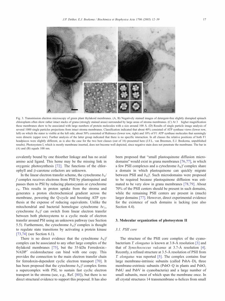
Fig. 3. Transmission electron microscopy of green plant thylakoid membranes. (A, B) Negatively stained images of detergent-free slightly disrupted spinach
chloroplasts often show rather intact stacks of grana (strongly stained areas) surrounded by large areas of stroma membranes. (C) At 3� higher magnification
these membranes show to be associated with large numbers of protein molecules with a size around 100 2. (D) Results of single particle image analysis of
several 1000 single particles projections from intact stroma membranes. Classification indicated that about 40% consisted of ATP synthase views (lower row,
left) on which the stator is visible at the left side; about 50% consisted of Rubisoco (lower row, right) and 10% of F1 ATP synthase molecules that seemingly
were dimeric (upper row). Further analysis of the latter group indicated that there is no specific interaction. In all classes the relative positions of both F1
headpieces were slightly different, as is also the case for the two best classes (out of 16) presented here (J.F.L. van Breemen, E.J. Boekema, unpublished
results). Photosystem I, which is mostly membrane inserted, does not become well depicted, since negative stain does not penetrate the membrane. The bar in
(A) and (B) equals 100 nm.
J.P. Dekker, E.J. Boekema / Biochimica et Biophysica Acta 1706 (2005) 12–39 17
covalently bound by one thioether linkage and has no axial
amino acid ligand. This heme may be the missing link in
oxygenic photosynthesis [72]. The functions of the chlor-
ophyll and h-carotene cofactors are unknown.
In the linear electron transfer scheme, the cytochrome b6/
f complex receives electrons from PSII by plastoquinol and
passes them to PSI by reducing plastocyanin or cytochrome
c6. This results in proton uptake from the stroma and
generates a proton electrochemical gradient across the
membrane, powering the Q-cycle and boosting ATP syn-
thesis at the expense of reducing equivalents. Unlike the
mitochondrial and bacterial homologue cytochrome bc1,
cytochrome b6/f can switch from linear electron transfer
between both photosystems to a cyclic mode of electron
transfer around PSI using an unknown pathway (see Section
5.5). Furthermore, the cytochrome b6/f complex is thought
to regulate state transitions by activating a protein kinase
[73,74] (see Section 6.1).
There is no direct evidence that the cytochtome b6/f
complex can be associated to any other large complex of the
thylakoid membranes [75], but the 35-kDa Ferredoxin:-
NADP+ oxidoreductase can bind with one copy. This
provides the connection to the main electron transfer chain
for ferredoxin-dependent cyclic electron transport [70]. It
has been proposed that the cytochrome b6/f complex forms
a supercomplex with PSI, to sustain fast cyclic electron
transport in the stroma (see, e.g., Ref. [80]), but there is no
direct structural evidence to support this proposal. It has also
been proposed that bsmall plastoquinone diffusion micro-
domainsQ would exist in grana membranes [76,77], in which
a few PSII complexes and a cytochrome b6/f complex share
a domain in which plastoquinone can quickly migrate
between PSII and b6/f. Such microdomains were proposed
to be required because plastoquinone diffusion was esti-
mated to be very slow in grana membranes [78,79]. About
70% of the PSII centers should be present in such domains,
while the remaining PSII centers are present in (much)
larger domains [77]. However, direct experimental evidence
for the existence of such domains is lacking (see also
Section 4.4).
3. Molecular organization of photosystem II
3.1. PSII core
The structure of the PSII core complex of the cyano-
bacterium T. elongatus is known at 3.8-2 resolution [3] and
that of Synechococcus vulcanus at 3.7-2 resolution [4].
Recently, a refined structure at 3.5-2 resolution of PSII from
T. elongatus was reported [5]. The complex contains four
large membrane-intrinsic subunits (called PsbA–D), three
membrane-extrinsic subunits (PsbO–Q in plants and PsbO,
PsbU and PsbV in cyanobacteria) and a large number of
small subunits, most of which span the membrane once. In
all crystal structures 14 transmembrane a-helices from small
J.P. Dekker, E.J. Boekema / Biochimica et Biophysica Acta 1706 (2005) 12–3918
subunits are observed [3–5]. Green plants contain probably
two additional small proteins that span the membrane once
[81].
PsbA (D1) and PsbD (D2) bind six chlorophyll a and
two pheophytin a molecules, while PsbB (CP47) and PsbC
(CP43) bind 16 and 14 chlorophyll a molecules, respec-
tively [5]. PsbA and PsbD constitute the photochemical
reaction center in which the charge separation and primary
electron transfer reactions take place [82,83], while PsbB
and PsbC have a light-harvesting function, i.e., they absorb
light and transfer the excitation energy to the reaction center.
Even more importantly, they also accept excitation energy
from the peripheral antenna and transfer this to the reaction
center as well [55,84]. It is of interest to note that there are
no indications yet that any of the small proteins binds
chlorophyll. Some may, however, be involved in the binding
of h-carotene [5]. This contrasts with the situation in PSI, in
which several of the small proteins do bind chlorophyll (see
Section 2.1). The extrinsic proteins do not bind chlorophyll
either.
In cyanobacteria, the basic unit of PSII that was
crystallized is a dimer [3–5]. The a-helices of four not yet
identified small subunits as well as helices of PsbA (D1) and
PsbB (CP47) are located in the dimerization domain [4]
(Fig. 4B). It has been shown that the PSII monomers can be
fully active [85] and that the organization of the dimers is
very similar in cyanobacteria and green plants [86,87]. Also
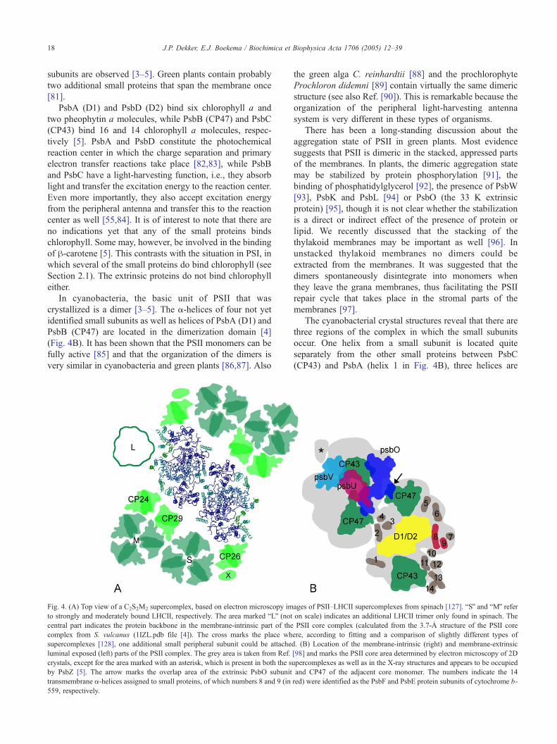
Fig. 4. (A) Top view of a C2S2M2 supercomplex, based on electron microscopy im
to strongly and moderately bound LHCII, respectively. The area marked bLQ (notcentral part indicates the protein backbone in the membrane-intrinsic part of the
complex from S. vulcanus (1IZL.pdb file [4]). The cross marks the place wh
supercomplexes [128], one additional small peripheral subunit could be attache
luminal exposed (left) parts of the PSII complex. The grey area is taken from Ref.
crystals, except for the area marked with an asterisk, which is present in both the s
by PsbZ [5]. The arrow marks the overlap area of the extrinsic PsbO subunit
transmembrane a-helices assigned to small proteins, of which numbers 8 and 9 (in
559, respectively.
the green alga C. reinhardtii [88] and the prochlorophyte
Prochloron didemni [89] contain virtually the same dimeric
structure (see also Ref. [90]). This is remarkable because the
organization of the peripheral light-harvesting antenna
system is very different in these types of organisms.
There has been a long-standing discussion about the
aggregation state of PSII in green plants. Most evidence
suggests that PSII is dimeric in the stacked, appressed parts
of the membranes. In plants, the dimeric aggregation state
may be stabilized by protein phosphorylation [91], the
binding of phosphatidylglycerol [92], the presence of PsbW
[93], PsbK and PsbL [94] or PsbO (the 33 K extrinsic
protein) [95], though it is not clear whether the stabilization
is a direct or indirect effect of the presence of protein or
lipid. We recently discussed that the stacking of the
thylakoid membranes may be important as well [96]. In
unstacked thylakoid membranes no dimers could be
extracted from the membranes. It was suggested that the
dimers spontaneously disintegrate into monomers when
they leave the grana membranes, thus facilitating the PSII
repair cycle that takes place in the stromal parts of the
membranes [97].
The cyanobacterial crystal structures reveal that there are
three regions of the complex in which the small subunits
occur. One helix from a small subunit is located quite
separately from the other small proteins between PsbC
(CP43) and PsbA (helix 1 in Fig. 4B), three helices are
ages of PSII–LHCII supercomplexes from spinach [127]. bSQ and bMQ referon scale) indicates an additional LHCII trimer only found in spinach. The
PSII core complex (calculated from the 3.7-2 structure of the PSII core
ere, according to fitting and a comparison of slightly different types of
d. (B) Location of the membrane-intrinsic (right) and membrane-extrinsic
[98] and marks the PSII core area determined by electron microscopy of 2D
upercomplexes as well as in the X-ray structures and appears to be occupied
and CP47 of the adjacent core monomer. The numbers indicate the 14
red) were identified as the PsbF and PsbE protein subunits of cytochrome b-

J.P. Dekker, E.J. Boekema / Biochimica et Biophysica Acta 1706 (2005) 12–39 19
found in the center of the dimer (helices 2–4 in Fig. 4B),
while the remaining 10 (including PsbE and PsbF which
together form cytochrome b-559) form an almost continu-
ous row of helices along the outer rim of the complex from
PsbB to PsbC (helices 5–14 in Fig. 4). This part of the
complex is therefore almost free of chlorophyll (the
peripheral chlorophyll molecule known as ChlZD2 seems
to be the only chlorophyll near the outer rim of the complex
[3–5]). We note that green plants probably contain two
additional small proteins that span the membrane once
(PsbR and PsbW), but it is not clear where these proteins are
located [81]. One of those could be located near helix 1
(Fig. 4B) [98]. It has been suggested that PsbW stabilizes
the PSII dimer [93]. It is, however, unlikely that this protein
occurs in the center of the dimer, because of the structural
similarity of the dimeric organization in green plants and
cyanobacteria, and of its chemically induced cross-link with
PsbE (the large subunit of cytochrome b-559, helix 9 in Fig.
4B), which is located at the opposite side of the complex
[99,100].
3.2. Peripheral antenna
In higher plants and eukaryotic algae, the peripheral
antenna of PSII consists of a number of pigment–protein
complexes belonging to the Lhc super-gene family [36]. In
green plants two types of peripheral antenna proteins
associated to PSII can be distinguished. The most abundant
complex is the so-called bmajorQ LHCII antenna complex.
This complex occurs in a trimeric association state [101] and
is not unique in composition. It consist of various
combinations of three very similar proteins, encoded by
the lhcb1, lhcb2 and lhcb3 genes, that usually occur in a
ratio of about 8:3:1 [35]. In addition, there are three bminorQantenna complexes, which are called Lhcb4 (CP29), Lhcb5
(CP26) and Lhcb6 (CP24) and usually occur in monomeric
aggregation states. All these complexes bind various
molecules of chlorophyll a and chlorophyll b and of the
xanthophylls lutein, violaxanthin and neoxanthin [84,102].
The structure of the major trimeric LHCII complex is known
at 2.72 2 [7]. It is generally believed that the minor
complexes adopt rather similar three-dimensional organiza-
tions [35,103,104].
A special case is given for PsbS. This very hydrophobic
protein also belongs to the Lhc super-gene family and has
four transmembrane a-helices, one more than most other
members from this family. This protein has by some authors
been considered as a PSII core protein (see, e.g., Ref. [105]),
but it has never been found in PSII crystals. It has also been
considered as a light-harvesting protein, but also this is
controversial, because it is not certain whether or not this
protein binds chlorophyll [84,106]. Fact is that unlike most
other members of the Lhc super-gene family, this protein is
stable in the absence of chlorophylls and carotenoids [107]
and that it is directly involved in one of the most important
regulation mechanisms of photosynthesis, the mechanism
by which excess excitation energy is harmlessly dissipated
into heat [108] (discussed in more detail in Section 6.2). It
has been suggested that a dimer-to-monomer conversion of
PsbS accompanies this nonphotochemical quenching of
excess absorbed excitation energy [109].
3.3. PSII–LHCII supercomplexes
3.3.1. General features
A variable number of the peripheral antenna proteins can
associate with dimeric PSII core complexes to form the so-
called PSII–LHCII supercomplexes. Supercomplexes were
first recognized in electron microscopic images by their
peculiar rectangular shape after a mild detergent solubiliza-
tion of grana membranes [87]. These rectangular super-
complexes, in the following denoted as dstandardT or C2S2supercomplexes (see below), contain almost all PSII core
proteins [98], but from the peripheral antenna only the
Lhcb1, Lhcb2, Lhcb4 and Lhcb5 gene products could be
detected [110]. The PsbS protein is absent as well [111],
though the absence of PsbS in PSII–LHCII supercomplexes
is not generally accepted [112]. A three-dimensional
structure of this supercomplex was constructed (at 24-2resolution) by single-particle analysis of images obtained by
cryoelectron microscopy [113]. It was shown that the three-
dimensional structures of PSII-LHCII complexes from the
green alga C. reinhardtii [88] and the liverwort Marchantia
polymorpha [114] are very similar to that of higher plants.
Combining this work with the X–ray crystallography work
on cyanobacterial PSII [3] revealed quite some detail of the
structure of the core part of the PSII–LHCII supercomplex
[90].
An important question is whether or not the (rectan-
gular) PSII–LHCII supercomplex represents the native
organization of PSII in the grana membranes. Some
authors [115] suggested that the dimeric supercomplex
represents an artefact induced by the solubilization of the
membranes. We do not share this opinion and note that
rectangular supercomplexes were not only observed after
detergent solubilization of PSII grana membranes, but also
(and with very high yield) after detergent solubilization of
complete thylakoid membranes [116]. In addition, EM
micrographs of partially unfolding grana membranes
clearly revealed the presence of rectangular supercom-
plexes in the membranes [117], which indicates that the
supercomplexes occur as such in the membranes. More
evidence for the occurrence of PSII–LHCII supercom-
plexes in grana membranes has been provided by analyses
of regular arrays of particles in grana membranes and of
mutants of Arabidopsis thaliana that express antisense
constructs (see Section 4).
An important consequence of the organization of the
PSII–LHCII supercomplexes is that the Lhcb4–LHCII–
Lhcb5 antenna structure needs a dimeric PSII core to
become attached to PSII. Lhcb4 (CP29) binds to one PSII
core monomer, Lhcb5 (CP26) to the other, while the

J.P. Dekker, E.J. Boekema / Biochimica et Biophysica Acta 1706 (2005) 12–3920
trimeric LHCII unit has clear contacts with both Lhcb4 and
Lhcb5 (Fig. 4A). Indeed, monomeric cores with attached
LHCII have never been observed in mixed populations of
disrupted grana. This also explains why the peripheral
antenna proteins easily detach from PSII during the
transition from dimer to monomer. Our analyses did not
reveal other types of association of PSII and LHCII than the
supercomplex. Other associations can, however, not be
completely ruled out if one looks at positions of core
complexes within the membrane [118]. These authors
claimed a rearrangement of the LHCII antenna proteins
around the core dimer, based on the finding of a new type of
crystal packing. However, after testing the packing of this
lattice with the standard C2S2 supercomplexes, we conclude
that such particles nicely can fit within the published lattice
and that the proposed LHCII rearrangement is an over-
interpretation of low-resolution data.
3.3.2. Extrinsic proteins
After removal of extrinsic proteins by Tris-washing
PSII–LHCII supercomplexes could be observed with either
an elongated or a shortened appearance [95]. These types of
supercomplexes could not be detected in the databases of
supercomplexes obtained from oxygen- evolving or salt-
washed PSII membranes, even when an elongated or
shrinked supercomplex was used as reference in the data
analysis. It was suggested that the removal of PsbO (the
extrinsic 33-kDa protein) causes a diminishing of the
interaction between the two PSII core units. It was also
shown that removal of PsbO and/or PsbP (the 23-kDa
protein) can induce a displacement of LHCII and/or Lhcb4
towards the PSII core complex [95]. This suggests that the
extrinsic proteins not only have roles in the cofactor
requirement for photosynthetic oxygen evolution [119],
but also may function to keep the peripheral antenna at a
proper distance to maintain sequestered domains of inor-
ganic cofactors required for oxygen evolution. The PSII
structure from S. vulcanus [4] provides some evidence for
this. The PsbO protein has a number of close contacts with
the CP47 subunit of the other PSII monomer and also
extends over the outer rim of the complex at a position
where in green plants trimeric LHCII is bound (Fig. 4B).
We note that the position of the PsbO protein in the
structures of the cyanobacterial PSII core complex [3–
5,120] differs from that anticipated earlier for the PSII core
complex from green plants [113,121,122] (see also Ref.
[123]). In this earlier view, the PsbO protein was thought to
be located at the most stain-excluding region of the PSII
core complex near the bCQ of CP47 in the left part of Fig.
4B, both in green plants and in cyanobacteria. This view
was based in part on the amount of staining in top views in
electron microscopy images, which appeared to be very
similar for PSII in green plants and cyanobacteria. The most
stain-excluding area of the cyanobacterial PSII core com-
plex is at a position with, according to the recent structures,
almost no extrinsic mass, so it seems that the extrinsic
proteins have only minor effects on the staining of the PSII
core complexes.
A matter of current debate is the number of PsbO
proteins per PSII monomer. The crystal structures of
cyanobacterial PSII core complexes reveal only one copy
[3–5], but binding studies of PsbO to plant PSII suggest
binding of two copies with markedly different binding
affinities [124]. A recent study on the binding domains of
PsbO from spinach suggested two different binding
domains, one of which is absent in the amino acid sequences
of cyanobacterial PsbO [125]. It is possible that a second
PsbO protein is present in green plant PSII, provided that it
has minor effects on the staining of PSII as seen in top views
in electron microscopy images.
3.3.3. Binding of trimeric antenna proteins
The rectangular supercomplex contains two trimeric
LHCII complexes per PSII core dimer. However, the total
number of trimeric LHCII per PSII dimer is generally about
eight, which immediately raises the question of the location
of the remaining population of LHCII trimers. Based on the
shortest (b5 min) and mildest (using the nonionic detergent
n-dodecyl-a,d-maltoside, a-DM) possible way of solubiliz-
ing PSII grana membranes from spinach, we found by
electron microscopy and single particle analysis of partially
solubilized complexes two additional binding sites for
trimeric LHCII at the PSII core dimers [126–128]. Accord-
ing to the frequency of occurrence the three binding sites
were designated bSQ, bMQ and bLQ (from strongly, moder-
ately and loosely bound LHCII, respectively) (Fig. 4A).
Analysis of the averaged images of C2S2M1–2 super-
complexes showed that the M trimer is rotationally shifted
by about 208 compared to the S trimer [128]. In Arabidopsis
thaliana, only S and M trimers were found, but in the same
position [129]. However, the M trimers were more abundant
in Arabidopsis than in spinach (see also below). Also in the
liverwort M. polymorpha the M trimers are more abundant
than in spinach [114].
The S-LHCII trimer consists predominantly of the Lhcb1
and Lhcb2 gene products, because only these were detected
in C2S2 supercomplexes [110]. In contrast, the M-LHCII
trimer consists most likely of the Lhcb1 and Lhcb3 gene
products, because the Lhcb3 gene product is present in the
larger supercomplexes [127] and because it occurs in a
supercomplex consisting of the Lhcb1, Lhcb3, Lhcb4 and
Lhcb6 gene products in a 2:1:1:1 ratio [130,131]. The M-
LHCII trimer and the Lhcb4 and Lhcb6 gene products are
very close together in the PSII–LHCII supercomplex (Fig.
4A). This suggests that one of the constituents of M-LHCII
is encoded by Lhcb3. The positions that Lhcb1 and Lhcb3
may occupy have not yet been determined.
Analysis of supercomplexes isolated from Arabidopsis
plants expressing an antisense construct to Lhcb2 revealed
that the LHCII binding sites are not unique for the various
types of trimers [132]. In these plants, not only the synthesis
of Lhcb2 was almost completely abolished, but also that of

J.P. Dekker, E.J. Boekema / Biochimica et Biophysica Acta 1706 (2005) 12–39 21
the strongly related Lhcb1 protein [133]. It appeared that in
these plants, the expression of the antisense Lhcb2 construct
resulted in strongly increased levels of Lhcb5 (CP26) and
(to a minor extent) Lhcb3, and that supercomplexes were
formed with trimers consisting of Lhcb5 and Lhcb3 at the S-
and M- binding positions [132]. This replacement is unique,
because expression of antisense constructs to the minor
peripheral antenna proteins and to the peripheral antenna
proteins of PSI did not lead to increased synthesis of other
proteins (discussed in detail below), and stresses the
importance of the particular organization of PSII and LHCII
in supercomplexes.
3.3.4. Binding of monomeric antenna proteins
Our work also gave new information on the location and
binding of the minor complexes Lhcb4 (CP29), Lhcb5
(CP26) and Lhcb6 (CP24). Based on cross-linking experi-
ments [134], it was assumed that Lhcb5 is located near PsbC
(CP43) on the tip of the PSII–LHCII supercomplex, whereas
Lhcb4 is located near PsbB (CP47) on the other side of the
complex (Fig. 4A). A recent structural analysis of super-
complexes prepared from antisense mutants of Arabidopsis
without Lhcb5 or Lhcb4 confirmed this view [135]. Lhcb6
is absent in the dstandardT C2S2 supercomplex, but present in
the larger complexes [127,134], which strongly suggests
that Lhcb6 represents the additional mass with the size of a
monomeric light-harvesting complex close to the M trimer
and Lhcb4 (Fig. 4A). A physical interaction between Lhcb6
and Lhcb4 became also obvious from Arabidopsis plants
with antisense constructs to Lhcb4 or Lhcb5 [136].
The different minor complexes seem to have unique and
different roles in the supramolecular organization of PSII
and LHCII. Supercomplexes with empty Lhcb5 binding site
but with otherwise normal appearance could be isolated
from Arabidopsis plants expressing antisense constructs to
Lhcb5 [135]. In addition, classification of projections of
supercomplexes from spinach [95,128] or Arabidopsis [129]
suggested the existence of many supercomplexes with
empty Lhcb5 binding sites. This indicates that Lhcb5 is
not needed for the formation of PSII–LHCII supercom-
plexes and that antisense removal of this protein does not
lead to replacement by one of the other members of the Lhc
super gene family. No intact supercomplexes could be
isolated from Arabidopsis plants expressing antisense
constructs to Lhcb4 [135], and PSII–LHCII supercomplexes
with empty Lhcb4 binding sites could not be found [128].
This suggests that also Lhcb4 occupies a unique position in
the PSII macrostructure and that, in contrast to Lhcb5, its
presence is essential for the formation of PSII–LHCII
supercomplexes. Both the Lhcb4 and Lhcb5 antisense
mutants showed a rather normal photosynthetic perform-
ance, although the mutants showed slightly different
fluorescence characteristics and an increased number of
PSII centers [133]. This suggests that the organization of
PSII and LHCII into supercomplexes is not absolutely
required for photosynthetic performance, at least under
normal physiological conditions and light levels. In the
field, however, most mutants showed decreased ecological
flexibility, an important parameter for plant fitness under
natural conditions [137,138].
3.3.5. Role of small PSII core subunits
Of special interest for the supramolecular organization
of the PSII core complex and its surrounding peripheral
antenna are those small subunits of the PSII core that
could play a role in the binding of the peripheral antenna.
The constructed figure of the PSII–LHCII supercomplex
suggests that of the 14–16 small proteins only a few seem
to be involved in contacts with the peripheral antenna. The
first is the one (or two in green plants) protein located
between PsbA and PsbC (helix 1 in Fig. 4B). This protein
may contribute to the binding of S-LHCII and may
therefore be important for the supramolecular organization
of PSII and LHCII. In the structure of S. vulcanus this
protein remained unassigned [4], whereas in the structure
of T. elongatus it was assigned to PsbI [5]. It is possible
(but by no way proven) that the PsbR protein (not present
in cyanobacteria—see Section 3.1) is also located at this
position. Two other proteins (helices 5 and 6 in Fig. 4B,
attributed to PsbH and PsbX in [5]) seem to have contacts
with CP24.
Another protein that may be involved in the association
of the peripheral antenna is the protein located at the outer
tip of PsbC in the supercomplex (helices 13 and 14 in Fig.
4B). It is possible that this protein is PsbZ (also known as
Ycf9 or Orf62), the only small protein in PSII that has two
transmemebrane a-helices, because mutants without PsbZ
appeared to have strongly reduced Lhcb5 levels [139,140].
The location as indicated in the figure is consistent with the
reduced level of Lhcb5. However, it appeared that without
PsbZ no PSII–LHCII supercomplexes could be isolated,
which is not consistent with results from an Arabidopsis
mutant with an antisense construct to Lhcb5 [135]. This and
other studies revealed that Lhcb5 is not required for the
stabilization of PSII–LHCII supercomplexes. Another pos-
sibility is that helix 14 arises from PsbJ, because also
without this small protein no stable PSII–LHCII super-
complexes could be detected, whereas it also seems to
interact with the extrinsic proteins PsbP and PsbQ [141],
which are expected near this position (Fig. 4B). Ferreira et
al. [5] assigned helix 10 to PsbJ, but this location is far from
the interaction domain with the peripheral antenna, and does
not easily explain the absence of supercomplexes if PsbJ is
absent.
If PsbZ is not responsible for helices 14 and 13, it can
also be located at the first interaction position (see above),
near helices V and VI of PsbC and helices A and B of PsbA
(helix 1 in Fig. 4B). Also two chlorophylls of PsbC and one
of PsbA (ChlZD1) are positioned near this helix, so this
protein could be important for energy transfer from the
peripheral antenna to the PSII core. A location of PsbZ at
this position is consistent with the absence of super-
J.P. Dekker, E.J. Boekema / Biochimica et Biophysica Acta 1706 (2005) 12–3922
complexes, and can be consistent with the absence of Lhcb5
if the absence of S-LHCII prevents the binding of Lhcb5. In
green plants two helices are modeled at this position [90],
but in cyanobacteria only one [3–5]. The amino acid
sequence of PsbZ from Synechocystis suggests two helices
[81], which is inconsistent with a location of PsbZ at helix
position 1 in the structural models of the cyanobacterial PSII
core complex. We conclude that more studies are required to
understand the role of PsbZ in the PSII–LHCII interaction.
3.3.6. Energy transfer
The constructed figure (Fig. 4A) gives some indication
on the possible energy transfer routes in the super-
complexes. CP43 (PsbC) can receive excitation energy
from both CP26 (Lhcb5) and S-LHCII. At the present
resolution it is impossible to estimate the distance between
the most nearby chlorophylls, but nevertheless it seems
reasonable to assume efficient energy transfer along these
routes. CP47 (PsbB) has excellent contacts with CP29
(Lhcb4). The distance between CP24 (Lhcb6) and CP47
seems slightly larger, so most energy transfer from CP24
may go via CP29. Interestingly, there seems to be a rather
large distance between the chlorophylls of L-LHCII and of
the PSII core complex, because L-LHCII is located behind
the rim of chlorophyll-free small subunits of the PSII core
complex (Fig. 4A). This suggests that energy transfer from
L-LHCII proceeds via CP26 or CP24. The only chlor-
ophyll that comes somewhat close is the peripheral
chlorophyll of PsbD (D2), so a function of this chlorophyll
could be to mediate energy transfer from L-LHCII to heart
of the PSII core complex. The peripheral chlorophyll of
PsbA (D1) also seems quite remote from the peripheral
antenna system, the S-LHCII trimer is the most nearby, but
this complex has much better contacts with CP43. For
more details on the energy transfer characteristics, we refer
to a recent review [84].
3.4. LHCII aggregates
Most PSII–LHCII supercomplexes contain two, three
of four LHCII trimers. Very few complexes with five
LHCII trimers could be detected, but complexes with
more trimers were never observed, despite a very large
data set of EM projections of supercomplexes [127].
However, there are usually about eight LHCII trimers per
PSII core dimer [130], so there is considerably more
LHCII than present in the PSII–LHCII supercomplexes.
This implies the presence of a pool of non-bound or very
loosely bound LHCII, in line with many earlier sugges-
tions (see, e.g., Ref. [142] and Section 4.3). Classification
of single-particle projections of partially solubilized PSII
grana membranes revealed small amounts (1–5%) of a very
characteristically shaped particle (Fig. 5) that was identified
as a heptamer of LHCII trimers [143]. The same or similar
LHCII aggregates were purified by Peter and Thornber
[130] and Ruban et al. [144].
3.5. PSII–Pcb supercomplexes
Prochlorophytes contain a Chl a/b binding protein called
Pcb that does not belong to the Lhc super-gene family, but
instead is related to PsbC (CP43) [59]. Recently, PSII–Pcb
supercomplexes from P. didemni and Prochlorococcus MIT
9313 were described. These complexes consist of a PSII
core dimer, organized in a similar way as in cyanobacteria
and green plants, flanked by five Pcb subunits at each side
of the dimer in Prochloron [89] or by four PcbA subunits at
each side of the dimer in Prochlorococcus [26]. The Pcb
subunits occur at similar positions as the Lhcb4–LHCII–
Lhcb5 subunits of green plants and thus also need a PSII
core dimer as a scaffold to bind rows of four or five Pcb
subunits.
4. Organization of supercomplexes in stacked grana
membranes
4.1. Crystalline arrays
Crystalline 2D arrays of PSII have been observed
decades ago after freeze-etching and freeze- breaking of
green plant photosynthetic membranes (see Refs.
[105,123,145] for recent reviews). It was noted that PSII
in such arrays is dimeric (see, e.g., Refs. [146–148]), but
details of the molecular composition of PSII in these
rows could not be provided because of the limited
resolution of the freeze-etching technique. In addition,
there appeared to be quite some variation in the size of
the repeating unit in these rows, which suggests that
several types of complexes can form repeating units,
depending on factors like plant species, growth condi-
tions, preparation method, etc.
Recently, it became possible to see details of the area
between the rows, using electron microscopy and image
analysis of negatively stained grana membranes isolated
from spinach thylakoids after a short treatment with a-DM
[149]. Two types of rows were found, called bsmall-spacedQand blarge-spacedQ rows, indicating a spacing of rows of 23
and 26 nm, respectively. The small-spaced semi-crystalline
macrodomains occurred in about 1% of the membranes, and
were shown to consist of a C2S2 repeating unit (see also
Table 1), while the large-spaced semi-crystalline macro-
domains occurred in about 50% of the membranes and were
suggested to consist of an asymmetric C2S2M repeating unit
[149]. The unit cell of the latter and most abundant regular
arrays (27.3�18.3 nm, angle 74.58, area 481 nm2)
resembles the unit cell of a frequently occurring regular
array in freeze-fractured thylakoid membranes from spinach
(26.5�18.7 nm, angle 698, area 462 nm2) [147], which
suggests that the regular arrays have not been induced by
the detergent treatment or by the negative stain used to
image the membranes by EM (discussed in more detail in
Section 4.4).

Fig. 5. The LHCII icosienamer. (A) Average image of 133 aligned complexes indicates the presence of seven LHCII trimers [143]. (B) Threefold symmetrized
projection of image A. (C) Fitting of the LHCII structure [6] in the lower three densities. The bar is 10 nm.
J.P. Dekker, E.J. Boekema / Biochimica et Biophysica Acta 1706 (2005) 12–39 23
Grana membranes from Arabidopsis thaliana prepared in
the same way as described above for spinach consistently
showed semi-crystalline macrodomains with larger unit cells
than in spinach [129]. In wild-type A. thaliana, the unit cell
was 25.6�21.4 nm (angle 778, area 534 nm2). Image
analysis revealed that these semi-crystalline macrodomains
are built up from C2S2M2 supercomplexes. The additional
M-LHCII in the unit cell of A. thaliana compared to spinach
Table 1
Unit cell and composition of regular arrays of PSII and LHCII in grana
preparations
Regular
arrays?
Unit Area
(nm2)
Protein
replaced?
Reference
Spinach yes C2S2 389 – [149]
Spinach yes C2S2M 481 – [149]
Arabidopsis yes C2S2M2 534 – [129]
CP29 no – – no [135]
CP26 yes C2S2M2 500 no [135]
Lhcb1+2 yes C2S2M2 524 yes [132]
PsbS yes C2S2M2 531 no [150]
is consistent with the more frequent occurrence of M-LHCII
in supercomplexes from A. thaliana than from spinach, in
line with the idea that the repeating unit in these semi-
crystalline arrays is a PSII–LHCII supercomplex. Mem-
branes from the npq4 mutant of A. thaliana, which lacks the
PsbS protein [108], revealed an identical unit cell as in the
wild-type [150], which strongly suggests that the relatively
large PsbS protein is not present in the regular arrays in
grana membranes of wild-type plants. A slightly different
unit cell was found in membranes from plants with an
antisense construct to Lhcb2 [132], which can be explained
by the fact that the trimers in these membranes consist of
Lhcb5 and Lhcb3 instead of Lhcb1 and Lhcb2. A smaller
unit cell was also found in membranes from plants with an
antisense construct to Lhcb5 [135], in which case the image
analysis indicated a supramolecular organization without
Lhcb5 but otherwise similar to that of the wild-type. No
regular arrays were found in membranes from plants with an
antisense construct to Lhcb4, but PSII–LHCII supercom-
plexes were not found either [135]. Table 1 summarizes the
J.P. Dekker, E.J. Boekema / Biochimica et Biophysica Acta 1706 (2005) 12–3924
properties of the regular arrays observed in the investigated
types of plants.
These results suggest that the basic unit of PSII and
LHCII in grana membranes differs in spinach and Arabi-
dopsis. In spinach, the basic unit is C2S2M, in which two
copies of S-LHCII, Lhcb4 and Lhcb5 and one copy of M-
LHCII and Lhcb6 are attached to a PSII core dimer, whereas
in A. thaliana the basic unit is C2S2M2, in which two copies
of S-LHCII, M-LHCII, Lhcb4, Lhcb5 and Lhcb6 are
attached (see also Fig. 4A). In view of the occurrence of
supercomplexes, it is likely that the basic unit is also
C2S2M2 in M. polymorpha [114]. We note that the protein
mass of the C2S2M2 unit is almost 1.1 MDa and that this
complex binds approximately 190 Chl a, 80 Chl b and 75
carotenoid molecules [84].
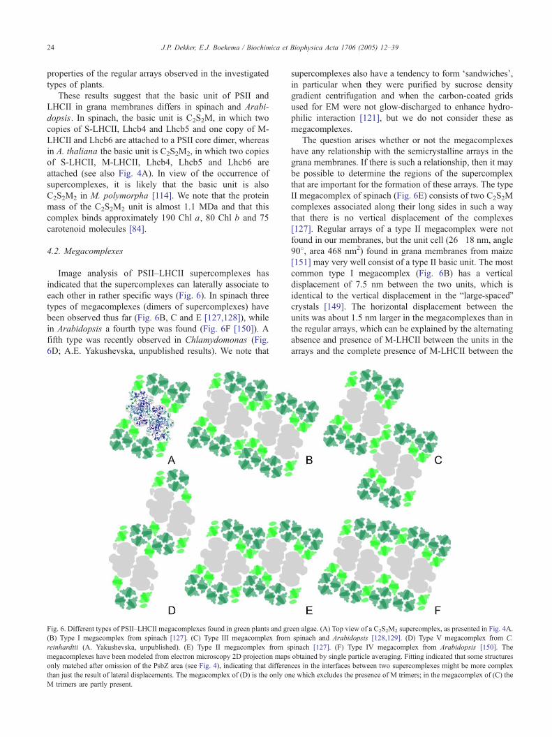
4.2. Megacomplexes
Image analysis of PSII–LHCII supercomplexes has
indicated that the supercomplexes can laterally associate to
each other in rather specific ways (Fig. 6). In spinach three
types of megacomplexes (dimers of supercomplexes) have
been observed thus far (Fig. 6B, C and E [127,128]), while
in Arabidopsis a fourth type was found (Fig. 6F [150]). A
fifth type was recently observed in Chlamydomonas (Fig.
6D; A.E. Yakushevska, unpublished results). We note that
Fig. 6. Different types of PSII–LHCII megacomplexes found in green plants and gr
(B) Type I megacomplex from spinach [127]. (C) Type III megacomplex from
reinhardtii (A. Yakushevska, unpublished). (E) Type II megacomplex from s
megacomplexes have been modeled from electron microscopy 2D projection maps
only matched after omission of the PsbZ area (see Fig. 4), indicating that differen
than just the result of lateral displacements. The megacomplex of (D) is the only on
M trimers are partly present.
supercomplexes also have a tendency to form dsandwichesT,in particular when they were purified by sucrose density
gradient centrifugation and when the carbon-coated grids
used for EM were not glow-discharged to enhance hydro-
philic interaction [121], but we do not consider these as
megacomplexes.
The question arises whether or not the megacomplexes
have any relationship with the semicrystalline arrays in the
grana membranes. If there is such a relationship, then it may
be possible to determine the regions of the supercomplex
that are important for the formation of these arrays. The type
II megacomplex of spinach (Fig. 6E) consists of two C2S2M
complexes associated along their long sides in such a way
that there is no vertical displacement of the complexes
[127]. Regular arrays of a type II megacomplex were not
found in our membranes, but the unit cell (26�18 nm, angle
908, area 468 nm2) found in grana membranes from maize
[151] may very well consist of a type II basic unit. The most
common type I megacomplex (Fig. 6B) has a vertical
displacement of 7.5 nm between the two units, which is
identical to the vertical displacement in the blarge-spacedQcrystals [149]. The horizontal displacement between the
units was about 1.5 nm larger in the megacomplexes than in
the regular arrays, which can be explained by the alternating
absence and presence of M-LHCII between the units in the
arrays and the complete presence of M-LHCII between the
een algae. (A) Top view of a C2S2M2 supercomplex, as presented in Fig. 4A.
spinach and Arabidopsis [128,129]. (D) Type V megacomplex from C.
pinach [127]. (F) Type IV megacomplex from Arabidopsis [150]. The
obtained by single particle averaging. Fitting indicated that some structures
ces in the interfaces between two supercomplexes might be more complex
e which excludes the presence of M trimers; in the megacomplex of (C) the

Fig. 7. Crystallinity of C2S2M complexes in paired inside-out grana
membranes from spinach. In one of the membrane layers, the one which is
stronger contrasted with negative stain; rows of crystalline core parts are
visible. In the other membrane layer, the crystallinity in the largest domain
is partly in the same direction (see arrows), but only a few complexes are
present, indicating that LHCII dominates in these areas. On the left and
right sides of the grana membrane single membrane layers are attached with
a different granularity, likely caused by aggregated PSI complexes. The bar
equals 100 nm.
J.P. Dekker, E.J. Boekema / Biochimica et Biophysica Acta 1706 (2005) 12–39 25
units in the megacomplexes. The minor type III mega-
complex from spinach (Fig. 6C [127]) has a vertical
displacement of 20 nm and appears to be the repeating unit
of the regular arrays in Arabidopsis [129]. However, the
megacomplex observed in Arabidopsis (Fig. 6F [150]) does
not form the basis of a known regular array.
The various structures suggest that the presence of L-
LHCII would prevent the formation of four of the five types
of megacomplexes observed thus far, while M-LHCII in
plant megacomplexes is either required for the formation of
these megacomplexes (Figs. 6B, E, and F) or does not
impose a steric hindrance (Fig. 6C). M-LHCII would
impose steric hindrance for the formation of the mega-
complex observed in Chlamydomonas (Fig. 6D), but this
type of LHCII was not observed at all in Chlamydomonas
supercomplexes (A.E. Yakushevska, unpublished observa-
tions), which may be related to the absence of an Lhcb6
homologue in Chlamydomonas [44,152]. It is possible that
L-LHCII is bound to supercomplexes located at the end of
the regular arrays. The data suggest that Lhcb5 is essential
for the formation of the type I and type III megacomplexes
(Figs. 6B and C), and thus for the most common regular
arrays in spinach and Arabidopsis, respectively. On the
other hand, Lhcb4 is not involved in the formation of
megacomplexes, because it is located more in the interior of
the supercomplex, but is essential for the formation of the
supercomplexes themselves.
4.3. Organization of PSII and LHCII in opposing
membranes
An advantage of the EM technique applied to negatively
stained grana membranes is that both layers of the grana can
be investigated simultaneously. This is not possible with
other structural techniques, such as atomic force microscopy
(AFM) and freeze fracture and freeze-etching EM techni-
ques. So with EM and additional image analysis, it is
possible to study the relation between the complexes in the
opposing membranes in terms of the positioning of the PSII
core dimer and the LHCII units.
We recently presented a detailed study of a-DM
prepared grana membranes from spinach [117,149]. There
appeared to be quite some heterogeneity in the number and
organization of the PSII complexes in these membranes.
Although all grana membranes consisted of two membrane
layers, some appeared to have a much lower density of
PSII complexes than others. This means that there is a
large variability in the number of LHCII antenna com-
plexes per PSII within a granum or between grana. A
small part of the membranes contained regular arrays in
both layers [149], and in these membranes the ratio of
LHCII to PSII will be not much higher than 1.5, the
number for the C2S2M repeating unit. Some other grana
contained surprisingly little PSII [117], and the ratio of
LHCII to PSII is in these membranes probably much
higher than 4 (the average ratio of LHCII to PSII in grana
membranes). The origin and significance of this hetero-
geneity is not yet understood.
It appeared that in those grana that have rows in both
membranes, there are preferential angles of rows in
opposing membranes with respect to each other, both in
spinach (Fig. 7) and in Arabidopsis (Fig. 8). At these angles
the overlap of LHCII trimers is optimized, at least for the
central part of the domains. For spinach the preferential
angles were 38 (F48) and 468 (F108) [149]; for Arabidopsisthey were 328 (F28) and 588 (F38) [129]. The differences
are most likely related to the different basic units. These
results indicate that the organization of the complexes in one
membrane affects the organization of complexes in the
opposing membrane.
Because the PSII–LHCII supercomplexes have a very
pronounced handedness, it is possible to judge if a certain
PSII complex in a granal membrane is located in the upper
membrane or in the lower membrane. The handedness was
analyzed in a relatively well-ordered membrane from
spinach [149], and it appeared that in one part of the
granum most PSII complexes were located in the upper
layer, in another part in the lower layer, while only in the
middle a region occurred with a row in both layers. Based
on this analysis, it was proposed that most (but certainly not
all) PSII–LHCII supercomplexes face an LHCII-only region
in the opposing membrane (Fig. 7). A variation of this
model was proposed by Ford et al. [115], who suggested
that one layer would consist entirely of PSII and the other
entirely of LHCII. We consider this possibility as less likely,
not only because this model does not accept the PSII–LHCII
supercomplex as basic unit for PSII in the membranes, but
also because this model needs a much larger amount of

Fig. 8. Preferential stacking as determined in paired inside-out grana membranes from A. thaliana. If two crystalline arrays face each other they are arranged in
such a way that there is optimal overlap of LHCII trimers (and hence also from core complexes) in at least the center of the sandwiched crystals. (A) Electron
micrograph of a negatively stained grana fragment of a 338-type crystal in which the two layers differ about 338 in their rotational orientation [129]. (B) Image
of a grana fragment of a 588-type in which the layers differ about 588. (C) Fourier filtering of the inner part of the 588-type crystal from B. The position of the
black dots on the core complex densities indicates dislocation in one of the layers; possibly to get a better match between LHCII complexes from adjacent
layers. (D, E) Simulation of the overlap pattern in the 338-type and 588-type crystals, respectively, based on 150 averaged crystals each. Asteriks indicate
positions where core complexes (in red) of adjacent layers match optimally. The bar in A is 100 nm.
J.P. Dekker, E.J. Boekema / Biochimica et Biophysica Acta 1706 (2005) 12–3926
reorganization of PSII and LHCII if a transition occurs from
a random organization to an organization in rows.
4.4. Do rows of PSII–LHCII supercomplexes represent
native structures?
It is important to know if PSII in semi-crystalline arrays
represents a fully functional photosystem or an inactive
complex. Freeze-fracture studies have indicated that rows
are largely absent in freshly prepared thylakoid membranes
but can be generated by prolonged storage at 48 C [153] or
by incubation in certain buffers [147,154]. It appeared that
thylakoids with rows of PSII are thermally more stable than
those without rows of PSII [154]. So it is possible that the
organization of PSII and LHCII in rows represents an in
vivo situation under at least some physiological conditions.
Cold acclimation could be one of those conditions [155].
However, even if an organization of PSII and LHCII in
rows would occur in vivo under some physiological
conditions, it is by no way certain that this organization
represents a fully functional photosynthetic unit. In such a
unit, not only efficient excitation energy transfer and
primary electron transport would have to occur, but also
the transfer of reduced plastoquinone to the cytochrome b6/f
complex needs to be fast and effective. It has been proposed
[76,77] that PSII centers with a functional plastoquinone
pool can only be found in bsmall plastoquinone diffusion
microdomainsQ (see Section 2.6). Such microdomains
cannot exist in the regular arrays of PSII and LHCII if they
have the same composition in our b6/f-free a-DM derived
grana membranes [117] and in thylakoids after prolonged
storage at 48 C [153] or by incubation in certain buffers
[147,154]. Also the image analysis of the rows did not give
any indication for the presence of the cytochrome complex.
So, if the concept of the small plastoquinone diffusion
microdomains is correct, then the rows represent a form of
PSII that is not directly involved in linear electron transport,
and could represent at least some of the 30% of PSII centers
that are thought to be inactive [77].
In our opinion it is not clear yet if a structural
organization of PSII in rows excludes a special long-
range plastoquinone diffusion pathway between PSII in
the rows and b6/f in another part of the membrane. For
instance, the organization in rows may permit a special
route in the LHCII areas of the rows along which
plastoquinone can migrate quickly. In Ref. [79] it was
suggested that most of the lipids in the grana will be
relatively immobile dboundaryT lipids. However, the
number of boundary lipids can be significantly smaller
than suggested in Ref. [79], because they will probably
not be present in the regions between the long sides of the
supercomplexes (as in the megacomplexes), while for a
C2S2M2 repeating unit the number of free LHCII trimers
and thus the number of boundary lipids is also lower than
calculated in Ref. [79]. Lower numbers of relatively
immobile boundary lipids implicate higher numbers of
highly mobile lipids and therefore plastoquinone diffusion
may not be as restricted as calculated. This suggests that
functional photosynthesis in semi-crystalline parts of the
grana cannot be excluded yet.

J.P. Dekker, E.J. Boekema / Biochimica et Biophysica Acta 1706 (2005) 12–39 27
4.5. Energy transfer and trapping in grana membranes
Efficient photosynthetic performance of PSII in grana
requires a delicate interplay between excitation energy
transfer, trapping of excitation energy by charge separation
in the reaction center and subsequent secondary electron
transfer reactions [156]. It is of interest to know to which
extent the structural organization of PSII in the grana
determines the kinetics and yield of these processes, and
thus determines the photosynthetic performance of PSII.
There are in principle three different processes which
could provide the rate-limiting step of energy transfer and
trapping, i.e., the rate of the primary charge separation
reaction, the rate of the energy transfer from the core
antenna to the reaction center, and the rate of energy transfer
among the various peripheral and core antenna proteins. If
the overall kinetics is determined by one of these processes,
the kinetics is said to be trap-limited, transfer-to-the-trap-
limited, or diffusion-limited, respectively. In intact PSII, the
overall kinetics occurs in two phases, with a main phase of
about 200 ps and a minor phase of about 500 ps [157],
provided that the quinone acceptor QA is oxidized. This
kinetics is considerably slower than that observed for intact
PSI (see Section 2).
There is currently no consensus on which of the
processes mentioned above is most important for PSII in
grana. A recent study on PSII complexes of different sizes
was interpreted to mean that energy transfer and trapping in
PSII is basically trap-limited [158]. However, it was recently
pointed out that there is a long distance between chlor-
ophylls of the PsbB or PsbC core antenna proteins and those
of the reaction center, which implies a quite slow energy
transfer reaction, and which, at least in the PSII core
complex itself, could limit the trapping process [159]. On
the other hand, it was pointed out that the orientations of the
chlorophylls of PsbB and PsbC are optimized for fast
excitation energy transfer [160] and a comparison between
the energy transfer and trapping dynamics in CP47-RC and
RC preparations suggested that the energy transfer between
CP47 and the RC does not limit the overall trapping of the
excitation energy [161].
Energy transfer within the peripheral antenna may in fact
also be rate-limiting, because energy transfer from one
LHCII to the next was suggested to occur in 32 ps [162]
and, depending on the supramolecular organization, more
than one LHCII complex needs to be passed before the
excitation reaches the reaction center. Also the time of
energy transfer between two opposing membranes in a
granum is considerable [163,164]. We conclude that there
are currently no data available that favor either a trap-limited
or a diffusion-limited trapping of excitation energy in PSII
in vivo (see also Ref. [84]). This implies that structural
rearrangements of the peripheral antenna (which should
primarily affect the diffusion-limited trapping rate) are likely
to have effects on the photosynthetic performance in vivo
(see also Section 6).
5. Overall chloroplast membrane topology
5.1. Vertical distance between membranes in grana stacks
The grana discs of green plant chloroplasts have
diameters of about 300–600 nm [165,166]. The sizes of
the grana and the proportions of grana and stroma appear
remarkably constant among plant species and among plants
grown under different environmental conditions [167].
In the vertical direction several to several dozens of
membranes may stack. The membranes are never equi-
distantly spaced, because on the stromal side membranes
are rather flat and seemingly form a pair spaced by about 2
nm, while at the lumenal side the distance between the
membranes is much larger because of the presence of large
protrusions of the PSII core and cytochrome b6/f
complexes (see also Fig. 9). The vertical distance from
one pair of membranes to the next can be extracted from
micrographs of thin-sectioned chloroplasts. Table 2 gives a
list of the average vertical repeat in the grana stacks,
presented in or calculated from 17 different papers. Some
of these numbers may give slight underestimations of the
spacing, because the pressure used in the thin-sectioning
technique to obtain slices of chloroplasts may induce some
shrinking. Table 2 shows that the vertical repeat varies
roughly from 14 to 24 nm, though distances around 16 and
21 nm seem to be the most common. A repeating unit of
21 nm would be just sufficient for the vertical height of
two PSII or cytochrome b6/f complexes on top of each
other, because each PSII and b6/f unit measures maximally
about 10.5 nm [10,11,113]. This would mean that not only
contacts between PSII and LHCII at their stromal sides
determine the morphology of the grana, but also contacts
between PSII and/or cytochrome b6/f units in opposing
membranes in the lumen. This is in line with studies on
inside-out vesicles with exposed lumenal side [168], which
revealed considerable interactions between the lumenal-
exposed sides of the membranes. The most exposed
proteins are the extrinsic PsbP and PsbQ proteins of PSII,
so these proteins may not only function to optimize the
inorganic cofactor requirement for water oxidation [119],
but also may contribute to the shape of the grana stacks. A
shorter distance than 21 nm would mean, if not artefactual,
that two PSII units cannot be located with their extrinsic
subunits directly opposed to each other in the lumen. This
could diminish the diffusion of PSII complexes in the
grana stacks and also limit the available volume for
lumenal proteins (see also below).
Interesting observations were made by Murakami and
Packer [169], who reported a considerable shortening of the
vertical distance by light, and by Albertsson [168], who
showed an increased interaction between the lumenal-
exposed sides of the membrane by the light-induced
acidification of the lumen. This could mean that the
repeating distances of about 16 and 21 nm reflect grana in
light- and dark-adapted conditions, respectively, and that

Fig. 9. A model of the green plant thylakoid membrane with dimensions derived from atomic models of the main protein components and cellular dimensions
from thin sectioning electron microscopy. Dimeric PSII–LHCII supercomplexes and PSII monomers are in dark green, single LHCII trimers in bright green,
PSI in dark blue, cytochrome b6/f dimers in orange and ATP synthase in red. The acidic lumen is indicated in bright blue. The model shows about a half of an
average grana stack in the horizontal dimension. PSII–LHCII supercomplexes can oppose both LHCII or other supercomplexes in opposing membranes. Note
that under many physiological conditions, the number of cytochrome b6/f dimers in the grana can be significantly higher than shown in this figure. The arrows
indicate the vertical repeat as discussed in Section 5.1. The bar is 50 nm.
J.P. Dekker, E.J. Boekema / Biochimica et Biophysica Acta 1706 (2005) 12–3928
light induces a shrinking of the volume of the lumen in the
stacks of up to 30% [170].
We note that the distance between the two membranes in
the non-stacked regions of the chloroplasts can be consid-
erably larger than in the grana (see, e.g., Ref. [166]), and
that the protein complexes in the grana have considerably
larger lumenal-exposed parts than those in the stroma,
which suggests that most of the water-soluble proteins
located in the lumen [171] will be present in the non-
Table 2
Vertical distance between pairs of membranes in grana stacks
Distance (2) Material Reference
144 Spinach, Light adapted [169]
147 Maize [222]
152 Spinach, Av. 2 figures [223]
155 Spinach, Av. 2 figures [224]
162 Barley [225]
165 [226]
173 Spinach [227]
179 [228]
180 Spinach [229]
189 [230]
195 Spinach [231]
200 [13]
206 Av. 2 figures [166]
214 Spinach, Dark adapted [169]
215 Arabidopsis, Av. 2 figures [232]
223 Barley, transversely fractured [225]
230 Spinach [233]
243 Spinach [234]
appressed parts. In fact, the close spacing in the lumen
between the opposing membranes in the stacks and the
bulky extrinsic parts of PSII and/or cytochrome b6/f may
hinder the diffusion of water-soluble proteins in the lumenal
compartments of the stacks.
5.2. Membrane curvature and margins
The question why the stacks appear flat is intriguing
(they are already flat before LHCII is associated to PSII in
the latest state of chloroplast development). Stroma mem-
branes may be somewhat tubular, which could have to do
with the F1F0 ATPase. The F1 headpiece is much larger
than the membrane-bound F0 part, so two molecules would
give steric hindrance if close together.
Another intriguing aspect of the morphology of the
stacked membranes is the extreme curvature of the
membranes in the margins, the part of the membranes that
connect two grana membranes at their lumenal sides. A
figure with all thylakoid components drawn on scale (Fig. 9)
makes clear that at least the PSII, PSI and cytochrome b6/f
complexes are too large to be located in the margins. Of all
thylakoid membrane complexes, the ATPase complex has
the smallest diameter in the plane of the membrane, but
there is no experimental evidence that this complex could be
located in the margins. One should also realize that a protein
complex that usually is located in a flat membrane will find
it difficult to locate itself in a very strongly curved
membrane. Based on these considerations, we conclude
J.P. Dekker, E.J. Boekema / Biochimica et Biophysica Acta 1706 (2005) 12–39 29
that the strongly curved margins of the stacked membranes
are essentially protein-free, as suggested earlier by Murphy
[172].
The idea that the grana margins are protein-free seems to
be in line with considerations of Staehelin and van der Staay
[13], but contradicts quantitative models based on press
treatment of thylakoid membranes followed by aqueous
polymer two-phase partition (see, e.g., Refs. [173,174]). In
these models, considerable numbers of at least three of the
four types of large complexes are placed in the margins,
which is inconsistent with the small volume of the strongly
curved parts of the membranes. Based on the dimensions of
grana and stroma, we calculate that the volume of the
margins is at most 10% of the volume of the membranes in
the grana, and at most 5% of the total volume of the
thylakoid membranes. However, if the vesicles obtained by
press treatment are just enriched in the parts of the
membrane that originate from the margins before the press
treatment, then the margins in the papers of Albertsson and
co-workers also include considerable parts of the mem-
branes adjacent to the strongly curved parts of the
membranes and thus reflect the protein composition close
to these curved parts.
5.3. Protein composition of grana and stroma
The PSI and ATP synthase complexes cannot be present
in the grana membanes because of their bulky stromal-
exposed parts and also not in the margins because of the
large volumes of their membrane-intrinsic parts. So these
complexes can only be located in unstacked thylakoid
membranes and in the end membranes of the stacks (Fig. 9).
PSII supercomplexes are probably located exclusively in the
stacked grana membranes, while monomeric PSII core
complexes can also occur in the stroma (see, e.g., Ref.
[96]). The occurrence of monomeric PSII in the stroma
thylakoids has physiological significance in view of the PSII
repair cycle. Of all thylakoid proteins, the D1 protein of
PSII is the most vulnerable for damage by light-induced
radicals and active oxygen species, and all green plants have
an efficient and rapid repair mechanism for damaged D1
[97]. This repair mechanism occurs in the stroma mem-
branes, and thus requires a movement of a PSII super-
complex with damaged D1 protein from the grana to the
stroma, a partial or complete disassembly of the complex
into its individual subunits, proteolysis of the damaged D1
protein, insertion of nascent D1 chains in the membrane,
followed by movement to the grana and assembly of the
PSII–LHCII supercomplex. LHCII occurs for a large part in
the grana, but to some extent also in the stroma, where it
may bind to PSI (especially in the so-called State 2—see
Section 6.1).
The location of PSII and PSI in physically separated parts
of the membranes imposes constraints on the electron
transfer between these systems, because long distances
may have to be bridged. In this respect, the location of the
cytochrome b6/f complex is crucial, just as the possibility or
impossibility of long-range electron transfer from PSII to
b6/f by plastoquinone and from b6/f to PSI by plastocyanin.
Currently, most models assume an about even distribution
of b6/f between the grana stacks and stroma membranes
(see, e.g., Refs. [173,175]) and a restricted diffusion of
plastoquinone [76–78,176], suggesting the impossibility of
long-range electron transfer by plastoquinone, at least in the
relatively protein-rich grana.
We would like to point out that the location of the
cytochrome b6/f complex is not as clear as has been
suggested in most studies. A large part of the experimental
evidence for the about even distribution of b6/f between
grana and stroma was provided by biochemical studies (see,
e.g., Refs. [177]) and electron microscopy of immunola-
beled thylakoids [178,179]. However, the biochemical
studies are rather indirect, and are in most cases based on
the b6/f contents of vesicles prepared by press treatment of
thylakoid membranes followed by aqueous polymer two-
phase partition [177]. It is unclear to which extent these
vesicles represent the in vivo situation (see Section 5.2). The
structural studies on immunolabeled thylakoids revealed a
fair amount of b6/f antibodies in the grana of broken
chloroplasts, but in intact chloroplasts it seems that b6/f in
the grana occurs for a large part in clusters [178], whereas in
another study a considerable amount of b6/f antibody
attributed to the grana may in fact arise from the end
membranes [179], which certainly will have a different
protein composition than membranes from the inner parts of
the grana.
There are also indications that grana can be essentially
depleted of the cytochrome b6/f complex. Grana membranes
isolated by Triton-X-100 treatment of stacked chloroplast
membranes [180] were shown to consist of paired grana
membranes with similar diameters as in native thylakoids
[181], but do not contain any cytochrome b6/f. A shorter
and milder method using a-dodecylmaltoside also resulted
in paired grana membranes which, however, also did not
contain appreciable amounts of cytochrome b6/f [117]. In
the latter membranes, regular arrays of PSII and LHCII were
frequently observed [149] with almost identical unit cells as
found in untreated thylakoid membranes (see, e.g., Ref.
[147] and Section 4.1), which suggests that under some
conditions areas of PSII and LHCII can occur in grana
membranes without significant numbers of cytochrome b6/f
complexes (see also Section 4.4). In the next section we
propose an explanation for the presence or absence of the
cytochrome b6/f complex in the various grana preparations.
5.4. Factors that sustain the grana and stroma division
There are three main driving forces for the appearance of
stacked and unstacked membranes, i.e., the interplay
between van der Waals attractive forces and cation-mediated
electrostatic interaction between membranes, lateral segre-
gation of protein complexes within the membranes, and
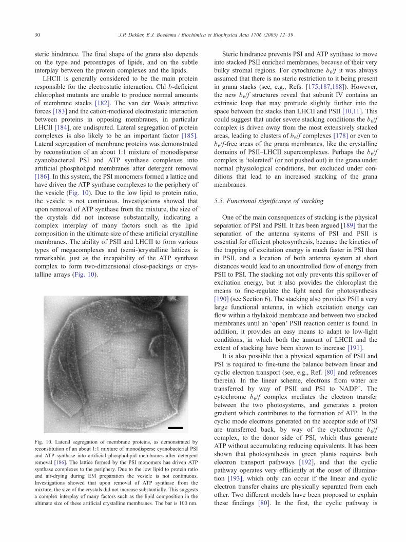
J.P. Dekker, E.J. Boekema / Biochimica et Biophysica Acta 1706 (2005) 12–3930
steric hindrance. The final shape of the grana also depends
on the type and percentages of lipids, and on the subtle
interplay between the protein complexes and the lipids.
LHCII is generally considered to be the main protein
responsible for the electrostatic interaction. Chl b-deficient
chloroplast mutants are unable to produce normal amounts
of membrane stacks [182]. The van der Waals attractive
forces [183] and the cation-mediated electrostatic interaction
between proteins in opposing membranes, in particular
LHCII [184], are undisputed. Lateral segregation of protein
complexes is also likely to be an important factor [185].
Lateral segregation of membrane proteins was demonstrated
by reconstitution of an about 1:1 mixture of monodisperse
cyanobacterial PSI and ATP synthase complexes into
artificial phospholipid membranes after detergent removal
[186]. In this system, the PSI monomers formed a lattice and
have driven the ATP synthase complexes to the periphery of
the vesicle (Fig. 10). Due to the low lipid to protein ratio,
the vesicle is not continuous. Investigations showed that
upon removal of ATP synthase from the mixture, the size of
the crystals did not increase substantially, indicating a
complex interplay of many factors such as the lipid
composition in the ultimate size of these artificial crystalline
membranes. The ability of PSII and LHCII to form various
types of megacomplexes and (semi-)crystalline lattices is
remarkable, just as the incapability of the ATP synthase
complex to form two-dimensional close-packings or crys-
talline arrays (Fig. 10).
Fig. 10. Lateral segregation of membrane proteins, as demonstrated by
reconstitution of an about 1:1 mixture of monodisperse cyanobacterial PSI
and ATP synthase into artificial phospholipid membranes after detergent
removal [186]. The lattice formed by the PSI monomers has driven ATP
synthase complexes to the periphery. Due to the low lipid to protein ratio
and air-drying during EM preparation the vesicle is not continuous.
Investigations showed that upon removal of ATP synthase from the
mixture, the size of the crystals did not increase substantially. This suggests
a complex interplay of many factors such as the lipid composition in the
ultimate size of these artificial crystalline membranes. The bar is 100 nm.
Steric hindrance prevents PSI and ATP synthase to move
into stacked PSII enriched membranes, because of their very
bulky stromal regions. For cytochrome b6/f it was always
assumed that there is no steric restriction to it being present
in grana stacks (see, e.g., Refs. [175,187,188]). However,
the new b6/f structures reveal that subunit IV contains an
extrinsic loop that may protrude slightly further into the
space between the stacks than LHCII and PSII [10,11]. This
could suggest that under severe stacking conditions the b6/f
complex is driven away from the most extensively stacked
areas, leading to clusters of b6/f complexes [178] or even to
b6/f-free areas of the grana membranes, like the crystalline
domains of PSII–LHCII supercomplexes. Perhaps the b6/f
complex is dtoleratedT (or not pushed out) in the grana under
normal physiological conditions, but excluded under con-
ditions that lead to an increased stacking of the grana
membranes.
5.5. Functional significance of stacking
One of the main consequences of stacking is the physical
separation of PSI and PSII. It has been argued [189] that the
separation of the antenna systems of PSI and PSII is
essential for efficient photosynthesis, because the kinetics of
the trapping of excitation energy is much faster in PSI than
in PSII, and a location of both antenna system at short
distances would lead to an uncontrolled flow of energy from
PSII to PSI. The stacking not only prevents this spillover of
excitation energy, but it also provides the chloroplast the
means to fine-regulate the light need for photosynthesis
[190] (see Section 6). The stacking also provides PSII a very
large functional antenna, in which excitation energy can
flow within a thylakoid membrane and between two stacked
membranes until an dopenT PSII reaction center is found. In
addition, it provides an easy means to adapt to low-light
conditions, in which both the amount of LHCII and the
extent of stacking have been shown to increase [191].
It is also possible that a physical separation of PSII and
PSI is required to fine-tune the balance between linear and
cyclic electron transport (see, e.g., Ref. [80] and references
therein). In the linear scheme, electrons from water are
transferred by way of PSII and PSI to NADP+. The
cytochrome b6/f complex mediates the electron transfer
between the two photosystems, and generates a proton
gradient which contributes to the formation of ATP. In the
cyclic mode electrons generated on the acceptor side of PSI
are transferred back, by way of the cytochrome b6/f
complex, to the donor side of PSI, which thus generate
ATP without accumulating reducing equivalents. It has been
shown that photosynthesis in green plants requires both
electron transport pathways [192], and that the cyclic
pathway operates very efficiently at the onset of illumina-
tion [193], which only can occur if the linear and cyclic
electron transfer chains are physically separated from each
other. Two different models have been proposed to explain
these findings [80]. In the first, the cyclic pathway is

J.P. Dekker, E.J. Boekema / Biochimica et Biophysica Acta 1706 (2005) 12–39 31
thought to take place in supercomplexes consisting of PSI,
b6/f, plastocyanin and ferredoxin. A problem of this
hypothesis is that there is as yet no experimental evidence
for the existence of such supercomplexes. In the second
model, no such supercomplex is required, but a localization
of both pathways in different parts of the membrane [80].
The linear pathway was suggested to occur preferentially
near the grana margins, making use of PSII and b6/f from
the grana and nearby PSI complexes. The cyclic pathway
would operate predominantly in stroma lamellae far away
from the grana, with b6/f located in the stroma. Ferredoxin-
NADP reductase (FNR) would play a key role in discrim-
inating between the linear and cyclic routes, by binding to
PSI for linear electron transport and to b6/f for cyclic
electron transport. In the stacks, the binding of FNR to b6/f
is unlikely because of steric hindrance. We conclude that the
occurrence of PSI and PSII in stroma and grana, respec-
tively, and the more even distribution of the cytochrome b6/f
complex facilitate the balance between linear and cyclic
electron transport.
6. Structural rearrangements of protein complexes upon
short-term adaptation
6.1. State transitions
When plants are exposed to light conditions that
preferably excite either PSI or PSII, then the plants are able
to redistribute the excitation energy by a mechanism called
dstate transitionsT (see Refs. [74,175,194,195] for recent
reviews). There is general consensus now about the basic
features of this process. When PSII is preferentially excited
by dlight 2T, the plastoquinone pool becomes more reduced,
which leads to a conformational change of the cytochrome
b6/f complex, which in turn activates a kinase bound to the
b6/f complex. This kinase is then released from b6/f, after
which it migrates to LHCII and promotes its phosphoryla-
tion. Phosphorylated LHCII is thought to have a decreased
affinity for PSII and an increased affinity for PSI, and thus a
lateral movement of phosphorylated LHCII from PSII to PSI
occurs that can explain the shift from dstate 1T (induced by
dlight 1T) to dstate 2T (induced by dlight 2T).There are several aspects of this mechanism that are
relevant for the understanding of the supramolecular
organization of the protein complexes in the thylakoid
membranes. The first relates to the identity of the kinases
that catalyze the phosphorylation. A number of different
types of kinases have now been described in Arabidopsis
and Chlamydomonas, called TAKs (thylakoid-associated
kinases) [196,197] and Stt7 (state transition thylakoid) [73].
Although these proteins are not sequence-related, their
overall structures are comparable, with most probably a
single transmembrane a-helix and a large extrinsic protein
loop at the stromal side of the membrane. The size of this
loop will most likely prevent the kinase to enter the region
between the stacks (Fig. 9), so only those LHCII complexes
can be phosphorylated that are either in the stroma or at the
interface between stroma and grana. This may explain why
in green plants only a relatively small part of LHCII is
phosphorylated, and perhaps also why high light intensities
further limit the accessibility of LHCII for the kinase [198],
because the observed decrease of the interthylakoid distance
upon illumination may further limit the accessibility of
LHCII (see Section 5.1).
It is not known if the LHCII that gets phosphorylated
originates from the PSII–LHCII supercomplexes or from the
LHCII-only part of the membranes. If phosphorylation only
occurs at the border between grana and stroma, then it is
possible that LHCII from both pools is actually involved. It
has been reported that phosphorylation induces a dissocia-
tion of the LHCII trimer into monomers and a conforma-
tional change at the stromal side of the complex [199] but
both statements require further experimental proof.
It is now clear which PSI subunits play an essential role
in the state transitions. Arabidopsis plants without the PSI-H
and PSI-L subunits [200] as well as those without the PSI-O
subunit [201] are highly deficient in state transitions. The
absence of these proteins did not have an effect on the
phosphorylation of LHCII, and evidence was presented that
phosphorylated LHCII remained bound to the grana in the
absence of PSI-H [200]. Similar results were obtained with a
PSI-deficient mutant from Chlamydomonas [202]. This
means that the dmolecular recognitionT between LHCII
and the PSI-H subunit of PSI forms the molecular basis for
the transition from state 1 to state 2 [175]. Biochemical
studies have shown that both phosphorylated and non-
phosphorylated LHCII bind to PSI [203], which suggests
that neither phosphorylation nor the monomeric or trimeric
aggregation state of LHCII affects the affinity of PSI for
LHCII, and that phosphorylation will primarily affect the
affinity of LHCII for the stacked grana membranes.
We note that, at least in Chlamydomonas, the transition
from state 1 to state 2 is not only accompanied by the
migration of LHCII from the grana to the stroma, but also
by a migration of the cytochrome b6/f complex [179] and by
an increase of the extent of cyclic electron transport around
PSI [204]. We also note that the extent of the state
transitions is much larger in Chlamydomonas than in green
plants: in green plants about 10–20% of LHCII is trans-
ferred from PSII to PSI upon the transition from state 1 to
state 2 [205], whereas in Chlamydomonas about 80% of
LHCII migrates from PSII to PSI [202]. These differences
may be explained by differences in membrane structure in
both types of organisms. Chlamydomonas membranes show
a much less pronounced stacking than green plant mem-
branes [206,207], which probably results in a much greater
accessibility of LHCII for the kinases responsible for the
phosphorylation of LHCII. This, in turn, could lead to a
much larger proportion of LHCII that is able to migrate to
PSI. It is not likely that the transition from state 1 to state 2
leads to a complete destacking of the thylakoid membranes

J.P. Dekker, E.J. Boekema / Biochimica et Biophysica Acta 1706 (2005) 12–3932
of Chlamydomonas [179], but involvement of the complete
granum in Chlamydomonas instead of only a small part in
green plants is likely. In this respect, it is interesting to note
that PSII–LHCII super- and megacomplexes could only be
isolated from Chlamydomonas membranes grown in state 1,
but not in state 2 (A.E. Yakushevska, unpublished observa-
tions), suggesting that the association of the PSII and LHCII
complexes is less tight in state 2 than in state 1. It is unlikely
that the two types of PSI–LHCI particles found in
Chlamydomonas (see Ref. [48] and Section 2.3) are related
to the state transitions, because the size of the additional
mass in the larger complexes is too small to explain the
additional light-harvesting ability of PSI in state 2.
6.2. High-energy quenching
In most habitats, plants are exposed to a wide variety of
irradiance intensities. Problems occur when the light
intensity exceeds the plant’s capacity for photosynthesis.
Without proper protection, the accumulation of excited
states will finally result in the generation of harmful oxygen
species, which can damage the membranes, pigments and
proteins of the photosynthetic organism [208]. In high light
conditions, plants are able to harmlessly dissipate excess
excitation energy into heat by a process known as high-
energy quenching (qE). This process is one of the
manifestations of the nonphotochemical quenching of
chlorophyll fluorescence, in which the rate of non-radiative
decay is increased, resulting in lower quantum yields of
fluorescence, intersystem crossing to the triplet state and
charge separation in the photosynthetic reaction centers. The
basic features of this process are now well understood
[209,210]. The process is triggered by acidification of the
thylakoid lumen, which activates violaxanthin de-epoxidase
(VDE), the enzyme that converts violaxanthin into zeax-
anthin [211]. The process requires the presence of the PsbS
protein [108] as well as its protonation by the acidification
of the thylakoid lumen [212]. It has been shown that isolated
PsbS is able to bind zeaxanthin, giving rise to spectroscopic
changes that are also observed for qE in vivo [213].
Whether the activated zeaxanthin directly deactivates
chlorophyll excited states (by means of energy transfer to
the low-lying dforbiddenT first electronically excited S1state) or induces conformational changes in the (super)
complexes of the grana (leading to new pigment–pigment
interactions and increased non-radiative decay rates) is still
a matter of debate [210].
The first aspect of this process relevant for the supra-
molecular organization of the photosynthetic apparatus is
the location of the violaxanthin de-epoxidase enzyme. This
enzyme is water-soluble and occurs in the thylakoid lumen.
It was suggested that this enzyme requires lipid inverted
hexagonal structures for its activity [214]. However, it was
also shown that the inverted hexagonal phase of the major
thylakoid lipid monogalactosyldiacylglycerol (MGDG) dis-
appears upon the addition of LHCII [215], and so it must be
questioned to which extent VDE can catalyze the con-
version of violaxanthin into zeaxanthin in a highly packed
membrane like the thylakoid membrane of the grana (Fig.
9). There is more space for inverted hexagonal structures in
the stroma lamellae, but in this case the zeaxanthin has to
diffuse over a considerable distance to reach PsbS, because
this protein is thought to be located exclusively in the grana.
Perhaps the possibility could be investigated that VDE
catalyzes the formation of zeaxanthin in the strongly curved
margins of the thylakoid membranes. If VDE is active at the
inner surface of these membranes, zeaxanthin would need to
move over a relatively short distance to the PsbS protein in
the grana membranes.
Another aspect relevant for the understanding of the
supramolecular organization is the location of the PsbS
protein. It was found that PsbS is not bound to PSII–LHCII
supercomplexes [111] and also that it does not occur in
crystalline arrays of C2S2M2 supercomplexes in Arabidop-
sis [150], which excludes the possibility that the protein was
lost from the PSII–LHCII supercomplexes during the
purification procedure. This suggests that the PsbS protein
is located in the LHCII-only regions of the grana mem-
branes. Chlorophylls bound to proteins in these regions are
presumably well able to transfer excitation energy to PSII
(see Section 5.5), and a diminishing of excited state
lifetimes in these areas of the membranes after zeaxanthin
activation will give a quenching of fluorescence and the
lower probability of exciting PSII. We note that part of the
current controversy on the location of PsbS may be caused
by the tendency of the isolated protein for self-aggregation
[106]. A dimer-to-monomer conversion has been proposed
upon acidification [109], but to which respect dimer
formation at higher pH values is due to artificial aggregation
is not clear. The finding that PsbS changes its conformation
upon binding of zeaxanthin [213] is better documented, and
a move of the activated protein from LHCII-only regions to
PSII–LHCII supercomplexes remains a possibility.
7. Outlook
We have tried to give an answer to the question how the
proteins in the thylakoid membranes are organized and how
these membranes form the grana and stroma in the
chloroplast. The classical electron microscopy techniques
of freeze-etching, freeze-fracturing and negative staining
have contributed a lot to the understanding, together with
biochemical approaches. However, they have some limi-
tations because the chloroplast is disrupted before imaging.
Thus, there always remains some bias about the credibility
of the observed positions of the membranes and their
individual components. An alternative would be to perform
tomographic reconstructions of intact chloroplasts just as
was carried out for mitochondria [216]. It is expected that
the resolution will increase to 2–3 nm in the near future by
improving data acquisition techniques and the use of

J.P. Dekker, E.J. Boekema / Biochimica et Biophysica Acta 1706 (2005) 12–39 33
helium-cooled electron microscopes. With the present state-
of-the-art tomography, it should for instance be possible to
visualize the membrane system of stacked and unstacked
thylakoid membranes. Application of cryo-EM at liquid
helium temperatures would also be advantageous for single-
particle averaging.
Another promising technique that was, as yet, not much
applied to the study of the organization of green plant
thylakoid membranes is AFM, with which a number of
spectacular results were obtained on membranes and two-
dimensional crystals of photosynthetic complexes from
purple bacteria [217–219]. With this technique topographs
can be obtained of membranes at a higher signal-to-noise
ratio than with electron microscopy. Also the combination
of this technique with two-photon fluorescence imaging
seems promising [220]. For oxygenic photosynthetic sys-
tems studies on two-dimensional crystals of cyanobacterial
PSI [221] and on envelope-free chloroplasts [165] were
presented. The latter study is particularly interesting,
because it allowed a first insight into rearrangements of
the photosynthetic membranes upon unstacking.
Acknowledgements
We thank Dr. A.E. Yakushevska and Mr. J.F.L van
Breemen for providing unpublished data. Our work was
supported by the Netherlands Organization for Scientific
Research (NWO), by grants from the Foundations of Life
and Earth Sciences (ALW) and Chemical Sciences (CW), by
the Foundation for Fundamental Research on Matter
(FOM), and by the European Union grants to the PSICO
Research Training Network (HPRN-CT-2002-00248) and
INTRO2 Marie Curie Research Training Network (MRTN-
CT-2003-505069).
References
[1] P. Jordan, P. Fromme, H.T. Witt, O. Kuklas, W. Saenger, N. Krauss,
Three-dimensional structure of cyanobacterial photosystem I at 2.5
angstrom resolution, Nature 411 (2001) 909–917.
[2] A. Ben-Shem, F. Frolow, N. Nelson, Crystal structure of plant
photosystem I, Nature 426 (2003) 630–635.
[3] A. Zouni, H.T. Witt, J. Kern, P. Fromme, N. Krauss, W. Saenger, P.
Orth, Crystal structure of photosystem II from Synechococcus
elongatus at 3.8 angstrom resolution, Nature 409 (2001) 739–743.
[4] N. Kamiya, J.R. Shen, Crystal structure of oxygen-evolving photo-
system II from Thermosynechococcus vulcanus at 3.7-angstrom
resolution, Proc. Natl. Acad. Sci. U. S. A. 100 (2003) 98–103.
[5] K.N. Ferreira, T.M. Iverson, K. Maghlaoui, J. Barber, S. Iwata,
Architecture of the photosynthetic oxygen-evolving center, Science
303 (2004) 1831–1838.
[6] W. Kqhlbrandt, D.N. Wang, Y. Fujiyoshi, Atomic model of plant
light-harvesting complex by electron crystallography, Nature 367
(1994) 614–621.
[7] Z. Liu, H. Yan, K. Wang, T. Kuang, J. Zhang, L. Gui, X. An, W.
Chang, Crystal structure of spinach light-harvesting complex at 2.72
2 resolution, Nature 428 (2004) 287–292.
[8] J.P. Abrahams, A.G. Leslie, R. Lutter, J.E. Walker, Structure at 2.8 2resolution of F1-ATPase from bovine heart mitochondria, Nature 370
(1994) 621–628.
[9] G. Groth, E. Pohl, The structure of the chloroplast F1-ATPase at 3.2
2 resolution, J. Biol. Chem. 276 (2001) 1345–1352.
[10] G. Kurisu, H. Zhang, J.L. Smith, W.A. Cramer, Structure of the
cytochrome b(6)f complex of oxygenic photosynthesis: tuning the
cavity, Science 302 (2003) 1009–1014.
[11] D. Stroebel, Y. Choquet, J.-L. Popot, D. Picot, An atypical haem in
the cytochrome b6f complex, Nature 426 (2003) 413–418.
[12] H. Sch7gger, Respiratory chain supercomplexes of mitochondria and
bacteria, Biochim. Biophys. Acta 1555 (2002) 154–159.
[13] L.A. Staehelin, G.W.M. van der Staay, in: D.R. Ort, C.F. Yocum
(Eds.), Oxygenic Photosynthesis: The light reactions, Kluwer,
Dordrecht, 1996, pp. 11–30.
[14] P. Fromme, P. Jordan, N. Krauss, Structure of photosystem I,
Biochim. Biophys. Acta 1507 (2001) 5–31.
[15] W. Saenger, P. Jordan, N. Krauss, The assembly of protein subunits
and cofactors in photosystem I, Curr. Opin. Struct. Biol. 12 (2002)
244–254.
[16] P. Heathcote, P.K. Fyfe, M.R. Jones, Reaction centres: the structure
and evolution of biological solar power, Trends Biochem. Sci. 27
(2002) 79–87.
[17] P. Fromme, J. Kern, B. Loll, J. Biesiadka, W. Saenger, H.T. Witt,
N. Krauss, A. Zouni, Functional implications on the mechanism of
the function of photosystem II including water oxidation based on
the structure of photosystem II, Philos. Trans. R. Soc. Lond., B 357
(2002) 1337–1345.
[18] H.V. Scheller, P.E. Jensen, A. Haldrup, C. Lunde, J. Knoetzel, Role
of subunits in eukaryotic Photosystem I, Biochim. Biophys. Acta
1507 (2001) 41–60.
[19] J. Knoetzel, A. Mant, A. Haldrup, P.E. Jensen, H.V. Scheller, PSI-O,
a new 10-kDa subunit of eukaryotic photosystem I, FEBS Lett. 510
(2002) 145–148.
[20] P.E. Jensen, A. Haldrup, L. Rosgaard, H.V. Scheller, Molecular
dissection of photosystem I in higher plants: topology, structure and
function, Physiol. Plant. 119 (2003) 313–321.
[21] B. Gobets, R. van Grondelle, Energy transfer and trapping in
photosystem I, Biochim. Biophys. Acta 1507 (2001) 80–99.
[22] E.J. Boekema, J.P. Dekker, M. van Heel, M. Rfgner, W. Saenger,
I. Witt, H.T. Witt, Evidence for a trimeric organization of the
photosystem I complex from the thermophilic cyanobacterium
Synechococcus sp, FEBS Lett. 217 (1987) 283–286.
[23] E.J. Boekema, A.F. Boonstra, J.P. Dekker, M. Rfgner, Electron-microscopical structural analysis of photosystem I, photosystem II
and the cytochrome b6f complex from green plants and cyanobac-
teria, J. Bioenerg. Biomembr. 26 (1994) 17–29.
[24] G.W.M. van der Staay, E.J. Boekema, J.P. Dekker, H.C.P.
Matthijs, Characterization of trimeric Photosystem I particles from
the prochlorophyte Prochlorothrix hollandica by electron micro-
scopy and image analysis, Biochim. Biophys. Acta 1142 (1993)
189–193.
[25] T.S. Bibby, J. Nield, F. Partensky, J. Barber, Oxyphotobacteria—
antenna ring around photosystem I, Nature 413 (2001) 590.
[26] T.S. Bibby, I. Mary, J. Nield, F. Partensky, Low-light-adapted
Prochlorococcus species possess specific antennae for each photo-
system. J. Barber, Nature 424 (2003) 1051–1054.
[27] E.J. Boekema, P.E. Jensen, E. Schlodder, J.F.L. van Breemen, H. van
Roon, H.V. Scheller, J.P. Dekker, Green plant photosystem I binds
light-harvesting complex I on one side of the complex, Biochemistry
40 (2001) 1029–1036.
[28] V.P. Chitnis, P.R. Chitnis, PsaL subunit is required for the formation
of photosystem I trimers in the cyanobacterium Synechocystis sp.
PCC 6803, FEBS Lett. 336 (1993) 330–334.
[29] A. Ben-Shem, F. Frolow, N. Nelson, Evolution of photosystem I—
from symmetry through pseudosymmetry to asymmetry, FEBS Lett.
564 (2004) 274–280.

J.P. Dekker, E.J. Boekema / Biochimica et Biophysica Acta 1706 (2005) 12–3934
[30] S. Satoh, A. Tanaka, Chlorophyll b inhibits the formation of
photosystem I trimer in Synechocystis sp. PCC 6803, FEBS Lett.
528 (2002) 235–240.
[31] J.A. Ihalainen, P.E. Jensen, A. Haldrup, I.H.M. van Stokkum, R. van
Grondelle, H.V. Scheller, J.P. Dekker, Pigment organization and
energy transfer dynamics in isolated, photosystem I (PSI) complexes
from Arabidopsis thaliana depleted of the PSI-G, PSI-K, PSI-L, or
PSI-N subunit, Biophys. J. 83 (2002) 2190–2201.
[32] P.E. Jensen, M. Gilpin, J. Knoetzel, H.V. Scheller, The PSI-K subunit
of photosystem I is involved in the interaction between light-
harvesting complex I and the photosystem I reaction center core, J.
Biol. Chem. 275 (2000) 24701–24708.
[33] S. Jansson, B. Andersen, H.V. Scheller, Nearest-neighbor analysis of
higher-plant photosystem I holocomplex, Plant Physiol. 112 (1996)
409–420.
[34] P.E. Jensen, L. Rosgaard, J. Knoetzel, H.V. Scheller, Photosystem I
activity is increased in the absence of the PSI-G subunit, J. Biol.
Chem. 277 (2002) 2798–2803.
[35] S. Jansson, The light-harvesting chlorophyll a/b binding proteins,
Biochim. Biophys. Acta 1184 (1994) 1–19.
[36] S. Jansson, A guide to the Lhc genes and their relatives in
Arabidopsis, Trends Plant Sci. 4 (1999) 236–240.
[37] V.H.R. Schmid, K.V. Cammarata, B.U. Bruns, G.W. Schmidt,
In vitro reconstitution of the photosystem I light-harvesting
complex LHCI-730: heterodimerization is required for antenna
pigment organization, Proc. Natl. Acad. Sci. U. S. A. 94 (1997)
7667–7672.
[38] J. Knoetzel, B. Bossmann, L.H. Grimme, Chlorina and viridis
mutants of barley (Hordeum vulgare L.) allow assignment of long-
wavelength chlorophyll forms to individual Lhca proteins of photo-
system I in vivo, FEBS Lett. 436 (1998) 339–342.
[39] V.H.R. Schmid, H. Paulsen, J. Rupprecht, Identification of N- and C-
terminal amino acids of Lhca1 and Lhca4 required for formation of
the heterodimeric peripheral photosystem I antenna LHCI-730,
Biochemistry 41 (2002) 9126–9131.
[40] U. Ganeteg, A. Strand, P. Gustafsson, S. Jansson, The properties of
the chlorophyll a/b-binding proteins Lhca2 and Lhca3 studied in
vivo using antisense inhibition, Plant Physiol. 127 (2001) 150–158.
[41] R. Croce, T. Morosinotto, S. Casteletti, J. Breton, R. Bassi, The Lhca
antenna complexes of higher plants photosystem I, Biochim.
Biophys. Acta 1556 (2002) 29–40.
[42] M. Hippler, J. Klein, A. Fink, T. Allinger, P. Hoerth, Towards
functional proteomics of membrane protein complexes: analysis of
thylakoid membranes from Chlamydomonas reinhardtii, Plant J. 28
(2001) 595–606.
[43] R. Bassi, S.Y. Soen, G. Frank, H. Zuber, J.-D. Rochaix, Character-
ization of chlorophyll a/b proteins from Chlamydomonas reinhardtii,
J. Biol. Chem. 267 (1992) 25714–25721.
[44] E.J. Stauber, A. Fink, C. Markert, O. Kruse, U. Johanningmeier,
M. Hippler, Proteomics of Chlamydomonas reinhardtii light-
harvesting proteins, Eukaryotic Cell 2 (2003) 978–994.
[45] G.R. Wolfe, F.X. Cunningham, D. Durnford, B.R. Green, E. Gantt,
Evidence for a common origin of chloroplasts with light-harvesting
complexes of different pigmentation, Nature 367 (1994) 566–568.
[46] T. Morosinotto, J. Breton, R. Bassi, R. Croce, The nature of a
chlorophyll ligand in Lhca proteins determines the far red
fluorescence emission typical of photosystem I, J. Biol. Chem. 278
(2003) 49223–49229.
[47] R. Croce, D. Dorra, A.R. Holzwarth, R.C. Jennings, Fluorescence
decay and spectral evolution in intact photosystem I of higher plants,
Biochemistry 39 (2000) 6341–6348.
[48] M. Germano, A.E. Yakushevska, W. Keegstra, H.J. van Gorkom,
J.P. Dekker, E.J. Boekema, Supramolecular organization of photo-
system I and light- harvesting complex I in Chlamydomonas
reinhardtii, FEBS Lett. 525 (2002) 121–125.
[49] J. Kargul, J. Nield, J. Barber, Three-dimensional reconstruction of a
light-harvesting complex I–photosystem I (LHCI–PSI) supercom-
plex from the green alga Chlamydomonas reinhardtii—insights into
light harvesting for PSI, J. Biol. Chem. 278 (2003) 16135–16141.
[50] Y. Takahashi, T.-A. Yasui, E.J. Stauber, M. Hippler, Comparison of
the subunit compositions of the PSI–LHCI supercomplex and the
LHCI in the green alga Chlamydomonas reinhardtii, Biochemistry
43 (2004) 7816–7823.
[51] T.S. Bibby, J. Nield, J. Barber, Iron deficiency induces the formation
of an antenna ring around trimeric photosystem I in cyanobacteria,
Nature 412 (2001) 743–745.
[52] E.J. Boekema, A. Hifney, A.E. Yakushevska, M. Piotrowski, W.
Keegstra, S. Berry, K.-P. Michel, E.K. Pistorius, J. Kruip, A giant
chlorophyll–protein complex induced by iron deficiency in cyano-
bacteria, Nature 412 (2001) 745–748.
[53] J. Nield, E.P. Morris, T.S. Bibby, J. Barber, Structural analysis of the
photosystem I supercomplex of cyanobacteria induced by iron
deficiency, Biochemistry 42 (2003) 3180–3188.
[54] K.-P. Michel, E.K. Pistorius, Adaptation of the photosynthetic
electron transport chain in cyanobacteria to iron deficiency:
The function of IdiA and IsiA, Physiol. Plant. 120 (2004)
36–50.
[55] T.M. Bricker, L.K. Frankel, The structure and function of CP47 and
CP43 in photosystem II, Photosynth. Res. 72 (2002) 131–146.
[56] E.G. Andrizhiyevskaya, T.M.E. Schwabe, M. Germano, S. DTHaene,J. Kruip, R. van Grondelle, J.P. Dekker, Spectroscopic properties of
PSI–IsiA supercomplexes from the cyanobacterium Synechococcus
PCC 7942, Biochim. Biophys. Acta 1187 (2002) 265–272.
[57] A.N. Melkozernov, T.S. Bibby, S. Lin, J. Barber, R.E. Blankenship,
Time-resolved absorption and emission show that the CP43 ’ antenna
ring of iron-stressed Synechocystis sp PCC6803 is efficiently
coupled to the photosystem I reaction center core, Biochemistry 42
(2003) 3893–3903.
[58] E.G. Andrizhiyevskaya, D. Frolov, R. van Grondelle, J.P. Dekker,
Energy transfer and trapping in the photosystem I complex of
Synechococcus PCC 7942 and in its supercomplex with IsiA,
Biochim. Biophys. Acta 1656 (2004) 104–113.
[59] J. LaRoche, G.W.M. van der Staay, F. Partensky, A. Ducret, R.
Aebershold, R. Li, S.S. Golden, R.G. Hiller, P.M. Wrench, A.W.D.
Larkum, B.R. Green, Independent evolution of the prochlorophyte
and green plant chlorophyll a/b light-harvesting proteins, Proc. Natl.
Acad. Sci. U. S. A. 93 (1996) 15244–15248.
[60] J. Duncan, T. Bibby, A. Tanaka, J. Barber, Exploring the ability of
chlorophyll b to bind to the CP43 ’ protein induced under iron
deprivation in a mutant of Synechocystis PCC 6803 containing the
cao gene, FEBS Lett. 541 (2003) 171–175.
[61] H. Xu, D. Vavilin, W. Vermaas, Chlorophyll b can serve as the major
pigment in functional photosystem II complexes of cyanobacteria,
Proc. Natl. Acad. Sci. U. S. A. 98 (2001) 14168–14173.
[62] J. Kruip, P.R. Chitnis, B. Lagoutte, M. Rfgner, E.J. Boekema,
Structural organization of the major subunits in cyanobacterial
photosystem 1—localization of subunits PsaC, -D, -E, -F, and -J, J.
Biol. Chem. 272 (1997) 17061–17069.
[63] R. Kouril, N. Yeremenko, S. DTHaene, A.E. Yakushevska, W.
Keegstra, H.C.P. Matthijs, J.P. Dekker, E.J. Boekema, Photosystem I
trimers from Synechocystis PCC 6803 lacking the PsaF and PsaJ
subunits bind an IsiA ring of 17 units, Biochim. Biophys. Acta 1607
(2003) 1–4.
[64] N. Yeremenko, R. Kouril, J.A. Ihalainen, S. D’Haene, N. van
Oosterwijk, E.G. Andrizhiveksya, W. Keegstra, H.L. Dekker, M.
Hagemann, E.J. Boekema, H.C.P. Matthijs, J.P. Dekker, Supra-
molecular organization and dual function of the IsiA chlor-
ophyll-binding protein in cyanobacteria, Biochemistry 43 (2004)
10308–10313.
[65] B.R. Green, in: B.R. Green, W.W. Parson (Eds.), Light-Harvesting
Antennas in Photosynthesis, Kluwer Academic Publishers, The
Netherlands, 2003, pp. 129–168.
[66] J.S. Lolkema, E.J. Boekema, The A-type ATP synthase subunit K of
Methanopyrus kandleri is deduced from its sequence to form a

J.P. Dekker, E.J. Boekema / Biochimica et Biophysica Acta 1706 (2005) 12–39 35
monomeric rotor comprising 13 hairpin domains, FEBS Lett. 543
(2003) 47–50.
[67] H. Seelert, A. Poetsch, N.A. Dencher, A. Engel, H. Stahlberg, D.L.
Mqller, Proton-powered turbine of a plant motor, Nature 405 (2000)
418–419.
[68] I. Arnold, K. Pfeiffer, W. Neupert, R.A. Stuart, H. Schagger, Yeast
mitochondrial F1F0-ATP synthase exists as a dimer: identification of
three dimer-specific subunits, EMBO J. 17 (1998) 7170–7178.
[69] H. Eubel, L. Jansch, H.P. Braun, New insights into the respiratory
chain of plant mitochondria. Supercomplexes and a unique
composition of complex II, Plant Physiol. 133 (2003) 274–286.
[70] H. Zhang, J.P. Whitelegge, W.A. Cramer, Ferredoxin:NADP+
oxidoreductase is a subunit of the chloroplast cytochrome b6 f
complex, J. Biol. Chem. 276 (2001) 38159–38165.
[71] S. Iwata, J.W. Lee, K. Okada, J.K. Lee, M. Iwata, B. Rasmussen,
T.A. Link, S. Ramaswamy, B.K. Jap, Complete structure of the 11-
subunit bovine mitochondrial cytochrome bc1 complex, Science 281
(1998) 64–71.
[72] J.F. Allen, Cytochrome b6f: structure for signalling and vectorial
metabolism, Trends Plant Sci. 9 (2004) 130–137.
[73] N. Depege, S. Bellafiore, J.-D. Rochaix, Role of chloroplast protein
kinase Stt7 in LHCII phosphorylation and state transition in
Chlamydomonas, Science 299 (2003) 1572–1575.
[74] F.-A. Wollman, State transitions reveal the dynamics and flexibility
of the photosynthetic apparatus, EMBO J. 20 (2001) 3623–3630.
[75] J. Heinemeyer, H. Eubel, D. Wehmhfner, L. J7nsch, H.-P. Braun,Proteomic approach to characterize the supramolecular organiza-
tion of photosystems in higher plants, Phytochemistry 65 (2004)
1683–1692.
[76] P. Joliot, J. Lavergne, D. Beal, Plastoquinone compartmentation in
chloroplasts: 1. Evidence for domains with different rates of
photoreduction, Biochim. Biophys. Acta 1101 (1992) 1–12.
[77] H. Kirchhoff, S. Horstmann, E. Weiss, Control of the photosynthetic
electron transport by PQ diffusion microdomains in thylakoids of
higher plants, Biochim. Biophys. Acta 1459 (2000) 148–168.
[78] J. Lavergne, J.-P. Bouchaud, P. Joliot, Plastoquinone compartmenta-
tion in chloroplasts. 2. Theoretical aspects, Biochim. Biophys. Acta
1101 (1992) 13–22.
[79] H. Kirchhoff, U. Mukherjee, H.J. Galla, Molecular architecture of
the thylakoid membrane: lipid diffusion space for plastoquinone,
Biochemistry 41 (2002) 4872–4882.
[80] P. Joliot, D. Beal, A. Joliot, Cyclic electron flow under saturating
excitation of dark-adapted Arabidopsis leaves, Biochim. Biophys.
Acta 1656 (2004) 166–176.
[81] L.-X. Shi, W.P. Schrfder, The low molecular mass subunits of the
photosynthetic supracomplex, photosystem II, Biochim. Biophys.
Acta 1608 (2004) 75–96.
[82] J.P. Dekker, R. van Grondelle, Primary charge separation in
photosystem II, Photosynth. Res. 63 (2000) 195–208.
[83] B.A. Diner, F. Rappaport, Structure, dynamics, and energetics of the
primary photochemistry of photosystem II of oxygenic photosyn-
thesis, Annu. Rev. Plant Biol. 53 (2002) 551–580.
[84] H. van Amerongen, J.P. Dekker, in: B.R. Green, W.W. Parson (Eds.),
Light-Harvesting Antennas in Photosynthesis, Kluwer Academic
Publishers, The Netherlands, 2003, pp. 219–251.
[85] J.P. Dekker, E.J. Boekema, H.T. Witt, M. Rfgner, Refined
purification and further characterization of oxygen-evolving and
Tris-treated photosystem II particles from the thermophilic cyano-
bacterium Synechococcus sp, Biochim. Biophys. Acta 936 (1988)
307–318.
[86] G.F. Peter, J.P. Thornber, Biochemical evidence that the higher plant
photosystem II core complex is organized as a dimer, Plant Cell
Physiol. 32 (1991) 1237–1250.
[87] E.J. Boekema, B. Hankamer, D. Bald, J. Kruip, J. Nield, A.F.
Boonstra, J. Barber, M. Rfgner, Supramolecular structure of the
photosystem II complex from green plants and cyanobacteria, Proc.
Natl. Acad. Sci. U. S. A. 92 (1995) 175–179.
[88] J. Nield, O. Kruse, J. Ruprecht, P. da Fonseca, C. Bqchel, J. Barber,Three-dimensional structure of Chlamydomonas reinhardtii and
Synechococcus elongatus photosystem II complexes allows for
comparison of their oxygen-evolving complex organization, J. Biol.
Chem. 275 (2000) 27940–27946.
[89] T.S. Bibby, J. Nield, M. Chen, A.W.D. Larkum, J. Barber, Structure
of a photosystem II supercomplex isolated from Prochlorion didemni
retaining its chlorophyll a/b light-harvesting system, Proc. Natl.
Acad. Sci. U. S. A. 100 (2003) 9050–9054.
[90] J. Barber, Photosystem II: a multisubunit membrane protein that
oxidises water, Curr. Opin. Struct. Biol. 12 (2002) 523–530.
[91] O. Kruse, D. Zheleva, J. Barber, Stabilization of photosystem two
dimers by phosphorylation: implication for the regulation of the
turnover of D1 protein, FEBS Lett. 408 (1997) 276–280.
[92] O. Kruse, B. Hankamer, C. Konczak, C. Gerle, E. Morris, A.
Radunz, G.H. Schmid, J. Barber, Phosphatidylglycerol is involved
in the dimerization of photosystem II, J. Biol. Chem. 275 (2000)
6509–6514.
[93] L.-X. Shi, Z.J. Lorkovic, R. Oelmqller, W.P. Schrfder, The low
molecular mass PsbW protein is involved in the stabilization of the
dimeric photosystem II complex in Arabidopsis thaliana, J. Biol.
Chem. 275 (2000) 37945–37950.
[94] D. Zheleva, J. Sharma, M. Panico, H.R. Morris, J. Barber, Isolation
and characterization of monomeric and dimeric CP47-reaction center
photosystem II complexes, J. Biol. Chem. 273 (1998) 16122–16127.
[95] E.J. Boekema, J.F.L. van Breemen, H. van Roon, J.P. Dekker,
Conformational changes in photosystem II supercomplexes
upon removal of extrinsic subunits, Biochemistry 39 (2000)
12907–12915.
[96] J.P. Dekker, M. Germano, H. van Roon, E.J. Boekema, Photosystem
II solubilizes as a monomer by mild detergent treatment of unstacked
thylakoid membranes, Photosynth. Res. 72 (2002) 203–210.
[97] E. Baena-Gonzalez, E.-M. Aro, Biogenesis, assembly and turnover
of photosystem II units, Philos. Trans. R. Soc. Lond., B 357 (2002)
1451–1460.
[98] B. Hankamer, E. Morris, J. Nield, A. Carne, J. Barber, Subunit
positioning and transmembrane helix organisation in the core dimer
of photosystem II, FEBS Lett. 504 (2001) 142–151.
[99] K.D. Irrgang, L.-X. Shi, C. Funk, W.P. Schrfder, A nuclear-encoded
subunit of the photosystem II reaction center, J. Biol. Chem. 270
(1995) 17588–17593.
[100] C. Bqchel, E. Morris, E. Orlova, J. Barber, Localisation of the PsbH
subunit in photosystem II: a new approach using labelling of His-tags
with a Ni2+-NTA gold cluster and single particle analysis, J. Mol.
Biol. 312 (2001) 371–379.
[101] J.P.G. Butler, W. Kqhlbrandt, Determination of the aggregate size in
detergent solution of the light-harvesting chlorophyll a/b complex
from chloroplast membranes, Proc. Natl. Acad. Sci. U. S. A. 85
(1988) 3797–3801.
[102] D. Sandona, R. Croce, A. Pagano, M. Crimi, R. Bassi, Higher plants
light harvesting proteins. Structure and function as revealed by
mutation analysis of either protein or chromophore moieties,
Biochim. Biophys. Acta 1365 (1998) 207–214.
[103] R. Bassi, R. Croce, D. Cugini, D. Sandona, Mutational analysis of a
higher plant antenna protein provides identification of chromophores
bound into multiple sites, Proc. Natl. Acad. Sci. U. S. A. 96 (1999)
10056–10061.
[104] R. Croce, G. Canino, F. Ros, R. Bassi, Chromophore organization in
the higher-plant photosystem II antenna protein CP26, Biochemistry
41 (2002) 7334–7343.
[105] B. Hankamer, J. Barber, E.J. Boekema, Structure and membrane
organization of photosystem II in green plants, Annu. Rev. Plant
Physiol. Plant Mol. Biol. 48 (1997) 641–671.
[106] P. Dominici, S. Caffari, F. Armenante, S. Ceoldo, M. Crimi, R. Bassi,
Biochemical properties of the PsbS subunit of photosystem II either
purified from chloroplast or recombinant, J. Biol. Chem. 277 (2002)
22750–22758.

J.P. Dekker, E.J. Boekema / Biochimica et Biophysica Acta 1706 (2005) 12–3936
[107] C. Funk, I. Adamska, B.R. Green, B. Andersson, G. Renger, The
nuclear-encoded chlorophyll-binding photosystem-II-S protein is
stable in the absence of pigments, J. Biol. Chem. 270 (1995)
30141–30147.
[108] X.-P. Li, O. Bjfrkman, C. Shih, A.R. Grossman, M. Rosenquist,
S. Jansson, K.K. Niyogi, A pigment-binding protein essential for
regulation of photosynthetic light harvesting, Nature 403 (2000)
391–395.
[109] E. Bergantino, A. Segalla, A. Brunetta, E. Teardo, F. Rigoni, G.M.
Giacometti, I. Szabo, Light- and pH-dependent structural changes in
the PsbS subunit of photosystem II, Proc. Natl. Acad. Sci. U. S. A.
100 (2003) 15265–15270.
[110] B. Hankamer, J. Nield, D. Zheleva, E.J. Boekema, S. Jansson, J.
Barber, Isolation and biochemical characterisation of monomeric and
dimeric photosystem II complexes from spinach and their relevance
to the organisation of photosystem II in vivo, Eur. J. Biochem. 243
(1997) 422–429.
[111] J. Nield, C. Funk, J. Barber, Supermolecular structure of photo-
system II and location of the PsbS protein, Proc. R. Soc. Lond. 355
(2000) 1337–1344.
[112] E. Thidholm, V. Lindstrfm, C. Tissier, C. Robinson, W.P. Schrfder,C. Funk, Novel approach reveals localisation and assembly pathway
of the PsbS and PsbW proteins into the photosystem II dimer, FEBS
Lett. 513 (2002) 217–222.
[113] J. Nield, E.V. Orlova, E.P. Morris, B. Gowen, M. van Heel, J. Barber,
3D map of the plant photosystem II supercomplex obtained by
cryoelectron microscopy and single particle analysis, Nat. Struct.
Biol. 7 (2000) 44–47.
[114] R. Harrer, Associations between light-harvesting complexes and
photosystem II from Marchantia polymorpha L. determined by two-
and three-dimensional electron microscopy, Photosynth. Res. 75
(2003) 249–258.
[115] R.C. Ford, S.S. Stoylova, A. Holzenburg, An alternative model for
photosystem II/light harvesting complex II in grana membranes
based on cryo-electron microscopy studies, Eur. J. Biochem. 269
(2002) 326–336.
[116] S. Eshagi, B. Andersson, J. Barber, Isolation of a highly active PSII–
LHCII supercomplex from thylakoid membranes by a direct method,
FEBS Lett. 446 (1999) 23–26.
[117] H. van Roon, J.F.L. van Breemen, F.L. deWeerd, J.P. Dekker, E.J.
Boekema, Solubilization of green plant thylakoid membranes with n-
dodecyl-a,d-maltoside. Implications for the structural organization
of the Photosystem II, Photosystem I, ATP synthase and cytochrome
b(6)f complexes, Photosynth. Res. 64 (2000) 155–166.
[118] S. Stoylova, T.D. Flint, R.C. Ford, A. Holzenburg, Structural
analysis of photosystem II in far-red-light-adapted thylakoid mem-
branes—new crystal forms provide evidence for a dynamic
reorganization of light-harvesting antennae subunits, Eur. J. Bio-
chem. 267 (2000) 207–215.
[119] R.J. Debus, The polypeptides of photosystem II and their influence
on manganotyrosyl-based oxygen evolution, Met. Ions Biol. Syst. 37
(2000) 657–711.
[120] P. da Fonseca, E.P. Morris, B. Hankamer, J. Barber, Electron
crystallographic study of photosystem II from the cyanobacterium
Synechococcus elongatus, Biochemistry 41 (2002) 5163–5167.
[121] E.J. Boekema, J. Nield, B. Hankamer, J. Barber, Localization of the
23-kDa subunit of the oxygen-evolving complex of photosystem II
by electron microscopy, Eur. J. Biochem. 252 (1998) 268–276.
[122] H. Kuhl, M. Rfgner, J.F.L. van Breemen, E.J. Boekema, Local-
ization of cyanobacterial photosystem II donor-side subunits by
electron microscopy and the supramolecular organization of photo-
system II in the thylakoid membrane, Eur. J. Biochem. 266 (1999)
453–459.
[123] L. Bumba, F. Vacha, Electron microscopy in structural studies of
photosystem II, Photosynth. Res. 77 (2003) 1–19.
[124] S.D. Betts, J.R. Ross, E. Pichersky, C.F. Yocum, Mutation
Val235Ala weakens binding of the 33-kDa manganese stabilizing
protein of photosystem II to one of two sites, Biochemistry 36
(1997) 4047–4053.
[125] H. Popelkova, M.M. Im, C.F. Yocum, N-terminal truncations of
manganese stabilizing protein identify two amino acid sequences
required for binding of the eukaryotic protein to photosystem II and
reveal the absence of one binding-related sequence in cyanobacteria,
Biochemistry 41 (2002) 10038–10045.
[126] E.J. Boekema, H. van Roon, J.P. Dekker, Specific association of
photosystem II and light-harvesting complex II in partially solubi-
lized photosystem II membranes, FEBS Lett. 424 (1998) 95–99.
[127] E.J. Boekema, H. van Roon, F. Calkoen, R. Bassi, J.P. Dekker,
Multiple types of association of photosystem II and its light-
harvesting antenna in partially solubilized photosystem II mem-
branes, Biochemistry 38 (1999) 2233–2239.
[128] E.J. Boekema, H. van Roon, J.F.L van Breemen, J.P. Dekker,
Supramolecular organization of photosystem II and its light-harvest-
ing antenna in partially solubilized photosystem II membranes, Eur.
J. Biochem. 266 (1999) 444–452.
[129] A.E. Yakushevska, P.E. Jensen, W. Keegstra, H. van Roon, H.V.
Scheller, E.J. Boekema, J.P. Dekker, Supermolecular organization of
photosystem II and its associated light-harvesting antenna in
Arabidopsis thaliana, Eur. J. Biochem. 268 (2001) 6020–6021.
[130] G.F. Peter, J.P. Thornber, Biochemical composition and organization
of higher plant photosystem II light-harvesting pigment proteins, J.
Biol. Chem. 266 (1991) 16745–16754.
[131] R. Bassi, P. Dainese, A supramolecular light-harvesting complex
from chloroplast photosystem II membranes, Eur. J. Biochem. 204
(1992) 317–326.
[132] A.V. Ruban, M. Wentworth, A.E. Yakushevska, J. Andersson, P.J.
Lee, W. Keegstra, J.P. Dekker, E.J. Boekema, S. Jansson, P. Horton,
Plants lacking the main light-harvesting complex retain photosystem
II macro-organization, Nature 421 (2003) 648–652.
[133] J. Andersson, M. Wentworth, R.G. Walters, C.A. Howard, A.V.
Ruban, P. Horton, S. Jansson, Absence of the Lhcb1 and Lhcb2
proteins of the light-harvesting complex of photosystem II - effects
on photosynthesis, grana stacking and fitness, Plant J. 35 (2003)
350–381.
[134] R. Harrer, R. Bassi, M.G. Testi, C. Sch7fer, Nearest-neighbor
analysis of a photosystem II complex from Marchantia polymorpha
L. (liverwort), which contains reaction center and antenna proteins,
Eur. J. Biochem. 255 (1998) 196–205.
[135] A.E. Yakushevska, W. Keegstra, E.J. Boekema, J.P. Dekker, J.
Andersson, S. Jansson, A.V. Ruban, P. Horton, The structure of
photosystem II in Arabidopsis: localization of the CP26 and CP29
antenna complexes, Biochemistry 42 (2003) 608–613.
[136] J. Andersson, R.G. Walters, P. Horton, S. Jansson, Antisense
inhibition of the photosynthetic antenna proteins CP29 and CP26:
Implications for the mechanism of protective energy dissipation,
Plant Cell 13 (2001) 1193–1204.
[137] C. Kqlheim, J. 2gren, S. Jansson, Rapid regulation of light
harvesting and plant fitness in the field, Science 297 (2002) 91–93.
[138] U. Ganeteg, C. Kqlheim, J. Andersson, S. Jansson, Is each light-
harvesting complex protein important for plant fitness? Plant
Physiol. 134 (2004) 502–509.
[139] S. Ruf, K. Biehler, R. Bock, A small chloroplast-encoded protein as a
novel architectural component of the light-harvesting antenna, J. Cell
Biol. 149 (2000) 369–378.
[140] M. Swiatek, R. Kuras, A. Sokolenko, D. Higgs, J. Olive, G.
Cinque, B. Mqller, L.A. Eichacker, D.B. Stern, R. Bassi, R.G.
Herrmann, F.-A. Wollman, The chloroplast gene ycf9 encodes a
photosystem II (PSII) core subunit, PsbZ, that participates in PSII
supramolecular architecture, Plant Cell 13 (2001) 1347–1367.
[141] M. Suorsa, R.E. Regel, V. Paakkarinen, N. Battchikova, R.G.
Herrmann, E.-M. Aro, Protein assembly of photosystem II and
accumulation of subcomplexes in the absence of low molecular
mass subunits PsbL and PsbJ, Eur. J. Biochem. 271 (2004)
96–107.

J.P. Dekker, E.J. Boekema / Biochimica et Biophysica Acta 1706 (2005) 12–39 37
[142] B. Andersson, J.M. Anderson, Lateral heterogeneity in the
distribution of chlorophyll–protein complexes of the thylakoid
membranes of spinach chloroplasts, Biochim. Biophys. Acta 593
(1980) 427–440.
[143] J.P. Dekker, H. van Roon, E.J. Boekema, Heptameric association of
light-harvesting complex II trimers in partially solubilized photo-
system II membranes, FEBS Lett. 449 (1999) 211–214.
[144] A.V. Ruban, P.J. Lee, M. Wentworth, A.J. Young, P. Horton,
Determination of the stoichiometry and strength of binding of
xanthophylls to the photosystem II light harvesting complexes,
J. Biol. Chem. 274 (1999) 10458–10465.
[145] L.A. Staehelin, Chloroplast structure: from chlorophyll granules to
supra-molecular architecture of thylakoid membranes, Photosynth.
Res. 76 (2003) 185–196.
[146] L.A. Staehelin, Reversible particle movements associated with
unstacking and restacking of chloroplast membranes in vitro, J. Cell
Biol. 71 (1976) 136–158.
[147] K.R. Miller, G.J. Miller, K.R. McIntyre, Light-harvesting chloro-
phyll–protein complex of photosystem 2. Its location in photo-
synthetic membrane, J. Cell Biol. 71 (1976) 624–638.
[148] D.J. Simpson, Freeze-fracture studies on barley plastid membranes.
2. Wild-type chloroplast, Carlsberg Res. Commun. 43 (1978)
365–389.
[149] E.J. Boekema, J.F.L. van Breemen, H. van Roon, J.P. Dekker,
Arrangement of photosystem II supercomplexes in crystalline
macrodomains within the thylakoid membrane of green plant
chloroplasts, J. Mol. Biol. 301 (2000) 1123–1133.
[150] A.E. Yakushevska, A.V. Ruban, P.E. Jensen, H. van Roon, K.K.
Niyogi, P. Horton, J.P. Dekker, E.J. Boekema, Supermolecular
organization of photosystem II and its associated light-harvesting
antenna in the wild- type and npq4 mutant of Arabidopsis
thaliana, PS2001 Proceedings: 12th International Congress on
Photosynthesis, CSIRO Publishing, Melbourne, Australia, 2001,
pp. S5–006.
[151] C. Santini, V. Tidu, G. Tognon, A. Ghiretti Magaldi, R. Bassi,
3-Dimensional structure of the higher plant photosystem II reaction
center and evidence for its dimeric organization in vivo, Eur. J.
Biochem. 221 (1994) 307–315.
[152] H. Teramoto, T.-A. Ono, J. Minagawa, Identification of Lhcb gene
family encoding the light-harvesting chlorophyll a/b proteins of
photosystem II in Chlamydomonas reinhardtii, Plant Cell Physiol.
42 (2001) 849–856.
[153] G.A. Semenova, Particle regularity on thylakoid fracture faces is
influenced by storage conditions, Can. J. Bot. 73 (1995) 1676–1682.
[154] N.M. Tsvetkova, E.L. Apostolova, A.P.R. Brain, W.P. Williams, P.J.
Quinn, Factors influencing PS II particle array formation in
Arabidopsis thaliana chloroplasts and the relationship of such arrays
to the thermostability of PS II, Biochim. Biophys. Acta 1228 (1995)
201–210.
[155] M.P. Garber, P.L. Steponkus, Alterations in chloroplast thylakoids
during cold acclimation, Plant Physiol. 57 (1976) 681–686.
[156] L.M.C. Barter, J.R. Durrant, D.R. Klug, A quantitative structure–
function relationship for the Photosystern II reaction center: super-
molecular behavior in natural photosynthesis, Proc. Natl. Acad. Sci.
U. S. A. 100 (2003) 946–951.
[157] R. van Grondelle, J.P. Dekker, T. Gillbro, V. Sundstrom, Energy
traansfer and trapping in photosynthesis, Biochim. Biophys. Acta
1187 (1994) 1–65.
[158] L.M.C. Barter, M. Bianchetti, C. Jeans, M.J. Schilstra, B. Hankamer,
B.A. Diner, J. Barber, J.R. Durrant, D.R. Klug, Relationship between
excitation energy transfer, trapping, and antenna size in photosystem
II, Biochemistry 40 (2001) 4026–4034.
[159] S. VasilTev, P. Orth, A. Zouni, T.G. Owens, D. Bruce, Excited-statedynamics in photosystem II: Insights from the X-ray crystal
structure, Proc. Natl. Acad. Sci. U. S. A. 98 (2001) 8602–8607.
[160] S. Vasil’ev, J.-R. Shen, N. Kamiya, D. Bruce, The orientations of
core antenna chlorophylls in photosystem II are optimized to
maximize the quantum yield of photosynthesis, FEBS Lett. 561
(2004) 111–116.
[161] E.G. Andrizhiyevskaya, D. Frolov, R. van Grondelle, J.P. Dekker,
On the role of the CP47 core antenna in the energy transfer and
trapping dynamics of photosystem. Phys. Chem. Chem. Phys.
(in press).
[162] V. Barzda, V. Gulbinas, R. Kananavicius, H. van Amerongen, R. van
Grondelle, L. Valkunas, Singlet–singlet annihilation kinetics in
aggregates and trimers of LHCII, Biophys. J. 80 (2001) 2409–2421.
[163] H.-W. Trissl, J. Breton, J. Deprez, W. Leibl, Primary electrogenic
reactions of photosystem II as probed by the light gradient method,
Biochim. Biophys. Acta 893 (1987) 305–319.
[164] W. Leibl, J. Breton, J. Deprez, H.-W. Trissl, Photoelectric study on
the kinetics of trapping and charge stabilization in oriented PSII
membranes, Photosynth. Res. 22 (1989) 257–275.
[165] D. Kaftan, V. Blumfeld, R. Nevo, A. Scherz, Z. Reich, From
chloroplasts to photosystems: in situ scanning force microscopy on
intact thylakoid membranes, EMBO J. 21 (2002) 6146–6153.
[166] L. Mustardy, G. Garab, Granum revisited. A three-dimensional
model—where things fall into place, Trends Plant Sci. 8 (2003)
117–122.
[167] P.2. Albertsson, E. Andreasson, The constant proportion of grana
and stroma lamellae in plant chloroplasts, Physiol. Plant. 121 (2004)
334–342.
[168] P.-2. Albertsson, Interaction between the lumenal sides of the
thylakoid membrane, FEBS Lett. 149 (1982) 186–190.
[169] S. Murakami, L. Packer, Protonation and chloroplast membrane
structure, J. Cell Biol. 47 (1970) 332–351.
[170] J.M. Anderson, W.S. Chow, Structural and functional dynamics of
plant photosystem II, Philos. Trans. R. Soc. Lond., B 357 (2002)
1421–1430.
[171] T. Kieselbach, W.P. Schrfder, The proteome of the chloroplast lumen
of higher plants, Photosynth. Res. 78 (2003) 249–264.
[172] D.J. Murphy, The molecular organization of the photosynthetic
membranes of higher plants, Biochim. Biophys. Acta 864 (1986)
33–94.
[173] P.-2. Albertsson, A quantitative model of the domain structure of the
photosynthetic membrane, Trends Plant Sci. 6 (2001) 349–354.
[174] R. Danielsson, P.-2. Albertsson, F. Mamedov, S. Styring,
Quantification of photosystem I and II in different parts of the
thylakoid membrane from spinach, Biochim. Biophys. Acta 1608
(2004) 53–61.
[175] J.F. Allen, J. Forsberg, Molecular recognition in thylakoid structure
and function, Trends Plant Sci. 6 (2001) 317–326.
[176] I.G. Tremmel, H. Kirchhoff, E. Weis, G.D. Farquhar, Dependence of
plastoquinol diffusion on the shape, size, and density of integral
thylakoid proteins, Biochim. Biophys. Acta 1607 (2003) 97–109.
[177] J.M. Anderson, Distribution of the cytochromes of spinach
chloroplasts between the appressed membranes of grana stacks and
stroma-exposed thylakoid regions, FEBS Lett. 138 (1982) 62–66.
[178] D.R. Allred, L.A. Staehelin, Lateral distribution of the cyto-
chrome b6 f complex and coupling factor ATP synthetase
complexes of chloroplast thylakoid membranes, Plant Physiol.
78 (1985) 199–202.
[179] O. Vallon, L. Bulte, P. Dainese, J. Olive, R. Bassi, F.-A. Wollman,
Lateral distribution of cytochrome b6 f complexes along thylakoid
membranes upon state transitions, Proc. Natl. Acad. Sci. U. S. A. 88
(1991) 8262–8266.
[180] D.A. Berthold, G.T. Babcock, C.F. Yocum, A highly-resolved,
oxygen-evolving photosystem II preparation from spinach thylakoid
membranes, FEBS Lett. 134 (1981) 231–234.
[181] T.G. Dunahay, L.A. Staehelin, M. Seibert, P.D. Ogilvie, S.P. Berg,
Structural, biochemical and biophysical characterization of four
oxygen-evolving photosystem II preparations from spinach, Bio-
chim. Biophys. Acta 764 (1984) 179–193.
[182] K.D. Allen, M.E. Duysen, L.A. Staehelin, Biogenesis of thylakoid
membranes is controlled by light intensity in the conditional

J.P. Dekker, E.J. Boekema / Biochimica et Biophysica Acta 1706 (2005) 12–3938
chlorophyll b-deficient CD3 mutant of wheat, J. Cell Biol. 107
(1988) 907–919.
[183] W.S. Chow, C. Miller, J.M. Anderson, Surface charges, the
heterogeneous lateral distribution of the two photosystems,
and thylakoid stacking, Biochim. Biophys. Acta 1057 (1991)
69–77.
[184] J. Barber, Influence of surface charges on thylakoid structure and
function, Annu. Rev. Plant Physiol. 33 (1982) 261–295.
[185] A. Borodich, I. Rojdestvenski, M. Cottam, G. Oquist, Segregation of
the photosystems in thylakoids depends on their size, Biochim.
Biophys. Acta 1606 (2003) 73–82.
[186] B. Bfttcher, P. Gr7ber, E.J. Boekema, The structure of photosystem I
from the thermophilic cyanobacterium Synechococcus sp. deter-
mined by electron microscopy of two-dimensional crystals, Biochim.
Biophys. Acta 1100 (1992) 125–136.
[187] Y. Choquet, O. Vallon, Synthesis, assembly and degradation of
thylakoid membrane proteins, Biochimie 82 (2000) 615–634.
[188] F.-A. Wollman, L. Minai, R. Nechustai, The biogenesis and
assembly of photosynthetic proteins in thylakoid membranes,
Biochim. Biophys. Acta 1411 (1999) 21–85.
[189] H.-W. Trissl, C. Wilhelm, Why do thylakoid membranes from higher
plants form grana stacks? Trends Biochem. Sci. 18 (1993) 415–419.
[190] P. Horton, Are grana necessary for regulation of light harvesting?
Aust. J. Plant Physiol. 26 (1999) 659–669.
[191] J.M. Anderson, Photoregulation of the composition, function and
structure of thylakoid membranes, Annu. Rev. Plant Physiol. Plant
Mol. Biol. 37 (1986) 93–136.
[192] Y. Munekage, M. Hashimoto, C. Miyake, K.-I. Tomizawa, T. Endo,
M. Tasaka, T. Shikanai, Cyclic electron flow around photosystem I is
essential for photosynthesis, Nature 429 (2004) 579–582.
[193] P. Joliot, A. Joliot, Cyclic electron transfer in plant leaf, Proc. Natl.
Acad. Sci. U. S. A. 99 (2002) 10209–10214.
[194] A. Haldrup, P.E. Jensen, C. Lunde, H.V. Scheller, Balance of power:
a view of the mechanism of photosynthetic state transitions, Trends
Plant Sci. 6 (2001) 301–305.
[195] J.F. Allen, State transitions—a question of balance, Science 299
(2003) 1530–1532.
[196] S. Snyders, B.D. Kohorn, TAKs, thylakoid membrane protein
kinases associated with energy transduction, J. Biol. Chem. 274
(1999) 9137–9140.
[197] S. Snyders, B.D. Kohorn, Disruption of thylakoid-associated kinase
1 leads to alteration of light-harvesting in Arabidopsis, J. Biol.
Chem. 276 (2001) 32169–32176.
[198] H. Zer, M. Vink, S. Schochat, R.G. Herrmann, B. Andersson, I.
Ohad, Light affects the accessibility of the thylakoid light harvesting
complex II (LHCII) phosphorylation site to the membrane protein
kinase(s), Biochemistry 42 (2003) 728–738.
[199] A. Nilsson, D. Stys, T. Drakenberg, M.D. Spangfort, S. Forsen, J.F.
Allen, Phosphorylation controls the three-dimensional structure of
plant light-harvesting complex II, J. Biol. Chem. 272 (1997) 18350–
18357.
[200] C. Lunde, P.E. Jensen, A. Haldrup, J. Knoetzel, H.V. Scheller, The
PSI-H subunit of photosystem I is essential for state transitions in
plant photosynthesis, Nature 408 (2000) 613–615.
[201] P.E. Jensen, A. Haldrup, S. Zhang, H.V. Scheller, The PSI-O subunit
of plant photosystem I is involved in balancing the excitation
pressure between the two photosystems, J. Biol. Chem. 279 (2004)
24212–24217.
[202] R. Delosme, J. Olive, F.-A. Wollman, Changes in light energy
distribution upon state transitions: an in vivo photoacoustic study of
the wild type and photosynthetic mutants from Chlamydomonas
reinhardtii, Biochim. Biophys. Acta 1273 (1996) 150–158.
[203] S. Zhang, H.V. Scheller, Light-harvesting complex II binds to several
small subunits of photosystem I, J. Biol. Chem. 279 (2004) 3180–
3187.
[204] G. Finazzi, F. Rappaport, A. Furia, M. Fleischmann, J.-D. Rochaix,
F. Zito, G. Forti, Involvement of state transitions in the switch
between linear and cyclic electron flow in Chlamydomonas
reinhardtii, EMBO Rep. 3 (2002) 280–285.
[205] J.F. Allen, Protein phosphorylation in regulation of photosynthesis,
Biochim. Biophys. Acta 1098 (1992) 275–335.
[206] J. Olive, O. Vallon, F.-A. Wollman, M. Recouvreur, P. Bennoun,
Studies on the cytochrome b6/f complex. II. Localization of the
complex in the thylakoid membranes from spinach and Chlamydo-
monas reinhardtii by immunocytochemistry and freeze-fracture
analysis of b6/f mutants, Biochim. Biophys. Acta 851 (1996)
239–248.
[207] J.-D. Rochaix, Assembly, function and dynamics of the photo-
synthetic machinery in Chlamydomonas reinhardtii, Plant Physiol.
127 (2001) 1394–1398.
[208] J. Barber, B. Andersson, Too much of a good thing: light can be bad
for photosynthesis, Trends Biochem. Sci. 17 (1992) 61–66.
[209] P. Horton, A.V. Ruban, R.G. Walters, Regulation of light-harvesting
in green plants, Annu. Rev. Plant Physiol. Plant Mol. Biol. 47 (1996)
655–684.
[210] N.E. Holt, G.R. Fleming, K.K. Niyogi, Toward an understanding of
the mechanism of non-photochemical quenching in green plants,
Biochemistry 43 (2004) (in press).
[211] B. Demmig-Adams, W.W. Adams III, The role of xanthophyll cycle
carotenoids in the protection of photosynthesis, Trends Plant Sci. 1
(1996) 21–26.
[212] X.-P. Li, A.M. Gilmore, S. Caffari, R. Bassi, T. Golan, D. Kramer,
K.K. Niyogi, Regulation of photosynthetic light harvesting involves
intrathylakoid lumen pH sensing by the PsbS protein, J. Biol. Chem.
279 (2004) 22866–22874.
[213] M. Aspinall-OTDea, M. Wentworth, A. Pascal, B. Robert, A. Ruban,
P. Horton, In vitro reconstitution of the activated zeaxanthin state
associated with energy dissipation in plants, Proc. Natl. Acad. Sci.
U. S. A. 99 (2002) 16331–16335.
[214] D. Latowski, H.-E. Kerlund, K. Strzalka, Violaxanthin de-epoxidase,
the xanthophyll cycle enzyme, requires lipid inverted hexagonal
structures for its activity, Biochemistry 43 (2004) 4417–4420.
[215] I. Simidjiev, S. Stoylova, H. Amenitsch, T. Javorfi, L. Mustardy, P.
Laggner, A. Holzenburg, G. Garab, Self-assembly of large, ordered
lamellae from non-bilayer lipids and integral membrane proteins in
vitro, Proc. Natl. Acad. Sci. U. S. A. 97 (2000) 1473–1476.
[216] D. Nicastro, A.S. Frangakis, D. Typke, W. Baumeister, Cryo-electron
tomography of Neurospora mitochondria, J. Struct. Biol. 129 (2000)
48–56.
[217] S. Scheuring, F. Reiss-Husson, A. Engel, J.-L. Rigaud, J.-L. Ranck,
High-resolution AFM topographs of Rubrivax gelatinosus light-
harvesting complex LH2, EMBO J. 20 (2001) 3029–3035.
[218] S. Scheuring, J. Seguin, S. Marco, D. Levy, B. Robert, J.-L. Rigaud,
Nanodissection and high-resolution imaging of the Rhodopseudo-
monas viridis photosynthetic core complex in native membranes by
AFM, Proc. Natl. Acad. Sci. U. S. A. 100 (2003) 1690–1693.
[219] S. Bahatyrova, R.N. Frese, C.A. Siebert, J.D. Olsen, K.O. van der
Werf, R. van Grondelle, R.A. Niederman, P.A. Bullough, C. Otto,
C.N. Hunter, The native architecture of a photosynthetic membrane,
Nature 430 (2004) 1058–1061.
[220] C.C. Gradinaru, P. Martinsson, T.J. Aartsma, T. Schmidt, Simulta-
neous atomic-force and two-photon fluorescence imaging of bio-
logical membranes, Ultramicroscopy 99 (2004) 235–245.
[221] D. Fotiadis, D.J. Mqller, G. Tsiotis, L. Hasler, P. Tittmann, T. Mini, P.
Jenf, H. Gross, A. Engel, Surface analysis of the photosystem I
complex by electron and atomic force microscopy, J. Mol. Biol. 283
(1998) 83–94.
[222] J.M. Anderson, in: J. Amesz (Ed.), Photosynthesis, Elsevier Science
Publishers, Amsterdam, 1987.
[223] C. Weibull, W. Villiger, B. Bohrmann, Ultrastructure of spinach
chloroplasts as revealed by freeze substitution, J. Ultrastruct. Res.
104 (1990) 139–143.
[224] C. Westphal, H. Bfhme, D. Frfsch, in: T. Imura, S. Maruse, T.
Suzuki (Eds.), Light-induced transient changes in photosynthetic
J.P. Dekker, E.J. Boekema / Biochimica et Biophysica Acta 1706 (2005) 12–39 39
membranes of aqueous-embedded spinach chloroplasts. Proc. XIth
Int. Cong. On Electron Microscopy, Kyoto, J. Electron Microsc., vol.
35, 1986, pp. 2555–2556 supplement.
[225] D.J. Simpson, Freeze-fracture studies on barley plastid membranes:
3. Location of the light-harvesting chlorophyll protein, Carlsberg
Res. Commun. 44 (1979) 305–336.
[226] L.A. Staehelin, P.A. Armond, K.R. Miller, Chloroplast membrane
organization at the supramolecular level and its functional implica-
tion, Brookhaven Symp. Biol. 28 (1977) 278–315.
[227] S. Murakami, Structural and functional organization of the thylakoid
membrane system in photosynthetic apparatus, J. Electron Microsc.
41 (1992) 424–433.
[228] J. Olive, O. Vallon, Structural organization of the thylakoid
membrane—freeze-fracture and immunocytochemical analysis, J.
Electron Microsc. Tech. 18 (1991) 360–374.
[229] I. Nir, D.C. Pease, Chloroplast organization and the ultrastructural
localization of photosystem I and II, J. Ultrastruct. Res. 42 (1973)
534–550.
[230] D. Branton, R.B. Park, Subunits in chloroplast lamellae, J. Ultra-
struct. Res. 19 (1967) 283–303.
[231] A. Ongun, W.W. Thomson, J.B. Mudd, Lipid fixation during
preparation of chloroplasts for electron microscopy, J. Lipid Res. 9
(1968) 416–424.
[232] A.V. Ruban, A.A. Pascal, B. Robert, P. Horton, Activation of
zeaxanthin is an obligatory event in the regulation of photosynthetic
light harvesting, J. Biol. Chem. 277 (2002) 7785–7789.
[233] G.L. Nicolson, Structure of the photosynthetic apparatus in protein-
embedded chloroplasts, J. Cell Biol. 50 (1971) 258–263.
[234] K.T. Tokuyasu, Membranes as observed in frozen sections, J.
Ultrastruct. Res. 55 (1976) 281–287.
[235] P.O. Arvidsson, C. Sundby, A model for the topology of the
chloroplast thylakoid membrane, Aust. J. Plant Physiol. 26 (1999)
678–694.