University of Groningen Protein secretion and disulfide ... · characterized Tat system is that of...
25
University of Groningen Protein secretion and disulfide bond handling in Bacillus subtilis Kouwen, Roelof Hendrik Matthijs IMPORTANT NOTE: You are advised to consult the publisher's version (publisher's PDF) if you wish to cite from it. Please check the document version below. Document Version Publisher's PDF, also known as Version of record Publication date: 2009 Link to publication in University of Groningen/UMCG research database Citation for published version (APA): Kouwen, R. H. M. (2009). Protein secretion and disulfide bond handling in Bacillus subtilis. Groningen: s.n. Copyright Other than for strictly personal use, it is not permitted to download or to forward/distribute the text or part of it without the consent of the author(s) and/or copyright holder(s), unless the work is under an open content license (like Creative Commons). Take-down policy If you believe that this document breaches copyright please contact us providing details, and we will remove access to the work immediately and investigate your claim. Downloaded from the University of Groningen/UMCG research database (Pure): http://www.rug.nl/research/portal. For technical reasons the number of authors shown on this cover page is limited to 10 maximum. Download date: 26-08-2019
Transcript of University of Groningen Protein secretion and disulfide ... · characterized Tat system is that of...
University of Groningen
Protein secretion and disulfide bond handling in Bacillus subtilisKouwen, Roelof Hendrik Matthijs
IMPORTANT NOTE: You are advised to consult the publisher's version (publisher's PDF) if you wish to cite fromit. Please check the document version below.
Document VersionPublisher's PDF, also known as Version of record
Publication date:2009
Link to publication in University of Groningen/UMCG research database
Citation for published version (APA):Kouwen, R. H. M. (2009). Protein secretion and disulfide bond handling in Bacillus subtilis. Groningen: s.n.
CopyrightOther than for strictly personal use, it is not permitted to download or to forward/distribute the text or part of it without the consent of theauthor(s) and/or copyright holder(s), unless the work is under an open content license (like Creative Commons).
Take-down policyIf you believe that this document breaches copyright please contact us providing details, and we will remove access to the work immediatelyand investigate your claim.
Downloaded from the University of Groningen/UMCG research database (Pure): http://www.rug.nl/research/portal. For technical reasons thenumber of authors shown on this cover page is limited to 10 maximum.
Download date: 26-08-2019

53
Chapter 3
Overflow of a hyper-produced secretory
protein from the Bacillus Sec pathway into the
Tat pathway for protein secretion as revealed
by proteogenomics
Thijs R.H.M. Kouwen, René van der Ploeg, Haike Antelmann, Michael Hecker,
Georg Homuth, Ulrike Mäder and Jan Maarten van Dijl.
Proteomics, in press

Chapter 3
54
SUMMARY
Bacteria secrete numerous proteins into their environment for growth and survival under complex and ever-changing conditions. The highly different characteristics of secreted proteins pose major challenges to the cellular protein export machinery and, accordingly, different pathways have evolved. While the main secretion (Sec) pathway transports proteins in an unfolded state, the twin-arginine translocation (Tat) pathway transports folded proteins. To date, these pathways were believed to act in strictly independent ways. Here, we have employed proteogenomics to investigate the secretion mechanism of the esterase LipA of Bacillus subtilis, using a serendipitously obtained hyper-producing strain. While LipA is secreted Sec-dependently under standard conditions, hyper-produced LipA is secreted predominantly Tat-dependently via an unprecedented overflow mechanism. Two previously identified B. subtilis Tat substrates, PhoD and YwbN, require each a distinct Tat translocase for secretion. In contrast, hyper-produced LipA is transported by both Tat translocases of B. subtilis, showing that they have distinct but overlapping specificities. The identified overflow secretion mechanism for LipA focuses interest on the possibility that secretion pathway choice can be determined by environmental and intracellular conditions. This may provide an explanation for the previous observation that many Sec-dependently transported proteins have potential twin-arginine signal peptides for export via the Tat pathway.

Tat-dependent esterase secretion in B. subtilis
55
INTRODUCTION
Microorganisms secrete numerous enzymes into their extracellular milieu, enabling them to degrade a wide variety of macromolecular substrates and survive in complex and continuously changing environments (Albers et al., 2006; Pugsley et al., 2004; Sibbald et al., 2006; Tjalsma et al., 2000; Tjalsma et al., 2004). Bacterial protein secretion is thus a highly relevant topic in both fundamental molecular microbiological research as well as in applied research, especially since many of the secreted enzymes have (potential) applications in industry. The Gram-positive bacterium Bacillus subtilis serves as an excellent model organism to study protein secretion for several reasons. B. subtilis lacks an outer membrane, which retains many proteins in the periplasm of Gram-negative bacteria, such as Escherichia coli. Accordingly, the majority of B. subtilis proteins that are transported across the cytoplasmic membrane end up in the extracellular milieu of this bacterium (van Wely et al., 2001; Tjalsma et al., 2004; Tjalsma et al., 2000). This property makes B. subtilis an extremely attractive organism to investigate the total flow of proteins from the cell to the environment by proteomic techniques (Antelmann et al., 2001; Antelmann et al., 2000; Antelmann et al., 2003; Hirose et al., 2000; Tjalsma et al., 2004). Furthermore, the availability of the complete genome sequence (Kunst et al., 1997), readily available strains with mutations in nearly all of the ~4100 genes (Kobayashi et al., 2003), industrial application of many native secreted proteins (Zeigler and Perkins, 2008) and numerous useful techniques for gene cloning and expression (Bron et al., 1999; Meima et al., 2004) have made B. subtilis the major paradigm for research into protein secretion by Gram-positive bacteria.
Previous studies have described the composition of the so-called secretome of B. subtilis which, by definition, includes all secreted proteins plus the protein secretion machinery itself (Tjalsma et al., 2000; Antelmann et al., 2003; Tjalsma et al., 2004). The results of proteomics studies were complemented by secretome predictions based on the presence of specific targeting sequences that many secreted proteins have in common (Tjalsma, 2007; von Heijne, 1990a; von Heijne, 1990b). Furthermore, it was established that at least four distinct pathways for protein export from the cytoplasm are active (Antelmann et al., 2001; Tjalsma et al., 2004). The majority of secreted proteins appear to be exported via the general secretory (Sec) pathway. A limited number of other proteins are transported via the twin-arginine translocation (Tat) pathway, the pseudopilin export pathway for competence development (Com pathway), and pathways using ABC transporters dedicated to the secretion of bacteriocins and pheromones (Bacteriocin pathway).
The Tat pathway has attracted particular interest due to its capability of translocating fully folded proteins across biological membranes. Active Tat translocases have been identified in Gram-negative and Gram-positive bacteria, as well as in archaea and chloroplasts (Dabney-Smith et al., 2006; Mori and Cline, 2001; Muller and Klosgen, 2005; Robinson and Bolhuis, 2004; Sargent et al., 2006; Widdick et al., 2006; Yen et al.,

Chapter 3
56
2002; Sargent, 2007). These translocases have been named after the conserved twin-arginine (RR) motif found in the signal peptides of proteins that they transport. This motif is located in the N-terminal region of twin-arginine signal peptides and its consensus sequence was originally defined as (S/T)-R-R-x-F-L-K (Berks, 1996; Chaddock et al., 1995; Stanley et al., 2000). The core sequence of functional RR-motifs seems to be R/K-R-x-#-# (where # is a hydrophobic residue; (Cristobal et al., 1999)). The currently best-characterized Tat system is that of E. coli, which consists of three integral membrane proteins known as TatA, TatB and TatC. The consensus opinion within the field is that TatB and TatC serve in the initial RR-signal peptide reception, while TatB and TatC in complex with multiple TatA components form a protein-conducting channel (Alami et al., 2003; Mangels et al., 2005; Orriss et al., 2007). Interestingly, B. subtilis, as well as most other Gram-positive bacteria, do not possess a TatB homologue (Jongbloed et al., 2000; Jongbloed et al., 2004; Pop et al., 2002). Instead, these organisms contain minimal TatAC translocases in which a bifunctional TatA subunit seems to perform not only the role of TatA, but also that of TatB (Jongbloed et al., 2006; Barnett et al., 2008). Notably, B. subtilis contains three TatA proteins (named TatAc, TatAd and TatAy) and two TatC proteins (named TatCd and TatCy) (Jongbloed et al., 2000). Previous studies have shown that these Tat proteins form two distinct TatAC translocases (named TatAdCd and TatAyCy) for the export of specific substrates. The TatAdCd translocase is required for secretion of the phosphodiesterase PhoD (Jongbloed et al., 2000), while the TatAyCy translocase is required for secretion of a Dyp-type peroxidase called YwbN (Jongbloed et al., 2004). A specific role for TatAc in translocation has so far not been demonstrated. In accordance with their coupled functions, the tatAd and tatCd genes are organized in an operon, as are the tatAy and tatCy genes.
Previous estimates have indicated that of ~200 predicted exported proteins with cleavable signal peptides in B. subtilis, 69 could potentially use the Tat pathway as their signal peptides carry RR- or KR-motifs that conform to the core consensus (Jongbloed et al., 2002). However, PhoD and YwbN have so far remained the only experimentally confirmed proteins that are secreted through this route. The signal peptides of two additional proteins, QcrA and YkuE, were recently shown to direct secretion of an agarase via the Tat system of Streptomyces coelicolor when fused to this protein (Widdick et al., 2008). These findings have led to the conclusion that the Tat system of B. subtilis is highly selective (Jongbloed et al., 2002). A so far unresolved question concerned the Tat-dependent or Tat-independent secretion of the esterase LipA. The signal sequence of LipA conforms well to the most stringent criteria that are currently available for the prediction of RR-signal peptides, and a moderate effect on LipA secretion was observed in a tatCd deletion strain (Jongbloed et al., 2002). However, efficient Tat-independent LipA secretion could be demonstrated in a B. subtilis mutant lacking all five tat genes, and the secretion of this protein was shown to depend on the motor component SecA of the Sec-dependent secretion pathway (Jongbloed et al., 2002).
Tat-dependent esterase secretion in B. subtilis
57
In the present studies, we have reinvestigated the Sec- or Tat-dependent secretion of LipA employing a serendipitously obtained mutant strain that hyper-secretes LipA. Using a proteogenomics approach, we show that under conditions of massive LipA overproduction, this protein is secreted predominantly Tat-dependently. To our knowledge, this is the first reported case of an authentic, non-recombinant, secretory protein that can employ both the Sec and Tat pathways for export from the cytoplasm in a Gram-positive bacterium.
RESULTS
The LipA hyper-producing strain XdsbA*
In a recent study, we compared the thiol-disulfide oxidoreductase activities of the S. aureus lipoprotein DsbA and the homologous B. subtilis membrane protein BdbD (Kouwen et al., 2007). For this purpose, we expressed the S. aureus DsbA with a xylose-inducible promoter from a cassette that was integrated into the amyE locus of the B. subtilis chromosome. The resulting strain was named XdsbA. Unexpectedly, when the secreted proteins of different XdsbA isolates were analyzed by proteomics, one of them was found to display a remarkably high secretion of LipA, while all other XdsbA isolates produced LipA at wild-type levels. Immunoblotting with specific antibodies against LipA clearly confirmed that this strain, annotated as XdsbA*, produced massively increased levels of LipA compared to the regular XdsbA strain (Fig. 1). In fact, the production of LipA was so high that the LipA produced by the parental strain 168, the regular XdsbA strain and a control strain with the empty XTC cassette is barely detectable on the same blot due to the strong signal obtained for the LipA overproduced by the XdsbA* strain. Interestingly, the level of LipA hyper-secretion by the XdsbA* strain was significantly increased when xylose was added to the growth medium (Fig. 1). For this reason, in all further experiments cells were grown in the presence of 1% xylose. Under these conditions, the processing of pre-DsbA to mature DsbA was reduced (Fig. 1), suggesting that LipA hyper-production interferes to some extent with the Sec-dependent pre-DsbA export and/or processing by the lipoprotein-specific signal peptidase II.
LipA hyper-production by the XdsbA* strain is caused by strongly increased
lipA transcription
To pinpoint the possible cause of the hyper-production of LipA by the XdsbA* strain, we first verified the correct integration of the XdsbA cassette in the amyE locus by PCR and sequencing (data not shown). This confirmed that no chromosomal rearrangement had occurred, at least with respect to the amyE locus and the integrated

Chapter 3
58
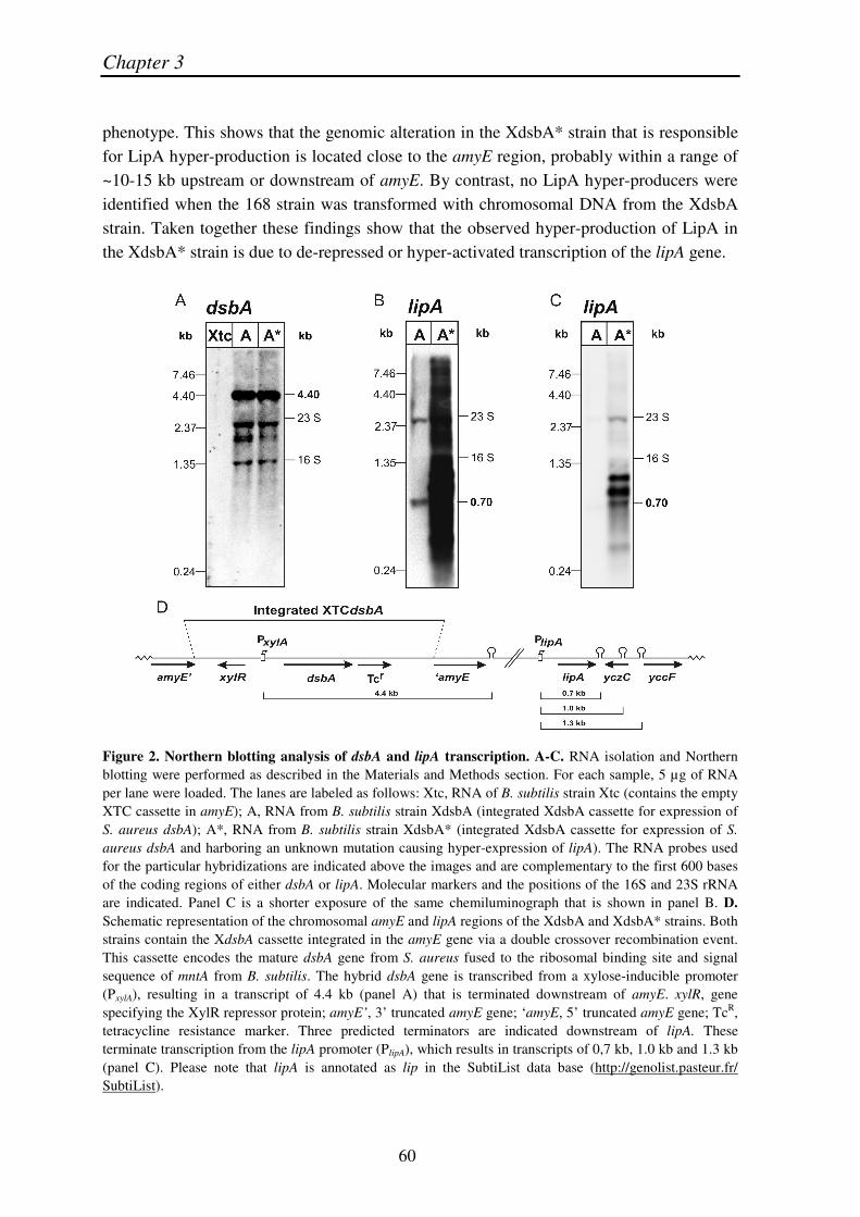
Figure 1. LipA secretion by the XdsbA* mutant. B. subtilis strains 168, Xtc, XdsbA and XdsbA* were grown overnight in LB medium in the absence (0%) or presence (1%) of xylose. Cells were separated from the growth medium by centrifugation and the presence of LipA or DsbA in both fractions was investigated by SDS-PAGE and subsequent Western blotting with specific antibodies against LipA or DsbA, respectively. Note that, due to the high concentrations of LipA, medium fractions were not concentrated. Arrows indicate the position of LipA. Bands with a higher mobility on SDS-PAGE are presumably degradation products of LipA. Molecular weight markers are indicated (kDa). XdsbA cassette. Next, we investigated whether the hyper-production of LipA might be related to possible differences in the expression of the dsbA gene as encoded by the XdsbA cassette. To this end, we performed Northern blotting experiments using mRNA extracted from the XdsbA and XdsbA* strains, using a specific probe complementary to the dsbA mRNA. mRNA from the Xtc strain was used as a negative control. The results of this analysis, shown in Figure 2A, confirmed that the S. aureus dsbA gene was equally well transcribed in both strains, whereas no dsbA transcript was detectable in the strain containing the empty XTC expression cassette. Notably, the band of ~4.4 kb representing the dsbA-specific mRNA was larger than expected on the basis of the dsbA gene alone (666 nucleotides). This indicates that the transcription of dsbA is terminated downstream of the amyE gene (Fig. 2D). As the level of dsbA transcription was comparable in the XdsbA and XdsbA* strains, we conclude that the observed difference in the production of LipA by these strains could not be attributed to indirect effects relating to the expression of dsbA. This view is in agreement with the fact that comparable amounts of DsbA (precursor plus mature) were produced in the XdsbA and XdsbA* strains when grown in the presence of xylose (Fig. 1). Thus, it seemed that LipA hyper-production was related to increased lipA gene expression.

Tat-dependent esterase secretion in B. subtilis
59
To assess whether the hyper-production of LipA did originate from increased lipA expression on the mRNA level, we performed a Northern blotting analysis using a specific lipA probe. As shown by the results in Figure 2 (B and C), the amount of lipA-specific mRNA was strongly increased in the XdsbA* strain. In order to resolve the different individual mRNA species, two images of the same Northern Blot exposed for different periods of time are shown. The longer exposure (Fig. 2B) reveals the presence of a 0.7 kb lipA-specific transcript in the XdsbA strain, representing the original monocistronic lipA transcript. This transcript seems to be terminated at the distinct transcriptional terminator, which can be derived from the DNA sequence immediately downstream of lipA. In the lane representing the XdsbA* strain only a dense black smear is visible due to the large amounts of lipA-specific mRNA in this strain. The shorter exposure reveals only very faint signals in the XdsbA lane, but allows detection of three lipA-specific mRNA species in the XdsbA* strain (Fig. 2C). The smallest among these again corresponds to the 0.7 kb mRNA, which is also detectable in the XdsbA strain. In addition, there are two lipA-specific mRNA species, which are not detectable in the XdsbA strain. The estimated sizes of these transcripts correspond to ~1.0 and ~1.3 kb. These additional bands most probably represent read-through transcripts that are not terminated at the transcriptional terminator immediately downstream of lipA, but somewhat further downstream. Indeed, a systematic search for stem-loop structures in the region downstream of lipA, using the program mfold (Mathews et al., 1999; Zuker, 2003) (http://helix.nih.gov/docs/gcg/mfold.html), revealed two distinct stem-loops: the first one located within the coding region of yczC (the gene downstream of lipA which is transcribed in the opposite direction) and the second located immediately upstream of yccF, within the promoter region of this gene. Both of these structures could function as transcriptional terminators or act as a 3 -̀stabilizer against 3 -̀exonucleases. The derived sizes of the mRNAs, which are predicted to terminate at these two sites, amount to 1.0 kb and 1.3 kb, therefore matching the sizes of the bands detected by Northern blotting (Fig 2D). Experimental confirmation of this in silico analysis was obtained by Northern blotting experiments using the D1 and D2 probes that were designed to hybridize to the two regions between the three stem-loops downstream of lipA. Using these probes, the 1.0 kb and 1.3 kb mRNAs were indeed detected again (results not shown), demonstrating that all the different lipA-specific mRNA species up-regulated in XdsbA* originate at the original lipA promoter. Taken together, these results demonstrate that the LipA hyper-production by the XdsbA* strain is correlated with strongly increased amounts of lipA-specific mRNAs, most probably due to strongly enhanced transcription of the lipA gene.
Sequencing of the 0.5 kb lipA promoter regions in the XdsbA and XdsbA* strains revealed that there are no alterations in the promoter sequence that could be responsible for the increased lipA transcription in the XdsbA* strain. Interestingly, when we transformed the parental strain B. subtilis 168 with genomic DNA of the XdsbA* strain and selected for tetracycline resistant transformants that contained the XdsbA cassette integrated in amyE, we observed that about 15% of these clones displayed the LipA hyper-production
Chapter 3
60
phenotype. This shows that the genomic alteration in the XdsbA* strain that is responsible for LipA hyper-production is located close to the amyE region, probably within a range of ~10-15 kb upstream or downstream of amyE. By contrast, no LipA hyper-producers were identified when the 168 strain was transformed with chromosomal DNA from the XdsbA strain. Taken together these findings show that the observed hyper-production of LipA in the XdsbA* strain is due to de-repressed or hyper-activated transcription of the lipA gene.
Figure 2. Northern blotting analysis of dsbA and lipA transcription. A-C. RNA isolation and Northern blotting were performed as described in the Materials and Methods section. For each sample, 5 µg of RNA per lane were loaded. The lanes are labeled as follows: Xtc, RNA of B. subtilis strain Xtc (contains the empty XTC cassette in amyE); A, RNA from B. subtilis strain XdsbA (integrated XdsbA cassette for expression of S. aureus dsbA); A*, RNA from B. subtilis strain XdsbA* (integrated XdsbA cassette for expression of S. aureus dsbA and harboring an unknown mutation causing hyper-expression of lipA). The RNA probes used for the particular hybridizations are indicated above the images and are complementary to the first 600 bases of the coding regions of either dsbA or lipA. Molecular markers and the positions of the 16S and 23S rRNA are indicated. Panel C is a shorter exposure of the same chemiluminograph that is shown in panel B. D. Schematic representation of the chromosomal amyE and lipA regions of the XdsbA and XdsbA* strains. Both strains contain the XdsbA cassette integrated in the amyE gene via a double crossover recombination event. This cassette encodes the mature dsbA gene from S. aureus fused to the ribosomal binding site and signal sequence of mntA from B. subtilis. The hybrid dsbA gene is transcribed from a xylose-inducible promoter (PxylA), resulting in a transcript of 4.4 kb (panel A) that is terminated downstream of amyE. xylR, gene specifying the XylR repressor protein; amyE’, 3’ truncated amyE gene; ‘amyE, 5’ truncated amyE gene; TcR, tetracycline resistance marker. Three predicted terminators are indicated downstream of lipA. These terminate transcription from the lipA promoter (PlipA), which results in transcripts of 0,7 kb, 1.0 kb and 1.3 kb (panel C). Please note that lipA is annotated as lip in the SubtiList data base (http://genolist.pasteur.fr/ SubtiList).

Tat-dependent esterase secretion in B. subtilis
61
Transcriptome analysis of the LipA hyper-producing XdsbA* strain
To determine which other genes had altered expression levels in the XdsbA* strain, we performed a genome-wide transcriptional analysis with microarrays. To distinguish between effects of the expression of dsbA and the unknown mutation causing lipA hyper-expression, two different sets of comparisons were carried out. For identifying the effects of the unknown mutation in the XdsbA* strain, we compared RNA from the XdsbA* and XdsbA strains. For visualization of the effects of dsbA expression we compared the RNA of the XdsbA strain with that of the Xtc control strain, harboring the empty integrated XTC expression cassette. The statistically significant effects are listed in Table 1. The first striking observation was that there are only a few genes up- or down-regulated in the XdsbA* strain compared to the XdsbA strain (six genes up-regulated and nine genes down-regulated). None of the affected genes however reside within the ~20-30 kb range around amyE gene, which indicates that the changed transcription of these genes is not likely the primary cause of the increased lipA transcription. Another striking observation is that most of the genes that are significantly up- or down-regulated in the XdsbA strain are inversely regulated in the XdsbA* strain (five of the seven down-regulated genes in XdsbA are up-regulated in XdsbA*; all seven up-regulated genes in XdsbA are down-regulated in XdsbA*; Table 1). For example, the manRPA gene cluster is down-regulated in the XdsbA strain, but up-regulated again in the XdsbA* strain. Conversely, the yddJ-rapI-phrI-yddM and ydcM-ydcN-ydcO gene clusters are up-regulated in the XdsbA strain, but down-regulated in the XdsbA* strain. Only two genes that are down-regulated in the XdsbA strain (yrhG and yrhE) are not up-regulated again in the XdsbA* strain, one gene up-regulated in the XdsbA* strain (lipA) is not affected in the XdsbA strain, and two genes down-regulated in the XdsbA* strain (yonBC) are not affected in the XdsbA strain. Thus, the effects of dsbA expression on the expression of other genes in B. subtilis seem to be largely reversed in the XdsbA* strain. This implies that the unidentified mutation in the XdsbA* strain that causes lipA hyper-expression is a mutation that counteracts certain possibly detrimental effects of DsbA production in B. subtilis. An important outcome of the transcriptome analyses is that the mutation causing LipA hyper-secretion by the XdsbA* strain has no detectable effect on any of the known genes for secretion machinery components. This includes the genes for proteins that target secretory precursor proteins to the protein export machinery (e.g. ffh, ftsY and csaA), genes for protein translocation across the membrane (e.g. the sec and tat genes; see Table 1), and genes for post-translocational modification and folding of secretory proteins (e.g. the bdb, sip, skf and prsA genes). Thus, it can be concluded that the mutation in the XdsbA* strain causing LipA hyper-production acts solely at the lipA transcriptional level. The XdsbA* strain can therefore be used to study the mechanism of LipA secretion under conditions of hyper-production.

Chapter 3
62
Table 1. Microarray analysis. To monitor the genome-wide effects on transcription of an unidentified mutation in the XdsbA* strain causing LipA hyper-production, or the expression of S. aureus dsbA in the XdsbA strain, microarray analyses were performed. Preparation of total RNA, cDNA synthesis, labelling, and DNA microarray hybridization and analysis were performed as indicated under Materials and Methods. The RNA samples obtained from three independent cultivations of the XdsbA*, XdsbA and Xtc strains were used for independent cDNA synthesis and competitive DNA array hybridization in two groups: XdsbA* versus XsbA, and XdsbA versus Xtc. The comparison of the XdsbA* and XsbA RNAs shows genome-wide effects of the mutation causing LipA hyper-production, the comparison of XdsbA and Xtc RNAs shows the effects of dsbA expression. Significantly up- or down-regulated genes, as displayed in this table, were considered as such when the mRNA abundance between two compared strains had Cyber-T Bayesian P values of < 0.001 and the individual fold change was at least 2. To show the absence of effects of LipA hyper-production on tat gene expression, the results obtained for the five B. subtilis tat genes are included in this Table. Results are sorted by the average RNA abundance levels in XdsbA*. Average changes of over two-fold are marked in bold.
Ratio of XdsbA* over XdsbA Ratio of XdsbA over Xtc
Gene Acc. nr.1 I II III Average I II III Average
lipA BG10679 152,95 118,15 112,57 127,89 0,75 0,91 0,85 0,84 manP BG13176 9,58 2,32 5,47 5,79 0,07 0,50 0,32 0,30 manA BG13177 6,17 2,14 3,51 3,94 0,14 0,79 0,34 0,42 manR BG13175 2,69 2,03 6,42 3,71 0,28 0,83 0,25 0,45 yxiE BG11134 1,00 1,47 5,00 2,49 0,45 0,39 0,35 0,40 bglH BG10935 1,47 1,28 3,78 2,17 0,42 0,20 0,36 0,33 yrhG BG12296 1,53 2,43 0,87 1,61 0,22 0,29 0,36 0,29 yrhE BG12294 1,39 0,73 1,02 1,05 0,39 0,28 0,24 0,30 ydcO BG12102 0,54 0,41 0,43 0,46 2,14 2,68 2,48 2,43 yonC BG13616 0,36 0,28 0,48 0,37 0,74 1,98 0,36 1,02 yonB BG13615 0,35 0,23 0,38 0,32 0,65 2,32 0,38 1,12 rapI BG12119 0,29 0,27 0,31 0,29 3,49 2,64 2,89 3,01 phrI BG12645 0,30 0,23 0,28 0,27 3,04 3,49 3,59 3,38 yddM BG12120 0,18 0,16 0,14 0,16 9,89 11,04 7,60 9,51 yddJ BG12117 0,13 0,16 0,13 0,14 5,65 6,03 7,83 6,50 ydcM BG12100 0,05 0,08 0,06 0,07 15,79 19,48 9,31 14,86 ydcN BG12101 0,05 0,04 0,05 0,04 19,37 19,16 22,48 20,34 tatAd BG12777 1,13 1,16 1,00 1,09 1,41 0,95 1,09 1,15 tatAy BG12206 1,37 0,89 1,01 1,09 1,03 0,89 1,09 1,00 tatAc BG13464 1,37 1,25 1,17 1,26 1,35 1,28 1,14 1,26 tatCd BG11175 0,89 0,99 1,05 0,98 1,19 1,04 0,97 1,07 tatCy BG12207 0,95 1,27 0,87 1,03 0,99 0,91 0,81 0,90 mntA2 BG13851 1,36 0,75 1,13 1,08 19,97 9,28 29,52 19,59
1Accession numbers were derived from the subtilist database (http://genolist.pasteur.fr/SubtiList). 2mntA appears to be up-regulated in the dsbA expressing strain, but this is due to the fact that the mntA signal sequence was fused to the dsbA gene for efficient targeting of DsbA to the membrane of B. subtilis. This value thus reflects the expression of the S. aureus dsbA gene rather than an altered expression of the B.
subtilis mntA gene
Tat-dependent esterase secretion in B. subtilis
63
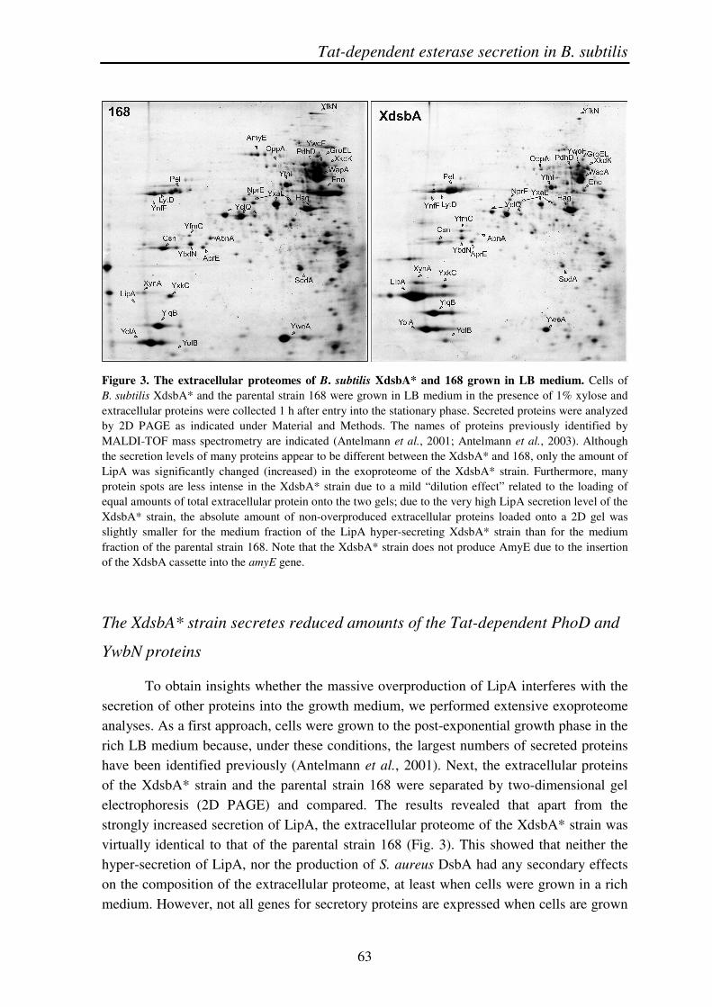
Figure 3. The extracellular proteomes of B. subtilis XdsbA* and 168 grown in LB medium. Cells of B. subtilis XdsbA* and the parental strain 168 were grown in LB medium in the presence of 1% xylose and extracellular proteins were collected 1 h after entry into the stationary phase. Secreted proteins were analyzed by 2D PAGE as indicated under Material and Methods. The names of proteins previously identified by MALDI-TOF mass spectrometry are indicated (Antelmann et al., 2001; Antelmann et al., 2003). Although the secretion levels of many proteins appear to be different between the XdsbA* and 168, only the amount of LipA was significantly changed (increased) in the exoproteome of the XdsbA* strain. Furthermore, many protein spots are less intense in the XdsbA* strain due to a mild “dilution effect” related to the loading of equal amounts of total extracellular protein onto the two gels; due to the very high LipA secretion level of the XdsbA* strain, the absolute amount of non-overproduced extracellular proteins loaded onto a 2D gel was slightly smaller for the medium fraction of the LipA hyper-secreting XdsbA* strain than for the medium fraction of the parental strain 168. Note that the XdsbA* strain does not produce AmyE due to the insertion of the XdsbA cassette into the amyE gene.
The XdsbA* strain secretes reduced amounts of the Tat-dependent PhoD and
YwbN proteins
To obtain insights whether the massive overproduction of LipA interferes with the secretion of other proteins into the growth medium, we performed extensive exoproteome analyses. As a first approach, cells were grown to the post-exponential growth phase in the rich LB medium because, under these conditions, the largest numbers of secreted proteins have been identified previously (Antelmann et al., 2001). Next, the extracellular proteins of the XdsbA* strain and the parental strain 168 were separated by two-dimensional gel electrophoresis (2D PAGE) and compared. The results revealed that apart from the strongly increased secretion of LipA, the extracellular proteome of the XdsbA* strain was virtually identical to that of the parental strain 168 (Fig. 3). This showed that neither the hyper-secretion of LipA, nor the production of S. aureus DsbA had any secondary effects on the composition of the extracellular proteome, at least when cells were grown in a rich medium. However, not all genes for secretory proteins are expressed when cells are grown

Chapter 3
64
in rich media such as LB (Tjalsma et al., 2004). Therefore, we also deployed two-dimensional gel electrophoresis to study the extracellular proteome of B. subtilis cells under phosphate-limiting conditions. Under these growth conditions, B. subtilis will secrete a set of phosphate starvation-induced proteins, which are not produced by cells grown in rich media (Antelmann et al., 2000). One of these proteins is the Tat-dependently secreted protein PhoD (Jongbloed et al., 2000). Figure 4 displays the extracellular proteomes of the XdsbA* and 168 strains grown under phosphate-limiting conditions. These results showed that LipA was also hyper-secreted by the XdsbA* strain under conditions of phosphate limitation. Interestingly though, the level of PhoD secretion was strongly reduced in the XdsbA* strain compared to the secretion of all other proteins that are secreted via the Sec pathway, such as PhoB (compare the intensities of the PhoD spots to the intensities of other spots in Figure 4). This suggested that the hyper-secretion of LipA somehow interfered specifically with that of PhoD, possibly through a competition for translocation via the same Tat translocase.
Figure 4. The extracellular proteomes of B. subtilis XdsbA* and 168 under phosphate-limiting conditions. B. subtilis XdsbA* and 168 were grown under conditions of phosphate starvation as previously described (Antelmann et al., 2000; Jongbloed et al., 2000). Secreted proteins were analyzed by 2D PAGE as indicated under Material and Methods. The names of proteins previously identified by MALDI-TOF mass spectrometry are indicated (Antelmann et al., 2000; Jongbloed et al., 2000). Note that the levels of many proteins secreted by the XdsbA* strain appear to be increased compared to the levels of the corresponding proteins secreted by the 168 strain. This effect is to a large extent caused by the loading of equal amounts of total extracellular protein onto the two gels; due to the very severe reduction of the PhoD secretion level of the XdsbA* strain, the absolute amount of other extracellular proteins of this strain loaded onto a 2D gel was slightly higher than was the case for the extracellular proteins of the parental strain 168. Importantly, a comparison of spot intensities per gel clearly reveals that, in the XdsbA* strain, PhoD is secreted at severely reduced levels relative to other abundantly secreted proteins such as GlpQ, Hag, Pel, PhoB, PstS, and YurI.
Tat-dependent esterase secretion in B. subtilis
65
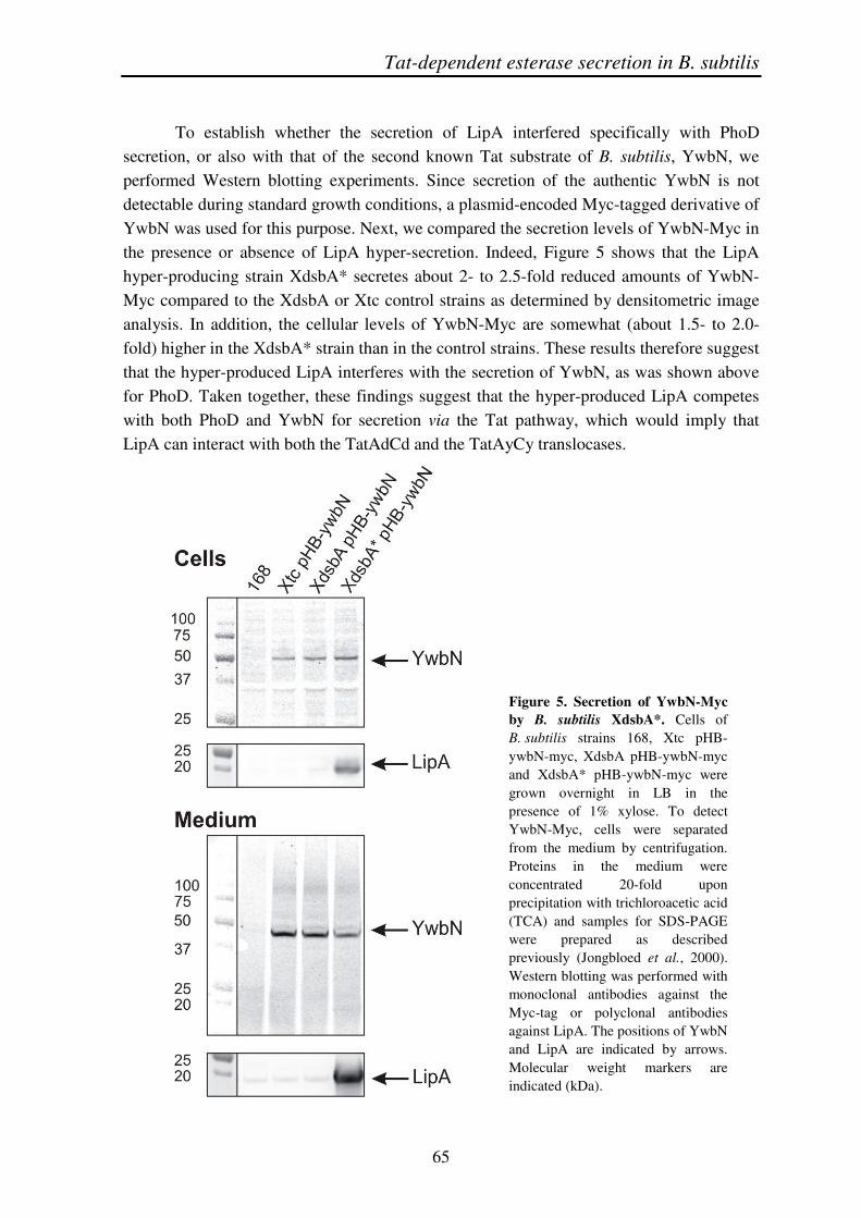
To establish whether the secretion of LipA interfered specifically with PhoD secretion, or also with that of the second known Tat substrate of B. subtilis, YwbN, we performed Western blotting experiments. Since secretion of the authentic YwbN is not detectable during standard growth conditions, a plasmid-encoded Myc-tagged derivative of YwbN was used for this purpose. Next, we compared the secretion levels of YwbN-Myc in the presence or absence of LipA hyper-secretion. Indeed, Figure 5 shows that the LipA hyper-producing strain XdsbA* secretes about 2- to 2.5-fold reduced amounts of YwbN-Myc compared to the XdsbA or Xtc control strains as determined by densitometric image analysis. In addition, the cellular levels of YwbN-Myc are somewhat (about 1.5- to 2.0-fold) higher in the XdsbA* strain than in the control strains. These results therefore suggest that the hyper-produced LipA interferes with the secretion of YwbN, as was shown above for PhoD. Taken together, these findings suggest that the hyper-produced LipA competes with both PhoD and YwbN for secretion via the Tat pathway, which would imply that LipA can interact with both the TatAdCd and the TatAyCy translocases.
Figure 5. Secretion of YwbN-Myc by B. subtilis XdsbA*. Cells of B. subtilis strains 168, Xtc pHB-ywbN-myc, XdsbA pHB-ywbN-myc and XdsbA* pHB-ywbN-myc were grown overnight in LB in the presence of 1% xylose. To detect YwbN-Myc, cells were separated from the medium by centrifugation. Proteins in the medium were concentrated 20-fold upon precipitation with trichloroacetic acid (TCA) and samples for SDS-PAGE were prepared as described previously (Jongbloed et al., 2000). Western blotting was performed with monoclonal antibodies against the Myc-tag or polyclonal antibodies against LipA. The positions of YwbN and LipA are indicated by arrows. Molecular weight markers are indicated (kDa).
Chapter 3
66
Secretion of hyper-produced LipA is predominantly Tat-dependent
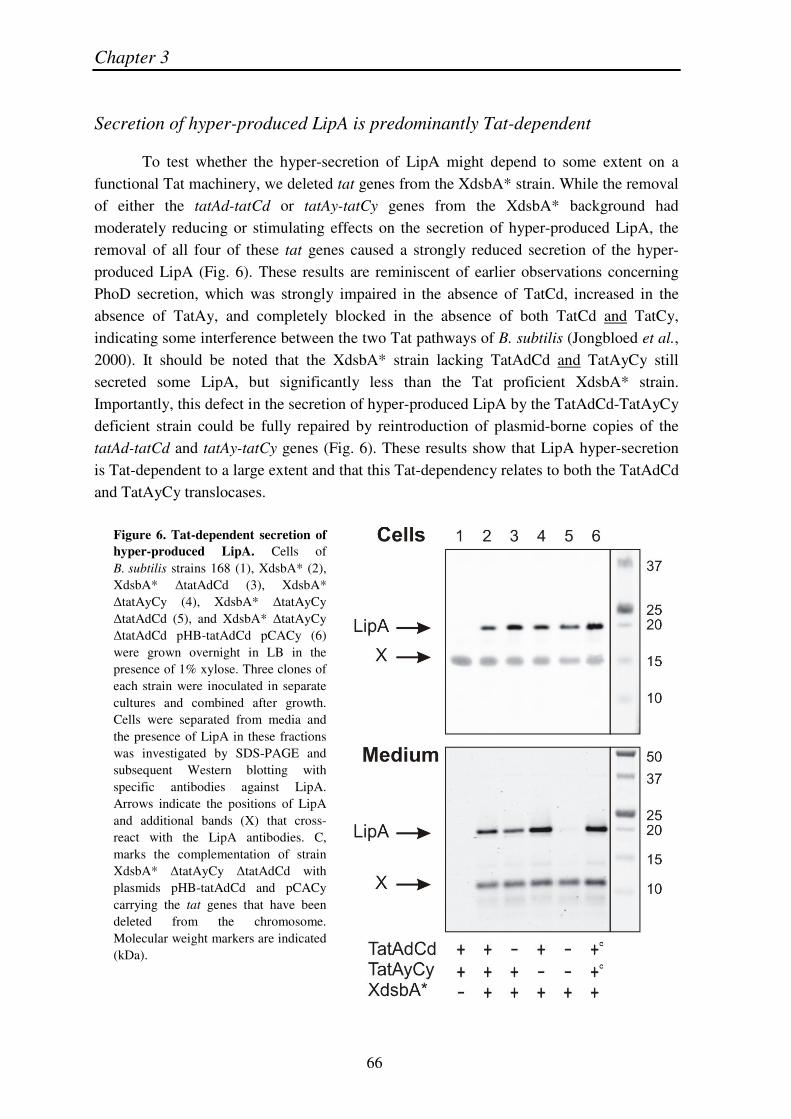
To test whether the hyper-secretion of LipA might depend to some extent on a functional Tat machinery, we deleted tat genes from the XdsbA* strain. While the removal of either the tatAd-tatCd or tatAy-tatCy genes from the XdsbA* background had moderately reducing or stimulating effects on the secretion of hyper-produced LipA, the removal of all four of these tat genes caused a strongly reduced secretion of the hyper-produced LipA (Fig. 6). These results are reminiscent of earlier observations concerning PhoD secretion, which was strongly impaired in the absence of TatCd, increased in the absence of TatAy, and completely blocked in the absence of both TatCd and TatCy, indicating some interference between the two Tat pathways of B. subtilis (Jongbloed et al., 2000). It should be noted that the XdsbA* strain lacking TatAdCd and TatAyCy still secreted some LipA, but significantly less than the Tat proficient XdsbA* strain. Importantly, this defect in the secretion of hyper-produced LipA by the TatAdCd-TatAyCy deficient strain could be fully repaired by reintroduction of plasmid-borne copies of the tatAd-tatCd and tatAy-tatCy genes (Fig. 6). These results show that LipA hyper-secretion is Tat-dependent to a large extent and that this Tat-dependency relates to both the TatAdCd and TatAyCy translocases.
Figure 6. Tat-dependent secretion of hyper-produced LipA. Cells of B. subtilis strains 168 (1), XdsbA* (2), XdsbA* �tatAdCd (3), XdsbA* �tatAyCy (4), XdsbA* �tatAyCy �tatAdCd (5), and XdsbA* �tatAyCy �tatAdCd pHB-tatAdCd pCACy (6) were grown overnight in LB in the presence of 1% xylose. Three clones of each strain were inoculated in separate cultures and combined after growth. Cells were separated from media and the presence of LipA in these fractions was investigated by SDS-PAGE and subsequent Western blotting with specific antibodies against LipA. Arrows indicate the positions of LipA and additional bands (X) that cross-react with the LipA antibodies. C, marks the complementation of strain XdsbA* �tatAyCy �tatAdCd with plasmids pHB-tatAdCd and pCACy carrying the tat genes that have been deleted from the chromosome. Molecular weight markers are indicated (kDa).
Tat-dependent esterase secretion in B. subtilis
67
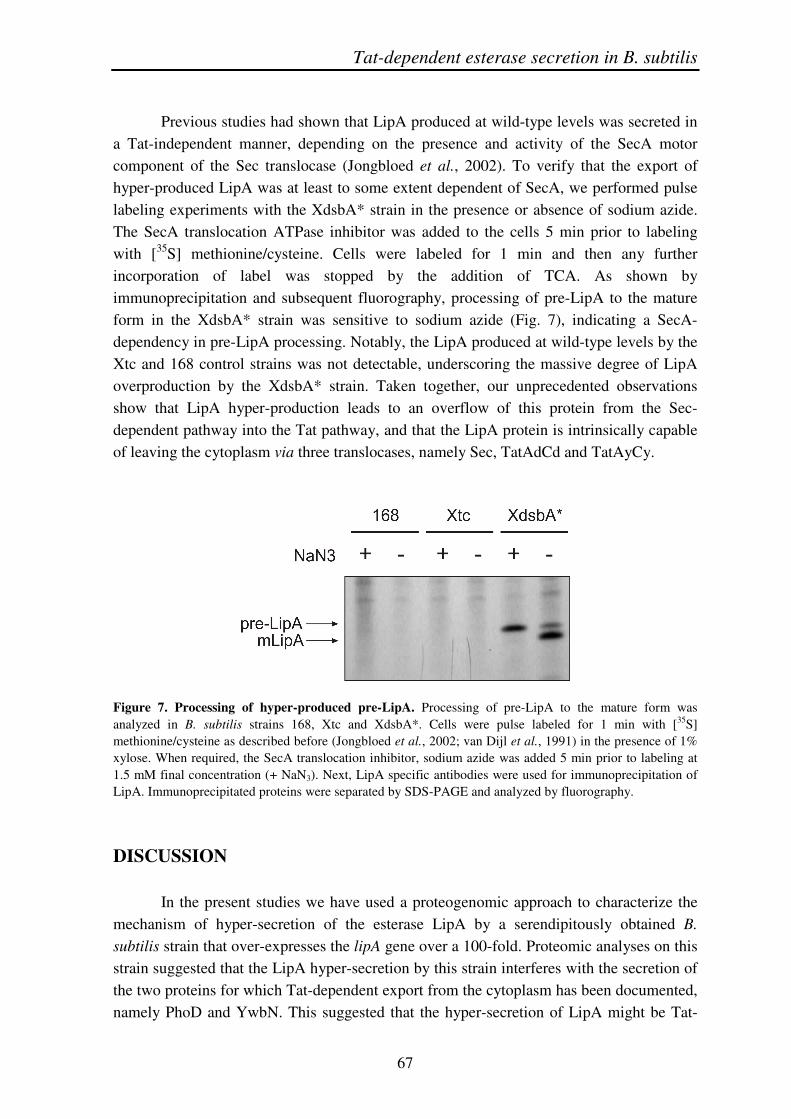
Previous studies had shown that LipA produced at wild-type levels was secreted in a Tat-independent manner, depending on the presence and activity of the SecA motor component of the Sec translocase (Jongbloed et al., 2002). To verify that the export of hyper-produced LipA was at least to some extent dependent of SecA, we performed pulse labeling experiments with the XdsbA* strain in the presence or absence of sodium azide. The SecA translocation ATPase inhibitor was added to the cells 5 min prior to labeling with [35S] methionine/cysteine. Cells were labeled for 1 min and then any further incorporation of label was stopped by the addition of TCA. As shown by immunoprecipitation and subsequent fluorography, processing of pre-LipA to the mature form in the XdsbA* strain was sensitive to sodium azide (Fig. 7), indicating a SecA-dependency in pre-LipA processing. Notably, the LipA produced at wild-type levels by the Xtc and 168 control strains was not detectable, underscoring the massive degree of LipA overproduction by the XdsbA* strain. Taken together, our unprecedented observations show that LipA hyper-production leads to an overflow of this protein from the Sec-dependent pathway into the Tat pathway, and that the LipA protein is intrinsically capable of leaving the cytoplasm via three translocases, namely Sec, TatAdCd and TatAyCy.
Figure 7. Processing of hyper-produced pre-LipA. Processing of pre-LipA to the mature form was analyzed in B. subtilis strains 168, Xtc and XdsbA*. Cells were pulse labeled for 1 min with [35S] methionine/cysteine as described before (Jongbloed et al., 2002; van Dijl et al., 1991) in the presence of 1% xylose. When required, the SecA translocation inhibitor, sodium azide was added 5 min prior to labeling at 1.5 mM final concentration (+ NaN3). Next, LipA specific antibodies were used for immunoprecipitation of LipA. Immunoprecipitated proteins were separated by SDS-PAGE and analyzed by fluorography.
DISCUSSION
In the present studies we have used a proteogenomic approach to characterize the mechanism of hyper-secretion of the esterase LipA by a serendipitously obtained B. subtilis strain that over-expresses the lipA gene over a 100-fold. Proteomic analyses on this strain suggested that the LipA hyper-secretion by this strain interferes with the secretion of the two proteins for which Tat-dependent export from the cytoplasm has been documented, namely PhoD and YwbN. This suggested that the hyper-secretion of LipA might be Tat-
Chapter 3
68
dependent even though it had previously been shown that LipA produced at wild-type levels was Sec-dependently secreted. Indeed, secretion studies with tat mutant strains revealed a strong Tat-dependency of the hyper-secreted LipA. Remarkably, both the TatAdCd and the TatAyCy translocase are involved in LipA hyper-secretion. Indirect effects on the transcription of genes for known Tat or Sec pathway components were ruled out through the results of transcriptome analyses. It thus seems that an overflow mechanism directs the hyper-produced LipA from the Sec pathway into the Tat pathway, which is an unprecedented finding.
Protein secretion is an important topic in current molecular microbiological research related both to pathogens and organisms that are used as cell factories for biotechnological applications. Although many pathways for protein secretion have been described in considerable detail, the full complexity of these pathways and their interdependencies are not very well understood. Over the past decade, we have employed proteomics technology to map the different pathways of the secretome of B. subtilis (Antelmann et al., 2001; Tjalsma et al., 2004). This has revealed a major role of the Sec pathway in protein secretion by this organism. Other pathways for protein secretion, such as the Tat pathway, were shown to serve highly selective purposes. Quite remarkably, merely two proteins (PhoD and YwbN) were shown to be exported Tat-dependently in B. subtilis, each of them requiring a distinct Tat translocase (Jongbloed et al., 2002; Jongbloed et al., 2004). This finding raised the intriguing question whether the Tat pathway in B. subtilis is really of minor importance, despite its intrinsic capacity to transport fully folded proteins (Barnett et al., 2008), or whether it also serves particular other functions under certain conditions that had not yet been identified. In line with the latter idea was the finding that many secreted B. subtilis proteins, like the esterase LipA, contain potential RR-motifs in their signal peptides. On the other hand however, our earlier proteomics studies clearly showed that all expressed proteins with potential RR-motifs (except PhoD and YwbN) were secreted Tat-independently, at least under the standard laboratory test conditions (Tjalsma et al., 2004; Jongbloed et al., 2002; Jongbloed et al., 2004). Our present studies have now identified one condition under which a normally Sec-dependently secreted protein with a potential RR-motif in its signal peptide can take a turn from the Sec pathway into the Tat pathway, namely the condition of hyper-secretion. Although this finding was unexpected, it is retrospectively perhaps not completely surprising since B. subtilis and related Bacillus species rely on the high-level secretion of degradative enzymes for their growth and survival in the soil and plant rhizosphere, for which they need to feed on decaying organic matter (Earl et al., 2008). Under certain natural conditions, regulated hyper-secretion of degradative enzymes is therefore likely to occur, and the fact that a spontaneous mutation actually resulted in the hyper-secretion of LipA is fully consistent with this view. Thus, we believe that the current observations may have a broader significance for protein secretion by bacilli and other bacteria under natural conditions.

Tat-dependent esterase secretion in B. subtilis
69
As yet, the exact location and nature of the mutation causing lipA hyper-expression from the original lipA promoter region is not known. It is however located in the vicinity of the amyE gene since frequent (~15%) co-transformation of the XdsbA cassette in the amyE gene and the mutation was observed. No obvious candidate genes for trans-acting factors in lipA expression located near the amyE gene could so far be identified. Clearly, sequence analysis identified no mutations in the upstream region of lipA, and we consider the distance between lipA and amyE (35 kb) too large to account for the frequent co-transformation of the XdsbA cassette in amyE with the mutation causing lipA overexpression. Transcriptome analyses of the XdsbA* strain did, unfortunately, not reveal potential locations for the unknown mutation since the transcription of none of the genes in the vicinity of amyE was altered. It thus seems most likely that a point mutation in a trans-acting transcriptional activator or repressor of lipA is responsible for the LipA hyper-production phenotype of the XdsbA* strain. Interestingly, our transcriptome analyses show that the unidentified mutation has by-and-large a counter-acting effect on the cellular response of B. subtilis to the expression of the S. aureus dsbA gene. This view is underscored by the fact that increasing the dsbA expression by the addition of xylose to the medium is followed by an increased production of LipA. Together, these findings suggest that expression of the dsbA gene is to some extent stressful for the B. subtilis cell and that this stress is relieved by the mutation in the XdsbA* strain. However, we have so far not observed any phenotypes of the XdsbA strain that suggest any severely detrimental effects of DsbA production. In any case, the hyper-expression of lipA in our mutant would be a side-effect of the compensatory mutation in the XdsbA* strain as the expression of lipA was not affected by dsbA expression in the original XdsbA strain. It should be noted that the mutation leading to LipA hyper-production seems to occur infrequently since we observed this phenomenon only once so far. Although we do not know the precise nature of the mutation causing LipA hyper-production in the XdsbA* strain, we conclude from the transcriptome analyses that this mutant can be regarded as a bona fide overproduction strain with no side effects on the expression of secretion machinery components. In fact, this mutant does not even display a secretion stress response despite the hyper-production of LipA (Antelmann et al., 2003). Thus, the XdsbA* strain can be used for studies on the LipA secretion mechanism, like any other strain overproducing a secretory protein.
Our proteomics analyses suggest that the hyper-produced LipA competes with PhoD and YwbN for secretion via the TatAdCd and TatAyCy translocases, respectively. These findings provided in fact the first incentive to investigate the Tat-dependency of LipA hyper-secretion, which was then confirmed by our experiments with multiple tat mutant strains hyper-producing LipA. Although a possible involvement of the Tat pathway in LipA secretion has been contemplated before (Jongbloed et al., 2002), conclusive evidence for Tat-dependent LipA secretion could not be obtained until the LipA hyper-secreting XdsbA* strain became available. Importantly, the finding that LipA hyper-secretion depends on both Tat translocases of B. subtilis (i.e. TatAdCd and TatAyCy) is unprecedented and has not been reported for any other Tat substrate in B. subtilis or other

Chapter 3
70
organisms with duplicated Tat systems. This finding shows for the first time that the two B. subtilis Tat translocases do not have strictly separated specificities as was previously suggested (Jongbloed et al., 2004; Pop et al., 2002), but that their specificities overlap at least to some extent. It should be noted that we currently do not know what determines the specificity of Tat translocation pathway choice in B. subtilis. This may relate to the RR-sequence motifs in signal peptides, the nature and folding state of the mature protein, or all these factors together. In this respect, we cannot exclude the possibility that, under non-hyper-producing conditions, LipA is a preferred substrate for only one of the two B. subtilis Tat translocases. This is however hard to assess due to the predominantly Sec-dependent translocation of LipA under non-hyper-producing conditions, as well as the essentiality of the Sec machinery for bacterial growth and life (Jongbloed et al., 2002). In the specific case of LipA hyper-production, we believe that the massive production of pre-LipA somehow saturates the Sec translocase for this protein. This would then lead to reduced LipA translocation rates and possibly folding of LipA in the cytoplasm. The folded pre-LipA would thus become a better substrate for the Tat machinery, which is known to accept mainly folded proteins. Alternatively, saturation of the Sec translocase might lead to a more effective recognition of the RR-motif in pre-LipA by the Tat machinery and, in this case, cytoplasmic folding of LipA might not be a strict prerequisite for Tat-dependent export. For example, unfolded DHFR can be translocated via the thylakoidal Tat system (Hynds et al., 1998) and unstructured, small, hydrophilic proteins can be exported Tat-dependently in E. coli (Richter et al., 2007). In any case, evidence for some degree of Sec pathway saturation by LipA hyper-production was indeed derived from the observation that the DsbA precursor accumulates in the XdsbA* strain, whereas this is not the case in the XdsbA strain. On the other hand, the secretion of the majority of proteins was not affected at the steady state level as was shown by proteomics. This is in fact consistent with the finding that LipA hyper-production did not interfere with cell growth and viability, which would have been the case if the Sec pathway had been jammed in a major way. Apparently, jamming of the Sec pathway is prevented by overflow of pre-LipA into the Tat pathway. Finally, our pulse labeling experiments suggest that the processing of newly synthesized hyper-produced pre-LipA is sensitive to sodium azide, indicating involvement of SecA in this precursor processing. This is consistent with our previous observations that LipA can be secreted Sec-dependently and with our present model that the observed Tat-dependency of hyper-produced LipA relates to an overflow mechanism from the Sec pathway into the Tat pathway. We do however not know the fate of the mature hyper-produced LipA that is detected by pulse labeling. In fact, it is quite conceivable that a substantial portion is degraded at the trans side of the membrane, as was previously shown for over-produced amylases that do not fold efficiently enough upon membrane translocation via the Sec pathway in an unfolded state (Stephenson et al., 1998; Stephenson et al., 2000). Notably, we can not completely exclude the possibility that sodium azide also affects the export of LipA via the B. subtilis Tat pathway. To our knowledge there are only two reported cases where the transport of Tat-dependent proteins

Tat-dependent esterase secretion in B. subtilis
71
was affected by sodium azide. These concern the Glucose-fructose oxidoreductase (GFOR) of the Gram-negative bacterium Zymomonas mobilis (Wiegert et al., 1996), and the 17-kD subunit of the photosynthetic O2-evolving complex (OE17) in intact chloroplasts (Leheny et al., 1998). It is however not entirely clear whether these are direct effects of sodium azide on the Tat machinery, or whether they should be regarded as an indication of some Sec-dependent translocation of the two proteins, like we have described here for LipA.
In conclusion, our present findings show for the first time that certain secretory proteins, such as the esterase LipA of B. subtilis, can be exported both via the Sec and the Tat pathways of a Gram-positive bacterium depending on the conditions applied. To date, a re-routing of proteins from the Sec to the Tat pathway or vice versa has been achieved only artificially by replacing Sec-type signal peptides with RR-signal peptides and/or by modulating the folding conditions for the exported proteins so that they fold in the cytoplasm prior to membrane translocation. This has been worked out especially well in E. coli for re-routed export of the alkaline phosphatase PhoA and the maltose-binding protein MalE from the Sec pathway into the Tat pathway, and for the re-routed export of the ribose-binding protein RbsB from the Tat pathway into the Sec pathway (Blaudeck et al., 2003; DeLisa et al., 2003; Tullman-Ercek et al., 2007; Pradel et al., 2003).
Although the overflow mechanism as presented in our paper has not been documented before, we believe that it may be a more common, but so far overlooked, mechanism for the secretion of proteins that can fulfill the requirements for transport via both the Sec pathway (i.e. channel passage in an unfolded state) and the Tat pathway (i.e. folding prior to channel passage). It will be an important challenge for future studies to identify the environmental or cellular conditions that influence the usage of particular secretory pathways in order to obtain a full understanding of the biological processes that require an active Tat pathway in bacteria, such as B. subtilis.
EXPERIMENTAL PROCEDURES
Plasmids, bacterial strains, media and growth conditions
The plasmids and bacterial strains used in this study are listed in Table 2. Strains were grown with agitation at 37ºC in either Luria Bertani (LB) medium, S7 minimal salt medium or Paris minimal (PM) medium (see (Kouwen et al., 2007) for exact media compositions). If appropriate, media were supplemented with antibiotics at the following concentrations: ampicillin (Ap), 100 �g/ml (E. coli); erythromycin (Em), 100 �g/ml (E. coli) or 2 �g/ml (B. subtilis); chloramphenicol (Cm), 5 �g/ml (B. subtilis); tetracycline (Tc), 10 �g/ml (B. subtilis); spectinomycin (Sp), 100 �g/ml (B. subtilis); kanamycin (Km), 50 �g/ml (E. coli) or 20 �g/ml (B. subtilis). To visualize �-amylase activity (specified by the amyE gene), LB plates were supplemented with 1% starch.
Chapter 3
72
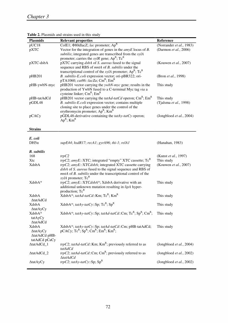
Table 2. Plasmids and strains used in this study
Plasmids Relevant properties Reference pUC18 ColE1; �80dlacZ; lac promoter; ApR (Norrander et al., 1983) pXTC Vector for the integration of genes in the amyE locus of B.
subtilis; integrated genes are transcribed from the xylA promoter; carries the xylR gene; ApR; TcR
(Darmon et al., 2006)
pXTC-dsbA pXTC carrying dsbA of S. aureus fused to the signal sequence and RBS of mntA of B. subtilis under the transcriptional control of the xylA promoter; ApR; TcR
(Kouwen et al., 2007)
pHB201 B. subtilis-E.coli expression vector; ori-pBR322; ori-pTA1060; cat86::lacZa; CmR; EmR
(Bron et al., 1998)
pHB-ywbN-myc pHB201 vector carrying the ywbN-myc gene; results in the production of YwbN fused to a C-terminal Myc tag via a cysteine linker; CmR; EmR
This study
pHB-tatAdCd pHB201 vector carrying the tatAd-tatCd operon; CmR; EmR This study pGDL48 B. subtilis-E.coli expression vector; contains multiple
cloning site to place genes under the control of the erythromycin promoter; ApR; KmR
(Tjalsma et al., 1998)
pCACy pGDL48-derivative containing the tatAy-tatCy operon; ApR; KmR
(Jongbloed et al., 2004)
Strains E. coli DH5� supE44; hsdR17; recA1; gyrA96; thi-1; relA1 (Hanahan, 1983) B. subtilis 168 trpC2 (Kunst et al., 1997) Xtc trpC2; amyE::XTC; integrated “empty” XTC cassette; TcR This study XdsbA trpC2; amyE::XTCdsbA; integrated XTC cassette carrying
dsbA of S. aureus fused to the signal sequence and RBS of mntA of B. subtilis under the transcriptional control of the xylA promoter; TcR
(Kouwen et al., 2007)
XdsbA* trpC2; amyE::XTCdsbA*; XdsbA derivative with an additional unknown mutation resulting in lipA hyper-production; TcR
This study
XdsbA �tatAdCd
XdsbA*; tatAd-tatCd::Km; TcR; KmR This study
XdsbA �tatAyCy
XdsbA*; tatAy-tatCy::Sp; TcR; SpR This study
XdsbA* tatAyCy �tatAdCd
XdsbA*; tatAy-tatCy::Sp; tatAd-tatCd::Cm; TcR; SpR; CmR; This study
XdsbA �tatAyCy �tatAdCd pHB- tatAdCd pCaCy
XdsbA*; tatAy-tatCy::Sp; tatAd-tatCd::Cm; pHB-tatAdCd; pCACy; TcR; SpR; CmR; EmR; KmR;
This study
�tatAdCd_1 trpC2; tatAd-tatCd::Km; KmR; previously referred to as tatAdCd
(Jongbloed et al., 2004)
�tatAdCd_2 trpC2; tatAd-tatCd::Cm; CmR; previously referred to as �tatAdCd
(Jongbloed et al., 2002)
�tatAyCy trpC2; tatAy-tatCy::Sp; SpR (Jongbloed et al., 2002)
Tat-dependent esterase secretion in B. subtilis
73
General DNA techniques
Procedures for DNA purification, restriction, ligation, agarose gel electrophoresis, and transformation of competent E. coli cells were carried out as previously described (Sambrook et al., 1989). Chromosomal DNA of B. subtilis was isolated according to Bron and Venema (Bron and Venema, 1972). PCR was carried out with the Pwo DNA polymerase, using chromosomal DNA as a template. All PCR fragments were ligated in pUC18 and subsequently introduced in E. coli DH5�. Plasmid DNA from E. coli was isolated using the alkaline lysis method (Sambrook et al., 1989), or the High Pure Plasmid Isolation Kit according to the protocol supplied by the manufacturer (Roche Applied Science). B. subtilis was transformed as described by Kunst and Rapoport (Kunst and Rapoport, 1995). All constructs were checked by sequencing.
Construction of B. subtilis mutant strains and plasmids
In a recent study, we have constructed the B. subtilis XdsbA strain to characterize the activity of the S. aureus lipoprotein DsbA in B. subtilis (Kouwen et al., 2007). This strain contains the XdsbA cassette inserted into the amyE locus for the xylose-inducible expression of S. aureus dsbA. It should be noted that the sequence coding for the mature S. aureus DsbA was fused to the signal sequence and ribosomal binding site of B. subtilis mntA to facilitate efficient translation and export of the DsbA lipoprotein. When a series of correct clones producing DsbA were checked by proteomics, one of these showed an unexpectedly high production of LipA. This serendipitously obtained LipA hyper-secreting mutant was annotated as XdsbA*.
The B. subtilis control strain Xtc, carrying the “empty” XTC cassette integrated in the amyE locus via double cross-over recombination was obtained by transformation of B. subtilis 168 with the pXTC vector. Correct integration of the XTC cassette was checked by selection for tetracycline resistance, and screening for an AmyE-negative phenotype on starch-containing plates.
To construct plasmid pHB-ywbN-myc, the B. subtilis ywbN gene was PCR-amplified from chromosomal DNA of strain 168 with the primers pHB-ywbN-bsu-SalI-F (5'-GGGGGGTCGA-CATGTGCTATAAAAGGAG-3') and pHB-ywbN-bsu-EcoRI-R (5'-CCCCCGAATTCTTAGTTCAAATCTTC-CTCACTGATCAATTTCTGTTCTGATTCCAGCAAACGCTGGGC-3') containing SalI-HindII and EcoRI restriction sites, respectively (underlined). In the reverse primer a c-Myc tag sequence was incorporated (marked in italics) allowing immunodetection of the protein. The amplified ywbN gene was digested with HindII-EcoRI and ligated to the SmaI-EcoRI cleaved plasmid pHB201. All plasmids thus obtained were checked by sequencing (ServiceXS; Leiden, the Netherlands). Next, the constructed pHB-ywbN-myc plasmid was introduced into the B. subtilis strains 168, Xtc and XdsbA*.
B. subtilis strain XdsbA* �TatAdCd was obtained by transformation of strain XdsbA* with chromosomal DNA of strain �TatAdCd_1 and selection for kanamycin resistance. B. subtilis strain XdsbA* �TatAyCy was obtained by transformation of strain XdsbA* with chromosomal DNA of strain �TatAyCy and selection for spectinomycin resistance. The combined mutant strain XdsbA* �TatAyCy �TatAdCd was constructed by transformation of strain XdsbA* �TatAyCy with chromosomal DNA of strain �TatAdCd_2 and selection for chloramphenicol resistance.
To construct plasmid pHB-tatAdCd, the B. subtilis tatAd-tatCd genes were PCR-amplified from chromosomal DNA of strain 168 with the primers TatAdCdBsF (5'-CCCCCACTAGTAAGCAATCCGA-TGAGGTCG-3') and TatAdCdBsR (5'-CCCCCCTCGAGGATGAGGATGTGAAGTCAC-3') containing SpeI and XhoI restriction sites, respectively (underlined). The amplified tatAd-tatCd genes were digested with SpeI-XhoI and ligated to the corresponding restriction sites of pHB201. Obtained plasmids were checked by sequencing. Next, the constructed pHB-tatAdCd plasmid was introduced into the B. subtilis strain XdsbA* �TatAyCy �TatAdCd. Finally, plasmid pCACy, carrying the tatAy-tatCy genes, was also introduced into the B. subtilis XdsbA* �TatAyCy �TatAdCd strain.
Chapter 3
74
SDS-PAGE and Western blotting
The presence of LipA, DsbA and YwbN-Myc in growth medium and/or cell lysates was detected by Western blotting. Cellular or secreted proteins were separated by SDS-PAGE (using pre-cast Bis-Tris NuPAGE gels from Invitrogen), and proteins were then semi-dry blotted (75 min at 1 mA / Cm2) onto a nitrocellulose membrane (Protran®, Schleicher & Schuell). Subsequently, the DsbA proteins were detected with specific polyclonal antibodies raised in rabbits (Eurogentec). LipA was detected with polyclonal antibodies kindly provided by Dr Y. L. Boersma. The YwbN-Myc protein was detected with monoclonal antibodies against the Myc-tag (Gentaur). The detection of these antibodies was performed with fluorescent IgG secondary antibodies (IRDye 800 CW goat anti-rabbit or goat anti-mouse from LiCor Biosciences) in combination with the Odyssey Infrared Imaging System (LiCor Biosciences). Densitometric image analysis to quantify relative protein amounts as detected by Western blotting was performed with the program ImageJ (http://rsbweb.nih.gov/ij/).
Northern blot analysis
Preparation of total RNA was carried out as described by Eymann et al. (Eymann et al., 2002). Northern blot analyses using specific RNA probes were performed as described by Homuth et al. (Homuth et al., 1997). Chemiluminescence was detected using a Lumi-Imager (Roche Diagnostics). Transcript sizes were determined by comparison with an RNA size marker (Invitrogen). The digoxygenin-labelled specific RNA probes were synthesized by in vitro transcription using T7 RNA polymerase and specific PCR products as templates. Synthesis of the DNA templates was performed by PCR using the following pairs of oligonucleotides: for the lip probe, lip-for (5'-ATGAAATTTGTAAAAAGAAG-3') and lip-rev (5'-CTAATACGACTCACTATAGGGAGAAATCAGGCTGTTGACTTGGC-3'); for dsbA, dsbA-for (5'-TGCGGTAAAAAAGAATCAGC-3') and dsbA-rev (5'-CTAATACGACTCACTATAGGGAGA-CTATTTGATTTTATCTTTTA-3'); for D1, D1-for (5'-GTAAATGCGGCAGTCAAATA-3') and D1-rev (5'-CTAATACGACTCACTATAGGGAGA-TGGGTCCGCCGGTGTCATTA-3'); for D2, D2-for (5'-GCAGCATGAAACCAGCTAGT-3') and D2-rev (5'-CTAATACGACTCACTATAGGGAGA-CTTCTGAGGGCTTGGTTTCG-3'). The underlined sequences indicate the T7 promoter region.
Transcriptome analysis
Cell harvesting and preparation of total RNA were performed as described previously (Eymann et al., 2002). RNA samples were DNAse-treated with the RNAse-free DNAse kit (Qiagen) according to the manufacturer’s instructions and purified using RNeasy mini columns (Qiagen). The quality control of the RNA preparations was performed with the RNA 6000 Nano LabChip Kit (Agilent Technologies) on the Agilent 2100 Bioanalyzer according to the manufacturer’s instructions. The RNA samples obtained from three independent cultivations were used for independent cDNA synthesis and DNA array hybridization. Generation of the Cy3/Cy5-labeled cDNAs and hybridization to B. subtilis whole-genome DNA microarrays (Eurogentec) were performed as previously described by Jürgen et al. (Jürgen et al., 2005). The slides were scanned with a ScanArray Express scanner (PerkinElmer Life and Analytical Sciences). Quantification of the signal and background intensities of individual spots was carried out using the ScanArray Express image analysis software.
Data were analyzed using the GeneSpring software (Agilent Technologies). Raw signal intensities were first transformed by intensity dependent LOWESS normalization. The normalized array data were subjected to a statistical analysis using Cyber-T, a program based on a t-test combined with a Bayesian statistical framework (Baldi and Long, 2001). The software is accessible through a Web interface at http://cybert.microarray.ics.uci.edu. The mRNA abundance was considered to be significantly different between the wild type and the respective mutant strain if (i) the Cyber-T Bayesian P value was < 0.001 and (ii) the individual fold change was at least 2. The potential and known functions of the encoded proteins were predominantly inferred from the SubtiList database (http://genolist.pasteur.fr/SubtiList/).
Tat-dependent esterase secretion in B. subtilis
75
Proteomics
Cells of B. subtilis were grown at 37°C under vigorous agitation in 1 liter of LB medium, or a synthetic medium containing 0.16 mM KH2PO4 to induce a phosphate starvation response (Antelmann et al., 1997). At the onset of and after 1 hour of post-exponential growth, cells were separated from the growth medium by centrifugation. The secreted proteins in the growth medium were collected for two-dimensional gel electrophoresis (2D PAGE), gels were stained with the SYPRO Ruby protein gel stain (Molecular Probes Inc.). All detected protein spots were identified previously by matrix-assisted laser desorption/ionization – time of flight mass spectrometry (MALDI-TOF MS) (Antelmann et al., 2001; Antelmann et al., 2003; Jongbloed et al., 2000; Jongbloed et al., 2002). To visualize possible differences in extracellular protein composition, dual channel image analysis of stained gels was performed using the DECODON Delta 2D software (http://www.decodon.com). Each experiment was performed at least twice.
Pulse labeling of proteins, immunoprecipitation, and fluorography
Pulse labeling of B. subtilis, immunoprecipitation, and fluorography were performed as described previously (Jongbloed et al., 2002; van Dijl et al., 1991). To inhibit the translocation ATPase activity of
SecA, sodium azide (1.5 mM final concentration) was added to the cells 5 min prior to labeling (Klein et al., 1994). Immunoprecipitation was performed with specific antibodies against LipA.
ACKNOWLEDGMENTS
The authors wish to thank Ykelien Boersma and Wim Quax for providing antibodies against LipA, and Anja Hoffmann, Julia Kretschmer and Sebastian Grund for excellent technical assistance. T.R.H.M.K., R.v.d.P., H.A., M.H., U.M. and J.M.v.D. were in parts supported by the CEU projects LSHG-CT-2004-503468, LSHG-CT-2004-005257, LSHM-CT-2006-019064 and LSHG-CT-2006-037469, the transnational SysMO initiative through project BACELL SysMO, the European Science Foundation under the EUROCORES Programme EuroSCOPE, and grant 04-EScope 01-011 from the Research Council for Earth and Life Sciences of the Netherlands Organization for Scientific Research. H.A. and M.H. were supported by the “Deutsche Forschungsgemeinschaft”, the “Bundesministerium für Bildung und Forschung”, and the “Fonds der Chemischen Industrie”. G.H. and U.M. were supported by the “Bundesministerium für Bildung und Forschung” (03ZIK012).

Chapter 3
76