University of Groningen Pemphigus pathogenesis Sokol, Ena
21
University of Groningen Pemphigus pathogenesis Sokol, Ena IMPORTANT NOTE: You are advised to consult the publisher's version (publisher's PDF) if you wish to cite from it. Please check the document version below. Document Version Publisher's PDF, also known as Version of record Publication date: 2016 Link to publication in University of Groningen/UMCG research database Citation for published version (APA): Sokol, E. (2016). Pemphigus pathogenesis: Insights from light and electron microscopy studies. University of Groningen. Copyright Other than for strictly personal use, it is not permitted to download or to forward/distribute the text or part of it without the consent of the author(s) and/or copyright holder(s), unless the work is under an open content license (like Creative Commons). The publication may also be distributed here under the terms of Article 25fa of the Dutch Copyright Act, indicated by the “Taverne” license. More information can be found on the University of Groningen website: https://www.rug.nl/library/open-access/self-archiving-pure/taverne- amendment. Take-down policy If you believe that this document breaches copyright please contact us providing details, and we will remove access to the work immediately and investigate your claim. Downloaded from the University of Groningen/UMCG research database (Pure): http://www.rug.nl/research/portal. For technical reasons the number of authors shown on this cover page is limited to 10 maximum. Download date: 03-01-2022
Transcript of University of Groningen Pemphigus pathogenesis Sokol, Ena

University of Groningen
Pemphigus pathogenesisSokol, Ena
IMPORTANT NOTE: You are advised to consult the publisher's version (publisher's PDF) if you wish to cite fromit. Please check the document version below.
Document VersionPublisher's PDF, also known as Version of record
Publication date:2016
Link to publication in University of Groningen/UMCG research database
Citation for published version (APA):Sokol, E. (2016). Pemphigus pathogenesis: Insights from light and electron microscopy studies. Universityof Groningen.
CopyrightOther than for strictly personal use, it is not permitted to download or to forward/distribute the text or part of it without the consent of theauthor(s) and/or copyright holder(s), unless the work is under an open content license (like Creative Commons).
The publication may also be distributed here under the terms of Article 25fa of the Dutch Copyright Act, indicated by the “Taverne” license.More information can be found on the University of Groningen website: https://www.rug.nl/library/open-access/self-archiving-pure/taverne-amendment.
Take-down policyIf you believe that this document breaches copyright please contact us providing details, and we will remove access to the work immediatelyand investigate your claim.
Downloaded from the University of Groningen/UMCG research database (Pure): http://www.rug.nl/research/portal. For technical reasons thenumber of authors shown on this cover page is limited to 10 maximum.
Download date: 03-01-2022

501164-L-bw-Sokol501164-L-bw-Sokol501164-L-bw-Sokol501164-L-bw-Sokol
4
Chapter 1c
Pemphigus: disease and pathogenesis
E. Sokol
Center for Blistering Diseases, Department of Dermatology and Department of Cell BiologyUniversity Medical Center Groningen, University of Groningen,
Groningen, the Netherlands

501164-L-bw-Sokol501164-L-bw-Sokol501164-L-bw-Sokol501164-L-bw-Sokol
Chapter 1C
40
Contents
1. Introduction 41 2. Forms of pemphigus 43
2.1. Classical forms of pemphigus 44 Pemphigus vulgaris 44 Pemphigus foliaceus 44
2.2. Non-classical forms of pemphigus 46 Pemphigus herpetiformis 46 IgA pemphigus 46 Paraneoplastic pemphigus 46 Drug induced pemphigus 46
3. Pemphigus pathogenesis 47 3.1. Pathogenicity of pemphigus autoantibodies 48 3.2. Autoreactive T cells in pemphigus 48 3.3. Antigens in pemphigus 48 3.4. Molecular mechanism of acantholysis in pemphigus 49
Steric hindrance 49 Cell signaling theory 51 Desmoglein non assembly depletion theory 52
3.5. Desmoglein compensation theory 54 4.Diagnosis of pemphigus 56 5.Therapy of pemphigus 56

501164-L-bw-Sokol501164-L-bw-Sokol501164-L-bw-Sokol501164-L-bw-Sokol
Pemphigus: disease and pathogenesis
41
1. Introduction
Autoimmune bullous diseases are a heterogeneous group of disorders associated with auto-antibodies directed against desmosomal proteins (pemphigus group) or hemidesmosomal proteins (pemphigoid group including epidermolysis bullosa aquisita) or against epidermal transglutaminase (dermatitis herpetiformis) (Baum et al., 2014). The main clinical manifestation of these disorders is a blister, caused by loss of cell-cell or cell-matrix adhesion, and depending on its histological localization the diseases are divided into those with intraepidermal (intraepithelial) and those with subepidermal (subepithelial) loss of adhesion [Figure 1] (Baum et al., 2014; Kneisel and Hertl, 2011). Intraepidermal (intraepithelial) blistering diseases are caused by antibodies against desmosomal proteins and compromise the pemphigus group, a group of mucocutaneous autoimmune blistering disorders with loss of cell-cell adhesion (acantholysis) in the skin and/or mucous membranes (Joly and Litrowski, 2011). Where blisters form depends on the antigens targeted as these have different distributions over the layers of the different stratified epithelia [Table 1]. Why pemphigus patients produce auto-antibodies and how these cause blisters is still unknown and several hypotheses about the pathomechanism can be found in the literature. In this chapter we will discuss the different forms of pemphigus and the current theories on pathogenesis.

501164-L-bw-Sokol501164-L-bw-Sokol501164-L-bw-Sokol501164-L-bw-Sokol
Chapter 1C
42
Table 1. Pemphigus forms, their targeted antigens and clinical symptoms. Taken with permission (Jonkman, 2016).
Disease Target antigens
Clinical symptoms
Pemphigus vulgaris, mucosal dominant
Dsg3 Painful erosions of the oral mucosa
Pemphigus vulgaris, mucocutaneous
Dsg1, Dsg3 Painful blisters and erosions of the oral mucosa and skin
Pemphigus vegetans, Hallopeau type
Dsg3 Pustules accumulate in body folds and around orifices, easily secondarily infected
Pemphigus vegetans, Neumann type
Dsg3 Papillomas accumulate in body and around orifices easily secondarily infected
Pemphigus foliaceus Dsg1 Crusted plaques with multiple layers of scaling, which easily erodes at the scalp, temples, periorbicular area, neck, upper chest and back
Endemic pemphigus Dsg1 Localized form (form fruste) and generalized form (bullous invasion, keratotic, hyperpigmented, pemhigus herpetiformis and exfoliative erythroderma)
Pemphigus erythematosus
Dsg1 Lupus-like butterfly rash and seborrheic distribution. Evoked by UV light
Pemphigus herpetiformis Dsg1, Dsg3 Grouped (herpetiform) distribution of itching erythematous vesicular/bullous/papular lesions, often in an annular-shaped pattern. Nikolsky’s sign is negative
IgA pemhigus, subcorneal pustular dermatosis type
Dsc1 Erythematous skin lesions with tiny superficial circinate pustules, desqumation from the edges surfacing the entire body, particularly in the intertriginous areas
IgA pemhigus intraepidermal neutrophilic IgA dermatosis type
Unknown Annular erythematous plaques with circinate pustules and crusts that spread outwards and heal inwards in a sunflower-like appearance
Drug-induced pemphigus Dsg1, Dsg3 Prodromal stage with pruritus and nonspecific lesions preceding the genuine pemphigus lesions, mimicking all variants of pemphigus
Paraneoplasitc pemphigus Envoplakin, periplakin, desmoplakin, BP230, A2ML1, Dsg1, Dsg3
Painful severe oral stomatitis, with hemorrhagic crusts. Flaccid to tense blisters at the face, trunk and extremities. Generalized lichenoid erythema. Sproadically shortness of breath. Underlying neoplasm.

501164-L-bw-Sokol501164-L-bw-Sokol501164-L-bw-Sokol501164-L-bw-Sokol
Pemphigus: disease and pathogenesis
43
2. Forms of pemphigus
Pemphigus disorders can be divided into the classical forms pemphigus vulgaris and pemphigus foliaceus and the non-classical forms pemphigus herpetiformis, paraneoplastic pemphigus, IgA pemphigus and drug-included pemphigus. Based on the histological localization of acantholysis (cell-cell separation) we distinguish the superficial forms, where acantholysis is located in the subcorneal layers (subcorneal blistering diseases) and the deep forms with suprabasal acantholysis (suprabasal blistering diseases) [Figure 1]. Superficial forms include sporadic and endemic pemphigus foliaceus and pemphigus erythematosus, while deep forms are pemphigus vulgaris and its subforms (Ioannides et al., 2008). The localization of acantholysis in the non-classical forms is not well defined.
Figure 1. Schematic presentation of epidermis and localization of the blisters in the autoimmune blistering diseases. In the intraepidermal blistering diseases acantholysis occurs in the epidermis since autoantibodies target desmosomal proteins. In the subepidermal blistering diseases acanthoylsis occurs below the epidermis.

501164-L-bw-Sokol501164-L-bw-Sokol501164-L-bw-Sokol501164-L-bw-Sokol
Chapter 1C
44
2.1. Classical forms of pemphigus
Pemphigus vulgaris Pemphigus vulgaris (PV) is the most common form of pemphigus with an incidence of 0.1-0.5/100 000 (Kneisel and Hertl, 2011). It is more common among Jewish or Mediterranean ancestry and associated with certain HLA types (HLA-DRB*1 0402 and 1401/04), but it is not a hereditary disease. The disease manifests mainly between the fourth and sixth life decade. PV compromises two main forms: mucosal dominant (mdPV) and mucocutaneous pemphigus vulgaris (mcPV). In addition to classical PV, special PV forms exist: pemphigus vegetans and neonatal pemphigus.
In mdPV form autoantibodies against desmoglein 3 (Dsg3) are present and these cause blisters and erosions on the mucous membranes (Amagai and Stanley, 2012; Amagai et al., 1991). Blistering can occur in the oral and nasal cavity [Figure 2], in the mucosa of the cornea, laryngo-pharynx, esophagus, anal canal, vagina and internal portion of the lips with non-keratinized epithelium. Blisters rapidly rupture leading to chronic lesions. Histologically acantholysis is seen in the suprabasal layers. Loss of cell-cell contact of the basal cells leads to the so-called row of tombstones formed out of the cells that have lost contact with their neigbours but remain attached to basement membrane zone.
In mcPV forms, autoantibodies against Dsg1 and Dsg3 are present what leads to blistering of both the skin and mucous membranes. Cutaneous lesions can occur on the entire skin surface; but mechanically strained and intertriginous sites are predilection. Acantholysis is similar to mdPV located in the suprabasal layers.
Pemphigus vegetans received its name from the marked tendency for the development of papillomatous and verrucous vegetations (Kneisel and Hertl, 2011). The lesions are mainly located in the intertriginous areas. Two types of pemphigus vegetans can be distinguished: the Neumann type and the Hallopeau type (Ahmed and Blose, 1984). The Neumann type takes an aggressive course with the formation of whitish, macerated plaques, while in the Hallopeau type initially pustules appear that transform into warty lesions.
Neonatal pemphigus occurs by transplacental transmission of autoantibodies produced by a pregnant woman with PV (Gushi et al., 2008).
Pemphigus foliaceus Pemphigus foliaceus (PF) is characterized by superficial skin lesions with intact mucous membranes, and it is classified into sporadic PF (Cazenave type), endemic PF (Fogo Selvagem) and pemphigus erythematosus. The autoantibodies in PF are directed against Dsg1 and the lesions are histologically located in the upper layer of the epidermis (Amagai and Stanley, 2012; Dasher et al., 2008). Lesions in PF mainly have a seborrheic

501164-L-bw-Sokol501164-L-bw-Sokol501164-L-bw-Sokol501164-L-bw-Sokol
Pemphigus: disease and pathogenesis
45
distribution on face, scalp and upper trunk. In central and southern Brazil, an endemic form of pemphigus called ‘’Fogo selvagem’’ (wild fire) is found. It is believed to be induced by bites of black flies of the Simuliidae (Crosby and Diaz, 1993; Aoki et al., 2004). Other regions of endemic PF are reported in Tunisia, Tanzania and Tibet (Meyer and Misery, 2010) Pemphigus erythematosus (Senear-Usher syndrome) is associated with lesions on the face that have a butterfly like distribution as lupus erythematosus (Steffen and Thomas, 2003). Autoantibodies are deposited on the surface of the keratinocytes as in PF, but in PE complement and IgG is also found at the basement membrane zone. This ‘lupus band’ is described to contain the shedded ectodomain of Dsg1 (Oktarina et al., 2012).
Figure 2. Clinical presentation of pemphigus. a) Eroded blister on the skin of pemphigus foliaceus patient. b) Erosions on the buccal mucosa of mucosal dominant pemphigus vulgaris patient. c) Blisters, erosions and crusts on the skin of the mucocutaneous pemphigus vulgaris patient.

501164-L-bw-Sokol501164-L-bw-Sokol501164-L-bw-Sokol501164-L-bw-Sokol
Chapter 1C
46
2.2. Non-classical forms of pemphigus
Pemphigus herpetiformis Pemphigus herpetiformis (PH) is one of the less common forms of pemphigus and it is characterized by clinical features of dermatitis herpetiformis and the immunologic characteristics of pemphigus (Kasperkiewicz et al., 2014). Skin lesions are erythematous, vesicular, bullous or papular, often annular shaped and with severe pruritus. Mucous membranes are rarely affected. Histologically PH presents with spongiosis and microabscesses in the mid or subcorneal epidermis, mostly without acantholysis. Autoantibodies are mostly against Dsg1, less frequently against Dsg3. Recently autoantibodies against Dsc were described (Ishii et al., 2015). IgA pemphigus IgA pemphigus is a rare entity among pemphigus and clinically it is presented by blisters and pustules on erythematous or on normal skin. There are two distinct types of IgA pemphigus: the subcorneal pustular dermatosis (SPD) type and the intraepidermal neutrophilic (IEN) type (Kneisel and Hertl, 2011; Hashimoto, 2001). In SPD type superficial pustular lesions are present on the entire body, with predilection of the intertriginous regions, while the IEN type presents with atypical regions of pustules in an annular or circinate arrangement with a crust in the middle (sunflower-like configuration) (Hashimoto, 2001). Histologically, neutrophil infiltration and acantholysis are observed and tissue-bound and circulating IgA antibodies are found that target target desmosomal (SPD) or nondesmosomal (IEN) cell surface components. In SPD type desmocollin 1 is identified as the autoantigen (Hashimoto et al., 1997), while in the IEN type auto-antigen is still not known, although in one case Dsg1 was identified (Karpati et al., 2000). Paraneoplastic pemphigus Paraneoplastic pemphigus (PNP) is a distinct autoimmune blistering disease associated with the presence of certain mainly lymphoproliferative cancers. Autoantibodies found in PNP are against desmoplakin I, desmoplakin II, envoplakin, periplakin, plectin and alpha-2-macroglobulin-like-1 protein, a broad range protease inhibitor expressed in stratified epithelia (Yong and Tey, 2013). Clinically PNP manifests with painful mucosal blisters that can be accompanied by skin lesions and systemic involvement. The skin eruptions can be diverse and may manifest as lesions of pemphigus, pemphigoid, erythema multiforme or graft versus host disease (Yong and Tey, 2013). Drug induced pemphigus Drug induced pemphigus (DIP) is commonly induced by drugs that contain thiol compounds like penicillamine, captopril, bucillamine and thiopropin, as well as by drugs

501164-L-bw-Sokol501164-L-bw-Sokol501164-L-bw-Sokol501164-L-bw-Sokol
Pemphigus: disease and pathogenesis
47
that contain sulfur in their molecule that may undergo metabolic changes to form active thiol groups, like penicillins, cephalosporins and piroxicam (Brenner et al., 1998). Also some non-thiol drugs like enalapril are reported to induce DIP. Clinically DIP starts with non-specific manifestations and later resembles PV or PF (Brenner et al., 1998). Antibodies to Dsg1 and Dsg3 are reported (Feng et al., 2011). After the discontinuation of the responsible medication lesions may heal.
3. Pemphigus pathogenesis
The hallmark of autoimmune disorders is the activation of self-reactive T and B cells. Self-reactive T and B cells arise routinely in healthy individuals and are normally eliminated by negative selection by antigen ligation of the T cell receptor (TCR) and B cell receptor (BCR). In autoimmune disorders self-reactive cells however escape negative selection and tissue injury is then caused by auto-reactive T cells or autoantibodies produced by auto-reactive B cells (Rose and Mackay, 2006). Autoantibodies can directly bind the targeted antigen and induce tissue damage by recruitment of destructive immune cells and other immunological components. Pemphigus is an exception as in an in vitro skin model purified pemphigus patient IgG in the absence of immune cells was sufficient to cause acantholysis (Oktarina et al., 2011)
Soluble antibodies produced by effector B cells belong to the protein class of immunoglobulins (Ig). The basic structure of the Ig molecule consists of two heavy and two light chains which are held together by a combination of covalent and non-covalent bonds to from a Y shaped molecule. Each chain in Ig molecule has a variable (V) and a constant (C) region (Lipman et al., 2005). The variable parts of heavy and light chains pair to form the part that binds the antigen. The whole Y molecule structure can be cleaved by papain giving two Fab fragments (Fragment antigen binding) and one FC fragment that has no antigen binding capability. These three fragments are connected by flexible hinge region that allows the arms of the Y molecule to adopt different angles.
There are five classes of antibodies IgA, IgD, IgE, IgG and IgM. The IgG class has four subclasses (IgG1-4). In pemphigus the main antibodies are of the IgG class although approximately half of the patients also have some IgA autoantibodies (Mentink et al., 2007). Also IgE and IgM autoantibodies have been reported (Nagel et al., 2010). In IgA pemphigus so far only IgA autoantibodies are reported. Most autoantibodies in patient with active PV and PF are from the IgG4 subclass, while in the patients in remission they are often of the IgG1 subclass (Bhol et al., 1995; Kricheli et al., 2000).

501164-L-bw-Sokol501164-L-bw-Sokol501164-L-bw-Sokol501164-L-bw-Sokol
Chapter 1C
48
3.1. Pathogenicity of pemphigus autoantibodies It is possible to demonstrate the pathogenicity of autoantibodies in vivo and in vitro models (van der Wier et al., 2010). In vivo injection of pemphigus autoantibodies into the peritoneum of mice produces the clinical, histological and immunological characteristics of pemphigus (Anhalt et al., 1982; Futamura et al., 1989). The same can be induced in mice models with grafted human skin where pemphigus IgG is injected into the dermis of the grafted human skin (Zillikens et al., 2001). When anti-Dsg1 and anti-Dsg3 IgG were removed by immuno-adsorption against the recombinant extracellular domains of Dsg1 and Dsg3 the sera lost their blister inducing capacity when injected in neonatal mice (Amagai et al., 1994). In vitro pemphigus auto-antibodies cause acantholysis in organ cultures of normal human skin (van der Wier et al., 2010; Michel and Ko, 1977; Hashimoto, 1988). Incubation of normal human skin with either heated serum or purified IgG leads to acantholysis which indicates that this reaction is independent of complement (Michel and Ko, 1977; Oktarina et al., 2011; Schiltz and Michel, 1976). Fab fragments of pemphigus IgG molecules induce acantholysis indicating that this reaction does not depend on the subclass of IgG (Oktarina et al., 2011).
3.2. Autoreactive T cells in pemphigus Autoreactive T cells are important in pathogenesis of antibody and cell-mediated autoimmune disorders. It has been shown that autoreactive T cells play an important role in helping B cells to produce pathogenic auto-antibodies in PV (Hertl and Riechers, 1999). In animal models proof was obtained that both T- and B-cells recognizing Dsg3 are needed for the pathogenesis of PV (Tsunoda et al., 2002). Auto-reactive T cells in pemphigus secrete both Th2 and Th1 cytokines and recognize epitopes of the extracellular domain of Dg3 (Lin et al., 1997; Hertl et al., 1998; Hertl and Veldman, 2003). Autoreactive T cells have also been identified in endemic form of pemphigus foliaceus (Lin et al., 2000). 3.3. Antigens in pemphigus In 1964 Beutner and Jordon discovered circulating autoantibodies in pemphigus sera that were directed against the cell surface of keratinocytes (Beutner and Jordon, 1964). Then in 1986 Stanley et al proved that the antigen of pemphigus foliaceus was Dsg1. Some years later Dsg3 was found as the antigen of pemphigus vulgaris (Amagai et al., 1991). Dsg1 and Dsg3 are considered to be the main pemphigus antigens and are transmembrane adhesion molecules present in desmosomes (see chapter 1a and 1b). Other desmosomal proteins have incidentally also been identified as pemphigus autoantibodies [Table 1] Dsc2, Dsc3 and DP are also recognized as target antigen is some patients with PV (Hisamatsu et al., 2004; Mao et al., 2010; Mimouni et al., 2004). Dsc1 and seems to be the relevant target antigens in IgA pemphigus. A few studies

501164-L-bw-Sokol501164-L-bw-Sokol501164-L-bw-Sokol501164-L-bw-Sokol
Pemphigus: disease and pathogenesis
49
suggest a possible role for non-desmosomal antigens in pemphigus: E-cadherin (Evangelista et al., 2008); PERP- peripheral myelin protein (Kalantari-Dehaghi et al., 2011) and cholinergic receptors (Nguyen et al., 2004).
In paraneoplastic pemphigus the immune response can be directed to multiple antigens. Besides Dsg3 and Dsg1 these include BP230, desmoplakin, envoplakin, periplakin, plectin, alpha-2-macroglobulin-like-1 protein, Dsc3, Pkp3 and other unknown proteins (Anhalt et al., 1990; Kim et al., 1997; Proby et al., 1999; Brandt et al., 2012; Lambert et al., 2010)
3.4. Molecular mechanism of acantholysis in pemphigus Blisters in pemphigus patients are result of acantholysis. Various studies proved the capability of pemphigus sera or purified IgG to induce acantholysis without activation of other parts of the immune response. The question how antibodies are capable of doing this is still not solved and to this day several theories that try to explain the pathomechanism exist. The main theories are the steric hindrance, the cell signaling and the desmoglein non assembly depletion theory [Figure 3]. Steric hindrance The steric hindrance theory states that acantholysis is caused by the direct interference of pemphigus IgG with the amino-terminal extracellular domain of desmogleins. As these domains are the actual binding domains that interconnect two opposite desmogleins with each other it is thought that the binding of IgG would weaken this binding and that desmosomes therefore would split lengthwise [Figure 3a]. Immunomapping of pathogenic antibodies has demonstrated that they indeed do bind to the amino-terminal extracellular domain of Dsgs (Tsunoda et al., 2003; Sekiguchi et al., 2001). Furthermore in a mouse model PV IgG targeting epitopes at the amino-terminal part of the extracellular region of Dsg3 induced, but not PV IgG that targets the extracellular region more close to the cell membrane (EC3-5) (Amagai et al., 1992). Electron microscopy showed split half-desmosomes around the blisters in PF and PV (Wang et al., 2009).

501164-L-bw-Sokol501164-L-bw-Sokol501164-L-bw-Sokol501164-L-bw-Sokol
Chapter 1C
50
Figure 3. Schematic presentation of the main pemphigus pathogenesis theories. a) Steric hindrance theory: loss of cell-cell adhesion occurs due to the direct interference of IgG with desmoglein transinteraction. b) Signaling theory: loss of cell-cell adhesion occurs due to activation of signaling pathways RoA, P38MAPK, c-Myc or apoptosis after binding of IgG to the targeted desmogleins. c) Desmoglein depletion theory: loss of cell-cell adhesion occurs due to endocytosis of free non-desmosomal desmogleins which are not able to incorporate into desmosomes.

501164-L-bw-Sokol501164-L-bw-Sokol501164-L-bw-Sokol501164-L-bw-Sokol
Pemphigus: disease and pathogenesis
51
Cell signaling theory Several studies have demonstrated that modulation of certain signaling pathways prevents keratinocyte dissociation induced by pemphigus autoantibodies and that inhibition of these pathways prevents loss of cell-cell adhesion. p38MAPK, Rho family GTPase, c-Myc and plakoglobin, protein kinase C, phospholipase C signaling pathways and apoptosis have been suggested to be involved in pemphigus pathogenesis [Figure 3b]. RhoA signaling pathway. Rho is a member of the Ras superfamily of small GTP-binding proteins that play a central role in diverse biological processes such as actin cytoskeleton organization, microtubule dynamics, gene transcription, oncogenic transformation, cell cycle progression, adhesion and epithelial wound repair. It was shown on an ex vivo living human skin model and in cultured HaCat (immortalized human keratinocytes) cells that effects of PF and PV IgG were abolished by activation of RhoA 60 which suggests that RhoA signaling is important in the pathogenesis (Waschke et al., 2006).
P38 MAPK signaling pathway. The p38 mitogen activated protein kinase (MAPK) pathway is activated upon external stress and leads to diverse biological effects (Cuadrado and Nebreda, 2010). Incubation with PV IgG led to rapid phosphorylation of heat shock protein 27 (HSP27) and p38MAPK in a cell line model and inhibition of p38MAPK prevented PV IgG-induced HSP27 phosphorylation, keratin filament retraction and actin reorganization (Berkowitz et al., 2005). p38MAPK inhibition prevented also formation of blisters in mice by both PV and PF antibodies (Berkowitz et al., 2006). Increased phosphorylation was also found in PV and PF patient skin (Berkowitz et al., 2008). When pemphigus serum is added to cultured keratinocytes it induces internalization of Dsg3 and this internalization can be prevented by adding a p38MAPK inhibitor (Jolly et al., 2010). However pathogenic monoclonal antibodies did not cause loss of cell-cell adhesion in cultured cells and p38MAPK was not activated what Mao et al. made hypothesize that this was due to compensation by Dsg1 (Mao et al., 2011). Pathogenic monoclonal PV antibodies can cause loss of cell-cell adhesion without activation of p38MAPK kinase and alteration of Dsg3 trafficking, while polyclonal patient antibodies alter Dsg3 and activate p38MAPK kinase. This and the previous study therefore questions if P38MAPK activation is a primary event or rather a result of blistering (Saito et al., 2012).
C-Myc signaling. C-Myc is known to induce stem cells proliferation and terminal differentiation in keratinocytes. Plakoglobin as an important protein of desmosomes is a crucial suppressor of c-Myc in keratinocytes and PV antibodies have been shown to induce depletion of PG from soluble pools of Dsg3 and reduce nuclear PG. By this PV antibodies abolished PG/Lef-1-mediated c-Myc suppression. Furthermore c-Myc inhibitors suppress PV pathogenesis in a mouse model system (Williamson et al., 2006).

501164-L-bw-Sokol501164-L-bw-Sokol501164-L-bw-Sokol501164-L-bw-Sokol
Chapter 1C
52
High nuclear c-Myc is found in keratinocytes of PV patient skin (Williamson et al., 2007). Experiments with PG knock-out keratinocytes suggest that PG is a main effector of the PV IgG-induced signals downstream of c-Myc that disrupts the desmosomal plaque at the plasma membrane (de Bruin et al., 2007).
Protein kinase C. Protein kinase C isoenzymes are translocated from the cytosol to the particulate/cytoskeleton fraction upon adding PV IgG to cultured cells indicating that they might play a role in signaling cascades in pemphigus pathogenesis (Osada et al., 1997).
Apoptotic pathways. Apoptosis occurs normally during ageing and development and as a homeostatic mechanism to maintain a population of cells in tissues. Apoptosis occurs also as a defense mechanism and during diseases. Two main apoptotic pathways are the intrinsic or mitochondrial pathway and the extrinsic or dead receptor pathway (Elmore, 2007). In cultured keratinocytes Fas ligand, p53, Bax are expressed as well as the activation of caspase 8 when PV IgG is added (Wang et al., 2004). Fas ligand was demonstrated in PV sera, while skin organ cultures incubated with PV sera showed activation of Fas ligand, Fas receptor and p53 (Wang et al., 2004). Positive Tunel staining is also reported in PV skin and in keratinocytes incubated with PV serum (Frusic-Zlotkin et al., 2005; Puviani et al., 2003; Wang et al., 2004). However a recent extensive study on patient skin that included several apoptosis important enzymes but also the gold standard electron microscopy could find not a hint of evidence that apoptosis is part of the acantholytic process (Janse et al., 2014).
Desmoglein non assembly depletion theory Pemphigus non assembly depletion theory explains that pemphigus auto-antibodies bind the non-junctional desmoglein that cannot be incorporated into desmosomes leading to depletion of the targeted desmoglein. If neither Dsg1 nor Dsg3 is available for incorporation desmosmes cannot form anymore leading to loss of cell-cell adhesion (Oktarina et al., 2011) [Figure 3c].
Supportive evidence for this theory was already early obtained from research on PV IgG and cell culture models. Two pools of Dsg3 are recognized the non-junctional membrane (Triton X soluble) and the desmosomal (Triton X- insoluble) (Aoyama and Kitajima, 1999). Upon incubation with PV auto-antibodies, the non-junctional pool of Dsg3 is first depleted (20 min), while the desmosomal pool of Dsg3 is depleted at a much later stage (Aoyama and Kitajima, 1999; Cirillo et al., 2006; Jennings et al., 2011). The non-junctional Dsg3 is rapidly depleted by endocytosis (Jennings et al., 2011) and this endocytosis is independent of clathrin and dynamin (Delva et al., 2008). Labeling for
501164-L-bw-Sokol501164-L-bw-Sokol501164-L-bw-Sokol501164-L-bw-Sokol
Pemphigus: disease and pathogenesis
53
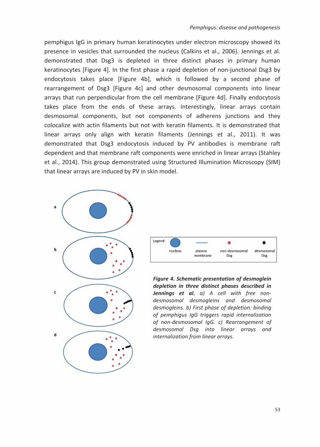
pemphigus IgG in primary human keratinocytes under electron microscopy showed its presence in vesicles that surrounded the nucleus (Calkins et al., 2006). Jennings et al. demonstrated that Dsg3 is depleted in three distinct phases in primary human keratinocytes [Figure 4]. In the first phase a rapid depletion of non-junctional Dsg3 by endocytosis takes place [Figure 4b], which is followed by a second phase of rearrangement of Dsg3 [Figure 4c] and other desmosomal components into linear arrays that run perpendicular from the cell membrane [Figure 4d]. Finally endocytosis takes place from the ends of these arrays. Interestingly, linear arrays contain desmosomal components, but not components of adherens junctions and they colocalize with actin filaments but not with keratin filaments. It is demonstrated that linear arrays only align with keratin filaments (Jennings et al., 2011). It was demonstrated that Dsg3 endocytosis induced by PV antibodies is membrane raft dependent and that membrane raft components were enriched in linear arrays (Stahley et al., 2014). This group demonstrated using Structured Illumination Microscopy (SIM) that linear arrays are induced by PV in skin model.
Figure 4. Schematic presentation of desmoglein depletion in three distinct phases described in Jennings et al. a) A cell with free non-desmosomal desmogleins and desmosomal desmogleins. b) First phase of depletion: binding of pemphigus IgG triggers rapid internalization of non-desmosomal IgG. c) Rearrangement of desmosomal Dsg into linear arrays and internalization from linear arrays.

501164-L-bw-Sokol501164-L-bw-Sokol501164-L-bw-Sokol501164-L-bw-Sokol
Chapter 1C
54
In skin of PF and PV patients the targeted desmoglein and IgG do not have the smooth membrane localization as seen normal human skin, but are relocalized into a clustered pattern (Oktarina et al., 2011). In PV patient skin clusters of Dsg3 contain IgG, while in PF patient skin clusters of Dsg1 contain both IgG and plakoglobin. It is important to notice that these clusters do not contain other desmosomal components and their abundancy is different within patients. In some pemphigus patient biopsies they are highly abundant, in others less, but mostly they are localized in the lower layers of the epidermis. This thus is very different from the situation in cultured cells where all desmosomal components gather in linear arrays.
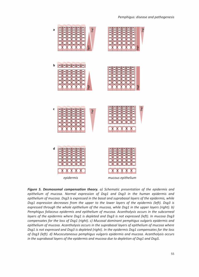
3.5. Desmoglein compensation theory It is well known that levels of blistering within stratified epithelium differ between main forms of pemphigus. In PF blisters are located in the upper layers of the epidermis while in both forms of PV, blisters are suprabasal. Alterations are reported as well. Desmoglein compensation hypothesis explains that level of acantholysis depends on the expression patter of two main pemphigus antigens desmoglein 1 and 3 (Mahoney et al., 2006; Mahoney et al., 1999) [Figure 5]. Dsg1 is expressed through all the layers of epidermis, but its expression decreases from the upper layers to the lower layers, while Dsg3 is expressed in the lower layers of the epidermis and through all the mucosa [Figure 5a]. Targeting Dsg1, PF autoantibodies will induce acantholysis in the upper layer of the epidermis because of the absence of Dsg3 which will not compensate for the loss for Dsg1 [Figure 5b]. Targeting Dsg3 acantholysis will occur only in suprabasal layers of mucosa where Dsg3 is the only Dsg expressed, but not in the skin [Figure 5c]. Skin lesions in PV will occur in the situations when both Dsgs are targeted [Figure 5d]. However this theory is not able to explain why in mcPV acantholysis occurs in the suprabasal layers. Possible explanation is the diffusion of IgG which starts from the lower layers, and suprabasal layers would be the first layers to be reached by IgG. Desmoglein compensation hypothesis is supported by mice model lacking Dsg3. In these mice PF antibodies induced extensive blisters both in the skin and mucosa (Mahoney et al., 2006).
501164-L-bw-Sokol501164-L-bw-Sokol501164-L-bw-Sokol501164-L-bw-Sokol
Pemphigus: disease and pathogenesis
55
Figure 5. Desmosomal compensation theory. a) Schematic presentation of the epidermis and epithelium of mucosa. Normal expression of Dsg1 and Dsg3 in the human epidermis and epithelium of mucosa. Dsg3 is expressed in the basal and suprabasal layers of the epidermis, while Dsg1 expression decreases from the upper to the lower layers of the epidermis (left). Dsg3 is expressed through the whole epithelium of the mucosa, while Dsg1 in the upper layers (right). b) Pemphigus foliaceus epidermis and epithelium of mucosa. Acantholysis occurs in the subcorneal layers of the epidermis where Dsg1 is depleted and Dsg3 is not expressed (left). In mucosa Dsg3 compensates for the loss of Dsg1 (right). c) Mucosal dominant pemphigus vulgaris epidermis and epithelium of mucosa. Acantholysis occurs in the suprabasal layers of epithelium of mucosa where Dsg1 is not expressed and Dsg3 is depleted (right). In the epidermis Dsg1 compensates for the loss of Dsg3 (left). d) Mucocutaneous pemphigus vulgaris epidermis and mucosa. Acantholysis occurs in the suprabasal layers of the epidermis and mucosa due to depletion of Dsg1 and Dsg3.
epidermis mucous epithelium

501164-L-bw-Sokol501164-L-bw-Sokol501164-L-bw-Sokol501164-L-bw-Sokol
Chapter 1C
56
4. Diagnosis of pemphigus
The diagnosis of pemphigus is based on three criteria: clinical features, histopathological findings and immunological tests. The clinical features of the various pemphigus forms have been addressed in the beginning of this chapter. To possibly fit pemphigus the histology should demonstrate acantholysis. Positive evidence of pemphigus can then be obtained by the demonstration of autoantibodies by direct immunofluorescence (analysis of biopsy) or indirect immunofluorescence (analysis of serum for antibodies capable of binding to epithelium). By direct immunofluorescence presence of tissue bound auto-antibodies is shown in 90% patients with pemphigus (Ioannides et al., 2008). By indirect immunofluorescence serum of patients is tested on human skin, monkey esophagus or guinea pig as epithelial a substrate. Enzyme-linked immunosorbent (ELISA) assay detects the presence of anti-Dsg1 and/or anti-Dsg3 IgG in serum.
5. Therapy of pemphigus
If not treated pemphigus has a severe rate of mortality of 90%. There is no specific treatment for pemphigus and current therapies consist of drugs proven beneficial in other auto-immune disorders: corticosteroids, adjuvants, removal of pathogenic antibodies and rituximab.
Corticosteroids. Corticosteroid drugs are chemical modifications of natural glucocorticosteroids. They have inhibitory effects on specific immune responses mediated by T cells and B cells. Corticosteroids diffuse passively through the cell membrane and they bind to the intercellular glucocorticoid receptor. Binding of the drug to this receptor results in translocation of the complex into the nucleus where it effects gene transcription or posttranslational events. Corticosteroids in pemphigus improved prognosis and are mostly used in combination with adjuvants.
Removal of pathogenic antibodies. By immunoabsorption and plasmapheresis pathogenic antibodies can be removed from the serum. Both methods however remove all antibodies what may have unwanted side effects. Experiments are currently going on that investigate the specific removal of anti-Dsg antibodies by adsorption of serum on resins containing recombinant ectodomains of Dsg.
Rituximab. Rituximab is a chimeric monoclonal immunoglobulin G1 antibody that targets B-cell differentiation marker CD-20. The binding of rituximab to cell surface CD20 destructs the lymphocyte by three potential mechanisms: complement-dependent cytotoxicity, apoptosis, or antibody-dependent cytotoxicity. Today it is recognized as first choice for treating pemphigus.

501164-L-bw-Sokol501164-L-bw-Sokol501164-L-bw-Sokol501164-L-bw-Sokol
Pemphigus: disease and pathogenesis
57
Thesis aim
The overall aim of this thesis is to contribute to unraveling the pathomechanism of acantholysis in pemphigus at the ultrastructural level. One of the main questions in pemphigus research remains how the antibodies to desmogleins induce desmosomal dysfunction. As discussed before a number of hypotheses exist next to each other. These are however mainly based on data obtained from experiments with cultured cells and to a lesser degree with data obtained from mouse models. Data on patient skin are scarce while these contain clues on the actual pathogenesis. Moreover the current information on ultrastructural changes in patient skin and the fate of desmosomes are mainly based on older electron microscopy studies when the antigens were still unknown, let alone today’s knowledge on the relationship between the different antigens, antibody specificities and split-level.
To better understand the ultrastructural changes in relation to disease type we will use in Chapter 2 newly developed large scale electron microscopy to investigate skin and mucosa of patients with various forms of pemphigus. As this allows easy and quick analysis of all cells of the epithelium this will give an unbiased detailed approach to study patient tissue. From previous immunofluorescence studies it is known that in patient tissue the IgG induces a sequestering of the targeted desmoglein from the other desmosomal components.
In Chapter 3 we therefore will investigate this phenomenon at the ultrastructural level using immuno-electron microscopy in combination with large scale electron microscopy. To understand how cell and skin observations can be integrated cell culture findings have to be compared with skin findings. All ideas about internalization and cell signaling that evolved from culture models have had a great impact on ideas on pathology for the last fifteen years.
Recently it was hypothesized that there were several phases in the pathogenesis, starting with internalization and then the forming of so-called linear arrays, gatherings of desmosomal components where a second different phase of internalization would take place. In Chapter 4 we aim at investigating how these two phases could possibly contribute to pathogenesis in time, and if possibly we can find observations that would fit observations with skin tissue. A great advancing step in understanding the real time effect of pemphigus IgG on human skin would be to follow the effect of pemphigus IgG on desmosomes in living skin.

501164-L-bw-Sokol501164-L-bw-Sokol501164-L-bw-Sokol501164-L-bw-Sokol
58


![Oral Manifestations of Pemphigus Vulgaris: Clinical ... · bullous pemphigus, and paraneoplastic pemphigus [4]. The differential diagnosis includes other dermatological diseases with](https://static.fdocuments.us/doc/165x107/5cbb138688c9930c5f8bb27d/oral-manifestations-of-pemphigus-vulgaris-clinical-bullous-pemphigus-and.jpg)