University of Groningen Gravimetric Viral Diagnostics: Afzal, … · chemosensors Review...
26
University of Groningen Gravimetric Viral Diagnostics: Afzal, Adeel; Mujahid, Adnan; Schirhagl, Romana; Bajwa, Sadia Z.; Latif, Usman; Feroz, Saima Published in: Chemosensors DOI: 10.3390/chemosensors5010007 IMPORTANT NOTE: You are advised to consult the publisher's version (publisher's PDF) if you wish to cite from it. Please check the document version below. Document Version Publisher's PDF, also known as Version of record Publication date: 2017 Link to publication in University of Groningen/UMCG research database Citation for published version (APA): Afzal, A., Mujahid, A., Schirhagl, R., Bajwa, S. Z., Latif, U., & Feroz, S. (2017). Gravimetric Viral Diagnostics:: QCM Based Biosensors for Early Detection of Viruses. Chemosensors, 5(1), 1-25. DOI: 10.3390/chemosensors5010007 Copyright Other than for strictly personal use, it is not permitted to download or to forward/distribute the text or part of it without the consent of the author(s) and/or copyright holder(s), unless the work is under an open content license (like Creative Commons). Take-down policy If you believe that this document breaches copyright please contact us providing details, and we will remove access to the work immediately and investigate your claim. Downloaded from the University of Groningen/UMCG research database (Pure): http://www.rug.nl/research/portal. For technical reasons the number of authors shown on this cover page is limited to 10 maximum. Download date: 14-06-2018
Transcript of University of Groningen Gravimetric Viral Diagnostics: Afzal, … · chemosensors Review...

University of Groningen
Gravimetric Viral Diagnostics:Afzal, Adeel; Mujahid, Adnan; Schirhagl, Romana; Bajwa, Sadia Z.; Latif, Usman; Feroz,SaimaPublished in:Chemosensors
DOI:10.3390/chemosensors5010007
IMPORTANT NOTE: You are advised to consult the publisher's version (publisher's PDF) if you wish to cite fromit. Please check the document version below.
Document VersionPublisher's PDF, also known as Version of record
Publication date:2017
Link to publication in University of Groningen/UMCG research database
Citation for published version (APA):Afzal, A., Mujahid, A., Schirhagl, R., Bajwa, S. Z., Latif, U., & Feroz, S. (2017). Gravimetric ViralDiagnostics:: QCM Based Biosensors for Early Detection of Viruses. Chemosensors, 5(1), 1-25. DOI:10.3390/chemosensors5010007
CopyrightOther than for strictly personal use, it is not permitted to download or to forward/distribute the text or part of it without the consent of theauthor(s) and/or copyright holder(s), unless the work is under an open content license (like Creative Commons).
Take-down policyIf you believe that this document breaches copyright please contact us providing details, and we will remove access to the work immediatelyand investigate your claim.
Downloaded from the University of Groningen/UMCG research database (Pure): http://www.rug.nl/research/portal. For technical reasons thenumber of authors shown on this cover page is limited to 10 maximum.
Download date: 14-06-2018

chemosensors
Review
Gravimetric Viral Diagnostics: QCM BasedBiosensors for Early Detection of Viruses
Adeel Afzal 1,2,*, Adnan Mujahid 3,*, Romana Schirhagl 4, Sadia Zafar Bajwa 5, Usman Latif 2
and Saima Feroz 6,7
1 Department of Chemistry, College of Science, University of Hafr Al Batin, P.O. Box 1803, Hafr Al Batin 31991,Saudi Arabia
2 Interdisciplinary Research Centre in Biomedical Materials, COMSATS Institute of Information Technology,1.5 KM Defence Road, Off. Raiwind Road, Lahore 54000, Pakistan; [email protected]
3 Institute of Chemistry, University of Punjab, Quaid-i-Azam Campus, Lahore 54590, Pakistan4 Groningen University, University Medical Center Groningen, Antonius Deusinglaan 1, 9713 AV Groningen,
The Netherlands; [email protected] National Institute for Biotechnology and Genetic Engineering (NIBGE), Faisalabad 38000, Pakistan;
[email protected] Department of Biosciences, COMSATS Institute of Information Technology, Park Road, Chak Shahzad,
Islamabad 45550, Pakistan; [email protected] Department of Biology, College of Science, University of Hafr Al Batin, Female Campus, Hafr Al Batin 31991,
Saudi Arabia* Correspondence: [email protected] (A.A.); [email protected] (A.M.);
Tel.: +966-13-720-3426 (ext. 1675) (A.A.)
Academic Editor: Peter LieberzeitReceived: 18 December 2016; Accepted: 9 February 2017; Published: 13 February 2017
Abstract: Viruses are pathogenic microorganisms that can inhabit and replicate in human bodiescausing a number of widespread infectious diseases such as influenza, gastroenteritis, hepatitis,meningitis, pneumonia, acquired immune deficiency syndrome (AIDS) etc. A majority of these viraldiseases are contagious and can spread from infected to healthy human beings. The most importantstep in the treatment of these contagious diseases and to prevent their unwanted spread is to timelydetect the disease-causing viruses. Gravimetric viral diagnostics based on quartz crystal microbalance(QCM) transducers and natural or synthetic receptors are miniaturized sensing platforms that canselectively recognize and quantify harmful virus species. Herein, a review of the label-free QCM virussensors for clinical diagnostics and point of care (POC) applications is presented with major emphasison the nature and performance of different receptors ranging from the natural or synthetic antibodiesto selective macromolecular materials such as DNA and aptamers. A performance comparison ofdifferent receptors is provided and their limitations are discussed.
Keywords: antibodies; aptamer; epitope imprinting; molecularly imprinted polymers; quartz crystalmicrobalance; virus sensor
1. Introduction
Early diagnosis of infectious disease-causing agents such as viruses is essential for clinical andpoint of care (POC) applications [1–3]. Since the viruses have extremely small size and can infect allliving beings, i.e., humans, animals, and plants. Therefore, their precise and accurate detection is ofsignificant interest. Viruses usually live in host’s living cells, where they replicate. Thus, their detectionis a complicated and challenging task due to the complex nature of the medium in which they exist.Since the discovery of first virus, i.e., tobacco mosaic virus (TMV) in 1892, a large number of testsand toolkits have been developed for different types of viruses [4,5]. Especially, modern tools like
Chemosensors 2017, 5, 7; doi:10.3390/chemosensors5010007 www.mdpi.com/journal/chemosensors

Chemosensors 2017, 5, 7 2 of 25
enzyme-linked immunosorbent assay (ELISA) and polymerized chain reaction (PCR) amplification arehighly sensitive and selective protocols for virus recognition [6,7]. Disposable kits having immobilizedenzymes or DNA offer simple and rapid results in minimal cost [8,9]. However, such tests are moresuited for qualitative detection of viruses in remote areas.
Regardless of various established techniques for the detection and investigation of infectiousviruses, the development of smart biosensors and diagnostic devices for quantitative determinationof viruses is mandatory [10–13]. The major impetus in the research and development of such virussensors is their practical features such as the ease of operation, simple and straightforward devicefabrication, possibility to integrate synthetic or natural antibodies, rapid response, high selectivityand cross-sensitivity, portability, and miniaturization capability. It is due to these advantages thatbiosensors have not only been widely employed for virus recognition [14], but have also been appliedfor the detection of microorganism such as pathogenic bacteria [15,16] and yeast [17–20], living bloodcells [21–23], and different diseases biomarkers [24,25].
A typical chemical sensor is comprised of natural and-or synthetic receptors coated on asuitable transducer such as optical, electrochemical, magnetic, gravimetric or mass-sensitive etc. [26].Among many other factors, the selection of a transduction device mainly depends on the nature andphysicochemical properties of the sensitive layer material that undergoes changes when exposed tothe analyte. For instance, in case of an optical sensor, the optical properties of the selective material (orreceptor layer) change due to its interactions with the analyte of interest. However, if the receptor layeris optically inactive, a labeling species or indicator is introduced that translates the receptor-analyteinteractions into a recognizable optical signal [27]. Thus, the inclusion of a labeling molecule to thereceptor layer introduces the desired sensing feature. Since all receptor surfaces do not necessarilypossess the optical, electrochemical, or magnetic characteristics, the addition of a labeling agent causesmore complexity in the receptor’s binding mechanism and may also affect its sensitivity, which areobvious disadvantages of the labeling techniques.
In this bargain, acoustic or mass-sensitive devices offer label-free detection of analytes, becausemass is a fundamental property of any analyte. Thus, the acoustic wave devices are consideredas universal mass-sensitive or gravimetric transducers. These transducers include: (a) the bulkacoustic wave devices such as quartz crystal microbalance (QCM) [28,29]; (b) the surface acousticwave (SAW) resonators [30,31]; and (c) the shear transverse wave (STW) resonators [32,33], whichare frequently studied in a combination with different receptor materials for molecular recognitionand biomedical diagnosis. The mass-sensitive biosensors produce direct shift in frequency in theevent of receptor-analyte interaction and analyte recognition. Among the aforementioned acousticdevices, QCM based chemical and biosensors have been extensively studied for the detection andquantification of a wide range of analytes from small molecules and ions to biological macromoleculesand pathogenic species, e.g., viruses.
Figure 1 shows the principle of a QCM based gravimetric sensor for virus recognition. A QCMtransducer, if coated with a suitably selective receptor, is capable of binding virus particles as well asvirus proteins. In principle, QCM is highly sensitive to the changes in mass loading and dependingupon the nature of analyte (e.g., virus particles) these little changes in mass due to selective virusbinding can be detected easily. The selective receptors therefore play a major role in virus recognitionand quantification due to their ability to interact and bind with viruses. Hereby, we present acomprehensive review of the gravimetric or mass-sensitive viral diagnostic devices based on QCMand a broad spectrum of synthetic and natural receptors.
The key objective of this work is to deliver an up-to-date literature review and critical analysis ofdifferent approaches employed in the design and application of QCM based biosensors and diagnosticdevices for the exclusive recognition of different types of viruses. The substance is divided into twosections in which the basic QCM device design and assembly, and different types of receptor materialswith special emphasis on the nature, fabrication method, and sensor performance are discussed. In the

Chemosensors 2017, 5, 7 3 of 25
end a comparison of different receptor layers is provided along with the benefits and limitations ofeach receptor. This work also highlights the major achievements and the future research viewpoint.
Chemosensors 2017, 5, 7 3 of 24
with the benefits and limitations of each receptor. This work also highlights the major achievements
and the future research viewpoint.
Figure 1. The principle of a QCM-based gravimetric virus sensor. The change in mass in response to
virus binding with the selective receptor is detected as a change in frequency of QCM transducer.
2. Quartz Crystal Microbalance: Transducer Design and Fabrication
This section presents a brief description of the principle, design, and fabrication of QCM-based
gravimetric transducer. During the last few decades, QCM measuring the frequency shift and the
electrochemical QCM, i.e., EQCM [34,35] simultaneously computing the electrochemical and
frequency changes have been broadly designed for sensing different analytes in the gaseous and
liquid phases. The pioneering work of Sauerbrey [36] on calculating the frequency changes as a
function of mass adsorbed or deposited on the surface of gravimetric devices has laid the foundation
for their applications in chemical and biosensors.
In a typical QCM sensor construction, a thin AT-cut quartz wafer having metal electrodes on
opposite sides is connected with oscillator circuit which drives QCM to resonate at characteristic
frequency. A bulk transverse wave is generated that propagates in perpendicular direction of quartz
surface. The crystal surface is usually covered with a certain receptor material which binds with
target analyte and thus leads to increase in the rigid mass of crystal. This results a change in
fundamental resonance frequency of crystal and may be used to precisely determine the adsorbed
mass on crystal surface therefore, referring it as sensor signal.
QCM, due to its extremely high sensitivity towards minor changes in mass, is considered as one
of the best platforms for such applications. If combined with suitable selective material or receptor
layer, QCM transforms into an exceptionally smart tiny balance capable of detecting miniscule
alterations in surface mass, i.e., usually in the range of ng/cm2 [37].
The sensor response of a typical QCM device, i.e., shift in frequency (Δf), is directly
proportional to the square of its fundamental resonating frequency and the mass deposited [36], as
given in the Equation (1) below:
∆𝑓 = −𝑓𝑜2 ∙ ∆𝑚
𝐴√ρ𝑞μ𝑞⁄ (1)
where fo is the fundamental resonant frequency of QCM, Δm is the change in mass, A is the
piezoelectrically active area of QCM in cm2, ρq is the density of quartz (i.e., 2.648 g/cm3), and µ q is the
shear modulus of an AT-cut quartz crystal (i.e., 2.947 × 10¹¹ g/cm·s2).
Figure 1. The principle of a QCM-based gravimetric virus sensor. The change in mass in response tovirus binding with the selective receptor is detected as a change in frequency of QCM transducer.
2. Quartz Crystal Microbalance: Transducer Design and Fabrication
This section presents a brief description of the principle, design, and fabrication of QCM-basedgravimetric transducer. During the last few decades, QCM measuring the frequency shift andthe electrochemical QCM, i.e., EQCM [34,35] simultaneously computing the electrochemical andfrequency changes have been broadly designed for sensing different analytes in the gaseous and liquidphases. The pioneering work of Sauerbrey [36] on calculating the frequency changes as a function ofmass adsorbed or deposited on the surface of gravimetric devices has laid the foundation for theirapplications in chemical and biosensors.
In a typical QCM sensor construction, a thin AT-cut quartz wafer having metal electrodes onopposite sides is connected with oscillator circuit which drives QCM to resonate at characteristicfrequency. A bulk transverse wave is generated that propagates in perpendicular direction of quartzsurface. The crystal surface is usually covered with a certain receptor material which binds with targetanalyte and thus leads to increase in the rigid mass of crystal. This results a change in fundamentalresonance frequency of crystal and may be used to precisely determine the adsorbed mass on crystalsurface therefore, referring it as sensor signal.
QCM, due to its extremely high sensitivity towards minor changes in mass, is considered as one ofthe best platforms for such applications. If combined with suitable selective material or receptor layer,QCM transforms into an exceptionally smart tiny balance capable of detecting miniscule alterations insurface mass, i.e., usually in the range of ng/cm2 [37].
The sensor response of a typical QCM device, i.e., shift in frequency (∆f ), is directly proportionalto the square of its fundamental resonating frequency and the mass deposited [36], as given in theEquation (1) below:
∆ f = − f 2o ·∆m/A
√ρqµq (1)
where fo is the fundamental resonant frequency of QCM, ∆m is the change in mass, A is thepiezoelectrically active area of QCM in cm2, ρq is the density of quartz (i.e., 2.648 g/cm3), and µq is theshear modulus of an AT-cut quartz crystal (i.e., 2.947 × 10¹¹ g/cm·s2).

Chemosensors 2017, 5, 7 4 of 25
The Equation (1) is modified for measurements in liquid medium [38,39], i.e., when one face ofQCM is in contact with a liquid, the liquid loading effect on frequency change can be summarized asgiven in Equation (2) below:
∆ f = − f 3/2o
√ρlηl
πρqµq(2)
where ρl is the density of the liquid, and ηl is the viscosity of the liquid. It is important to mention herewhenever QCM devices are studied in liquid phase, the acoustic properties of liquid medium have tobe taken into account for sensor measurements.
Nonetheless, these equations show that the sensitivity of the QCM device can be improved byincreasing its primary frequency, i.e., if the fundamental frequency of a QCM is doubled, its responsewould be increased by a factor of four. However, this can only be achieved by reducing the thicknessof QCM wafer that will result in mechanically fragile devices. Thus, this parameter practically limitsthe fabrication of devices beyond certain frequency (fo). The other way to increase the sensitivity ofQCM devices is to work with overtones, which require special oscillator circuit having band passfilter for attenuation of harmonic resonances. However, this would also lead to an increase in noiselevel and ultimately making small improvement in signal to noise ratio. Therefore, an increase in thefundamental resonance frequency of crystal is the more appropriate method of tuning sensitivity.
Typically, QCM having frequency in the range of 5–20 MHz are used in liquid phase measurements.Although, a few reports suggest the use of QCM with frequency as high as 110 MHz for liquid phaseoperation [40]. This is remarkably high frequency and the authors report that substantially highdetection limit is achieved in the detection of M13-phages in liquid medium, i.e., the sensitivityimproved by a factor of 200 while using 56 MHz QCM as compared to 19 MHz QCM device [40].
Another important aspect in determining the sensitivity of a QCM device is the nature and designof electrodes on QCM wafers [41,42]. If the diameter of the electrode is bigger, the available surface forselective receptor layer integration would be large, which ultimately leads to higher sensor response.For electrode fabrication on QCM wafer, gold or any other noble metal paste can be applied via screenprinting. Many of the commercially available QCM have fused metal electrodes that afford bettersupport on QCM surface. Furthermore, the integration of receptor layer with QCM electrode shouldbe firm and stable, i.e., the receptor material should not be deteriorated during the fabrication andtesting processes.
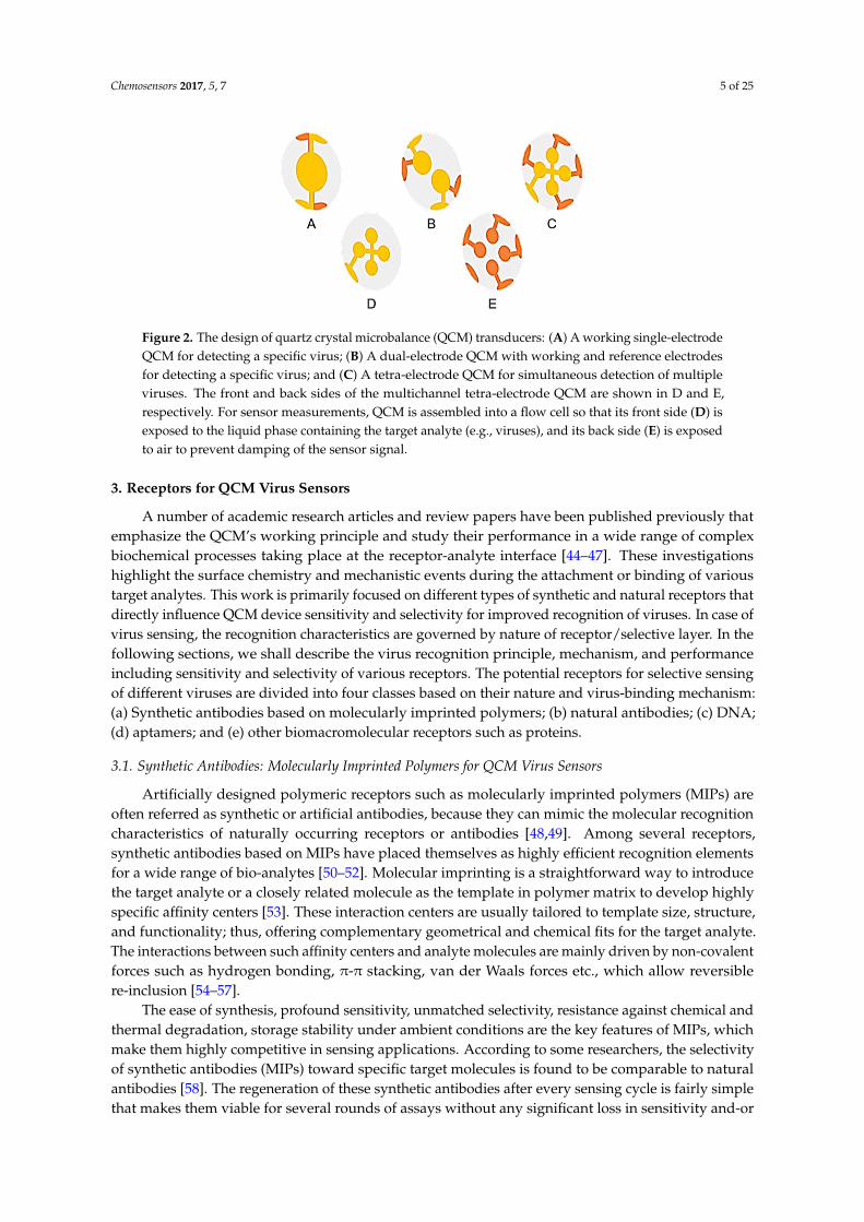
Figure 2 shows the design of a single, dual, and tetra-electrode QCM wafer for detecting viruses.A multichannel QCM having more than one electrodes for sensing different analytes is an innovativedesign [43]. Albeit this unique tetra-electrode, QCM design has not been implemented for simultaneousrecognition of different viruses or virus sub-types, but it could be adopted as a single transducerplatform for different viruses. However, it is important to mention that there should be a minimaldistance between the multiple electrodes to prevent the cross-talk between electrical signals and therebyreducing signal-to-noise ratio for improved sensitivity. Furthermore, in liquid phase measurements,one side of the QCM remains in air to avoid excessive damping problems.
These are some general but imperative design and fabrication parameters that regulate theultimate QCM device sensitivity. The following study emphasizes a variety of synthetic receptorsbased on imprinted polymers, natural antibodies, DNA and aptamers for the detection of differentviruses in combination with QCM devices.
Chemosensors 2017, 5, 7 5 of 25Chemosensors 2017, 5, 7 5 of 24
Figure 2. The design of quartz crystal microbalance (QCM) transducers: (A) A working
single-electrode QCM for detecting a specific virus; (B) A dual-electrode QCM with working and
reference electrodes for detecting a specific virus; and (C) A tetra-electrode QCM for simultaneous
detection of multiple viruses. The front and back sides of the multichannel tetra-electrode QCM are
shown in D and E, respectively. For sensor measurements, QCM is assembled into a flow cell so that
its front side (D) is exposed to the liquid phase containing the target analyte (e.g., viruses), and its
back side (E) is exposed to air to prevent damping of the sensor signal.
3. Receptors for QCM Virus Sensors
A number of academic research articles and review papers have been published previously that
emphasize the QCM’s working principle and study their performance in a wide range of complex
biochemical processes taking place at the receptor-analyte interface [44–47]. These investigations
highlight the surface chemistry and mechanistic events during the attachment or binding of various
target analytes. This work is primarily focused on different types of synthetic and natural receptors
that directly influence QCM device sensitivity and selectivity for improved recognition of viruses. In
case of virus sensing, the recognition characteristics are governed by nature of receptor/selective
layer. In the following sections, we shall describe the virus recognition principle, mechanism, and
performance including sensitivity and selectivity of various receptors. The potential receptors for
selective sensing of different viruses are divided into four classes based on their nature and
virus-binding mechanism: (a) Synthetic antibodies based on molecularly imprinted polymers;
(b) natural antibodies; (c) DNA; (d) aptamers; and (e) other biomacromolecular receptors such as
proteins.
3.1. Synthetic Antibodies: Molecularly Imprinted Polymers for QCM Virus Sensors
Artificially designed polymeric receptors such as molecularly imprinted polymers (MIPs) are
often referred as synthetic or artificial antibodies, because they can mimic the molecular recognition
characteristics of naturally occurring receptors or antibodies [48,49]. Among several receptors,
synthetic antibodies based on MIPs have placed themselves as highly efficient recognition elements
for a wide range of bio-analytes [50–52]. Molecular imprinting is a straightforward way to introduce
the target analyte or a closely related molecule as the template in polymer matrix to develop highly
specific affinity centers [53]. These interaction centers are usually tailored to template size, structure,
and functionality; thus, offering complementary geometrical and chemical fits for the target analyte.
The interactions between such affinity centers and analyte molecules are mainly driven by
non-covalent forces such as hydrogen bonding, π-π stacking, van der Waals forces etc., which allow
reversible re-inclusion [54–57].
The ease of synthesis, profound sensitivity, unmatched selectivity, resistance against chemical
and thermal degradation, storage stability under ambient conditions are the key features of MIPs,
which make them highly competitive in sensing applications. According to some researchers, the
selectivity of synthetic antibodies (MIPs) toward specific target molecules is found to be comparable
to natural antibodies [58]. The regeneration of these synthetic antibodies after every sensing cycle is
Figure 2. The design of quartz crystal microbalance (QCM) transducers: (A) A working single-electrodeQCM for detecting a specific virus; (B) A dual-electrode QCM with working and reference electrodesfor detecting a specific virus; and (C) A tetra-electrode QCM for simultaneous detection of multipleviruses. The front and back sides of the multichannel tetra-electrode QCM are shown in D and E,respectively. For sensor measurements, QCM is assembled into a flow cell so that its front side (D) isexposed to the liquid phase containing the target analyte (e.g., viruses), and its back side (E) is exposedto air to prevent damping of the sensor signal.
3. Receptors for QCM Virus Sensors
A number of academic research articles and review papers have been published previously thatemphasize the QCM’s working principle and study their performance in a wide range of complexbiochemical processes taking place at the receptor-analyte interface [44–47]. These investigationshighlight the surface chemistry and mechanistic events during the attachment or binding of varioustarget analytes. This work is primarily focused on different types of synthetic and natural receptors thatdirectly influence QCM device sensitivity and selectivity for improved recognition of viruses. In case ofvirus sensing, the recognition characteristics are governed by nature of receptor/selective layer. In thefollowing sections, we shall describe the virus recognition principle, mechanism, and performanceincluding sensitivity and selectivity of various receptors. The potential receptors for selective sensingof different viruses are divided into four classes based on their nature and virus-binding mechanism:(a) Synthetic antibodies based on molecularly imprinted polymers; (b) natural antibodies; (c) DNA;(d) aptamers; and (e) other biomacromolecular receptors such as proteins.
3.1. Synthetic Antibodies: Molecularly Imprinted Polymers for QCM Virus Sensors
Artificially designed polymeric receptors such as molecularly imprinted polymers (MIPs) areoften referred as synthetic or artificial antibodies, because they can mimic the molecular recognitioncharacteristics of naturally occurring receptors or antibodies [48,49]. Among several receptors,synthetic antibodies based on MIPs have placed themselves as highly efficient recognition elementsfor a wide range of bio-analytes [50–52]. Molecular imprinting is a straightforward way to introducethe target analyte or a closely related molecule as the template in polymer matrix to develop highlyspecific affinity centers [53]. These interaction centers are usually tailored to template size, structure,and functionality; thus, offering complementary geometrical and chemical fits for the target analyte.The interactions between such affinity centers and analyte molecules are mainly driven by non-covalentforces such as hydrogen bonding, π-π stacking, van der Waals forces etc., which allow reversiblere-inclusion [54–57].
The ease of synthesis, profound sensitivity, unmatched selectivity, resistance against chemical andthermal degradation, storage stability under ambient conditions are the key features of MIPs, whichmake them highly competitive in sensing applications. According to some researchers, the selectivityof synthetic antibodies (MIPs) toward specific target molecules is found to be comparable to naturalantibodies [58]. The regeneration of these synthetic antibodies after every sensing cycle is fairly simplethat makes them viable for several rounds of assays without any significant loss in sensitivity and-or

Chemosensors 2017, 5, 7 6 of 25
selectivity. Furthermore, the cost of MIPs for a typical analyte is in the range of 0.1–0.5 $/mg, whereasthe natural antibodies depending upon the target molecules are offered in 100–1000 $/mg [59].
The foremost advantage of MIPs in chemical and/biosensing applications is the possibility toeasily integrate MIPs with different transducer devices such as QCM [60,61]. Due to their exceptionalcharacteristics, MIPs are considered as potential candidate for virus sensing applications and oftenexhibit high recognition proficiency and selectivity [62]. There are a variety of imprinting strategies formicroorganisms’ detection [63]; however, in the following sub-sections, we shall focus on the selectedapproaches that have been typically employed in QCM based viral diagnostics. Table 1 shows theselected examples of virus-MIP based QCM biosensors and their detection limits.
Table 1. The selected examples of the molecularly imprinted polymers (MIP) as synthetic receptors forQCM-based viral diagnostics.
Receptor Template/Target Polymer DetectionLimit Reference
Soft-lithographically(surface) imprintedMIP
TMV Polyurethane 8 ng/mL [64]
PPOV Polyurethane 5 × 10⁵ virusparticles/mL
[65]
HRV 1A, HRV 2,HRV 14 Polyurethane 100 µg/mL [66]
H5N1, H5N3,H1N1, H1N3,H6N1
Poly(acrylamide-co-methacrylicacid-co-methylmethacrylate-co-N-vinylpyrrolidone)with N,N-(1,2-dihydroxyethylene)bisacrylamide
105
particles/mL[67]
Epitope imprintedMIP
Dengue virusNS1 protein
Poly(acrylic acid-co-acrylamide) with ethyleneglycoldimethylacrylate 1 µg/L [68]
HIV-1 GP41 Polydopamine 2 ng/mL [69]
Plastic antibodyreplica HRV 14 Poly(vinylpyrrolidone-co-methacrylic acid) with
N,N′-(1,2-dihydroxyethylene)bisacrylamide2.5 × 1012 virusparticles/mL
[70]
3.1.1. Soft-Lithography
Owing to the delicate nature of microorganisms, soft-lithography is an appropriate stampingtechnique for patterning polymeric materials, thus generating selective biomimetic surfaces [71,72].It is a surface imprinting technique that has been extensively applied for macromolecular imprintingin the last two decades [73,74]. In this case, a suitable monolayer of targeted microorganism orbiomacromolecule is deposited as template on an inert substrate to make the template-stamp, whichis subsequently pressed on the surface of a pre-polymer. The stamp-pressed pre-polymer layer isallowed to undergo polymerization under moderate conditions, and then washed with mild solventsto remove template from the polymer surface [75]. A schematic of the soft-lithography based surfaceimprinting procedure is shown in Figure 3A. This method allows transfer of precise structural detailsof bioanalytes onto synthetic polymer surface. Consequently, this biomimetic interface is capable ofselectively recognizing target analytes through non-covalent chemical interactions.
Soft-lithography allows faster mass transfer of bioanalytes with enhanced reversibility.Furthermore, the pre-polymer can be coated on QCM electrodes before soft-lithographic stampingprocedure for simple and easy integration of the selective layer with the transducer. Soft-lithographicimprinting has been extensively studied in combination with QCM devices for different types ofviruses. For instance, Dickert et al. [76,77] first patterned a pre-polymer coated on QCM electrodesurface with tobacco mosaic viruses (TMV) using soft-lithography, and the resulting sensor wasreported to be highly sensitivity towards TMV. The device also exhibited reversible sensor signalindicating the complete removal of adsorbed species and perfect layer regeneration for further analyses.In a subsequent work [64], the sensitivity of QCM devices coated with surface imprinted polyurethanemonolayer was substantially improved and the selectivity was investigated revealing high affinityof stamped polymers for the targeted virus. The detection of parapox ovis virus (PPOV) is another

Chemosensors 2017, 5, 7 7 of 25
example of the sensing platform using a combination of QCM and soft-lithographically patternedpolymer surfaces [65].
In a later study [66], this strategy was extended to imprint different strains of human rhinovirus(HRV) as three serotypes, i.e., HRV 1A, HRV 2, and HRV 14, were used as the templates to producethe respective biomimetic selective layers on QCM. The sensing measurements revealed that eachtype of imprinted surface offered the highest sensor response to its templated strain virus as shown inFigure 3B, thus proving excellent selectivity. Similarly, the surface imprinted polymers designed bysoft-lithography procedure are successfully employed in QCM-based viral diagnostics for influenzaA virus sub-types, i.e., H5N1, H5N3, H1N1, H1N3, and H6N1 [67,78]. The fabricated sensorsdemonstrated considerable selectivity for screening of influenza A virus sub-types as each sub-typewas best recognized by its own imprinted surface. The reported detection limit was 105 virusparticles/mL [67]. In view of these examples, it is obvious that soft-lithography approach to designsurface MIPs makes them capable of selectively distinguishing different serotypes of the same virus,which may be termed as intra-group selectivity.
Chemosensors 2017, 5, 7 7 of 24
virus (PPOV) is another example of the sensing platform using a combination of QCM and
soft-lithographically patterned polymer surfaces [65].
In a later study [66], this strategy was extended to imprint different strains of human rhinovirus
(HRV) as three serotypes, i.e., HRV 1A, HRV 2, and HRV 14, were used as the templates to produce
the respective biomimetic selective layers on QCM. The sensing measurements revealed that each
type of imprinted surface offered the highest sensor response to its templated strain virus as shown
in Figure 3B, thus proving excellent selectivity. Similarly, the surface imprinted polymers designed
by soft-lithography procedure are successfully employed in QCM-based viral diagnostics for
influenza A virus sub-types, i.e., H5N1, H5N3, H1N1, H1N3, and H6N1 [67,78]. The fabricated
sensors demonstrated considerable selectivity for screening of influenza A virus sub-types as each
sub-type was best recognized by its own imprinted surface. The reported detection limit was 105
virus particles/mL [67]. In view of these examples, it is obvious that soft-lithography approach to
design surface MIPs makes them capable of selectively distinguishing different serotypes of the
same virus, which may be termed as intra-group selectivity.
Figure 3. (A) A schematic representation of soft-lithography: A stamp of assembled virus particles is
pressed onto the pre-polymer coated on a QCM electrode. After polymerization, the stamp is
removed and the template is washed away to obtain the surface imprinted synthetic antibodies;
(B) Relative sensor response of different human rhinovirus (HRV) serotype-imprinted polymers
towards different strains of HRV. It is evident that each sub-type of HRV is preferentially identified
by the respective MIP surface. Figure 3B is reproduced with permission from Jenik et al. [66].
Copyright by the American Chemical Society, 2009.
3.1.2. Epitope Imprinting
The living cells, viruses and other microorganisms are sometimes difficult to fit and imprint as
templates due to their large size and the lack of binding site accessibility [79]. To overcome such
challenges in biomacromolecular imprinting, epitope imprinting is introduced as an efficient
macromolecular imprinting method, especially for proteins [80,81]. Epitope imprinting is also a
method of choice for imprinting and recognition of viral proteins and different species of viruses.
This technique, unlike whole-cell imprinting or soft-lithographic patterning of viruses on polymer
surfaces, introduces a small peptide fragment as the template for imprinting polymers. The resultant
imprinted material interacts with target protein through its epitope, i.e., a small part of the antigen.
A schematic of the epitope imprinting process in shown in Figure 4A.
The natural antibody-antigen binding inspired epitope imprinting approach is useful in
producing artificial receptors for bioanalytes recognition. Albeit it has been extensively studied for
protein recognition [59,82], some researchers also investigated its applications in diagnosis of viruses
and viral proteins. The selection of peptide fragment as template and its sequence are important to
achieve improved recognition as the surface groups on the epitope yield specific structural and
Figure 3. (A) A schematic representation of soft-lithography: A stamp of assembled virus particles ispressed onto the pre-polymer coated on a QCM electrode. After polymerization, the stamp is removedand the template is washed away to obtain the surface imprinted synthetic antibodies; (B) Relativesensor response of different human rhinovirus (HRV) serotype-imprinted polymers towards differentstrains of HRV. It is evident that each sub-type of HRV is preferentially identified by the respective MIPsurface. Figure 3B is reproduced with permission from Jenik et al. [66]. Copyright by the AmericanChemical Society, 2009.
3.1.2. Epitope Imprinting
The living cells, viruses and other microorganisms are sometimes difficult to fit and imprintas templates due to their large size and the lack of binding site accessibility [79]. To overcomesuch challenges in biomacromolecular imprinting, epitope imprinting is introduced as an efficientmacromolecular imprinting method, especially for proteins [80,81]. Epitope imprinting is also amethod of choice for imprinting and recognition of viral proteins and different species of viruses. Thistechnique, unlike whole-cell imprinting or soft-lithographic patterning of viruses on polymer surfaces,introduces a small peptide fragment as the template for imprinting polymers. The resultant imprintedmaterial interacts with target protein through its epitope, i.e., a small part of the antigen. A schematicof the epitope imprinting process in shown in Figure 4A.
The natural antibody-antigen binding inspired epitope imprinting approach is useful in producingartificial receptors for bioanalytes recognition. Albeit it has been extensively studied for proteinrecognition [59,82], some researchers also investigated its applications in diagnosis of viruses and viralproteins. The selection of peptide fragment as template and its sequence are important to achieve

Chemosensors 2017, 5, 7 8 of 25
improved recognition as the surface groups on the epitope yield specific structural and functionalmemory in polymers. Theses surface groups are suited for epitope imprinting due to their accessibilityand functionality that leads to the selective recognition of targeted species.
Tai and coworkers [68,83] adopted the epitope imprinting approach for QCM-based serologicalassay of dengue virus, i.e., mosquito-borne virus, infections. They used acrylic polymer systemfor epitope imprinting of 15-mer peptide that is known as the linear epitope of dengue virus NS1protein [68]. The resulting epitope-imprinted polymer (EIP) QCM sensor achieved the direct andquantitative detection of the NS1 protein, as shown in Figure 4B. Furthermore, the EIP-QCM sensorsexhibited a good correlation with enzyme-linked immunosorbent assay (ELISA) indicating reliabledetection of dengue virus. The sensor also correctly identified samples for the presence or absenceof virus species. The EIP sensor coatings remained stable for a period of one month and could beregenerated five times. The analysis time of one sample was 20–30 min with the detection limit of1–10 µg/L. Such devices are useful in clinical diagnosis of dengue virus infections as they prevent theuse of monoclonal antibodies [84,85].
Chemosensors 2017, 5, 7 8 of 24
functional memory in polymers. Theses surface groups are suited for epitope imprinting due to their
accessibility and functionality that leads to the selective recognition of targeted species.
Tai and coworkers [68,83] adopted the epitope imprinting approach for QCM-based serological
assay of dengue virus, i.e., mosquito-borne virus, infections. They used acrylic polymer system for
epitope imprinting of 15-mer peptide that is known as the linear epitope of dengue virus NS1
protein [68]. The resulting epitope-imprinted polymer (EIP) QCM sensor achieved the direct and
quantitative detection of the NS1 protein, as shown in Figure 4B. Furthermore, the EIP-QCM sensors
exhibited a good correlation with enzyme-linked immunosorbent assay (ELISA) indicating reliable
detection of dengue virus. The sensor also correctly identified samples for the presence or absence of
virus species. The EIP sensor coatings remained stable for a period of one month and could be
regenerated five times. The analysis time of one sample was 20–30 min with the detection limit of
1–10 µg/L. Such devices are useful in clinical diagnosis of dengue virus infections as they prevent the
use of monoclonal antibodies [84,85].
Figure 4. (A) A schematic representation of the epitope imprinting procedure; (B) The frequency
shifts as a function of the epitope-imprinted polymer’s (EIP) complexation with dengue virus NS1
protein. The EIP-coated QCM chip is capable to bind NS1 protein in the range of 5 ng/mL to
50 μg/mL with 100 μL injection of the unpurified NS1 protein solution. Figure 4B is reproduced with
permission from Tai et al. [68]. Copyright by the American Chemical Society, 2005.
Chen and coworkers [69] used a synthetic peptide, i.e., 35 amino acid residue as template to
developed epitope-imprinted polydopamine coated on QCM for human immunodeficiency virus
(HIV) type-1 detection. The selection of epitope was made by similar amino acid residues of HIV-1
glycoprotein 41, i.e., amino acid numbers 579–613. The device exhibited high sensitivity and
selectivity for HIV-1 glycoprotein (GP41) with the estimated detection limit of 2 ng/mL. The
highpoint of this approach is its applicability to real samples, where the sensor exhibits recovery
values of 86.5%–94.1% of HIV-1 GP41 in spiked human urine. These findings are interesting in view of
their potential for clinical tests of target viruses and-or viral proteins without using labeling indicator.
3.1.3. Plastic Antibody Replica
Dickert and coworkers [70,86] employed an innovative strategy to produce plastic antibody
replica for virus sensing via two-step soft-lithography technique. Firstly, they used natural
antibodies for a specific target as the template in synthetic polymer system, which could be
Figure 4. (A) A schematic representation of the epitope imprinting procedure; (B) The frequency shiftsas a function of the epitope-imprinted polymer’s (EIP) complexation with dengue virus NS1 protein.The EIP-coated QCM chip is capable to bind NS1 protein in the range of 5 ng/mL to 50 µg/mL with100 µL injection of the unpurified NS1 protein solution. Figure 4B is reproduced with permission fromTai et al. [68]. Copyright by the American Chemical Society, 2005.
Chen and coworkers [69] used a synthetic peptide, i.e., 35 amino acid residue as template todeveloped epitope-imprinted polydopamine coated on QCM for human immunodeficiency virus(HIV) type-1 detection. The selection of epitope was made by similar amino acid residues of HIV-1glycoprotein 41, i.e., amino acid numbers 579–613. The device exhibited high sensitivity and selectivityfor HIV-1 glycoprotein (GP41) with the estimated detection limit of 2 ng/mL. The highpoint of thisapproach is its applicability to real samples, where the sensor exhibits recovery values of 86.5%–94.1%of HIV-1 GP41 in spiked human urine. These findings are interesting in view of their potential forclinical tests of target viruses and-or viral proteins without using labeling indicator.
3.1.3. Plastic Antibody Replica
Dickert and coworkers [70,86] employed an innovative strategy to produce plastic antibodyreplica for virus sensing via two-step soft-lithography technique. Firstly, they used natural antibodies

Chemosensors 2017, 5, 7 9 of 25
for a specific target as the template in synthetic polymer system, which could be precipitated inappropriate solvent to produce nanoparticles. Then, natural antibody-imprinted nanoparticles wereused as the master stamp on the surface of pre-polymer already coated on QCM electrode. Thus,two-step soft-lithography leads to the formation of precisely patterned polymer interface that perfectlymimics natural antibodies used as template.
The authors compared QCM sensor results of natural antibodies and their plastic antibody replicafor HRV (the template and target analyte) and foot-and-mouth disease virus (FMDV; the interferent),as shown in Figure 5. Astonishingly, plastic antibody replica exhibited higher response, i.e., six timeshigher, as compared to natural receptors. It was suggested that high sensitivity of plastic antibodyreplica might be attributed to the higher surface area of MIP-nanoparticles. Furthermore, the sensorresponse of both natural and synthetic receptors towards FMDV was negligible showing very lowcross-sensitivity and excellent selectivity. This approach provides an alternative and innovative way toprecisely pattern synthetic receptors that are plastic copies of natural antibodies and that can be usedin microorganisms’ detection and bioassay.
Chemosensors 2017, 5, 7 9 of 24
precipitated in appropriate solvent to produce nanoparticles. Then, natural antibody-imprinted
nanoparticles were used as the master stamp on the surface of pre-polymer already coated on QCM
electrode. Thus, two-step soft-lithography leads to the formation of precisely patterned polymer
interface that perfectly mimics natural antibodies used as template.
The authors compared QCM sensor results of natural antibodies and their plastic antibody
replica for HRV (the template and target analyte) and foot-and-mouth disease virus (FMDV; the
interferent), as shown in Figure 5. Astonishingly, plastic antibody replica exhibited higher
response, i.e., six times higher, as compared to natural receptors. It was suggested that high
sensitivity of plastic antibody replica might be attributed to the higher surface area of
MIP-nanoparticles. Furthermore, the sensor response of both natural and synthetic receptors
towards FMDV was negligible showing very low cross-sensitivity and excellent selectivity. This
approach provides an alternative and innovative way to precisely pattern synthetic receptors that
are plastic copies of natural antibodies and that can be used in microorganisms’ detection
and bioassay.
Figure 5. A comparison of the sensor response of natural antibodies and their synthetic replica
prepared via imprinting for human rhinovirus (HRV) and foot-and-mouth disease virus (FMDV)
recognition. HRV is the template as well as the target analyte, while FMDV is used for
cross-sensitivity measurements. Reproduced with permission from Schirhagl et al. [70]. Copyright
by WILEY-VCH Verlag GmbH & Co. KGaA, Weinheim, 2009.
3.2. Natural Antibodies for QCM Virus Sensors
This section reviews strategies for fast and reliable detection of viruses with the help of natural
antibody (NAb)-based QCM biosensors. NAb-QCM biosensors for viral recognition have versatile
advantageous features compared to other sensing devices. They are eminently suitable for the rapid
diagnosis of viruses with high sensitivity and specificity. Natural antibodies are proteins produced
by the immune system to protect the body by identifying and neutralizing pathogens [87]. Natural
antibodies function as the selective receptors in gravimetric viral diagnostic devices and offer
site-specific affinity with either viral shell proteins or with the proteins released by the viruses when
they enter the host body. In fact, the antigenic amino acids occur as patches and function as protein
binding sites on the virus coat.
The accumulation of bioanalyte (the targeted virus) on the surface of QCM biosensor coated
with NAb results in the resonant frequency shift that is used as the sensor signals. The sensitivity of
the NAb-QCM biosensor depends on the type of antibody used and its orientation on the electrode
surface. Although considerable data has been documented on NAb-based methods for the detection
of pathogenic viruses by conventional techniques [88,89], relatively little work has been done on
specifically utilizing NAb-QCM biosensors as viral diagnostics. Nonetheless, NAb-QCM based
detection systems have been developed to detect harmful virus species causing fatal diseases to
Figure 5. A comparison of the sensor response of natural antibodies and their synthetic replicaprepared via imprinting for human rhinovirus (HRV) and foot-and-mouth disease virus (FMDV)recognition. HRV is the template as well as the target analyte, while FMDV is used for cross-sensitivitymeasurements. Reproduced with permission from Schirhagl et al. [70]. Copyright by Wiley-VCHVerlag GmbH & Co. KGaA, Weinheim, Germany, 2009.
3.2. Natural Antibodies for QCM Virus Sensors
This section reviews strategies for fast and reliable detection of viruses with the help of naturalantibody (NAb)-based QCM biosensors. NAb-QCM biosensors for viral recognition have versatileadvantageous features compared to other sensing devices. They are eminently suitable for therapid diagnosis of viruses with high sensitivity and specificity. Natural antibodies are proteinsproduced by the immune system to protect the body by identifying and neutralizing pathogens [87].Natural antibodies function as the selective receptors in gravimetric viral diagnostic devices and offersite-specific affinity with either viral shell proteins or with the proteins released by the viruses whenthey enter the host body. In fact, the antigenic amino acids occur as patches and function as proteinbinding sites on the virus coat.
The accumulation of bioanalyte (the targeted virus) on the surface of QCM biosensor coatedwith NAb results in the resonant frequency shift that is used as the sensor signals. The sensitivity ofthe NAb-QCM biosensor depends on the type of antibody used and its orientation on the electrodesurface. Although considerable data has been documented on NAb-based methods for the detectionof pathogenic viruses by conventional techniques [88,89], relatively little work has been done onspecifically utilizing NAb-QCM biosensors as viral diagnostics. Nonetheless, NAb-QCM based

Chemosensors 2017, 5, 7 10 of 25
detection systems have been developed to detect harmful virus species causing fatal diseases to humanbeings, plants, and animals in the last two decades [90–92]. Table 2 shows the selected examples ofNAb-QCM biosensors for viruses.
Table 2. The selected examples of the natural antibodies (NAb) as receptors for QCM-basedviral diagnostics.
Receptor Target Fabrication/Immobilization Method Detection Limit Reference
Anti-H5 NAb AIV H5N1 Anti-H5 attached to nanobeads immobilizedon 16-mercaptohexadecanoic acid monolayer 0.128 HAU [93]
Anti-MCMV NAb MCMV3-mercaptopropanoic acid and11-mercaptoundecanoic acid (10:1 ratio)crosslinked with anti-MCMV
250 ng/mL [94]
Monoclonal anti-CIVNP antibody CIV H3N2 ProLinker™ B immobilized anti-CIV
monoclonal antibody 14 nM [95]
Secondary antibodies HepBV Secondary antibodies linked throughcarboxylated hyper-branched polymer 2 ng/mL [96]
For instance, Li et al. [93] fabricated a QCM immunosensor using magnetic nanobeads andpolyclonal anti-H5 antibodies for the detection of avian influenza virus (AIV) H5N1 in agricultural,food, environmental, and clinical samples. The surface antigen hemagglutinin (HA) was depositedon QCM through self-assembled monolayer of 16-mercaptohexadecanoic acid. The target AI H5N1viruses were then captured by the immobilized anti-H5 antibodies attached to magnetic nanobeads.The addition of magnetic nanobeads coated with anti-H5 results in amplification of binding reactionbetween antibody and virus antigens. This anti-H5 coated NAb-QCM immunosensor exhibited goodsensitivity (limit of detection: 0.128 HAU [97]) due to nanobeads amplification. It was also noticed thatsignal amplification was more significant at lower virus concentration that could be favorable for earlystage screening of H5N1 virus.
This setup was also used to quantify viruses from chicken tracheal swab samples. Authors did notobserve any significant interference with AIV subtypes H3N2, H2N2, and H4N8 [93]. It is imperativeto mention here that by increasing the number of active binding sites at sensor interfacial coatings,amplified mass loading can be achieved by QCM based immunosensors. In this perspective, thefabrication of nanomaterials with NAb could lead to enhanced mass deposition on electrode surfacethus, amplifying sensor response. This has also been demonstrated in a report [98], where authorsdeveloped Au nanoparticles functionalized with antibodies to amplify the recognition process. It hasbeen shown that Au modified receptors increase the detection limit to three orders of magnitude higheras compared to direct QCM sensing without amplification.
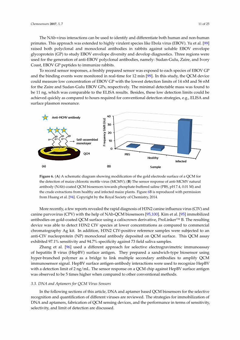
Huang et al. [94] reported a NAb-QCM biosensor for the selective recognition of maize chloroticmottle virus (MCMV). They mixed 3-mercaptopropanoic acid and 11-mercaptoundecanoic acid(10:1 ratio) to prepare a self-assembled monolayer on QCM gold electrodes. Then, anti-MCMVantibody was used as the cross-linking agent for specific recognition of MCMV. The MCMV wascultivated in corn, and the infected tissues were collected after 14 days for tests. Figure 6A shows thesurface modification of QCM electrodes to fabricate NAb-QCM biosensor for MCMV detection.
The anti-MCMV coated NAb-QCM biosensor successfully detected MCMV in the concentrationrange of 250 ng/mL to 10 µg/mL. The detection limit was reported to be 250 ng/mL, which was veryclose to that achieved by conventional ELISA method. The device was also found to be 45 fold moreselective towards MCMV as compared to other viruses such as maize dwarf mosaic virus (MDMV),sugarcane mosaic virus (SCMV), and wheat streak mosaic virus (WSMV) at the same concentration.Furthermore, the NAb-QCM biosensor was not only capable to recognize MCMV in a mixture ofMCMV, MDMV, WSMV, and SCMV, but also distinguished between healthy and infected corn leafsamples with high accuracy [94], as shown in Figure 6B.
Chemosensors 2017, 5, 7 11 of 25
The NAb-virus interactions can be used to identify and differentiate both human and non-humanprimates. This approach was extended to highly virulent species like Ebola virus (EBOV). Yu et al. [99]raised both polyclonal and monoclonal antibodies in rabbits against soluble EBOV envelopeglycoprotein (GP) to study EBOV envelope diversity and develop diagnostics. Three regions wereused for the generation of anti-EBOV polyclonal antibodies, namely: Sudan-Gulu, Zaire, and IvoryCoast, EBOV GP peptides to immunize rabbits.
To record sensor responses, a freshly prepared sensor was exposed to each species of EBOV GPand the binding events were monitored in real-time for 12 min [99]. In this study, the QCM devicecould measure low concentration of EBOV GP with the lowest detection limits of 14 nM and 56 nMfor the Zaire and Sudan-Gulu EBOV GPs, respectively. The minimal detectable mass was found tobe 11 ng, which was comparable to the ELISA results. Besides, these low detection limits could beachieved quickly as compared to hours required for conventional detection strategies, e.g., ELISA andsurface plasmon resonance.
Chemosensors 2017, 5, 7 11 of 24
regions were used for the generation of anti-EBOV polyclonal antibodies, namely: Sudan-Gulu,
Zaire, and Ivory Coast, EBOV GP peptides to immunize rabbits.
To record sensor responses, a freshly prepared sensor was exposed to each species of EBOV GP
and the binding events were monitored in real-time for 12 min [99]. In this study, the QCM device
could measure low concentration of EBOV GP with the lowest detection limits of 14 nM and 56 nM
for the Zaire and Sudan-Gulu EBOV GPs, respectively. The minimal detectable mass was found to
be 11 ng, which was comparable to the ELISA results. Besides, these low detection limits could be
achieved quickly as compared to hours required for conventional detection strategies, e.g., ELISA
and surface plasmon resonance.
Figure 6. (A) A schematic diagram showing modification of the gold electrode surface of a QCM for
the detection of maize chlorotic mottle virus (MCMV); (B) The sensor response of anti-MCMV
natural antibody (NAb)-coated QCM biosensors towards phosphate-buffered saline (PBS, pH 7.4,
0.01 M) and the crude extractions from healthy and infected maize plants. Figure 6B is reproduced
with permission from Huang et al. [94]. Copyright by the Royal Society of Chemistry, 2014.
More recently, a few reports revealed the rapid diagnosis of H3N2 canine influenza virus (CIV)
and canine parvovirus (CPV) with the help of NAb-QCM biosensors [95,100]. Kim et al. [95]
immobilized antibodies on gold-coated QCM surface using a calixcrown derivative, ProLinker™ B.
The resulting device was able to detect H3N2 CIV species at lower concentrations as compared to
commercial chromatography Ag kit. In addition, H3N2 CIV-positive reference samples were
subjected to an anti-CIV nucleoprotein (NP) monoclonal antibody deposited on QCM surface. This
QCM assay exhibited 97.1% sensitivity and 94.7% specificity against 73 field saliva samples.
Zhang et al. [96] used a different approach for selective electrogravimetric immunoassay of
hepatitis B virus (HepBV) surface antigen. They prepared a sandwich-type biosensor using
hyper-branched polymer as a bridge to link multiple secondary antibodies to amplify QCM
immunosensor signal. HepBV surface antigen-antibody interactions were used to recognize HepBV
with a detection limit of 2 ng/mL. The sensor response on a QCM chip against HepBV surface
antigen was observed to be 5 times higher when compared to other conventional methods.
3.3. DNA and Aptamers for QCM Virus Sensors
In the following sections of this article, DNA and aptamer based QCM biosensors for the
selective recognition and quantification of different viruses are reviewed. The strategies for
immobilization of DNA and aptamers, fabrication of QCM sensing devices, and the performance in
terms of sensitivity, selectivity, and limit of detection are discussed.
One of the most important challenges in utilizing DNA and aptamers as the selective receptor
layers in QCM-based virus sensors is their anchoring to the surface of QCM electrode. This requires
the modification of Au-electrodes by functional thiols which can bind to the electrode surface
Figure 6. (A) A schematic diagram showing modification of the gold electrode surface of a QCM forthe detection of maize chlorotic mottle virus (MCMV); (B) The sensor response of anti-MCMV naturalantibody (NAb)-coated QCM biosensors towards phosphate-buffered saline (PBS, pH 7.4, 0.01 M) andthe crude extractions from healthy and infected maize plants. Figure 6B is reproduced with permissionfrom Huang et al. [94]. Copyright by the Royal Society of Chemistry, 2014.
More recently, a few reports revealed the rapid diagnosis of H3N2 canine influenza virus (CIV) andcanine parvovirus (CPV) with the help of NAb-QCM biosensors [95,100]. Kim et al. [95] immobilizedantibodies on gold-coated QCM surface using a calixcrown derivative, ProLinker™ B. The resultingdevice was able to detect H3N2 CIV species at lower concentrations as compared to commercialchromatography Ag kit. In addition, H3N2 CIV-positive reference samples were subjected to ananti-CIV nucleoprotein (NP) monoclonal antibody deposited on QCM surface. This QCM assayexhibited 97.1% sensitivity and 94.7% specificity against 73 field saliva samples.
Zhang et al. [96] used a different approach for selective electrogravimetric immunoassayof hepatitis B virus (HepBV) surface antigen. They prepared a sandwich-type biosensor usinghyper-branched polymer as a bridge to link multiple secondary antibodies to amplify QCMimmunosensor signal. HepBV surface antigen-antibody interactions were used to recognize HepBVwith a detection limit of 2 ng/mL. The sensor response on a QCM chip against HepBV surface antigenwas observed to be 5 times higher when compared to other conventional methods.
3.3. DNA and Aptamers for QCM Virus Sensors
In the following sections of this article, DNA and aptamer based QCM biosensors for the selectiverecognition and quantification of different viruses are reviewed. The strategies for immobilization ofDNA and aptamers, fabrication of QCM sensing devices, and the performance in terms of sensitivity,selectivity, and limit of detection are discussed.
Chemosensors 2017, 5, 7 12 of 25
One of the most important challenges in utilizing DNA and aptamers as the selective receptorlayers in QCM-based virus sensors is their anchoring to the surface of QCM electrode. This requiresthe modification of Au-electrodes by functional thiols which can bind to the electrode surface forminga self-assembled monolayer at thiol-end [101,102], while the next-end functionality, e.g., a carboxylicacid, can bind to the receptor DNA and aptamers.
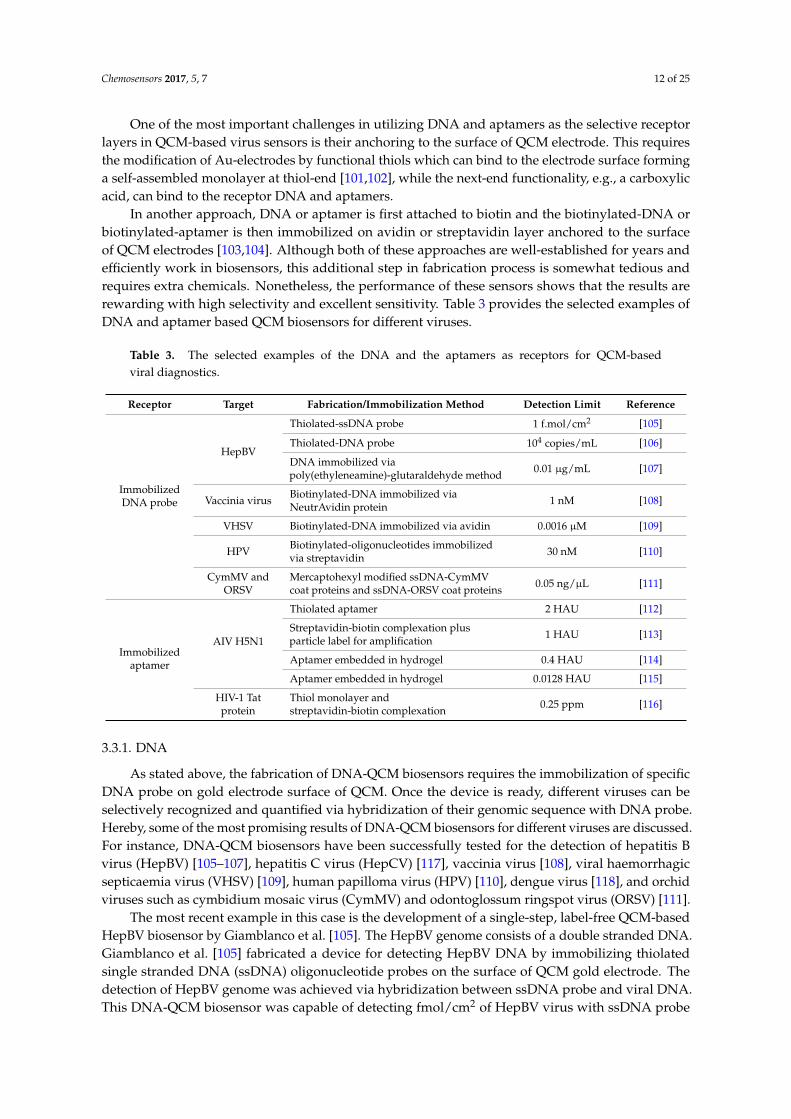
In another approach, DNA or aptamer is first attached to biotin and the biotinylated-DNA orbiotinylated-aptamer is then immobilized on avidin or streptavidin layer anchored to the surfaceof QCM electrodes [103,104]. Although both of these approaches are well-established for years andefficiently work in biosensors, this additional step in fabrication process is somewhat tedious andrequires extra chemicals. Nonetheless, the performance of these sensors shows that the results arerewarding with high selectivity and excellent sensitivity. Table 3 provides the selected examples ofDNA and aptamer based QCM biosensors for different viruses.
Table 3. The selected examples of the DNA and the aptamers as receptors for QCM-basedviral diagnostics.
Receptor Target Fabrication/Immobilization Method Detection Limit Reference
ImmobilizedDNA probe
HepBV
Thiolated-ssDNA probe 1 f.mol/cm2 [105]
Thiolated-DNA probe 104 copies/mL [106]
DNA immobilized viapoly(ethyleneamine)-glutaraldehyde method 0.01 µg/mL [107]
Vaccinia virus Biotinylated-DNA immobilized viaNeutrAvidin protein 1 nM [108]
VHSV Biotinylated-DNA immobilized via avidin 0.0016 µM [109]
HPV Biotinylated-oligonucleotides immobilizedvia streptavidin 30 nM [110]
CymMV andORSV
Mercaptohexyl modified ssDNA-CymMVcoat proteins and ssDNA-ORSV coat proteins 0.05 ng/µL [111]
Immobilizedaptamer
AIV H5N1
Thiolated aptamer 2 HAU [112]
Streptavidin-biotin complexation plusparticle label for amplification 1 HAU [113]
Aptamer embedded in hydrogel 0.4 HAU [114]
Aptamer embedded in hydrogel 0.0128 HAU [115]
HIV-1 Tatprotein
Thiol monolayer andstreptavidin-biotin complexation 0.25 ppm [116]
3.3.1. DNA
As stated above, the fabrication of DNA-QCM biosensors requires the immobilization of specificDNA probe on gold electrode surface of QCM. Once the device is ready, different viruses can beselectively recognized and quantified via hybridization of their genomic sequence with DNA probe.Hereby, some of the most promising results of DNA-QCM biosensors for different viruses are discussed.For instance, DNA-QCM biosensors have been successfully tested for the detection of hepatitis Bvirus (HepBV) [105–107], hepatitis C virus (HepCV) [117], vaccinia virus [108], viral haemorrhagicsepticaemia virus (VHSV) [109], human papilloma virus (HPV) [110], dengue virus [118], and orchidviruses such as cymbidium mosaic virus (CymMV) and odontoglossum ringspot virus (ORSV) [111].
The most recent example in this case is the development of a single-step, label-free QCM-basedHepBV biosensor by Giamblanco et al. [105]. The HepBV genome consists of a double stranded DNA.Giamblanco et al. [105] fabricated a device for detecting HepBV DNA by immobilizing thiolatedsingle stranded DNA (ssDNA) oligonucleotide probes on the surface of QCM gold electrode. Thedetection of HepBV genome was achieved via hybridization between ssDNA probe and viral DNA.This DNA-QCM biosensor was capable of detecting fmol/cm2 of HepBV virus with ssDNA probe

Chemosensors 2017, 5, 7 13 of 25
density of ~4 × 10¹² molecules per cm2 without using any amplification or labeling technique. ThessDNA probe density determines the sensitivity and selectivity of the fabricated QCM-DNA biosensor,because the diffusion of target HepBV genome to the hybridization site, its conformation, and spacingbetween the ssDNA probes are dependent on probe density [119,120]. Thus, the receptor surfaces withlower DNA probe density may produce a strongly selective and irreversible adsorption and vice versa.
Earlier, Skladal et al. [117] fabricated a QCM-DNA biosensor by immobilizing biotinylated-DNA(via interaction with avidin or streptavidin) on QCM surface for detecting HepCV in serum. Theimmobilization of DNA probe was achieved by modifying gold electrode with cysteamine andsubsequent activation with glutaraldehyde followed by anchoring streptavidin or avidin. They foundout that immobilization efficiency of avidin was significantly higher as compared to streptavidin,and the biotinylated-DNA-avidin based QCM was regenerated 30 times without any loss of bindingcapacity. Figure 7 summarizes the mechanism for the detection of viruses using DNA-QCM biosensors.
Chemosensors 2017, 5, 7 13 of 24
Earlier, Skladal et al. [117] fabricated a QCM-DNA biosensor by immobilizing
biotinylated-DNA (via interaction with avidin or streptavidin) on QCM surface for detecting HepCV
in serum. The immobilization of DNA probe was achieved by modifying gold electrode with
cysteamine and subsequent activation with glutaraldehyde followed by anchoring streptavidin or
avidin. They found out that immobilization efficiency of avidin was significantly higher as
compared to streptavidin, and the biotinylated-DNA-avidin based QCM was regenerated 30 times
without any loss of binding capacity. Figure 7 summarizes the mechanism for the detection of
viruses using DNA-QCM biosensors.
Figure 7. A schematic representation of the DNA-QCM virus sensor. DNA probe is attached to the
QCM gold electrodes through an immobilizer that often uses thiol linkages, avidin-biotin, or
streptavidin-biotin complexation. DNA probe then selectively binds to the target.
Kleo et al. [108] fabricated a DNA-QCM biosensor for indirect determination of vaccinia virus
DNA using polymerase chain reaction (PCR) amplification. The biotinylated-capture-DNA was
immobilized via affinity binding to NeutrAvidin, while NeutrAvidin was anchored to the QCM
electrode by thiol moieties. The synthetic vaccinia virus complementary ssDNA generated by PCR
technique was hybridized with recognition sites of capture-DNA probe. The change in frequency of
QCM triggered by hybridization of ssDNA complementary strand onto the ssDNA capture strand
was monitored. The DNA-QCM biosensor signal was significantly enhanced by denaturation of
PCR amplified target DNA due to improved hybridization efficiency. The sensor response and
specificity of sensor were further increased by using a gold nanoparticles tagged enhancer sequence.
In this method, the analysis time was reduced to 15 min in comparison to classical techniques.
Dell’Atti et al. [110] also coupled the DNA-QCM multi-sensor (based on three biosensors) with
PCR amplification for the simultaneous detection HPV genotypes in human samples. They detected
sixteen strains of the high-risk HPV by immobilizing a degenerate DNA probe on QCM electrodes.
The degenerate DNA probe was selected in a conserved region of the viral genome for detecting
different viral strains. The DNA-QCM multi-sensor by immobilizing degenerate HPV, HPV 16 and
HPV 18 biotinylated-DNA probes for simultaneously detecting and genotyping various HPV strains.
The sensor exhibited excellent sensitivity and specificity with a detection limit of 30 nM.
3.3.2. Aptamers
Aptamers are pieces of single stranded nucleic acids (DNA or RNA) typically 20–90 nucleotides
in length [121]. They are particularly attractive for sensor applications since they are relatively easily
to produce with selectivity for a wide variety of analytes. These range from ions [122,123] or small
molecules [124] to a wide range of proteins [125] or even entire cells [126,127]. As a result, aptamers
Figure 7. A schematic representation of the DNA-QCM virus sensor. DNA probe is attached tothe QCM gold electrodes through an immobilizer that often uses thiol linkages, avidin-biotin, orstreptavidin-biotin complexation. DNA probe then selectively binds to the target.
Kleo et al. [108] fabricated a DNA-QCM biosensor for indirect determination of vaccinia virusDNA using polymerase chain reaction (PCR) amplification. The biotinylated-capture-DNA wasimmobilized via affinity binding to NeutrAvidin, while NeutrAvidin was anchored to the QCMelectrode by thiol moieties. The synthetic vaccinia virus complementary ssDNA generated by PCRtechnique was hybridized with recognition sites of capture-DNA probe. The change in frequency ofQCM triggered by hybridization of ssDNA complementary strand onto the ssDNA capture strandwas monitored. The DNA-QCM biosensor signal was significantly enhanced by denaturation of PCRamplified target DNA due to improved hybridization efficiency. The sensor response and specificity ofsensor were further increased by using a gold nanoparticles tagged enhancer sequence. In this method,the analysis time was reduced to 15 min in comparison to classical techniques.
Dell’Atti et al. [110] also coupled the DNA-QCM multi-sensor (based on three biosensors) withPCR amplification for the simultaneous detection HPV genotypes in human samples. They detectedsixteen strains of the high-risk HPV by immobilizing a degenerate DNA probe on QCM electrodes.The degenerate DNA probe was selected in a conserved region of the viral genome for detectingdifferent viral strains. The DNA-QCM multi-sensor by immobilizing degenerate HPV, HPV 16 andHPV 18 biotinylated-DNA probes for simultaneously detecting and genotyping various HPV strains.The sensor exhibited excellent sensitivity and specificity with a detection limit of 30 nM.

Chemosensors 2017, 5, 7 14 of 25
3.3.2. Aptamers
Aptamers are pieces of single stranded nucleic acids (DNA or RNA) typically 20–90 nucleotidesin length [121]. They are particularly attractive for sensor applications since they are relatively easilyto produce with selectivity for a wide variety of analytes. These range from ions [122,123] or smallmolecules [124] to a wide range of proteins [125] or even entire cells [126,127]. As a result, aptamerscan be used for detecting viruses directly as well as molecules produced by the affected host inresponse to the viral infection (as for instance antibodies or certain molecules produced on the hostcell surface [128]). Thus, they are potentially useful sensing molecules for both late and early stagesof a viral disease. Aptamers have already been produced for a wide variety of viruses. A fewprominent examples are HIV [129,130], HepBV [131], EBOV [132], severe acute respiratory syndrome(SARS) [133,134], norovirus [135], rabies virus [136], vaccinia virus [137,138], dengue virus [128], andinfluenza viruses [139,140].
Aptamers are produced in a process called SELEX (Systematic Evolution of Ligands byExponential Enrichment) in vitro [141,142]. A schematic representation of the process is shown inFigure 8. The SELEX process for production of aptamers offers the advantage over natural antibodiesthat the time required for aptamers’ selection is only a few weeks rather than a few months needed toproduce monoclonal antibodies [121]. Aptamers are also much more stable than antibodies makingthem suitable in applications requiring harsh conditions, (e.g., high temperature or extreme pH) [143].General review articles about aptamer-based sensors [144] and aptamer-based sensors for viruses areavailable in literature [145–147].
Combining aptamers with QCMs as transducers is useful: for instance, an advantage that isspecific for QCM or mass-sensitive detection systems, but is also positive for other transductionapplications, is that aptamers are relatively small. Thus, there are no problems with swelling ofthe selective layer after the aptamers are attached to the surface of a QCM device. Despite theseadvantages, the QCM aptasensors are not widely studied and reported yet for viruses. However, thereare some promising results, mainly for sensing AIV H5N1 and HIV-1, which indicate the potential ofaptamer-QCM viral diagnostic systems. A few such examples are discussed below.
To utilize QCM as a transducer, the aptamer has to be attached to the electrode (usually gold)surface of the QCM. Before coating, it is common to clean the QCM surface with a (1:1:5) solution ofH2O2, NH3 and distilled water. Due to the high affinity between gold and sulfur, attaching a thiol isgenerally a method of choice for immobilization of apatamers. Then, streptavidin-biotin binding isexploited to attach and immobilize the aptamer (ala DNA, as discussed earlier). Such an approach wasestablished by Tombelli and coworkers [116,148,149] for the detection of HIV-1 Tat (trans-activator oftranscription) protein.
They first attached 11-mercaptoundecanol and carboxylated dextran to the gold surface.Then the surface of the crystal was activated with N-hydroxysuccinimide (50 mM) and1-ethyl-3-(dimethylaminopropyl)carbodiimide (200 mM) in water. After 5 min, the activating solutionwas replaced by a streptavidin solution in acetate buffer. After blocking the remaining active siteswith ethanolamine hydrochloride, the biotinylated aptamer solution was added to immobilize theaptamers. The QCM aptasensor exhibited excellent specificity with a detection limit of 0.25 ppm forHIV-1 Tat protein [116]. Furthermore, the authors directly compared QCM aptasensor and monoclonalanti-Tat antibody based immunosensor for HIV-1 Tat protein and received almost identical results(same sensitivity, slightly better dynamic range for antibodies, same regeneration procedure) [116].
A slightly different approach was used by Wang et al. [112] for label-free detection of AIV H5N1in which they increased the active surface area by nanostructuring. By doing so they successfullyachieved improved aptamer attachment and thus, sensitivity. Instead of using streptavidin-biotincomplexation, they bound the aptamer directly to a thiol monolayer via an NHS linker. The resultingQCM-aptasensor demonstrated 2−4 HAU [97] per 50 µL detection limit for AIV H5N1, and did notexhibit noticeable interference with non-target AIV sub-types H1N1, H2N2, H7N2 and H5N3.

Chemosensors 2017, 5, 7 15 of 25
Brockman et al. [113] established a method to amplify the signal generated by a QCM-aptasensorfor AIV H5N1. They first immobilized streptavidin directly to the QCM surface and then boundbiotinylated aptamers to it to detect the viruses. Finally, the QCM-aptasensor signal was amplified byadding aptamer coated magnetic nanobeads. The nanobead amplification of the sensor signal waseffective at low AIV H5N1 concentrations.
Recently, a slightly more complex approach was developed and utilized by somescientists [114,115], who implemented the aptamers for AIV detection into a hydrogel. The hydrogelapproach has the inherent advantages: for example, the hydrogel can enhance the measuring effect; theaptamer is more protected against degradation inside a hydrogel; and some other functionalitiescan also be implemented in the hydrogel. Wang et al. [115] demonstrated that the developedhydrogel-based QCM aptasensor was capable of detecting AIV H5N1, and the device achievedhigh sensitivity with the detection limit of 0.0128 HAU [97]. Thus, it is confirmed that highly specificand label-free QCM aptasensors have great potential for fast and selective recognition of differentviruses, e.g., AIV H5N1 and HIV.
Chemosensors 2017, 5, 7 15 of 24
Recently, a slightly more complex approach was developed and utilized by some
scientists [114,115], who implemented the aptamers for AIV detection into a hydrogel. The hydrogel
approach has the inherent advantages: for example, the hydrogel can enhance the measuring effect;
the aptamer is more protected against degradation inside a hydrogel; and some other functionalities
can also be implemented in the hydrogel. Wang et al. [115] demonstrated that the developed
hydrogel-based QCM aptasensor was capable of detecting AIV H5N1, and the device achieved high
sensitivity with the detection limit of 0.0128 HAU [97]. Thus, it is confirmed that highly specific and
label-free QCM aptasensors have great potential for fast and selective recognition of different
viruses, e.g., AIV H5N1 and HIV.
Figure 8. A schematic representation of the SELEX process for producing aptamers: (A) The process
starts with a library containing nucleotides with varying length and sequences; (B) The target
molecule (in this example a virus particle) is added and favorable conditions are provided for
binding. (C) Once, the binding between the virus particles and the nucleotides is achieved; (D) the
non-binding nucleotides are removed; (E) The bound nucleotides are subsequently separated from
the virus particles, and (F) amplified; (G) Finally, the product is used as a new library for another
cycle. The cycle is repeated (typically 5–20 times) until only strongly binding nucleotides are present.
4. Summary and Outlook
This article presents a review of the gravimetric viral diagnostic systems consisting of a
selective layer or receptor that captures viruses and a QCM transducer that translates viral binding
events into legible sensor signals. This work provides a comparative study of the assembly and
performance of different types of receptors such as synthetic antibodies, natural antibodies, DNA
probes, and aptamers. In the past 15–20 years, a number of reports have been published on
combining these receptors with QCM to develop rapid, low-cost, reliable, sensitive, and specific
biosensors for label-free recognition of viruses, or viral DNA and surface proteins. In summary, we
report the competing advantages and drawbacks of various receptors based on their nature,
immobilization and fabrication strategies, cost-effectiveness, stability, and sensing performance.
Firstly, gravimetric or mass-sensitive biosensors based on QCM are one of the very few devices
that can virtually detect and report anything that has mass ranging from small molecules to
microorganisms. A QCM is particularly useful for sensing viruses, which are neither fluorescent nor
electrically conducting and thus are difficult to detect by optical or electrochemical methods. QCM is
also highly sensitive to very small changes in surface mass, i.e., in the range of ng/cm2 [37]. Moreover,
QCM can be combined with synthetic or naturally occurring receptors easily for selective and
Figure 8. A schematic representation of the SELEX process for producing aptamers: (A) The processstarts with a library containing nucleotides with varying length and sequences; (B) The target molecule(in this example a virus particle) is added and favorable conditions are provided for binding. (C) Once,the binding between the virus particles and the nucleotides is achieved; (D) the non-binding nucleotidesare removed; (E) The bound nucleotides are subsequently separated from the virus particles, and(F) amplified; (G) Finally, the product is used as a new library for another cycle. The cycle is repeated(typically 5–20 times) until only strongly binding nucleotides are present.
4. Summary and Outlook
This article presents a review of the gravimetric viral diagnostic systems consisting of a selectivelayer or receptor that captures viruses and a QCM transducer that translates viral binding events intolegible sensor signals. This work provides a comparative study of the assembly and performanceof different types of receptors such as synthetic antibodies, natural antibodies, DNA probes, andaptamers. In the past 15–20 years, a number of reports have been published on combining thesereceptors with QCM to develop rapid, low-cost, reliable, sensitive, and specific biosensors for label-freerecognition of viruses, or viral DNA and surface proteins. In summary, we report the competingadvantages and drawbacks of various receptors based on their nature, immobilization and fabricationstrategies, cost-effectiveness, stability, and sensing performance.

Chemosensors 2017, 5, 7 16 of 25
Firstly, gravimetric or mass-sensitive biosensors based on QCM are one of the very few devices thatcan virtually detect and report anything that has mass ranging from small molecules to microorganisms.A QCM is particularly useful for sensing viruses, which are neither fluorescent nor electricallyconducting and thus are difficult to detect by optical or electrochemical methods. QCM is alsohighly sensitive to very small changes in surface mass, i.e., in the range of ng/cm2 [37]. Moreover,QCM can be combined with synthetic or naturally occurring receptors easily for selective and specificrecognition of any target, e.g., viruses in this case. Table 4 provides a qualitative comparison of themost commonly used virus recognition technologies with QCM based gravimetric viral diagnostics.
Apart from many advantages of QCM devices as a general gravimetric transducer, it suffers fromcertain inherent problems. For example, in order to enhance sensitivity, fundamental frequency of QCMhas to be increased and correspondingly the thickness of quartz sheet has to be reduced. Decreasingthickness of quartz sheet would make QCM device more fragile and mechanically unstable thus,limiting its practical use when extremely high sensitivity is desired in chemical sensing. The viscosityof the in-contact medium also has strong influence on QCM damping as highly viscous analytesolution would lead to increase frequency fluctuations that make measurements difficult at trace levels.Furthermore, the integration of QCM with micro/nano electromechanical systems is still in its infancydue to material constraints and other issues. In future, some of these problems need to be addressed toachieve the true potential of QCM-based gravimetric virus sensors.
Table 4. A comparison of different virus recognition techniques with QCM based gravimetric sensors.
Technique Detection Principle Time Cost Remarks
Viral plaque assay Measuring virusinfective particles Lengthy Inexpensive Conventional, Simple,
Poor reproducibility
Hemagglutinationassay Virus protein assay Lengthy Inexpensive Conventional, Simple,
Poor reproducibility
Immunofluorescenceassay Virus protein assay Moderately fast Expensive Modern, Sensitive, Poor
reproducibility
ELISA Virus Protein Bindingwith Enzymes Rapid Inexpensive Modern, Highly Sensitive,
Good reproducibility
PCR Nucleic AcidAmplification Rapid Inexpensive Modern, Highly Sensitive,
Excellent reproducibility
Virus FlowCytometry
Biophysical method ofcounting virus particles Rapid Expensive Modern, Highly Sensitive,
Excellent reproducibility
QCM gravimetricSensor
Selective binding ofvirus particlesor proteins
Fast InexpensiveSimple and reversible, Goodreproducibility, Highlysensitive and selective
Secondly, positive and negative aspects of different receptors are briefly discussed. Syntheticantibodies based on MIPs can be fabricated easily using different approaches such as soft-lithographyand epitope imprinting. The primary advantage of soft-lithographic surface imprinting procedureis to transmit complete geometrical and surface chemical characteristics into MIPs that leads to highspecificity. On the other hand, typical challenge in this technique is to produce a good numberof recognition centers for viruses that can lead to readable sensor signal. Another limitation ofsoft-lithography is the difficulty to imprint smaller biomacromolecules such as viral DNA or surfaceproteins, which can undergo conformational changes.
Conversely, the epitope approach can be used to imprint epitopes of viral surface proteins todetect different viruses. The difficulty in epitope imprinting however originates from the choice ofspecific fragments that can be imprinted and are capable of selectively capturing the target [150].In addition, certain reports suggest that whole-protein imprinting is more efficient as compared toepitope imprinting [151]. Synthetic antibody replica designed via dual imprinting process presentan exceptional case of selectivity and sensitivity for viruses and small molecules, but they have not
Chemosensors 2017, 5, 7 17 of 25
gained popular backing for their application to other viruses or bioanalytes. Thus, it is difficult toconclude about its future based on few examples.
Natural antibodies (NAb) are proteins with inherent sensitivity and specific binding capacitytowards antigens, i.e., viruses. They are extremely selective and possess high binding affinity fortarget viruses. However, they have limited life span and cannot be used in harsh environmentsfearing their decomposition. For instance, the fragile structure of NAb limits their applications inreal-life samples such as serum due to the presence of enzymes that may damage their structure [152].Furthermore, it is believed that their protein character may lead to their deactivation during repeatedbinding-and-regeneration cycles [153,154].
When compared to fully synthetic antibodies (MIPs), DNA and aptamers are also less stable.DNA and aptamers are biomolecules, which consist of nucleic acids. They can be degraded bymicroorganisms. Also, the degradation by RNases and DNases can occur. RNases and DNasesare problematic since they are an important part of the immune systems’ defense against virusesin numerous organisms and thus are almost omnipresent. As a result, special care has to be takento make them RNase/DNase free. Currently there are two strategies which have been developedto circumvent this problem. The first approach is to synthesize a “mirror” analog of the aptamerthat retains the original properties, but is not cleaved by nucleases [155]. The second approach is toinclude local modifications of the ribose 2’ sites in the aptamer chain which prevents cleavage by theenzymes [156,157].
3D structure of aptamers for instance in comparison with other NAb depends more on thesurrounding medium. As a consequence, the sensing parameters and the quality of the sensors variesmore when different solutions are used for sensing. Additionally, the high affinity of the NAb, DNA,and aptamers with their target can be challenging. Although high binding affinities are usually desired,it may lead to irreversible sensor signal preventing the receptor layer to be regenerated. Generally,longer regeneration times or harsher conditions are needed in that case than e.g., for MIP, whichusually have a lower affinity for their target. Specifically, the aptamers are composed only of 4 differentnucleic acids, while proteins have 20 amino acid building blocks and thousands of monomers areavailable commercially [158]. As a result there is less chemical flexibility, when using aptamers [159].
Finally, we believe that MIPs, NAb, DNA as well as aptamers are of great value forviral recognition systems due to their excellent sensitivity and selectivity towards a variety ofdisease-causing viruses. However, the NAb, DNA, and aptamers are worthy of further investigationsto enhance their long-term stability, to develop new immobilization and fabrication technologies withthe objective of lowering cost and effort, to optimize their concentration, orientation, binding efficiencyfor reversible binding, and to advance their easy regeneration preventing any functionality damagesand-or degradation.
Synthetic antibodies based on MIPs, on the other hand, fairly deserve a more concentratedresearch effort to make them a viable alternative of natural receptors, and more so to commercializeMIP-QCM based gravimetric viral diagnostic systems because the MIPs are relatively more stable,selective and show comparable sensitivity, and are easy to fabricate and regenerate thus loweringtheir overall cost and analysis time. In future, these approaches may lead to commercial availability ofcheaper and more reliable viral diagnostics.
Acknowledgments: A.A. and A.M. thank Franz L. Dickert (University of Vienna, Vienna, Austria) for hiscontinuous support and erudite discussions.
Author Contributions: A.A. and A.M. planned and supervised this work. A.A. organized and edited the contentof this review paper, and contributed the introduction, summary and outlook sections. A.M. reviewed QCMdevice design and synthetic antibodies, R.S. reviewed aptamers, S.Z.B. reviewed natural antibodies, and U.L.reviewed DNA receptors for QCM-based viral diagnostics. S.F. helped in literature survey, review, and editing ofthe manuscript.
Conflicts of Interest: The authors declare no conflict of interest.

Chemosensors 2017, 5, 7 18 of 25
References and Note
1. Wang, J. Electrochemical biosensors: Towards point-of-care cancer diagnostics. Biosens. Bioelectron. 2006, 21,1887–1892. [CrossRef] [PubMed]
2. Choi, S.; Goryll, M.; Sin, L.Y.M.; Wong, P.K.; Chae, J. Microfluidic-based biosensors toward point-of-caredetection of nucleic acids and proteins. Microfluid. Nanofluid. 2011, 10, 231–247. [CrossRef]
3. Tokel, O.; Inci, F.; Demirci, U. Advances in Plasmonic Technologies for Point of Care Applications. Chem. Rev.2014, 114, 5728–5752. [CrossRef] [PubMed]
4. Wiedbrauk, D.L.; Farkas, D.H. Molecular Methods for Virus Detection; Academic Press, Inc.: San Diego, CA,USA, 1995.
5. Mahony, J.B. Detection of Respiratory Viruses by Molecular Methods. Clin. Microbiol. Rev. 2008, 21, 716–747.[CrossRef] [PubMed]
6. Jartti, T.; Söderlund-Venermo, M.; Hedman, K.; Ruuskanen, O.; Mäkelä, M.J. New molecular virus detectionmethods and their clinical value in lower respiratory tract infections in children. Paediatr. Respir. Rev. 2013,14, 38–45. [CrossRef] [PubMed]
7. Boonham, N.; Kreuze, J.; Winter, S.; van der Vlugt, R.; Bergervoet, J.; Tomlinson, J.; Mumford, R. Methodsin virus diagnostics: From ELISA to next generation sequencing. Virus Res. 2014, 186, 20–31. [CrossRef][PubMed]
8. Hashimoto, K.; Ito, K.; Ishimori, Y. Microfabricated disposable DNA sensor for detection of hepatitis B virusDNA. Sens. Actuators B Chem. 1998, 46, 220–225. [CrossRef]
9. Xu, C.; Feng, Y.; Chen, Y.; Gao, J.; Lu, Y. Rapid detection of measles virus using reverse transcriptionloop-mediated isothermal amplification coupled with a disposable lateral flow device. Diagn. Microbiol.Infect. Dis. 2016, 85, 168–173. [CrossRef] [PubMed]
10. Guo, D.; Zhuo, M.; Zhang, X.; Xu, C.; Jiang, J.; Gao, F.; Wan, Q.; Li, Q.; Wang, T. Indium-tin-oxide thin filmtransistor biosensors for label-free detection of avian influenza virus H5N1. Anal. Chim. Acta 2013, 773,83–88. [CrossRef] [PubMed]
11. Wu, Y.-M.; Cen, Y.; Huang, L.-J.; Yu, R.-Q.; Chu, X. Upconversion fluorescence resonance energytransfer biosensor for sensitive detection of human immunodeficiency virus antibodies in human serum.Chem. Commun. 2014, 50, 4759–4762. [CrossRef] [PubMed]
12. Choi, Y.-H.; Lee, G.-Y.; Ko, H.; Chang, Y.W.; Kang, M.-J.; Pyun, J.-C. Development of SPR biosensor for thedetection of human hepatitis B virus using plasma-treated parylene-N film. Biosens. Bioelectron. 2014, 56,286–294. [CrossRef] [PubMed]
13. Altintas, Z.; Gittens, M.; Pocock, J.; Tothill, I.E. Biosensors for waterborne viruses: Detection and removal.Biochimie 2015, 115, 144–154. [CrossRef] [PubMed]
14. Diouani, M.F.; Helali, S.; Hafaid, I.; Hassen, W.M.; Snoussi, M.A.; Ghram, A.; Jaffrezic-Renault, N.;Abdelghani, A. Miniaturized biosensor for avian influenza virus detection. Mater. Sci. Eng. C 2008,28, 580–583. [CrossRef]
15. Bajwa, S.Z.; Mustafa, G.; Samardzic, R.; Wangchareansak, T.; Lieberzeit, P.A. Nanostructured materials withbiomimetic recognition abilities for chemical sensing. Nanoscale Res. Lett. 2012, 7, 328. [CrossRef] [PubMed]
16. Jiang, H.; Jiang, D.; Shao, J.; Sun, X. Magnetic molecularly imprinted polymer nanoparticles basedelectrochemical sensor for the measurement of Gram-negative bacterial quorum signaling molecules(N-acyl-homoserine-lactones). Biosens. Bioelectron. 2016, 75, 411–419. [CrossRef] [PubMed]
17. Dickert, F.L.; Hayden, O. Bioimprinting of Polymers and Sol-Gel Phases. Selective Detection of Yeasts withImprinted Polymers. Anal. Chem. 2002, 74, 1302–1306. [CrossRef] [PubMed]
18. Seidler, K.; Polreichová, M.; Lieberzeit, P.A.; Dickert, F.L. Biomimetic Yeast Cell Typing—Application ofQCMs. Sensors 2009, 9, 8146–8157. [CrossRef] [PubMed]
19. Seidler, K.; Lieberzeit, P.A.; Dickert, F.L. Application of yeast imprinting in biotechnology and process control.Analyst 2009, 134, 361–366. [CrossRef] [PubMed]
20. Idil, N.; Hedström, M.; Denizli, A.; Mattiasson, B. Whole cell based microcontact imprinted capacitivebiosensor for the detection of Escherichia coli. Biosens. Bioelectron. 2017, 87, 807–815. [CrossRef] [PubMed]
21. Hayden, O.; Mann, K.-J.; Krassnig, S.; Dickert, F.L. Biomimetic ABO Blood-Group Typing. Angew. Chem.Int. Ed. 2006, 45, 2626–2629. [CrossRef] [PubMed]

Chemosensors 2017, 5, 7 19 of 25
22. Mujahid, A.; Dickert, F.L. Blood Group Typing: From Classical Strategies to the Application of SyntheticAntibodies Generated by Molecular Imprinting. Sensors 2016, 16, 51. [CrossRef] [PubMed]
23. Mujahid, A.; Aigner, S.; Dickert, F.L. Micro-structured interdigital capacitors with synthetic antibodyreceptors for ABO blood-group typing. Sens. Actuators B Chem. 2017, 242, 378–383. [CrossRef]
24. Washburn, A.L.; Luchansky, M.S.; Bowman, A.L.; Bailey, R.C. Quantitative, Label-Free Detection of FiveProtein Biomarkers Using Multiplexed Arrays of Silicon Photonic Microring Resonators. Anal. Chem. 2010,82, 69–72. [CrossRef] [PubMed]
25. Qureshi, A.; Gurbuz, Y.; Niazi, J.H. Biosensors for cardiac biomarkers detection: A review. Sens. ActuatorsB Chem. 2012, 171–172, 62–76. [CrossRef]
26. Zhang, J.X.J.; Hoshino, K. Molecular Sensors and Nanodevices: Principles, Designs and Applications in BiomedicalEngineering; Elsevier Science: Waltham, MA, USA, 2013.
27. Heller, D.A.; Baik, S.; Eurell, T.E.; Strano, M.S. Single-Walled Carbon Nanotube Spectroscopy in Live Cells:Towards Long-Term Labels and Optical Sensors. Adv. Mater. 2005, 17, 2793–2799. [CrossRef]
28. Lin, T.-Y.; Hu, C.-H.; Chou, T.-C. Determination of albumin concentration by MIP-QCM sensor.Biosens. Bioelectron. 2004, 20, 75–81. [CrossRef] [PubMed]
29. Zheng, J.; Li, G.; Ma, X.; Wang, Y.; Wu, G.; Cheng, Y. Polyaniline-TiO2 nano-composite-based trimethylamineQCM sensor and its thermal behavior studies. Sens. Actuators B Chem. 2008, 133, 374–380. [CrossRef]
30. Mujahid, A.; Dickert, F.L. SAW and Functional Polymers. In Gas Sensing Fundamentals; Kohl, C.-D.,Wagner, T., Eds.; Springer Series on Chemical Sensors and Biosensors; Springer: Berlin/Heidelberg,Germany, 2013.
31. Afzal, A.; Iqbal, N.; Mujahid, A.; Schirhagl, R. Advanced vapor recognition materials for selective and fastresponsive surface acoustic wave sensors: A review. Anal. Chim. Acta 2013, 787, 36–49. [CrossRef] [PubMed]
32. Dickert, F.L.; Hayden, O.; Halikias, K.P. Synthetic receptors as sensor coatings for molecules and living cells.Analyst 2001, 126, 766–771. [CrossRef] [PubMed]
33. Mujahid, A.; Afzal, A.; Glanzing, G.; Leidl, A.; Lieberzeit, P.A.; Dickert, F.L. Imprinted sol-gel materials formonitoring degradation products in automotive oils by shear transverse wave. Anal. Chim. Acta 2010, 675,53–57. [CrossRef] [PubMed]
34. Buttry, D.A.; Ward, M.D. Measurement of interfacial processes at electrode surfaces with the electrochemicalquartz crystal microbalance. Chem. Rev. 1992, 92, 1355–1379. [CrossRef]
35. Singh, A.K.; Singh, M. Molecularly imprinted Au-nanoparticle composite-functionalized EQCM sensor forl-serine. J. Electroanal. Chem. 2016, 780, 169–175. [CrossRef]
36. Sauerbrey, G. Verwendung von Schwingquarzen zur Wägung dünner Schichten und zur Mikrowägung.Z. Phys. 1959, 155, 206–222. (In Deutsch) [CrossRef]
37. Höök, F.; Kasemo, B.; Nylander, T.; Fant, C.; Sott, K.; Elwing, H. Variations in Coupled Water, ViscoelasticProperties, and Film Thickness of a Mefp-1 Protein Film during Adsorption and Cross-Linking: A QuartzCrystal Microbalance with Dissipation Monitoring, Ellipsometry, and Surface Plasmon Resonance Study.Anal. Chem. 2001, 73, 5796–5804. [CrossRef] [PubMed]
38. Kanazawa, K.K.; Gordon, J.G. Frequency of a quartz microbalance in contact with liquid. Anal. Chem. 1985,57, 1770–1771. [CrossRef]
39. Sohna Sohna, J.E.; Cooper, M.A. Does the Sauerbrey equation hold true for binding of peptides andglobular proteins to a QCM? A systematic study of mass dependence of peptide and protein bindingwith a piezoelectric sensor. Sens. Bio-Sens. Res. 2016, 11, 71–77. [CrossRef]
40. Uttenthaler, E.; Schräml, M.; Mandel, J.; Drost, S. Ultrasensitive quartz crystal microbalance sensors fordetection of M13-Phages in liquids. Biosens. Bioelectron. 2001, 16, 735–743. [CrossRef]
41. Zeng, H.; Jiang, Y.; Xie, G.; Yu, J. Polymer coated QCM sensor with modified electrode for the detection ofDDVP. Sens. Actuators B Chem. 2007, 122, 1–6. [CrossRef]
42. Korposh, S.; Selyanchyn, R.; Lee, S.-W. Nano-assembled thin film gas sensors. IV. Mass-sensitive monitoringof humidity using quartz crystal microbalance (QCM) electrodes. Sens. Actuators B Chem. 2010, 147, 599–606.[CrossRef]
43. Latif, U.; Mujahid, A.; Afzal, A.; Sikorski, R.; Lieberzeit, P.A.; Dickert, F.L. Dual and tetraelectrode QCMsusing imprinted polymers as receptors for ions and neutral analytes. Anal. Bioanal. Chem. 2011, 400,2507–2515. [CrossRef]

Chemosensors 2017, 5, 7 20 of 25
44. Rodahl, M.; Höök, F.; Fredriksson, C.; Keller, C.A.; Krozer, A.; Brzezinski, P.; Voinova, M.; Kasemo, B.Simultaneous frequency and dissipation factor QCM measurements of biomolecular adsorption and celladhesion. Faraday Discuss. 1997, 107, 229–246. [CrossRef]
45. Marx, K.A. Quartz Crystal Microbalance: A Useful Tool for Studying Thin Polymer Films and ComplexBiomolecular Systems at the Solution-Surface Interface. Biomacromolecules 2003, 4, 1099–1120. [CrossRef][PubMed]
46. Arnau, A. A Review of Interface Electronic Systems for AT-cut Quartz Crystal Microbalance Applications inLiquids. Sensors 2008, 8, 370–411. [CrossRef] [PubMed]
47. Tuantranont, A.; Wisitsora-at, A.; Sritongkham, P.; Jaruwongrungsee, K. A review of monolithic multichannelquartz crystal microbalance: A review. Anal. Chim. Acta 2011, 687, 114–128. [CrossRef] [PubMed]
48. Haupt, K.; Linares, A.V.; Bompart, M.; Bui, B.T.S. Molecularly imprinted polymers. In Molecular Imprinting;Springer: Berlin, Germany, 2011; pp. 1–28.
49. Li, S.; Ge, Y.; Piletsky, S.A.; Turner, A.P.F. A Zipper-Like On/Off-Switchable Molecularly Imprinted Polymer.Adv. Funct. Mater. 2011, 21, 3344–3349. [CrossRef]
50. Haupt, K.; Mosbach, K. Molecularly imprinted polymers and their use in biomimetic sensors. Chem. Rev.2000, 100, 2495–2504. [CrossRef] [PubMed]
51. Bolisay, L.D.; Culver, J.N.; Kofinas, P. Optimization of Virus Imprinting Methods To Improve Selectivity andReduce Nonspecific Binding. Biomacromolecules 2007, 8, 3893–3899. [CrossRef] [PubMed]
52. Schirhagl, R. Bioapplications for molecularly imprinted polymers. Anal. Chem. 2014, 86, 250–261. [CrossRef][PubMed]
53. Mosbach, K. Molecular imprinting. Trends Biochem. Sci. 1994, 19, 9–14. [CrossRef]54. Sellergren, B. Important considerations in the design of receptor sites using noncovalent imprinting.
Am. Chem. Soc. 1998, 703, 49–81.55. Wulff, G.; Biffis, A. Molecular imprinting with covalent or stoichiometric non-covalent interactions.
Tech. Instrum. Anal. Chem. 2001, 23, 71–111.56. Sellergren, B. The non-covalent approach to molecular imprinting. Tech. Instrum. Anal. Chem. 2001, 23,
113–184.57. Dickert, F.L.; Hayden, O. Non-covalent molecularly imprinted sensors for vapours, polyaromatic
hydrocarbons and complex mixtures. Tech. Instrum. Anal. Chem. 2001, 23, 503–525.58. Schirhagl, R.; Latif, U.; Podlipna, D.; Blumenstock, H.; Dickert, F.L. Natural and Biomimetic Materials for the
Detection of Insulin. Anal. Chem. 2012, 84, 3908–3913. [CrossRef] [PubMed]59. Whitcombe, M.J.; Chianella, I.; Larcombe, L.; Piletsky, S.A.; Noble, J.; Porter, R.; Horgan, A. The rational
development of molecularly imprinted polymer-based sensors for protein detection. Chem. Soc. Rev. 2011,40, 1547–1571. [CrossRef] [PubMed]
60. Dickert, F.L.; Hayden, O. Molecular imprinting in chemical sensing. TrAC Trends Anal. Chem. 1999, 18,192–199. [CrossRef]
61. Mujahid, A.; Lieberzeit, P.A.; Dickert, F.L. Chemical sensors based on molecularly imprinted sol-gel materials.Materials 2010, 3, 2196–2217. [CrossRef]
62. Mujahid, A.; Iqbal, N.; Latif, U. Molecularly Imprinted Polymers (MIPs): Virus Sensing Applications. InEncyclopedia of Biomedical Polymers and Polymeric Biomaterials; CRC Press: Boca Raton, FL, USA, 2015.
63. Perez, N.; Alexander, C.; Vulfson, E.N. Surface imprinting of microorganisms. Tech. Instrum. Anal. Chem.2001, 23, 295–304.
64. Hayden, O.; Lieberzeit, P.A.; Blaas, D.; Dickert, F.L. Artificial Antibodies for Bioanalyte Detection—SensingViruses and Proteins. Adv. Funct. Mater. 2006, 16, 1269–1278. [CrossRef]
65. Lieberzeit, P.A.; Gazda-Miarecka, S.; Halikias, K.; Schirk, C.; Kauling, J.; Dickert, F.L. Imprinting as a versatileplatform for sensitive materials—Nanopatterning of the polymer bulk and surfaces. Sens. Actuators B Chem.2005, 111–112, 259–263. [CrossRef]
66. Jenik, M.; Schirhagl, R.; Schirk, C.; Hayden, O.; Lieberzeit, P.; Blaas, D.; Paul, G.; Dickert, F.L. SensingPicornaviruses Using Molecular Imprinting Techniques on a Quartz Crystal Microbalance. Anal. Chem. 2009,81, 5320–5326. [CrossRef] [PubMed]
67. Wangchareansak, T.; Thitithanyanont, A.; Chuakheaw, D.; Paul Gleeson, M.; Lieberzeit, P.; Sangma, C.A novel approach to identify molecular binding to the influenza virus H5N1: Screening using molecularlyimprinted polymers (MIPs). MedChemComm 2014, 5, 617–621. [CrossRef]

Chemosensors 2017, 5, 7 21 of 25
68. Tai, D.-F.; Lin, C.-Y.; Wu, T.-Z.; Chen, L.-K. Recognition of dengue virus protein using epitope-mediatedmolecularly imprinted film. Anal. Chem. 2005, 77, 5140–5143. [CrossRef] [PubMed]
69. Lu, C.-H.; Zhang, Y.; Tang, S.-F.; Fang, Z.-B.; Yang, H.-H.; Chen, X.; Chen, G.-N. Sensing HIV related proteinusing epitope imprinted hydrophilic polymer coated quartz crystal microbalance. Biosens. Bioelectron. 2012,31, 439–444. [CrossRef] [PubMed]
70. Schirhagl, R.; Lieberzeit, P.A.; Dickert, F.L. Chemosensors for Viruses Based on Artificial ImmunoglobulinCopies. Adv. Mater. 2010, 22, 2078–2081. [CrossRef] [PubMed]
71. Xia, Y.; Whitesides, G.M. Soft lithography. Annu. Rev. Mater. Sci. 1998, 28, 153–184. [CrossRef]72. Mujahid, A.; Iqbal, N.; Afzal, A. Bioimprinting strategies: From soft lithography to biomimetic sensors and
beyond. Biotechnol. Adv. 2013, 31, 1435–1447. [CrossRef] [PubMed]73. Kane, R.S.; Takayama, S.; Ostuni, E.; Ingber, D.E.; Whitesides, G.M. Patterning proteins and cells using soft
lithography. Biomaterials 1999, 20, 2363–2376. [CrossRef]74. Kryscio, D.R.; Peppas, N.A. Critical review and perspective of macromolecularly imprinted polymers.
Acta Biomater. 2012, 8, 461–473. [CrossRef] [PubMed]75. Qin, D.; Xia, Y.; Whitesides, G.M. Soft lithography for micro-and nanoscale patterning. Nat. Protoc. 2010, 5,
491–502. [CrossRef] [PubMed]76. Dickert, F.L.; Hayden, O.; Lieberzeit, P.; Haderspoeck, C.; Bindeus, R.; Palfinger, C.; Wirl, B. Nano- and
micro-structuring of sensor materials—From molecule to cell detection. Synth. Met. 2003, 138, 65–69.[CrossRef]
77. Hayden, O.; Bindeus, R.; Haderspöck, C.; Mann, K.-J.; Wirl, B.; Dickert, F.L. Mass-sensitive detection of cells,viruses and enzymes with artificial receptors. Sens. Actuators B Chem. 2003, 91, 316–319. [CrossRef]
78. Wangchareansak, T.; Thitithanyanont, A.; Chuakheaw, D.; Gleeson, M.P.; Lieberzeit, P.A.; Sangma, C.Influenza A virus molecularly imprinted polymers and their application in virus sub-type classification.J. Mater. Chem. B 2013, 1, 2190–2197. [CrossRef]
79. Ge, Y.; Turner, A.P.F. Too large to fit? Recent developments in macromolecular imprinting. Trends Biotechnol.2008, 26, 218–224. [CrossRef] [PubMed]
80. Rachkov, A.; Minoura, N. Towards molecularly imprinted polymers selective to peptides and proteins. Theepitope approach. Biochim. Biophys. Acta BBA Protein Struct. Mol. Enzymol. 2001, 1544, 255–266. [CrossRef]
81. Nishino, H.; Huang, C.-S.; Shea, K.J. Selective protein capture by epitope imprinting. Angew. Chem. Int. Ed.2006, 45, 2392–2396. [CrossRef] [PubMed]
82. Hansen, D.E. Recent developments in the molecular imprinting of proteins. Biomaterials 2007, 28, 4178–4191.[CrossRef] [PubMed]
83. Tai, D.-F.; Lin, C.-Y.; Wu, T.-Z.; Huang, J.-H.; Shu, P.-Y. Artificial Receptors in Serologic Tests for the EarlyDiagnosis of Dengue Virus Infection. Clin. Chem. 2006, 52, 1486–1491. [CrossRef] [PubMed]
84. Piletsky, S.A.; Turner, N.W.; Laitenberger, P. Molecularly imprinted polymers in clinical diagnostics—Futurepotential and existing problems. Med. Eng. Phys. 2006, 28, 971–977. [CrossRef] [PubMed]
85. Zhang, H.; Zhao, M.; Yang, Y. Molecular Imprinting Technique for Biomimetic Sensing and Diagnostics.In Biomedical Nanomaterials; Zhao, Y., Shen, Y., Eds.; Wiley-VCH Verlag GmbH & Co. KGaA: Weinheim,Germany, 2016.
86. Schirhagl, R.; Podlipna, D.; Lieberzeit, P.A.; Dickert, F.L. Comparing biomimetic and biological receptors forinsulin sensing. Chem. Commun. 2010, 46, 3128. [CrossRef] [PubMed]
87. Ochsenbein, A.F.; Zinkernagel, R.M. Natural antibodies and complement link innate and acquired immunity.Immunol. Today 2000, 21, 624–630. [CrossRef]
88. Cheng, X.; Chen, G.; Rodriguez, W.R. Micro- and nanotechnology for viral detection. Anal. Bioanal. Chem.2009, 393, 487–501. [CrossRef] [PubMed]
89. Caygill, R.L.; Blair, G.E.; Millner, P.A. A review on viral biosensors to detect human pathogens.Anal. Chim. Acta 2010, 681, 8–15. [CrossRef] [PubMed]
90. Uttenthaler, E.; Kößlinger, C.; Drost, S. Quartz crystal biosensor for detection of the African Swine Feverdisease. Anal. Chim. Acta 1998, 362, 91–100. [CrossRef]
91. Uttenthaler, E.; Kößlinger, C.; Drost, S. Characterization of immobilization methods for African swine fevervirus protein and antibodies with a piezoelectric immunosensor1. Biosens. Bioelectron. 1998, 13, 1279–1286.[CrossRef]

Chemosensors 2017, 5, 7 22 of 25
92. Makaraviciute, A.; Ruzgas, T.; Ramanavicius, A.; Ramanaviciene, A. A QCM-D Study of Reduced AntibodyFragments Immobilized on Planar Gold and Gold Nanoparticle Modified Sensor Surfaces. Key Eng. Mater.2014, 605, 340–343. [CrossRef]
93. Li, D.; Wang, J.; Wang, R.; Li, Y.; Abi-Ghanem, D.; Berghman, L.; Hargis, B.; Lu, H. A nanobeads amplifiedQCM immunosensor for the detection of avian influenza virus H5N1. Biosens. Bioelectron. 2011, 26, 4146–4154.[CrossRef] [PubMed]
94. Huang, X.; Xu, J.; Ji, H.-F.; Li, G.; Chen, H. Quartz crystal microbalance based biosensor for rapid andsensitive detection of maize chlorotic mottle virus. Anal. Methods 2014, 6, 4530–4536. [CrossRef]
95. Kim, Y.K.; Lim, S.-I.; Cho, Y.-Y.; Choi, S.; Song, J.-Y.; An, D.-J. Detection of H3N2 canine influenza virus usinga Quartz Crystal Microbalance. J. Virol. Methods 2014, 208, 16–20. [CrossRef] [PubMed]
96. Zhang, L.-Q.; Shen, H.-X.; Cheng, Q.; Liu, L.-C. Sensitive electrogravimetric immunoassay of hepatitis Bsurface antigen through hyperbranched polymer bridge linked to multiple secondary antibodies. Chem. Pap.2016, 70, 1031–1038. [CrossRef]
97. HAU is a unit, which is commonly used to quantify avian influenza virus (AIV) sub-types. It is based on thehemagglutination assay (HA). It relies on the fact that hemagglutinin, a surface protein of AIV, agglutinatesthe red blood cells. HAU indicates at which dilution of virus solution, the agglutination takes place afterincubating for one hour with 1% erythrocyte solution.
98. Chu, X.; Zhao, Z.-L.; Shen, G.-L.; Yu, R.-Q. Quartz crystal microbalance immunoassay with dendriticamplification using colloidal gold immunocomplex. Sens. Actuators B Chem. 2006, 114, 696–704. [CrossRef]
99. Yu, J.-S.; Liao, H.-X.; Gerdon, A.E.; Huffman, B.; Scearce, R.M.; McAdams, M.; Alam, S.M.; Popernack, P.M.;Sullivan, N.J.; Wright, D.; et al. Detection of Ebola virus envelope using monoclonal and polyclonal antibodiesin ELISA, surface plasmon resonance and a quartz crystal microbalance immunosensor. J. Virol. Methods2006, 137, 219–228. [CrossRef] [PubMed]
100. Kim, Y.K.; Lim, S.-I.; Choi, S.; Cho, I.-S.; Park, E.-H.; An, D.-J. A novel assay for detecting canine parvovirususing a quartz crystal microbalance biosensor. J. Virol. Methods 2015, 219, 23–27. [CrossRef] [PubMed]
101. Karpovich, D.S.; Blanchard, G.J. Direct Measurement of the Adsorption Kinetics of AlkanethiolateSelf-Assembled Monolayers on a Microcrystalline Gold Surface. Langmuir 1994, 10, 3315–3322. [CrossRef]
102. Tominaga, M.; Ohira, A.; Yamaguchi, Y.; Kunitake, M. Electrochemical, AFM and QCM studies on ferritinimmobilized onto a self-assembled monolayer-modified gold electrode. J. Electroanal. Chem. 2004, 566,323–329. [CrossRef]
103. Lee, T.M.-H.; Li, L.-L.; Hsing, I.-M. Enhanced Electrochemical Detection of DNA Hybridization Based onElectrode-Surface Modification. Langmuir 2003, 19, 4338–4343. [CrossRef]
104. Su, X.; Wu, Y.-J.; Robelek, R.; Knoll, W. Surface Plasmon Resonance Spectroscopy and Quartz CrystalMicrobalance Study of Streptavidin Film Structure Effects on Biotinylated DNA Assembly and Target DNAHybridization. Langmuir 2005, 21, 348–353. [CrossRef] [PubMed]
105. Giamblanco, N.; Conoci, S.; Russo, D.; Marletta, G. Single-step label-free hepatitis B virus detection by apiezoelectric biosensor. RSC Adv. 2015, 5, 38152–38158. [CrossRef]
106. Yao, C.; Xiang, Y.; Deng, K.; Xia, H.; Fu, W. Sensitive and specific HBV genomic DNA detection usingRCA-based QCM biosensor. Sens. Actuators B Chem. 2013, 181, 382–387. [CrossRef]
107. Zhou, X.; Liu, L.; Hu, M.; Wang, L.; Hu, J. Detection of hepatitis B virus by piezoelectric biosensor. J. Pharm.Biomed. Anal. 2002, 27, 341–345. [CrossRef]
108. Kleo, K.; Kapp, A.; Ascher, L.; Lisdat, F. Detection of vaccinia virus DNA by quartz crystal microbalance.Anal. Biochem. 2011, 418, 260–266. [CrossRef] [PubMed]
109. Hong, S.-R.; Jeong, H.-D.; Hong, S. QCM DNA biosensor for the diagnosis of a fish pathogenic virus VHSV.Talanta 2010, 82, 899–903. [CrossRef] [PubMed]
110. Dell’Atti, D.; Zavaglia, M.; Tombelli, S.; Bertacca, G.; Cavazzana, A.O.; Bevilacqua, G.; Minunni, M.;Mascini, M. Development of combined DNA-based piezoelectric biosensors for the simultaneous detectionand genotyping of high risk Human Papilloma Virus strains. Clin. Chim. Acta 2007, 383, 140–146. [CrossRef][PubMed]
111. Eun, A.J.-C.; Huang, L.; Chew, F.-T.; Fong-Yau Li, S.; Wong, S.-M. Detection of two orchid viruses usingquartz crystal microbalance-based DNA biosensors. Phytopathology 2002, 92, 654–658. [CrossRef] [PubMed]
112. Wang, R.; Wang, L.; Callaway, Z.T.; Lu, H.; Huang, T.J.; Li, Y. A nanowell-based QCM aptasensor for rapidand sensitive detection of avian influenza virus. Sens. Actuators B Chem. 2017, 240, 934–940. [CrossRef]

Chemosensors 2017, 5, 7 23 of 25
113. Brockman, L.; Wang, R.; Lum, J.; Li, Y. QCM Aptasensor for Rapid and Specific Detection of Avian InfluenzaVirus. Open J. Appl. Biosens. 2013, 2, 97–103. [CrossRef]
114. Xu, L.; Wang, R.; Kelso, L.C.; Ying, Y.; Li, Y. A target-responsive and size-dependent hydrogel aptasensorembedded with QD fluorescent reporters for rapid detection of avian influenza virus H5N1. Sens. ActuatorsB Chem. 2016, 234, 98–108. [CrossRef]
115. Wang, R.; Li, Y. Hydrogel based QCM aptasensor for detection of avian influenzavirus. Biosens. Bioelectron.2013, 42, 148–155. [CrossRef] [PubMed]
116. Minunni, M.; Tombelli, S.; Gullotto, A.; Luzi, E.; Mascini, M. Development of biosensors with aptamers asbio-recognition element: The case of HIV-1 Tat protein. Biosens. Bioelectron. 2004, 20, 1149–1156. [CrossRef][PubMed]
117. Skládal, P.; Riccardi, C.; dos, S.; Yamanaka, H.; da Costa, P.I. Piezoelectric biosensors for real-time monitoringof hybridization and detection of hepatitis C virus. J. Virol. Methods 2004, 117, 145–151. [CrossRef] [PubMed]
118. Chen, S.-H.; Chuang, Y.-C.; Lu, Y.-C.; Lin, H.-C.; Yang, Y.-L.; Lin, C.-S. A method of layer-by-layer goldnanoparticle hybridization in a quartz crystal microbalance DNA sensing system used to detect denguevirus. Nanotechnology 2009, 20, 215501. [CrossRef] [PubMed]
119. Peterson, A.W.; Heaton, R.J.; Georgiadis, R.M. The effect of surface probe density on DNA hybridization.Nucleic Acids Res. 2001, 29, 5163–5168. [CrossRef] [PubMed]
120. Boozer, C.; Ladd, J.; Chen, S.; Yu, Q.; Homola, J.; Jiang, S. DNA Directed Protein Immobilization on MixedssDNA/Oligo(ethylene glycol) Self-Assembled Monolayers for Sensitive Biosensors. Anal. Chem. 2004, 76,6967–6972. [CrossRef] [PubMed]
121. Wandtke, T.; Wozniak, J.; Kopinski, P. Aptamers in Diagnostics and Treatment of Viral Infections. Viruses2015, 7, 751–780. [CrossRef] [PubMed]
122. Huang, C.-C.; Chang, H.-T. Aptamer-based fluorescence sensor for rapid detection of potassium ions inurine. Chem. Commun. 2008, 12, 1461–1463. [CrossRef] [PubMed]
123. Li, L.; Li, B.; Qi, Y.; Jin, Y. Label-free aptamer-based colorimetric detection of mercury ions in aqueous mediausing unmodified gold nanoparticles as colorimetric probe. Anal. Bioanal. Chem. 2009, 393, 2051–2057.[CrossRef] [PubMed]
124. Jo, M.; Ahn, J.-Y.; Lee, J.; Lee, S.; Hong, S.W.; Yoo, J.-W.; Kang, J.; Dua, P.; Lee, D.; Hong, S.; et al. Developmentof Single-Stranded DNA Aptamers for Specific Bisphenol A Detection. Oligonucleotides 2011, 21, 85–91.[CrossRef] [PubMed]
125. Xiao, Y.; Lubin, A.A.; Heeger, A.J.; Plaxco, K.W. Label-Free Electronic Detection of Thrombin in Blood Serumby Using an Aptamer-Based Sensor. Angew. Chem. 2005, 117, 5592–5595. [CrossRef]
126. Shangguan, D.; Li, Y.; Tang, Z.; Cao, Z.C.; Chen, H.W.; Mallikaratchy, P.; Sefah, K.; Yang, C.J.; Tan, W.Aptamers evolved from live cells as effective molecular probes for cancer study. Proc. Natl. Acad. Sci. USA2006, 103, 11838–11843. [CrossRef] [PubMed]
127. Wiraja, C.; Yeo, D.; Lio, D.; Labanieh, L.; Lu, M.; Zhao, W.; Xu, C. Aptamer technology for tracking cells’status & function. Mol. Cell. Ther. 2014, 2, 33. [PubMed]
128. Fletcher, S.J.; Phillips, L.W.; Milligan, A.S.; Rodda, S.J. Toward specific detection of Dengue virus serotypesusing a novel modular biosensor. Biosens. Bioelectron. 2010, 26, 1696–1700. [CrossRef] [PubMed]
129. Pavski, V.; Le, X.C. Detection of Human Immunodeficiency Virus Type 1 Reverse Transcriptase UsingAptamers as Probes in Affinity Capillary Electrophoresis. Anal. Chem. 2001, 73, 6070–6076. [CrossRef][PubMed]
130. Rahim Ruslinda, A.; Tanabe, K.; Ibori, S.; Wang, X.; Kawarada, H. Effects of diamond-FET-based RNAaptamer sensing for detection of real sample of HIV-1 Tat protein. Biosens. Bioelectron. 2013, 40, 277–282.[CrossRef] [PubMed]
131. Suh, S.-K.; Song, S.; Oh, H.-B.; Hwang, S.-H.; Hah, S.S. Aptamer-based competitive binding assay for one-stepquantitation of hepatitis B surface antigen. Analyst 2014, 139, 4310–4314. [CrossRef] [PubMed]
132. Binning, J.M.; Wang, T.; Luthra, P.; Shabman, R.S.; Borek, D.M.; Liu, G.; Xu, W.; Leung, D.W.; Basler, C.F.;Amarasinghe, G.K. Development of RNA aptamers targeting Ebola virus VP35. Biochemistry (Mosc.) 2013,52, 8406–8419. [CrossRef] [PubMed]
133. Shum, K.T.; Tanner, J.A. Differential Inhibitory Activities and Stabilisation of DNA Aptamers against theSARS Coronavirus Helicase. ChemBioChem 2008, 9, 3037–3045. [CrossRef] [PubMed]
Chemosensors 2017, 5, 7 24 of 25
134. Jang, K.J.; Lee, N.-R.; Yeo, W.-S.; Jeong, Y.-J.; Kim, D.-E. Isolation of inhibitory RNA aptamers against severeacute respiratory syndrome (SARS) coronavirus NTPase/Helicase. Biochem. Biophys. Res. Commun. 2008,366, 738–744. [CrossRef] [PubMed]
135. Escudero-Abarca, B.I.; Suh, S.H.; Moore, M.D.; Dwivedi, H.P.; Jaykus, L.-A. Selection, Characterizationand Application of Nucleic Acid Aptamers for the Capture and Detection of Human Norovirus Strains.PLoS ONE 2014, 9, e106805. [CrossRef] [PubMed]
136. Liang, H.-R.; Hu, G.-Q.; Xue, X.-H.; Li, L.; Zheng, X.-X.; Gao, Y.-W.; Yang, S.-T.; Xia, X.-Z. Selection of anaptamer against rabies virus: A new class of molecules with antiviral activity. Virus Res. 2014, 184, 7–13.[CrossRef] [PubMed]
137. Nitsche, A.; Kurth, A.; Dunkhorst, A.; Pänke, O.; Sielaff, H.; Junge, W.; Muth, D.; Scheller, F.; Stöcklein, W.;Dahmen, C.; et al. One-step selection of Vaccinia virus-binding DNA aptamers by MonoLEX. BMC Biotechnol.2007, 7, 48. [CrossRef] [PubMed]
138. Zhang, Y.; Yu, Z.; Jiang, F.; Fu, P.; Shen, J.; Wu, W.; Li, J. Two DNA Aptamers against Avian Influenza H9N2Virus Prevent Viral Infection in Cells. PLoS ONE 2015, 10, e0123060. [CrossRef] [PubMed]
139. Wongphatcharachai, M.; Wang, P.; Enomoto, S.; Webby, R.J.; Gramer, M.R.; Amonsin, A.; Sreevatsan, S.Neutralizing DNA Aptamers against Swine Influenza H3N2 Viruses. J. Clin. Microbiol. 2013, 51, 46–54.[CrossRef] [PubMed]
140. Shiratori, I.; Akitomi, J.; Boltz, D.A.; Horii, K.; Furuichi, M.; Waga, I. Selection of DNA aptamers that bind toinfluenza A viruses with high affinity and broad subtype specificity. Biochem. Biophys. Res. Commun. 2014,443, 37–41. [CrossRef] [PubMed]
141. Ellington, A.D.; Szostak, J.W. In vitro selection of RNA molecules that bind specific ligands. Nature 1990,346, 818–822. [CrossRef] [PubMed]
142. Tuerk, C.; Gold, L. Systematic evolution of ligands by exponential enrichment: RNA ligands to bacteriophageT4 DNA polymerase. Science 1990, 249, 505–510. [CrossRef] [PubMed]
143. Iliuk, A.B.; Hu, L.; Tao, W.A. Aptamer in Bioanalytical Applications. Anal. Chem. 2011, 83, 4440–4452.[CrossRef] [PubMed]
144. Cho, E.J.; Lee, J.-W.; Ellington, A.D. Applications of Aptamers as Sensors. Annu. Rev. Anal. Chem. 2009, 2,241–264. [CrossRef] [PubMed]
145. Van den Kieboom, C.H.; van der Beek, S.L.; Mészáros, T.; Gyurcsányi, R.E.; Ferwerda, G.; de Jonge, M.I.Aptasensors for viral diagnostics. TrAC Trends Anal. Chem. 2015, 74, 58–67. [CrossRef]
146. Leija-Montoya, A.G.; Benítez-Hess, M.L.; Alvarez-Salas, L.M. Application of Nucleic Acid Aptamers toViral Detection and Inhibition. In Nucleic Acids—From Basic Aspects to Laboratory Tools, Edited by Marcelo L.Larramendy and Sonia Soloneski; InTech: Rijeka, Croatia, 2016; pp. 93–119.
147. Kumar, P.K.R. Monitoring Intact Viruses Using Aptamers. Biosensors 2016, 6, 40. [CrossRef] [PubMed]148. Tombelli, S.; Mascini, M.; Braccini, L.; Anichini, M.; Turner, A.P. Coupling of a DNA piezoelectric biosensor
and polymerase chain reaction to detect apolipoprotein E polymorphisms. Biosens. Bioelectron. 2000, 15,363–370. [CrossRef]
149. Tombelli, S.; Minunni, M.; Luzi, E.; Mascini, M. Aptamer-based biosensors for the detection of HIV-1 Tatprotein. Bioelectrochemistry 2005, 67, 135–141. [CrossRef] [PubMed]
150. Yang, K.; Zhang, L.; Liang, Z.; Zhang, Y. Protein-imprinted materials: Rational design, application andchallenges. Anal. Bioanal. Chem. 2012, 403, 2173–2183. [CrossRef] [PubMed]
151. Brown, M.E.; Puleo, D.A. Protein binding to peptide-imprinted porous silica scaffolds. Chem. Eng. J. 2008,137, 97–101. [CrossRef] [PubMed]
152. Hamilton, A.D.; Hamuro, Y. Synthetic Antibody Mimics-Multiple Peptide Loops Attached to a MolecularScaffold. US 5770380 A, 23 June 1998.
153. Wu, A.M.; Senter, P.D. Arming antibodies: Prospects and challenges for immunoconjugates. Nat. Biotechnol.2005, 23, 1137–1146. [CrossRef] [PubMed]
154. Zhou, J.; Soontornworajit, B.; Wang, Y. A Temperature-Responsive Antibody-Like Nanostructure.Biomacromolecules 2010, 11, 2087–2093. [CrossRef] [PubMed]
155. Eulberg, D.; Klussmann, S. Spiegelmers: Biostable Aptamers. ChemBioChem 2003, 4, 979–983. [CrossRef][PubMed]
Chemosensors 2017, 5, 7 25 of 25
156. Pagratis, N.C.; Bell, C.; Chang, Y.-F.; Jennings, S.; Fitzwater, T.; Jellinek, D.; Dang, C. Potent 2′-amino-, and2′-fluoro-2′- deoxyribonucleotide RNA inhibitors of keratinocyte growth factor. Nat. Biotechnol. 1997, 15,68–73. [CrossRef] [PubMed]
157. Kusser, W. Chemically modified nucleic acid aptamers for in vitro selections: Evolving evolution.Rev. Mol. Biotechnol. 2000, 74, 27–38. [CrossRef]
158. Schirhagl, R.; Ren, K.; Zare, R.N. Surface-imprinted polymers in microfluidic devices. Sci. Chin. Chem. 2012,55, 469–483. [CrossRef]
159. Potyrailo, R.A.; Conrad, R.C.; Ellington, A.D.; Hieftje, G.M. Adapting Selected Nucleic Acid Ligands(Aptamers) to Biosensors. Anal. Chem. 1998, 70, 3419–3425. [CrossRef] [PubMed]
© 2017 by the authors; licensee MDPI, Basel, Switzerland. This article is an open accessarticle distributed under the terms and conditions of the Creative Commons Attribution(CC BY) license (http://creativecommons.org/licenses/by/4.0/).