University of Groningen Coping style & stressor ... filecDivisi on of Medical Phar macol gy,...
22
University of Groningen Coping style & stressor susceptibility Veenema, Alexandra Hendrika IMPORTANT NOTE: You are advised to consult the publisher's version (publisher's PDF) if you wish to cite from it. Please check the document version below. Document Version Publisher's PDF, also known as Version of record Publication date: 2003 Link to publication in University of Groningen/UMCG research database Citation for published version (APA): Veenema, A. H. (2003). Coping style & stressor susceptibility: Neuroendocrine and neurochemical studies with genetically selected mouse lines. Groningen: s.n. Copyright Other than for strictly personal use, it is not permitted to download or to forward/distribute the text or part of it without the consent of the author(s) and/or copyright holder(s), unless the work is under an open content license (like Creative Commons). Take-down policy If you believe that this document breaches copyright please contact us providing details, and we will remove access to the work immediately and investigate your claim. Downloaded from the University of Groningen/UMCG research database (Pure): http://www.rug.nl/research/portal. For technical reasons the number of authors shown on this cover page is limited to 10 maximum. Download date: 07-03-2019
Transcript of University of Groningen Coping style & stressor ... filecDivisi on of Medical Phar macol gy,...

University of Groningen
Coping style & stressor susceptibilityVeenema, Alexandra Hendrika
IMPORTANT NOTE: You are advised to consult the publisher's version (publisher's PDF) if you wish to cite fromit. Please check the document version below.
Document VersionPublisher's PDF, also known as Version of record
Publication date:2003
Link to publication in University of Groningen/UMCG research database
Citation for published version (APA):Veenema, A. H. (2003). Coping style & stressor susceptibility: Neuroendocrine and neurochemical studieswith genetically selected mouse lines. Groningen: s.n.
CopyrightOther than for strictly personal use, it is not permitted to download or to forward/distribute the text or part of it without the consent of theauthor(s) and/or copyright holder(s), unless the work is under an open content license (like Creative Commons).
Take-down policyIf you believe that this document breaches copyright please contact us providing details, and we will remove access to the work immediatelyand investigate your claim.
Downloaded from the University of Groningen/UMCG research database (Pure): http://www.rug.nl/research/portal. For technical reasons thenumber of authors shown on this cover page is limited to 10 maximum.
Download date: 07-03-2019

5-HT1A receptor gene expression and binding in the brain
The two left pictures show 5-HT1a receptor gene expression in a brain slice of a LAL (upper left picture) and a SAL mouse (lower right picture). In particular in the CA1 region of the hippocampus (moustache shape) the expression is clearly lower in the LAL than in the SAL mouse. The two right pictures show 5-HT1A receptor binding with 3H-8-OH-DPAT in a brain slice of a LAL (upper right picture) and a SAL mouse (lower right picture). 5-HT1A receptor binding is lower in the hippocampus of the LAL mouse compared to the SAL mouse. For further details see chapter 6.

Chapter 6
DIFFERENCES IN THE EFFECTS OF 5-HT1A
RECEPTOR AGONISTS ON FORCED SWIMMING
BEHAVIOUR AND IN BRAIN 5-HT METABOLISM
BETWEEN LOW AND HIGH AGGRESSIVE MICE
A.H. Veenemaa, T.I.F.H. Cremersb, M. Jongsmab, P. Steenbergenc,
S.F. de Boera, E.R. de Kloetc, J.M. Koolhaasa
aDepartment of Animal Physiology, Center for Behavioural and Cognitive Neurosciences, University of Groningen, The Netherlands
bDepartment of Psychiatry, Groningen University Hospital, The Netherlands cDivision of Medical Pharmacology, Leiden/Amsterdam Center for Drug Research,
Leiden University Medical Center, The Netherlands

116 Chapter 6
Abstract
Wild house mice genetically selected for low (LAL) and high (SAL) aggression show divergent behavioural coping responses in the forced swim test (FST, i.e., immobility by LAL mice versus swimming/climbing by SAL mice). The two mouse lines also differ in structural and functional properties of postsynaptic serotonergic-1A (5-HT1A) receptors. In the present study, it was first investigated whether this difference in 5-HT1A receptors is associated with a difference in brain 5-HT metabolism in LAL and SAL mice. Secondly, the behavioural response of LAL and SAL mice to two different types of 5-HT1A receptor agonists (8-OH-DPAT and S-15535) was examined in the FST. In addition, 5-HT metabolism was measured in several brain regions of vehicle treated mice and 8-OH-DPAT treated mice in an attempt to explain behavioural differences in the forced swim test. Results show that in most brain regions, levels of 5-HT, 5-HIAA and 5-HIAA/5-HT ratio (index of 5-HT turnover) were not significantly different between LAL and SAL mice. Only in brain stem 5-HT content was higher, and only in striatum and amygdaloid region 5-HT turnover was lower in LAL compared to SAL mice. Hence, it is unlikely that the observed difference in 5-HT1A properties between LAL and SAL mice is an adaptive compensatory reaction to changes in 5-HT metabolism. The full 5-HT1A receptor agonist, 8-OH-DPAT, abolished the behavioural line-differences in the FST by reducing immobility behaviour in LAL mice and reducing climbing behaviour in SAL mice. The somatodendritic 5-HT1A autoreceptor agonist, S-15535, induced a similar behavioural effect as 8-OH-DPAT in SAL mice, but did not alter the behaviour of LAL mice. This suggests that the behavioural change was induced by predominant activation of postsynaptic 5-HT1A receptors in LAL mice and of presynaptic 5-HT1A receptors in SAL mice. Enhanced somatodendritic 5-HT1A autoreceptor sensitivity in SAL mice is further supported by the finding that 8-OH-DPAT (5 mg/kg) potently attenuated the forced swimming-enhanced 5-HT turnover in SAL but not in LAL mice. Interestingly, however, forced swimming did not elicit an increase in 5-HT turnover in LAL mice compared to SAL mice. In conclusion, the present study demonstrates that genetic selection for trait aggression in male wild house-mice is associated with region-specific differences in basal 5-HT metabolism that were not related to the region-specific differences in 5-HT1A receptor properties. The anti-immobility effect of 8-OH-DPAT together with signs of lower brain 5-HT responsiveness and previously demonstrated HPA hyperreactivity, suggests that these LAL mice may be of clinical significance for the study of affective disorders.
Serotonergic functioning in LAL and SAL mice 117
Introduction
Dysregulation of central serotonergic (5-HT) neurotransmission, particular at 5-HT1A receptors, has been implicated in several psychiatric disorders such as anxiety, depression and aggression (Baldwin and Rudge, 1995; Maes and Meltzer, 1995; Van Praag, 2001; Blier and Ward, 2003). Therefore, 5-HT1A receptors are considered as a relevant target for the therapeutic treatment of these disorders (Lesch, 1991; Kurtz, 1992; De Vry, 1995). The potent anxiolytic and anti-aggressive effects induced by acute treatment of 5-HT1A receptor agonists are generally believed to be mediated by activation of somatodendritic 5-HT1A autoreceptors in the raphe nuclei which lead to a decrease in 5-HT release in terminal areas of the limbic system, such as the hippocampus (Hutson et al., 1989; Sharp et al., 1990). Indeed, it was found that activation of forebrain postsynaptic 5-HT1A receptors mediate anxiogenic effects in rats (File et al., 1996). In contrast, functional desensitisation of the somatodendritic 5-HT1A autoreceptors, which contributes to enhanced 5-HT neurotransmission, is currently thought to play a key role in the therapeutic efficacy of most antidepressant drugs (Blier and de Montigny, 1994; Le Poul et al., 1999).
A difference in postsynaptic 5-HT1A receptor expression and binding capacity was found in male wild house-mice genetically selected for low and high aggressive behaviour. LAL (Long Attack Latency; low to non aggressive) male mice showed lower 5-HT1A receptor expression and binding capacity in hippocampus, frontal cortex and lateral septum than SAL (Short Attack Latency; high aggressive) male mice, whereas no difference was found in the binding capacity to somatodendritic 5-HT1A autoreceptors in the dorsal raphe nucleus (Korte et al., 1996). Furthermore, LAL mice showed a lower sensitivity of postsynaptic 5-HT1A receptors (Van der Vegt et al., 2001) and an attenuated 5-HT responsiveness in the hippocampus (Van Riel et al., 2002) compared to SAL mice. Currently, it is not known, however, whether the observed difference in postsynaptic 5-HT1A receptors is related to a difference in brain 5-HT metabolism between LAL and SAL mice. Down-regulation and/or functional desensitisation of 5-HT1A receptors may constitute an adaptive compensatory response to chronically elevated levels of synaptic 5-HT that may be expected to be present in the non-aggressive LAL mice. A previous study by Olivier et al. (1990) has demonstrated that the high-aggressive SAL mice had lower whole brain 5-HT levels than the low-aggressive LAL mice, thereby confirming the well-established link between low 5-HT metabolism with high trait-like aggressiveness. In the first part of the present study it was, therefore, hypothesized that LAL and SAL mice show differences in 5-HT metabolism in several brain regions under baseline conditions.
When LAL and SAL mice are subjected to the forced swim test (FST), they show distinctly different behavioural responses. LAL mice display high immobility behaviour, whereas SAL mice show high climbing and swimming behaviour (Veenema et al., 2003a,b). These behavioural responses are representative for the

118 Chapter 6
‘passive’ behavioural coping style in LAL mice and the ‘active’ behavioural coping style in SAL mice (Benus et al., 1989, 1991a,b; Sluyter et al., 1996). Acute treatment with 5-HT1A receptor agonists was shown to effectively decrease immobility behaviour in the FST (Wieland and Lucki, 1990; Singh and Lucki, 1993; Schreiber and De Vry, 1993; Detke et al., 1995; O’Neill and Conway, 2001). In the second part of this study, the effect of two different types of 5-HT1A receptor agonists, 8-OH-DPAT (a prototypical full 5-HT1A receptor agonist) and S-15535 (a preferential somatodendritic 5-HT1A autoreceptor agonist) on the behavioural response of LAL and SAL mice in the FST was examined. In an attempt to explain the behavioural differences in the forced swim test, 5-HT metabolism in several brain regions was measured in vehicle treated mice and 8-OH-DPAT treated mice. Methods Mice
The two mouse lines, genetically selected for long attack latency (LAL) and short attack latency (SAL), originated from a colony of wild house-mice (Mus musculus domesticus) maintained at the University of Groningen, The Netherlands, since 1971. The mice were housed in plexiglass cages (17 x 11 x 13 cm) in a room with a 12:12 light/dark cycle (lights on from 0030 to 1230). Standard laboratory chow and water were available ad libitum. The mice were weaned at 3-4 weeks of age, and were paired male-female at the age of 6-8 weeks. At 14 weeks of age, male mice were tested for their attack latency as described by Van Oortmerssen and Bakker (1981). Briefly, LAL and SAL male mice are confronted with a standard non-aggressive male opponent at the border of their home cage. The time it takes before a LAL or SAL mouse attacks the non-aggressive opponent is measured on three consecutive days. The attack latency score is the mean of these daily scores. Neither LAL nor SAL mice experienced a social defeat. Only non-attacking LAL males and SAL males with an attack latency of less than 50 s were used for the experiments. The LAL males came from the 36-38th generation of selection and the SAL males from the 62-63rd generation and were 17 weeks of age (+/- 2 weeks). All experiments were in accordance with the regulations of the Committee for Use of Experimental Animals of the University of Groningen (DEC nr 2326). Experiment 1: 5-HT1A receptor mRNA expression and binding and brain 5-HT metabolism in LAL and SAL mice
To determine 5-HT1A receptor mRNA expression and binding capacity, LAL and SAL mice (n = 8 per line) were decapitated under CO2 anaesthesia in the late light phase (between 0900 and 1200). The brains were rapidly removed, quickly frozen in ice-cold p-heptane and stored in –80oC for subsequent in situ hybridisation and autoradiography. To measure 5-HT and 5-HIAA brain contents, LAL and SAL mice were decapitated under CO2 anaesthesia in the late light phase (between 0900 and 1200; n = 8 per line) and in the early dark phase (between 1500 and 1800; n = 8 per line). The brains were rapidly removed and dissected in nine regions and stored in –80oC for subsequent measurements of contents of 5-HT and its major metabolite 5-hydroxyindoleacetic acid (5-HIAA) with HPLC.

Serotonergic functioning in LAL and SAL mice 119
Experiment 2: Behavioural effects of 8-OH-DPAT and S-15535 in FST, and the effect of 8-OH-DPAT on forced swim-induced 5-HT metabolism
Mice received an intraperitoneal (i.p.) injection with saline (vehicle 8-OH-DPAT, n = 6 per line), 2mg/kg 8-OH-DPAT (LAL n = 7; SAL n = 6) 5 mg/kg 8-OH-DPAT (LAL n = 7; SAL n = 6), distilled water (vehicle S-15535, n = 9 per line), 5 mg/kg S-15535 (LAL n = 6; SAL n = 8) or 20 mg/kg S-15535 (LAL n = 6; SAL n = 8) 1 h before they were subjected to the forced swim test. To exclude the possibility that the behavioural effects of the 5-HT1A receptor agonists were due to changes in HPA reactivity, mice were decapitated under CO2 anaesthesia 15 min after the FST and trunk blood was collected to measure plasma corticosterone. The brains of the 8-OH-DPAT (5 mg/kg) group and the vehicle group were rapidly removed, dissected in nine regions and stored in –80oC for subsequent 5-HT and 5-HIAA content measurements with HPLC. Drugs
S-15535-3 methanesulfonate (4-(benzodioxan-5-yl)1-(indan-2-yl)piperazine, lot nr. E1798), molecular weight 432.5, was provided by Institut de Recherches Internationales Servier, France. 8-OH-DPAT (8-hydroxy-2-(di-n-dipropylamino)tetralin) was dissolved in sterile saline (0.9% NaCl) and S-15535 was dissolved in distilled water. The injections were given i.p. in a volume of 0.2 ml/20 g body weight. Forced swim test
The present procedure was a modified version of the test described by Porsolt et al (1977a). Briefly, mice were given a single trial in which they were forced to swim inside a narrow plexiglass cylinders (diameter of 10 cm) in soiled water for 5 min. The temperature of the water was 25oC. The following three behaviours were recorded using The Observer, version 3.0 (Noldus Information Technology, Wageningen, The Netherlands): Immobility (floating in the water without struggling, making only those movements necessary to keep the head above the water), swimming (making active swimming motions and moving around in the cylinder), climbing (making active movements with the forepaws in and out the water, usually directed against the wall). The behavioural scoring was done by a researcher blinded to the treatment condition. Experiments were performed in the late light phase between 1000 and 1200.
HPLC
For determination of 5-HT and 5-HIAA contents, the brains were dissected on a chilled plate. Each brain was divided in nine regions (frontal cortex, septum, striatum, parietal and occipital cortex, hippocampus, amygdaloid region, hypothalamus, cerebellum and brain stem). Brain sections were put in eppendorf tubes, frozen in nitric oxide and stored in –80oC until the time of assay. Brain monoamine levels were determined using HPLC method with electrochemical detection. Samples were homogenized in 1 ml 0.1 M perchloric acid and centrifuged at 14000 rpm for 10 min at 4oC. The supernatant was removed and assayed for 5-HT and 5-HIAA by injecting 50 µl onto a reversed phase Supelcosil LC-18-CB column (150 x 4.6 mm, 3 microm particle size) connected to a detector. The detector was equipped with a glassy carbon electrode set at a potential of 500 mV relative to the Ag/AgCl reference electrode. The mobile phase consisted of 30 mM sodium acetate, 0.8 mM 1-octanesulfonic acid, 0.5 mM EDTA, 12% methanol and 0.8 mM TMA at pH 4.1 delivered a flow of 1 ml/min. Known amounts of 5-HT and 5-HIAA (Sigma Chemical, St.

120 Chapter 6
Louis, USA) were run in parallel throughout the whole procedure for standardisation. Monoamine levels were calculated as ng/g tissue. In situ hybridisation
20 µm brain tissue sections were cut on a cryostat and thaw-mounted on poly-L-lysine coated slides. These slides were stored at –80oC until the time of hybridisation. The hybridisation protocol was adopted from Meijer et al. (2000). Hybridisation was performed using a specific oligonucleotide for the 5-HT1A receptor (45-mer): 5´-TGG-AGA-TGA-GAA-AGC-CAA-TGA-GCC-AAG-TGA-GCG-AGA-TCA-GCG-CAG-3´. To control for specificity of hybridisation, an oligonucleotide was used that was identical except for 6 point mutations: 5´-TGT-AGA-TGA-TAA-AGC-AAA-TGA-TCC-AAG-GGA-GCG-CGA-TCA-TCG-CAG-3´. Hybridised slices were exposed to a X-Omat AR film (Kodak, Rochester, NY, USA) for 14 days (5-HT1A receptor). Optical density was determined by using an automatic image analysis system (Quantimet 500, Leica, Cambridge). The optical density of hippocampal CA1 region and of dorsal raphe nucleus was determined in three and two sections of each mouse, respectively. The values of the sections were averaged per region for each mouse. For tissue background, the optical density of a small area between the CA1 and dentate gyrus and a nonhybridized region outside the dorsal raphe nucleus were measured. Autoradiography
20 µm brain tissue sections were cut on a cryostat and thaw-mounted on gelatine coated slides. These slides were stored at –80oC until the time of radioligand binding. 3H-8-OH-DPAT binding to brain sections was performed according to Sijbesma et al. (1991), with some minor modifications. Briefly, after 3 x 10 min pre-incubation at room temperature, the mounted sections were incubated in 0.17 M Tris-HCl, pH 7.6, containing 4 mM CaCl2, 0.01% ascorbinic acid and 10 µM parglyline in the presence of 1.5 nM 3H-8-OH-DPAT (2(N,N-di[2,3(n)-3H]propylamino)-8-hydroxy-1,2,3,4-tetrahydronaphtalene, s.a. 221 Ci/mmol, Amersham TRK 850) for 60 min at room temperature. Following incubation, the slides were washed at 4oC in incubation buffer (3 x 90 s), in distilled water (10 s) and dried in a stream of cold air. Non-specific binding was determined in the presence of 1 µM 5-HT. Sections were exposed to 3H-sensitive film (Hyperfilm, Amersham) together with a standard scale (3H-micro-scales, Amersham) and exposed at room temperature for 8 weeks. An automatic image analysis system (Quantimet 500, Leica, Cambridge) was used to measure optical density. Optical density was determined in lateral septum, frontal cortex, and hippocampus (CA1 region and dentate gyrus) in three sections per mouse and dorsal raphe nucleus in two sections per mouse. The values of the sections were averaged per region for each mouse. 3H-8-OH-DPAT binding was calculated in fmol/mg tissue. Radioimmunoassay for corticosterone
Blood samples were centrifuged at 2600g for 10 min at 4oC. Plasma samples were stored at –20oC until assayed. Plasma corticosterone was determined in duplo using a commercially available radioimmunoassay kit (Mouse Corticosterone RIA Kit, ICN Biomedicals, Costa Mesa, CA, USA). The detection limit of the assay was 3 ng corticosterone/ml with an intra-assay variance of 4.4% and inter-assay variance 6.5%.
Serotonergic functioning in LAL and SAL mice 121
Statistics A Student t-test was used to compare 5-HT1A receptor mRNA expression and binding
in several brain regions between LAL and SAL mice. Analysis of Variance (ANOVA) was used to determine line-, time- and interaction effects of 5-HT, 5-HIAA and 5-HIAA/5-HT ratio in LAL and SAL mice. The behavioural effects of 8-OH-DPAT and S-15535 in the forced swim test were determined using a multivariate ANOVA. Effects of 8-OH-DPAT and S-15535 on plasma corticosterone, and the effects of 8-OH-DPAT on 5-HT, 5-HIAA and 5-HIAA/5-HT ratio were analysed using ANOVA. When significance was revealed, appropriate pairwise comparisons (LSD test) were executed based on the estimated marginal means. For all tests the software package SPSS (version 11) was used and the level of significance was P < 0.05. Data are presented as mean ± S.E.M. Results Experiment 1: 5-HT1A receptor mRNA expression and binding and brain 5-HT metabolism in LAL and SAL mice 5-HT1A receptor mRNA expression and binding capacity (Fig. 1).
The mRNA expression of the 5-HT1A receptor in the CA1 region of the hippocampus was significantly lower in LAL mice than in SAL mice (P < 0.005), whereas no difference was observed in the dorsal raphe nucleus (Fig. 1A). Furthermore, a similar line difference was found in hippocampal 5-HT1A receptor binding capacity. 3H-8-OH-DPAT binding in LAL mice was significantly lower in CA1 region (P < 0.001) and in dentate gyrus (P < 0.05) than in SAL mice (Fig. 1B). No line difference in 3H-8-OH-DPAT binding was observed in lateral septum, frontal cortex and dorsal raphe nucleus (Fig. 1B). Baseline 5-HT and 5-HIAA concentrations in several brain regions (Fig. 2).
No significant line effect was observed for 5-HIAA content in any brain region. A significant time effect was found for 5-HIAA content in all brain regions except the hypothalamus: frontal cortex (F(1,26)= 17.437, P < 0.001), septum (F(1,27)= 6.742, P < 0.05), striatum (F(1,25)= 11.091, P < 0.005), parietal and occipital cortex (F(1,26)= 5.016, P < 0.05), hippocampus (F(1,26)= 5.016, P < 0.05), amygdaloid region (F(1,23)= 5.909, P < 0.05), cerebellum (F(1,23)= 5.367, P < 0.05) and brain stem (F(1,24)= 11.557, P < 0.005). 5-HIAA concentrations were significant higher during the late light phase than during the early dark phase in frontal cortex (P < 0.01, both mouse lines), septum (P < 0.05, only LAL), striatum (P < 0.01, only SAL), parietal and occipital cortex (P < 0.05, only LAL), hippocampus (P < 0.05, only SAL) and brain stem (P < 0.05, both mouse lines) (Fig. 2A).
A significant line effect was observed for 5-HT content only in brain stem (F(1,24)= 5.273, P < 0.05). LAL mice showed significantly higher 5-HT levels in the

122 Chapter 6
brain stem than SAL mice when decapitated in the light phase (P < 0.05) (Fig. 2B). A significant time effect was found for 5-HT content in all brain regions except for the hypothalamus and cerebellum: frontal cortex (F(1,26)= 14.711, P < 0.005), septum (F(1,27)= 8.834, P < 0.01), striatum (F(1,25)= 10.487, P < 0.005), parietal and occipital cortex (F(1,26)= 7.493, P < 0.05), hippocampus (F(1,26)= 7.344, P < 0.05), amygdaloid region (F(1,23)= 6.623, P < 0.05), brain stem (F(1,24)= 13.168, P < 0.005). Subsequent pairwise comparisons revealed that 5-HT concentrations were significantly higher in the late light phase than in the early dark phase in frontal cortex (P < 0.01, both mouse lines), septum (P < 0.05, only LAL), striatum (P <0.05, both mouse lines), parietal and occipital cortex (P < 0.05, only LAL) and in brain stem (P < 0.005, only LAL) (Fig. 2B).
The 5-HIAA/5-HT ratio showed a line effect in striatum (F(1,25)= 18.555, P < 0.001), hippocampus (F(1,26)= 4.339, P < 0.05), amygdaloid region (F(1,23)= 33.104, P < 0.001) hypothalamus (F(1,19)= 6.942, P < 0.05) and cerebellum (F(1,23)= 5.077, P < 0.05). Pairwise comparisons, however, only revealed a significantly lower 5-HIAA/5-HT ratio in LAL than SAL mice in striatum (P < 0.005, light; P < 0.05, dark) and amygdaloid region (P < 0.001, light; P < 0.005, dark) (Fig. 2C).
To investigate the specificity of these line and time differences in 5-HT and 5-HIAA, norepinephrine was also measured in the same mice. No line or time difference was found for norepinephrine in any of the brain regions measured (data not shown).
A
5-H
T1aR
mR
NA
(arb
. uni
ts)
0.0
0.1
0.2
0.3
0.4
0.5
CA1DRN
∗∗
LALSAL
B
L. Septum
F. Cortex
CA1 DGDRN
3 H-8
-OH
-DPA
T bi
ndin
g(fm
ol/m
g tis
sue)
0
20
40
60
80∗∗∗LAL
SAL
∗
Fig. 1. 5-HT1A receptor mRNA expression and binding in several brain regions of LAL and SAL mice. (A) LAL mice showed lower 5-HT1A receptor mRNA expression in the CA1 region of the hippocampus than SAL mice, whereas no difference was found in the dorsal raphe nucleus (DRN). (B) 3H-8-OH-DPAT binding was lower in LAL mice in the CA1 region and dentate gyrus (DG) than in SAL mice. No significant line differences were found in the lateral septum, frontal cortex or DRN. * P < 0.05, ** P < 0.005, *** P < 0.001, Student t-test.

Serotonergic functioning in LAL and SAL mice 123
A
5-H
IAA
(ng/
g tis
sue)
0
200
400
600
B
5-H
T (n
g/g
tissu
e)
0
400
800
1200
C
frontal cortex
septumstri
atum
par+occ cortex
hippocampus
amygdala
hypothalamus
cerebellum
brain stem
5-H
IAA/
5-H
T
0.0
0.2
0.4
0.6
0.8
1.0
LAL lightLAL darkSAL lightSAL dark
LAL lightLAL darkSAL lightSAL dark
LAL lightLAL darkSAL lightSAL dark
∗ ∗
∗
∗∗
∗∗
∗
∗∗
∗∗∗
∗
∗∗
∗∗
∗∗

124 Chapter 6
Fig. 2. (previous page) 5-HT and 5HIAA contents were measured in homogenate of several brain regions of LAL and SAL mice, sacrificed in the late light phase or early dark phase. 5-HIAA (A) and 5-HT (B) contents were significantly higher in the late light phase than in the early dark phase in several brain regions in both mouse lines. (B) LAL mice showed significantly higher 5-HT contents in the brain stem than SAL mice during the late light phase. (C) 5-HIAA/5-HT ratio was significantly lower in LAL mice than in SAL mice in striatum and amygdaloid region. * P at least < 0.05, pairwise comparisons (LSD test) following multivariate ANOVA. Experiment 2: Behavioural effects of 8-OH-DPAT and S-15535 in FST, and the effect of 8-OH-DPAT on forced swim-induced 5-HT metabolism. Effect of 8-OH-DPAT on forced swimming behaviour (Fig. 3)
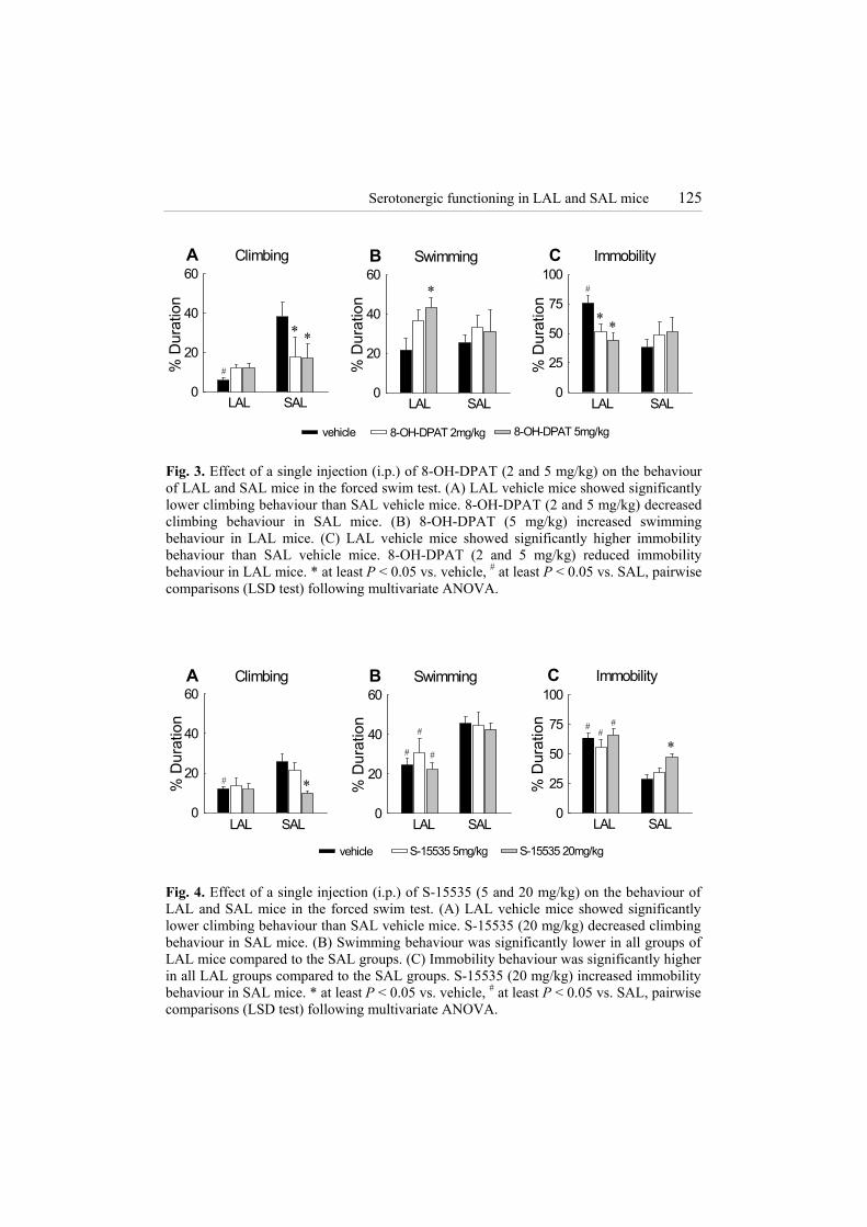
A line effect was found for climbing (F(1,32)= 11.101, P < 0.005) and a line × treatment interaction was observed for climbing (F(2,32)= 4.358 , P < 0.05) and immobility (F(2,32) = 4.772, P < 0.05). Significantly lower climbing (P < 0.005) (Fig. 3A) and higher immobility (P < 0.005) (Fig. 3C) behaviour was observed in vehicle-treated LAL mice compared to vehicle-treated SAL mice. These line differences were abolished by treatment with 8-OH-DPAT. This 5-HT1A receptor agonist induced in LAL mice a significant increase in swimming (P < 0.05, 5 mg/kg) (Fig. 3B) and a decrease in immobility (P < 0.05, 2 mg/kg; P < 0.01, 5 mg/kg) (Fig. 3C) compared to LAL vehicle. In SAL mice, 8-OH-DPAT induced a significant decrease in climbing (P < 0.05, 2 mg/kg; P < 0.01, 5 mg/kg) (Fig. 3A) compared to SAL vehicle. Effect of S-15535 on forced swimming behaviour (Fig. 4)
Line effects were observed for climbing (F(1,40)= 5.944, P < 0.05), swimming (F(1,40)= 27.511, P < 0.001) and immobility (F(1,40)= 57.976, P < 0.001) behaviour. Vehicle-treated LAL mice showed less climbing (P < 0.005) (Fig. 4A) and swimming (P < 0.001) (Fig. 4B) and more immobility (P < 0.001) (Fig. 4C) behaviour than vehicle-treated SAL mice. LAL mice treated with S-15535 showed less swimming (P < 0.05, 5mg/kg; P < 0.005 20mg/kg) (Fig. 4B) and more immobility (P < 0.005, both doses) (Fig. 4C) behaviour than SAL mice treated with S-15535.
A treatment effect was observed for climbing (F(2,40)= 3.458, P < 0.05) and immobility (F(2,40)= 5.042, P < 0.05) behaviour. In SAL mice, S-15535 (20 mg/kg) induced a significant decrease in climbing (P < 0.005 vs. SAL vehicle; P < 0.01 vs. SAL S-15535 [5 mg/kg]) (Fig. 4A) and an increase in immobility (P < 0.005, vs. SAL vehicle; P < 0.05, vs. SAL S-15535 [5 mg/kg]) (Fig. 4C) behaviour. No effect of S-15535 (5 mg/kg and 20 mg/kg) was observed on the behaviour of LAL mice (Fig. 4A-C).
Serotonergic functioning in LAL and SAL mice 125
A Climbing
% D
urat
ion
0
20
40
60
LAL SAL
∗ ∗
#
B Swimming
% D
urat
ion
0
20
40
60C Immobility
% D
urat
ion
0
25
50
75
100
LAL SALLAL SAL
∗
∗∗
#
vehicle 8-OH-DPAT 2mg/kg 8-OH-DPAT 5mg/kg Fig. 3. Effect of a single injection (i.p.) of 8-OH-DPAT (2 and 5 mg/kg) on the behaviour of LAL and SAL mice in the forced swim test. (A) LAL vehicle mice showed significantly lower climbing behaviour than SAL vehicle mice. 8-OH-DPAT (2 and 5 mg/kg) decreased climbing behaviour in SAL mice. (B) 8-OH-DPAT (5 mg/kg) increased swimming behaviour in LAL mice. (C) LAL vehicle mice showed significantly higher immobility behaviour than SAL vehicle mice. 8-OH-DPAT (2 and 5 mg/kg) reduced immobility behaviour in LAL mice. * at least P < 0.05 vs. vehicle, # at least P < 0.05 vs. SAL, pairwise comparisons (LSD test) following multivariate ANOVA.
vehicle
A Climbing
% D
urat
ion
0
20
40
60
LAL SAL
∗#
B Swimming
% D
urat
ion
0
20
40
60
LAL SAL
##
#
LAL SAL
#
∗#
#
C Immobility
% D
urat
ion
0
25
50
75
100
S-15535 20mg/kgS-15535 5mg/kg
##
#
∗
Fig. 4. Effect of a single injection (i.p.) of S-15535 (5 and 20 mg/kg) on the behaviour of LAL and SAL mice in the forced swim test. (A) LAL vehicle mice showed significantly lower climbing behaviour than SAL vehicle mice. S-15535 (20 mg/kg) decreased climbing behaviour in SAL mice. (B) Swimming behaviour was significantly lower in all groups of LAL mice compared to the SAL groups. (C) Immobility behaviour was significantly higher in all LAL groups compared to the SAL groups. S-15535 (20 mg/kg) increased immobility behaviour in SAL mice. * at least P < 0.05 vs. vehicle, # at least P < 0.05 vs. SAL, pairwise comparisons (LSD test) following multivariate ANOVA.

126 Chapter 6
Effect of 8-OH-DPAT and S-15535 on the release of plasma corticosterone in the FST (Table 1)
No treatment effect was observed for 8-OH-DPAT or S-15535 on plasma corticosterone concentrations. A significant line effect was found in the experiment with S-15535 (F(1,39)= 8.508, P < 0.001). Here, vehicle-treated LAL mice showed significant higher plasma corticosterone concentrations than vehicle-treated SAL mice (P < 0.05) (Table 1), and LAL mice treated with S-15535 (5mg/kg) showed significant higher plasma corticosterone concentrations than their SAL counterparts (P < 0.05). Table 1. Effect of 8-OH-DPAT and S-15535 on the release of plasma corticosterone (µg/dL) in LAL and SAL mice 15 min after the forced swim test.
LAL SAL
8-OH-DPAT 0 mg/kg 48.9 ± 3.6 36.6 ± 5.8
2 mg/kg 44.5 ± 4.6 52.5 ± 6.9
5 mg/kg 45.4 ± 2.6 44.3 ± 5.4
S-15535 0 mg/kg 52.1 ± 6.1a 39.6 ± 3.0
5 mg/kg 54.0 ± 6.2b 38.8 ± 4.6
20 mg/kg 50.1 ± 5.3 45.3 ± 3.1 a P < 0.05 vs. SAL S-15535 (0 mg/kg), b P < 0.05 vs. SAL S-15535 (5 mg/kg), pairwise comparisons (LSD) following univariate ANOVA. Effect of 8-OH-DPAT on 5-HT metabolism in LAL and SAL mice after forced swimming (Fig. 5)
A line effect for 5-HIAA was found in hippocampus (F(1,21)= 9.802, P < 0.01) and amygdaloid region (F(1,21)= 8.719, P < 0.01). LAL mice treated with 8-OH-DPAT showed significantly lower 5-HIAA contents than SAL mice treated with 8-OH-DPAT in both brain regions (P < 0.05) (Fig. 5A). A treatment effect for 5-HIAA was found in hypothalamus (F(1,21)= 10.322, P < 0.005), where SAL mice treated with 8-OH-DPAT showed lower 5-HIAA contents than SAL vehicle (P < 0.005) (Fig. 5A).
A line effect for 5-HT was found in all brain regions measured: frontal cortex (F(1,21)= 5.386, P < 0.05), septum (F(1,21)= 7.907, P < 0.05), striatum (F(1,21)= 15.036, P < 0.005), parietal and occipital cortex (F(1,19)= 6.520, P < 0.05), hippocampus (F(1,20)= 15.502, P < 0.005), hypothalamus (F(1,21)= 4.549, P < 0.05), amygdaloid region (F(1,21)= 20.138, P < 0.005), cerebellum (F(1,20)= 7.564, P < 0.05) and brain stem (F(1,20)= 28.074, P < 0.005). LAL vehicle showed higher 5-HT

Serotonergic functioning in LAL and SAL mice 127
contents than SAL vehicle in frontal cortex, striatum, amygdaloid region (all P < 0.05), hippocampus and brain stem (P < 0.005) (Fig. 5B). LAL mice treated with 8-OH-DPAT showed higher 5-HT contents in septum, striatum, cerebellum (all P < 0.05), amygdaloid region (P < 0.005), and brain stem (P < 0.01) (Fig. 5B). A treatment effect for 5-HT was found only in brain stem (F(1,20)= 7.604, P < 0.05), where SAL mice treated with 8-OH-DPAT showed higher 5-HT contents than SAL vehicle (P < 0.05) (Fig. 5B).
A treatment effect for 5-HIAA/5-HT ratio was found in septum (F(1,21)= 6.827, P < 0.05), hypothalamus (F(1,21)= 7.460, P < 0.05), amygdaloid region (F(1,21)= 7.708, P < 0.05), cerebellum (F(1,20)= 6.411, P < 0.05) and brain stem (F(1,20)= 10.529, P < 0.005). A line × treatment interaction was found in septum (F(1,21)= 4.378, P < 0.05), parietal and occipital cortex (F(1,19)= 6.898, P < 0.05), hippocampus (F(1,20)= 4.392, P < 0.05) and brain stem (F(1,20)= 4.738, P < 0.05). LAL vehicle showed a lower 5-HIAA/5-HT ratio than SAL vehicle in septum, parietal and occipital cortex and brain stem (all P < 0.05) (Fig. 5C). SAL mice treated with 8-OH-DPAT showed a lower 5-HIAA/5-HT ratio than SAL vehicle in all brain regions except frontal cortex and striatum (P < 0.005, septum, hypothalamus, brain stem; P < 0.05 all other regions) (Fig. 5C). Fig. 5. (next page) Effect of 8-OH-DPAT (5 mg/kg) on 5-HT and 5-HIAA contents in several brain regions of SAL and LAL mice, 15 min after the FST. (A) 8-OH-DPAT induced a decrease in 5-HIAA content in SAL mice in hippocampus and amygdaloid region compared to LAL mice, and in hypothalamus compared to SAL vehicle. (B) 5-HT contents were higher in several brain regions in LAL mice (both groups) compared to SAL mice (both groups). 8-OH-DPAT in SAL mice increased 5-HT content in brain stem compared to vehicle SAL mice. (C) Vehicle LAL mice showed a lower 5-HIAA/5-HT ratio in septum, parietal and occipital cortex and brain stem than vehicle SAL mice. 8-OH-DPAT induced a decrease in 5-HIAA/5-HT ratio in SAL mice in seven out of nine brain regions. * at least P < 0.05, pairwise comparisons (LSD test) following ANOVA.
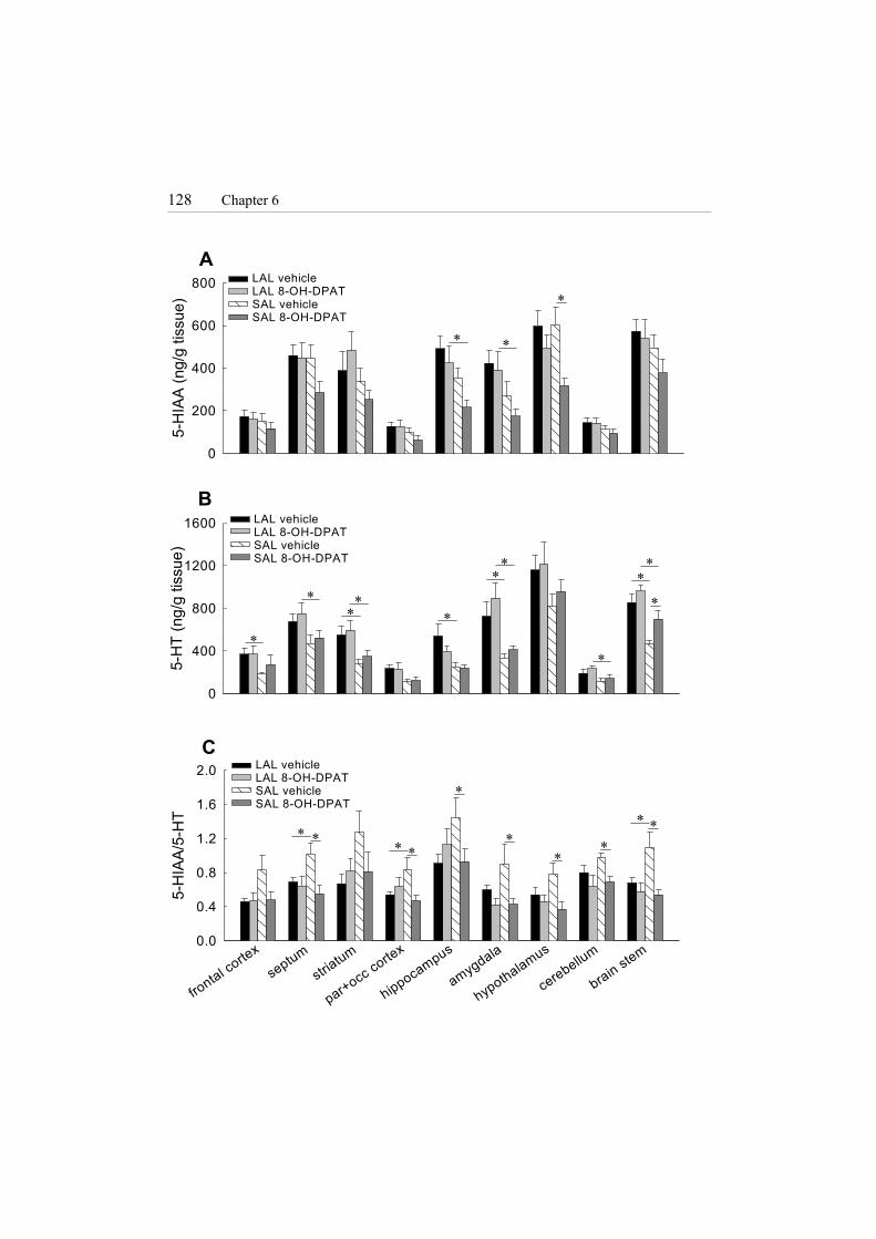
128 Chapter 6
A
5-H
IAA
(ng/
g tis
sue)
0
200
400
600
800
B
5-H
T (n
g/g
tissu
e)
0
400
800
1200
1600
C
frontal corte
x
septumstria
tum
par+occ cortex
hippocampus
amygdala
hypothalamus
cerebellum
brain stem
5-H
IAA/
5-H
T
0.0
0.4
0.8
1.2
1.6
2.0
LAL vehicleLAL 8-OH-DPATSAL vehicleSAL 8-OH-DPAT
LAL vehicleLAL 8-OH-DPATSAL vehicleSAL 8-OH-DPAT
LAL vehicleLAL 8-OH-DPATSAL vehicleSAL 8-OH-DPAT
∗ ∗
∗
∗
∗ ∗∗ ∗
∗∗
∗
∗∗
∗
∗ ∗∗∗
∗∗
∗
∗
∗∗

Serotonergic functioning in LAL and SAL mice 129
Discussion
In the present study, it was shown that low-aggressive LAL mice and high-aggressive SAL mice had region-specific differences in 5-HT1A receptor properties and 5-HT metabolism. LAL mice had lower 5-HT1A receptor mRNA expression and binding capacity in the CA1 region and dentate gyrus of the hippocampus than high-aggressive SAL mice. No significant line difference was found in two other forebrain regions nor in the dorsal raphe nucleus. In the brain stem 5-HT contents were significantly higher, and in striatum and amygdaloid region 5-HIAA/5-HT ratio (as an index of 5-HT turnover) was significantly lower in LAL than in SAL mice. Both mouse lines showed higher 5-HT and 5-HIAA contents in the light compared to the dark phase. It was further shown that the full 5-HT1A receptor agonist 8-OH-DPAT (2 and 5 mg/kg) was able to abolish the behavioural line-differences in the FST by reducing immobility behaviour in LAL mice and reducing climbing behaviour in SAL mice. The somatodendritic 5-HT1A autoreceptor agonist S-15535 (20 mg/kg) did not alter forced swimming behaviour in LAL mice, but induced a decrease in climbing and an increase in immobility behaviour in SAL mice. None of the 5-HT1A receptor agonists affected the release of plasma corticosterone concentrations. In response to forced swimming, LAL mice showed higher 5-HT contents in striatum, amygdaloid region and brain stem and lower 5-HIAA/5-HT ratio in septum, parietal and occipital cortex and brain stem than SAL mice. 8-OH-DPAT (5 mg/kg) did not affect monoamine contents in LAL mice, whereas in SAL mice a clear decrease in 5-HIAA/5-HT ratio was observed in seven out of nine brain regions.
The line-difference found for 5-HT1A receptor mRNA expression and binding capacity in the CA1 region and dentate gyrus of the hippocampus, with lower levels in LAL than in SAL mice, is consistent with previous findings (Korte et al., 1996, Van Riel et al., 2002). No line-difference was found in two other forebrain regions, the prefrontal cortex and lateral septum, nor in the dorsal raphe nucleus. The former two findings are in contrast with the report by Korte et al. (1996). This discrepancy is likely due to the difference in time of the day prior to tissue preparation. Furthermore, a circadian fluctuation was found for 5-HT and 5-HIAA contents (contents were higher during the light than during the dark phase) in both the LAL and SAL mice, which have been described in other rodent species as well (Collu et al., 1973; Philo et al., 1977), and seem to be associated with the circadian release of HPA hormones (Aldegunde et al., 1984). Interestingly, in most brain regions, levels of 5-HT, 5-HIAA and 5-HT turnover were not significantly different between LAL and SAL mice. Only in brain stem 5-HT content was higher and only in striatum and amygdaloid region the 5-HT turnover was lower in LAL compared to SAL mice. Thus, the line-differences found in 5-HT turnover are not related to the line-differences in 5-HT1A receptor gene expression and binding. It is, therefore, unlikely that the observed difference in 5-HT1A properties between SAL and LAL mice is an adaptive compensatory reaction to changes in 5-HT
130 Chapter 6
metabolism. These data also signify that we could not confirm that low 5-HT metabolism is linked with high trait aggression as displayed by the SAL mice. These contradictory results suggests a rather complicated pattern of the involvement of 5-HT in low and high trait aggression in these mice. This phenomenon could, however, be specific for these selection lines. Conversely, rats with low and high trait aggression (derived from an unselected strain of wild-type rats) displayed a similar difference in sensitivity of postsynaptic 5-HT1A receptors as LAL and SAL mice (Van der Vegt et al., 2001). This suggests that the differences found between LAL and SAL mice are not unique but may rather be a more general characteristic of individuals with low and high trait aggression. Obviously, microdialysis should be applied in future studies to measure extracellular 5-HT in specific brain regions to determine the status of 5-HT neurotransmission in these two mouse lines. Yet, the confirmation of lower hippocampal 5-HT1A receptor gene expression and binding in LAL mice together with lower hippocampal 5-HT responsiveness (Van Riel et al., 2002), lower sensitivity of postsynaptic 5-HT1A receptors (Van der Vegt et al., 2001), and signs of lower 5-HT turnover in striatum and amygdaloid region may indicate a tendency towards lower 5-HT activity in specific brain regions in LAL versus SAL mice.
In agreement with previous findings (Veenema et al., 2003a,b), forced swimming induced a differential behavioural response in LAL and SAL mice. LAL mice displayed high immobility behaviour, whereas SAL mice showed high climbing and/or swimming behaviour. High immobility behaviour is presumably related to a state of depression-like behaviour, as antidepressant drugs are able to decrease immobility behaviour in the FST (Porsolt et al., 1977a,b, 1979). Interestingly, the high immobility behaviour of LAL mice was associated with higher 5-HT contents in frontal cortex, striatum, hippocampus, amygdaloid region and brain stem than in SAL mice. Moreover, 5-HT turnover was lower in all brain regions in LAL compared to SAL mice reaching significance in septum, parietal and occipital cortex and brain stem. Accordingly, LAL mice showed signs of an attenuated 5-HT responsiveness to forced swimming compared to SAL mice.
Acute administration with the full 5-HT1A receptor agonist, 8-OH-DPAT (2 mg/kg and 5mg/kg), effectively abolished the line-differences in forced swimming behaviour. 8-OH-DPAT induced a significant decrease in immobility behaviour in LAL mice and a decrease in climbing behaviour in SAL mice. In general, 8-OH-DPAT decreases immobility behaviour in the FST (Wieland and Lucki, 1990; Singh and Lucki, 1993; Schreiber and De Vry, 1993; Detke et al., 1995, O’Neill and Conway, 2001), although strain differences in the behavioural effects of 8-OH-DPAT have also been reported (Lopez-Rubalcava and Lucki, 2000). Moreover, it was found that the antidepressant, fluoxetine, decreased immobility behaviour in rats showing ‘passive’ behaviour, whereas the same drug increased immobility behaviour in rats showing ‘active’ behaviour (Taghzouti et al., 1999). Hence, individual differences in passive and active behaviours in the forced swim test determined the effectiveness of drugs like 8-OH-DPAT and fluoxetine. This may

Serotonergic functioning in LAL and SAL mice 131
underly a differential activation or functioning of the 5-HT system and 5-HT1A receptors during forced swimming. Indeed, in the present study it was found that the differential behavioural effects of 8-OH-DPAT (5 mg/kg) in the FST were associated with differential changes in 5-HT metabolism of LAL and SAL mice. In LAL mice, 8-OH-DPAT had no effect on brain 5-HT metabolism, whereas 8-OH-DPAT in SAL mice potently attenuated the forced swimming-enhanced 5-HT turnover. Thus, the decrease in climbing behaviour induced by 8-OH-DPAT in SAL mice was associated with a decrease in 5-HT metabolism. This could have been mediated by activation of presynaptic 5-HT1A autoreceptors.
To determine this latter suggestion, the 5-HT1A receptor agonist S-15535 was used. S-15535 acts as an agonist on the somatodendritic 5-HT1A autoreceptor and has antagonistic properties on postsynaptic 5-HT1A receptors (Millan et al., 1993, 1997; Schreiber et al., 1995). If activation of primarily postsynaptic 5-HT1A receptors induced the behavioural change in LAL mice, it was expected that S-15535 further enhanced immobility behaviour or had no effect at all. Indeed, S-15535 (5 and 20 mg/kg) did not alter forced swimming behaviour in LAL mice. In contrast, S-15535 (20 mg/kg) in SAL mice had a similar behavioural effect as 8-OH-DPAT. This suggests that in SAL mice, the behavioural effects of S-15535 and of 8-OH-DPAT were mediated by predominant activation of somatodendritic 5-HT1A autoreceptors. The behavioural effects of S-15535 or 8-OH-DPAT could not be attributed to changes in HPA activity as neither ligand affected the release of plasma corticosterone. The observed higher release of plasma corticosterone in LAL compared to SAL mice is, however, in agreement with earlier findings (Veenema et al., 2003a).
In conclusion, this study demonstrates that genetic selection for trait aggression in male wild house-mice is associated with region-specific differences in 5-HT1A receptor properties and region-specific differences in basal and stress-induced 5-HT metabolism. These line-differences may also provide a basis for understanding the distinct behavioural responses to forced swimming and the differential behavioural effects of the two 5-HT1A receptor ligands in LAL and SAL mice. Considering the line-differences in behavioural coping responses, 5-HT1A receptor properties, 5-HT metabolism, and previously found line-differences in HPA regulation (Veenema et al., 2003b,c), these LAL and SAL mice represent an interesting animal model to study the genetic basis of individual variation in susceptibility to affective disorders like depression and anxiety. Acknowledgements The authors would like to thank Gerardus Zuidema and Auke Meinema for excellent animal care. Jan Bruggink and Jan Keijser are appreciated for their technical assistance.

132 Chapter 6
References Aldegunde M, Arnaez E, Miguez I, Fernandez P. Variations in monoamine contents in
discrete brain regions and their concomitance with plasma corticosteroids during the day. Intern J Neurosci 1984 24, 233-238.
Baldwin D, Rudge S. The role of serotonin in depression and anxiety. Int Clin Psychopharmacol 1995, 9:41-45.
Benus RF, Bohus B, Koolhaas JM, van Oortmerssen GA. Behavioural strategies of aggressive and non-aggressive male mice in active shock avoidance. Behavioural processes 1989, 20:1-12.
Benus RF, Bohus B, Koolhaas JM, van Oortmerssen GA. Behavioural differences between artificially selected aggressive and non-aggressive mice: response to apomorphine. Behav Brain Res 1991a, 43:203-208.
Benus RF, Bohus B, Koolhaas JM, van Oortmerssen GA. Heritable variation for aggression as a reflection of individual coping strategies. Experientia 1991b, 47:1008-19.
Blier P, de Montigny C. Current advances and trends in the treatment of depression. Trends Pharmacol Sci 1994, 15:220-226.
Blier P, Ward NM. Is there a role for 5-HT1A agonists in the treatment of depression? Biol Psychiatry 2003, 53:193-203.
Collu R, Jequier JC, Letarte J, Leboeuf G, Ducharme JR. Diurnal variations of plasma growth hormone and brain monoamines in adult male rats. Can J Physiol Pharmacol 1973, 51:890-892.
Detke MJ, Wieland S, Lucki I. Blockade of the antidepressant-like effects of 8-OH-DPAT, buspirone and desipramine in the rat forced swim test by 5HT1A receptor antagonists. Psychopharmacology 1995, 119:47-54.
De Vry J. 5-HT1A receptor agonists: recent developments and controversial issues. Psychopharmacology 1995, 121:1-26.
File SE, Gonzalez LE, Andrews N. Comparative study of pre- and postsynaptic 5-HT1A receptor modulation of anxiety in two ethological animal tests. J Neurosci 1996, 16:4810-4815.
Hutson PH, Sarna GS, O’Connell MT, Curzon G. Hippocampal 5-HT synthesis and release in vivo is decreased by infusion of 8-OH-DPAT into the nucleus raphe dorsalis. Neurosci Lett 1989, 100:276-280.
Korte SM, Meijer OC, de Kloet ER, Buwalda B, Keijser J, Sluyter F, van Oortmerssen G, Bohus B. Enhanced 5-HT1A receptor expression in forebrain regions of aggressive house mice. Brain Res 1996, 736:338-343.
Kurtz N. Efficacy of azapirones in depression. In Stahl SM, Gastpar M, Keppel Hesselink JM, Traber J. (Eds.) Serotonin1A receptor in depression and anxiety. Raven Press, New York 1992, pp. 163-170.
Le Poul E, Laaris N, Doucet E, Fattaccini CM, Mocaër E, Hamon M, Lanfumey L. Chronic alnespirone-induced desensitisation of somatodendritic 5-HT1A autoreceptors in the rat dorsal raphe nucleus. Eur J Pharmacol 1999, 365:165-173.
Lesch KP. 5-HT1A receptor responsivity in anxiety disorders and depression. Prog Neuropsychopharmacol Biol Psychiatry 1991, 15: 723-733.
Lopez-Rubalcava C, Lucki I. Strain differences in the behavioral effects of antidepressant drugs in the rat forced swimming test. Neuropsychopharmacology 2000, 22:191-199.

Serotonergic functioning in LAL and SAL mice 133
Maes M, Meltzer H. The serotonin hypothesis of depression. In Bloom F. and Kupfer D (Eds.). Psychopharmacology: The fourth generation of progress. Raven Press, New York 1995, pp. 933-944.
Meijer OC, Steenbergen PJ, de Kloet ER. Differential expression and regional distribution of steroid receptor coactivators SRC-1 and SRC-2 in brain and pituitary. Endocrinology 2000, 141:2192-2199.
Millan MJ, Rivet JM, Canton H, Lejeune F, Gobert A, Widdowson P, Bervoets K, Brocco M, Peglion JL. S 15535: a highly selective benzodioxopiperazine 5-HT1A receptor ligand which acts as an agonist and an antagonist at presynaptic and postsynaptic sites respectively. Eur J Pharmacol 1993, 230: 99-102.
Millan MJ, Hjorth S, Samanin R, Schreiber R, Jaffard R, De Ladonchamps B, Veiga S, Goument B, Peglion JL, Spedding M, Brocco M. S 15535, a novel benzodioxopiperazine ligand of serotonin (5-HT) 1A receptors: II. Modulation of hippocampal serotonin release in relation to potential anxiolytic properties. J Pharmacol Exp Ther 1997, 282: 148-161.
Olivier B, Mos J, Tulp M, Schipper J, Den Daas S, Van Oortmerssen G. Serotonergic involvement in aggressive behavior in animals. In Van Praag HM, Plutchik R, Apter A. (Eds.) Violence and suicidality – perspectives in clinical and psychobiological research. Brunner/Mazel Publishers New York 1990, pp. 79-137.
O'Neill MF, Conway MW. Role of 5-HT(1A) and 5-HT(1B) receptors in the mediation of behavior in the forced swim test in mice. Neuropsychopharmacology 2001, 24:391-398.
Philo R, Rudeen PK, Reitter RJ. A comparison of the circadian rhythms and concentrations of serotonin and norepinephrine in the telencephalon of four rodent species. Comp Biochem Physiol C 1977, 57:127-130.
Porsolt RD, Le Michon M, Jalfre M. Depression: a new animal model sensitive to antidepressant treatments. Nature 1977a, 266: 730-732.
Porsolt RD, Bertin A, Jalfre M. Behavioral despair in mice: a primary screening test for antidepressants. Arch Int Pharmacodyn 1977b, 229:327-336.
Porsolt RD. Animal model of depression. Biomedicine 1979, 30:139-140. Schreiber R, De Vry J. Neuroanatomical basis for the antidepressant-like effects of the 5-
HT(1A) receptor agonists 8-OH-DPAT and ipsapirone in the rat forced swimming test. Behav Pharmacol 1993, 4:625-636.
Schreiber R, Brocco M, Lefebvre DL, Monneyron S, Millan MJ. A drug discrimination analysis of the actions of novel serotonin1A receptor ligands in the rat using the 5-HT1A receptor agonist, 8-hydroxy-2-(di-n-propylamino)tetralin. J Pharmacol Exp Ther 1995, 275: 822-831.
Sharp T, Bramwell SR, Clark D, Grahame-Smith DG. In vivo measurements of extracellular 5-hydroxytryptamine in hippocampus of the anaesthetized rat using microdialysis: changes in relation to 5-hydroxytryptaminergic neuronal activity. J Neurochem 1990, 53:234-240.
Singh A, Lucki I. Antidepressant-like activity of compounds with varying efficacy at 5-HT1A receptors. Neuropharmacology 1993, 32:331-340.
Sijbesma H, Schipper J, De Kloet ER, Mos J, Van Aken A, Olivier B. Post-synaptic 5-HT1 receptors and offensive aggression in rats: a combined behavioral and autoradiographic study with eitoprazine. Pharmacol Biochem Behav 1991, 38:447-458.

134 Chapter 6
Sluyter F, Korte SM, Bohus B, van Oortmerssen GA. Behavioral stress response of genetically selected aggressive and non-aggressive wild house mice in the shock-probe/defensive burying test. Pharmacol Biochem Behav 1996, 54:113-116.
Taghzouti K, Lamarque S, Kharouby M, Simon H. Interindividual differences in active and passive behaviors in the forced-swimming test: implications for animal models of psychopathology. Biol Psychiatry 1999, 45:750-758.
Van der Vegt BJ, de Boer SF, Buwalda B, de Ruiter AJ, de Jong JG, Koolhaas JM. Enhanced sensitivity of postsynaptic serotonin-1A receptors in rats and mice with high trait aggression. Physiol Behav 2001 74:205-11.
Van Oortmerssen GA, Bakker TC. Artificial selection for short and long attack latencies in wild Mus musculus domesticus. Behav Genet 1981, 11:115-126.
Van Praag HM. Anxiety/aggression-driven depression. A paradigm of functionalization and verticalization of psychiatric diagnosis. Prog Neuropsychopharmacol Biol Psychiatry 2001, 25:893-924.
Van Riel E, Meijer OC, Veenema AH, Joëls M. Hippocampal serotonin responses in short and long attack latency mice. J Neuroendocrinol 2002, 14:234-240.
Veenema AH, Meijer OC, De Kloet ER, Koolhaas JM, Bohus BG. Differences in basal and stress-induced HPA regulation of wild house mice selected for high and low aggression. Horm Behav 2003a, 54:197-204.
Veenema AH, Meijer OC, De Kloet ER, Koolhaas JM. Genetic selection for coping style predicts stressor susceptibility. J Neuroendocrinol 2003b, 15: 256-267.
Veenema AH, Feldker DEM, Schmidt M, Sijtsma B, Koolhaas JM, De Kloet ER. Stressor susceptibility in low and high aggressive mice: effects of time and of type of opponent in sensory contact model. Psychoneuroendocrinology 2003c, submitted.
Wieland S, Lucki I. Antidepressant-like activity of 5-HT1A agonists measured with the forced swim test. Psychopharmacology 1990, 101:497-504.