University of Groningen 4-Hydroxyacetophenone ... · Eur. J. Biochem. 268, 2547–2557 (2001) q...
12
University of Groningen 4-Hydroxyacetophenone monooxygenase from Pseudomonas fluorescens ACB - A novel flavoprotein catalyzing Baeyer-Villiger oxidation of aromatic compounds Kamerbeek, Nanne M.; Moonen, Mariëlle J.H.; Ven, Jos G.M. van der; Berkel, Willem J.H. van; Fraaije, Marco W.; Janssen, Dick B. Published in: European Journal of Biochemistry DOI: 10.1046/j.1432-1327.2001.02137.x IMPORTANT NOTE: You are advised to consult the publisher's version (publisher's PDF) if you wish to cite from it. Please check the document version below. Document Version Publisher's PDF, also known as Version of record Publication date: 2001 Link to publication in University of Groningen/UMCG research database Citation for published version (APA): Kamerbeek, N. M., Moonen, M. J. H., Ven, J. G. M. V. D., Berkel, W. J. H. V., Fraaije, M. W., & Janssen, D. B. (2001). 4-Hydroxyacetophenone monooxygenase from Pseudomonas fluorescens ACB - A novel flavoprotein catalyzing Baeyer-Villiger oxidation of aromatic compounds. European Journal of Biochemistry, 268(9), 2547-2557. https://doi.org/10.1046/j.1432-1327.2001.02137.x Copyright Other than for strictly personal use, it is not permitted to download or to forward/distribute the text or part of it without the consent of the author(s) and/or copyright holder(s), unless the work is under an open content license (like Creative Commons). Take-down policy If you believe that this document breaches copyright please contact us providing details, and we will remove access to the work immediately and investigate your claim. Downloaded from the University of Groningen/UMCG research database (Pure): http://www.rug.nl/research/portal. For technical reasons the number of authors shown on this cover page is limited to 10 maximum. Download date: 26-12-2019
Transcript of University of Groningen 4-Hydroxyacetophenone ... · Eur. J. Biochem. 268, 2547–2557 (2001) q...
University of Groningen
4-Hydroxyacetophenone monooxygenase from Pseudomonas fluorescens ACB - A novelflavoprotein catalyzing Baeyer-Villiger oxidation of aromatic compoundsKamerbeek, Nanne M.; Moonen, Mariëlle J.H.; Ven, Jos G.M. van der; Berkel, Willem J.H.van; Fraaije, Marco W.; Janssen, Dick B.Published in:European Journal of Biochemistry
DOI:10.1046/j.1432-1327.2001.02137.x
IMPORTANT NOTE: You are advised to consult the publisher's version (publisher's PDF) if you wish to cite fromit. Please check the document version below.
Document VersionPublisher's PDF, also known as Version of record
Publication date:2001
Link to publication in University of Groningen/UMCG research database
Citation for published version (APA):Kamerbeek, N. M., Moonen, M. J. H., Ven, J. G. M. V. D., Berkel, W. J. H. V., Fraaije, M. W., & Janssen, D.B. (2001). 4-Hydroxyacetophenone monooxygenase from Pseudomonas fluorescens ACB - A novelflavoprotein catalyzing Baeyer-Villiger oxidation of aromatic compounds. European Journal of Biochemistry,268(9), 2547-2557. https://doi.org/10.1046/j.1432-1327.2001.02137.x
CopyrightOther than for strictly personal use, it is not permitted to download or to forward/distribute the text or part of it without the consent of theauthor(s) and/or copyright holder(s), unless the work is under an open content license (like Creative Commons).
Take-down policyIf you believe that this document breaches copyright please contact us providing details, and we will remove access to the work immediatelyand investigate your claim.
Downloaded from the University of Groningen/UMCG research database (Pure): http://www.rug.nl/research/portal. For technical reasons thenumber of authors shown on this cover page is limited to 10 maximum.
Download date: 26-12-2019

Eur. J. Biochem. 268, 2547±2557 (2001) q FEBS 2001
4-Hydroxyacetophenone monooxygenase fromPseudomonas fluorescens ACBA novel flavoprotein catalyzing Baeyer±Villiger oxidation of aromatic compounds
Nanne M. Kamerbeek1, MarieÈ lle J. H. Moonen2, Jos G. M. van der Ven1,*, Willem J. H. van Berkel2,Marco W. Fraaije1 and Dick B. Janssen1
1Laboratory of Biochemistry, Groningen Biomolecular Sciences and Biotechnology Institute, University of Groningen, the Netherlands;2Laboratory of Biochemistry, Department of Agrotechnology and Food Sciences, Wageningen University, the Netherlands
A novel flavoprotein that catalyses the NADPH-dependent
oxidation of 4-hydroxyacetophenone to 4-hydroxyphenyl
acetate, was purified to homogeneity from Pseudomonas
fluorescens ACB. Characterization of the purified enzyme
showed that 4-hydroxyacetophenone monooxygenase
(HAPMO) is a homodimer of < 140 kDa with each subunit
containing a noncovalently bound FAD molecule. HAPMO
displays a tight coupling between NADPH oxidation and
substrate oxygenation. Besides 4-hydroxyacetophenone a
wide range of other acetophenones are readily converted
via a Baeyer±Villiger rearrangement reaction into the
corresponding phenyl acetates. The P. fluorescens HAPMO
gene (hapE) was characterized. It encoded a 640 amino-
acid protein with a deduced mass of 71 884 Da. Except
for an N-terminal extension of < 135 residues, the sequence
of HAPMO shares significant similarity with two known
types of Baeyer±Villiger monooxygenases: cyclohexanone
monooxygenase (27±33% sequence identity) and steroid
monooxygenase (33% sequence identity). The HAPMO
sequence contains several sequence motifs indicative for
the presence of two Rossman fold domains involved in FAD
and NADPH binding. The functional role of a recently
identified flavoprotein sequence motif (ATG) was explored
by site-directed mutagenesis. Replacement of the strictly
conserved glycine (G490) resulted in a dramatic effect on
catalysis. From a kinetic analysis of the G490A mutant it is
concluded that the observed sequence motif serves a struc-
tural function which is of importance for NADPH binding.
Keywords: aromatic ketones; Baeyer±Villiger monooxy-
genase; flavoprotein; 4-hydroxyacetophenone monooxy-
genase; mutagenesis.
The aerobic degradation of aromatic compounds by soilbacteria depends on the activity of oxygenases [1]. So fartwo classes of flavoprotein hydroxylases have been shownto act as aromatic monooxygenases: the single componentaromatic hydroxylases [2] and two-component hydroxy-lases [3,4]. The single component aromatic hydroxylasesshare a typical dinucleotide binding fold for complexationof the FAD cofactor while lacking a NAD(P) binding fold
[5]. An extensively studied member of this widespreadclass of flavoenzymes is p-hydroxybenzoate hydroxylase[6,7]. Mechanistic studies have shown that the monooxy-genation reactions catalysed by single component aromatichydroxylases are preceded by NAD(P)H-mediated flavinreduction. The reduced flavin then reacts with dioxygen toform a reactive electrophilic hydroperoxyflavin intermedi-ate which is able to introduce a hydroxyl group into anactivated aromatic ring [8]. The two-component hydroxy-lases typically consist of a relatively small flavin reductasewhich generates reduced flavin at the expense of NAD(P)H.After intermolecular transfer, the reduced flavin is used bythe large oxygenating component to catalyse substratehydroxylation [9,10]. Although the genes of several of thesetwo-component hydroxylases have been identified, mech-anistic data on this type of monooxygenases are scarce.
Except for the above-mentioned monooxygenation reac-tions another route of flavoenzyme-mediated oxygenationof aromatic compounds has been suggested in the literature.For several bacteria it was found that their ability todegrade aromatic compounds depends on an enzyme-mediated Baeyer±Villiger reaction in which an oxygenatom is inserted between the aromatic ring and a ketoneside chain (Scheme 1). These Baeyer±Villiger reactionshave been detected during studies on the microbial degra-dation of several ring-substituted acetophenones [11±13]and 4-ethylphenol [14,15]. Recently, a Baeyer±Villigermonooxygenase from Pseudomonas putida JD1, involved
Correspondence to D. B. Janssen, Laboratory of Biochemistry,
Department of Chemistry, University of Groningen, Nijenborgh 4,
9747 AG Groningen, the Netherlands. Fax: 131 503634165,
Tel.: 131 503634209, E-mail: [email protected]
Abbreviations: DIG, digoxigenin; HAPMO, 4-hydroxyacetophenone
monooxygenase.
Enzymes: catalase (EC. 1.11.1.6); cyclohexanone monooxygenase (EC.
1.14.13.22); 4-hydroxyacetophenone monooxygenase (EC. 1.14.13.x);
4-hydroxyphenyl acetate hydrolase (EC. 3.1.1.x); steroid monooxy-
genase (EC. 1.14.13.54).
*Present address: Diosynth B.V., Akzo Nobel, Apeldoorn, the
Netherlands.
Note: a web site is available at URL: www.chem.rug.nl/biotechnology
Note: The novel nucleotide sequence data published here have been
submitted to Genbank and are available under accession number
AF355751.
(Received 22 December 2000, revised 14 February 2001, accepted
5 March 2001)

in 4-ethylphenol metabolism, was purified and partiallycharacterized [16]. However, enzymes involved in theseatypical metabolic pathways have never been cloned.
All Baeyer±Villiger monooxygenases identified so farwere shown to be flavoproteins [17]. As for the singlecomponent hydroxylases, also for these enzymes a per-oxygenated flavin has been proposed as the oxygenatingspecies [18]. However, in the Baeyer±Villiger reaction thisreactive flavin intermediate is supposed to act as anucleophile indicating that in these monooxygenases aperoxyflavin anion is involved in substrate attack [19,20].
Pseudomonas fluorescens ACB is able to use 4-hydro-xyacetophenone as sole carbon and energy source [13].Since it was suggested that the microbial degradation of thisphenolic compound is initiated by a Baeyer±Villigerreaction (Scheme 1), we started a study to identify thisatypical aromatic monooxygenase. We report here on thepurification, gene cloning, sequence analysis and charac-terization of 4-hydroxyacetophenone monooxygenase(HAPMO). It is shown that the enzyme is a FAD-dependentBaeyer±Villiger monooxygenase that is active with a widerange of aromatic ketones. A preliminary report on thepurification of HAPMO from P. fluorescens ACB has beenpresented elsewhere [21].
M A T E R I A L S A N D M E T H O D S
Chemicals
Restriction enzymes, 7-deaza-dGTP, and isopropyl thio-b-d-galactoside were obtained from Roche. NADH, NADPH,dithiothreitol, glucose oxidase (grade II) and catalase werefrom Boehringer. Tris, FAD, FMN and riboflavin were fromSigma. Q-Sepharose Fast Flow, phenyl-Sepharose FastFlow, Superdex 200 Prep Grade, Superdex PG200 HR10/30 and molecular weight markers were from Pharmacia.Bio-Gel P-6DG and Macro-Prep Ceramic Hydroxyapatite(Type I, particle size 20 mm) were from Bio-Rad. Acryl-amide, bisacrylamide and Coomassie brilliant blue G250were from Serva. Aromatic ketones were purchased fromAcros, Aldrich, Fluorochem and Lancaster. Stock ketonesolutions were prepared in dimethyl formamide. H18
2 O (97atom % 18O) was from Campro (Elst, the Netherlands). Allother chemicals were of commercially available analyticalgrade.
Bacterial strains and culture conditions
P. fluorescens ACB was kindly provided by D. D. Focht(University of California, Riverside, USA). P. fluorescensACB was isolated from activated sewage sludge byenrichment and serial transfer with 4-hydroxyacetophenoneas growth substrate [13] and grown in 0.8% Nutrient Broth(Difco) at 30 8C or in minimal medium based on KroÈckeland Focht [22], containing per litre demineralized water:
3.5 g Na2HPO4, 1.4 g KH2PO4, 0.5 g (NH4)2SO4, 0.2 gMgSO4.7H2O, 10 mg yeast extract and 5 mL of a traceelements solution. The trace elements solution containedper litre: 780 mg Ca(NO3)2´4H2O, 200 mg FeSO4´7H2O,10 mg ZnSO4´7H2O, 10 mg H3BO3, 11.8 mgCoSO4´7H2O, 4 mg MnSO4´1H2O, 3 mg Na2MoO4´2H2O,2 mg NiCl2´6H2O, 10 mg CuSO4´5H2O and 2 mgNa2WO4´2H2O (adjusted to pH 2 with H2SO4). The finalpH of the medium was 7.0. Cells were grown on 10 mm4-hydroxyacetophenone at 30 8C in 2 L Erlenmeyer flasksagitated at 270 r.p.m. on an orbital shaker to providesuitable aeration. Large-scale growth, with 4-hydroxy-acetophenone as the sole carbon and energy source, wasperformed batchwise in a 200-L fermentor (BioengineeringAG) at 30 8C. During growth, the medium was stirred at150 r.p.m. and flushed with 30 L air´min21 and the pH waskept constant at pH 7.0 with NaOH. The fermentor wasinoculated with 2 L cell culture, and growth was induced bythe addition of 5 mm 4-hydroxyacetophenone, followed bytwo portions of 10 mm 4-hydroxyacetophenone at 8-hintervals. The cells were harvested after 24 h and the cellpaste (< 235 g wet weight) was frozen and stored at280 8C. Under these conditions, no significant loss ofHAPMO activity occurred over a period of 6 months.
Escherichia coli strains HB101, TOP10F 0 and BL21(DE3)pLysS were grown in Luria±Bertani medium supplementedwith the appropriate antibiotic at 20, 30 or 37 8C.
Enzyme purification from P. fluorescens ACB
HAPMO was purified from P. fluorescens ACB to apparenthomogeneity in two chromatographic steps. All purificationsteps were performed at 4 8C. Cells (200 g wet weight)were suspended in 21 mm potassium phosphate pH 7.1, anddisrupted through a precooled Manton Gaulin press. Theclarified cell extract was treated with 0.25% (w/v)protamine sulfate and the supernatant obtained aftercentrifugation was loaded onto a Q-Sepharose column(5 � 25 cm), equilibrated in 21 mm potassium phosphatepH 7.1. After washing with 2 vol. starting buffer, theHAPMO activity was eluted with a linear gradient of0±0.7 m KCl in starting buffer. Active fractions wereconcentrated by ultrafiltration (Amicon YM-30 membrane)and diluted with 3 vol. 10 mm potassium phosphate,pH 7.1. The enzyme solution was then adjusted to 1 mammonium sulfate and loaded onto a phenyl Sepharosecolumn (2.5 � 40 cm), equilibrated in 50 mm potassiumphosphate pH 7.1, containing 1 m ammonium sulfate. Afterwashing with starting buffer, the HAPMO activity waseluted with a linear descending gradient of 1±0 m ammoniumsulfate in 50 mm potassium phosphate, pH 7.1. Activefractions were concentrated by ultrafiltration, dialysed in50 mm potassium phosphate, pH 7.1, and stored at 280 8C.
The 4-hydroxyphenyl acetate hydrolase was purifiedfrom P. fluorescens ACB in three chromatographic steps.Cell extract was loaded on a DEAE±Sepharose column(6 � 2.5 cm) equilibrated in 21 mm phosphate pH 7.1.Esterase activity was eluted with a linear gradient of0±0.7 m KCl in starting buffer. Active fractions werepooled, dialysed against 1 mm phosphate pH 7.1, andloaded on a hydroxyapatite column (7 � 3 cm) equili-brated in 1 mm phosphate pH 7.1. Esterase activity waseluted with a linear gradient of 1±100 mm phosphate
Scheme 1.
2548 N. M. Kamerbeek et al. (Eur. J. Biochem. 268) q FEBS 2001

pH 7.1. Active fractions were pooled, concentrated andloaded on a Sephacryl S-200 column (34 � 1 cm) runningin 21 mm potassium phosphate, pH 7.1. The fractions withthe highest esterase activity were pooled, resulting in anenzyme preparation which was more than 75% pure. ForN-terminal sequencing, the esterase was isolated from aSDS-containing polyacrylamide gel.
Preparation of the genomic library
All DNA isolation and cloning procedures were carried outessentially as described by Sambrook et al. [23]. A genomiclibrary of P. fluorescens ACB was constructed in thecosmid vector pLAFR3 according to the strategy describedby Staskawicz et al. [24]. CsCl gradient centrifugation wasused to purify pLAFR3 isolated from E. coli HB101.Constructed vector arms and inserts were ligated andsubsequently packaged in vitro with a DNA-packaging kit(Boehringer). E. coli HB101 cells were infected with thepackaging mix and transducts were selected on Luria±Bertani plates supplemented with tetracycline. From 10transducts the plasmids were isolated and checked for insertby restriction analysis. All contained insert with anestimated average size of 17 kb. A total number of 2400colonies was transferred to 96-well microtiter platescontaining Luria±Bertani and tetracycline. After overnightgrowth, glycerol was added and the library was stored at280 8C until use.
`Touch down' PCR on total DNA
All PCR amplifications were performed as described byInnis and Gelfand [25]. The 50-mL reaction mixturescontained PCR buffer (Roche), 200 mm of each dNTP,20±30 pmol primer, 1±5 units Taq or Pwo polymerase and10±100 ng template.
The following degenerated primers were designed on theN-terminal amino-acid sequences of the purified enzymes:HAPMO, PAMOACB1 (forward) 5 0-GAAACCATGGCNGCNTTYAAYACNACNYTNCC-3 0 and PAMOACB2(reverse) 5 0-GAAAACCATGGRTCRTCRTCRTARTCNARNGWNGG-3 0; esterase (4-hydroxyphenyl acetate hydro-lase), PESACB1 (forward) 5 0-GAAACCATGGCNCTNGAYGTNGARWCNGCNCARCTNCTNGG-30 and PESACB2(reverse) 5 0-GAAAACCATGGYTCNGCNAGYTGNCCNAGNAGYTG 3 0 (NcoI sites are underlined and thesubstituted nucleotide in PAMOACB1 is shown in bold).Touch-down PCR was performed on total DNA ofP. fluorescens ACB [26]. After initial denaturation for5 min at 94 8C, the cycling program was as follows: 1 min94 8C, 1 min 60 8C (2 8C decrease after every two cycles to48 8C) and elongation at 72 8C for 2 min. In addition, 25cycles were performed at a hybridization temperature of48 8C and elongation for 1 min at 72 8C. The fragmentobtained using the PESACB1 and PAPMO2 primers wasligated into the pCR2.1-TOPO vector and then transformedinto E. coli TOP10F 0 cells according to the recommenda-tions of the manufacturer (Invitrogen). Plasmids containinginsert were purified from overnight cultures with the HighPureTM plasmid isolation kit (Roche).
Preparation of a digoxigenin-labelled probe
To prepare the probe to screen the genomic library, thehapD gene in pCR2.1 was amplified with Taq polymerase(Roche) using the primers PESACBf (5 0-GAAACCATGGCTCTTGACGTCGAGACGGCACAGCTG-3 0) andPESACBr (5 0-GAAAGGATCCTAAAAAGGGTTTCAGCCAAGGCTTGAT-3 0) during 30 cycles of 1 min 94 8C,1 min 60 8C and 1 min at 72 8C. The amplified fragmentwas isolated from gel and labelled with digoxigenin (DIG)by random-priming according to the method described bythe DIG system user's guide for filter hybridization(Boehringer) yielding the DIG-labelled probe HAP2.
Screening of the genomic library
For screening, the constructed library was plated from thefrozen stock on Luria±Bertani plates supplemented with12.5 mg´mL21 tetracycline and incubated overnight at37 8C. Colonies were transferred to cellulose nitrate mem-branes. The membranes were processed and subsequentlyhybridized with the DIG-labelled HAP2 probe at 68 8C.Two primers, designed on the nondegenerated part of thehapD sequence were used to check E. coli clones whichgave a positive signal after blotting: PESACB3 (5 0-GAAGCCGTTCCATTTCT-3 0) and PESACB4 (5 0-GCCAAGGCTTGATCTGATC-3 0). Cycling parameters were as fol-lows: 1 min at 94 8C, 1 min at 57 8C and 1 min at 72 8C.As templates the pLAFR3 cosmids were isolated from thepositive clones.
Cloning, expression and purification of recombinantHAPMO
The hapE gene was amplified with Pwo polymerase usingthe total DNA of P. fluorescens ACB as template. There-fore, two primers were designed: PAPMO1 (5 0-CACGGCATATGAGCGCCTTCAATACC-3 0, NdeI site underlined)and PAPMO2 (5 0-CACGGGGATCCTCAACCTAGTTGGTAATCAGTCGG-3 0, BamHI site in italic type). Cyclingparameters were as follows: 30 cycles of 1 min at 94 8C,1 min at 56 8C and 1 min at 72 8C. After amplification, thegene was isolated from gel, digested with NdeI and BamHIand ligated behind the T7 promoter of the NdeI/BamHIdigested expression vector pET-5a (Promega) yieldingpHAPE1.
E. coli BL21(DE3)pLysS was transformed with thisconstruct and grown in Luria±Bertani medium supple-mented with 100 mg´mL21 ampicillin and 35 mg´mL21
chloramphenicol at 30 8C. When the D600 reached 0.8,0.4 mm isopropyl thio-b-d-galactoside was added and thecells were grown for another 12±14 h at 20 8C. Cells (6 g)were harvested by centrifugation, resuspended in 50 mmpotassium phosphate, pH 7.0, containing 0.1 mm EDTAand 0.1 mm dithiothreitol, and sonicated to prepare cellextract. After ultracentrifugation for 60 min at 110 000 g,the supernatant was applied onto a ceramic hydroxyapatitecolumn (2.5 � 12 cm), equilibrated with 50 mm potassiumphosphate, pH 7.0, and eluted with the same buffer.Fractions containing HAPMO activity were pooled andloaded onto a Q-Sepharose column (2.5 � 18 cm) equili-brated with 50 mm potassium phosphate, pH 7.0. Afterwashing with starting buffer, the enzyme was eluted with a
q FEBS 2001 Cloning of 4-hydroxyacetophenone monooxygenase (Eur. J. Biochem. 268) 2549

linear gradient of 0±1 m KCl in the same buffer. HAPMOeluted at a concentration of 0.6 m KCl. Active fractionswere concentrated by ultrafiltration, dialysed in 50 mmpotassium phosphate, pH 7.0, and stored at 280 8C.
PCR amplification to create truncated HAPMO variants
Two primers were designed to amplify truncated hapEgenes. Primer PHAPMO3, 5 0-CACGGCATATGGCCAGCGGCCGCGACTTCAAGGTGGTG-3 0 annealed justupstream the part of the gene encoding the first Rossmanfold sequence motif. Val136 was mutated to a methionine tocreate a NdeI site. Primer PHAPMO8, 5 0-CACGGCATATGGCCGAAGAAGCCGTGACCGCC-3 0 was used tocreate a mutant lacking the part encoding the first 114amino acids. Due to the introduction of a NdeI site, Ile115was replaced by a methionine (NdeI sites underlined, startcodon in bold, substituted codons in italic type). PCR withPwo polymerase, using either PHAPMO3 or PHAPMO8 incombination with PAPMO2 as the reverse primer, under thesame conditions as described for hapE amplification,yielded fragments which were cloned with NdeI/BamHIin the pET5-a vector yielding pHAPE2 and pHAPE3,respectively. Successful cloning was confirmed by plasmidsequencing.
Site-directed mutagenesis
The Quickchange site-directed mutagenesis kit (Stratagene)was used to introduce a point mutation into HAPMOchanging the glycine at position 490 into an alanine. Twoprimers were used, Primer 1 (5 0-GTGTATGGCACGGCGTTCCATGCCTCG-3 0) and primer 2 (5 0-CGAGGCATGGAACGCCGTGCCATACAC-3 0). The mutagenic codon isshown in italic. The PCR reaction, with pHAPE1 astemplate, was performed under the conditions recom-mended by the manufacturer. Successful mutagenesis wasconfirmed by plasmid sequencing.
Steady-state kinetics
HAPMO activity was determined spectrophotometricallyby monitoring the decrease of NADPH at 370 nm(:370 � 2.7 mm21´cm21). Reaction mixtures (1 mL) typi-cally contained 50 mm potassium phosphate pH 8.0,0.25 mm NADPH and 50±100 nm enzyme. The reactionwas started by the addition of 10 mL 0.1 m 4-hydroxy-acetophenone in dimethylformamide. Specific activitieswere corrected for endogenous NADPH oxidase activity.One unit of HAPMO activity is defined as the amount ofprotein that oxidizes 1 mmol´min21 NADPH. All kineticmeasurements were performed at 30 8C using air-saturatedbuffers.
Steady-state kinetic parameters for acetophenone deriva-tives were estimated using a concentration range of 2.0 mmto 5.0 mm and a fixed concentration of 0.25 mm NADPH.The K 0m and V 0max for NADPH were estimated using afixed concentration of 200 mm 4-hydroxyacetophenone andNADPH concentration range of 30±300 mm.
Formation of hydrogen peroxide was measured indirectlyby adding a catalytic amount of catalase after completion ofa HAPMO-catalysed conversion. Oxygen consumption and
formation were monitored using an optical oxygen sensor`Mops-1' (Prosense, Germany).
4-Hydroxyphenyl acetate hydrolase activity was deter-mined spectrophotometrically by monitoring the productionof p±nitrophenol at 405 nm (:405 � 7745 m21´cm21, asdetermined experimentally at pH 7.0). Reaction mixturescontained 1±100 mg protein in 1 mL of a saturated solutionof p-nitrophenyl acetate in phosphate buffer (pH 7.0).
Isotope labelling experiment
For 18O incorporation experiments, 150 mL H182 O was
added to 327.5 mL 1.0 mm aromatic substrate in 50 mmphosphate buffer pH 7.0. After addition of HAPMO(2.5 mL, 200 mm) and NADPH (20 mL, 25 mm) thesamples were incubated for 10 min and subsequentlyextracted with 1 mL diethylether. GC-MS analysis of theether extracts was performed on a Hewlett Packard HP5890 series II gas chromatograph and a Hewlett Packard HP5971 mass spectrometer equipped with an HP-5 column.Samples (1 mL) were injected without derivatization, andthe temperature program was 1 min isothermal at 80 8Cfollowed by an increase to 150 8C at 10 8C´min21 andfinally 4 min at this temperature.
Analytical methods
Protein content was determined with Coomassie BrilliantBlue using BSA as the standard. SDS/PAGE was carriedout on 12.5% slab gels. The Amersham Pharmacia Biotechlow molecular mass calibration kit containing phospho-rylase b (94 kDa), BSA (67 kDa), ovalbumin (43 kDa),carbonic anhydrase (30 kDa), soybean trypsin inhibitor(20.1 kDa), and a-lactalbumin (14.4 kDa) served as areference. Proteins were stained with Coomassie BrilliantBlue G250. The relative molecular mass of native HAPMOwas determined by FPLC gel filtration using a Superdex200 HR 10/30 column (Pharmacia Biotech) running with50 mm potassium phosphate buffer pH 7.0 containing150 mm KCl. The column was calibrated with blue dextran2000, thyroglobulin (669 kDa), ferritin (440 kDa), catalase(232 kDa), aldolase (158 kDa), lipoamide dehydrogenase(100 kDa), p-hydroxybenzoate hydroxylase (88 kDa),BSA (67 kDa), ovalbumin (43 kDa), chymotrypsinogen A(25 kDa), myoglobin (17.8 kDa) and cytochrome c(12.3 kDa).
The flavin prosthetic group of HAPMO was isolatedand identified by reverse-phase HPLC according to theprocedure described for 4-hydroxybenzoate 1-hydroxylase[27].
Absorption spectra were recorded at 25 8C on an AmincoDW-2000 spectrophotometer or a PerkinElmer LambdaBio40 spectrophotometer. Flavin fluorescence emissionspectra were recorded on an SPF-500 spectrofluorimeteressentially as described elsewhere [28].
DNA was cycle-sequenced with the Amersham ThermoSequenase cycle-sequencing kit with 7-deaza-dGTP andCy5 labelled fluorescent primers. Sequencing reactionmixtures were run on the Pharmacia ALF-Express DNAsequencer. The N-terminal amino-acid sequences of puri-fied HAPMO and 4-hydroxyphenyl acetate hydrolase weredetermined by automatic Edman degradation at Euro-sequence BV (Groningen, the Netherlands). The blast
2550 N. M. Kamerbeek et al. (Eur. J. Biochem. 268) q FEBS 2001

program [29] at the National Center for BiotechnologyInformation (www.ncbi.nlm.nih.gov/BLAST/) was usedto search for proteins showing sequence similarity.Multiple sequence alignments were made with theclustal w program at the European BioinformaticsInstitute (www.ebi.ac.uk/clustalw) [30].
R E S U LT S
Purification of HAPMO from P. fluorescens ACB
Purification of HAPMO by anion exchange and hydro-phobic chromatography resulted in a homogeneous enzymepreparation which is free of esterase activity. Table 1summarizes a typical purification from 200 g 4-hydro-xyacetophenone-grown P. fluorescens ACB cells. Theenzyme was purified 25-fold with an overall yield of nearly50%. SDS/PAGE revealed the presence of a singlepolypeptide chain, corresponding to an apparent molecularmass of < 70 kDa. The purified enzyme elutes from ananalytical Superdex 200 gel filtration column in onesymmetrical peak with an apparent molecular mass of140 ^ 5 kDa. This suggests that native HAPMO fromP. fluorescens ACB occurs as a homodimer in solution.
Spectral properties and identification of prostheticgroup
The optical spectrum of HAPMO shows maxima at 382 and440 nm, indicative of a flavoprotein (Fig. 1A). The ratio ofthe absorbance at 280 nm relative to that at 440 nm was15.5. Unfolding of the enzyme revealed that the flavincofactor is noncovalently bound. Incubation for 10 min ofnative HAPMO in the presence of 0.1% SDS leads to a finalspectrum which corresponds to the absorbance of free FAD(Fig. 1A). The identity of the flavin prosthetic group wasconfirmed by HPLC and fluorescence analysis. From theabsorbance difference between free and protein-boundFAD, a value of 12.4 mm21´cm21 is estimated for themolar absorption coefficient (:440) of HAPMO at pH 7.0.Moreover, from the absorbance at 440 nm and proteincontent, it is concluded that each HAPMO subunit containsone molecule of FAD. The flavin fluorescence in HAPMOis strongly quenched. Comparison with flavin standardsrevealed that the relative fluorescence quantum yield ofprotein-bound FAD is less than 1% of free FAD (pH 7.0).
Catalytic properties
HAPMO from P. fluorescens ACB catalyses the FAD-dependent conversion of 4-hydroxyacetophenone to
4-hydroxyphenyl acetate with consumption of stoichio-metric amounts of NADPH and molecular oxygen. Almostzero activity was found with NADH. The pH and tem-perature optima for enzyme catalysis are 7.5 and 30 8C,respectively. The specific activity of the purified enzymeis < 5.5 mmol NADPH oxidized´min21´mg21 under theconditions of the standard assay (pH 8.0). Addition of freeFAD to the assay mixture did not stimulate HAPMOactivity, indicating that the flavin cofactor is tightly bound.To asses whether the FAD cofactor is involved in catalysisthe enzyme was titrated with NADPH under anaerobicconditions. It was found that the flavin is readily reduced byNADPH (Fig. 1B). The full two-electron reduction of theFAD cofactor, as evidenced by the absorption decrease inthe region 400±500 nm, indicates that all cofactor mol-ecules participate in the reduction reaction. Upon restoringaerobic conditions by flushing the enzyme solution with air,the FAD cofactor is reoxidized within 5 min showing theability of molecular oxygen to reoxidize the flavin cofactor.Oxygen consumption experiments performed in the absenceor presence of catalase confirmed that in the absence ofsubstrate the enzyme can act as an NADPH oxidase,
Fig. 1. Spectral properties of HAPMO from P. fluorescens ACB.
The absorption spectra of HAPMO were recorded in 50 mm potassium
phosphate buffer pH 7.0. (A) Absorption spectra of native (1) and
unfolded HAPMO (2) and of the G490A mutant (3). HAPMO was
unfolded by incubation of the enzyme in 0.1% SDS. (B) Spectral
properties of oxidized and reduced HAPMO (3.0 mm).Oxidized
HAPMO (1) was fully reduced by addition of 15 mm NADPH (2).
Upon restoring aerobic conditions, the flavin becomes fully reoxidized
(3).
Table 1. Purification of 4-hydroxyacetophenone monooxygenase
from P. fluorescens ACB.
Step
Volume
(mL)
Protein
(mg)
Activity
(U)
Specific
activity
(U´mg21)
Yield
(%)
Cell extract 800 30210 6552 0�.22 100
Q-Sepharose 35 2610 4959 1�.90 76
Phenyl-Sepharose 25 569 3129 5�.50 48
q FEBS 2001 Cloning of 4-hydroxyacetophenone monooxygenase (Eur. J. Biochem. 268) 2551

forming hydrogen peroxide. Such an uncoupling reactionhas also been observed for other flavin-dependent mono-oxygenases [6,31]. To determine the rate of the observedoxidase activity, consumption of NADPH was monitored inthe absence of aromatic substrate revealing a low reactivity(k 0cat � 0.11´s21, K 0m(NADPH) � 5.0 mm). Furthermore,analysis of the amount of hydrogen peroxide producedduring conversion of 4-hydroxyacetophenone or aceto-phenone showed that in the presence of these aromaticsubstrates the uncoupling reaction does not occur as nohydrogen peroxide could be detected.
Substrate specificity
HAPMO catalyses the conversion of a wide range ofacetophenone derivatives. The highest catalytic efficiencyis observed with compounds bearing an electron donatingsubstituent at the para position of the aromatic ring(Table 2). The apparent kinetic parameters listed inTable 2 indicate that the preference for these compoundsis related not only to the rate of substrate conversion butalso to substrate affinity. Table 2 also shows that theenzyme is rather active with the substrate analogues4-hydroxybenzaldehyde and 4-hydroxypropiophenone.Cyclohexanone and cyclopentanone, which are substratesfor some known Baeyer±Villiger monooxygenases [17], arenot converted by HAPMO.
Isotope labelling experiments
To confirm the Baeyer±Villiger monooxygenation reactionand the formation of ester product, the enzymatic conver-sion was carried out with acetophenone and 4-hydroxy-acetophenone as substrates in the presence of 30% (v/v)H18
2 O. After extraction of the reaction mixture withdiethylether, samples were analysed by GC-MS. As aproduct from acetophenone conversion, phenyl acetate wasidentified giving a M1 of m/z 136 and a base peak of m/z94. The fragmentation pattern of the phenyl acetatespectrum was identical to that obtained after performingthe enzymatic reaction with acetophenone in nonlabeledH2O. This excludes the possibility that the oxygen atominserted is derived from water and confirms the nature of
the enzymatic Baeyer±Villiger monooxygenation reaction.Incubation with 4-hydroxyacetophenone showed only theformation of nonlabeled hydroquinone. This observation isattributed to the instability of the formed 4-hydroxyphenylacetate.
Cloning strategy
During growth of P. fluorescens ACB on minimal mediumwith 4-hydroxyacetophenone as sole source of carbon andenergy, it was observed that production of HAPMOcoincided with an increase in esterase activity, causinghydrolysis of the formed 4-hydroxyphenyl acetate (data notshown). As both enzymes are metabolically coupled [13], itwas expected that the corresponding genes might be locatedin one operon. Therefore, both enzymes were purified andthe N-terminal amino-acid sequences were determined. ForHAPMO, 20 residues were identified (Ser-Ala-Phe-Asn-Thr-Thr-Leu-Pro-Ser-Leu-Asp-Tyr-Asp-Asp-Asp-Thr-Leu-Arg-Glu-His) whereas for the esterase 21 residues (Thr-Leu-Asp-Val-Glu-Ser-Ala-Gln-Leu-Leu-Gly-Gln-Leu-Ala-Glu-Arg-Gly-Ala-Lys-Pro-Phe) were identified.
Four degenerated primers based on the N-terminalamino-acid sequences were designed; one forward primerand one reverse primer for each terminus. Touch-down PCRon total DNA of P. fluorescens ACB with the four logicalprimer combinations resulted in a fragment of 1035 bp,designated HAP1, when the primers PESACB1 andPAPMO2 were used. Taking the orientation of the primersinto account it could be concluded that the esterase genehad been amplified. Analysis of the nucleotide sequence,after cloning HAP1 in the pCR2.1 TOPO vector, revealedan ORF of which the deduced amino-acid sequence showedhigh similarity with those of various lipases and esterases.Furthermore, the calculated molecular mass of < 33 kDaand the N-terminal amino-acid residues confirmed that theORF encoded the esterase. Downstream of the esterasegene, a partial ORF was identified that encodes a peptidefully consistent with the determined N-terminal peptidesequence of HAPMO. These findings prompted us to usethe esterase gene, designated hapD, as a probe to screen agenomic library.
Table 2. Steady-state kinetic parameters of HAPMO. All experiments were performed at 30 8C in air-saturated 50 mm potassium phosphate
buffer pH 8.0. Apparent turnover rates (k 0cat) and apparent Michaelis constants (K 0m) were determined at fixed concentrations of either NADPH
(250 mm) or 4-hydroxyacetophenone (200 mm).
Substrate
K 0mmm
k 0cat
s21
k 0cat /K0m
103 s21´m21
NADPH 64 �^ 14 9.3 �^ 0.7 145
4-Hydroxyacetophenone 39 �̂ 9.0 10.1 �^ 1.1 259
4-Aminoacetophenone 3.0 �^ 1.0 12.3 �^ 0.7 4100
4-Methylacetophenone 161 �̂ 36 6.3 �^ 0.5 39
4-Methoxyacetophenone 541 �̂ 119 1.7 �^ 0.2 3�.1
4-Fluoroacetophenone 1040 �^ 301 0.6 �^ 0.1 0�.6
acetophenone 2270 �̂ 873 4.5 �^ 1.2 2�.0
4-Hydroxy-3-methylacetophenone 380 �̂ 27 5.4 �^ 0.1 14
4-Hydroxypropiophenone 12 �̂ 0.5 10.6 �^ 0.4 883
4-Hydroxybenzaldehyde 101 �̂ 16 7.6 �^ 0.3 75
2552 N. M. Kamerbeek et al. (Eur. J. Biochem. 268) q FEBS 2001

Genomic library screening and sequence of the hapEgene
A genomic library of P. fluorescens ACB was constructedusing the cosmid vector pLAFR3 as described under
Materials and methods. The hapD gene was amplifiedfrom pCR2.1, DIG-labelled and used as a probe. Screeningof 1440 clones yielded two positive clones. PCR analysisconfirmed that the hapD sequence was present on bothplasmids and therefore the flanking regions of the esterase
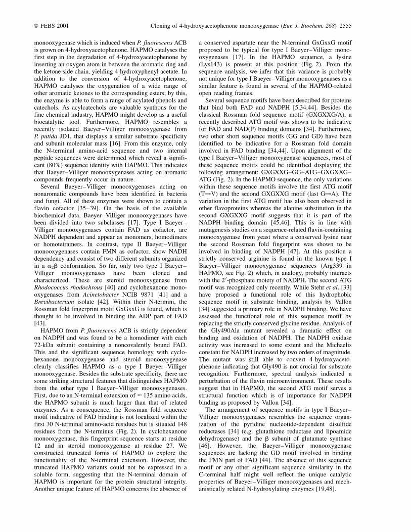
Fig. 2. Multiple sequence alignment of HAPMO from P. fluorescens ACB with other Baeyer±Villiger monooxygenases. The N-terminal
truncated HAPMO sequence is aligned with cyclohexanone monooxygenase from Acinetobacter sp. NCIB 9871 (Acin, P12015) [41], steroid
monooxygenase from Rhodococcus rhodochrous (Rhod, BAA24454) [40] and two cyclohexanone monooxygenases from a Brevibacterium isolate
[42]. The regions indicative for FAD and NADPH binding are marked with asterisks and circles, respectively [34].
q FEBS 2001 Cloning of 4-hydroxyacetophenone monooxygenase (Eur. J. Biochem. 268) 2553

gene were sequenced. Sequencing of a 14-kb fragmentrevealed that the gene encoding HAPMO is the fifth ORF ofan operon encoding genes involved in the degradation of4-hydroxyacetophenone (data not shown). We thereforedesignated it hapE. The sequence has been deposited in theGenbank. The hapE gene encodes a protein of 640 aminoacids with a deduced molecular mass of 71 884 Da, whichis in line with the subunit molecular mass determined forthe native enzyme.
Protein sequence comparison
Alignment with the known Baeyer±Villiger monooxygenasesshowed that HAPMO possesses 33 and 27% sequenceidentity with steroid monooxygenase from Rhodococcusrhodochrous and cyclohexanone monooxygenase fromAcinetobacter sp. NCIB 9871, respectively. Sequenceidentities with the two newly identified cyclohexanonemonooxygenases from a Brevibacterium isolate were 31%for Bre1 and 26% for Bre2 (Fig. 2). Intriguingly,HAPMO has an extension of <135 residues at theN-terminus which is not found in the other Baeyer±Villigermonooxygenase sequences. Furthermore, relatively highdegrees of sequence identity were found for ORFs fromP. fluorescens (38%), Streptomyces coelicolor (33%),Mycobacterium tuberculosis (33%), and a Rhizobium sp.(32%). This suggests that Baeyer±Villiger monooxy-genases are widespread throughout the eubacteria. Lowerdegrees of sequence identity (20±25%) were found with theeukaryotic flavin-containing monooxygenases. The mostconserved regions, located at the N-terminal half of thehomologous sequences, contain two typical GXGXX(G/A)Rossman fold fingerprints (Fig. 2). These two sequencemotifs are indicative of the presence of two bab dinucleo-tide binding folds [32] binding the adenylate parts of theFAD and NAD(P)H cofactors.
Expression and purification of recombinant HAPMO
For expression in E. coli, the hapE gene was placed undercontrol of a T7 promoter in the expression vector pET5-a.When expression of HAPMO was induced in cells grown at30 8C, part of the enzyme was produced as inclusion bodiesas evidenced by SDS/PAGE analysis of whole cells. Bylowering the temperature to 20 8C, formation of inclusionbodies could be prevented while yielding reasonableexpression of soluble and active enzyme. RecombinantHAPMO was purified from E. coli BL21(DE3)pLysSharbouring pHAPE1 in a two-column procedure (Fig. 3).From 1-L culture, 30 mg enzyme could be purified with ayield of 70%. The A280 /A440 ratio of purified recombinantHAPMO was 15.6 which is similar to the value found forthe enzyme isolated from P. fluorescens ACB. Moreover,the hydrodynamic properties and catalytic features of therecombinant enzyme are identical when compared toHAPMO purified from the wild-type strain.
Truncation of HAPMO
Because the N-terminal region of HAPMO does not showany similarity with known protein sequences it wasanticipated that this part of the protein might not beessential for catalysis. To test this hypothesis, two mutant
constructs, pHAPE2 and pHAPE3, were created that lackthe coding region for the first 135 and 114 N-terminalamino-acid residues, respectively. However, SDS/PAGEanalysis of cell extracts and whole cells transformedwith these constructs showed that there was no visibleexpression of the truncated proteins at an inductiontemperature of 20 8C. Raising the induction temperatureto 30 8C resulted in the formation of inclusion bodies.Activity was no detected in the cell extracts at eitherinduction temperatures. Apparently, the N-terminal part ofHAPMO is of importance for the structural integrity of theenzyme.
Properties of the G490A mutant
Recently, an ATG sequence motif was described to becommon for FAD and NAD(P)H binding proteins [33,34].Since this sequence motif is also found in HAPMO (Fig. 2)and its function is still unclear, we decided to mutate thestrictly conserved glycine 490 into an alanine. The mutantwas overexpressed and purified according to the protocolfor wild-type HAPMO. Gel filtration showed that themutant enzyme was purified as a dimer. The ratio of theabsorbance at 280 nm relative to that at 440 nm was 17.5,indicating that the enzyme is saturated with FAD. However,the shape of the flavin spectrum of the Gly490Ala mutantdiffers significantly from that of the wild-type enzyme(Fig. 1A). The oxidase activity of this mutant is signifi-cantly higher than that of the wild-type enzyme(k 0cat � 0.4´s21) but the Michaelis constant for NADPHis rather high (K 0m(NADPH) � 700 mm). In contrast with thewild-type enzyme, addition of 4-hydroxyacetophenone didnot stimulate the rate of NADPH oxidation. However,GC-MS analysis revealed that the Gly490Ala mutant is stillable to catalyse the Baeyer±Villiger oxidation of thearomatic substrate.
D I S C U S S I O N
This paper describes the purification, characterization andcloning of HAPMO, a novel FAD-dependent Baeyer±Villiger
Fig. 3. SDS/PAGE of purification steps of recombinant 4-hydroxy-
acetophenone monooxygenase from E. coli BL21(DE3)pLysS. Lane
1 and 6, marker proteins; lane 2 and 3, cell extract at two
concentrations; lane 4, hydroxyapatite fraction; lane 5 and 7,
Q-Sepharose fraction at two concentrations.
2554 N. M. Kamerbeek et al. (Eur. J. Biochem. 268) q FEBS 2001
monooxygenase which is induced when P. fluorescens ACBis grown on 4-hydroxyacetophenone. HAPMO catalyses thefirst step in the degradation of 4-hydroxyacetophenone byinserting an oxygen atom in between the aromatic ring andthe ketone side chain, yielding 4-hydroxyphenyl acetate. Inaddition to the conversion of 4-hydroxyacetophenone,HAPMO catalyses the oxygenation of a wide range ofother aromatic ketones to the corresponding esters; by this,the enzyme is able to form a range of acylated phenols andcatechols. As acylcatechols are valuable synthons for thefine chemical industry, HAPMO might develop as a usefulbiocatalytic tool. Furthermore, HAPMO resembles arecently isolated Baeyer±Villiger monooxygenase fromP. putida JD1, that displays a similar substrate specificityand subunit molecular mass [16]. From this enzyme, onlythe N-terminal amino-acid sequence and two internalpeptide sequences were determined which reveal a signifi-cant (80%) sequence identity with HAPMO. This indicatesthat Baeyer±Villiger monooxygenases acting on aromaticcompounds frequently occur in nature.
Several Baeyer±Villiger monooxygenases acting onnonaromatic compounds have been identified in bacteriaand fungi. All of these enzymes were shown to contain aflavin cofactor [35±39]. On the basis of the availablebiochemical data, Baeyer±Villiger monooxygenases havebeen divided into two subclasses [17]. Type I Baeyer±Villiger monooxygenases contain FAD as cofactor, areNADPH dependent and appear as monomers, homodimersor homotetramers. In contrast, type II Baeyer±Villigermonooxygenases contain FMN as cofactor, show NADHdependency and consist of two different subunits organizedin a a2b conformation. So far, only two type I Baeyer±Villiger monooxygenases have been cloned andcharacterized. These are steroid monooxygenase fromRhodococcus rhodochrous [40] and cyclohexanone mono-oxygenases from Acinetobacter NCIB 9871 [41] and aBrevibacterium isolate [42]. Within their N-termini, theRossman fold fingerprint motif GxGxxG is found, which isthought to be involved in binding the ADP part of FAD[43].
HAPMO from P. fluorescens ACB is strictly dependenton NADPH and was found to be a homodimer with each72-kDa subunit containing a noncovalently bound FAD.This and the significant sequence homology with cyclo-hexanone monooxygenase and steroid monooxygenaseclearly classifies HAPMO as a type I Baeyer±Villigermonooxygenase. Besides the substrate specificity, there aresome striking structural features that distinguishes HAPMOfrom the other type I Baeyer±Villiger monooxygenases.First, due to an N-terminal extension of < 135 amino acids,the HAPMO subunit is much larger than that of relatedenzymes. As a consequence, the Rossman fold sequencemotif indicative of FAD binding is not localized within thefirst 30 N-terminal amino-acid residues but is situated 148residues from the N-terminus (Fig. 2). In cyclohexanonemonooxygenase, this fingerprint sequence starts at residue12 and in steroid monooxygenase at residue 27. Weconstructed truncated forms of HAPMO to explore thefunctionality of the N-terminal extension. However, thetruncated HAPMO variants could not be expressed in asoluble form, suggesting that the N-terminal domain ofHAPMO is important for the protein structural integrity.Another unique feature of HAPMO concerns the absence of
a conserved aspartate near the N-terminal GxGxxG motifproposed to be typical for type I Baeyer±Villiger mono-oxygenases [17]. In the HAPMO sequence, a lysine(Lys143) is present at this position (Fig. 2). From thesequence analysis, we infer that this variance is probablynot unique for type I Baeyer±Villiger monooxygenases as asimilar feature is found in several of the HAPMO-relatedopen reading frames.
Several sequence motifs have been described for proteinsthat bind both FAD and NADPH [5,34,44]. Besides theclassical Rossman fold sequence motif (GXGXXG/A), arecently described ATG motif was shown to be indicativefor FAD and NAD(P) binding domains [34]. Furthermore,two other short sequence motifs (GG and GD) have beenidentified to be indicative for a Rossman fold domaininvolved in FAD binding [34,44]. Upon alignment of thetype I Baeyer±Villiger monooxygenase sequences, most ofthese sequence motifs could be identified displaying thefollowing arrangement: GXGXXG±GG±ATG±GXGXXG±ATG (Fig. 2). In the HAPMO sequence, the only variationswithin these sequence motifs involve the first ATG motif(T!V) and the second GXGXXG motif (last G!A). Thevariation in the first ATG motif has also been observed inother flavoproteins whereas the alanine substitution in thesecond GXGXXG motif suggests that it is part of theNADPH binding domain [45,46]. This is in line withmutagenesis studies on a sequence-related flavin-containingmonooxygenase from yeast where a conserved lysine nearthe second Rossman fold fingerprint was shown to beinvolved in binding of NADPH [47]. At this position astrictly conserved arginine is found in the known type IBaeyer±Villiger monooxygenase sequences (Arg339 inHAPMO, see Fig. 2) which, in analogy, probably interactswith the 2 0-phosphate moiety of NADPH. The second ATGmotif was recognized only recently. While Stehr et al. [33]have proposed a functional role of this hydrophobicsequence motif in substrate binding, analysis by Vallon[34] suggested a primary role in NADPH binding. We haveassessed the functional role of this sequence motif byreplacing the strictly conserved glycine residue. Analysis ofthe Gly490Ala mutant revealed a dramatic effect onbinding and oxidation of NADPH. The NADPH oxidaseactivity was increased to some extent and the Michaelisconstant for NADPH increased by two orders of magnitude.The mutant was still able to convert 4-hydroxyaceto-phenone indicating that Gly490 is not crucial for substraterecognition. Furthermore, spectral analysis indicated aperturbation of the flavin microenvironment. These resultssuggest that in HAPMO, the second ATG motif serves astructural function which is of importance for NADPHbinding as proposed by Vallon [34].
The arrangement of sequence motifs in type I Baeyer±Villiger monooxygenases resembles the sequence organ-ization of the pyridine nucleotide-dependent disulfidereductases [34] (e.g. glutathione reductase and lipoamidedehydrogenase) and the b subunit of glutamate synthase[46]. However, the Baeyer±Villiger monooxygenasesequences are lacking the GD motif involved in bindingthe FMN part of FAD [44]. The absence of this sequencemotif or any other significant sequence similarity in theC-terminal half might well reflect the unique catalyticproperties of Baeyer±Villiger monooxygenases and mech-anistically related N-hydroxylating enzymes [19,48].
q FEBS 2001 Cloning of 4-hydroxyacetophenone monooxygenase (Eur. J. Biochem. 268) 2555

Expression of HAPMO, under the control of the T7promotor [49], in E. coli BL21(DE3)pLysS resulted inrelatively large amounts of soluble and active enzyme. Thisallows us to address in the near future the catalyticmechanism and structure±function relationship of thisnovel aromatic Baeyer±Villiger monooxygenase in furtherdetail.
A C K N O W L E D G E M E N T S
This work was supported by the Council for Chemical Sciences of the
Netherlands Organization for Scientific Research (CW-NWO), division
`Procesvernieuwing voor een schoner milieu' and by the European
Community (Framework IV, project BIO4-CT98-0267).
R E F E R E N C E S
1. Harayama, S., Kok, M. & Neidle, E.L. (1992) Functional and
evolutionary relationships among diverse oxygenases. Annu. Rev.
Microbiol. 46, 565±601.
2. Suske, W.A., Held, M., Schmid, A., Fleischmann, T., Wubbolts,
M.G. & Kohler, H.-P.E. (1997) Purification and characterization of
2-hydroxybiphenyl 3-monooxygenase, a novel NADH-dependent,
FAD-containing aromatic hydroxylase from Pseudomonas aze-
laica HBP1. J. Biol. Chem. 272, 24257±24265.
3. Arunachalam, U., Massey, V. & Miller, S.M. (1994) Mechanism
of p-hydroxyphenylacetate-3-hydroxylase. A two-protein enzyme.
J. Biol. Chem. 269, 150±155.
4. Becker, D., Schrader, T. & Andreesen, J.R. (1997) Two-component
flavin-dependent pyrrole-2-carboxylate monooxygenase from
Rhodococcus sp. Eur. J. Biochem. 249, 739±747.
5. Eppink, M.H.M., Schreuder, H.A. & van Berkel, W.J.H. (1997)
Identification of a novel conserved sequence motif in flavoprotein
hydroxylases with a putative dual function in FAD/NAD(P)H
binding. Protein Sci. 6, 2454±2458.
6. Entsch, B. & van Berkel, W.J.H. (1995) Structure and mechanism
of para-hydroxybenzoate hydroxylase. FASEB J. 9, 476±483.
7. Eppink, M.H.M., Overkamp, K.M., Schreuder, H.A. & Van Berkel,
W.J.H. (1999) Switch of coenzyme specificity of p-hydroxy-
benzoate hydroxylase. J. Mol. Biol. 292, 87±96.
8. Entsch, B., Ballou, D.P. & Massey, V. (1976) Flavin-oxygen
derivatives involved in hydroxylation by p-hydroxybenzoate
hydroxylase. J. Biol. Chem. 251, 2550±2563.
9. Xun, L.Y. & Sandvik, E.R. (2000) Characterization of 4-hydroxy-
phenylacetate 3-hydroxylase (HpaB) of Escherichia coli as a
reduced flavin adenine dinucleotide-utilizing monooxygenase.
Appl. Environ. Microbiol. 66, 481±486.
10. Galan, B., Diaz, E., Prieto, M.A. & Garcia, J.L. (2000) Functional
analysis of the small component of the 4-hydroxyphenylacetate
3-monooxygenase of Escherichia coli W: a prototype of a new
flavin: NAD(P)H reductase subfamily. J. Bacteriol. 182, 627±636.
11. Cripps, R.E. (1975) The microbial metabolism of acetophenone.
Metabolism of acetophenone and some chloroacetophenones by an
Arthrobacter species. Biochem. J. 152, 233±241.
12. Havel, J. & Reineke, W. (1993) Microbial degradation of
chlorinated acetophenones. Appl. Environ. Microbiol. 59,
2706±2712.
13. Higson, F.K. & Focht, D.D. (1990) Bacterial degradation of
ring-chlorinated acetophenones. Appl. Environ. Microbiol. 56,
3678±3685.
14. Darby, J.M., Taylor, D.G. & Hopper, D.J. (1987) Hydroquinone as
the ring-fission substrate in the catabolism of 4-ethylphenol and
4-hydroxyacetophenone by Pseudomonas putida JD1. J. Gen.
Microbiol. 133, 2137±2146.
15. Jones, K.H., Trudgill, P.W. & Hopper, D.J. (1994) 4-Ethylphenol
metabolism by Aspergillus fumigatus. Appl. Environ. Microbiol.
60, 1978±1983.
16. Tanner, A. & Hopper, D.J. (2000) Conversion of 4-hydroxyaceto-
phenone into 4-phenyl acetate by a flavin adenine dinucleotide-
containing Baeyer±Villiger-type monooxygenase. J. Bacteriol.
182, 6565±6569.
17. Willetts, A. (1997) Structural studies and synthetic applications of
Baeyer-Villiger monooxygenases. Trends Biotechnol. 15, 55±62.
18. Ryerson, C.C., Ballou, D.P. & Walsh, C. (1982) Mechanistic
studies on cyclohexanone oxygenase. Biochemistry 21, 2644±2655.
19. Sheng, D., Ballou, D.P. & Massey, V. (1999) The intermediates
involved in the catalytic reaction of cyclohexanone monooxy-
genase. In Flavins and Flavoproteins, 13th International Congress
on Flavins and Flavoproteins (Ghisla, S., Kroneck, P., Macheroux,
P. & Sund, H., eds), pp. 367±370. Agency for Scientific
Publication, Berlin.
20. Massey, V. (1994) Activation of molecular oxygen by flavins and
flavoproteins. J. Biol. Chem. 269, 22459±22462.
21. Moonen, M.J.H., Rietjens, I.M.C.M. & van Berkel, W.J.H. (1999)
Purification and some properties of acetophenone monooxygenase.
In Flavins and Flavoproteins, 13th International Congress on
Flavins and Flavoproteins (Ghisla, S., Kroneck, P., Macheroux, P.
& Sund, H., eds), pp. 375±378. Agency for Scientific Publication,
Berlin.
22. Krockel, L. & Focht, D.D. (1987) Construction of chlorobenzene-
utilizing recombinants by progenitive manifestation of a rare
event. Appl. Environ. Microbiol. 53, 2470±2475.
23. Sambrook, J., Fritsch, E.F. & Maniatis, T. (1989) Molecular
Cloning: A Laboratory Manual, 2nd edn. Cold Spring Harbor
Laboratory Press, New York.
24. Staskawicz, B., Dahlbeck, D., Keen, N. & Napoli, C. (1987)
Molecular characterization of cloned avirulence genes from race 0
and race 1 of Pseudomonas syringae pv. glycinea. J. Bacteriol.
169, 5789±5794.
25. Innis, M.A. & Gelfand, D.H. (1990) A guide to methods and
amplification. In PCR Protocols (Innis, M.A., Gelfand, D.H.,
Sninsky, J.J. & White, T.J., eds), pp. 3±12. Academic Press, San
Diego.
26. Don, R.H., Cox, P.T., Wainwright, B.J., Baker, K. & Mattick, J.S.
(1991) `Touchdown' PCR to circumvent spurious priming during
gene amplification. Nucleic Acids Res. 19, 4008.
27. Eppink, M.H.M., Boeren, S.A., Vervoort, J. & van Berkel, W.J.H.
(1997) Purification and properties of 4-hydroxybenzoate 1-hydro-
xylase (decarboxylating), a novel flavin adenine dinucleotide-
dependent monooxygenase from Candida parapsilosis CBS604.
J. Bacteriol. 179, 6680±6687.
28. Eschrich, K., van der Bolt, F.J.T., de Kok, A. & van Berkel, W.J.H.
(1993) Role of Tyr201 and Tyr385 in substrate activation by
p-hydroxybenzoate hydroxylase from Pseudomonas fluorescens.
Eur. J. Biochem. 216, 137±146.
29. Altschul, S.F., Madden, T.L., Schaeffer, A.A., Zhang, J., Zhang, Z.,
Miller, W. & Lipman, D.J. (1997) Gapped BLAST and
PSI-BLAST: a new generation of protein database search
programs. Nucleic Acids Res. 25, 3389±3402.
30. Thompson, J.D., Higgins, D.G. & Gibson, T.J. (1994) CLUSTAL
W: improving the sensitivity of progressive multiple sequence
alignment through sequence weighting, position-specific gap
penalties and weight matrix choice. Nucleic Acids Res. 22,
4673±4680.
31. Suske, W.A., van Berkel, W.J.H. & Kohler, H.P.E. (1999) Catalytic
mechanism of 2-hydroxybiphenyl 3-monooxygenase, a flavo-
protein from Pseudomonas azelaica HBP1. J. Biol. Chem. 274,
33355±33365.
32. Wierenga, R.K., Terpstra, P. & Hol, W.G. (1986) Prediction of the
occurrence of the ADP-binding beta alpha beta-fold in proteins,
2556 N. M. Kamerbeek et al. (Eur. J. Biochem. 268) q FEBS 2001

using an amino-acid sequence fingerprint. J. Mol. Biol. 187,
101±107.
33. Stehr, M., Diekmann, H., Smau, L., Seth, O., Ghisla, S., Singh, M.
& Macheroux, P. (1998) A hydrophobic sequence motif common
to N-hydroxylating enzymes. Trends Biochem. Sci. 23, 56±57.
34. Vallon, O. (2000) New sequence motifs in flavoproteins: Evidence
for common ancestry and tools to predict structure. Proteins 38,
95±114.
35. Donoghue, N.A., Norris, D.B. & Trudgill, P.W. (1976) The
purification and properties of cyclohexanone oxygenase from
Nocardia globerula CL1 and Acinetobacter NCIB 9871. Eur. J.
Biochem. 63, 175±192.
36. Itagaki, E. (1986) Studies on steroid monooxygenase from
Cylindrocarpon radicicola ATCC 11011. Purification and charac-
terization. J. Biochem. 99, 815±824.
37. Miyamoto, M., Matsumoto, J., Iwaya, T. & Itagaki, E. (1995)
Bacterial steroid monooxygenase catalyzing the Baeyer±Villiger
oxidation of C21-ketosteroids from Rhodococcus rhodochrous: the
isolation and characterization. Biochim. Biophys. Acta 1251,
115±124.
38. Ougham, H.J., Taylor, D.G. & Trudgill, P.W. (1983) Camphor
revisited: involvement of a unique monooxygenase in metabolism
of 2-oxo-delta 3-4,5,5-trimethylcyclopentenylacetic acid by
Pseudomonas putida. J. Bacteriol. 153, 140±152.
39. van der Werf, M.J. (2000) Purification and characterization of a
Baeyer-Villiger mono-oxygenase from Rhodococcus erythropolis
DCL14 involved in three different monocyclic monoterpene
degradation pathways. Biochem. J. 3, 693±701.
40. Morii, S., Sawamoto, S., Yamauchi, Y., Miyamoto, M., Iwami, M.
& Itagaki, E. (1999) Steroid monooxygenase of Rhodococcus
rhodochrous: Sequencing of the genomic DNA, and hyperexpres-
sion, purification, and characterization of the recombinant enzyme.
J. Biochem. 126, 624±631.
41. Chen, Y.C., Peoples, O.P. & Walsh, C.T. (1988) Acinetobacter
cyclohexanone monooxygenase: gene cloning and sequence
determination. J. Bacteriol. 170, 781±789.
42. Brzostowicz, P.C., Gibson, K.L., Thomas, S.M., Blasko, M.S. &
Rouviere, P.E. (2000) Simultaneous identification of two cyclo-
hexanone oxidation genes from an environmental Brevibacterium
isolate using mRNA differential display. J. Bacteriol. 182,
4241±4248.
43. Wierenga, R.K., Drenth, J. & Schulz, G.E. (1983) Comparison of
the three-dimensional protein and nucleotide structure of the
FAD-binding domain of p-hydroxybenzoate hydroxylase with the
FAD- as well as NADPH-binding domains of glutathione
reductase. J. Mol. Biol. 167, 725±739.
44. Eggink, G., Engel, H., Vriend, G., Terpstra, P. & Witholt, B.
(1990) Rubredoxin reductase of Pseudomonas oleovorans. Struc-
tural relationship to other flavoprotein oxidoreductases based on
one NAD and two FAD fingerprints. J. Mol. Biol. 212, 135±142.
45. Bork, P. & Grunwald, C. (1990) Recognition of different
nucleotide-binding sites in primary structures using a property-
pattern approach. Eur. J. Biochem. 191, 347±358.
46. Morandi, P., Valzasina, B., Colombo, C., Curti, B. & Vanoni, M.A.
(2000) Glutamate synthase: identification of the NADPH-binding
site by site-directed mutagenesis. Biochemistry 39, 727±735.
47. Suh, J.K., Poulsen, L.L., Ziegler, D.M. & Robertus, J.D. (1999)
Lysine 219 participates in NADPH specificity in a flavin-
containing monooxygenase from Saccharomyces cerevisiae.
Arch. Biochem. Biophys. 372, 360±366.
48. van Berkel, W.J.H., Eppink, M.H.M. & Schreuder, H.A. (1999)
Coenzyme recognition by flavoprotein aromatic hydroxylases. In
Flavins and Flavoproteins, 13th International Congress on
Flavins and Flavoproteins (Ghisla, S., Kroneck, P., Macheroux,
P. & Sund, H., eds), pp. 343±349. Agency for Scientific
Publication, Berlin.
49. Studier, F.W. & Moffatt, B.A. (1986) Use of bacteriophage T7
RNA polymerase to direct selective high-level expression of
cloned genes. J. Mol. Biol. 189, 113±130.
q FEBS 2001 Cloning of 4-hydroxyacetophenone monooxygenase (Eur. J. Biochem. 268) 2557