UNIVERSITl SAINS MALAYSIA -...
43
PREVALANCE AND MOLECULAR EPIDEMIOLGY OF Clostridium difficile INFECTION IN HOSPITAL UNIVERSITI SAINS MALAYSIA PATIENTS AND ELDERLY COMMUNITY SUBJECTS IN KELANTAN DR NADIAH HANIM ZAINUL DISSERTATION SUBMITTED IN PARTIAL FULLFILLMENT OF THE REQUIREMENT FOR THE DEGREE OF MASTER OF MEDICINE (INTERNAL MEDICINE) UNIVERSITl SAINS MALAYSIA UNJVERSITI SAIN S MALAYS IA 201 6
Transcript of UNIVERSITl SAINS MALAYSIA -...

PREV ALANCE AND MOLECULAR EPIDEMIOLGY OF
Clostridium difficile INFECTION
IN HOSPITAL UNIVERSITI SAINS MALAYSIA PATIENTS
AND ELDERLY COMMUNITY SUBJECTS IN KELANTAN
DR NADIAH HANIM ZAINUL
DISSERTATION SUBMITTED IN PARTIAL FULLFILLMENT OF THE
REQUIREMENT FOR THE DEGREE OF MASTER OF MEDICINE
(INTERNAL MEDICINE)
UNIVERSITl SAINS MALAYSIA
UNJVERSITI SAINS MALAYSIA
201 6
PREVALANCE AND MOLECULAR EPIDEMIOLGY OF Clostridium difficile
INFECTION IN HOSPITAL UNIVERSITI SAINS MALAYSIA PATIENTS AND
ELDERLY COMMUNITY SUBJECTS IN KELANTAN
Dr Nadiah Hanim Zainul
MMed Internal Medicine
Department of Internal Medicine
School of Medical Sciences, Universiti Sains Malaysia
Health Campus, 16150 Kelantan, Malaysia
Introduction: An increase in the incidence of Clostridium difficile infection (CDI) in
Western countries has come to prominence over the last 15 years. However awareness and
surveillance of CDI in Asia remained poor with epidemiological data being scanty in Asia and
in particular Malaysia. CDI is commonly associated with nosocomial infections, but
community acquired CDI has been reported with increasing frequency lately. Despite the
increase in incidence and severity of CDI, a recent survey found awareness of CDI being poor,
with underestimation of its contribution to antibiotic-associated disease and recurrence rates.
Objective: The aims of this study were to explore the prevalence and associated risk
factors of CDI in hospitalized patients in HUSM, to explore the carriage rate among the elderly
in the community in Kelantan, and to determine the level of awareness of CDI among staff and
students in HUSM.
ACKNOWLEDGEMENT
I would like to begin by expressing my gratitude to the Almighty for his guidance and blessing
without which I would not be able to embark on this mammoth task of beginning and
completing this dissertation.
I would like to extend my upmost gratitude to my supervisor, Assoc Prof Dr Lee Yeong Yeh
for his extremely valuable guidance, criticism and suggestion in the completion of this
dissertation. He has always been there to guide me patiently and despite all my shortcomings,
he still kept supporting and pushing me patiently to complete this dissertation. For this, I am
forever grateful to his efforts and guidance.
I would like to thank Professor Dato Dr Zurkurnai Yusof, Head of Department of Internal
Medicine, School of Medical Sciences, Universiti Sains Malaysia (USM) for his support,
suggestion, guidance and encouragement during the completion of this dissertation.
Last but not least I would like to thank my family. My father Dato Dr Zainul Hamzah and my
mother Datin Shamsol Fahar Shawal who have always been the pillars of my strength and have
supported me through thick and thin. Without their love and understanding, I would not be
where I am now. Thank you to my brother, Muhammad Faris and my sister, Suraya Hanim
who were always there for me with their support and love.
NADIAH HANIM ZAINUL
TABLE OF CONTENTS
CONTENTS
PAGE NUMBER
List of tables and figures i-iii
List of abbreviations iv-v
Abstract in English vi-viii
Abstract in Malay ix-xi
CHAPTER 1: INTRODUCTION AND LITERATURE REVIEW
1.0 Introduction
1.1 Clostridium difficile – The organism 1
1.2 Epidemiology of Clostridium difficile infection 1-4
1.2.1 Nosocomial Clostridium difficile infection
1.2.2 Community acquired Clostridium difficile infection
1.2.3 Epidemiology in Malaysia
1.2.4 PCR Ribotypes in Asia
1.3 Pathogenesis of Clostridium difficile infection 4-8
1.3.1 Factors affecting virulence
1.3.1.1 Toxins
1.3.1.2 Sporulation and germination
1.3.1.3 Surface layer proteins and adherence
1.3.1.4 Toxin variant strains
1.3.1.5 Host immune response

1.4 Risk factors of Clostridium difficile infection 8-14
1.4.1 Drug related risk factors
1.4.1.1 Antibiotics use
1.4.1.2 Proton pump inhibitors
1.4.1.3 Cancer chemotherapy
1.4.1.4 Immunosuppressive agents
1.4.2 Non-drug related risk factors
1.4.2.1 Age
1.4.2.2 Prolonged Hospital Stay
1.4.2.3 Co-morbidity
1.4.2.4 Charlson Comorbidity Index
1.5 Clinical presentation of Clostridium difficile infection 14-17
1.5.1 Asymptomatic carriage
1.5.2 Mild to moderate CDI
1.5.3 Severe CDI
1.5.4 Fulminant CDI
1.6 Diagnosis of Clostridium difficile infection 17-20
1.6.1 Laboratory diagnosis
1.6.2 Endoscopic and radiologic diagnosis
1.7 Treatment of Clostridium difficile infection 20-21

CHAPTER 2: STUDY OBJECTIVES
2.1 General Objective 22
2.2 Specific Objectives 22-23
2.2.1 Hospital study
2.2.2 Community study
2.2.3 Awareness study
2.3 Research Questions 23
CHAPTER 3: METHODOLOGY
3.1 Study Design 24
3.2 Study Location and Study Duration 24 – 25
3.3 Reference Population and Source Population 25
3.4 Sampling Frame 26
3.5 Inclusion and Exclusion Criteria 26 – 27
3.6 Sample Size Calculation 27 – 30
3.7 Sampling Method 30
3.8 Research Tools and Operational Definitions 31 – 36
3.8.1 Study tools
3.8.2 Stool sample, antigen and toxin detection
3.8.3 Stool culture and ribotyping
3.8.4 Variables recorded in data entry form
3.8.5 Definition of recorded variables
3.8.6 Research and ethics committee
3.9 Statistical Analysis 36 – 37
3.10 Study Flow Chart 38 – 40
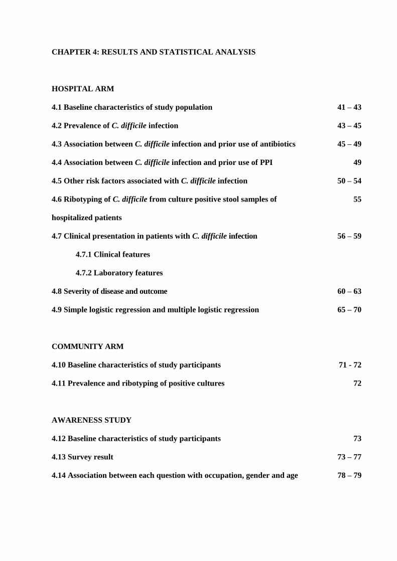
CHAPTER 4: RESULTS AND STATISTICAL ANALYSIS
HOSPITAL ARM
4.1 Baseline characteristics of study population 41 – 43
4.2 Prevalence of C. difficile infection 43 – 45
4.3 Association between C. difficile infection and prior use of antibiotics 45 – 49
4.4 Association between C. difficile infection and prior use of PPI 49
4.5 Other risk factors associated with C. difficile infection 50 – 54
4.6 Ribotyping of C. difficile from culture positive stool samples of
hospitalized patients
55
4.7 Clinical presentation in patients with C. difficile infection 56 – 59
4.7.1 Clinical features
4.7.2 Laboratory features
4.8 Severity of disease and outcome 60 – 63
4.9 Simple logistic regression and multiple logistic regression 65 – 70
COMMUNITY ARM
4.10 Baseline characteristics of study participants 71 - 72
4.11 Prevalence and ribotyping of positive cultures 72
AWARENESS STUDY
4.12 Baseline characteristics of study participants 73
4.13 Survey result 73 – 77
4.14 Association between each question with occupation, gender and age 78 – 79

CHAPTER 5: DISCUSSION
HOSPITAL ARM
5.1 Prevalence of C. difficile infection 80
5.2 Association between C. difficile infection and prior use of antibiotics 81 – 82
5.3 Association between C. difficile infection and prior use of PPI 82
5.4 Other risk factors associated with C. difficile infection 83 – 84
5.5 Ribotyping of C. difficile from culture positive stool samples of
hospitalized patients
85
5.6 Clinical presentation in patients with C. difficile infection 85 – 87
5.7 Severity of disease and outcome 87
COMMUNITY ARM
5.8 Carriage rate of C. difficile among the elderly in the community 88
5.9 Ribotyping of C. difficile from culture positive stool samples of
elderly in the community
89
AWARENESS STUDY
5.10 Awareness of C. difficile infection among medical staff and students in HUSM 90 – 91
CHAPTER 6: CONCLUSION 92
CHAPTER 7: LIMITATION AND RECOMMENDATIONS 93 - 94
REFERENCES 95 – 107
APPENDIX 108
i
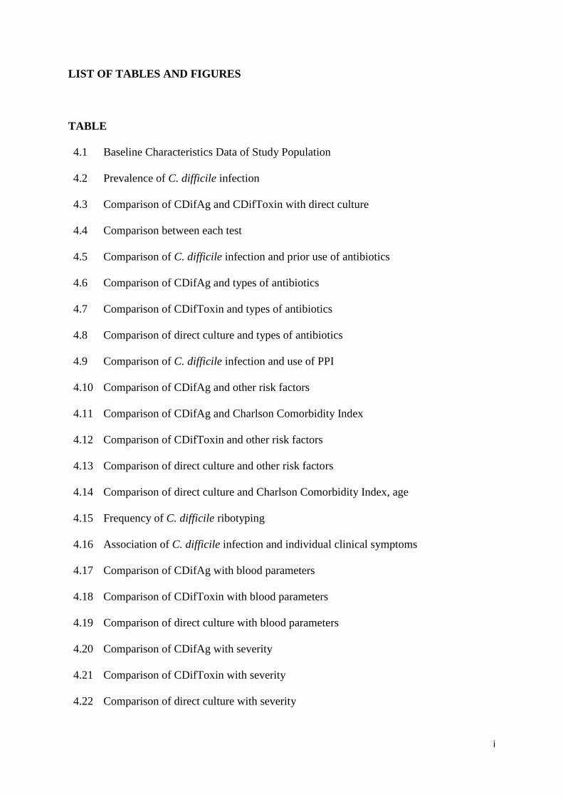
LIST OF TABLES AND FIGURES
TABLE
4.1
4.2
4.3
4.4
4.5
4.6
4.7
4.8
4.9
4.10
4.11
4.12
4.13
4.14
4.15
4.16
4.17
4.18
4.19
4.20
4.21
4.22
Baseline Characteristics Data of Study Population
Prevalence of C. difficile infection
Comparison of CDifAg and CDifToxin with direct culture
Comparison between each test
Comparison of C. difficile infection and prior use of antibiotics
Comparison of CDifAg and types of antibiotics
Comparison of CDifToxin and types of antibiotics
Comparison of direct culture and types of antibiotics
Comparison of C. difficile infection and use of PPI
Comparison of CDifAg and other risk factors
Comparison of CDifAg and Charlson Comorbidity Index
Comparison of CDifToxin and other risk factors
Comparison of direct culture and other risk factors
Comparison of direct culture and Charlson Comorbidity Index, age
Frequency of C. difficile ribotyping
Association of C. difficile infection and individual clinical symptoms
Comparison of CDifAg with blood parameters
Comparison of CDifToxin with blood parameters
Comparison of direct culture with blood parameters
Comparison of CDifAg with severity
Comparison of CDifToxin with severity
Comparison of direct culture with severity

ii
4.23
4.24
4.25
4.26
4.27
4.28
4.29
4.30
4.31
4.32
4.33
4.34
5.1
Comparison of C. difficile infection with outcome of disease
Summary of characteristics of study population with C. difficile infection
Factors associated with prevalence of C. difficile infection using Simple Logistic
Regression
Factors associated with C difficile infection using Multiple Logistic Regression
Area under ROC curve
Baseline characteristics of participants.
Prevalence of C. difficile carriage and ribotyping in the community
Baseline characteristics of respondents
Comparison of Question 1 with gender and occupation
Comparison of Question 2 with gender and occupation
Comparison of Question 3 with gender and occupation
Comparison of Question 4 with gender and occupation
Comparison of correct result for each question
FIGURE
3.1
3.2
4.1
4.2
4.3
4.4
C. DIFF QUIK CHEK COMPLETE ® for C. difficile antigen and toxin detection.
C. DIFF QUIK CHEK COMPLETE ® result interpretation
The percentage of respondents’ total number of correct answers
The percentage of respondents’ answers to question regarding frequency of CDAD
treatment failure or recurrence
The percentage of respondents’ answers to question regarding antibiotic
associated colitis attributed to C. difficile
The percentage of respondents’ answers to question regarding types of antibiotics
associated with CDAD
iii
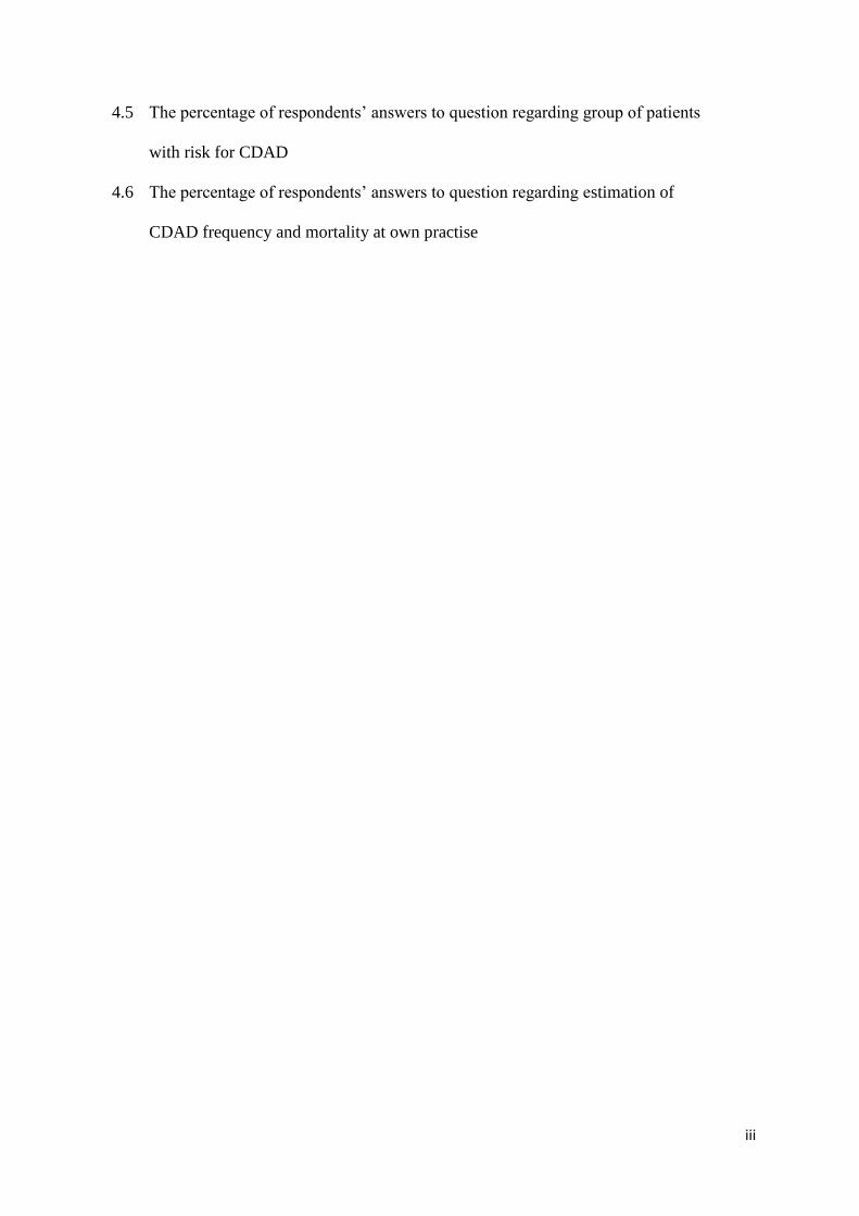
4.5
4.6
The percentage of respondents’ answers to question regarding group of patients
with risk for CDAD
The percentage of respondents’ answers to question regarding estimation of
CDAD frequency and mortality at own practise

iv
LIST OF ABBREVIATIONS
AAD
C. difficile
CCCNA
CDAD
CDI
CDifAg
CDifToxin
CDT
CKD
COPD
CRP
CPE
CT
DM
DNA
EIA
FMT
HIV
HUSM
IBD
IDSA
IgG
IHD
Antibiotics Associated Diarrhoea
Clostridium difficile
C. difficile Culture Cytotoxin Neutralization Assay
Clostridium difficile Associated Diarrhoea
Clostridium difficile Infection
Clostridium difficile Antigen
Clostridium difficile Toxin
Binary Toxin
Chronic Kidney Disease
Chronic Obstructive Pulmonary Disease
C-Reactive Protein
Cytopathic Effect
Computed Tomography
Diabetes Mellitus
Deoxyribonucleic Acid
Enzyme Immunoassay
Faecal microbiota transplantation
Human Immunodeficiency Virus
Hospital Universiti Sains Malaysia
Inflammatory Bowel Disease
Infectious Diseases Society of America
Immunoglobulin G
Ischaemic Heart Disease

v
LTCF
NAAT
NG
NHS
PCR
PPI
RT
TC
TcdA
TcdB
WBC
Long-Term Care Facilities
Nucleic Acid Amplification Test
Nasogastric
National Health Service
Polymerase Chain Reaction
Proton pump inhibitor
Ribotype
Toxigenic Culture
Toxin A
Toxin B
White Blood Cells
vi
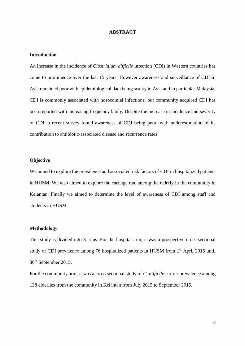
ABSTRACT
Introduction
An increase in the incidence of Clostridium difficile infection (CDI) in Western countries has
come to prominence over the last 15 years. However awareness and surveillance of CDI in
Asia remained poor with epidemiological data being scanty in Asia and in particular Malaysia.
CDI is commonly associated with nosocomial infections, but community acquired CDI has
been reported with increasing frequency lately. Despite the increase in incidence and severity
of CDI, a recent survey found awareness of CDI being poor, with underestimation of its
contribution to antibiotic-associated disease and recurrence rates.
Objective
We aimed to explore the prevalence and associated risk factors of CDI in hospitalized patients
in HUSM. We also aimed to explore the carriage rate among the elderly in the community in
Kelantan. Finally we aimed to determine the level of awareness of CDI among staff and
students in HUSM.
Methodology
This study is divided into 3 arms. For the hospital arm, it was a prospective cross sectional
study of CDI prevalence among 76 hospitalized patients in HUSM from 1st April 2015 until
30th September 2015.
For the community arm, it was a cross sectional study of C. difficile carrier prevalence among
138 elderlies from the community in Kelantan from July 2015 to September 2015.

vii
For both arms, stools were tested for C. difficile antigen and toxin detection using C. DIFF
QUIK CHEK COMPLETE®. The samples were then sent to Western Australia for culture and
PCR for toxin genes and ribotyping for molecular epidemiology.
For the awareness survey, it was a cross sectional study of C. difficile awareness among 154
participants comprised of HUSM staff and students during an awareness campaign for C.
difficile in HUSM on 6th August 2015. Data was obtained thru a self-administered questionnaire
which was based from a previous international internet-based awareness study.
Result
For the hospital arm, 20 samples (26.3%) were positive for C. difficile antigen (CDifAg), 7
samples (9.2%) were positive for C. difficile toxin (CDifToxin) and 19 samples (25%) were
positive from direct culture. Significant ribotype diversity with six distinct ribotype groups
(QX001, UK 017, QX 002, QX 107, QX 117 and QX 463) were identified. Charlson
Comorbidity Index, urea, creatinine, albumin and CRP level, duration of hospitalization, use
of antibiotics, use of chemotherapy, underlying medical illness and fulminant severity were
significantly associated with CDI using Simple Logistic Regression (P-value < 0.25). Further
analysis with Multiple Logistic Regression showed significant association between CDI with
age, duration of hospitalization and use of antibiotics (P-value < 0.05).
For the community arm, 2 samples (1.6%) were positive for both CDifAg and direct culture
while negative for CDifToxin. Ribotying of the 2 samples showed unknown strain. From the
study, it was found that the study population did not have high PPI and antibiotics use which
were known CDI risk factors.
For awareness study arm, there was a low level of awareness on CDI with only 2.6% of 154
respondents able to correctly answer all questions correctly. Ironically a large proportion of the

viii
participants (n = 73; 47.4%) considered C. difficile to be overestimated in their current practise.
There was no significant association between level of awareness on CDI with age, gender and
occupation i.e. being a clinician.
Conclusion
In conclusion, this study demonstrated that the prevalence rate for CDI in hospitalized patients
in HUSM were 26.3% for CDifAg, 9.2% for CDifToxin and 25% for direct culture with 6
distinct ribotype strains; QX001, UK 017, QX 002, QX 107, QX 117 and QX 463 identified.
Independent risk factors for CDI were age, duration of hospitalization and use of antibiotics.
The carrier rate for C. difficile was 1.6% among the elderly in the community with unknown
strain identified from PCR ribotyping. Low usage of PPI and antibiotics were seen in our study
population and could explain the low prevalence rate of CDI in our study population.
Low awareness on CDI was seen among healthcare professionals in HUSM which was also
seen internationally. Therefore CDI being an underdiagnosed and under recognised issue in the
healthcare system is an issue that needs to be addressed by all parties.
ix
ABSTRAK
Pengenalan
Peningkatan kes jangkitan C. difficile (CDI) di negara Barat dapat diperhatikan beberapa tahun
kebelakangan ini. Walaupun CDI menunjukkan peningkatan yang ketara di seluruh dunia, data
epidemiologi berkaitan CDI di Asia terutamanya Malaysia sangat mengecewakan. CDI
biasanya dikaitkan sebagai jangkitan nosokomial, namun kes di dalam komuniti juga telah
dilaporkan meningkat sejak kebelakangan ini. Kajian kesedaran di kalangan pengamal
perubatan seluruh dunia menunjukkan kesedaran tentang CDI yang rendah.
Objektif
Untuk mengenalpasti kekerapan dan faktor risiko CDI di kalangan pesakit yang dimasukkan
ke HUSM . Kajian ini juga bertujuan untuk mengkaji kekerapan pembawa C. difficile di
kalangan warga emas di Kelantan. Kami juga bertujuan untuk menentukan tahap kesedaran
mengenai CDI di kalangan staf dan pelajar di HUSM .
Methodologi
Kajian ini dibahagikan kepada 3 bahagian. Untuk kajian hospital , ia adalah satu kajian keratan
rentas kelaziman di kalangan 76 pesakit yang dimasukkan ke hospital di HUSM dengan CDI
dari 1 April 2015 dan akan berakhir pada 30 September 2015.
Untuk kajian masyarakat , ia adalah satu kajian keratan rentas kelaziman pembawa kuman C.
difficile di kalangan 138 warga emas di dalam masyarakat di Kelantan dari bulan Julai 2015
hingga September 2015.

x
Sampel najis telah diuji untuk C. difficile antigen dan toxin menggunakan C. DIFF QUIK
CHEK COMPLETE®. Sampel najis kemudiannya telah dihantar ke makmal di Western
Australia dan seterusnya dikultur dan menjalani ujian ribotaip bagi epidemiolgi molekular.
Bagi kajian kaji selidik kesedaran CDI, ia merupakan satu kajian keratan rentas di kalangan
154 peserta yang terdiri daripada kakitangan HUSM dan pelajar yang menghadiri kempen
kesedaran C. difficile di HUSM pada 6 Ogos 2015. Data telah diperoleh menerusi borang soal
selidik yang berdasarkan daripada kajian kesedaran peringkat antarabangsa yang telah
berlangsung sebelumnya.
Keputusan
Untuk kajian hospital, 20 sampel (26.3%) didapati positif C. difficile antigen, 7 sampel (9.2%)
positif untuk C. difficile toksin dan 19 sampel (25%) positif daripada kultur secara terus. Enam
kumpulan ribotype berbeza ( QX001 , UK 017, QX 002, QX 107, 117 dan QX QX 463) telah
dikenal pasti. Indeks ‘Comorbidity Charlson’, urea, kreatinine, albumin dan CRP, tempoh
rawatan di hospital, penggunaan antibiotik, penggunaan kemoterapi, pesakit yang mengalami
penyakit perubatan kronik dan tahap penyakit fulminan mempunyai hubungan yang signifikan
dengan CDI menggunakan Simple Logistik Regresion (Nilai-P < 0.25). Analisis lanjut dengan
Multiple Logistic Regression menunjukkan hubungan yang signifikan antara CDI dengan usia,
tempoh rawatan di hospital dan penggunaan antibiotik (Nilai-P < 0.05).
Untuk kajian masyarakat, 2 sampel (1.6%) adalah positif bagi CdifAg dan kultur secara terus
manakala negatif untuk CDifToxin. Ribotying daripada 2 sampel tersebut menunjukkan ia
tergolong didalam strain yang tidak dikenalpasti. Dari kajian ini, didapati bahawa populasi
kajian mempunyai penggunaan antibiotik serta PPI yang rendah.

xi
Untuk kajian kaji selidik kesedaran CDI, terdapat tahap kesedaran yang rendah mengenai CDI
dengan hanya 2.6% daripada 154 responden dapat menjawab semua soalan dengan betul.
Sebahagian besar daripada peserta (n = 73; 47.4%) menganggap C. difficile tidak penting di
dalam amalan semasa mereka. Tidak ada hubungan yang signifikan antara tahap kesedaran
mengenai CDI dengan usia, jantina dan pekerjaan.
Kesimpulan
Pada kesimpulannya, kajian ini menunjukkan bahawa kadar kelaziman untuk CDI adalah
26.3% untuk CDifAg, 9.2% untuk CDifToxin dan 25% untuk kultur secara terus untuk pesakit
di dalam wad. Enam jenis ribotaip yang berbeza iaitu QX 001 , UK 017, QX 002, QX 107, QX
117 dan QX 463 telah dikenal pasti. Faktor-faktor risiko yang mempunyai kaitan dengan CDI
adalah umur, tempoh rawatan di hospital dan penggunaan antibiotik.
Kadar kelaziman pembawa C. difficile didapati sebanyak 1.6% di kalangan warga tua di dalam
masyarakat. Ribotype dari PCR menunjukkan ia tergolong didalam strain yang tidak dapat
dikenal pasti. Penggunaan PPI dan antibiotik yang rendah dapat dilihat di dalam populasi kajian
ini dan dapat menjelaskan kadar kelaziman yang rendah di dalam populasi kajian ini.
Tahap kesedaran yang rendah mengenai C. difficile di kalangan warga HUSM juga dapat
dilihat di peringkat antarabangsa. Oleh itu kepentingan mencegah CDI kurang diberi tumpuan
dan pengiktirafan di dalam sistem penjagaan kesihatan dan ini merupakan satu isu yang perlu
diberi perhatian oleh semua pihak.

1
CHAPTER 1: INTRODUCTION AND LITERATURE REVIEW
1.1 Clostridium difficile (C. difficile) – The organism
C. difficile is an opportunistic, gram-positive, rod-shaped, spore-forming anaerobic bacterium
that exists in the soil and the gastrointestinal tract of animal and humans. It is part of the normal
intestinal microbiota in 1–3% of healthy adults and 15–20% of infants (Goudarzi et al., 2014).
It was first discovered as a new species of bacteria in 1935 by Hall and O’Toole and was named
Bacillus difficilis due to its difficult anaerobic isolation from human faeces. At that time it was
still not identified as a causative agent of human disease and was noted as a component of
normal faecal microbiota of newborn infants (Hall and O'Toole, 1935). Forty years later in the
1970s, it was then discovered that the microorganism was able to produce toxins causing toxin-
mediated infection thus its name was subsequently changed to Clostridium difficile (Kuipers
and Surawicz, 2008).
The bacteria can exist in spore form where it can survive harsh environments and common
sterilization techniques making it resistant to high temperatures, ultraviolet light, harsh
chemicals and antibiotics. Pathogenic strain of C. difficile produces cytotoxin (toxin A and
toxin B) which results in varieties of pathology from being asymptomatic to mild or moderate
diarrhoea, to fulminant and sometimes fatal pseudomembranous colitis.
1.2 Epidemiology of Clostridium difficile infection (CDI)
1.2.1 Nosocomial Clostridium difficile infection
C. difficile is one of the most common nosocomial infections in recent decades, with increasing
prevalence, morbidity and mortality being reported worldwide (Kachrimanidou and
Malisiovas, 2011). Colonisation occurs in 20-40% of hospitalised adults compared with 2-3%
in healthy adults (Heinlen and Ballard, 2010). Studies have also found high rates of

2
asymptomatic Colonisation in neonates and elderly with prevalence ranging from 40-84%
(Karasawa et al., 2005, Hensgens et al., 2012b).
The incidence of CDI has progressively increased. Prevalence of CDI has been reported at 8.75
cases/1,000 adult admissions in United States hospitals. (Chung et al., 2010). Data from the
Canadian nosocomial surveillance between November 2004 and April 2005 showed incidence
of 65 cases per 100,000 patient-days, or 4.6 cases per 1,000 admissions with a large outbreak
in 2003 showing a 4-fold increase in the incidence of CDI (22.2 cases per 100,000 population
in 1991 to 92.2 cases per 100,000 in 2003) (Gilca et al., 2010).
Since 2001, outbreaks in North America have been attributed to a hypervirulent strain of C.
difficile, referred to as ribotype BI/NAP1/027 (toxinotype III). Ribotype 027 has since spread
beyond North America to Europe and Australia, and has been reported in several more
developed Asian countries (Clements et al., 2010, Rupnik et al., 2009). The increased incidence
of infections is accompanied by greater severity of disease caused by this strain, with higher
case-fatality rates and related morbidity (Clements et al., 2010).
Unfortunately data on the incidence of C. difficile infection in Asia in particular are limited but
the available reports suggest an increasing number in countries including Singapore, Taiwan,
Korea, India and Japan. Limited studies indicate that CDI may also be a significant pathogen
in this region, but the true prevalence of CDI remains unknown particularly in South-East Asia.
In the Asia Pacific region, CDI prevalence had increased over the years. In Singapore, the
prevalence increased from 1.49 cases per 10,000 patient-days in year 2001 to 6.64 cases per
10,000 patient-days in 2006 (Lim et al., 2008), while in Malaysia, a study observed the
incidence of CDI cases in northern eastern coast of Malaysia at 13.7% (Hassan et al., 2012a).
A study in Thailand in 2012 among 175 patients showed that 26.9% were positive for antigen
while 12.6% were positive for toxins (Putsathit et al., 2015).

3
1.2.2 Community acquired Clostridium difficile infection
C. difficile has traditionally been linked to disease in hospitalised populations. However
community acquired CDI also occurs and recently has been reported with increasing frequency
(Hensgens et al., 2012b). An estimated 20 % to 28 % of CDI is community associated with an
incidence of 20 to 50 cases per 100 000 population in the United States, Sweden and England
(Kutty et al., 2010, Karlström et al., 1998, Wilcox et al., 2008). Previous studies have shown
that approximately 40% of patients acquiring community-associated CDI were not exposed to
traditional risk factors for CDI such as advanced age, antibiotic exposure, and medications to
suppress gastric acid suggesting that additional factors may contribute to infection. Possible
reservoirs for community-associated disease include soil, water, pets, meat and vegetables
(Heinlen and Ballard, 2010). Transmission in food has been proposed as an explanation for
community-acquired disease with C. difficile being isolated from food. The predominant strain
isolated in food are ribotypes 027 and 078, strains well established to cause human disease
(Carroll, 2011). Community-acquired infections are more commonly associated with binary
toxin-producing strains such as 078 (Riley, 2006).
1.2.3 Epidemiology in Malaysia
Systematic search of PubMed currently yields only four papers mentioning isolation of C.
difficile in Malaysia, three of which were published prior to 1997 (Boey et al., 1997, Parasakhti
et al., 1988, Hassan and Cheng, 1991). The most recent publications introduced systematic
testing for CDI in a hospital in North Eastern Malaysia (Hassan et al., 2012a, Hassan et al.,
2012b). The prevalence of C. difficile was 13.7% among 175 stool samples tested by assay for
toxin A and B. No data on carriage of C. difficile among elderly in the community are available
in Malaysia but this data may be important to explain the high prevalence of C. difficile
infection in hospitals.
4
1.2.4 PCR ribotypes in Asia
Ribotyping data with internationally recognised nomenclature are available for China, Japan,
Singapore, Hong Kong, Taiwan and Korea. Overall, the most prevalent ribotypes in Asia
appear to be 017, 018, 014, 002, and 001 (Collins et al., 2013). These ribotypes are among the
top ten most commonly found ribotypes in Europe.
RT 027, which is still the major ribotype in North America, has been reported only sporadically
in Hong Kong, Japan, South Korea, Singapore and more recently, China. Similarly, RT 078
has only been reported in South Korea and China. RT 017, which is a toxin A-negative, toxin
B-positive (A−B+) strain, is the predominant strain in China and South Korea and is prevalent
in Japan, Taiwan and Hong Kong. This ribotype has also caused epidemics in The Netherlands
and Ireland and is an emerging ribotype in Australia (Collins et al., 2013).
1.3 Pathogenesis of Clostridium difficile infection
C. difficile spores are transmitted from person to person via the faecal-oral route. Bacterial
spores are metabolically dormant and are resistant to desiccation, chemicals and extreme
temperatures. Spores frequently contaminate the environment around patients with CDI,
potentially persisting for months and even years.
There are two forms of the organism, a dormant spore form that is resistant to antibiotics and
a vegetative form that can produce toxins and is susceptible to the activity of antibiotics. The
vegetative form of C. difficile is killed at normal gastric pH (defined as a pH < 4.0), whereas
C. difficile spores may survive exposure to acid in the stomach (McFarland et al., 2007). Spores
that do pass through the stomach germinate to their vegetative form in the small intestine.
Indigenous colonic flora is the first line of defence against Colonisation by pathogens such as
C. difficile. This can be disrupted by antimicrobial drugs, several medications (i.e.,
chemotherapy drugs, proton pump inhibitors), illness or surgical procedures. This disruption

5
allows C. difficile to colonize the intestinal tract, reproduce and cause clinical disease
(Sunenshine and McDonald, 2006). However, only toxigenic strains are associated with the
development of C. difficile diarrhoea as some C. difficile strains do not produce toxins and
therefore do not cause disease.
In summary the pathogenesis of CDI consist of alteration of the normal faecal flora,
Colonisation with toxigenic C. difficile and growth of the organism with elaboration of its
toxins.
1.3.1 Factors affecting virulence
The virulence of the infecting strain and the host’s immune response determine whether a
person develops clinical disease and also determines the severity of disease. C. difficile
virulence factors include toxin production, sporulation, surface layer proteins and adherence,
and toxin variant strain.
1.3.1.1 Toxins
The primary virulence factor of C. difficile is its ability to produce and release two toxins
namely Toxin A (TcdA) and Toxin B (TcdB). Both toxins are cytotoxic and stimulate
production of tumour necrosis factor and pro-inflammatory interleukins which result in
inflammation and increased vascular permeability in the colon, the release and accumulation
of neutrophils and pseudo-membrane formation (Poutanen and Simor, 2004). TcdA play a
more critical role in the pathogenesis of C. difficile diarrhoeal disease than TcdB. TcdA has
been shown to be more closely associated with tissue damage and fluid accumulation
(Bongaerts and Lyerly, 1994, Johnson et al., 1990). These models also suggest that TcdB may
contribute to disease only after TcdA has damaged the gastrointestinal wall. However, they
also observed that either one of the two toxins alone can cause disease (Poxton et al., 2001,

6
Bongaerts and Lyerly, 1994, Barbut et al., 2002). It was evidenced by outbreaks of severe
infection caused by TcdA negative; TcdB positive strains.
Approximately 6 to 12.5% of strains of C. difficile produce another toxin, called C. difficile
transferase (CDT) or binary toxin that is unrelated to either TcdA or TcdB. CDT alone does
not appear to cause disease. The clinical significance of CDT in CDI remains uncertain (Karen
C and John G, 2011). More recent findings however indicate that CDT contributes to an
increased severity of disease as CDT production appears to be epidemiologically associated
with strains producing higher fatality rates (McDonald et al., 2005, Shen, 2012, Dingle et al.,
2011).
1.3.1.2 Sporulation and germination
The rate of C. difficile sporulation is an important virulence factor. ‘Hyper-sporulation’, in
addition to the ability of spores to survive in the environment, propagates the spread of C.
difficile from person-to-person. Therefore it has been postulated that increased sporulation may
be associated with hypervirulence (Merrigan et al., 2010, Dawson et al., 2011).
1.3.1.3 Surface layer proteins and adherence
Surface proteins are integral to the adherence of the organism to the gut mucosa and can induce
both inflammatory and antibody responses in the host (Drudy et al., 2004, Calabi and
Fairweather, 2002, Ausiello et al., 2006). There is considerable variability between the surface
proteins of different strains. Therefore the differences in these proteins may alter a particular
strain’s ability to adhere to intestinal epithelial cells (Calabi and Fairweather, 2002, Drudy et
al., 2004, Péchiné et al., 2005).

7
1.3.1.4 Toxin variant strains, Ribotype 027 , Ribotype 078
C. difficile can be divided into 5 genetic groups or clades (designated clades 1 to 5) that
continue to evolve at the strain level giving rise to hypervirulent types. Examples of these
strains are ribotype 027 (toxinotype III, ST-1, BI/NAP1) and ribotype 078 (ST-11) strains
which are associated with more severe disease and higher mortality rate (He et al., 2013).
Since early 2000, outbreaks in healthcare facilities have spread across the United States,
Canada and Europe attributable to this new highly virulent strain aka C. difficile ribotype 027.
Mortality rates in outbreaks caused by this strain have been three-times higher than in outbreaks
caused by less virulent strains (Pépin et al., 2005, Loo et al., 2005, Warny et al., 2005). This
strain has unique characteristics that may explain the virulence with higher levels of toxin
production, fluoroquinolone resistance and the production of binary toxin. It produces a binary
toxin and has a partial deletion in a toxin regulator gene i.e. TcdC that cause hyperproduction
of TcdA and TcdB in vitro (Warny et al., 2005, Åkerlund et al., 2008). Aside from having
altered TcdC, epidemic 027 strains have five unique genetic regions not present in historical
027 strains (Stabler et al., 2006). These genes include mutations that explain enhanced toxicity,
motility, survival and increased sporulation. Epidemic isolates of C. difficile ribotype 027 were
resistant to fluoroquinolones (McDonald et al., 2005, Loo et al., 2005), which suggests that the
increased use of quinolones may have influenced the emergence of this strain.
Ribotype 078 (toxinotype V) has also contributed to the increased incidence of CDI over the
past 10 years. It is the most common strain isolated from pigs and cattle in the USA (Keel et
al., 2007) and is now the third most common ribotype causing disease in humans in Europe
(Freeman et al., 2010). These strains also produce CDT and carry a 39-bp deletion in TcdC in
combination with a point mutation at position 184 resulting in a stop codon (Freeman et al.,
2010).

8
Ribotypes 027 and 078 produce TcdA and TcdB in higher quantities than other strains,
probably due to the mutated TcdC protein, which has been shown to be non-functional in
ribotype 078 strains (Carter et al., 2011).
1.3.1.5 Host immune response
Host immune response influences the clinical expression of C. difficile infection. Human
immune response to C. difficile develops during infancy. Infants who carry C. difficile develop
antibodies to TcdA and to TcdB. In adults, high titres of serum immunoglobulin G (IgG)
against TcdA promote the development of an asymptomatic carrier state rather than infection
(Kyne et al., 2000). When infection develops, person with high antibody concentrations tend
to have shorter durations of illness and less risk of recurrence than person who lack these
antibodies. Individuals without prompt development of these antibodies to TcdA are more
likely to experience more severe symptoms and have an increased risk for recurrence of CDI
(Kyne et al., 2001, Katchar et al., 2007).
1.4 Risk factors of C. difficile infection
Risk factors for development of CDI are advanced age of 65 years old and more, duration of
hospitalization, exposure to antimicrobial agents, use of acid suppressing medications,
chemotherapy, underlying comorbidity and gastrointestinal surgery or manipulation of the
gastrointestinal tract (including tube feeding) (Cohen et al. 2010).
1.4.1 Drug related risk factors
Drugs that have been implicated in C. difficile infections are antibiotics, immunosuppressive
agents, proton pump inhibitors and cancer therapeutics.

9
1.4.1.1 Antibiotics use
Exposure to antibiotics is the preeminent risk factor. More than 90% of healthcare associated
C. difficile infections are associated with antibiotic use (Sunenshine and McDonald, 2006).
Administration of broad-spectrum antimicrobials causes disruption to the normal intestinal
flora and subsequently promote proliferation of toxigenic C. difficile. Historically, clindamycin
was the first antibiotic implicated in CDAD when it was associated with pseudomembranous
colitis in the early 1970s (Pear et al., 1994). Now it is well known that penicillin, ampicillin,
cephalosporin and fluoroquinolones can all precipitate CDAD. Studies carried out after 1980
showed that cephalosporin is the most common agent implicated in nosocomial CDAD. A
recent meta-analysis by Riley et al. indicate that third-generation cephalosporin remain the
strongest antibiotic risk factor (Slimings and Riley, 2014). In outpatient settings, antibiotics
such as ampicillin, amoxicillin or amoxicillin-clavulanate combination are important and
common causes. Less commonly implicated antibiotics are macrolides, tetracyclines,
sulphonamides, trimethoprim, chloramphenicol and penicillin other than
ampicillin/amoxicillin (Vaishnavi, 2009).
Risk of development of CDI is also increased with multiple antibiotics use and longer course
of therapy. The number of administered antibiotics, their dosage and the duration of therapy
have been identified as factors determining the risk for CDI (Owens et al., 2008). CDI risk is
elevated 7 to 10 fold during antibiotic therapy and the first month after cessation of antibiotics.
It remains elevated for at least 3 months after administration of antibiotics (Hensgens et al.,
2012a).
1.4.1.2 Proton pump inhibitors
Colonisation of normally sterile upper gastrointestinal tract can be a consequence of gastric
acid suppressive use. Lower acidity environment allows vegetative forms of C. difficile to

10
survive. Patients are about twice as likely to develop CDAD with PPI, due to increased survival
of spores. Recent meta-analyses confirm the association of PPI use with an increased risk of
CDI. A meta-analysis by Janarthanan showed a 65% increase in the incidence of CDAD among
PPI users while another meta-analysis by Kwok et al showed an increase of 85% (Janarthanan
et al., 2012, Kwok et al., 2012).
1.4.1.3 Cancer chemotherapy
Patients undergoing antineoplastic chemotherapy are at increased risk for CDI. Administration
of cancer chemotherapeutic agents possessing antibacterial properties may also result in
sufficient disturbance of the intestinal micro flora to allow Colonisation with C. difficile. Some
research have suggested that this association may be related to concurrent use of antimicrobials
and immunosuppression rather than to the use of chemotherapeutic drugs alone (Toor et al.,
2001, Arango et al., 2006).
1.4.1.4 Immunosuppressive agents
Immunosuppressive drugs have been reported to be associated with the development of CDAD.
Patients receiving immunosuppressive drugs are debilitated and therefore are unable to mount
an effective IgG antibody response against C. difficile toxin thereby increasing the risk for
CDAD. Though the ability to mount an immune response is not protective against C. difficile
Colonisation, it is associated with decreased morbidity, mortality and recurrence of CDAD
(Kyne et al., 2001). Patients at highest risk for fulminant disease among others include those
who have recently received immunosuppressive therapy besides have undergone surgical
procedures or those with a history of CDAD (Bartlett, 2002, Dallal et al., 2002).
Exposure to corticosteroids is significantly associated with an increased risk of CDAD relapse
warranting a longer treatment course. C. difficile Colonisation is more frequent in intensive

11
care and oncology units where use of broad spectrum antibiotic and immunosuppression are
wide spread. The use of immunosuppressive may account for a large number of CDAD patients
without prior use of antibiotics. As the use of immunosuppressive increases, the incidence of
CDAD will also rise further (Vaishnavi, 2009).
1.4.2 Non-drug related risk factors
Non-drug related risk factors can be further divided into host risk factors, environmentally
related risk factors and pathogen related risk factors. Host risk factors include age, underlying
co-morbidity, impaired immunity and prolonged hospital stay.
1.4.2.1 Age
The elderly population is at higher risk of developing CDI. Advanced age is one of the most
commonly-cited risk factors for CDI. A study reported that patients over 65 years of age had a
10-fold higher risk for CDI during an outbreak than did younger patients (Pépin et al., 2005).
There is also a trend of increasing CDI related hospital discharges among persons greater than
65 years of age compared to those of other age group (McDonald et al., 2006). Collectively,
higher incidence and severity of infection among older persons are most likely related to the
increased likelihood for older persons to have a greater number of comorbid conditions, more
severe illness, suppressed immune systems and hospitalization or residence in long-term-care
settings as compared with younger persons.
1.4.2.2 Prolonged Hospital Stay
The spread of C. difficile within hospitals is well-documented. Early studies showed that
hospitalised populations exhibit much higher rates of Colonisation, with one study reporting
that hospitalised adults have a 20-40% rate of Colonisation compared with a rate of 2-3%

12
among healthy adults (McFarland et al., 1989, Viscidi et al., 1983). The risk of C. difficile
Colonisation increased proportionately with length of hospital stay. It is estimated to be 13%
in patients with hospital stays of up to two weeks and 50% in those with hospital stays longer
than four weeks (Clabots et al., 1992). Patients who share a room with a C. difficile–positive
patient acquire the organism after an estimated hospital stay of 3.2 days, compared with a
hospital stay of 18.9 days for other patients (McFarland et al., 1989).
1.4.2.3 Co-morbidity
A number of specific comorbid conditions are associated with CDI. Specific comorbid
conditions that have been associated with CDI include gastrointestinal diseases, COPD,
malignancy, renal disease or failure, diabetes, HIV and conditions resulting in an
immunocompromised state (Morris et al., 2002, Dial et al., 2008). Agency for Healthcare
Research and Quality (AHRQ) data showed that multiple co-morbidities put patients at risk for
CDI (Elixhauser and Jhung, 2008).
A study within a hospital with endemic CDI found that myocardial infarction, COPD, liver
disease, renal failure, and leukaemia and/or lymphoma were associated with increased risk for
CDI (Dubberke et al., 2007b). However, it is unclear whether the increased risk for CDI is due
to the actual condition or due to treatment for sequela of their chronic illness such as
antimicrobials use thus increasing risk for CDI among these persons (Cunney et al., 1998,
Pituch, 2009). Co-morbidity that contribute to the risk of dying within 30 days include
cognitive impairment and liver, renal and ischemic heart diseases (Welfare et al., 2011).
Persons with chronic underlying conditions are also likely to seek medical care in healthcare
facilities more often than persons who do not have comorbid conditions. These exposures to
healthcare facilities may increase the likelihood for them to be exposed to surfaces and persons

13
contaminated with C. difficile, thus increasing their risk for CDI for a reason other than their
specific underlying illness.
Persons with gastrointestinal conditions are considered to be at particular risk for CDI.
Identifiable risk factors involving gastrointestinal diseases are IBD, bowel ischaemia,
mechanical bowel cleansing, enteric infections that change colonic microflora, prolonged
presence of a nasogastric tube for enteral feeding, use of electronic rectal thermometers, use of
enemas, gastrointestinal stimulants and stool softeners. Studies have found increasing
incidence rates among patients with IBD exceed those in the general hospitalised population
(Rodemann et al., 2007). C. difficile is estimated to be the cause of 5-19% of IBD flares.
Between 1998 and 2004, the highest prevalence rate of CDI occurred among patients with
ulcerative colitis (37.3 per 1,000) followed by patients with Crohn’s disease, patients with non-
IBD gastrointestinal conditions, and finally, the general medical population within the National
Health Service (NHS) (Meyer et al., 2004). The mortality rate was four times higher among
hospitalised patients who had IBD and CDI than among patients hospitalised for IBD alone
and was two times higher among patients hospitalised with CDI alone (Koss et al., 2006,
Ananthakrishnan et al., 2008).
Another established gastrointestinal related risk factor for CDI is manipulation of the
gastrointestinal tract which includes tube feeding. Bliss et al. studied the incidence of C.
difficile acquisition and CDAD in tube-fed and non-tube fed patients and reported that tube-
fed patients, especially those receiving post pyloric tube feeding are at greater risk for
development of CDAD compared with hospitalised, non-tube-fed patients (Bliss et al., 1998).
Renal impairment is also one of the risk factor for CDI. Declining renal function is associated
with impairments in adaptive and innate immunity with resultant increased susceptibility to
infections and infection-related morbidity and mortality. Uraemia affects cell-mediated as well
as innate and adaptive immunity Therefore they are at increased risk of infection and more

14
likely to receive antibiotic therapy. Additionally, individuals with renal insufficiency have
reduced gastric acid secretion which may increase the risk of Colonisation with C. difficile
(Mullane et al., 2013).
1.4.2.4 Charlson Comorbidity Index
Comorbidities, in general, are medical conditions that underlie the primary illness for which a
person is seeking medical attention. These medical conditions increase a person’s total burden
of disease, are likely to contribute to risk of complications or death, and may affect physician
choice of treatment for other illness (Foley et al., 1992). The collective effect of multiple
comorbid conditions was assessed through the use of the Charlson Comorbidity Index. The
Charlson Comorbidity Index was first developed as a weighted index which was shown to
predict one-year mortality in a small cohort of hospitalised patients (Charlson et al., 1987). The
index assigns a weight to each of the 19 conditions based on their potential for increasing the
likelihood of death. Each patient’s specific conditions are identified, at which point the weights
for comorbidities are added to serve as a summary score. This summary score takes into
account both the number of conditions and the risk associated with these conditions into
account. A higher score represents higher levels of comorbidity (Charlson et al., Mackenzie,
1987).
1.5 Clinical presentation of Clostridium difficile infection
CDI manifest with a spectrum of clinical conditions which range from asymptomatic carriage,
mild or moderate diarrhoea, to fulminant and sometimes fatal pseudomembranous colitis, toxic
megacolon and death (Cohen et al., 2010). Patients with clinical symptoms can be stratified
into mild to moderate illness, severe illness and fulminant disease (Bartlett and Gerding, 2008,
Cohen et al., 2010).

15
1.5.1 Asymptomatic carriage
Colonisation with C. difficile is the presence of the organism in a person with no clinical
symptoms e.g. diarrhoea. Symptomatic disease is less often seen in carriers, despite the fact
that most of the C. difficile isolates are toxin producing (Shim et al., 1998). They however may
be a reservoir of C. difficile and contribute to disease transmission especially in long-term care
facilities (Riggs et al., 2007). Based on several studies, the frequency of carrier stage in healthy
adults, hospitalised patients, and patients with long hospital stays are approximately 1-3%, 20-
30%, and 50% respectively (Nakamura et al., 1981, Karasawa et al., 2005, Riggs et al., 2007).
1.5.2 Mild to moderate CDI
Mild disease is characterized by diarrhoea in the absence of signs and symptoms of colitis.
Patients with moderate disease have diarrhoea with evidence of colitis characterized by fever
and abdominal cramps, usually in the lower quadrants. Laboratory abnormalities in mild and
moderate disease include a leucocytosis level of 15,000 cells/μl or lower and a serum creatinine
level less than 1.5 times the premorbid level (Cohen et al., 2010, Bartlett and Gerding, 2008).
For mild-to-moderate disease, diarrhoea is usually the only symptom. The incubation period
from ingestion of C. difficile to onset of symptoms has been estimated to be a median of 2–3
days (McFarland et al., 1989). Patients may experience multiple episodes of diarrhoea but
usually considerably less than 10 per day. Watery stool with a characteristic foul odour is the
usual presentation although mucoid or soft stools may also occur. Presence of gross blood in
the stool is rare (Bartlett, 2002). Systemic symptoms are usually absent in mild disease and
physical examination is remarkable only for mild abdominal tenderness (Sunenshine and
McDonald, 2006). Other clinical features consistent with CDI include abdominal cramps,
fever, leucocytosis, and hypoalbuminemia. Fever occurs in 28%, leucocytosis in 50% and
abdominal pain in 22% of cases (Bartlett et al., 1980).
16
1.5.3 Severe CDI
Infectious Diseases Society of America (IDSA) 2010 guidelines criteria for severe CDI also
took into account blood investigation as part of the criteria on the basis of WBC greater than
15 x 109/L, or a level of creatinine 1.5-fold above the patient’s baseline value (Cohen et al.,
2010). Other characteristics of severe disease include markedly elevated temperature reaching
40°C, pseudomembranous colitis and hypoalbuminemia (serum albumin level of <25 g/L).
Around 10% of cases of CDI have clinical features consistent with severe CDI (Muto et al.,
2005). Severe disease usually present with profuse, non-bloody diarrhoea, abdominal pain,
fever, nausea, anorexia, malaise and abdominal tenderness. It may also cause paralytic ileus
that can evolve into toxic megacolon (Sunenshine and McDonald, 2006).
In up to 20% of patients with severe CDI, diarrhoea and fluid loss are minimal and patients
present instead with abdominal distention and ileus which is in contrast to mild form of CDI,
therefore often leading to misdiagnosis (Dallal et al., 2002). This occurs when the infection
causes paralytic ileus, preventing the passage of stool and most common in postoperative
patients who are receiving narcotics for pain (Bartlett and Gerding, 2008). Therefore even in
the absence of diarrhoea, symptoms such as unexplained fever, leucocytosis and abdominal
pain in a patient with recent antibiotic exposure should raise suspicion of CDI.
A study in Boston showed that patients with severe C. difficile colitis were more likely to have
abdominal pain, tenderness and distention, peritonitis, hemoconcentration, hypoalbuminemia
and an elevated (> 25 x 109/L) or suppressed (< 1.5 x 10⁹/L) white blood cell count (Rubin et
al., 1995). Another study showed C-reactive protein and leukocytes can be moderately or even
highly elevated in 58 % of patients with unexplained leucocytosis but had CDI (Wanahita et
al., 2003). Hypoalbuminemia is also a common feature as CDI is a protein-losing enteropathy
and low albumin is considered a marker of inflammatory states.

17
1.5.4 Fulminant CDI
Fulminant colitis occurs in approximately 3% of CDI patients and is associated with severe
complications which include perforation, peritonitis, prolonged ileus, megacolon and death. It
presents with signs and symptoms of severe toxicity i.e. fever, colitis with severe lower
quadrant or even diffuse abdominal pain, diarrhoea, distension and marked leucocytosis
(Triadafilopoulos and Hallstone, 1991, Kelly et al., 1994). The timing from onset of any CDI
symptoms to fulminate colitis varies from weeks to just a couple of hours of which patients
with rapid progression have worse outcomes (Dallal et al., 2002). CDI is associated with
exacerbation of ulcerative colitis and it was observed that fulminant colitis was reported more
frequently during outbreaks of C. difficile in patients with IBD which carries higher mortality
than those without underlying IBD (Hookman and Barkin, 2009).
1.6 Diagnosis of Clostridium difficile infection
Diagnosis of CDI is based on both clinical and microbiological methods with the presence of
diarrhoea and stool test positive for toxigenic C. difficile or its toxin, colonoscopy, or
histopathologic findings demonstrating pseudomembranous colitis (Cohen et al. 2010). A
recent guideline by the SHEA/IDSA recommended all the laboratory tests be done on the
unformed stool specimens unless ileus is suspected (Cohen et al., 2010). Only watery or loose
stools should be tested for C. difficile because the rate of asymptomatic Colonisation is
relatively high; therefore, testing in persons who do not have diarrhoeal symptoms may identify
patients who are colonized but not infected.
1.6.1 Laboratory diagnosis
Diagnosis of CDI is a clinical diagnosis supported by laboratory findings. Routine laboratory
tests for CDI diagnosis include a cytotoxin assay for Toxin B, a rapid enzyme immunoassay

18
(EIA), a latex agglutination test to detect bacterial antigen and anaerobic stool culture. For the
past 30 years, the two primary reference tests are the C. difficile culture cytotoxin neutralization
assay (CCCNA) and toxigenic culture (TC) (Planche and Wilcox, 2010, Sambol et al., 2000).
CCCNA detects the presence of C. difficile toxins, toxin B and toxin A, in cell culture. If toxin-
induced cytopathic effect (CPE) is observed, C. difficile is confirmed as the cause of infection
by performing a neutralization assay to ensure that the CPE is attributable to C. difficile toxins
rather than nonspecific toxicity by using either C. sordellii or C. difficile antiserum. The
reported sensitivities for CCCNAs range from 65 to 90% (Burnham and Carroll, 2013).
However the downside of the test is the relatively long turnaround time due to the technical
demands of the lab procedures (O'Connor et al., 2001).
Toxigenic culture (TC) is based upon isolating the organism from faecal specimens and
determining if the recovered isolate is a toxin-producing strain (Burnham and Carroll, 2013).
C. difficile culture alone is not sufficient because not all C. difficile strains produce toxin (Rea
et al., 2012, Viscidi et al., 1983, Burnham and Carroll, 2013). In studies that have evaluated
the use of TC performed after a negative direct toxin test, an increased yield of C. difficile by
15-23% has been seen (Carroll, 2011). TC has several utilities. It is important for organism
characterization in the setting of an outbreak or other epidemiological studies; when evaluating
a new test method; for surveillance of drug resistance and to evaluate new therapies; and
occasionally for difficult patient management. The SHEA/IDSA guidelines support the use of
toxigenic culture as the gold standard in method comparison studies (Cohen et al., 2010) but
the labour requirement and turnaround time are not usually considered practical for routine
diagnostic use (Burnham and Carroll, 2013).
The enzyme immunoassay (EIA) for detection of toxins A and B has been the most widely
used diagnostic test for CDI because of its rapid turnaround, low cost and simplicity. There are
a number of commercially available EIAs for C. difficile toxin. However the sensitivity and

19
specificity that have been reported for these assays vary widely, from approximately 40% to
100% (Burnham and Carroll, 2013). Compared with TC, EIAs for toxins A and B have low
sensitivity from 60% to 80% and a specificity from 91% to 99.4% (Eastwood et al., 2009) while
in comparison with CCCNA, sensitivity of 75% to 95% and a specificity of 83% to 98% have
been reported (Goudarzi et al., 2014). Different strains of C. difficile can also provide different
results in toxin EIA assays (Tenover et al., 2010). Due to the inadequate sensitivity of toxin
EIAs for C. difficile testing, it is not considered the best way to make a diagnosis of CDI
(Eastwood et al., 2009, Cohen et al., 2010, Carroll, 2011). The SHEA/IDSA guideline outlines
that EIAs should no longer be considered adequate stand-alone tests for the diagnosis of C.
difficile infection (Cohen et al., 2010).
C. difficile nucleic acid amplification test (NAATs) is the most recent method for detection of
C. difficile. It identifies genes (not the toxin) that encode the toxins (usually toxin B) by using
PCR or loop-mediated isothermal amplification of DNA. These assays have a short turnaround
time, and sensitivities range from 84% to 96% and specificities range from 94 to 99%.
(Deshpande et al., 2011).
1.6.2 Endoscopy and radiology
C. difficile most often causes a nonspecific colitis. In more severe cases, the distinct
macroscopic appearance of pseudomembranous colitis at endoscopy or by histopathologic
examination may be seen. At least 90 % of patients with pseudomembranous colitis
demonstrate either C. difficile or its toxins in stool samples (Wolfhagen et al., 1994). Milder
cases may only reveal nonspecific findings of erythema and oedema.
CT scan is not required for diagnosing CDI, especially for mild-to-moderate disease, but it can
be useful for recognizing more severe forms. Colonic inflammation can also be shown on CT
20
as increased thickening of the colonic wall (Ash et al., 2006) with trapping of contrast material,
pancolitis, pericolonic fat changes and ascites.
1.7 Treatment of Clostridium difficile infection
Treatment should be based on disease severity or whether one is treating initial or recurrent
CDI. In asymptomatic carriers, treatment is not indicated as available data suggest that
treatment in these individuals would not prevent symptomatic transmission or infection
(Goudarzi et al., 2014).
Metronidazole and oral vancomycin are recommended for the treatment of initial episode.
Patients with mild-to-moderate CDI should be treated with metronidazole 500 mg orally 3
times a day for 10 days. Metronidazole has similar efficacy as vancomycin for treatment of
mild to moderate CDI. Unlike vancomycin, metronidazole is well absorbed and its faecal
concentration is very low or none in healthy volunteers and asymptomatic C. difficile carriage
(Vecchio and Zacur, 2012, Khanna and Pardi, 2012). Routine use of vancomycin is not
recommended due to the risk of development of vancomycin resistance in other organisms
especially enterococci (Vecchio and Zacur, 2012, Khanna and Pardi, 2012, Apisarnthanarak et
al., 2002). However failure to respond to metronidazole therapy within 5-7 days should prompt
consideration of a change in therapy to vancomycin at standard dosing. (Musher et al. 2005,
Surawicz et al. 2013). The time to resolution of diarrhoea might be shorter with vancomycin
than with metronidazole therapy (Belmares et al. 2007).
Patients with severe CDI should be treated with vancomycin 125 mg orally four times per day
for 10 days (Cohen et al., 2010). In treatment failure with low dose oral vancomycin or
complicated CDI, it is recommended to use high-dose (500mg every 6 hours) oral vancomycin
plus intravenous metronidazole, 500 mg 3 times a day (Khanna and Pardi, 2012, Cohen et al.,

21
2010). Administration of vancomycin via enema is used for patients with surgical or anatomic
abnormalities.
Treatment of the first recurrence of CDI is the same as the treatment of first episode of CDI. In
patients with a second recurrence of CDI, vancomycin should be the treatment of choice.
Tapered or pulse-dosage vancomycin may reduce the risk of a subsequent recurrence
(McFarland et al., 2002).
Fidaxomicin is a new macrocyclic that might be favoured over oral vancomycin in patients
with multiple recurrences. Fidaxomicin can be applied for treatment of patients at high risk of
recurrent CDI, patients infected with non-hypervirulent strain, patients with multiple episodes
of recurrence, and patients who are not able to tolerate oral vancomycin (Khanna and Pardi,
2012, Knight and Surawicz, 2013).
Faecal microbiota transplantation (FMT) is an alternative therapy for treatment of recurrent
cases of CDI. In this method, normal faecal microbiota in patients is restored using intestinal
microorganisms from a healthy donor stool. FMT has high success rate of in treating CDI with
rapid and enduring response with effectiveness of 92% of cases while the success rate of FMT
via enema, nasogastric route, and colonoscopy was 95%, 76%, and 89%, respectively (Gough
et al., 2011).
22
CHAPTER 2: STUDY OBJECTIVES
2.1 General Objectives
2.1.1 To explore the prevalence and its associated risk factors for C. difficile infection
in hospitalised patients with loose stools or suspected cases in HUSM.
2.1.2 To explore the carriage rate and its associated risk factors for C. difficile among
the elderly in the community from Tumpat and Kota Bharu district in Kelantan.
2.1.3 To determine the awareness of C. difficile infection among hospital staff and
students in HUSM.
2.2 Specific Objectives
2.2.1 Hospital study
2.2.1.1 To determine the prevalence of C. difficile infection in hospitalised patients
with loose stools or suspected cases in HUSM.
2.2.1.2 To determine the association between C. difficile infection and prior use of
antibiotics.
2.2.1.3 To determine the ribotyping of C. difficile from culture positive stool
samples of hospitalised patients.
2.2.1.4 To determine the initial clinical presentation in patients with C. difficile
infection.
2.2.1.5 To determine the severity of disease and outcome in infected hospitalised
patients.

23
2.2.2 Community study
2.2.2.1 To determine the carriage rate of C. difficile among the elderly in the
community.
2.2.2.2 To determine the risk factors associated with C. difficile carriage.
2.2.2.3 To determine the ribotyping of C. difficile from culture positive stool
samples of elderly in the community.
2.2.3 Awareness study
2.2.3.1 To determine the awareness among medical staff and students within HUSM
with regards to C. difficile infection.
2.3 Research Questions
2.3.1 What is the prevalence of C. difficile infection in hospitalised patients in HUSM
and the carriage rate in the elderly community in Kelantan?
2.3.2 Are there any associations between antibiotics and proton pump inhibitor
exposure with the prevalence of C. difficile infection in HUSM and also C.
difficile carriage in the community?
2.3.3 What are other associated factors in the prevalence of C. difficile infection or
carriage?
2.3.4 What is the awareness among medical staff with regards to C. difficile infection?

24
CHAPTER 3: METHODOLOGY
3.1 Study Design
Hospital arm
This is a prospective cross sectional study among hospitalised patients in HUSM with C.
difficile infection. Similar studies are also being conducted concurrently involving three other
centres in Malaysia i.e. Universiti Kebangsaan Malaysia (UKM), Universiti Malaya (UM) and
Universiti Teknologi Mara (UiTM) in collaboration with The University of Western Australia.
Community arm
This is a cross sectional study of C. difficile carrier prevalence among the elderly in the
community in Kelantan.
Awareness Survey
This is a cross sectional study of C. difficile awareness among hospital staff (doctors, non-clinical
medical professionals, paramedics) and students (postgraduate, undergraduate) in HUSM.
3.2 Study Location and Study Duration
Hospital study
This study was carried out in hospitalised C. difficile patients in HUSM from 1st April 2015
until 30th September 2015.