UNIVERSITÀ DEGLI STUDI DI TORINO · mass of the blastocyst. Their properties include the capacity...
67
UNIVERSITÀ DEGLI STUDI DI TORINO Dipartimento di Scienze Oncologiche Dottorato di Ricerca in SISTEMI COMPLESSI APPLICATI ALLA BIOLOGIA POST-GENOMICA XXI CICLO TITOLO DELLA TESI Endothelial and Neural Commitment in Differentiating Embryonic Stem Cells Cndidato Alessio Noghero Tutor Prof. Federico Bussolino Coordinatore del Dottorato Prof. Federico Bussolino Anni Accademici 2005-2008 Settore Scientifico Disciplinare BIO/11
Transcript of UNIVERSITÀ DEGLI STUDI DI TORINO · mass of the blastocyst. Their properties include the capacity...
UNIVERSITÀ DEGLI STUDI DI TORINO
Dipartimento di Scienze Oncologiche
Dottorato di Ricerca inSISTEMI COMPLESSI APPLICATI ALLA BIOLOGIA
POST-GENOMICA
XXI CICLO
TITOLO DELLA TESIEndothelial and Neural Commitment in Differentiating
Embryonic Stem Cells
CndidatoAlessio Noghero
TutorProf. Federico Bussolino
Coordinatore del DottoratoProf. Federico Bussolino
Anni Accademici 2005-2008
Settore Scientifico Disciplinare BIO/11

2
Table of Contents
Abstract .......................................................................................................................................................... 4
Preface ........................................................................................................................................................... 5
Introduction ................................................................................................................................................... 6 1. Generation of embryonic stem cell lines .......................................................................................... 6 2. Defining stemness: pluripotency and self-renewal........................................................................... 8 3. Differentiation of embryonic stem cells...........................................................................................12
3.1. Differentiation of ES cells via embryoid bodies formation ...................................................12 3.2. Direct differentiation of ES cells ...........................................................................................13
4. Maturation of endothelial cells in the embryo: vasculogenesis and angiogenesis..........................15 4.1. Differentiation of ES cells into endothelium by EBs formation or by 2D cultures ...............17
5. Origin of neural cells and neural stem cells in the embryo and in the adult...................................18 5.1. Differentiation of ES cells into neurons by EBs formation or by 2D cultures.......................19
6. Parallelism between endothelial and neural cells differentiation...................................................20
Results ...........................................................................................................................................................22 1. Analysis of Nrp1 expression in differentiating ES cells ..................................................................22 2. Characterization of EBs-derived Nrp1+ cells..................................................................................22 3. Characterization of EBs-derived Nrp1+/Flk1+ and Nrp1+/Flk1- cells populations.........................23 4. In vitro analysis of the potential of differentiation of Nrp1+/Flk1+ and Nrp1+/Flk1- cells..............24
4.1. Endothelial commitment........................................................................................................24 4.2. Neural commitment ...............................................................................................................25
5. In vivo analysis of the potential of differentiation of Nrp1+/Flk1+ and Nrp1+/Flk1- cells ..............26 5.1. Endothelial commitment........................................................................................................26 5.2. Neural commitment ...............................................................................................................27
6. Generation of an in vitro model specific for the differentiation of both endothelial and neural cells 27
6.1. Direct differentiation of endothelial and neural cells from ES cells ......................................28 6.2. Effect of cytokines on the acquirement of endothelial and neural phenotypes......................29
7. Gene expression regulation by cytokine stimulation in the neuro-endothelial model of ES cells differentiation ...........................................................................................................................................30 8. Endothelial and neural differentiation in the vascular niche context .............................................31
Figures ..........................................................................................................................................................32

3
Discussion .....................................................................................................................................................47 Commitment of Nrp1+ cells derived from embryoid body.......................................................................47 In vitro simultaneous endothelial and neural differentiation: a model for vascular niche ..................49
Materials and Methods .................................................................................................................................51
Bibliography .................................................................................................................................................57
4
Abstract
Mouse embryonic stem (ES) cells are pluripotent cell lines derived from the inner cell
mass of the blastocyst. Their properties include the capacity to self renew and to in vitro
differentiate into embryo-like structures, Embryoid Bodies (EB), that contain derivatives
of the ectodermal, mesodermal and endodermal lineages.
In the sphere of vascular biology studies it has been shown in recent years that several
signalling pathways involved in angiogenesis and endothelial cell migration are indeed
regulated by molecules whose role was formerly identified in the nervous system. These
findings suggest a possible interplay between vascular and nervous system during
development. To this regard, by using as surface markers Flk-1 and Nrp-1, the two
receptors of VEGF (Vascular Endothelial Growth Factor), we purified from EBs a
population of cells which can differentiate into endothelial or neural cells upon
appropriate culture conditions.
Since the EB is a heterogeneous structure containing many different cell types, we shifted
to a more defined ES culture system that allows the simultaneous differentiation mainly
of endothelial cells and neurons. The analysis by real-time PCR and immunofluorescence
for the expression of endothelial and neural markers confirmed that this model is suitable
to study endothelial and neural cell interactions. Moreover, our culture system can be
viewed as a microenvironment similar to vascular niche, as many of its components
develop upon appropriate stimuli.

5
Preface
Embryonic stem (ES) cells are pluripotent cells derived from the inner cell mass of
blastocyst-stage embryos. Their importance to modern biology and medicine derives
from two unique characteristics that distinguish them from all other cells identified to
date. First, they have the capacity to self-renew, meaning that they can be expanded and
maintained in culture as pure populations of undifferentiated cells for extended periods of
time. Second, they are pluripotent, possessing the capacity to generate every cell type in
the body under appropriate conditions or stimuli. The pluripotent nature of mouse ES
cells was formally demonstrated by their ability to contribute to all tissues of adult mice,
including the germline, following their injection into host blastocysts (Bradley et al.,
1984). Therefore, ES cells are used as experimental model in order to obtain specific
precursor cells and to analyze early differentiation events that are difficult to study in the
mouse embryo and inaccessible in the humans. Moreover, the ES cell differentiation
system can be viewed as a novel and unlimited source of cells and tissues for
transplantation in support of the treatment of a broad spectrum of diseases.
The first part of this work was aimed to assess the commitment of embryoid bodies
derived cells toward endothelial or neural fate. We showed that the Nrp1+/Flk1+ cell
population can adopt either neural or endothelial phenotype, depending on appropriate
stimuli coming from the microenvironment, both in vitro and in vivo. These data led to
the publication Microenvironment drives the endothelial or neural fate of
differentiating embryonic stem cells coexpressing neuropilin-1 and Flk-1, FASEB
Journal (2008).

6
Introduction
1. Generation of embryonic stem cell lines
In the early 80s, pluripotent cells called embryonic stem cells were isolated from the
inner cell mass (ICM) of developing murine blastocysts (Evans and Kaufman, 1981;
Martin, 1981). In order to derive an ES cell line, embryos of early pre-implantation stage
(3.5 days after fertilization) are required. At this developmental time the outer layer of the
blastocyst is surrounded by the cells of the trophoblast, which will give rise to the
embryonic annexes. Within the blastocyst is present a cavity, the blastocoele, and one
side is occupied by a cellular mass, the ICM, separated from blastocoele by the cells of
the primitive endoderm. The ICM is the source of the epiblast, from which the three
embryonic layers will later originate: the definitive endoderm, mesoderm and ectoderm
and the germ cells as well (Fig. 1). Hence, the peculiar population of cells that compose
the ICM exists in vivo for a very short period of time, and only at this stage is possible to
efficiently derive ES cell lines.
The derivation of ES cells from ICM requires a relatively simple protocol. Embryos at
the expanded blastocyst stage are plated onto a feeder layer composed by murine
embryonic fibroblast. After several days of culture, the epiblast-derived cell mass is
disaggregated and replated. Various types of differentiated colonies arise along with
colonies of a characteristic undifferentiated morphology. The latter are individually
dissociated and replated, thus establishing the ES cell line. They proliferate rapidly in
culture, and clonal populations can be easily initiated from single cell (Smith, 2001).

7
Fig. 1. Lineage diagram of mouse development. Embryonic stem cells are derived from the inner cell mass,
thus they are able to enter all the lineages that are generated in vivo by the epiplast (blue lines). ES cells
can produce also hypoblast derivatives in vitro but rarely do so in vivo (black lines). Modified from Smith,
A.G. (2001). Embryo-derived stem cells: Of Mice and Men. Annu Rev Cell Dev Biol.
Similar techniques were employed in order to derive ES from many species other than
mouse, i.e. hamster (Doetschman et al., 1988), rabbit (Graves and Moreadith, 1993),
mink (Sukoyan et al., 1993), chicken (Pain et al., 1996), rat (Iannaccone et al., 1994), pig
(Shim et al., 1997), cow (First et al., 1994), rhesus monkey (Thomson et al., 1995), and
also human. In this case, human embryos originally produced by in vitro fertilization for
clinical purpose were cultured to the blastocyst stage and the inner cell masses were then
isolated (Thomson et al., 1998). Although basically similar, there are some differences
between mouse and human ES (hES) cells in their culture requirements, the morphology
of the undifferentiated cells in vitro, and the expression of surface antigens (Brivanlou et
al., 2003). Furthermore, hES cells have the potential to differentiate into trophoblast in
vitro, a property not observed for mouse ES cells (Odorico et al., 2001; Thomson and
Odorico, 2000). The generation of hES cell lines has represented a determinant turn in the
cell therapy field. The motivations for this interest consist in the possibility to provide a
virtually unlimited source of cells for tissue repair, to investigate and manipulate specific
gene function in human cells, and to provide large number of phenotypically defined cell

8
types for drug screening. Nevertheless, because of serious ethical concerns in the
derivation and use of hES from embryos the mouse still remains the organism more
easily accessible for developmental biology studies.
2. Defining stemness: pluripotency and self-renewal
In order to routinely expand ES cells in vitro as an undifferentiated population, the
presence of leukemia inhibitory factor (LIF) in the culture medium and/or a feeder layer
of murine embryonic fibroblasts (MEF) are required. Upon these conditions, ES cells can
be expanded indefinitely in culture as a homogeneous population without a requirement
of immortalization. The reproducible production of chimeric embryos by injecting few
ES cells into blastocysts combined to the unlimited generation of identical subclones,
confirm that ES cells undergo symmetrical self-renewal. In fact, in contrast to other
primary cultures, ES cells appear to be immortal and show no evidence of senescence.
This behavior correlates to the high activity of the telomerase, a ribonucleoprotein that,
by maintaining telomere length, plays an important role in replicative life-span (Counter,
1996). In addition, mouse and human-derived ES cells express several exclusive proteins
that can be considered distinctive of the undifferentiated status. These markers include
the stage-specific embryonic antigen (SSEA)-1, SSEA-3, SSEA-4, Oct-3/4, CD9 and
alkaline phosphatase (Koestenbauer et al., 2006), with some differences between mouse
and human ES cells.
Maintenance of the undifferentiated stem cell phenotype is not a cell-autonomous
process: at each ES cell division, the alternative outcomes of self-renewal and
differentiation are decided by the interplay between intrinsic and extrinsic factors. Media
containing all necessary metabolites and nutrients are not sufficient to support either
derivation or maintenance of ES cells. Even though co-culture with a feeder layer was
originally considered essential to maintain ES cells undifferentiated, it was later
discovered that feeders are dispensable when using conditioned media from Buffalo rat
liver cells (Smith and Hooper, 1987). This suggested that the crucial requirement is to
provide key signals coming from the milieu, without which the ES cells differentiate.

9
This finding lead to the identification of a diffusible inhibitor of differentiation, the
cytokine leukemia inhibitory factor (LIF), that could alone sustain murine ES cell self-
renewal in the absence of feeders (Smith et al., 1988). LIF is also produced by feeder
cells, and its expression is stimulated by the presence of ES cells (Rathjen et al., 1990).
On withdrawal of LIF (or feeders), proliferation continues but differentiation occurs, and
stem cells do not persist in the culture beyond a few days. It was demonstrated that the
effect of LIF is mediated via heterodimerization of two members of the class I cytokine
receptors, the low-affinity LIF receptor (LIF-R) and gp130, the signal transducer of
interleukin-6 (Davis et al., 1993; Gearing et al., 1991).
Besides this, ES cells can also be derived and maintained using a combination of
interleukin-6 and soluble interleukin-6 receptor (IL-6/sIL-6R). In this case, signalling is
initiated via formation of gp130 homodimers without involvement of LIF-R (Nichols et
al., 1994; Yoshida et al., 1994). Signals that derive from gp130 are therefore sufficient
for self-renewal. The pathway downstream gp130 is initiated by the recruitment of JAK
kinases, which consequently leads to activation of STAT3 and to the MAP kinase
signaling cascade. In particular, STAT3 is essential to the LIF signalling pathway, and
appears to play a central role in the balance between self-renewal and differentiation
(Burdon et al., 1999; Niwa et al., 1998). However, STAT3 is not specific of ES cells but
is found in a variety of other cell types, and often is associated with differentiation.
Nevertheless, the requirement for gp130 activation appears to be facultative in vivo,
because early embryogenesis can proceed in the absence of gp130 (Nichols et al., 2001).
The POU-domain transcription factor Oct-3/4 is expressed in totipotent and pluripotent
cells, including oocytes, early-cleavage-stages embryos, the ICM of the blastocyst,
epiblast, germ cells and cultured ES, but it is absent from all differentiated somatic cell
types in vitro and in vivo (Okamoto et al., 1990; Rosner et al., 1990). Oct-3/4-deficient
embryos fail to initiate foetal development because the ICM founder cells do not acquire
pluripotency and are restricted to the extraembryonic trophectoderm lineage (Nichols et
al., 1998). Oct-3/4 is a critical regulator for maintenance and differentiation of pluripotent
cells. Indeed, the amount of Oct-3/4 molecule determines three distinct fates of ES cells:
optimal Oct-3/4 level is necessary to maintain the pluripotent status, less than twofold
increase of the optimal level causes differentiation into primitive endoderm and

10
mesoderm, while repression induces loss of pluripotency and dedifferentiation to
trophectoderm (Niwa et al., 2000). A few target genes activated by Oct-3/4 have been
identified to date, among which the most relevant are Fgf4 and Zfp42/Rex1 (Niwa, 2001),
both involved in maintaining self-renewal. However, Oct-3/4, as other transcription
factors belonging to the POU family, can act as both transcriptional activator and
repressor, depending on which cofactor(s) is recruited.
As described so far, Oct-3/4 can be considered a molecular rheostat able to switch the cell
fate from self-renewal to differentiation. It cooperates with two other transcription
factors, namely Nanog and Sox2, creating the regulatory network responsible for the
maintenance of the ES cells’ undifferentiated status.
Nanog is a homeodomain protein whose enhanced expression confers constitutive self-
renewal on ES cells. Nanog mRNA is present in ICM cells of the living embryo and
during development its expression is further restricted to the epiblast. By implantation
stage, Nanog mRNA is downregulated, but it becomes readily detectable in primordial
germ cells. In vitro, Nanog overexpression can relieve completely of LIF-mediated
STAT3 activation, unless Oct-3/4 is deleted. In fact, continued expression of Oct-3/4 is
essential for Nanog-mediated self renewal, suggesting cooperation between these two
transcription factors (Chambers et al., 2003). Furthermore, in cell fusion experiments
Nanog has shown the ability to transfer pluripotency from ES to neural stem cells, thus
being able to re-organize a differentiated epigenome (Silva et al., 2006).
Sox2 is a member of the Sox (SRY-related HMG box) gene family that encodes
transcription factors with a single High-Mobility-Group DNA-binding domain. Sox2, as
Oct-3/4 and Nanog, behave as master regulator of pluripotency and is essential for
mammalian embryogenesis. Sox2 expression is associated with uncommitted dividing
stem and precursor cells of the developing central nervous system. Indeed Sox2 is one of
the earliest known transcription factors expressed in the developing neural tube (Zappone
et al., 2000). In early stages of development, Sox2 marks the pluripotent lineage of the
mouse embryo meaning that, like Oct-3/4, it is expressed in the ICM, epiblast, and germ
cells. However, unlike Oct-3/4, Sox2 is also expressed by the multipotent cells of the
extraembryonic ectoderm. In both lineages Sox2 expression is downregulated as soon as
differentiation proceeds (Avilion et al., 2003). In vitro, it was demonstrated that Sox2 can

11
act synergistically with Oct3/4 to activate target genes containing Sox2:Oct-3/4
enhancers, including Fgf4, Nanog, Oct-3/4 and Sox2 itself (Kuroda et al., 2005;
Okumura-Nakanishi et al., 2005; Yuan et al., 1995). Moreover, Sox2 is necessary for
regulating multiple transcription factors that affect Oct3/4 expression (Masui et al.,
2007), and moderate increases in the level of Sox2 protein in ES cells reduces their self-
renewal and promotes their differentiation (Kopp et al., 2008; Rizzino, 2008). These
observations together indicates that, like Oct-3/4, Sox2 expression must be preserved
within narrow limits, and its essential function is to stabilize ES cells in a pluripotent
state by maintaining the required level of Oct3/4 expression. All these data, taken
together, allowed the drawing of a map depicting the interacting molecular networks
responsible of ES self-renewal (Zhou et al., 2007), (Fig. 2).
Fig. 2. The regulatory network in mouse ES cells self-renewal. The network is anchored on the master
regulators Oct3/4, Sox2 and Nanog, and represents the interactions among the core regulators (pink) and
their protein-interaction partners (yellow). Blue and pink arrows indicate regulatory interactions inferred by
ChIP-chip analysis. From Zhou et al. (2007). A gene regulatory network in mouse embryonic stem cells.
Proc Natl Acad Sci U S A.

12
3. Differentiation of embryonic stem cells
Differentiation of ES cells in vitro provides a powerful model for addressing questions
related to development and lineage commitment, and offers several advantages over
comparable approaches in the whole embryo. First, the possibility to generate committed
cells provides access to populations otherwise difficult to study in vivo. Second, in vitro
differentiation of ES cells in which both alleles of a specific gene have been disrupted by
targeted mutations represents a good tool to investigate the impact of a null mutation,
especially if such deletion provokes early embryo death.
Differentiation of ES cells can be achieved in a number of different ways, all based on
the removal from contact with feeder cells and from LIF in the culture medium. The
principal techniques used to induce ES cells differentiation are the formation of cellular
aggregates called embryoid bodies and cultures onto stromal cells or onto extracellular
matrix protein-coated dishes, especially if the purpose is to induce specific lineages.
3.1. Differentiation of ES cells via embryoid bodies formation
The principal method to induce differentiation of ES cells is represented by cell
aggregation in suspension culture. The culture is initiated by the removal of feeder cells
and LIF from the medium and by seeding ES cells in bacterial petri dishes either in liquid
or in semisolid medium containing methyl cellulose. Under these conditions, ES cells are
unable to adhere to the surface of the culture dish, therefore forming floating spheroid
aggregates called embryoid bodies (EBs). Alternatively, the method of culturing ES cells
in ‘hanging drops’ still forces cell aggregation and allows the control on cells number and
EBs’ dimension. Once formed, the EBs can be transferred to standard tissue cultures in
order to complete their development (Keller, 1995). EBs contain ectodermal, mesodermal
and endodermal tissues reminding of the embryo at egg-cylinder stage. When kept in
suspension, EBs structure consists of an outer layer of endoderm, sometimes surrounding
a large cystic yolk sac-like cavity, while the inner part is occupied by ectoderm- and
mesoderm-like cells (Abe et al., 1996; Doetschman et al., 1985). EBs, however, differ
13
from the embryo in the lack of positional cues, thus resulting into a heterogeneous
cellular mass often resembling to a teratoma.
Cells within developing EBs can differentiate into a variety of cellular types, including
primitive and definitive hematopoietic cells (Doetschman et al., 1985; Nakano et al.,
1996; Nishikawa et al., 1998; Wiles and Keller, 1991), endothelial cells (Risau et al.,
1988; Wang et al., 1992; Yamashita et al., 2000), cardiomyocites (Maltsev et al., 1993;
Miller-Hance et al., 1993), skeletal and smooth muscle cells (Rohwedel et al., 1994;
Sanchez et al., 1991; Yamashita et al., 2000), neurons (Bain et al., 1995; Strubing et al.,
1995) and glial cells (Brustle et al., 1999; Fraichard et al., 1995).
Therefore, embryoid bodies formation can be used as a powerful tool to study the early
phases of embryonic development thanks to their ability to recapitulate the successive
maturation steps that occurs in vivo (Rohwedel et al., 1994; Vittet et al., 1996). However,
the cellular composition of EBs is very heterogeneous and differentiation take place with
the major disadvantage that some EB derivatives are poorly represented, as is the case for
neural and muscular cells. This indicates that factors required for the differentiation of
these lineages are reduced or almost absent during EB development.
3.2. Direct differentiation of ES cells
The developmental fate of differentiating ES cells depends on genetic programs that are
induced by extrinsic factors such as signaling molecules, growth factors, cell-cell
contacts and extracellular matrix proteins present in the microenvironment in which ES
cells develops. To obtain insights related to this process, methods alternative to EBs
formation were developed during time in order to preferentially induce those specific
lineages that are difficult to obtain with EBs. One of these methods relies on the use of
stromal cell lines that are able to direct, by cell-surface or soluble factors, the
differentiation of ES cells. This process is called ‘direct differentiation’ because a
suspension of ES cells is seeded upon a layer of stromal cells and differentiation occurs
without formation of EBs. For instance, the brain calvaria-derived OP9 stromal cells are
reported to be optimal feeders for promoting hematopoietic commitment (Hirashima et

14
al., 1999), while PA6 stromal cells are useful to induce neuronal differentiation
(Kawasaki et al., 2000). It is not well clear however which is the factor(s) produced by
these cells promoting the differentiation of one specific lineage at the expense of the
others.
The use of extracellular matrix proteins has also been reported to be effective to favor
single lineage commitment. ES cells are seeded onto tissue culture dishes coated either
with gelatin, collagen or laminin in a defined medium and differentiation occurs within
few days. Notably, collagen IV is proficiently used to induce mesodermal subsets like
endothelial and hematopoietic precursors (Nishikawa et al., 1998), while neurons arise
from ES cells plated onto gelatin-coated dishes when cultured in absence of serum (Ying
et al., 2003).
Therefore, the direct differentiation culture system is very useful to obtain precursors or
committed cells of the lineage of interest and to understand the molecular mechanisms at
the cutting edge of differentiation studies. Furthermore, the possibility to generate large
quantities of selected cells is pivotal when the purposes are downstream applications in
the transplantation and regenerative medicine field.
Fig. 3. The three protocols employed to induce ES cells differentiation.

15
4. Maturation of endothelial cells in the embryo: vasculogenesis and angiogenesis.
During murine embryogenesis, one fundamental process is the formation of the vascular
system through the organization of endothelial cells and their angioblastic precursors.
Early in the embryo, mesodermic cells from the primitive streak migrate into the
extraembryonic yolk sac to form a scattered vascular plexus. Some of these cells are
organized in clusters called blood islands, structures composed by an external layer of
endothelial cells and an inner part of hematopoietic precursors. At about E7.5, this is the
site in charge of the primitive hematopoiesis. In the embryo itself, the primitive
embryonic vascular network is formed by the growth of endothelial precursors, called
angioblasts, dispersed throughout the mesenchime. Posteriorly, dorsal aorta and cardinal
veins develop directly from angioblast aggregation. These early events of de novo blood
vessels formation constitute a process called vasculogenesis. Later on during
embryogenesis, the vascular tree will mature by slow remodeling through successive
steps, including sprouting, branching and migration, in a process termed angiogenesis
(Risau, 1997) (Fig. 4).
The primary signal that promotes endothelial development is given by the vascular-
endothelial growth factor (VEGF). In particular, VEGF-A and its receptors belong to one
of the majors signaling pathways active during developmental angiogenesis. VEGF-A
displays strong mitogenic activity and promotes migration and sprouting of endothelial
cells by binding to its receptors VEGFR-1 (Flt-1) and VEGFR-2 (KDR in human and
Flk-1 in mouse) (Ferrara and Kerbel, 2005). Flk-1 is the major mediator of the mitogenic
and angiogenic effects of VEGF, while Flt-1 seems to act as a decoy that modulates
VEGF activity on the vascular endothelium by preventing its binding to Flk-1 (Park et al.,
1994). Embryos lacking Flk-1 die at around E9 of development, showing no evidence of
blood vessels or hematopoietic cells formation (Shalaby et al., 1995).
The members of the family of FGFs (fibroblast growth factor) are involved into the
growth and differentiation of many tissues during development, including the
vasculature. FGF2, in particular, is responsible for mesodermic specification and
angioblast differentiation and proliferation. FGF2, acting through the receptor FGFR-1,
promotes endothelial cell assembly during vasculogenesis and induces sprouting

16
angiogenesis. Moreover, FGF2 is synthesized by many tumors and is involved in tumor
angiogenesis (Poole et al., 2001).
Other molecules required for vascular development and remodeling comprise
angiopoietin1, which binds to its receptor Tie-2, and angiopoietin2, which mainly
functions as an antagonist of Tie-2 activation. Tie-2 can heterodimerize with the orphan
receptor Tie-1, which is a regulator of the angiopoietin1/Tie2 activity (Yuan et al., 2007).
Fig. 4. Formation of a circulatory network from endothelial progenitors. a, Vascular progenitors appear in
response to bFGF and BMP4 in the posterior primitive streak (PPS) as Flk1+ mesodermal cells. b, Flk1+
cells in the primitive streak give rise to haemangioblasts, and c, in the yolk sac, these progenitors aggregate
into endothelial-lined blood islands that then fuse to generate a primary capillary plexus. e, Intra-embryonic
angioblasts migrate along distinct pathways before (f) aggregating directly into the dorsal aorta or cardinal
vein, without a plexus intermediate. g, The primary vessels (capillary plexus, dorsal aorta and cardinal
vein) then remodel, together with the extraembryonic plexus, to form a mature vasculature. Modified from
Coultas et al. (2002). Endothelial cells and VEGF in vascular development. Nature.

17
4.1. Differentiation of ES cells into endothelium by EBs formation or by 2D cultures
Cell differentiation within the EB is a powerful in vitro model suitable to track the onset
of the angioblasts and the subsequent formation of a vascular-like structure. The
appearance of endothelial-specific markers like Flk-1, Tie-1, Tie-2, CD31 and VE-
cadherin arise at different time during EB differentiation, in a way that recapitulates the
in vivo vasculogenesis process (Vittet et al., 1996). Furthermore, the EB vascular plexus
is remodeled after six days of differentiation by sprouting angiogenesis and the effect of
cytokines as VEGFA or FGF2 can be tested. Treatment with VEGFA induces the
formation of a compact peripheral plexus, while FGF2 promotes vessel elongation
(Magnusson et al., 2004). Sprouting angiogenesis become particularly evident when EBs
are formed in a 3D collagen gel. This environment favors sprouting of structure
composed by endothelial cells surrounded by perivascular cells which resemble, for
morphology and marker expression, to the pericytes that develop in vivo (Jakobsson et
al., 2007).
Other than in embryoid bodies, mesodermal commitment and successive endothelial
cells’ differentiation can be induced in two-dimensional culture by using collagen IV-
coated dishes (Nishikawa et al., 1998). Flk-1, the early differentiation marker for
endothelial and blood cells (Shalaby et al., 1997), is induced in about 20% of the cells
after few days of culture, thus indicating the onset of mesodermal induction. These cells
can subsequently be isolated from the bulk population by fluorescence activated cell
sorting (FACS) and replated onto collagen IV matrix to achieve the full endothelial
development. The appearance of the specific markers VE-cadherin, CD31 and CD34
indicates that this differentiation pathway is similar to that occurring during mouse
embryogenesis (Hirashima et al., 1999). The progeny of the Flk-1+ cells includes also
mural cells expressing the alpha-smooth muscle actin (SMA). Flk-1+ population is
therefore considered a common progenitor for both endothelial and mural cells. When
injected into chicken embryo vasculature, Flk-1+ cells contribute to the developing
vessels as both differentiated endothelial and mural cells (Yamashita et al., 2000).
18
5. Origin of neural cells and neural stem cells in the embryo and in the adult
After gastrulation, the three embryonic germ layers have been formed. The surface layer,
or ectoderm, originates skin and nervous system. Signals coming from the notochord, a
mesoderm-derived structure that underlies ectoderm, are responsible to induce the
formation of a strip of neuroepithelial (NE) cells, called neural plate, which is the origin
of the entire nervous system. The edges of the neural plate fold together thus forming the
neural tube. Within this structure, NE cells are in contact both with the ventricular surface
and the pial surface through radial processes. Initially, they divide symmetrically at the
ventricular surface to increase the number of stem cells. Later on, NE cells divide
asymmetrically generating a daughter cell that migrates radially to generate both
differentiated neurons and a stem cell that remains in the ventricular zone. With the onset
of neurogenesis, NE cells are transformed into cells called radial glia, which can be
considered as a specialized type of NE cells (Kriegstein and Götz, 2003; Noctor et al.,
2002). Like the NE cells proper, radial glial cells divide at the apical surface of the
ventricular zone and can generate neurons (Anthony et al., 2004; Malatesta et al., 2000;
Noctor et al., 2001). It was found, however, that during telencephalon development a
population of proliferating cells that appear as the major source of cortical neurons, arise
(Haubensak et al., 2004).
During adulthood, neurogenesis occurs only in two regions of the mammalian brain: the
subventricular zone (SVZ) and the subgranular zone (SGZ) of the hyppocampal
formation. These specialized structures support self-renewal and differentiation of neural
precursors through cell to cell signalling, interaction with extracellular matrix and
through basal lamina and vasculature (Doetsch, 2003). Within the adult SVZ, a subset of
GFAP-expressing astrocytes divides to give rise to transit-amplifying cells, which in
turns generate neuroblasts (Doetsch et al., 1999), (Fig. 5). Moreover, the fact that SVZ
stem cells and transit-amplifying cells directly contact blood vessels in sites devoid of
astrocytes endfeet, make these structures unique regarded to non-neurogenic regions
(Tavazoie et al., 2008).
19
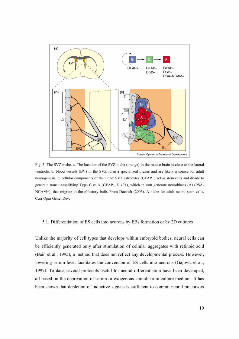
Fig. 5. The SVZ niche. a. The location of the SVZ niche (orange) in the mouse brain is close to the lateral
ventricle. b. blood vessels (BV) in the SVZ form a specialized plexus and are likely a source for adult
neurogenesis. c. cellular components of the niche: SVZ astrocytes (GFAP+) act as stem cells and divide to
generate transit-amplifying Type C cells (GFAP-, Dlx2+), which in turn generate neuroblasts (A) (PSA-
NCAM+), that migrate to the olfactory bulb. From Doetsch (2003). A niche for adult neural stem cells.
Curr Opin Genet Dev.
5.1. Differentiation of ES cells into neurons by EBs formation or by 2D cultures
Unlike the majority of cell types that develops within embryoid bodies, neural cells can
be efficiently generated only after stimulation of cellular aggregates with retinoic acid
(Bain et al., 1995), a method that does not reflect any developmental process. However,
lowering serum level facilitates the conversion of ES cells into neurons (Gajovic et al.,
1997). To date, several protocols useful for neural differentiation have been developed,
all based on the deprivation of serum or exogenous stimuli from culture medium. It has
been shown that depletion of inductive signals is sufficient to commit neural precursors
20
in adherent monoculture, but interestingly, intact autocrine FGF signaling is necessary
(Ying et al., 2003). Alternatively, the differentiation process can be carried out via the co-
culture of ES with a monolayer of bone marrow-derived stromal cells (PA6 cells), in
serum free conditions (Kawasaki et al., 2000). The factors that are responsible for the
neural induction activity of PA6 cells, however, still remain unknown.
6. Parallelism between endothelial and neural cells differentiation
The vertebrate vascular system consists of a highly organized network of capillaries,
arteries and veins that penetrates all the parts of the body, thus allowing oxygen
exchange, delivery of nutrients and removal of waste products. The complex sequence of
events that direct vascular network formation, including branching, sprouting
angiogenesis and vascular patterning, resembles to that occurring during nervous system
development. Vessels and nerves ramify throughout all the domains of developing body
by following similar pathways of migration (Martin and Lewis, 1989) and, in some
instances, they are mutually dependent: in embryonic mouse limb skin sensory nerves are
templates for blood vessels branching (Mukouyama et al., 2002). Moreover, as with the
growth cone is for neurons the sensory structure that sensitize growing axons to
directional information, also nascent capillary sprouts are guided by special endothelial
cells called tip cells (Gerhardt et al., 2003).
During the past two decades it has become increasingly evident that families of
molecules first described in neuronal patterning also play significant role in the
developing vascular system. So far, four families of classical neuronal guidance
molecules were found to be also involved in vascular development: Netrins, Ephrins,
Slits and Semaphorins. These signals act either as attractive or repelling cues on the tip
cells.
Semaphorins are a large family of secreted or cell-anchored proteins with a bifunctional
role, attractive or repulsive, depending on the receptor complex to which semaphorins
bind and on the crosstalk between receptors (Tamagnone and Comoglio, 2004).

21
Semaphorins bind to the receptor families neuropilins (Nrp) and plexins. In particular, the
secreted semaphorin 3A (Sema3A) controls both vascular patterning and axon guidance
through activation of a receptor complex formed by Nrp1 and plexinA, serving as ligand-
binding and signal-transducing component respectively (Neufeld and Kessler, 2008).
Besides being localized on axons and growth cones where it modulates nerves trajectory
during embryonic development (Fujisawa et al., 1995), the glycoprotein Nrp1 possess
also the ability to bind the two VEGFA isoforms VEGF165 and VEGF121 in human
endothelial cells and to form complexes with VEGFR-2, thus enhancing VEGF-induced
signal transduction (Shraga-Heled et al., 2007; Soker et al., 2002). According to that, the
embryonic lethality of Nrp1 knock-out mice is mainly due to an impaired neural
vascularization, defects in great vessels and in the capillary networks of the yolk sac
(Kawasaki et al., 1999; Kitsukawa et al., 1997). Finally, the Semaphorin/Neuropilin
system is involved in the development of the cardiovascular and nervous systems as well
(Carmeliet and Tessier-Lavigne, 2005; Serini and Bussolino, 2004).
The parallelism between vascular and nervous system is also mirrored at the level of cell
commitment and differentiation. In vitro, cells purified from bone marrow and cord blood
can originate neurons and glia (Buzanska et al., 2002; Goolsby et al., 2003; Reali et al.,
2006). On the other hand, mouse neural stem cells (NSCs) acquire an endothelial
phenotype when co-cultured with human mature endothelial cells (Wurmser et al., 2004)
or when inoculated in collagen gels (Oishi et al., 2004). In vivo, bone marrow-derived
cells enter the brain and differentiate into neural cells not only in a mouse model but also
in patients that underwent bone marrow transplantation (Mezey et al., 2000; Mezey et al.,
2003). Moreover, murine NSCs can engraft into the hematopoietic system of irradiated
hosts to produce blood cells (Bjornson et al., 1999), and in the quail-chick chimera model
the avian cranial neuroectoderm originates smooth muscle cells (Korn et al., 2002). All
together these data indicate that there is a plastic correlation between the endothelial and
neural commitments.

22
Results
1. Analysis of Nrp1 expression in differentiating ES cells
Embryonic stem cells (ES) were chosen as in vitro model to analyze the role played by
Nrp1 during cell differentiation in the embryoid bodies. ES cells were grown for 5 days
in hanging drop culture in LIF deprived medium to induce EBs formation, and then
allowed to attach to a substrate (day 5) where the EBs continued their expansion (Fig.
6A). To analyze Nrp1 expression in differentiating ES cells, 6 days-old EBs were fixed
and immunostained with anti-Nrp1 antibody. As shown in Fig. 6A, Nrp1 was widely
expressed in scattered areas throughout the growing EBs. Furthermore, Nrp1 protein was
immunoprecipitated from lysates purified from 7 days-old EBs but not from
undifferentiated W4-ES cells (Fig. 6B).
To quantify Nrp1 expression in differentiating ES cells, the EBs were analyzed by FACS.
As shown in Fig. 6C, Nrp1 expression was absent in undifferentiated ES cells and during
the first three days of differentiation; it became significant only at day 4 to finally
stabilize at 45-50% of the total EB cells population in the next days.
2. Characterization of EBs-derived Nrp1+ cells
In order to characterize the phenotype of Nrp1+ cells in the EBs, we performed a time-
course FACS analysis of EB cells double-labeled for Nrp1 and for markers of either
endothelial or neural cell precursors (Fig. 6D). Being the canonical marker of
hemangioblasts, Flk1 is expressed from day 4, which is the very first day of appearance
of Nrp1 expression. The amount of Nrp1+ cells within the Flk1 component progressively
increased over time to almost cover the totality of the Flk1 expressing cells at day 12,
time in which the Flk1+ endothelial cells are organized into mature vessels (Fig. 6D,

23
numbers above bars) (Gualandris et al., 2000). At these late time points, ICAM-2
expression and its Nrp1+ fraction became significant, whereas CD133 expression, which
identifies both endothelial (Fons et al., 2004) and neural precursors (Lee et al., 2005),
maintained a significant percentage of Nrp1-expressing cells throughout the entire EB
growth. The polysialylated embryonic form of the neural molecule NCAM (PSA-
NCAM) is characteristic of neural progenitors in the forebrain (Gago et al., 2003), and its
expression was shared almost entirely by Nrp1+ cells at all the time points examined.
CD31, which is expressed by undifferentiated ES cells (Li et al., 2005), included a
significant component of Nrp1+ cells at its peak of expression (day 7) to later stabilize at
a lower level at day 12.
3. Characterization of EBs-derived Nrp1+/Flk1+ and Nrp1+/Flk1- cells populations
Because endothelial cells and neurons share the expression of Nrp1 and VEGFR2/Flk1
(Fujisawa et al., 1995; Soker et al., 2002), we focused our attention on the cell
populations defined by these two markers, the Nrp1+/Flk1+, Nrp1+/Flk1- and Nrp1-/Flk1+
cell types (Fig. 7B, in green, red, magenta respectively). The potential of differentiation
towards neuronal lineages was investigated by analyzing the expression of the neuronal
markers Nestin, Sox1 and PSA-NCAM. The colocalization of nestin with both Flk1 and
Nrp1 (arrow and arrowhead respectively in Fig. 7A) was quantified at 50% of the cells of
both Nrp1+ populations by intracellular FACS analysis (Fig. 7B). Since the intermediate
filament protein nestin is expressed not only by neural precursors but also by the
endothelial cells during development (Wiese et al., 2004) the more specific markers sox1
and PSA-NCAM were next investigated. Sox1 is a SRY-related transcription factor
whose expression is characteristic of proliferating neural precursors (Bylund et al., 2003;
Kiefer, 2007). Nrp1+/Flk1+ and Nrp1+/Flk1- cells that expressed sox1 or PSA-NCAM
were detected by immunofluorescence in sparse areas of 7 days-old EBs (Fig. 7A, arrow
and arrowhead). The expression of PSA-NCAM was quantified at 42% of Nrp1+/Flk1+
cells and at 25% of Nrp1+/Flk1- cells by FACS analysis (Fig. 7B, green and red). The
24
lack of PSA-NCAM expression in the Nrp1-/Flk1+ population thus suggested that neural
orientation was not contemplated by the differentiation potential of those cells that did
not carry Nrp1 (Fig. 7B, magenta). To summarize, the molecular characterization of the
populations defined by Nrp1 and Flk1 revealed an intriguing coexistence of markers of
neural precursors with markers of mesoderm/endothelial precursors that made the fate of
Nrp1+ cells worth of further investigations.
4. In vitro analysis of the potential of differentiation of Nrp1+/Flk1+ and Nrp1+/Flk1-
cells
4.1. Endothelial commitment
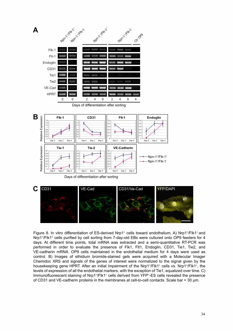
To better study the differentiation potential of Nrp1+ cells outside the EB environment,
Nrp1+/Flk1+ and Nrp1+/Flk1- cells were isolated at 95% of purity from 7 days-old EBs by
flow cytometry sorting and then cultured upon different conditions. To support the
endothelial differentiation in vitro, Nrp1+/Flk1+ and Nrp1+/Flk1- cells were seeded onto
confluent monolayers of OP9 stromal cells, described to be optimal feeders for
endothelial differentiation (Hirashima et al., 1999), and grown in the presence of EGM2
medium; the maturation into endothelium was evaluated by analyzing the expression of
different endothelial markers by RT-PCR at different days (Fig. 8A and 8B). Immediately
after sorting (day 0), the Nrp1+/Flk1+ cells expressed all the endothelial genes tested at
levels from two up to eight times higher than those of the Nrp1+/Flk1- cells as shown by
the quantification data plotted in Fig. 8B. High expression of CD31 messenger at day 0 in
both cell types could reflect an inheritance from undifferentiated ES cells (Li et al.,
2005). During the next days, the levels of expression of the majority of the endothelial
markers in the two cell populations became comparable. These last observations indicate
that the co-culture with OP9 feeders could fill-in the initial impairment of the Nrp1+ cells
sorted as Flk1-. Immunofluorescent stainings performed on Nrp1+/Flk1+ cells derived
from ES cell expressing the tracer yellow fluorescent protein (EYFP) (Hadjantonakis et

25
al., 2002) and co-cultured for 2 days with OP9 cells in presence of EGM2 medium
proved that CD31 and VE-cadherin mRNAs, previously detected by RT-PCR, were
actually translated into the corresponding functional proteins located at cell to cell
contacts (Fig. 8C).
4.2. Neural commitment
Previous observations indicated that within the 7 days-old EBs the Nrp1+/Flk1+ and
Nrp1+/Flk1- cells are neural committed (Fig. 7A and 7B). To better examine such
potential outside the EB environment, purified Nrp1+/Flk1+ and Nrp1+/Flk1- cells were
cultured in a neuronal medium and, as control, in endothelial medium. At different days
after sorting, Nrp1+/Flk1+ and Nrp1+/Flk1- cells were analyzed by immunofluorescence
for the expression of early and late neuronal markers. Sox2, the earliest transcription
factor expressed in the neural tube during development (Zappone et al., 2000) and nestin
were expressed by 60-80% of the Nrp1+/Flk1+ population already after 24 hours of
growth, not only in neuronal medium, but also in the unfavorable endothelial medium
(Fig. 9A and B). At the latest days of growth nestin and sox2 expression started to
decline, whereas the level of expression of the mature neuronal marker βIII-tubulin
increased, suggesting that neuronal differentiation was progressing. Indeed, as soon as
FGF-2 was withdrawn from the neural medium (Materials and Methods) Nrp1+ sorted
cells underwent a significant change in morphology characterized by cellular βIII-
tubulin+ processes resembling dendrites and axons (Fig. 9C, arrows). Similar results were
obtained also with Nrp1+/Flk1- cells (data not shown). Indeed, neuronal maturation is
fully accomplished only upon the neuron-specific culture conditions as confirmed by the
almost absent expression of βIII-tubulin when cells are grown in endothelial medium
(Fig. 9B). An analogous analysis was performed on sorted cells to detect the expression
of NeuN, a nuclear factor exclusively found in mature neurons (Sarnat et al., 1998):
positive nuclei were detected by immunofluorescence when cells were grown only in
neuronal medium (Fig. 9E). The results of the quantitative analysis are shown in Fig. 9D.
Similar results were obtained with Nrp1+/Flk1- cells (data not shown). All together these

26
data demonstrate that in response to the appropriate exogenous microenvironment the
bulk of ES-derived Nrp1+ cell population gives rise to endothelial or to neural cells.
5. In vivo analysis of the potential of differentiation of Nrp1+/Flk1+ and Nrp1+/Flk1-
cells
5.1. Endothelial commitment
Even with the limits of an in vitro culture system, the data shown so far represented a
supporting indication of the double endothelial and neuronal commitment of the ES-
derived Nrp1+ cells. We next wondered if a more physiological environment, such as a
living embryo, could allow the Nrp1+ cells to fully accomplish their commitments. On
these basis, Nrp1+/Flk1+ and Nrp1+/Flk1- cells were purified from 7 days-old YFP+-EBs
by cell sorting and microinjected into chicken embryos at stage HH19 of development.
To drive the Nrp1+/Flk1+ population towards the developing vascular system the
microinjection was performed in the beating heart of the embryos. Three days after
injection, the host vessels were visualized in embryo tissue sections by using Sambucus
Nigra Agglutinin (SNA). Fig. 10A shows that the Nrp1+/Flk1+ injected cells integrated
within the new forming vessels as indicated by the alignment of the YFP signal with the
signal given by the chicken endothelial specific SNA (Nanka et al., 2001). Identical
results were obtained by using the W4 ES cell line (data not shown). Out of 79 embryos
that survived the injection, 34 embryos had integrated the injected YFP+ cells within the
vasculature (43% of engraftment). Furthermore, the integrated cells also differentiated
into mature endothelium expressing CD31 and VE-Cadherin as demonstrated by
immunofluorescent staining of the tissue sections performed with antibodies specific for
the murine antigens (Fig. 10B). Similar results were obtained with the Nrp1+/Flk1- cells
with the exception that the vessels-integrated cells did expressed CD31 but not the more
mature marker VE-Cadherin (Fig. 10C), thus confirming the slight delay in pursuing

27
endothelial maturation by the Nrp-1+ cells sorted as Flk-1- previously suggested by the in
vitro data (Fig. 8B).
5.2. Neural commitment
When the purpose was to address the injected cells towards a neural fate, the
microinjection was performed into the mesencephalic cavity of embryos. Once injected,
cells tend to aggregate into spherical clusters that initially make contacts with the inner
face of the developing cerebral tissues. From this time on, cells leave the clusters and
start to invade the host tissues (Fig. 11A). Fig 11B shows the location of injected cells
five days after injection: the YFP+ Nrp1+/Flk1- cells have moved from the inner cavity,
entered the neurogenesis area of the developing brain, and started to express MAP2
antigen along with the surrounding MAP2+ chicken neurons thus becoming part of the
developing cerebral tissue. Analogous results were obtained with Nrp1+/Flk1+ cells (Fig.
11C). Notably, in some cases, even if injected in the encephalic cavity, the Nrp1+/Flk1+
cells were also found fully integrated within the cerebral vessels (data not shown),
suggesting that the bi-potentiality of commitment is an intrinsic characteristic of the
Nrp1+ cell populations ready to be driven towards endothelial or neural phenotype
whenever the exogenous conditions are permissive.
6. Generation of an in vitro model specific for the differentiation of both endothelial and
neural cells
Embryoid bodies, as illustrated before, are complicated 3D structures in which many
different cell types arise altogether. This property can be useful for a variety of studies,
but could be also a drawback when the purpose is to assess the differentiation potential of
a cell population that is not well represented in the EB, e. g., neurons. To overcome this
problem, several differentiation protocols were developed in order to selectively induce
the specific lineage of interest. In particular, we took advantage from neural

28
differentiation protocols to generate a culture system able to specifically induce the
endothelial and neural lineages at the same time.
6.1. Direct differentiation of endothelial and neural cells from ES cells
The differentiation of neural cells takes place when ES cells grow in absence of serum or
cytokines (Ying et al., 2003). Furthermore, it has been shown that several stromal cell
lines, such as PA6 cells, are able to promote neural differentiation when used as feeders
(Kawasaki et al., 2000). In order to induce neural commitment, YFP expressing-ES cells
were seeded at low density onto a PA6 cells monolayer in a serum free medium. After
few days of culture, some neurons started to appear, as indicated by the acquirement of
the neural specific marker β–III tubulin. The differentiation process was accomplished at
around day 7, when colonies of neurons developed nerve projection (Fig. 12A). Cells
within colonies were also positive for neurofilament H and for the synaptic marker SV2
(Fig. 12, C and D). The expression of YFP by the ES-derived progeny demonstrates that
the immunofluorescent staining is not present on PA6 cells, but it is confined to the YFP+
cells. Cells within the neural colonies also expressed the neural precursor marker Sox2
(Fig. 13A). Moreover, ramified cells resembling to radial glia were identified by the
antibody raised against the radial glia marker RC2 (Fig. 13B). These cells could represent
neural progenitors (Anthony et al., 2004). Endothelial cell differentiation is not
comprised in these culture conditions, as indicated by the complete absence of the
endothelial specific marker VE-cadherin (Fig. 12A). Conversely, by adding a cocktail of
cytokines containing FGF and EGF at the initiation of the culture, endothelial cells
developed along with neurons, as shown by the appearance of a network of cells
detectable by using an anti-VE-cadherin antibody (Fig. 12B and 14A). These cells
expressed a set of canonical endothelial markers, such as Flk1, ICAM2, and CD31 (Fig.
14 B, C, and D, respectively) though the latter was also found in colonies of cells with
undifferentiated morphology (Fig 14E, arrow). CD31 is indeed present in ES cells as cell
adhesion molecule; its expression is down-regulated during differentiation, then restored
in mature endothelial cells (Li et al., 2005). In order to understand if growth factors

29
induced different cell types other then endothelial and neural cells, we subsequently
performed a screening with several antibodies raised against markers of differentiated
cells. We were not able to detect any cell labeled with anti-keratins or GFAP antibodies
(data not shown). On the contrary, α-smooth muscle actin (α-SMA) was expressed in
PA6 cells especially when the differentiation process occurred in absence of growth
factors; indeed, FGF and EGF inhibited α-SMA expression in feeder cells (Fig. 15).
Beside this, very few ES-derived cells expressed α-SMA regardless of the presence of
FGF and EGF (data not shown). These data, taken together, indicate that the combination
of the stromal PA6 cells with a serum-free medium and the addition of growth factors
represents an efficient neuro-endothelial model to in vitro differentiate ES cells into
endothelium and neurons at the same time.
6.2. Effect of cytokines on the acquirement of endothelial and neural phenotypes
As general effect, the addition of FGF and EGF together stimulated cell proliferation,
thus resulting in increased size of colonies. In order to better understand the role of
cytokines in the induction of endothelial cell commitment, we stimulated ES cells with
FGF, EGF, or VEGF singularly at the onset of the culture system. Each growth factor
alone was able to promote the generation of endothelial cells, but induced different
cellular morphologies. FGF generated a network of elongated cells, while EGF promoted
the formation of sheets of cells VE-cadherin positive (Fig. 16 A and B). On the contrary,
VEGF was less effective in inducing the endothelial network, although some cells did
develop (Fig. 16C). With regard to neurons, the addition of growth factors did not alter
the induction of the neural commitment, compared to the condition without growth
factors. However, most likely FGF had an effect on the specification of specific subsets
of neurons. In particular, dopaminergic neurons were detected only after FGF
stimulation, as shown by the presence of tyrosine hydroxylase positive cells (Fig. 17A).
These data suggest that each growth factor gives a different contribution to address the
fate of the ES cells that are differentiating within this co-culture system.

30
7. Gene expression regulation by cytokine stimulation in the neuro-endothelial model of
ES cells differentiation
To better characterize the neuro-endothelial model of ES cells differentiation with
particular regard to the effects caused by the addition of growth factors, a gene
expression analysis was performed by using Taqman low density arrays. We studied the
expression of 83 genes chosen among markers of mature endothelial or neural cells,
glial/neural precursor markers, stem cell markers, factors involved during germ layer
specification, muscle, epithelial and pancreatic markers as well. ES cells were cultured in
serum-free medium without any growth factor in order to induce neuronal differentiation,
or stimulated with FGF or VEGF for the first three days in order to induce endothelial
commitment. After 6 days of culture, we compared the gene expression level between the
two experimental conditions. We performed two independent experiments and those
genes found differentially expressed with a fold increase above 2.5 in both experiments
were considered regulated by growth factors (Fig. 18). As expected, many of the
endothelial markers were upregulated after stimulation with either FGF or VEGF. In
particular, FGF was able to induce the expression of the endothelial/hematopoietic
precursors’ marker Tal1 (Scl-1), along with Pecam1 (CD31), angiotensin I converting
enzyme (Ace), Endoglin and Angiopoietin1. Conversely, VEGF was more efficient in
inducing Cdh5 (Ve-cadherin) and Tie1, compared to FGF (Fig. 19A). Neural
differentiation was less prone to be differentially regulated by cytokines in terms of fold
change; however, FGF induced in particular the expression of glutammic acid
decarboxylase (Gad) and tyrosine hydroxylase (TH), while VEGF showed no effect or
even an inhibitory effect on some neural markers. Interestingly, Sema3A and Nrp1, that
are two molecules shared by endothelial and neural cells, were upregulated after
stimulation with either FGF or VEGF (Fig. 19B). Genes representative for the three
embryonic germ layers were also strongly upregulated by FGF but not by VEGF, except
the definitive endoderm marker Cxcr4 that was downregulated by both growth factors
(Fig. 19C). The stem cell markers Zfp42 (Rex1) and Pou5f1 (Oct3/4) were strongly
upregulated after FGF stimulation, thus indicating a possible proliferation within the stem

31
cell compartment. On the other hand, genes representative for smooth muscle, epidermis,
liver and pancreas were not regulated or not detected at all, with the exception of
myogenin and keratin 14, which were upregulated by FGF and downregulated by VEGF
respectively (Fig. 19D). A control experiment performed with PA6 cells alone, stimulated
or not with FGF, showed that more then a half of the genes taken into account were not
detected (gray boxes in Fig. 18), and that among the remaining genes just few were
slightly upregulated or downregulated. Along with the fact that mRNA content coming
from feeder cells represents only about one tenth of the total mRNA extracted from the
coculture with ES-derived cells, we consider negligible the presence of feeders mRNA in
our analysis.
Taken together, these data reinforced the observations obtained with immunofluorescent
analysis, and indicates that the emergence of endothelial cells is dependent on cytokines
stimulation. Moreover, this coculture system seems to be specific for the induction of
endothelial and neural cells only, as demonstrated by the lack of markers of other cell
types.
8. Endothelial and neural differentiation in the vascular niche context
As recently well documented, the microenvironment of the vascular niche comprises
neural cells, proliferating neural precursors and specialized endothelial cells (Tavazoie et
al., 2008). In our model of neuro-endothelial differentiation, endothelial cells and neurons
develop simultaneously form ES cells in a multi-step process. In fact, cells expressing the
neural precursor marker nestin were found in proximity with mature neurons. Nestin was
expressed also by some endothelial cells, as this marker is shared by a variety of
precursor cells (Fig. 20A). The pluripotent stem cell marker CD133 was also expressed
by some cells within colonies (Fig. 20B). Therefore, despite the absence of GFAP+ cells,
the presence of endothelial cells, neurons and neural precursors, along with the
supporting mesenchymal cells, suggests that many of the elements of the vascular niche
are included.

A BNrp-1 IP Nrp1 IP IgG
WB Nrp1
200
150
100
M ES EB ES EBDIC
Day1 1% Day2 4% Day3 7%
Day4 17% Day5 27% Day7 55% Day9 52%
Cou
nts
Cou
nts
Neuropilin-1 expression
ES 0%
C
D
010
203040
506070
8090
4 5 6 7 8 12
Flk-1CD31ICAM-2PSA-NCAMAC133
% p
ositi
ve c
ells
Day
13
23 31 2835
183
19
3240
44
9
0 05 5
8 118
1518
42
4343
4
7 9
1418
16
Figure 6. Nrp1 expression in differentiating ES cells. A) 6 day-old EBs (DIC, differential interference contrast) were analyzed by immunofluorescence with an anti-Nrp1 antibody and by using a Leica DM IRB inverted microscope. Scale bar = 1 mm. B) Lysates from undifferentiated ES cells and 7-day-old EBs were precleared with irrelevant IgG (IP IgG) before Nrp1 immunoprecipitation (IP Nrp1) and Western blot analysis (WB Nrp1). Immunocomplexes were detected only in lysates purified from EBs. C) Single-cell suspensions were prepared from both undifferentiated ES cells and EBs and analyzed by FACS using the anti-Nrp1 antibody or its isotype IgG (gray). Nrp1 is not expressed by ES cells and by the EBs until day 4. The experiment shown is representative of 5 experiments. D) FACS analysis of EB-derived cells immunolabeled with anti-Nrp1 antibody in association with anti-Flk1, CD31, ICAM2, PSA-NCAM, or CD133 antibodies. Each colored bar represents the cells positive for the corresponding marker, whereas the number placed on the top is the percentage value of double-labeled Nrp1+ cells. Data are averages ± SD of 5 different experiments.
32

A Nrp1 Flk1 Nestin Merge
B
Flk-
1
Nrp-1
427
30 21
Cou
nts
Cou
nts
Cou
ntsNrp-1+/Flk-1+ 56% Nrp-1+/Flk-1- 51% Nrp-1-/Flk-1+ 53%
Nestin expression
Cou
nts Nrp-1+/Flk-1+ 42%
Cou
nts Nrp-1+/Flk-1- 25%
Cou
nts Nrp-1-/Flk-1+ 4.4%
PSA-NCAM expression
Nrp1 Flk1 Sox1 MergeMerge
Nrp1 Flk1 PSA-NCAM Merge
Figure 7. Characterization of EB-derived Nrp1+ cells. A) Confocal microscopy analysis of 7-day-old EBs immunostained with the anti-Nrp1, anti-Flk1, and anti-nestin or anti-sox1 or anti-PSA-NCAM antibodies. Nrp1+/Flk1+ cells (arrow) and Nrp1+/Flk1+ cells (arrowhead) expressing nestin, sox1, and PSA-NCAM are indicated. Scale bars = 50 μm. B) Quantification by FACS analysis of nestin and PSA-NCAM expression by the Nrp1+/Flk1+ cells (green), Nrp1+/Flk1+ cells (red) and Nrp1+/Flk1+ cells (magenta) vs. control (white). Percentage values of cells belonging to each of the four detected populations are indicated (dot plot, top right).
33
B Endoglin
0
0,1
0,2
0,3
0,4
0,5
0,6
Day 0 Day 2 Day 4
Tie-2
00,10,20,30,40,50,60,7
Day 0 Day 2 Day 4
VE-Cadherin
00,10,20,30,40,50,60,7
Day 0 Day 2 Day 4
Tie-1
00,10,20,30,40,50,60,7
Day 0 Day 2 Day 4
Flt-1
0
0,1
0,2
0,3
0,4
0,5
0,6
Day 0 Day 2 Day 4
Flk-1
00,20,40,60,8
11,21,41,6
Day 0 Day 2 Day 4
CD31
00,20,40,60,8
11,21,41,6
Day 0 Day 2 Day 4
Npn-1+/Flk-1+
Npn-1+/Flk-1-
Rel
ativ
e E
xpre
ssio
nR
elat
ive
Exp
ress
ion
Days of differentiation after sorting
CD31 VE-Cad CD31/Ve-Cad YFP/DAPIC
A
Days of differentiation after sorting
Npn-1
+ /Flk-
1+
Npn-1
+ /Flk-
1-
Npn-1
+ /Flk-
1-
Npn-1
+ /Flk-
1+
Ctr O
P9
2 4 9 2 4 9 400
Flk-1
Flt-1
Endoglin
CD31
Tie1
Tie2
VE-Cad
HPRT
Figure 8. In vitro differentiation of ES-derived Nrp1+ cells toward endothelium. A) Nrp1+/Flk1+ and Nrp1+/Flk1+ cells purified by cell sorting from 7-day-old EBs were cultured onto OP9 feeders for 4 days. At different time points, total mRNA was extracted and a semi-quantitative RT-PCR was performed in order to evaluate the presence of Flk1, Flt1, Endoglin, CD31, Tie1, Tie2, and VE-cadherin mRNA. OP9 cells maintained in the endothelial medium for 4 days were used as control. B) Images of ethidium bromide-stained gels were acquired with a Molecular Imager Chemidoc XRS and signals of the genes of interest were normalized to the signal given by the housekeeping gene HPRT. After an initial impairment of the Nrp1+/Flk1+ cells vs. Nrp1+/Flk1+, the levels of expression of all the endothelial markers, with the exception of Tie1, equalized over time. C) Immunofluorescent staining of Nrp1+/Flk1+ cells derived from YFP+-ES cells revealed the presence of CD31 and VE-cadherin proteins in the membranes at cell-to-cell contacts. Scale bar = 30 μm.
34

0
10
20
30
40
50
60
5 7
A B
E
Endothelial medium
Neuronal medium
Day
D
DAPINeuN Merge
DAPINeuN Merge
% p
ositi
ve c
ells
100
80
60
40
20
01 2 3 5 7
Endothelial medium
% p
ositi
ve c
ells
100
80
60
40
20
01 2 3 5 7
NestinSox2βIII tubulin
Neuronal medium
Day
% p
ositi
ve c
ells
Day
C βIII tubulin βIII tubulin Endothelial mediumNeuronal medium
NestinSox2βIII tubulin
Figure 9. In vitro differentiation of ES-derived Nrp1+ cells toward neurons. A, B) Nrp1+/Flk1+ cells purified by cell sorting from 7-day-old EBs were cultured for 7 days in a neuronal medium to favor neural commitment (A) and, as control, in an endothelial-suited medium (B). At different days the expression of nestin, sox2 and of βIII-tubulin was examined by immunofluorescent staining. Positive cells were counted in 5 different fields randomly chosen from the coverslips of 4 different experi-ments. The number of positive cells was expressed as percentage value of the total cells counted ± SD. C) Expression of βIII-tubulin by Nrp1+/Flk1+ sorted cells. Note the branched cell morphology (arrows). D) Nrp1+/Flk1+ cells were cultured for 7 days in neuronal or in endothelial medium. At differ-ent days, the expression of NeuN was examined by immunofluorescent staining. Positive cells were counted over the total cells present in 5 different fields randomly chosen from the coverslips of 4 different experiments. The number of the positive cells was expressed as a percentage value of the total cells counted ± SD. E) Expression of NeuN by Nrp1+/Flk1+ cells cultured in neuronal or endothe-lial medium. Scale bars = 25 μm (C, E).
35

Ve-Cad
YFP
WGA
Merge
C YFP
WGA
Merge
CD31
B
CD31 VE-Cad
YFP YFP
WGA WGA
MergeMerge
A YFP SNA Merge
Figure 10. In vivo differentiation of ES-derived Nrp1+ cells into endothelial cells. A) Nrp1+/Flk1+ cells purified from 7-day-old YFP+ EBs were injected into the heart of HH19-stage chicken embryos; 3 days after injection, tissue sections were analyzed by confocal microscopy. The embryo vasculature was visualized with SNA. Inset: injected cells that integrated within the labeled host vessels. B) Confocal images of tissue sections obtained from chicken embryos injected with YFP+-ES-derived Nrp1+/Flk1+ cells whose vessels were visualized by Alexa-Fluor 405-conjugated wheat germ agglutinin (WGA). CD31 and VE-cadherin were detected by immunofluorescent staining with murine specific anti-CD31 and anti-VE-cadherin antibodies. Note how the injected cells integrate and differentiate within the host vessels to perfectly match the vessel lumen border (arrow). C) Confocal images showing the integration and CD31 expression of Nrp1+/Flk1- cells within the embryonic vasculature 3 days after injection. Contrary to what observed with the Nrp1+/Flk1+ cells, no immunoreactivity was detected with the anti-VE-cadherin antibody. Scale bars = 30 µm.
36

B
YFP
MAP
2M
erge
DIC
M M
A YFP MAP2
DICMergeM
N
YFP
MAP
2M
erge
DIC
C
Figure 11. In vivo differentiation of ES-derived Nrp1+ cells into neural cells. A) Nrp1+/Flk1- cells purified from 7-day-old YFP+ EB cells were injected into the mesencephalic cavity of HH19-stage chicken embryos; 3 days after injection, tissue sections of the developing brain were analyzed by confocal microscopy by using an anti-MAP2 antibody that recognizes both mouse and chicken neurons. Differential interference contrast (DIC) image shows a cluster of injected cells within the mesencephalic cavity (M) that makes contact with the inner face of the developing tissues: inset shows an enlargement of cells entering the MAP2-labeled neurogenesis area of the developing brain (N). Invading YFP+ cells express MAP2 as well. B) Confocal images of serial optical sections of the same field showing a group of Nrp1+/Flk1- cells 5 days after injection. The YFP+-injected cells also express MAP2 along with the surrounding chicken neurons. C) Confocal images of serial optical sections of the same field, showing Nrp1+/Flk1+ cells 5 days after being injected into the mesencephalic cavity of HH19-stage chicken embryos. Scale bars = 50 µm.
37

VE-cadβIII-tubulin Merge
βIII-tubulin VE-cad Merge
NF-H YFP Merge
SV2 YFP Merge
A
B
D
C
Figure 12. In vitro differentiation of ES cells into neurons and endothelial cells. A) After 7 days of culture in the neural-suited differentiation medium, ES-derived cells form rounded colonies of neurons with outgrowing neural processes. No endothelial cells are detected. B) Addition of FGF and EGF stimulates the differentiation of a network of endothelial cells that can be detected with an anti-VE-cadherin antibody. C, D) Expression of the neural markers neurofilament H and SV2 is confined to the progeny of ES-derived YFP+ cells. Scale bars = 200 µm (A, B); 50 µm (C, D).
38

Sox2 Sox2/DAPI
RC2 RC2/DAPI
A
B
Figure 13. Expression of neural precursor markers during differentiation. A) The neural precursor marker Sox2 is detected in some nuclei of cells within differentiating colonies after 6 days of differentiation. B) Cells with radial glia morphology are labelled with the anti-RC2 antibody. Scale bars = 100 µm.
39

YFP MergeVE-Cad
ICAM-2 YFP Merge
YFPFlk-1 Merge
A
C
B
Figure 14. Expression of endothelial markers by ES-derived YFP+ cells. After 6 days of differentiation in medium containing FGF and EGF, endothelial cells can be detected with antibodies against VE-cadherin (A), Flk1 (B), ICAM2 (C) or CD31 (D). CD31 is also expressed within colonies containing undifferentiated cells (E, arrow). Scale bars = 100 µm.
Merge
Merge
YFP
YFP
CD31
CD31
E
D
→
40

Mergeα-SMA YFP
α-SMA YFP Merge
A
B
Figure 15. Expression of smooth muscle actin is dependent on culture conditions. A) PA6 cells differentiate into α-SMA expressing cells that surround colonies of ES-derived YFP+ cells, only if the culture medium is avoided of growth factors. B) Addition of FGF and EGF prevents the expression of α-SMA in PA6 cells. Scale bar = 100 µm.
41

VE-cad MergeβIII-tubulin
MergeβIII-tubulinVE-cad
VE-cad βIII-tubulin Merge
A
C
B
Figure 16. Effect of FGF, EGF, or VEGF on endothelial commitment and cell morphology. Growth factors were added to the culture medium at day 0. A) Endothelial cells generated after FGF stimulation acquire an elongated morphology, while (B) EGF promotes the formation of VE-cadherin sheets. C) Stimulation with VEGF at the onset of the culture is less efficient in the induction of endothelial commitment, although some scattered cells appear. Scale bars = 200 µm (A, B); 100 µm (C).
42

Merge
MergeTH
THMAPs
MAPsA
B
Figure 17. Effect of FGF on neural phenotipe acquisition. A) Clusters of TH+ cells develop when cells are stimulated with FGF, indicating an effect on the specification of dopaminergic neurons. On the contrary, in the absence of FGF no TH+ cells were detected (B). Note that neural differentiation is not affected by the presence of FGF, as neurons expressing the microtubulin associated proteins (MAPs) develop in a similar way. Scale bars = 100 µm.
43

FGF VEGF PA6
Figure 18. Gene expression analysis of ES-derived cells cultured in presence of citokynes. Gene expression data were analyzed with GEDAS software in order to visualize upregulated (red) or downregu-lated (green) genes. Cells were stimulated either with FGF or VEGF, and two indepen-dent experiments were compared. A hierar-chical clustering was performed in order to better visualize groups of up- or downregu-lated genes. The control experiment performed with PA6 cells alone stimulated with FGF, shows that most of the genes taken into account are not expressed by these cells (gray boxes), and among the expressed ones, only few are regulated by FGF.
44

A
DC
Figure 19. Genes regulated upon FGF or VEGF addition after 6 days of coculture of ES and PA6 cells. A) FGF induces the expression of several endothelial markers, such as Pecam1, Ace, Endoglin, Angiopoietin1 and the endothelial/hematopoietic marker Tal1. VEGF on the contrary is more efficient in inducing the more mature marker Cdh5 (VE-cadherin). B) Induction of neural markers by cytokines is less pronounced compared to endothelial ones; however FGF is able to induce several markers including tyrosine hydroxylase, confirming data from immunofluorescent analysis. Note that VEGF, like FGF, upregulates Sema3a and Nrp1, two genes expressed also by endothelial cells. C) FGF stimulates the expression of markers of the three embryonic germ layers, except the endoderm marker Cxcr4. D) The remaining regulated genes comprise the muscular marker Myogenin and the two stem cell markers Zfp42 and Pou5f1. Error bars indicates SEM of one representative experiment performed in duplicate.
B
45

VE-cadβIII-tubulin Nestin Merge
βIII-tubulin CD133 Merge
A
B
Figure 20. Neural and endothelial differentiation in the vascular niche context. A) Expression of the precursor marker nestin is detected in cells that have not yet acquired the neural marker βIII-tubulin, and in some endothelial cells (arrowhead). B) Some rounded cells expressing the pluripotent stem cell marker CD133 develop within differentiating colonies. Scale bars = 200 μm (A); 100 μm (B).
▼▼
46

47
Discussion
Commitment of Nrp1+ cells derived from embryoid body
In this study we used Nrp1 and Flk1 to identify a discrete, non-clonal population of
uncommitted ES-derived cells that can differentiate into endothelial or neuronal cells
depending on the suitable environmental conditions, both in vitro and in vivo. Within the
EBs we identified, characterized and isolated the Nrp1+/Flk1+ and Nrp1+/Flk1- lineages
and we analyzed their potential of differentiation in respect to endothelial and neural
commitments. Nrp1 was selected for its pivotal role in the vascular and neural systems
and for its activity on stem cell differentiation. Indeed, Nrp1 was suggested to be
involved in neural commitment of adult NSCs (Maurer et al., 2003) and in VEGF-A
mediated endothelial differentiation of bone marrow-derived AC133+ cells (Fons et al.,
2004). Flk1 is the canonical marker of multipotent mesodermal progenitors that can
differentiate into endothelial, haematopoietic, mural cells and cardiomyocytes (Kattman
et al., 2006; Yamashita et al., 2000).
The comparative analysis of Nrp1+/Flk1+ and Nrp1+/Flk1- cells indicates that the
presence of Flk1 favors the endothelial commitment of the Nrp1+ cells both in vitro in
OP9 co-cultures, and in vivo, where we observed that the Nrp1+/Flk1- cells seem to be
delayed in integrating within the embryonic host vessels and in differentiating into
mature VE-Cadherin+ endothelium (Fig. 8B and 10C). Thus, Flk1 presence matters in
terms of endothelial commitment, as previously well documented. If the presence of Flk1
facilitates the endothelial maturation, conversely the absence of Nrp1 seems to impair the
neural differentiation since the Nrp1-/Flk1+ cells did not express neuronal markers as
extensively as the two Nrp1+ counterparts both when included in the EB (Fig. 7B) and
when isolated as pure cell population (data not shown).

48
Even though the presence of markers of neural precursors such as nestin, sox1, sox2 and
PSA-NCAM was suggestive of a neural potential, only the presence of the proper
neuronal environment allowed both Nrp1+/Flk1+ and Nrp1+/Flk1- cells to definitively
maturate into βIII-tubulin+ and NeuN+ neurons. In vivo, the Nrp1+ sorted cells integrated
into the developing brain of chicken embryos where they differentiated into MAP2+
neurons independently of their initial production of Flk1.
The topic of the bi or multi-potentiality of progenitor cells has recently become
controversial. Despite the numerous indirect evidences supporting the existence of the
hemangioblast (Choi et al., 1998; Huber et al., 2004; Kattman et al., 2006), recent in vivo
studies argue against its bipotentiality. Cell-lineage tracing of cells from the primitive
streak to the yolk sac failed to reveal a common haematopoietic and endothelial
progenitor (Kinder et al., 2001). Moreover, the analysis of tetrachimeras derived from
different ES cell lines stably expressing separate fluorescent tracers showed that each
blood island has contributions from multiple clonal progenitors (Ueno and Weissman,
2006). Our results are more in line with these studies especially considering that a
putative neuron-endothelial precursor, by trespassing the ectoderm-mesoderm
boundaries, would represent a concept of bipotent progenitor more challenging than the
hemangioblast. The idea of a bipotent progenitor within our Nrp1+/Flk1+ cells was
justified by the model used, the ES cells, and by the presence of Flk1. By using the same
model with alternative protocols different groups showed that ES-derived Flk1+ cells
could differentiate into multiple mesoderm-derived lineages (Kattman et al., 2006; Wu et
al., 2006; Yamashita et al., 2000). Regarding these data, the unique presence of Nrp1
along with other neural markers gives more challenges to our model. It was proposed that
Flk1 marks a common mesodermal precursor that segregates to successive subsets of
Flk1-expressing or non-expressing cells whose fate is then determined by coexpression of
lineage specific transcription factors (Ema and Rossant, 2003). Hence, our Nrp1+/Flk1+
population could represent a transient intermediate pool of cells in which the committed
progenitors arise thanks to the loss or acquisition of specific molecules.
The possibility to purify this pool of cells by using Nrp1 and Flk1, along with the
encouraging in vivo transplantation data, opens new perspectives for the treatment of
49
disorders characterized by defects of both the neural and vascular systems, including
stroke, diabetic neuropathy, and Alzheimer's disease (Murry and Keller, 2008).
In vitro simultaneous endothelial and neural differentiation: a model for vascular
niche
In vitro differentiation of ES cells is a powerful tool to generate specific precursor cells.
Here, we describe a new differentiation system of ES cells that allows the generation of a
mixed culture of neurons and endothelial cells, without any other cell types. The analysis
of neural markers expression by immunofluorescent stainings showed that neural
differentiation in our conditions proceeds in a way similar to that described in other
differentiation protocols (Kawasaki et al., 2000; Ying et al., 2003). However, this system
is different from others published to date, since thanks to the use of feeder cells and
cytokines, endothelial cells arise among neurons (Fig. 12B, 16 and 20A). With particular
regard to the induction of endothelial commitment, our data indicate that stimulation with
FGF of EGF promotes endothelial differentiation, making this consistent with the well
demonstrated role of FGF during early vascular development (Risau and Flamme, 1995).
Even though it has been shown that PA6 cells produce VEGF (Hirashima et al., 1999),
this is not sufficient to trigger endothelial commitment, as almost no VE-cadherin+ cells
differentiate in absence of FGF or EGF (Fig. 12B). Moreover, exogenous addition of
VEGF at the onset of the culture is less efficient in inducing endothelial differentiation
compared to FGF (Fig. 16C). Nonetheless, the presence of feeder cells seems to be
crucial to support endothelial differentiation, by factors that at the present remain
unknown. Gene expression analysis revealed that endothelial markers increased their
expression when cells were stimulated with FGF, and, to a lesser extent, with VEGF,
while neural markers were little induced by cytokines. The increased size of the colonies
after FGF stimulation reflected its proliferating activity. As a consequence, we found that
the stem cell markers Zfp42 (Rex1) and Pou5f1 (Oct3/4) were strongly upregulated,
suggesting the presence of a compartment of undifferentiated stem cells. This was
confirmed by the labeling of some cell clusters with an anti-SSEA1 antibody, a marker

50
that specifically recognize undifferentiated stem cells (data not shown). On the contrary,
both real time PCR and immunofluorescent analysis demonstrated that markers for cell
types other than neurons and endothelial cells were not detected, or even their expression
were downregulated, as occurs, for instance, for α-SMA expression (Fig. 17).
The system of differentiation described here display several key properties of a
developing vascular niche. As previously described (Doetsch, 2003; Shen et al., 2008), a
typical example of vascular niche that is found in SVZ includes a basal lamina,
ependymal cells, neuroblasts, a transit amplifying population, and neural precursors with
glial phenotype, together with blood vessels. In our model of differentiation, after few
days of culture are visible cells at different stages of differentiation. Among neurons
expressing β–III tubulin and mature endothelial cells (Fig. 20), are present cells with glial
phenotype expressing the radial glia marker RC2 (Fig. 13B), precursor cells expressing
the pluripotent cell markers nestin (Wiese et al., 2004) (Fig. 20A) or CD133 (Fons et al.,
2004; Lee et al., 2005) (Fig. 20B), and few clusters of SSEA1+ cells (data not shown).
Contrarily to what has been demonstrated in vivo, in our culture system we did not
observed any GFAP+ cells, but this can be explained with the fact that growth factors
such as FGF and EGF inhibit the expression of such marker in neural precursors in vitro
(Conti et al., 2005). However, radial glia cells expressing RC2 and BLBP (data not
shown) develops within four days of culture, that is consistent with their role of neural
precursors (Anthony et al., 2004). Finally, the underlying layer of PA6 cells provides
physical support and probably instructing signals to differentiating cells.
The characterization of this neuro-endothelial differentiation model shows that neurons
and endothelial cells can develop in vitro in the same culture condition and temporal
window. Therefore, this model can be viewed as a useful tool to study the mutual
interaction between endothelial and neural cells during early phases of development, or in
structures such as the vascular niche, the latter receiving increasing consideration as
potential clinical target (Aliev et al., 2008).
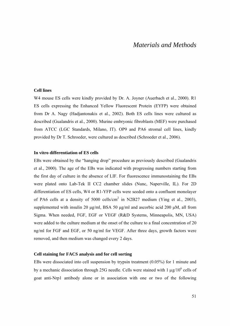
51
Materials and Methods
Cell lines
W4 mouse ES cells were kindly provided by Dr. A. Joyner (Auerbach et al., 2000). R1
ES cells expressing the Enhanced Yellow Fluorescent Protein (EYFP) were obtained
from Dr A. Nagy (Hadjantonakis et al., 2002). Both ES cells lines were cultured as
described (Gualandris et al., 2000). Murine embryonic fibroblasts (MEF) were purchased
from ATCC (LGC Standards, Milano, IT). OP9 and PA6 stromal cell lines, kindly
provided by Dr T. Schroeder, were cultured as described (Schroeder et al., 2006).
In vitro differentiation of ES cells
EBs were obtained by the “hanging drop” procedure as previously described (Gualandris
et al., 2000). The age of the EBs was indicated with progressing numbers starting from
the first day of culture in the absence of LIF. For fluorescence immunostaining the EBs
were plated onto Lab-Tek II CC2 chamber slides (Nunc, Naperville, IL). For 2D
differentiation of ES cells, W4 or R1-YFP cells were seeded onto a confluent monolayer
of PA6 cells at a density of 5000 cells/cm2 in N2B27 medium (Ying et al., 2003),
supplemented with insulin 20 μg/ml, BSA 50 μg/ml and ascorbic acid 200 μM, all from
Sigma. When needed, FGF, EGF or VEGF (R&D Systems, Minneapolis, MN, USA)
were added to the culture medium at the onset of the culture to a final concentration of 20
ng/ml for FGF and EGF, or 50 ng/ml for VEGF. After three days, growth factors were
removed, and then medium was changed every 2 days.
Cell staining for FACS analysis and for cell sorting
EBs were dissociated into cell suspension by trypsin treatment (0.05%) for 1 minute and
by a mechanic dissociation through 25G needle. Cells were stained with 1 μg/106 cells of
goat anti-Nrp1 antibody alone or in association with one or two of the following

52
antibodies: anti-Flk1, CD31, ICAM2, CD133, PSA-NCAM, and PE-anti-Flk1. Antibody
brands and dilutions are given in table 1. The secondary antibodies used were: FITC or
APC-conjugated-anti-goat to detect Nrp1, PE-conjugated-anti-rat to detect Flk1 and
CD133 and FITC–conjugated anti-mouse IgG or IgM to detect nestin and PSA-NCAM
(Southern Biotech., Birmingham, AL). To prepare cells for cell sorting the secondary
antibodies used were PE-conjugated anti-rat and biotin-conjugated anti-goat followed by
an AlexaFluor 647-PE-conjugated streptavidin (Invitrogen, Carlsbad CA). Cell sorting
was performed with Beckman Coulter Epics Altra. The accuracy of the sorting procedure
was confirmed by analyzing representative samples of the two sorted populations through
a Becton Dickinson FACS Calibur purposely reserved for routine analysis only. Such
analysis revealed that cell populations were sorted at 95-97% of purity.
Culturing sorted cells
Once sorted, cells were seeded onto OP9 monolayers and cultured in EGM2 medium
composed by endothelial cell basal medium-2 and the EGM-2 bullet kit (Bio Whittaker,
Walkersville, MD, USA).
For the neural differentiation, sorted cells were seeded onto laminin-1-coated coverslips
and grown for the first 2 days in a serum-free DMEM/F12 medium supplemented with
N2 supplement (Invitrogen), laminin-1 1 μg/ml (Sigma, St. Louis, MO, USA) and FGF-2
10 ng/ml (R&D Systems). FGF-2 was withdrawn from the medium for the following 48
hr after that medium was replaced with Neurobasal medium plus B27 supplement, 2%
horse serum, NGF 50 ng/ml (Roche, Mannheim, DE) and BDNF 10 ng/ml (R&D
Systems).
RNA extraction and RT-PCR
For the semi-quantitative RT-PCR, total RNAs from the co-cultures of YFP sorted cells
and OP9 cells, or OP9 cells alone were purified at the times indicated using Trizol
reagent (Invitrogen) and DNA-free DNase I (DNA-freeTM, Ambion, Austin, TX, USA).
RNAs were reverse transcribed using SuperscrptII cDNA synthesis kit (Invitrogen).
Control reactions without reverse transcriptase were performed for each RNA sample.
PCR reactions were carried out with 100 ng of first strand cDNA using Platinum Taq

53
polymerase (Invitrogen) and optimized to allow semi-quantitative comparisons within the
log phase of amplification. Comprehensive list of the primer sequences is given in the
table 2. Images of ethidium bromide-stained gels were acquired with a Molecular Imager
Chemidoc XRS (Biorad, Hercules, CA, USA) and densitometric analysis was performed
with Quantity One software (Biorad); results were normalized over the expression of the
HPRT housekeeping gene and expressed as relative units.
For the real-time PCR analysis, total RNAs from the co-culture of W4 ES cells and PA6
cells, or PA6 cells alone, were purified after 6 days of differentiation using Trizol
reagent, then treated with DNA-free DNase I. RNAs were reverse transcribed with High-
Capacity cDNA reverse transcription kit (Applied Biosystems, Foster City, CA, USA),
and cDNA was loaded on a Taqman Custom Array (Applied Biosystems) following the
manufactory instructions. Expression data were analyzed with SDS 2.3 and GEDAS
software.
Indirect immunofluorescence
The list of the primary antibodies used in these experiments is given in table 1. All the
images were captured by using a Leica TCS SP2 AOBS confocal microscope and
analyzed with Leica Confocal Software (LCS).
EBs and cultured cells were fixed with PBS, 4% paraformaldehyde. Incubation with the
primary antibodies was carried out for 1 hr at 37°C in moist chamber. Chicken frozen
sections were incubated in moist chamber overnight at 4°C with primary antibodies and,
only when required, they were incubated with biotinylated Sambucus Nigra Agglutinin
(Vector, CA, USA) and AlexaFluor-555-streptavidin (Invitrogen).
Immunoprecipitation and western blot analysis
Cell lysates of W4 undifferentiated ES cells and of 7 days-old EBs were obtained on cold
with 10 mM Tris/HCL pH 7.5, 150 mM NaCl, 5 mM EDTA, 1% Triton-X-100, 0.1 mM
ZnCl2 supplemented with protease inhibitors cocktail (Sigma). Protein content was
valuated by BCA assay (Pierce, Rockford, IL, USA). Nrp1 protein was
immunoprecipitated with 1 μg of rabbit polyclonal anti-Nrp1 antibody (Santa Cruz, CA,
USA) and immunocomplexes were loaded onto 7.5% SDS-PAGE and transferred to

54
PVDF membrane (Millipore Co., Bedford, MA, USA). The filter was decorated with the
goat polyclonal anti-Nrp1 antibody C19 (Santa Cruz). Immunocomplexes were then
visualized by ECL system (Pharmacia-Amersham Biotech., UK).
Microinjection experiments of chick embryos
Purified Nrp1+/Flk1+ or Nrp1+/Flk1- cells were microinjected or in the beating heart or in
the mesencephalic cavity of HH19 chicken embryos by using a manual micro-injector
(CellTram Oil, Eppendorf, Hamburg, DE). At different days after injection, the embryo
vasculature was labeled with AlexaFluor 405-conjugated wheat germ agglutinin that was
injected into an artery of the vitelline sac. Immediately after, embryos were sacrificed,
fixed with zinc saline formalin (Bio-Optica, Milano, IT) for 48 h at 4°C or with PBS 4%
paraformaldehyde for 24 hours at 4°C and cryoprotected in 15% sucrose for 8 hours and
then in 30% sucrose overnight at 4°C. After embedding and freezing them in Killik
compound (Bio-Optica), 12-20 μm sections were cut using a Leica CM3050 S cryostat.

55
Table 1. Antibodies used for immunofluorescence and FACS analysis
Primary Antibodies Host Dilution for IF
CD133 clone 13A4 (eBioscience) Mouse monoclonal
CD31 clone Mec13.3 (BD Pharmingen) Rat monoclonal 1:100
Flk1 clone Avas 12α1 (BD Pharmingen) Rat monoclonal 1:100
GFP (Invitrogen) Rabbit 1:1500
ICAM2 clone 3C4 (BD Pharmingen) Rat monoclonal 1:100
MAPs (Sigma) Rabbit policlonal 1:200
Nestin (BD Pharmingen) Mouse monoclonal 1:100
NeuN (Chemicon) Mouse monoclonal 1:50
Nrp1 (R&D Systems) Goat 1:200
PSA-NCAM clone 2-2B (Chemicon) Mouse monoclonal
RC2 (Dev. Studies Hybridoma Bank) Mouse monoclonal 1:20
Sox1 (Abcam) Rabbit 1:100
Sox2 (Chemicon) Rabbit 1:100
VE-Cadherin (R&D Systems) Goat 1:100
βIII-tubulin (Sigma) Mouse monoclonal 1:1000
Cytokeratin (Dako) Mouse monoclonal 1:400
TH (Chemicon) Mouse monoclonal 1:100
Neurofilament H (Affiniti) Mouse monoclonal 1:100
GFAP (Sigma) Mouse monoclonal 1:200
SV2 (Dev. Studies Hybridoma Bank) Mouse monoclonal 1:100
PE-Flk1 (BD Pharmingen) Rat monoclonal
APC-Flk1 (BD Pharmingen) Rat monoclonal
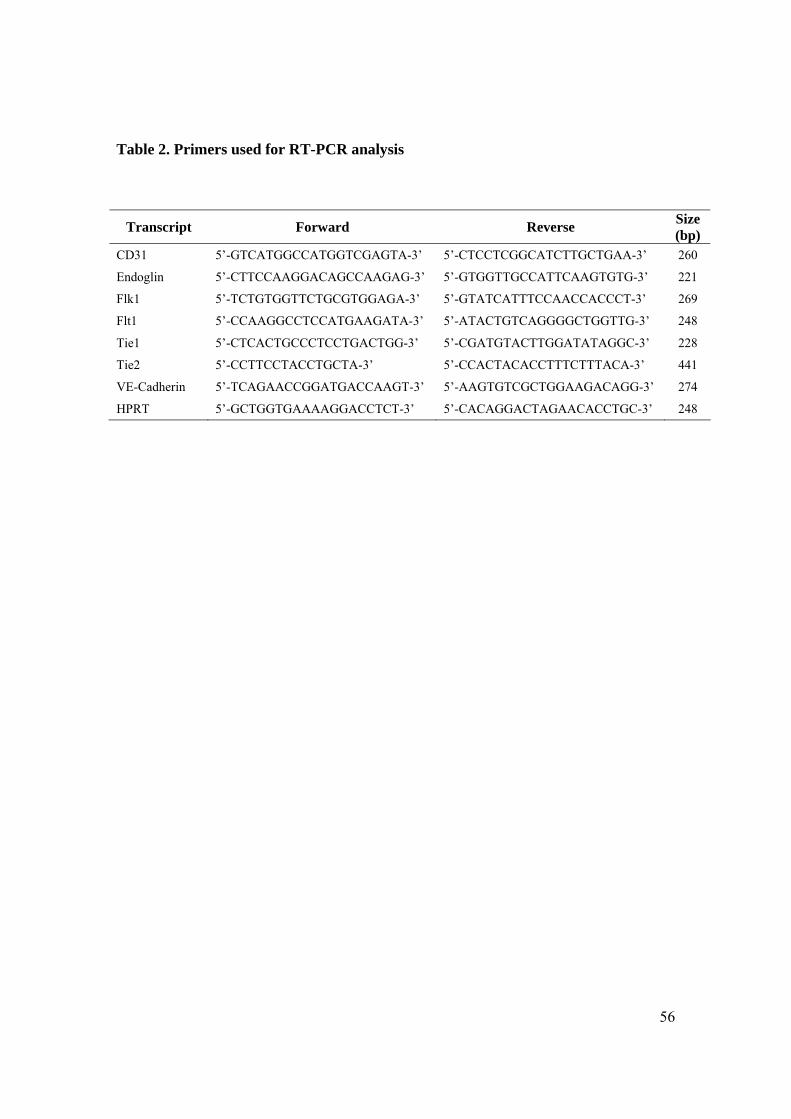
56
Table 2. Primers used for RT-PCR analysis
Transcript Forward Reverse Size (bp)
CD31 5’-GTCATGGCCATGGTCGAGTA-3’ 5’-CTCCTCGGCATCTTGCTGAA-3’ 260
Endoglin 5’-CTTCCAAGGACAGCCAAGAG-3’ 5’-GTGGTTGCCATTCAAGTGTG-3’ 221
Flk1 5’-TCTGTGGTTCTGCGTGGAGA-3’ 5’-GTATCATTTCCAACCACCCT-3’ 269
Flt1 5’-CCAAGGCCTCCATGAAGATA-3’ 5’-ATACTGTCAGGGGCTGGTTG-3’ 248
Tie1 5’-CTCACTGCCCTCCTGACTGG-3’ 5’-CGATGTACTTGGATATAGGC-3’ 228
Tie2 5’-CCTTCCTACCTGCTA-3’ 5’-CCACTACACCTTTCTTTACA-3’ 441
VE-Cadherin 5’-TCAGAACCGGATGACCAAGT-3’ 5’-AAGTGTCGCTGGAAGACAGG-3’ 274
HPRT 5’-GCTGGTGAAAAGGACCTCT-3’ 5’-CACAGGACTAGAACACCTGC-3’ 248

57
Bibliography
Abe, K., Niwa, H., Iwase, K., Takiguchi, M., Mori, M., Abe, S.I., and Yamamura, K.I. (1996). Endoderm-specific gene expression in embryonic stem cells differentiated to embryoid bodies. Exp Cell Res 229, 27-34.
Aliev, G., Shenk, J.C., Fischbach, K., and Perry, G. (2008). Stem cell niches as clinical targets: the future of anti-ischemic therapy? Nat Clin Pract Cardiovasc Med 5, 590-591.
Anthony, T.E., Klein, C., Fishell, G., and Heintz, N. (2004). Radial glia serve as neuronal progenitors in all regions of the central nervous system. Neuron 41, 881-890.
Auerbach, W., Dunmore, J.H., Fairchild-Huntress, V., Fang, Q., Auerbach, A.B., Huszar, D., and Joyner, A.L. (2000). Establishment and chimera analysis of 129/SvEv- and C57BL/6-derived mouse embryonic stem cell lines. Biotechniques 29, 1024-1028, 1030, 1032.
Avilion, A.A., Nicolis, S.K., Pevny, L.H., Perez, L., Vivian, N., and Lovell-Badge, R. (2003). Multipotent cell lineages in early mouse development depend on SOX2 function. Genes & development 17, 126-140.
Bain, G., Kitchens, D., Yao, M., Huettner, J.E., and Gottlieb, D.I. (1995). Embryonic stem cells express neuronal properties in vitro. Dev Biol 168, 342-357.
Bjornson, C.R., Rietze, R.L., Reynolds, B.A., Magli, M.C., and Vescovi, A.L. (1999). Turning brain into blood: a hematopoietic fate adopted by adult neural stem cells in vivo. Science 283, 534-537.
Bradley, A., Evans, M., Kaufman, M.H., and Robertson, E. (1984). Formation of germ-line chimaeras from embryo-derived teratocarcinoma cell lines. Nature 309, 255-256.
Brivanlou, A.H., Gage, F.H., Jaenisch, R., Jessell, T., Melton, D., and Rossant, J. (2003). Stem cells. Setting standards for human embryonic stem cells. Science 300, 913-916.
Brustle, O., Jones, K.N., Learish, R.D., Karram, K., Choudhary, K., Wiestler, O.D., Duncan, I.D., and McKay, R.D. (1999). Embryonic stem cell-derived glial precursors: a source of myelinating transplants. Science 285, 754-756.
Burdon, T., Stracey, C., Chambers, I., Nichols, J., and Smith, A. (1999). Suppression of SHP-2 and ERK Signalling Promotes Self-Renewal of Mouse Embryonic Stem Cells. Developmental Biology 210, 30-43.

58
Buzanska, L., Machaj, E.K., Zablocka, B., Pojda, Z., and Domanska-Janik, K. (2002). Human cord blood-derived cells attain neuronal and glial features in vitro. J Cell Sci 115, 2131-2138.
Bylund, M., Andersson, E., Novitch, B.G., and Muhr, J. (2003). Vertebrate neurogenesis is counteracted by Sox1-3 activity. Nat Neurosci 6, 1162-1168.
Carmeliet, P., and Tessier-Lavigne, M. (2005). Common mechanisms of nerve and blood vessel wiring. Nature 436, 193-200.
Chambers, I., Colby, D., Robertson, M., Nichols, J., Lee, S., Tweedie, S., and Smith, A. (2003). Functional expression cloning of Nanog, a pluripotency sustaining factor in embryonic stem cells. Cell 113, 643-655.
Choi, K., Kennedy, M., Kazarov, A., Papadimitriou, J.C., and Keller, G. (1998). A common precursor for hematopoietic and endothelial cells. Development 125, 725-732.
Conti, L., Pollard, S.M., Gorba, T., Reitano, E., Toselli, M., Biella, G., Sun, Y., Sanzone, S., Ying, Q.L., Cattaneo, E., et al. (2005). Niche-independent symmetrical self-renewal of a mammalian tissue stem cell. PLoS biology 3, e283.
Counter, C.M. (1996). The roles of telomeres and telomerase in cell life span. Mutation research 366, 45-63.
Davis, S., Aldrich, T.H., Stahl, N., Pan, L., Taga, T., Kishimoto, T., Ip, N.Y., and Yancopoulos, G.D. (1993). LIFR beta and gp130 as heterodimerizing signal transducers of the tripartite CNTF receptor. Science 260, 1805-1808.
Doetsch, F. (2003). A niche for adult neural stem cells. Curr Opin Genet Dev 13, 543-550.
Doetsch, F., Caille, I., Lim, D.A., Garcia-Verdugo, J.M., and Alvarez-Buylla, A. (1999). Subventricular zone astrocytes are neural stem cells in the adult mammalian brain. Cell 97, 703-716.
Doetschman, T., Williams, P., and Maeda, N. (1988). Establishment of hamster blastocyst-derived embryonic stem (ES) cells. Dev Biol 127, 224-227.
Doetschman, T.C., Eistetter, H., Katz, M., Schmidt, W., and Kemler, R. (1985). The in vitro development of blastocyst-derived embryonic stem cell lines: formation of visceral yolk sac, blood islands and myocardium. J Embryol Exp Morphol 87, 27-45.
Ema, M., and Rossant, J. (2003). Cell fate decisions in early blood vessel formation. Trends Cardiovasc Med 13, 254-259.
Evans, M.J., and Kaufman, M.H. (1981). Establishment in culture of pluripotential cells from mouse embryos. Nature 292, 154-156.

59
Ferrara, N., and Kerbel, R.S. (2005). Angiogenesis as a therapeutic target. Nature 438, 967-974.
First, N.L., Sims, M.M., Park, S.P., and Kent-First, M.J. (1994). Systems for production of calves from cultured bovine embryonic cells. Reproduction, fertility, and development 6, 553-562.
Fons, P., Herault, J.P., Delesque, N., Tuyaret, J., Bono, F., and Herbert, J.M. (2004). VEGF-R2 and neuropilin-1 are involved in VEGF-A-induced differentiation of human bone marrow progenitor cells. J Cell Physiol 200, 351-359.
Fraichard, A., Chassande, O., Bilbaut, G., Dehay, C., Savatier, P., and Samarut, J. (1995). In vitro differentiation of embryonic stem cells into glial cells and functional neurons. J Cell Sci 108 ( Pt 10), 3181-3188.
Fujisawa, H., Takagi, S., and Hirata, T. (1995). Growth-associated expression of a membrane protein, neuropilin, in Xenopus optic nerve fibers. Dev Neurosci 17, 343-349.
Gago, N., Avellana-Adalid, V., Evercooren, A.B., and Schumacher, M. (2003). Control of cell survival and proliferation of postnatal PSA-NCAM(+) progenitors. Mol Cell Neurosci 22, 162-178.
Gajovic, S., St-Onge, L., Yokota, Y., and Gruss, P. (1997). Retinoic acid mediates Pax6 expression during in vitro differentiation of embryonic stem cells. Differentiation 62, 187-192.
Gearing, D.P., Thut, C.J., VandeBos, T., Gimpel, S.D., Delaney, P.B., King, J., Price, V., Cosman, D., and Beckmann, M.P. (1991). Leukemia inhibitory factor receptor is structurally related to the IL-6 signal transducer, gp130. EMBO J 10, 2839-2848.
Gerhardt, H., Golding, M., Fruttiger, M., Ruhrberg, C., Lundkvist, A., Abramsson, A., Jeltsch, M., Mitchell, C., Alitalo, K., Shima, D., et al. (2003). VEGF guides angiogenic sprouting utilizing endothelial tip cell filopodia. J Cell Biol 161, 1163-1177.
Goolsby, J., Marty, M.C., Heletz, D., Chiappelli, J., Tashko, G., Yarnell, D., Fishman, P.S., Dhib-Jalbut, S., Bever, C.T., Jr., Pessac, B., et al. (2003). Hematopoietic progenitors express neural genes. Proc Natl Acad Sci U S A 100, 14926-14931.
Graves, K.H., and Moreadith, R.W. (1993). Derivation and characterization of putative pluripotential embryonic stem cells from preimplantation rabbit embryos. Molecular reproduction and development 36, 424-433.
Gualandris, A., Annes, J.P., Arese, M., Noguera, I., Jurukovski, V., and Rifkin, D.B. (2000). The latent transforming growth factor-beta-binding protein-1 promotes in vitro differentiation of embryonic stem cells into endothelium. Mol Biol Cell 11, 4295-4308.

60
Hadjantonakis, A.K., Macmaster, S., and Nagy, A. (2002). Embryonic stem cells and mice expressing different GFP variants for multiple non-invasive reporter usage within a single animal. BMC Biotechnol 2, 11.
Haubensak, W., Attardo, A., Denk, W., and Huttner, W.B. (2004). Neurons arise in the basal neuroepithelium of the early mammalian telencephalon: a major site of neurogenesis. Proc Natl Acad Sci U S A 101, 3196-3201.
Hirashima, M., Kataoka, H., Nishikawa, S., Matsuyoshi, N., and Nishikawa, S. (1999). Maturation of embryonic stem cells into endothelial cells in an in vitro model of vasculogenesis. Blood 93, 1253-1263.
Huber, T.L., Kouskoff, V., Fehling, H.J., Palis, J., and Keller, G. (2004). Haemangioblast commitment is initiated in the primitive streak of the mouse embryo. Nature 432, 625-630.
Iannaccone, P.M., Taborn, G.U., Garton, R.L., Caplice, M.D., and Brenin, D.R. (1994). Pluripotent embryonic stem cells from the rat are capable of producing chimeras. Dev Biol 163, 288-292.
Jakobsson, L., Kreuger, J., and Claesson-Welsh, L. (2007). Building blood vessels--stem cell models in vascular biology. J Cell Biol 177, 751-755.
Kattman, S.J., Huber, T.L., and Keller, G.M. (2006). Multipotent flk-1+ cardiovascular progenitor cells give rise to the cardiomyocyte, endothelial, and vascular smooth muscle lineages. Dev Cell 11, 723-732.
Kawasaki, H., Mizuseki, K., Nishikawa, S., Kaneko, S., Kuwana, Y., Nakanishi, S., Nishikawa, S.I., and Sasai, Y. (2000). Induction of midbrain dopaminergic neurons from ES cells by stromal cell-derived inducing activity. Neuron 28, 31-40.
Kawasaki, T., Kitsukawa, T., Bekku, Y., Matsuda, Y., Sanbo, M., Yagi, T., and Fujisawa, H. (1999). A requirement for neuropilin-1 in embryonic vessel formation. Development 126, 4895-4902.
Keller, G.M. (1995). In vitro differentiation of embryonic stem cells. Curr Opin Cell Biol 7, 862-869.
Kiefer, J.C. (2007). Back to basics: Sox genes. Dev Dyn 236, 2356-2366.
Kinder, S.J., Loebel, D.A., and Tam, P.P. (2001). Allocation and early differentiation of cardiovascular progenitors in the mouse embryo. Trends Cardiovasc Med 11, 177-184.
Kitsukawa, T., Shimizu, M., Sanbo, M., Hirata, T., Taniguchi, M., Bekku, Y., Yagi, T., and Fujisawa, H. (1997). Neuropilin-semaphorin III/D-mediated chemorepulsive signals play a crucial role in peripheral nerve projection in mice. Neuron 19, 995-1005.

61
Koestenbauer, S., Zech, N.H., Juch, H., Vanderzwalmen, P., Schoonjans, L., and Dohr, G. (2006). Embryonic Stem Cells: Similarities and Differences Between Human and Murine Embryonic Stem Cells. AJRI 55, 169-180.
Kopp, J.L., Ormsbee, B.D., Desler, M., and Rizzino, A. (2008). Small increases in the level of Sox2 trigger the differentiation of mouse embryonic stem cells. Stem Cells 26, 903-911.
Korn, J., Christ, B., and Kurz, H. (2002). Neuroectodermal origin of brain pericytes and vascular smooth muscle cells. J Comp Neurol 442, 78-88.
Kriegstein, A.R., and Götz, M. (2003). Radial glia diversity: A matter of cell fate. Glia 43, 37-43.
Kuroda, T., Tada, M., Kubota, H., Kimura, H., Hatano, S.-y., Suemori, H., Nakatsuji, N., and Tada, T. (2005). Octamer and Sox Elements Are Required for Transcriptional cis Regulation of Nanog Gene Expression. Mol Cell Biol 25, 2475-2485.
Lee, A., Kessler, J.D., Read, T.A., Kaiser, C., Corbeil, D., Huttner, W.B., Johnson, J.E., and Wechsler-Reya, R.J. (2005). Isolation of neural stem cells from the postnatal cerebellum. Nat Neurosci 8, 723-729.
Li, Z.J., Wang, Z.Z., Zheng, Y.Z., Xu, B., Yang, R.C., Scadden, D.T., and Han, Z.C. (2005). Kinetic expression of platelet endothelial cell adhesion molecule-1 (PECAM-1/CD31) during embryonic stem cell differentiation. J Cell Biochem 95, 559-570.
Magnusson, P., Rolny, C., Jakobsson, L., Wikner, C., Wu, Y., Hicklin, D.J., and Claesson-Welsh, L. (2004). Deregulation of Flk-1/vascular endothelial growth factor receptor-2 in fibroblast growth factor receptor-1-deficient vascular stem cell development. J Cell Sci 117, 1513-1523.
Malatesta, P., Hartfuss, E., and Gotz, M. (2000). Isolation of radial glial cells by fluorescent-activated cell sorting reveals a neuronal lineage. Development 127, 5253-5263.
Maltsev, V.A., Rohwedel, J., Hescheler, J., and Wobus, A.M. (1993). Embryonic stem cells differentiate in vitro into cardiomyocytes representing sinusnodal, atrial and ventricular cell types. Mech Dev 44, 41-50.
Martin, G.R. (1981). Isolation of a pluripotent cell line from early mouse embryos cultured in medium conditioned by teratocarcinoma stem cells. Proc Natl Acad Sci U S A 78, 7634-7638.
Martin, P., and Lewis, J. (1989). Origins of the neurovascular bundle: interactions between developing nerves and blood vessels in embryonic chick skin. Int J Dev Biol 33, 379-387.
62
Masui, S., Nakatake, Y., Toyooka, Y., Shimosato, D., Yagi, R., Takahashi, K., Okochi, H., Okuda, A., Matoba, R., Sharov, A.A., et al. (2007). Pluripotency governed by Sox2 via regulation of Oct3/4 expression in mouse embryonic stem cells. Nat Cell Biol 9, 625-635.
Maurer, M.H., Tripps, W.K., Feldmann, R.E., Jr., and Kuschinsky, W. (2003). Expression of vascular endothelial growth factor and its receptors in rat neural stem cells. Neurosci Lett 344, 165-168.
Mezey, E., Chandross, K.J., Harta, G., Maki, R.A., and McKercher, S.R. (2000). Turning blood into brain: cells bearing neuronal antigens generated in vivo from bone marrow. Science 290, 1779-1782.
Mezey, E., Key, S., Vogelsang, G., Szalayova, I., Lange, G.D., and Crain, B. (2003). Transplanted bone marrow generates new neurons in human brains. Proc Natl Acad Sci U S A 100, 1364-1369.
Miller-Hance, W.C., LaCorbiere, M., Fuller, S.J., Evans, S.M., Lyons, G., Schmidt, C., Robbins, J., and Chien, K.R. (1993). In vitro chamber specification during embryonic stem cell cardiogenesis. Expression of the ventricular myosin light chain-2 gene is independent of heart tube formation. J Biol Chem 268, 25244-25252.
Mukouyama, Y.S., Shin, D., Britsch, S., Taniguchi, M., and Anderson, D.J. (2002). Sensory nerves determine the pattern of arterial differentiation and blood vessel branching in the skin. Cell 109, 693-705.
Murry, C.E., and Keller, G. (2008). Differentiation of embryonic stem cells to clinically relevant populations: lessons from embryonic development. Cell 132, 661-680.
Nakano, T., Kodama, H., and Honjo, T. (1996). In vitro development of primitive and definitive erythrocytes from different precursors. Science 272, 722-724.
Nanka, O., Peumans, W.J., Van Damme, E.J., Pfuller, U., Valasek, P., Halata, Z., Schumacher, U., and Grim, M. (2001). Lectin histochemistry of microvascular endothelium in chick and quail musculature. Anat Embryol (Berl) 204, 407-411.
Neufeld, G., and Kessler, O. (2008). The semaphorins: versatile regulators of tumour progression and tumour angiogenesis. Nature reviews 8, 632-645.
Nichols, J., Chambers, I., and Smith, A. (1994). Derivation of germline competent embryonic stem cells with a combination of interleukin-6 and soluble interleukin-6 receptor. Exp Cell Res 215, 237-239.
Nichols, J., Chambers, I., Taga, T., and Smith, A. (2001). Physiological rationale for responsiveness of mouse embryonic stem cells to gp130 cytokines. Development 128, 2333-2339.

63
Nichols, J., Zevnik, B., Anastassiadis, K., Niwa, H., Klewe-Nebenius, D., Chambers, I., Scholer, H., and Smith, A. (1998). Formation of pluripotent stem cells in the mammalian embryo depends on the POU transcription factor Oct4. Cell 95, 379-391.
Nishikawa, S.I., Nishikawa, S., Hirashima, M., Matsuyoshi, N., and Kodama, H. (1998). Progressive lineage analysis by cell sorting and culture identifies FLK1+VE-cadherin+ cells at a diverging point of endothelial and hemopoietic lineages. Development 125, 1747-1757.
Niwa, H. (2001). Molecular mechanism to maintain stem cell renewal of ES cells. Cell Struct Funct 26, 137-148.
Niwa, H., Burdon, T., Chambers, I., and Smith, A. (1998). Self-renewal of pluripotent embryonic stem cells is mediated via activation of STAT3. Genes & development 12, 2048-2060.
Niwa, H., Miyazaki, J., and Smith, A.G. (2000). Quantitative expression of Oct-3/4 defines differentiation, dedifferentiation or self-renewal of ES cells. Nat Genet 24, 372-376.
Noctor, S.C., Flint, A.C., Weissman, T.A., Dammerman, R.S., and Kriegstein, A.R. (2001). Neurons derived from radial glial cells establish radial units in neocortex. Nature 409, 714-720.
Noctor, S.C., Flint, A.C., Weissman, T.A., Wong, W.S., Clinton, B.K., and Kriegstein, A.R. (2002). Dividing Precursor Cells of the Embryonic Cortical Ventricular Zone Have Morphological and Molecular Characteristics of Radial Glia. J Neurosci 22, 3161-3173.
Odorico, J.S., Kaufman, D.S., and Thomson, J.A. (2001). Multilineage differentiation from human embryonic stem cell lines. Stem Cells 19, 193-204.
Oishi, K., Kobayashi, A., Fujii, K., Kanehira, D., Ito, Y., and Uchida, M.K. (2004). Angiogenesis in vitro: vascular tube formation from the differentiation of neural stem cells. J Pharmacol Sci 96, 208-218.
Okamoto, K., Okazawa, H., Okuda, A., Sakai, M., Muramatsu, M., and Hamada, H. (1990). A novel octamer binding transcription factor is differentially expressed in mouse embryonic cells. Cell 60, 461-472.
Okumura-Nakanishi, S., Saito, M., Niwa, H., and Ishikawa, F. (2005). Oct-3/4 and Sox2 Regulate Oct-3/4 Gene in Embryonic Stem Cells. J Biol Chem 280, 5307-5317.
Pain, B., Clark, M., Shen, M., Nakazawa, H., Sakurai, M., Samarut, J., and Etches, R. (1996). Long-term in vitro culture and characterisation of avian embryonic stem cells with multiple morphogenetic potentialities. Development 122, 2339-2348.

64
Park, J.E., Chen, H.H., Winer, J., Houck, K.A., and Ferrara, N. (1994). Placenta growth factor. Potentiation of vascular endothelial growth factor bioactivity, in vitro and in vivo, and high affinity binding to Flt-1 but not to Flk-1/KDR. J Biol Chem 269, 25646-25654.
Poole, T.J., Finkelstein, E.B., and Cox, C.M. (2001). The role of FGF and VEGF in angioblast induction and migration during vascular development. Dev Dyn 220, 1-17.
Rathjen, P.D., Toth, S., Willis, A., Heath, J.K., and Smith, A.G. (1990). Differentiation inhibiting activity is produced in matrix-associated and diffusible forms that are generated by alternate promoter usage. Cell 62, 1105-1114.
Reali, C., Scintu, F., Pillai, R., Cabras, S., Argiolu, F., Ristaldi, M.S., Sanna, M.A., Badiali, M., and Sogos, V. (2006). Differentiation of human adult CD34+ stem cells into cells with a neural phenotype: role of astrocytes. Exp Neurol 197, 399-406.
Risau, W. (1997). Mechanisms of angiogenesis. Nature 386, 671-674.
Risau, W., and Flamme, I. (1995). Vasculogenesis, pp. 73-91.
Risau, W., Sariola, H., Zerwes, H.G., Sasse, J., Ekblom, P., Kemler, R., and Doetschman, T. (1988). Vasculogenesis and angiogenesis in embryonic-stem-cell-derived embryoid bodies. Development 102, 471-478.
Rizzino, A. (2008). Transcription factors that behave as master regulators during mammalian embryogenesis function as molecular rheostats. Biochem J 411, e5-7.
Rohwedel, J., Maltsev, V., Bober, E., Arnold, H.H., Hescheler, J., and Wobus, A.M. (1994). Muscle cell differentiation of embryonic stem cells reflects myogenesis in vivo: developmentally regulated expression of myogenic determination genes and functional expression of ionic currents. Dev Biol 164, 87-101.
Rosner, M.H., Vigano, M.A., Ozato, K., Timmons, P.M., Poirie, F., Rigby, P.W.J., and Staudt, L.M. (1990). A POU-domain transcription factor in early stem cells and germ cells of the mammalian embryo. Nature 345, 686-692.
Sanchez, A., Jones, W.K., Gulick, J., Doetschman, T., and Robbins, J. (1991). Myosin heavy chain gene expression in mouse embryoid bodies. An in vitro developmental study. J Biol Chem 266, 22419-22426.
Sarnat, H.B., Nochlin, D., and Born, D.E. (1998). Neuronal nuclear antigen (NeuN): a marker of neuronal maturation in early human fetal nervous system. Brain Dev 20, 88-94.
Schroeder, T., Meier-Stiegen, F., Schwanbeck, R., Eilken, H., Nishikawa, S., Hasler, R., Schreiber, S., Bornkamm, G.W., Nishikawa, S., and Just, U. (2006). Activated Notch1 alters differentiation of embryonic stem cells into mesodermal cell lineages at multiple stages of development. Mech Dev 123, 570-579.
65
Serini, G., and Bussolino, F. (2004). Common cues in vascular and axon guidance. Physiology (Bethesda) 19, 348-354.
Shalaby, F., Ho, J., Stanford, W.L., Fischer, K.D., Schuh, A.C., Schwartz, L., Bernstein, A., and Rossant, J. (1997). A requirement for Flk1 in primitive and definitive hematopoiesis and vasculogenesis. Cell 89, 981-990.
Shalaby, F., Rossant, J., Yamaguchi, T.P., Gertsenstein, M., Wu, X.F., Breitman, M.L., and Schuh, A.C. (1995). Failure of blood-island formation and vasculogenesis in Flk-1-deficient mice. Nature 376, 62-66.
Shen, Q., Wang, Y., Kokovay, E., Lin, G., Chuang, S.M., Goderie, S.K., Roysam, B., and Temple, S. (2008). Adult SVZ stem cells lie in a vascular niche: a quantitative analysis of niche cell-cell interactions. Cell stem cell 3, 289-300.
Shim, H., Gutierrez-Adan, A., Chen, L., BonDurant, R., Behboodi, E., and Anderson, G. (1997). Isolation of pluripotent stem cells from cultured porcine primordial germ cells. Biol Reprod 57, 1089-1095.
Shraga-Heled, N., Kessler, O., Prahst, C., Kroll, J., Augustin, H., and Neufeld, G. (2007). Neuropilin-1 and neuropilin-2 enhance VEGF121 stimulated signal transduction by the VEGFR-2 receptor. Faseb J 21, 915-926.
Silva, J., Chambers, I., Pollard, S., and Smith, A. (2006). Nanog promotes transfer of pluripotency after cell fusion. Nature 441, 997-1001.
Smith, A.G. (2001). EMBRYO-DERIVED STEM CELLS: Of Mice and Men. Annual Review of Cell and Developmental Biology 17, 435-462.
Smith, A.G., Heath, J.K., Donaldson, D.D., Wong, G.G., Moreau, J., Stahl, M., and Rogers, D. (1988). Inhibition of pluripotential embryonic stem cell differentiation by purified polypeptides. Nature 336, 688-690.
Smith, A.G., and Hooper, M.L. (1987). Buffalo rat liver cells produce a diffusible activity which inhibits the differentiation of murine embryonal carcinoma and embryonic stem cells. Developmental Biology 121, 1-9.
Soker, S., Miao, H.Q., Nomi, M., Takashima, S., and Klagsbrun, M. (2002). VEGF165 mediates formation of complexes containing VEGFR-2 and neuropilin-1 that enhance VEGF165-receptor binding. J Cell Biochem 85, 357-368.
Strubing, C., Ahnert-Hilger, G., Shan, J., Wiedenmann, B., Hescheler, J., and Wobus, A.M. (1995). Differentiation of pluripotent embryonic stem cells into the neuronal lineage in vitro gives rise to mature inhibitory and excitatory neurons. Mech Dev 53, 275-287.

66
Sukoyan, M.A., Vatolin, S.Y., Golubitsa, A.N., Zhelezova, A.I., Semenova, L.A., and Serov, O.L. (1993). Embryonic stem cells derived from morulae, inner cell mass, and blastocysts of mink: comparisons of their pluripotencies. Mol Reprod Dev 36, 148-158.
Tamagnone, L., and Comoglio, P.M. (2004). To move or not to move? Semaphorin signalling in cell migration. EMBO reports 5, 356-361.
Tavazoie, M., Van der Veken, L., Silva-Vargas, V., Louissaint, M., Colonna, L., Zaidi, B., Garcia-Verdugo, J.M., and Doetsch, F. (2008). A specialized vascular niche for adult neural stem cells. Cell Stem Cell 3, 279-288.
Thomson, J.A., Itskovitz-Eldor, J., Shapiro, S.S., Waknitz, M.A., Swiergiel, J.J., Marshall, V.S., and Jones, J.M. (1998). Embryonic Stem Cell Lines Derived from Human Blastocysts. Science 282, 1145-1147.
Thomson, J.A., Kalishman, J., Golos, T.G., Durning, M., Harris, C.P., Becker, R.A., and Hearn, J.P. (1995). Isolation of a primate embryonic stem cell line. Proceedings of the National Academy of Sciences of the United States of America 92, 7844-7848.
Thomson, J.A., and Odorico, J.S. (2000). Human embryonic stem cell and embryonic germ cell lines. Trends in Biotechnology 18, 53-57.
Ueno, H., and Weissman, I.L. (2006). Clonal analysis of mouse development reveals a polyclonal origin for yolk sac blood islands. Dev Cell 11, 519-533.
Vittet, D., Prandini, M.H., Berthier, R., Schweitzer, A., Martin-Sisteron, H., Uzan, G., and Dejana, E. (1996). Embryonic stem cells differentiate in vitro to endothelial cells through successive maturation steps. Blood 88, 3424-3431.
Wang, R., Clark, R., and Bautch, V.L. (1992). Embryonic stem cell-derived cystic embryoid bodies form vascular channels: an in vitro model of blood vessel development. Development 114, 303-316.
Wiese, C., Rolletschek, A., Kania, G., Blyszczuk, P., Tarasov, K.V., Tarasova, Y., Wersto, R.P., Boheler, K.R., and Wobus, A.M. (2004). Nestin expression--a property of multi-lineage progenitor cells? Cell Mol Life Sci 61, 2510-2522.
Wiles, M.V., and Keller, G. (1991). Multiple hematopoietic lineages develop from embryonic stem (ES) cells in culture. Development 111, 259-267.
Wu, S.M., Fujiwara, Y., Cibulsky, S.M., Clapham, D.E., Lien, C.L., Schultheiss, T.M., and Orkin, S.H. (2006). Developmental origin of a bipotential myocardial and smooth muscle cell precursor in the mammalian heart. Cell 127, 1137-1150.
Wurmser, A.E., Nakashima, K., Summers, R.G., Toni, N., D'Amour, K.A., Lie, D.C., and Gage, F.H. (2004). Cell fusion-independent differentiation of neural stem cells to the endothelial lineage. Nature 430, 350-356.

67
Yamashita, J., Itoh, H., Hirashima, M., Ogawa, M., Nishikawa, S., Yurugi, T., Naito, M., Nakao, K., and Nishikawa, S.-I. (2000). Flk1-positive cells derived from embryonic stem cells serve as vascular progenitors. Nature 408, 92-96.
Ying, Q.L., Stavridis, M., Griffiths, D., Li, M., and Smith, A. (2003). Conversion of embryonic stem cells into neuroectodermal precursors in adherent monoculture. Nat Biotechnol 21, 183-186.
Yoshida, K., Chambers, I., Nichols, J., Smith, A., Saito, M., Yasukawa, K., Shoyab, M., Taga, T., and Kishimoto, T. (1994). Maintenance of the pluripotential phenotype of embryonic stem cells through direct activation of gp130 signalling pathways. Mechanisms of Development 45, 163-171.
Yuan, H., Corbi, N., Basilico, C., and Dailey, L. (1995). Developmental-specific activity of the FGF-4 enhancer requires the synergistic action of Sox2 and Oct-3. Genes Dev 9, 2635-2645.
Yuan, H.T., Venkatesha, S., Chan, B., Deutsch, U., Mammoto, T., Sukhatme, V.P., Woolf, A.S., and Karumanchi, S.A. (2007). Activation of the orphan endothelial receptor Tie1 modifies Tie2-mediated intracellular signaling and cell survival. FASEB J 21, 3171-3183.
Zappone, M.V., Galli, R., Catena, R., Meani, N., De Biasi, S., Mattei, E., Tiveron, C., Vescovi, A.L., Lovell-Badge, R., Ottolenghi, S., et al. (2000). Sox2 regulatory sequences direct expression of a (beta)-geo transgene to telencephalic neural stem cells and precursors of the mouse embryo, revealing regionalization of gene expression in CNS stem cells. Development 127, 2367-2382.
Zhou, Q., Chipperfield, H., Melton, D.A., and Wong, W.H. (2007). A gene regulatory network in mouse embryonic stem cells. Proc Natl Acad Sci U S A 104, 16438-16443.