
UNIVERSITA' DEGLI STUDI DI PADOVA - Padua@Research
77
UNIVERSITA' DEGLI STUDI DI PADOVA Università degli Studi di Padova Dipartimento di Biologia SCUOLA DI DOTTORATO DI RICERCA IN BIOSCIENZE INDIRIZZO: GENETICA E BIOLOGIA MOLECOLARE DELLO SVLUPPO CICLO XX TESI DI DOTTORATO ARRHYTHMOGENIC RIGHT VENTRICULAR CARDIOMYOPATHY: MUTATION SCREENING OF CANDIDATE GENES AND IN VITRO FUNCTIONAL STUDIES Direttore della Scuola : Ch.mo Prof. TULLIO POZZAN Supervisore :Ch.mo Prof. GIAN ANTONIO DANIELI Dottoranda: MARZIA DE BORTOLI 31 gennaio 2008
Transcript of UNIVERSITA' DEGLI STUDI DI PADOVA - Padua@Research

UNIVERSITA' DEGLI STUDI DI PADOVA
Università degli Studi di Padova
Dipartimento di Biologia
SCUOLA DI DOTTORATO DI RICERCA IN BIOSCIENZE
INDIRIZZO: GENETICA E BIOLOGIA MOLECOLARE DELLO SVLUPPO
CICLO XX
TESI DI DOTTORATO
ARRHYTHMOGENIC RIGHT VENTRICULAR CARDIOMYOPATHY:
MUTATION SCREENING OF
CANDIDATE GENES AND IN VITRO FUNCTIONAL STUDIES
Direttore della Scuola : Ch.mo Prof. TULLIO POZZAN
Supervisore :Ch.mo Prof. GIAN ANTONIO DANIELI
Dottoranda: MARZIA DE BORTOLI
31 gennaio 2008


CONTENTS INTRODUCTION
CLINICAL ASPECTS OF ARVC
GENETICS OF ARVC
MOLECULAR PATHOGENESIS OF ARVC
PERP: A NOVEL CANDIDATE GENE
AIM OF THE STUDY
RESULTS
MUTATION SCREENING OF PERP GENE
MUTATION SCREENING OF DSC2 GENE
FUNCTIONAL ANALYSIS OF MUTANT DESMOCOLLINS
DISCUSSION
MUTATION SCREENING OF PERP GENE
MUTATION SCREENING OF DSC2 GENE
DSC2 FUNCTIONAL STUDIES
CONCLUSIONS
MATHERIALS AND METHODS
REFERENCES
SUMMARY
RIASSUNTO
1
1
2
7
9
11
13
17
17
27
31
31
32
36
39
41
55
67
71


1
INTRODUCTION CLINICAL ASPECTS OF ARVC
Arrhythmogenic right ventricular cardiomyopathy (ARVC) is an inherited heart disease
which may result in arrhythmia, heart failure, and sudden cardiac death. In fact, ARVC
is a major cause of sudden death in the young and athletes; its prevalence has been
estimated to vary from 1:2,500 to 1:5,000. Familial occurrence of ARVC is rather
common. The trait shows autosomal dominant inheritance with about 50% penetrance
(Nava A. et al., 1988). The main pathologic feature is progressive loss of right
ventricular myocardium, which is replaced by adipose and fibrous tissue (Thiene G. et
al., 1988). These changes may be localized; in early disease they are often confined to
the so-called “triangle of dysplasia”: the inflow, outflow, and apical regions of the right
ventricle. Aneurysm formation may occur. With the progress of the disease, diffuse
myocardial involvement leads to global right ventricular dilation. Histological
examination of affected myocardial tissue shows sparse myocytes interspersed among
adipocytes and fibrous tissue. The process begins from epicardium and gradually
extends through myocardium towards subendocardium. Fibrofatty substitution of the
left ventricle is rather frequent in the advances state of the disease.
Figure 1: A typical case of ARVC in a 25-years old man who died suddenly at rest.
Noticeable isolated fatty replacement of the right ventricular free wall and translucent infundibulum. Endomyocardial biopsy of the right ventricle free wall from another patient affected with ARVC shows rare myocytes embedded in fatty and fibrous tissues (From Nava A. et al., 1997).
Clinical manifestations of the disease occur most often between the second and fourth
decade of life; they include structural and functional abnormalities of the right ventricle,
electrocardiographic depolarization/repolarization changes and arrhythmias of right
ventricular origin (Marcus F.I. et al., 1982; Nava A. et al., 2000).

2
Natural history of the disease may be subdivided into four phases, on the basis of
clinical and pathological findings (Corrado D. et al., 2000). Early ARVC is often
described as “concealed” owing to frequent absence of clinical findings, although minor
ventricular arrhythmia and subtle structural changes are sometimes discernible. Patients
tend to be asymptomatic but nonetheless they may be at risk of sudden death, notably
during strong physical exercise. The “overt electrical disorder” which subsequently
develops is characterized by symptomatic ventricular arrhythmia; patients typically
present with palpitation, syncope, and pre-syncope. Morphological abnormalities are
more obvious at this stage and usually detectable by imaging. In the third phase, further
extension of disease through the right ventricular myocardium causes impaired
contractility and isolated right heart failure. Left ventricular involvement with
consequent biventricular failure occurs in the end-stage, which may be difficult to
distinguish from dilated cardiomyopathy (DCM) (Nemec J. et al., 1999).
GENETICS OF ARVC
From early ‘90s, linkage analysis started to reveal the existence of 9 genetic loci
independently involved in the determination of ARVC; however, only 4 disease genes
(RYR2, JUP, DSP and TGFβ3) have been identified so far within these regions (Tab.1).
Recently, genetic analysis shifted from linkage studies to candidate gene approach, thus
leading to the discovery of additional genes involved in ARVC (PKP2, DSG2 and
DSC2), which escaped detection by linkage approach (Tab.1).
Until now, out of the seven genes found associated to ARVC, RYR2 is the only one
directly involved in Ca2+ homeostasis (Tiso N. et al., 2001). RYR2 is one of the largest
human genes, including 105 exons and encoding a 565 Kda monomer, which is part of a
homo-tetrameric sarcoplasmic reticulum membrane protein. The homo-tetrameric
structure, known as cardiac ryanodine receptor, plays a pivotal role in intracellular
calcium homeostasis and excitation-contraction coupling in cardiomyocytes (Stokes
D.L. and Wagenknecht T., 2000; Missiaen L. et al., 2000). Mutations in the human
RYR2 gene have been associated with ARVC2 but also with catecholaminergic
polymorphic ventricular tachycardia (CPVT; OMIM 604772) (Priori S.G. et al., 2001;
Priori S.G. et al., 2002) and familial polymorphic ventricular tachycardia (FPVT;
OMIM 604772) (Laitinen P.J. et al., 2001; Laitinen P.J. et al., 2003). ARVC2 is a form
characterized by the presence of polymorphic, effort-induced arrhythmias, but less
pronounced fibro-fatty substitution of the myocardial tissue than in classical ARVC. All

3
RYR2 mutations causing ARVC2 were missense resulting in substitutions involving
amino acids highly conserved in critical domains of the protein (Tiso N. et al., 2001;
Bagattin A. et al., 2004). It has been proposed that RYR2 mutations affect regulation of
calcium channel. Intense adrenergic stimulation due to emotional or physical stress can
lead to calcium overload, thus triggering severe arrhythmias.
Table 1: Known ARVC loci and disease-genes.
Although ARVC is most commonly inherited as autosomal dominant trait, identification
of the first disease-causing gene occurred for a recessive variant known as Naxos
disease. This syndrome is characterised by arrhythmogenic right ventricular
cardiomyophaty associated with palmoplantar keratoderma and peculiar woolly hair. In
1998, Coonar et al. mapped the genetic locus to 17q21 (Coonar A.S. et. al., 1998). Two
years later, McKoy et al. reported a 2 base pair deletion (c.2157del2TG) in the
plakoglobin gene (JUP) as the cause of Naxos disease. This mutation results in a
frameshift and premature termination of the protein (McKoy G. et al., 2000).
Interestingly, a recent paper reported a novel dominant mutation (c.118_119insGCA) in
JUP 17q21
AD AR
ARVD12 Naxos Disease
DSC2 18q12.1 AD ARVD11
DSG2 18q12.1ADARVD10
PKP2 12p11 AD ARVD9
DSP 6p24ADARVD8
10q22.3ADARVD7
10p12-p14 AD ARVD6
3p23ADARVD5
2q32.1-q32.2ADARVD4
14q12-q22ADARVD3
RYR2 1q42-q43 AD ARVD2
TGF-β3 14q23-q24 AD ARVD1
Gene ChromosomeInheritance pattern
Locus

4
the gene encoding Plakoglobin, causing ARVC. This dominant mutation doesn’t disrupt
the frame of translation but it would cause the insertion of an additional serine residue at
amino acid position 39 within the N-terminal domain of plakoglobin (S39_K40insS)
(Asimaki A. et al., 2007). Plakoglobin is an armadillo family member contains 12 arm
reapts and is a major component of cell-cell adhesion complexes, which are abundant in
many tissues. It is also a signaling molecule with role in desmosome assembly and
development and in the regulation of gene expression (Rubenstein A. et al., 1997). In
fact, Plakoglobin is found in junctions as well as in the nucleus where it may have a role
in transcriptional regulation (Getsios et al., 2004). It is now accepted that plakoglobin
plays a role in the Wnt/β-catenin signalling pathway and that it also could interact with
Tcf/Lef protein. However it is not yet clear whether plakoglobin activates or represses
Wnt/β-catenin target genes and whether it acts on distinct set of genes (Garrod D. and
Chidgey M., 2007).
The first disease gene linked to autosomal dominant ARVC showing typical right
ventricular phenotype was Desmoplakin (DSP). In 2002, a genome scan in a Italian
family with ARVC detected linkage with a region of chromosome 6 short arm, which
includes DSP gene. A missense mutation S299R was identified in exon 7 of all affected
persons of this family; the resulting amino acid substitution modifies a putative
phosphorylation site in the N-terminal plakoglobin binding domain (Rampazzo A. et al.,
2002). Desmoplakin, together with plakoglobin, anchors to desmosomal cadherins,
forming an ordered array of nontransmembrane proteins, which bind to desmin
intermediate filaments in cardiomyocytes. Desmoplakin contains three main functional
domains: the N-terminal which binds to the desmosome via connection with
plakoglobin and plakophilin; a ROD segment which is predicted to form a dimeric coil
and the C-terminal important for intermediate filaments binding (Choi H.J. et al., 2002).
There are two isoforms of desmoplakin (DPI and DPII) which are generated by
alternative splicing and differ only in the length of the central rod domain. Mutations in
DSP gene have been shown to be responsible for some cases of an autosomal dominant
skin disorder (striate palmoplantar keratoderma) (Armstrong D.K. et al., 1999; Whittock
N.V. et al., 1999; Whittock N.V. et al., 2002); an autosomal recessive condition
characterized by dilated cardiomyopathy, woolly hair, and keratoderma (so-called
Carvajal syndrome) (Norgett E.E. et al., 2000), another autosomal recessive condition
characterized by ARVC, woolly hair, and keratoderma (Alcalai R. et al., 2003) and a

5
left-sided ARVC named arrhythmogenic left ventricular cardiomyopathy (ALVC)
(Norman M. et al., 2005).
The discovery of plakoglobin and desmoplakin mutations led to the idea that ARVC is
due to cellular-adhesion defects, thus prompting the candidate gene approach for
identifying additional genes involved in ARVC.
In 2004, Gerull et al. selected plakophilin-2 (PKP2) as a candidate gene for ARVC
because it encodes an essential protein of cardiac desmosomes; in fact, a homozygous
deletion in PKP2 gene caused a lethal cardiac defect in mice (Grossmann K.S. et al.,
2004). The authors identified 25 different heterozygous mutations in 32 of 120
unrelated ARVC patients (Gerull B. et al., 2004). Plakophilin-2 is an armadillo-related
protein, located in the outer dense plaque of desmosomes. It links desmosomal
cadherins to desmoplakin and the intermediate filament system (Fig.2). There are two
isoforms of plakophilin-2, a shorter ‘a’ variant and a longer ‘b’ form, generated by
alternative splicing. PKP2a and PKP2b differ by the insertion of 44 amino acids
between armadillo repeats 2 and 3 (Mertens C. et al., 1996). Plakophilin-2 are also
present in the nucleus, where it may play a role in transcriptional regulation because it
has been associated with RNA polymerase III (Mertens C. et al., 2001).
Figure 2: Schematic representation of relationships between desmosomal proteins.
Transmembrane desmosomal cadherins, Dsg and Dsc, bind the armadillo family protein PG, which in turn anchors the plakin family member DP and PKP. The cytoplasmic plaque, which is further stabilized by lateral interactions among these proteins, anchors the IF cytoskeleton to the desmosome (From: Green K.J. and Sympson C.L., 2007).
6
Gerull et al. speculated that lack of plakophilin-2 or incorporation of mutant
plakophilin-2 in the cardiac desmosomes might impair cell-cell contacts and might
disrupt association between adjacent cardiomyocytes.
Recent studies have reported mutations in a fourth desmosomal gene, desmoglein-2
(DSG2), in familial cases of ARVC (Pilichou K. et al., 2006; Awad M.M. et al., 2006).
After few months another desmosomal gene, desmoscollin-2 (DSC2) was identified as
involved in ARVC (Syrris P. et al., 2006; Heuser A. et al., 2006). Desmosomal
cadherins, DSGs and DSCs, are single-pass transmembrane glycoproteins, that mediate
Ca2+-dependent cell-cell adhesion (Yin T. and Green K.J., 2004), by interacting laterally
and transcellularly with each other and by recruiting cytoplasmic plaque proteins which
facilitate attachment of intermediate filaments. In humans there are four desmoglein
isoforms (DSG1-4) and three desmocollin isoforms (DSC1-3); the corresponding genes
cluster in the same region of chromosome 18 (Hunt D.M. et al., 1999). DSG2 and DSC2
are expressed in all desmosome-containing tissues but they are the only isoforms
expressed in cardiac myocytes (Schäfer S. et al., 1994; Nuber U.A. et al., 1995). Each of
the three desmocollin genes encodes a pair of proteins that are generated by alternative
splicing, a longer ‘a’ form and a shorter ‘b’ form that differ only in their C-terminal
tails. The desmocollin extracellular domains can divided into a number of subdomains,
four cadherin-like EC domains and an extracellular anchor domain (EA). Desmoglein
extracellular domains are organised in a similar fashion. Within the cell, both
desmocollin ‘a’ and ‘b’ proteins possess an intracellular anchor domain (IA) but only
‘a’ forms have an intracellular cadherin-like sequence domain (ICS). Desmoglein
cytoplasmic tails also have IA and ICS domains. Desmocollin and desmoglein ICS
domains provide binding sites for other desmosomal constituents such as plakoglobin.
Additional domains found in desmoglein cytoplasmic tails include the intracellular
proline-rich linker domain (IPL), a repeat unit domain (RUD) made by a variable
number of 29 amino acids repeats, and a glycine-rich desmoglein terminal domain
(DTD) (Green K.J. and Gaudry C.A., 2000; Huber O., 2003; Garrod D. and Chidgey
M., 2007).
On the other hand, different roles in the determination of ARVC have been suggested
by finding regulatory mutations of TGFβ3 gene associated with ARVC. In 1994,
linkage analysis identified a genetic locus for dominant ARVC at 14q23-q24, thereafter
termed ARVC1 (Rampazzo A. et al., 1994). One of the most promising candidate genes
mapped to this region was transforming growth factor β3 (TGFβ3). In fact a nucleotide

7
substitution c.-36G>A in 5’UTR of TGFβ3 gene was detected in all affected subjects
belonging to a large ARVC1 family and an additional mutation c.1723C>T was
identified in 3’UTR of one patient. In vitro expression assay of constructs containing
the mutations showed that mutated UTRs were two-fold more active than wild type
(Beffagna G. et al., 2005). Transforming growth factor β family of cytokines is known
to stimulate mesenchymal cells to proliferate and to produce extracellular matrix
components. It is therefore conceivable that enhanced TGFβ activity may lead to
myocardial fibrosis. Myocardial fibrosis may disrupt electrical and mechanical
behaviour of myocardium and extracellular matrix abnormalities may predispose re-
entrant ventricular arrhythmias. Moreover, it has been shown that TGFβs modulate
expression of genes encoding desmosomal proteins in different cell types (Kapoun A.M.
et al., 2004; Yoshida M. et al., 1992). Therefore, overexpression of TGFβ3, caused by
UTRs mutations, might affect cell to cell junctions stability, thus leading to disease
expression similar to that observed in ARVC due to mutations of genes encoding
desmosomal proteins.
Desmoplakin involved in ARVC8, plakophilin-2 involved in ARVC9, desmoglein-2
involved in ARVC10, desmocollin-2 involved in ARVC11 and plakoglobin involved in
Naxos syndrome and in ARVC12 are all desmosomal proteins. Based on present
evidence ARVC is considered a disease of the desmosome. For this reason, additional
components of desmosomal complex may be targets for pathogenic mutations leading to
ARVC.
MOLECULAR PATHOGENESIS OF ARVC While involvement of genes encoding desmosomal proteins in ARVC suggests that
disruption of desmosomal integrity might be among primary molecular defects (Yang Z.
et al, 2006), mechanisms leading to ARVC remain to be elucidated.
Studies reported so far point to the importance of desmosomes as intercellular adhesive
organelles, required for the integrity of epithelial and cardiac tissues. However,
desmosome components functions are not limited to their roles in desmosomes or in
mechanical integrity of tissues, but they extend to supra-adhesive functions in vivo. It
has been shown that mechanical forces applied to adherens junctions in ventricular
cardiomyocytes activate stretch-sensitive calcium channels via cadherin’ mechanical
intracellular signaling, thus suggesting the importance of these channels in transduction

8
of mechanical forces into a cellular electrochemical signal, via increase of intracellular
calcium concentration (Gannier F. et al., 1996; Tatsukawa Y. et al., 1997; Ko K. et al.,
2000; Knoll R. et al., 2003). Volume overload of the right ventricle in a patient with
genetically defective intercellular junctions (as in case of mutant plakoglobin,
desmoplakin, plakophilin, desmoglein, desmocollin or transforming growth factor β3)
could produce unusual stretching that might affect intracellular calcium concentration
and excitation-contraction coupling, thus producing arrhythmia. The existence of
ARVC2 due to RYR2 mutations supports the hypothesis of a key pathogenic role of
intracellular calcium overload in the molecular pathogenesis of the disease. (Marcus F.I.
et al., 2007).
On the other hand altering desmosome function could affect β-catenin signaling. This
notion is supported by the observed ability of the β-catenin binding partner PKP2 to
modulate β-catenin-dependent TOP-FLASH reporter activity in vitro (Chen X. et al.,
2002). Moreover, suppression of DP expression leads to nuclear localization of the
desmosomal protein plakoglobin and a 2-fold reduction in canonical Wnt/β-catenin
signaling through Tcf/Lef1 transcription factors (Garcia-Gras E. et al., 2006). Garcia-
Gras et al., show that heterozygous Dp-deficient mice exhibited excess adipocytes and
fibrosis in the myocardium, increased myocyte apoptosis, cardiac dysfunction, and
ventricular arrhythmias, thus recapitulating the phenotype of human ARVC. The
pathogenesis of ARVC described in such study is based on the essential role of Wnt/β-
catenin signaling in regulating the transcriptional switch between myogenesis versus
adipogenesis (Ross S.E. et al., 2000; Polesskaya A. et al., 2003; Chen A.E. et al., 2005).
Heart is likely to be made up of cardiac myoblasts and resident or circulating
mesenchymal stems cells, which in the absence of Wnt signaling could preferentially
differentiate into adipocytes (Ross S.E. et al., 2000). An alternative source of
adypocytes is fibrocytes, which are considered adipocyte progenitor cells (Nishikawa T.
et al. 1999). The latter possibility is supported by the predominant colocalization of
adipocytes and fibrosis in the myocardium of patients with ARVC (Garcia-Gras E. et
al., 2006).
Again, the question is whether ARVC is caused by defective adhesion or alterations in
differentiation and morphogenesis. Impaired desmosomal adhesion could lead to cell
detachment and death of cardiomyocytes, followed by inflammation and fibrofatty
replacement. However alterations of desmosomal constituents can have radical effects
on characteristics and behaviour of cells through alterations in intracellular signalling.

9
Whether all mutations involved in ARVC give rise to similar signalling defects, or
indeed whether a mechanical explanation such as weakened adhesion is ultimately
responsible for the phenotype in some or all of these cases remains to be seen.
PERP: A NOVEL CANDIDATE GENE
PERP, a tetraspan membrane protein originally identified as an apoptosis-associated
target of the p53 tumor suppressor (Attardi L.D. et al., 2000), localizes specifically to
desmosomes and is entirely absent from other regions of cell to cell contact (Fig.3).
Numerous structural defects in desmosomes are observed in Perp-deficient skin,
suggesting a role for PERP in promoting the stable assembly of desmosomal adhesive
complexes (Ihrie R.A. et al., 2005).
Figure 3: Immunogold EM using anti-Perp antibodies shows that Perp localizes specifically to desmosomes (From: Ihrie R.A. et al., 2005).
Two general models may explain how PERP might participate in desmosome assembly
function: PERP’s contribution to desmosomal integrity could be as core structural
component or, alternately, as a chaperone facilitating the transit of other critical
desmosome components to plasma membrane. Likewise transmembrane desmosomal
cadherin molecules, PERP could participate either in homophilic or heterophilic
interactions at plasma membrane, to provide relevant adhesive contacts. Another
potential structural role for PERP is to be anchoring point for connections to
intermediate filaments and cytoskeleton. As a chaperone, PERP might assist in the
trafficking or assembly of desmosomal subunits (Ihrie R.A. et al., 2005). Although
PERP’s exact molecular function is unknown, this protein is distantly related to
members of the claudin/PMP-22/EMP family of four-pass membrane proteins (Attardi
L.D. et al., 2000). This multiprotein family includes stargazin, claudins, and PMP-22,
which participate in a variety of cellular processes including ion channel function,
receptor trafficking, tight junction formation, and myelination (Jetten A.M. and Suter
U., 2000; Tsukita S. and Furuse M., 2002). As a plasma membrane protein, PERP could

10
act in a manner similar to any of these proteins to affect events important for tissue
development or architecture. PERP may play a role in the shuttling, assembly, or
stabilization of desmosomal proteins (Ihrie R.A. et al., 2005).
There are examples of tetraspan proteins acting as a molecular escorts or organizing
factors for membrane proteins. For instance, stargazing is involved in delivery of the
AMPA receptor to plasma membrane of cerebellar granular neurons, as well as in
clustering of these receptors at the synapse (Chen L. et al., 2000). PERP is also a p53
target gene involved in DNA damage-induced apoptosis (Attardi L.D. et al., 2000; Ihrie
R.A. et al., 2003), and during this process, the PERP promoter is bound not only by p53
but also by p63, indicating that PERP is responsive to signals from p63 (Flores E.R. et
al., 2002; Ihrie R.A. et al., 2005). p63 plays a vital role in the development of stratified
epithelia. While mechanism by which PERP participates in p53 mediated apoptosis is
not well understood, its activities in programmed cell death and adhesion may be
suspected. Alternatively, PERP may utilize distinct activities to enable the apoptotic and
adhesion responses. In the future, identification of functional domains within PERP will
help determine whether both activities are dependent on a common motif or different
regions of this multifaceted protein (Ihrie R.A. et al., 2005).
In situ hybridization analysis indicated that during embryogenesis, PERP message is
present in the heart (Ihrie R.A. et al., 2005). Analysis of Perp protein levels in newborn
mice demonstrated that it is indeed expressed in the heart; immunohistochemistry
localized Perp to intercalated discs of cardiac muscle, a site of known function for
desmosomes. This suggests that PERP could play a role in myocardial cells and that its
absence could cause a defect in heart function (Marques M.R. et al., 2006). These
characteristics of PERP lead to the idea that PERP gene may be a good candidate for
ARVC.
11
AIM OF THIS STUDY
Since mutations causing ARVC have been identified in genes encoding desmosomal
proteins, such as plakoglobin (JUP), desmoplakin (DSP), plakophilin-2 (PKP2),
desmoglein-2 (DSG2) and desmocollin-2 (DSC2), arrhythmogenic right ventricular
cardiomyopathy might be considered as “a disease of the desmosome”. However, since
no causative mutations in known ARVC genes have been detected in about 50% of
index cases, additional disease-genes have to be involved in the genetic determination
of the disease. Moreover identification of additional pathogenic mutations in known
genes associated with ARVC remains important, because it may result in early detection
of asymptomatic carriers and in increased diagnostic accuracy in the clinical evaluation
of family members.
PERP was selected as a valid candidate gene for ARVC. During this study a protocol
for DHPLC mutation screening of the human PERP gene was set up, and analysis by
direct sequencing and DHPLC was carried out on 90 ARVC index cases.
Moreover novel DSC2 mutations were identified by direct sequencing and DHPLC
analysis and their pathogenic effects were studied, by using an in vitro functional study.

12

13
RESULTS
MUTATION SCREENING OF PERP GENE
PERP gene (three exons) maps on chromosome 6q24. PERP encodes a tetraspan
membrane protein, localised to desmosomes of stratified epithelia and heart tissue (Fig.
1) (Ihrie R.A. et al., 2005). The gene encodes two different products; the isoform 2
lacks of four aminoacids (CGLA), encoded by exon1.
It is still unclear whether PERP protein is a structural constituent of desmosomes or it
plays an yet unknown role in desmosome assembly (Fig.1).
Figure 1: Hypothetical localization of PERP. As shown in the figure, PERP might
interact directly with plakoglobin (right) or with cadherins (left), or it may play a different, but yet undefined role, in the assembly of desmosomal complex (from: Ihrie R.A. et al ., 2005).
By RT-PCR on Human Multiple cDNA Tissue panel, tissue mRNA expression of PERP
gene was analyzed (Fig. 2). PERP gene appears to be expressed in skin, heart, liver and,
at lowest level, in kidney.

14
SkinSkeletalmusc le Liver kidney Heart CN
Figure 2: Expression pattern of PERP gene in some human tissues. PERP gene appears
to be transcribed in skin, heart, liver and, though at lowest level, in kidney, whereas there is no noticeable expression in skeletal muscle.
This expression profile is compatible with the hypothesis that PERP could play a role in
myocardial tissue. Therefore, PERP gene might be a candidate for ARVC. For this
reason, PERP gene was screened for mutations, by using DHPLC analysis and direct
sequencing of exons and UTR regions.
DNA samples from 90 unrelated Italian index cases affected with a classic form of
ARVC were screened for PERP mutations in coding sequences and untranslated regions
(UTRs). DHPLC analysis detected several abnormal elution profiles. Subsequent DNA
sequencing confirmed the presence of variations within these sequences. Only two
variants might be considered as putative novel mutations (G59R and c.1091C>T),
whereas the remaining nucleotide changes were either known SNPs or they were
detected in the control group of unrelated subjects from Italian population (Tab.1).
Amplicon Nucleotide Change Amino acid Change dbSNP rs#
Exon 2
c.203-41T>A
c.355+76A>G
c.355+83G>A
c.355+172C>G
c.355+190C>T
-
-
-
-
-
Novel
rs2484067
Novel
rs6903898
rs4896313
Exon 3 c.428C>G
c.492T>C
P143R
I164I
rs648802
rs648396
3’UTR
c.870C>T
c.960C>T
c.1146T>C
del1431-1433AGG
c.1444T>C
c.1486G>T
c.1687C>T
-
-
-
-
-
-
-
rs11557031
rs481438
rs8085
Novel
rs6588
rs11968569
rs8155
Table 1: SNPs detected in PERP gene.

15
EXON 1 MUTATION: c.175G>A → G59R
Nucleotide change c.175G>A, leading to a missense mutation Gly59Arg in exon1
(Fig.3), was detected in a ARVC patient carrying an additional mutation (S50fsX110) in
PKP2 gene. This patient showed a severe form of the disease. The PKP2 mutation was
identified as well in the DNA of the patient’s son, who, on the other hand, had not
inherited the variation in the PERP gene and resulted to be affected with a classical
form of ARVC.
Figure 3: The abnormal elution profile for the amplicon of PERP exon 1, detected by
DHPLC (left) was due to a mutation, as shown by DNA sequencing (right).
This missense mutation is not reported in the SNP database, but it was detected in 1 out
of 250 control subjects (500 chromosomes) screened by DHPLC analysis and direct
DNA sequencing. Accordingly, the frequency of such variant should be about 0,2%.
G59R mutation modifies a highly conserved amino acid residue, localized in an highly
conserved extracellular domain, for which a detailed function has not been reported yet
(Fig. 4).
Figure 4:PERP missense mutation G59R (arrow) involves an aminoacid highly
conserved among mammals. Also the entire extracellular domain appears to be highly conserved.
G G C A G C G G T C C N Gly Gly Gly Ser
Arg

16
3’UTR MUTATION: c.1091C>T
In a index case affected with a classical form of ARVC, DHPLC analysis revealed an
abnormal elution profile for the amplicon corresponding to one segment of the PERP
3’UTR. By direct sequencing of this amplicon, a novel nucleotide change c.1091C>T
was identified (Fig.5), which is not reported in the SNP database but it was detected in 2
out of 192 control subjects.
Figure 5: The abnormal elution profile for one segment of the PERP 3’UTR detected
by DHPLC (left) was due to a mutation, as shown by DNA sequencing (right).
Sequence alignment of the involved 3’ UTR segment of PERP gene with corresponding
sequences of 7 different mammals shows that mutation occurred in a conserved
sequence. (Fig. 6).
Figure 6: Variation c.1091C>T in 3’UTR region of PERP gene occurred in a conserved
region among mammals. The patient was carrier of a pathogenic mutation in DSP (Desmoplakin) gene
(R1113X). Analysis extended to additional members of the family (Fig.7) revealed that
R1113X in DSP gene and c.1091C>T in PERP gene were inherited by all the sibs. All
A T T T N C T A A
C T

17
family members screened for such mutations resulted negative for DSP mutation,
whereas subjects 8175, 8197 and 8176 showed to carry the PERP mutation.
+ 39years + 55 yea rs(ca nc er)+ 39years8195 8196(193 1)
7824(196 3)
8175(196 0)
8197(196 8)
(196 8)(195 9) (196 2) Adopted (196 1) (196 8)
8176(198 9)
8173(198 9)
8174(199 2)
8193(199 8)
+ 49years
*
** * *
-
--
-
-
-= DSP, R1113X= Negative for DSP, R1113X
- -
+ + +
+ + + +
- = Negative for Perp c.1091C> T+ = Perp, c.1091C> T
Figure 7: Family tree of the patient carrying PERP mutation (c.1091C>T) and DSP mutation (R1113X).
All additional family members carrying PERP mutation are asymptomatic and also the
members carrying both mutations (in PERP gene and in DSP gene) did not fulfilled the
current diagnostic criteria for ARVC.
MUTATION SCREENING OF DSC2 GENE
DSC2 gene encodes one of desmosomal cadherins, single-pass transmembrane
glycoproteins mediating Ca2+-dependent cell-cell adhesion. Desmosomal cadherins,
four desmogleins (DSG1-4) and three desmocollins (DSC1-3), tightly cluster on
chromosome 18. DSCs occur as “a” and “b” splice variants, with the “a” variant having
a slightly longer cytoplasmic domain with binding site for plakoglobin; DSCs might
support desmosomal assembly (Fig.8). Through their extracellular domains in a Ca2+-
dependent manner, desmocollins bind to desmosomal cadherins on the surface of
adjacent cells (Troyanovsky et al., 1993). Desmocollin-2 is almost ubiquitous in human
tissues, but it is the only isoform expressed in cardiac tissue (Nuber et al., 1995).
Mutations in four genes encoding major desmosomal proteins (plakoglobin,
desmoplakin, plakophilin-2, and desmoglein-2) have been associated to ARVC. For
such reasons DSC2 was considered a good candidate gene for ARVC.

18
Figure 8: Schematic structure of desmocollin-2 (isoform 2a and 2b). EI-EIV are extracellular amino terminal domains, EA and IA are extracellular and intracellular anchor domains, TM is a short transmembrane domain and ICS is the intracellular cadherin-binding domain.
Sixty-four unrelated Italian index cases affected with ARVC were screened for DSC2
mutations, by denaturing high-performance liquid chromatography (DHPLC) and by
subsequent direct DNA sequencing.
Six DSC2 mutations were identified (Tab.2) in seven patients.
Amplicon Nucleotide Change Amino acid Change
5’UTR c.-92G>T -
Exon 3 c.304G>A E102K
Exon 3 c.348A>G Q116Q
Exon 8 c.1034C>T I345T
Exon 17 c.2687_2688insGA E896fsX900
3’UTR c.3241A>T -
Table 2: DSC2 mutations detected in 7 ARVC index cases.
In two of them, two variations in UTR regions were detected: c.-92G>T in 5’UTR and
c.3241A>T in 3’UTR. In a different patient, a nucleotide substitution (c.348A>G) was
detected in exon 3; although this mutation corresponds to a synonymous variation
(Q116Q), it might create a cryptic splice site. Two heterozygous point mutations
c.304G>A (in exon 3) and c.1034C>T (in exon 8) were detected in two patients; they
result in predicted p.E102K and p.I345T amino acid substitutions. In exon 17,
c.2687_2688insGA was found in two patients; this variation causes a frameshift and
alteration of 4 aa residues before a termination codon is prematurely introduced
(E896fsX900). The screening among index cases detected nucleotide changes which

19
C G G N C C C G T
5’ UTR
A T T T T N A A A
3’ UTR
were identified as well among controls; moreover, additional variations were detected
which were already reported as SNPs (Tab.3).
Amplicon Nucleotide Change Amino acid Change dbSNP rs#
Exon 2 Ex2-155G>A
c.111A>G
-
L37L
rs28379678
Novel
Exon 5 c.536A>G D179G Novel
Exon 9 Ex9-40A>T - Novel
Exon 12 c.1787C>T A596V Novel
Exon 15
Ex15-27C>A
c.2326A>G
c.2393G>A
-
I776V
R798Q
Novel
rs1893963
Novel
Exon 17 Ex17-22G>A - rs1790682
Table 3: Non pathogenic nucleotide variations detected in the DSC2 gene. Three of
them were already reported in the SNPs database, whereas additional six were not reported there, but they were detected among controls.
5’UTR AND 3’UTR MUTATIONS
By direct DNA sequencing analysis two novel variations in 5’UTR (c.-92G>T) and
3’UTR (c.3241A>T) of DSC2 gene were identified (Fig.9) in two index cases with a
classic form of ARVC. They didn’t show familiar history of the disease. Both index
cases were also screened for the known ARVC genes and N76S was identified in PKP2
of patient carrying the variation in 3’UTR of DSC2 gene.
Figure 9: Nucleotide variations detected by DNA sequencing of PCR amplicons of
DSC2 5’UTR and of one segment of the 3’UTR. These changes were not identified in the SNP database and were never observed in 150
control subjects (300 chromosomes), thus suggesting that such nucleotide substitutions
might be pathogenic.

20
EXON 3 MUTATION: c.304G>A → E102K
By DHPLC analysis, an abnormal pattern was observed for the amplicon of exon 3 in
one patient affected with a classic form of ARVC. Direct DNA sequencing detected a
novel nucleotide change c.304G>A which causes an aminoacidic substitution: E102K
(Fig.10). This variation was not reported in the SNP database and it was not detected in
500 control chromosomes screened by DHPLC analysis.
Figure 10: DHPLC elution profiles of the amplicon of DSC2 exon3 of the patient (in
red) and of a control (in black). DNA sequence shows a nucleotide change c.304G>A resulting in a Glu102Lys substitution (E102K).
Mutation E102K replaces a negatively-charged residue by a positively-charged one.
Moreover, this mutation is located in the pro-peptide domain which is highly conserved
among species (Fig. 11). SFTILLSNTENQEKKKIFV Homo_Sapiens SFSILLSNTENQEEKKILV Canis_familiaris SFTILLSNTETQEEKEILV Bos_taurus SFTILLSNTENQEKKKIFV Macaca_mulatta SFTILLSNTENQEKKKIFV Pan_troglodytes SFTIWLFSTDSQEKREISV Mus_musculus SFTIWLFNTDSQEERELSV Rattus_norvegicus **:* * .*:.**:::: * E102K Figure 11: DSC2 missense mutation p.E102K occurred in sequence highly conserved
among mammals. In mouse and in rat dsc2 protein, E102 is replaced by aspartic acid (D), but glutamin (E) has physico-chemical properties similar to aspartic acid.
G A Glu Lys
A C T N A G A A C

21
C A T C A A C A A A G N
His Gln Thr Lys A G
The genetic study was extended to additional members of the family (Fig.12).
I
II
2
1 2 3
-1
I
II
I
II
2
1 2 3
-1 2
1 2 3
+ -1
+ ++
Figure 12: Family tree of the patient carrying E102K DSC2 mutation. Presence (+) or
absence (-) of DSC2 mutation is indicated. Arrow indicates the index case. The three additional family members carrying the same missense mutation did not
fulfilled the current diagnostic criteria for ARVC.
EXON 3 MUTATION: c.348A>G → Q116Q
An abnormal pattern of DHPLC elution profile was observed for the amplicon of DSC2
exon 3, obtained from DNA of one index case affected with a severe form of ARVC
and carrying two in cis DSP mutations (K470E and A566T). Direct DNA sequencing
detected a novel nucleotide change c.348A>G leading to a synonymous variation
Gln116Gln (Fig 13).
Figure 13: DHPLC elution profiles of DSC2 exon 3 amplicon: three peaks were
observed for the patient (in red), compared with the single peak of the control (in black). DNA sequence analysis revealed the presence of an heterozygous substitution (c.348A>G) in the individual with the abnormal peak detected by DHPLC analysis.
This nucleotide change was not detected in the SNP database and was never observed in
500 control chromosome (250 individuals) screened by DHPLC analysis. The mutation
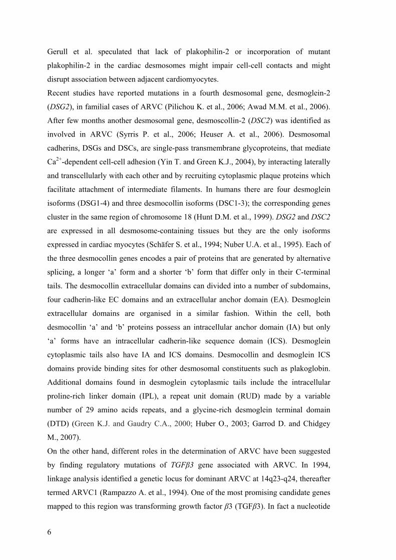
occurred in a sequence which appears highly conserved among mammals (Fig14).
22
Figure 14: The nucleotide change c.348A>G in the exon 3 of DSC2 gene occurred in a
sequence which shows high conservation among mammals. In this figure the minus strand is considered.
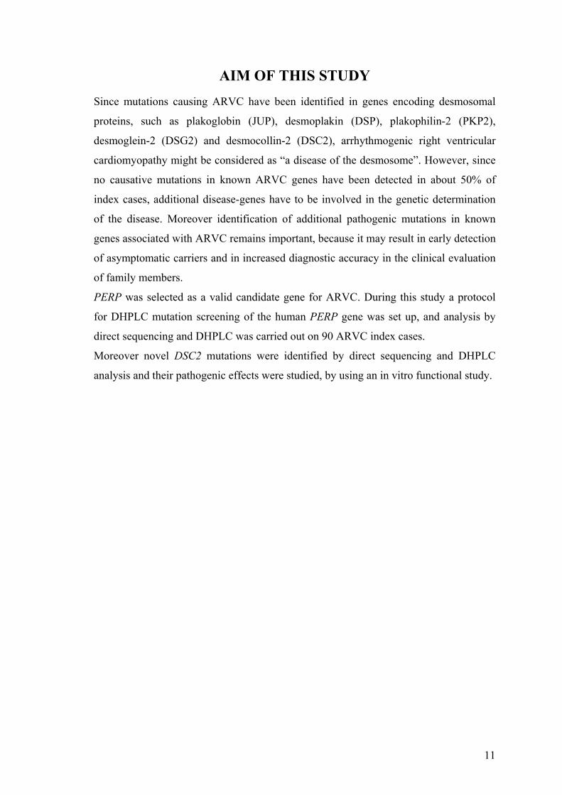
In order to understand the possible effect of this nucleotide change, RNA was extracted
from lymphocytes of the patient. cDNA obtained by RT-PCR was amplified with
different exon primers, but agarose electrophoresis and cDNA sequencing failed to
detect aberrant transcripts. RT-PCR products were cloned in pCR2.1-TOPO vector
using E.Coli cells (TOP 10 OneShot) as hosts. A total of 50 clones were randomly
picked up. DNA sequencing of such clones revealed 30 wild type cDNA and 20 mutant
cDNA (showing the nucleotide change c.348A>G). Only two of these mutant clones
(4%) showed an aberrant transcript, resulting from the skipping of 9 nucleotides just
few bases downstream the mutated nucleotide (Fig. 15).
Figure 15: Normal and aberrant transcripts detected by DNA sequencing among cDNA
clones (see the text for details). Although the 9-nucleotide deletion of aberrant spliced mRNA doesn’t alter the frame of
the sequence, it leads to the loss of three aminoacids very conserved among mammals
(Fig. 16).
A T C A A A C A A A G G T C C T A A A G A A A A G A C A T A
A T C A G A C A A A G A A A A G A C A T A C T A A A G A A A
WILD TYPE
cDNA PATIENT
Q116Q Del V119_K121
Q T K V L K K R H
Q T K K R H

23
Figure X:
Figure 16: Evolutionary conservation of DSC2 aminoacids since V119 to K121 among
8 mammalian species.
In mouse and in rat dsc2 protein, K121 (lysine) is replaced by asparagine (N), but the
two amino acids share similar physico-chemical properties.
The genetic study was extended also to additional members of the family (Fig. 17).
»»
»
»»
»
Figure 17: Family tree of the index case carrying Q116Q DSC2 mutation (») along with
K470E (*) and A566T (●) DSP mutations. By direct sequencing of DNA, DSC2 mutation Q116Q was detected in subjects 5658
and 5553 which are fully asymptomatic, whereas both DSP mutations (K470E and
A566T) resulted only in subject 7430 which presents a classical form of ARVC.
EXON 8 MUTATION: c.1034C>T → I345T
The index case affected with ARVC was diagnosed at the age of 50, due to a sustained
VT episode for which he received an implantable cardioverter defibrillator; he showed
segmentary involvement of left ventricle. The abnormal elution profile for the amplicon
IV
Del V119_K121
I
I
III
Homo_sapiens VFLEHQTKVLKKRHTKEKVLRRAKR Pan_troglodytes VFLEHQTKVLKKRHTKEKVLRRAKR Macaca_mulatta VFLEHQTKVLKKRQTKETVLRRAKR Canis_familiaris VLLQHQTKVLKKRHSKEKVLRRAKR Bos_taurus VLLEHQTKVLKKRHSQEKVLRRAKR Equus_caballus VLLEHQTKVLKKRHSKEKVLRRAKR Mus_musculus VHLEGPVEVLNKRPHTEKVLSRAKR Rattus_norvegicus VHLEGPVEVLNQRLHTEKVLRRAKR * *: .:**::* *.** ****

24
of exon 8 by DHPLC was due to a nucleotide substitution (c.1034C>T → I345T), as
shown by direct DNA sequencing (Fig. 18).
Figure 18: Results of the DHPLC and DNA sequencing analysis of DSC2 exon 8
amplicon from the index case (in red) compared with a control sample (in black).
This nucleotide change is not reported in the SNP database and was never found in 250
controls (500 control chromosomes) from Italian population. Mutation p.I345T replaced
a non polar hydrophobic amino acid by a polar hydrophilic amino acid. It is located in
the second extracellular cadherin (EC2) domain and alters the third amino acid of the
EC domain consensus sequence [L/I/V-x-L/I/V-x-D-x-N-D-N/H-x-P] (Takeichi M.,
1990). Cadherin domains are important for adhesive interactions and form Ca2+-
dependent rodlike structures. Moreover this change occurred in a residue highly
conserved among mammals (Fig. 19).
TTSTCIINIDDVNDHLPTFTR Homo_Sapiens TTSICIINIDDVNDNLPTFTR Canis_familiaris TTAICIINIEDVNDNLPTFTR Bos_taurus TTSTCIINIDDVNDHLPTFTR Macaca_mulatta TTSTCIINIDDVNDHLPTFTR Pan_troglodytes TTAKCIITIEDVNDNLPTFTR Mus_musculus TTATCVITIGDVNDNLPTFTR Rattus_norvegicus **: *:*.* ****:****** I345T Figure 19: DSC2 missense mutation I345T occurred in a sequence highly conserved
among mammals.
C T Ile Thr
A A C A N T G A T

25
The genetic study was extended to additional members of the family (Fig.20).
I 1 2
2 4 II
III 21
1 3
- -
- 5
SD(48)+
+
Figure 20: Family tree of the index case in whom I345T mutation was detected. Grey symbol represents an individual of unknown disease status. SD indicates sudden death. Presence (+) or absence (-) of the DSC2 mutation is indicated.
The 15 years-old daughter, although fully asymptomatic, was found to carry the same
DSC2 mutation detected in her father. However, due to the young age of individual III-
2, it cannot be excluded that later she could show clinical signs of the disease. All
family members not carrying DSC2 mutations were negative at clinical investigation.
EXON 17 MUTATION: c.2687_2688insGA → E896fsX900
Two unrelated index cases affected with a classical form of ARVC showed an abnormal
pattern of the DHPLC elution profile of DSC2 exon 17 amplicon. The DNA sequence
detected the presence of a mutation (c.2687_2688insGA) resulting in a premature stop
codon formation (E896fsX900) (Fig. 21).
One of the two patients is a woman carrying a mutation in the PKP2 gene (p.E58D)
while the other one is a man in whom a DSG2 mutation (p.Y87C) was previously
identified.
Figure 21: Results of the DHPLC and DNA sequencing analysis of DSC2 exon 17
amplicon from the index case (in red) compared with a control sample (in black).

26
The insertion c.2687_2688insGA is not reported in the SNP database but it was
observed in 6 out of 150 control subjects (300 chromosomes) screened by DHPLC
analysis and direct sequencing. Accordingly, the frequency of such variant should be
about 2%. This mutation would affect the C terminus of DSC2 by altering 4 aa residues
before a termination codon is prematurely introduced. If compared with the wild type,
only the last 5 amino acids were altered in the mutated protein, three were changed and
the last two were lost (Fig. 22). Wild type GCA GAA GCA TGC ATG AAG AGA TGA Protein ala glu ala cys met lys arg ter Mutated GCA GAG AAG CAT GCA TGA Protein ala glu lys his ala ter 896 900
Figure 22: Changes introduced in the amino acid sequence, caused by mutation
c.2687_2688insGA. Exon 17 encodes ICS domain in the DSC2a isoform. It is believed that the binding site
to plakoglobin is located within this functionally important domain. The change
occurred in five aa residues, which are non conserved among mammals, in contrast with
the high conservation of the upstream region (Fig. 23).
Figure 23: Last five aminoacids of DSC2 protein (involved by mutation
c.2687_2688insGA) show relative variance among mammalian species.
The genetic study was extended to the son of the patient, which resulted to carry the
same mutations and the same phenotype of his mother, on the contrary the daughter of
Homo_sapiens QEEDGLEFLDNLEPKFRTLAEACMKR Macaca_mulatta QEEDGLEFLDNLEPKFRTLAEACMKR Pan_troglodytes QEEDGLEFLDNLEPKFKTLAEACMKR Bos_taurus QEEDGLEFLDHLGPKFRTLAETCMKR Rattus_norvegicus QEEDGLEFLDHLEPKFRTLAEVCAKR Mus_musculus QEEDGLEFLDHLEPKFRTLAEVCAKR Canis_familiaris QEEDGLEFLDHLEPKFRTLAEACIKR Equus_caballus QDEDGLEFLDHLEPKFRTLAAACTTR *:********:* ***:*** .* .*

27
the second patient resulted negative for both mutations identified in her father and she is
fully asymptomatic.
FUNCTIONAL ANALYSIS OF MUTANT DESMOCOLLINS
To evaluate pathogenic potentials of the DSC2 missense mutations p.E102K and
p.I345T, full-length wild-type cDNA was directionally cloned in eukaryotic expression
vector to obtain a fusion protein with GFP. Mutated proteins carrying p.E102K and
p.I345T were obtained by site directed mutagenesis of the wild-type construct.
Constructs were transfected in the desmosome-forming cell line HL-1 having a
differentiated cardiomyocyte phenotype and contractile activity in vitro.
In transfected HL-1 cells, wild-type fusion protein was detected in the cell membrane,
into cell-cell contact regions (Figure 24, panel A), and co-localised with the endogenous
dsg, which was marked with monoclonal desmoglein antibody (Figure 24, panel A’ and
A’’). This co-localisation suggests that the wild-type fusion protein has been integrated
into normal-appearing desmosomes. By contrast, protein carrying the p.E102K and
p.I345T mutation were predominantly localised in the cytoplasm (Figure 24, panel B
and C) although a lower amount of GFP signal was detectable in membrane. Moreover
immunostaining with monoclonal desmoglein antibody showed both the presence of
well-assembled desmosomes in transfected HL-1 cells (Figure 24, panel B’, C’) and the
reduced co-localisation between endogenous dsg and mutated DSC2 (Figure 24, panel
B’’, C’’).
In addition, site-directed mutagenesis was performed on wild type construct in order to
study the functional effects of two novel DSC2 polymorphisms p.D179G, p.R798Q and
the putative pathogenic mutation p.E896fsX900. R798G is located in the cytoplasmic
region of DSC2, where also E896fsX900 maps, whereas D179G is localized in the
extracellular region of the protein, as the two missense DSC2 mutations mentioned
above. HL-1 cells were then transfected with these three new constructs.
Results suggest that both polymorphisms do not have functional effect as compared
with the wild type protein, whereas the frameshift mutation do have functional effect, as
shown by predominant localization in the cytoplasm of DSC2, similarly to the two
proteins carrying mutations p.E102K and p.I345T, respectively (Fig.25).

28
A
B B’ B’’
C C’ C’’
A’’A’
Dsc2-GFP-E102K
Dsc2-GFP-I345T
Dsc2-GFP-wt DG3.10
DG3.10
DG3.10
Merge
Merge
Merge
10 mμ
10 mμ
10 mμ
Figure 24: Transfection studies in HL-1 cells. Wild type DSC2 (WT-DSC2a-GFP) is localised at the cell membrane, border between two HL-1 cells (panel A), whereas E102K and I345T-DSC2a-GFP were mainly detected in the cytoplasm (panel B and C). Immunostaining with monoclonal desmoglein antibody DG3.10 showed both the presence of well-assembled desmosomes (panel A’, B’ and C’) and the reduced co-localisation between endogenous dsg and mutated DSC2 (yellow dots in panel B’’, C’’).

29
F
Dsc2-GFP-D179G
D’
DG3.10
E’
DG3.10
F’
DG3.10
D’’
Merge
E’’
Merge
F’’
Merge
D
Dsc2-GFP-E896fsX900
10 mμ
E
Dsc2-GFP-R798Q
10 mμ
10 mμ
Figure 25: Transfection studies in HL-1 cells. Note the D179G and R798Q-DSC2a-
GFP were localised at the cell membrane between two HL-1 cells (panel E and F), whereas E896fsX900-DSC2a-GFP were mainly detected in the cytoplasm (panel D). Immunostaining with monoclonal desmoglein antibody DG3.10 showed both the presence of well-assembled desmosomes (yellow dots in panel E’’ and F’’) and no co-localisation between endogenous dsg and DSC2a-GFP-E896fsX900 ( panel D’’).

30

31
DISCUSSION
Arrhythmogenic right ventricular cardiomyopathy (ARVC) is a dominant, degenerative
cardiomyopathy, frequently involved in sudden death of asymptomatic athletes and
teenagers. Human genetic studies over the last few years have offered insight into the
potential causes of ARVC. The involvement on ARVC of multiple desmosomal protein
genes, such as plakoglobin (JUP), desmoplakin (DSP), plakophilin-2 (PKP2),
desmoglein-2 (DSG2) and desmocollin-2 (DSC2), has led to the “desmosomal model”
hypothesis. Since mutation screening in such genes failed to detect causative mutations
in about 50% of patients affected with ARVC, current genetic research aims at
identifying novel genes involved in such disease and novel mutations in the known
genes.
MUTATION SCREENING OF PERP GENE
PERP gene encodes for a tetraspan membrane protein which is essential for
desmosomal adhesion in stratified epithelia (Ihrie R.A. et al., 2005) and localizes to the
intercalated discs of cardiac muscle, a site of known function for desmosomes (Marques
M.R. et al., 2006). For such reasons PERP was considered a good candidate gene for
ARVC.
Since the major studies on PERP were carried out on mice, in order to check the
expression of such gene in human myocardium, its expression pattern was preliminarily
verified by RT-PCR from RNA of different tissues. PERP resulted to be expressed in
human heart, therefore mutation screening of this gene was applied to a series of ninety
ARVC index cases.
Two putative novel mutations, G59R in exon 1 and c.1091C>T in 3’UTR, were
detected in two different ARVC index cases.
The missense mutation G59R replaced a small polar hydrophilic amino acid (glycine)
by a bigger positively-charged one (arginine). This change occurred in a residue highly
conserved among species and located in a conserved extracellular region for which a
function is still unknown, however the G59R PERP variant was identified as well in one
out of 250 controls. The index case resulted to carry as well a pathogenic mutation in
PKP2 gene (S50fsX110); the mutation was identified also in her son, who, on the
contrary resulted negative for the PERP mutation. It is interesting to notice that patient
32
appears to be affected with a severe form of ARVC, whereas her son (negative for
PERP mutation) seems to be affected with a classical form of ARVC. Therefore, the
possibility that PERP mutation could worsen the clinical presentation should be
considered.
Likewise, mutation c.1091C>T in 3’UTR changes a highly conserved nucleotide; this
mutation was detected in 2 out of 192 controls. The index case carrying c.1091C>T in
the PERP 3’UTR carries also a pathogenic mutation in DSP gene (R1113X). Both DSP
and PERP genes map to chromosome 6, respectively on 6p24 and 6q24. The patient’s
father is negative for both mutation, therefore it is probable that the two variations were
carried by the maternal chromosome 6. The patient and her sibs appear to have inherited
the two mutations in cis , due or to the absence of crossing-over, or to the presence of
double crossing-overs along the chromosome. A single crossing-over would lead to
inheritance of only one of the two mutations, as it actually happened in the sister and in
the brother of the patient (see Figure 7, on results section).
Genetic analysis of additional family members showed that individuals carrying only
PERP variant c.1091C>T were all healthy, and also the three patient’s sibs carrying
both PERP mutation and DSP mutation did not fulfilled the current diagnostic criteria
for ARVC, but due to their young age, it cannot be excluded that some of them could
later show major clinical signs.
Although two putative mutations in PERP gene were identified in index cases affected
with ARVC, it is impossible to establish whether these mutations might cause ARVC
when they occur not associated to additional pathogenic mutations. Data suggest that a
variation in Perp might worsen the clinical phenotype in patients carrying a pathogenic
mutation in a different gene involved in ARVC. For such reason, PERP still remains a
good candidate for ARVC and mutation screening should be extended to additional
series of index patients affected with ARVC.
MUTATION SCREENING OF DSC2 GENE
Two recent studies suggested that mutations in the gene encoding desmosomal
desmocollin-2 (DSC2) may cause ARVC (Syrris P. et al., 2006; Heuser A. et al., 2006).
In Syrris’s report, two heterozygous mutations were described: a deletion (c.1430delC)
and an insertion (c.2687_2688insGA). Both mutations result in frameshifts and
premature truncation of the desmocollin-2 protein. On the other hand Heuser et al.
identified a heterozygous splice acceptor–site mutation in DSC2 intron 5 (c.631-2A>G),

33
which activates a cryptic splice-acceptor site, leading to a downstream premature
termination codon.
In seven out of sixty-four ARVC unrelated Italian index cases screened in this study six
different DSC2 mutations were identified. Five of them (c.-92G>T, c.304G>A,
c.348A>G, c.1034T>C and c.3241A>T) were absent among controls, thus suggesting
that these genetic variants might be pathogenic. The sixth mutation
(c.2687_2688insGA) was detected in two different patients and in six control subjects,
suggesting the possibility of a polymorphism.
Two mutations in UTR regions were observed in two different patients: c.-92G>T in
5’UTR and c.3241A>T in 3’UTR. Although both nucleotide changes occurred in
sequences showing low conservation among mammals, they were unreported in the
SNP database and they were never detected among 150 Italian controls. However, to
exclude that such mutations could correspond to rare polymorphisms, the size of the
control group should be increased to 500 individuals. The patient carrying mutation
c.3241A>T in 3’UTR carries as well a mutation (N76S) in PKP2 gene. The missense
mutation N76S changes an asparagine (N) with a serine (S) which are both small polar
amino acids. This data suggests that the variation in PKP2 gene could be non
pathogenic; if so, the variation in 3’UTR of DSC2 gene might cause ARVC. This
hypothesis might be supported by the findings of a recent study (Beffagna G. et al.,
2004) in which it has been demonstrated that overexpression of TGFβ3, due to 5’UTR
and 3’UTR mutations, cause ARVC. Sequence alterations in untranslated regions of
genes were reported in several hereditary human diseases, such as hereditary
angioedema (Laimer M. et al., 2006) an autosomal dominant cerebellar ataxia (Ishikawa
K. et al., 2005) a distinctive form of cone dystrophy (Piri N. et al., 2005) hereditary
thrombophilia (Gehring N.H. et al., 2001), hereditary hyperferritinaemia-cataract
(Girelli D. et al., 1997) and fragile X mental retardation syndromes (Warren S.T. and
Nelson D.L., 1994). In fact 5’ and 3’UTRs contain several regulatory motifs, which
modify mRNA stability, localization, and degradation, thereby influencing gene
expression (Mitchell P. and Tollervey D., 2000; Amack J.D. et al., 2002; Mazumder B.
et al., 2003). Specific functional studies will be needed to understand the pathogenic
role of mutations affecting UTRs in DSC2.
A novel nucleotide change c.348A>G leading to a synonymous mutation Q116Q was
detected in DSC2 gene in a patient with a severe ARVC form. This patient carried two
in cis DSP mutations (K470E and A566T), which replace the corresponding wild type

34
amino acids with others with different physico-chemical properties. The nucleotide
change c.348A>G was never detected among 500 control chromosomes and it occurred
in a region highly conserved among species. This mutation might activate a cryptic
splice site: in fact, an abberant mRNA was identified by RT-PCR in patient’s RNA and
subsequent cloning of PCR products. Surprisingly, the resulting aberrant spliced
mRNA, showing a deletion of 9 nucleotides, was found only in 4% of screened clones
carrying the variation c.348A>G. It is possible that this percentage could have been
underestimated since mutant mRNA may have been degraded via nonsense-mediated
mRNA decay.
According to recent studies, desmoglein/desmocollin ratio is relevant to desmosomal
intercellular adhesion and must be finely regulated. L cell fibroblasts, in which the level
of Dsg1 was titrated against constant amounts of Dsc1, exhibited productive adhesion
only at the appropriate ratio of Dsg1 to Dsc1 (Dusek R.L. et al., 2006). This data
suggest that even if mutant DSC2 is present at low concentration, this could be
sufficient to alter the adhesive function of desmosomes. Moreover, although skipping of
9bp in the aberrant mRNA doesn’t alter the reading frame of DNA sequence, at protein
level it leads to loss of three amino acids very conserved among species and located in
the N-terminal pro-sequence domain; this region is involved in the maturation of
adhesive protein (Ozawa M. et al., 1990). Extension of mutation screening to additional
family members detected c.348A>G DSC2 mutation in the father and in the brother of
the patient; both these subjects were asymptomatic (see Figure 17, in results section).
On the contrary, one mother’s cousin, showing a classical form of ARVC, carried two
mutations in DSP gene (K470E and A566T, in cis). Only the index case, carrying two
DSP mutations in cis and one DSC2 mutation, resulted affected with a severe form of
ARVC. Taken together, these data suggest that the DSC2 mutation c.348A>G might
lead, in presence of other pathogenic mutations, to a more severe phenotype, but that,
per se, it would be insufficient to cause ARVC.
Two heterozygous point substitutions c.304G>A and c.1034T>C were detected in two
index cases. None of the detected nucleotide changes was found in a control group of
250 healthy and unrelated subjects (500 control chromosomes). c.304G>A and
c.1034T>C result in predicted p.E102K and p.I345T amino acid substitutions,
respectively. Physico-chemical properties of novel amino acids strongly differ from
wild type: mutation p.E102K replaced a negatively-charged residue by a positively-
charged one, whereas mutation p.I345T replaced a non polar hydrophobic amino acid

35
by a polar hydrophilic amino acid. Both these changes occurred in a residue highly
conserved among species. Only in mouse and rat E102 is replaced by aspartic acid, but
the two amino acids share similar physico-chemical properties.
Cadherins are synthesized as inactive precursor proteins containing a prosequence
followed by the cadherin domains. The N-terminal prosequence is proteolytically
cleaved off in the late Golgi and the mature cadherin is then transported to the plasma
membrane. Proteolytic removal of the prosequence results in structural rearrangements
within EC1 domain with the activation of adhesive properties (Ozawa M. et al., 1990).
Mutation p.E102K alters a conserved amino acid located in this propeptide region.
Mutation p.I345T is located in the EC2 domain, which forms, together with EC1, the
“minimal essential unit” to mediate cell adhesion, through cis and trans interactions
among desmosomal cadherins (Shan W. et al., 2004). These DSC2 mutations were
detected in two ARVC index cases and in four family members who met only minor
diagnostic criteria. This could be consistent with incomplete penetrance of such
mutations; incomplete penetrance is rather common in ARVC and it was reported for
several DSP, PKP2 and DSG2 mutations (Bauce B. et al., 2005; Syrris P. et al., 2006;
Van Tintelen J.P. et al., 2006; Pilichou K. et al., 2006). However, lack of clinical
manifestation could be due to the young age of most family members carrying the
DSC2 mutations.
To evaluate the pathogenic potentials of the two N-terminal DSC2 mutations, full-
length wild-type and mutated cDNAs were cloned in eukaryotic expression vectors to
obtain a fusion protein with green fluorescence protein (GFP); constructs were
transfected in HL-1 cells. This in vitro functional studies demonstrated that, unlike
wild-type DSC2, the two N-terminal mutants predominantly localise in the cytoplasm,
confirming the suspect that both mutations could have pathogenic effect.
An insertion of two bases (c.2687_2688insGA) was detected in exon 17 of DSC2 gene
in two unrelated index cases affected with ARVC; in one of them a missense mutation
(E58D) in PKP2 gene was previously detected and in the other one a missense mutation
(Y87C) in DSG2 gene was identified. These four amino acids (C=Cysteine, D=Aspartic
acid, E=Glutamic acid and Y=Tyrosine) have a polar side chains; therefore the two
missense mutations in PKP2 and DSG2 genes might be non pathogenic.
The c.2687_2688insGA DSC2 insertion was reported in the literature as a pathogenic
mutation (Syrris P. et al., 2006), since it results in a frameshift leading to a termination
codon 4 aa residues downstream (E896fsX900). In Syrris report, DSC2 E896fsX900

36
mutation was detected in three families, indicating either that this insertion is recurrent
in patients with ARVC, or that such families may share a common ancestor. This last
possibility was excluded, since analysis of microsatellite DNA markers (D18S847,
D18S49, and D18S457) in close proximity to the DSC2 locus demonstrated no allele
sharing in individuals carrying E896fsX900. Therefore, E896fsX900 might be a
recurrent mutation, possibly due to a “hot” mutational spot (Syrris et al., 2006).
However, in the present study the DSC2 insertion c.2687_2688insGA was identified in
2 out of 64 patients and it was detected as well in 6 out of 150 control subjects (300
chromosomes): this would suggest that, more likely, such variant could be a
polymorphism rather than a pathogenic mutation. This variant occurs in exon 17, which
encodes, in DSC2a isoform, the ICS domain. Such domain shows a high degree of
amino acid homology among various desmosomal and nondesmosomal cadherins.
Therefore, in theory, the alteration caused by the insertion is potentially pathogenic, but
in reality it affects only the last five amino acids of the protein, which are not highly
conserved among species in contrast with the highly conservation of the upstream
region. It is important to notice that the same region is untranslated in DSC2b isoform,
being included in its 3’UTR. Therefore, the insertion would affect only DSC2 isoform
a, leaving isoform b fully functional and possibly able to compensate in cardiac
myocytes the relative deficiency of DSC2a isoform. In vitro functional studies on HL-1
cells demonstrated that mutant DSC2 (E896fsX900) predominantly localizes to the
cytoplasm, in contrast with wild type DSC2 and DSC2 variants carrying polymorphisms
D179G and R798Q. These observations conflict with the hypothesis that
c.2687_2688insGA is a polymorphism; however it must be taken into account that such
sequence variation could have no pathogenic effect because of the presence of a correct
DSC2b isoform.
DSC2 FUNCTIONAL STUDIES
In vitro functional studies on HL-1 cells demonstrated that the two missense mutations
in the N-terminal domain (E102K and I345T) and the frameshift (E896fsX900) in C-
terminal domain affect the normal cellular localisation of DSC2. As previously
reported, wild-type DSC2a-GFP fusion protein was efficiently incorporated into
desmosomes and did not exert dominant-negative effect when overexpressed
(Windoffer R. et al., 2002). A lower amount of GFP signal was detected in the
cytoplasm, since proteins were still not fully trafficked to the membrane. Unlike wild-

37
type DSC2 and DSC2 variants carrying polymorphisms D179G and R798Q, mutants of
DSC2 were predominantly located in the cytoplasm suggesting a potential pathogenic
effect. Although all three mutations affect intracellular localization of desmocollin-2,
their effect probably differ, depending on the relative position of each mutation along
the protein.
Mutations p.E102K and p.I345T map to the N-terminal region, relevant to adhesive
function. Cadherins have been shown to be internalised and recycled back to the plasma
membrane in a constitutive manner, providing a mechanism for regulating the
availability of cadherins for junction formation and for maintaining a dynamic state of
cell-cell contacts (Le T.L. et al., 1999; Le T.L. et al., 2002; Kowalczyk A.P. et al., 2004;
D’Souza-Schorey C., 2005). Moreover, recent studies suggest that adhesive interactions
between cadherins as well as cytoskeletal associations prevent cadherin endocytosis
(Izumi G. et al., 2004). Based on these findings, it can be hypothesized that in the
absence of adhesive interactions, the two N-terminal mutants DSC2 might be
internalised from the cell surface by endocytosis. Interestingly, in Pemphigus vulgaris
(PV, MIM #169610), an autoimmune disease in which antibodies are directed against
DSG3, resulting in severe mucosal erosions and epidermal blistering, PV autoantibodies
trigger co-endocytosis of DSG3 and plakoglobin, leading to delivery of DSG3 to
lysosomal compartments and dramatic decrease in DSG3 protein level (Calkins C.C.et
al., 2006). Possibly, N-terminal DSC2 mutations could result in alteration of the
adhesion properties, leading to DSC2 internalisation.
On the other hand, mutation E896fsX900 maps to the C-terminal region, involving the
ICS domain of DSC2a isoform, and could impair the binding to plakoglobin or the
formation of desmosomal plaque and intermediate filaments anchorage, thus leading to
disruption of desmosome structure (Syrris P. et al., 2006). As previously mentioned,
DSC2 gene encodes for two different products (DSC2a and DSC2b); a mutation in the
C-terminal cytoplasmic domain could affect only the longer isoform a and
compensation by the other form might be possible. Further investigation will be needed
to understand whether DSC2 mutations could induce “null alleles” potentially leading to
haploinsufficiency (i.e. through cytoplasmic degradation) or mutant proteins could
remain within the cells, acting in a dominant form (i.e.toxic gain−of−function).
Recently, it was shown that suppression of desmoplakin expression with use of small
interfering RNA in atrial myocyte cell lines (HL-1 cells) or in heterozygous
desmoplakin-deficient mice leads to nuclear localization of plakoglobin, reduction in

38
canonical Wnt signaling through Tcf/Lef transcription factors, and increased myocyte
apoptosis (Garcia-Gras E. et al., 2006). Indeed, increased apoptosis of cardiomyocytes
was reported in patients with ARVC (Yamaji K. et al., 2005). Garcia-Gras et al. have
established a potential role for signaling defects in ARVC, leading to the idea that the
cell adhesion proteins are not only passive players in myocardial architecture, but they
might be considered as key regulators in cardiac patterning and development, in
myocyte differentiation, and in the maintenance of the cellular architecture of the adult
heart (MacRae C.A. et al., 2006).

39
CONCLUSIONS In this study two putative mutations in PERP gene were identified in index cases
affected with ARVC, but it was impossible to establish whether these mutations might
really cause ARVC. However, data suggested that a variation in PERP might produce a
very severe phenotype in patients carrying a pathogenic mutation in a different gene
involved in ARVC. Therefore, PERP still remains a good candidate for ARVC and
mutation screening should be extended to additional series of index patients.
Novel DSC2 mutations were detected in ARVC index cases and their pathogenic effects
were investigated using a cardiomyocytes cell line. In vitro functional studies
demonstrated that, unlike wild-type DSC2, the mutants are predominantly localised in
the cytoplasm, affecting transmembrane localisation of DSC2 and thus suggesting the
potential pathogenic effect of the reported mutations.
This method, based on transient transfection of HL-1 cell line with mutant constructs
for genes encoding desmosomal proteins, could allow to study potential pathogenic
effects of novel missense mutations suspected to cause ARVC.

40

41
MATERIALS AND METHODS
CLINICAL EVALUATION
The study involved subjects belonging to several ARVC families, sporadic cases of
Italian descent, all with a clinical diagnosis of ARVC. Clinical diagnosis for ARVC was
based on major and minor criteria, established by the European Society of
Cardiology/International Society and Federation of Cardiology Task Force (McKenna et
al., 1994). The patients were investigated at the Department of Clinical and
Experimental Medicine of the University of Padua, by Prof. Nava and colleagues.
Clinical investigations and blood sampling for DNA analysis were performed under
informed consent, according to the pertinent Italian legislation and in compliance with
Helsinki declaration. Each patients underwent 12-lead electrocardiography (ECG),
signal-averaged ECG (SAECG), 24 hour Holter ECG and two-dimensional
echocardiography.
DNA EXTRACTION
Genomic Dna was extracted from blood samples by a modified salting-out procedure,
evaluated at the Human Genetics Laboratory of Padua.
• Thaw blood sample (collected in K3E-EDTA tube and stored at -20°C) and
transferred to a sterile 50ml tube.
• Wash with erythrocyte lysing buffer (N-N solution: 0.9% NaCl, 0.1% Nonidet P40),
centrifuge for 30minutes at 4000rpm and 4°C. Wash and centrifuge are repeated twice.
• Add to the pellet 4ml of TEN solution ( 10mM TrisHcl; 2mM Na2EDTA pH 8;
400mM NaCl), and 300μl 20% SDS.
• Vortex to break up pellet
• Incubate to 80°C for 3hours with vigorous shaking. During this step the leukocytes
membranes are denatured and DNA goes out in solution.
• Add 1ml of saturated NaCl solution to precipitate proteins and cells membranes and
centrifuge for 10minutes at 4000rpm. Transfer the supernatant in a sterile 15ml
centrifuge tube.
• Add an equal volume of chloroform and shake by hand.

42
• Centrifuge for 10minutes at 4000rpm. Three phases are observed, the upper of which
containing the DNA. Transfer this phase to a fresh tube.
• Add an equal volume of isopropanol and invert the tube gently until the DNA has
precipitated.
• Centrifuge for 20minutes at 4000rpm and discard the supernatant.
• Wash pellet twice with 70% ethanol and then centrifuge for 10minutes at 4000rpm.
• Dry the pellet and resuspend in 300-500μl of TE buffer (10mM Tris-Hcl pH 8; 1mM
EDTA) and left overnight on a rotator.
RNA EXTRACTION (PAXgene blood Rna, PreAnalytiX)
The PAXgene Blood RNA Kit is for the purification of total RNA from 2.5ml human
whole blood collected in a PAXgene Blood RNA Tube, contains a proprietary reagent
composition able to protects RNA molecules from degradation by RNases and
minimizes induction of gene expression for up to 3 days at 18–25°C. Purification begins
with a centrifugation step to pellet nucleic acids in the PAXgene Blood RNA Tube. The
pellet is washed and resuspended, and incubated in optimized buffers together with
proteinase K to bring about protein digestion. An additional centrifugation through the
PAXgene Shredder spin column is carried out to homogenize the cell lysate and remove
residual cell debris, and the supernatant of the flow-through fraction is transferred to a
fresh microcentrifuge tube. Ethanol is added to adjust binding conditions, and the lysate
is applied to a PAXgene RNA spin column. During a brief centrifugation, RNA is
selectively bound to the PAXgene silica membrane as contaminants pass through.
Remaining contaminants are removed in several efficient wash steps. Between the first
and second wash steps, the membrane is treated with DNase I to remove bound DNA.
After the wash steps, RNA is eluted in Buffer BR5, denaturated at 65°C for downstream
applications and stored at -80°C. Typical yields of RNA isolated from 2.5ml human
blood are between 4 to 20µg. However, the yield is highly dono-dependent, and in some
cases higher or lower yields may be achieved.
DNA/RNA QUANTIFICATION
The concentration of DNA and RNA solution is determined by measuring at 260nm
against blank. The absorption of 1 OD is equivalent to approximately 50μl/ml dsDNA,

43
and approximately 40μl/ml RNA. The ratio A260/A280 is used to estimate the purity of
nucleic acid, since protein absorb at 280nm. A ratio higher than 2.0 indicates the
samples may be contaminated with chloroform or phenol. Pure DNA should have a
ratio of approximately 1.8, whereas pure RNA should give a value of approximately
2.0.
PCR PRIMERS DESIGN
All PCR primers used in this thesis were designed by PRIMER 3
(http://frodo.wi.mit.edu) and produced by SIGMA or MWG-Biotech. The primers
obtained from PRIMER 3 were chosen after analysis with BLAT
(http://genome.ucsc.edu) to avoid the presence of SNPs within their sequences and their
annealing in additional wrong regions.
DHPLC ANALYSIS FOR DSC2 AND PERP GENES
PCR amplifications
The analysis of the DSC2 gene in 64 ARVC index cases and of the PERP gene in 90
ARVC index cases, were performed by denaturing high-performance liquid
chromatography (DHPLC). Samples were amplified in a final volume of 25µl or 50µl
containing approximately 50-100ng of genomic DNA, 10X PCR buffer II (Applied
Biosystems), 1.5mM MgCl2 (Applied Biosystems), 10pmoli of each primer (Sigma
Genosys or MWG Genomics), 100µM deoxinucleotide triphosphates (Invitrogen) , and
1.25 unit of DNA TAQ GOLD polymerase. PCR conditions for all the DNA fragments
were as follows: an initial denaturation cycle of 95°C for 12 minutes followed by 37
cycles composed by a denaturation step (95°C) for 30 seconds, an annealing step for 30
seconds, and an extension step (72°C) for 45 seconds. A final extension of 72°C for 10
minutes complete the amplification. The amplification product was verified using 2%
agarose gel, and visualized using ethidium bromide and ultraviolet light. As the length
of fragment suitable for DHPLC analysis is less than 400bp, some longer exons were
splitted in shorter amplicons. All primer sequences and PCR conditions used are
reported in Appendix. A and Appendix B.
DHPLC analysis
DHPLC analysis was performed on a Transgenomic Wave DNA Fragment Analysis
System (Model 3500HT; Transgenomic) with a DNASep column (Transgenomic). PCR
fragments were denatured for 5min at 95°C and then left to reanneal slowly at room

44
temperature to promote the formation of heteroduplex. Separation was performed at a
flow rate of 0.9ml/min for Mutation Detection application (8min) and if necessary at
1.5ml/min for Rapid DNA application (3min). The acetonitrile gradient was adjusted to
elute the amplicon at half run, around 4min for Mutation Detection application and
1.5min for Rapid DNA application. The gradient was obtained by mixing Buffer A
(0.1M TEAA, pH 7.0) and Buffer B (0.1M TEAA, pH 7.0, 25% acetonitrile). Buffer B
increase was 2% per min (with flow rate 0.9ml/min) and 5% per min (with flow rate
1.5ml/min). Column temperatures were calculated by NAVIGATOR software
(Transgenomic). Whenever fragments showed different melting domains, additional
analyzing temperatures were used, to optimize resolution. These temperatures should
give the 75-80% of double strand DNA within the fragments. DHPLC conditions used
to analyse DSC2 and PERP genes are listed in Appendix A and Appendix B.
CONTROLS Whenever a putative mutation was detected, a search was performed on a single-
nucleotide polymorphism database (dbSNP:
http://www.ncbi.nlm.nih.gov/projects/SNP/). In case of negative results, DHPLC
analysis on genomic DNA from unrelated control individuals was performed with Rapid
DNA application, to exclude DNA polymorphisms.
EXPRESSION ANALYSIS FOR PERP GENE
Expression of PERP in different human tissues was assessed by RT-PCR amplification
of MTC (Multiple Tissue cDNA) panel (Clontech), including heart, skeletal muscle,
liver, kidney and skin. Amplification was performed by using the primers mentioned
below:
PERPexp0F CAGGCCACTCTCTGCTGTC
PERPexp0R CTCTGACAGCCCTCCTCGTA
The amplification products were verified using 2% agarose gel, and visualized using
ethidium bromide and ultraviolet light.
RT-PCR
This procedure outlines the synthesis of cDNA for subsequent amplification using PCR.
Reverse transcription reaction of up to 1µg of total RNA, poly(A)+ mRNA is performed
in 25µl reaction. Experimental RNA is combined with 2µl of random primer (500µg/ml,

45
Promega) and Rnase-free water to a final volume of 5µl. The primer/template mix is
thermally denatured at 70°C for 5 minutes and chilled on ice. A reverse transcription
reaction mix is assembled on ice to contain 5.6µl of nuclease-free water, 4µl of
ImProm-II 5X Reaction Buffer (Promega), 1µl of ImProm-II Reverse Transcriptase
(Promega), 2.4µl magnesium chloride (MgCl2 25mM, Promega), 1µl of dNTPs (10mM,
Invitrogen) and 1µl of Recombinant RNasin® Ribonuclease Inhibitor (40u/µl
Promega). As a final step, the template-primer combination is added to the reaction mix
on ice. Following an initial annealing at 25°C for 5 minutes, the reaction is incubated at
42°C for up to one hour and then at 70°C for 15 minutes to inactivate the reverse
transcriptase. The first strand of cDNA generated by reverse transcription of mRNA can
be used as a template for PCR or stored at -20°C.
RT-PCR experiments were carried out in a reaction buffer (12.5µl) containing 2µl
cDNA, 5pmoles oligonuclotide pimers, 100µM each of dGTP, dATP, dCTP, and dTTP,
10X PCR buffer II (Applied Biosystems), 1.5mM MgCl2 (Applied Biosystems), and
1.25unit of DNA TAQ GOLD polymerase. All polymerase chain reaction were carried
out in a thermal cycler, under the following conditions: 12min 95°C and 37 cycles of
denaturation (at 95°C for 1minute), annealing (at 65°C for 1minute), and extension (at
72°C for 2 minute). RT-PCR were performed by using the primers mentioned below:
dsc2-2F espr GTGATGCCTGCAAAAATGTG
dsc2-5R espr ATAGTTTTGGGCCGTGTCAG
dsc2-4R espr AAGGAATTGGAGCCCATCTT
Aliquots of the applied products were separated by 2% agarose gel electrophoresis an
stained by ethidium bromide. PURIFICATION OF PCR PRODUCTS AND DNA SEQUENCING
Primers and dNTPs unconsumed during the PCR amplification can interfere with DNA
sequencing methods. The PCR Product pre-Sequencing Kit provides two Hydrolytic
enzymes, Shrimp Alkaline Phosphatase (SAP) and Exonuclease I (EXO), which remove
excess of primers and dNTPs. In particular, SAP dephosphorylates dNTPs, while EXO
remove primers and any extraneous single-stranded DNA produced by PCR.
• PCR amplification mixture 5µl
EXO 0.5µl
SAP 0.5µl
Mix and incubate at 37°C for 15min.
46
• Incubate at 80°C for 15min to inactivate the enzymes.
• Add to purified PCR 1µl of primer for sequencing (10pmoli).
• Dry at 70°C for 20minutes.
PCR amplicons were sequenced at the BMR genomics, University of Padua, by an ABI
3730XL DNA sequencer (Applied Biosystems) with the BIG DYE dideoxy-terminator
chemistry (Applied Biosystems). CHROMAS software (Version 1.55; Technelysium)
and the LASERGENE software package (SeqMan II, DNASTAR) were used to edit,
assemble, and translate sequences. Amplicons showing putative mutations were re-
sequencing, by using as template the product of an new PCR reaction. DNA EXTRACTION FROM AGAROSE GEL (QIAEX II Gel Extraction kit, QIAGEN) A particular good purification of the PCR product before cloning is necessary to remove
the excess of dNTPs and primers and any aspecific DNA produced by the PCR.
Extraction of DNA fragments with QIAEX II kit is based on solubilization of agarose
and absorption of nucleic acids to silica-gel particles in the presence of high salt. All
impurities such as agarose, proteins, salts and ethidium bromide are removed during
washing steps. The DNA band is excised from the agarose gel, solubilized and washed
with buffers provided by the kit. The pure DNA is eluted in 20µl of water and is
suitable foe subsequent applications, such as sequencing or ligation.
PCR CLONING (TOPO TA Cloning, invitrogen)
The cDNA fragments obtained from RT-PCR were cloned in pCR2.1-TOPO vector
using E.Coli cells (TOPO10 OneShot) as hosts. TOPO TA Cloning vectors contain 3’-T
overhangs that enable the direct ligation of Taq-amplified PCR products with 3’-A
overhangs. Principal vector characteristics are: EcoRI sites flanking the PCR product
insertion site for removal of inserts; kanamycin and ampicillin resistance genes for
selection in E.Coli; blue/white screening of recombinant colonies; M13 forward and
reverse priming sites for sequencing or PCR screening.

47
Ligation
Mix reaction gently and incubate for 5 minutes at room temperature. For large products
(>1Kb) increasing reaction time will yield more colonies. Place the reaction on ice and
follow the chemical transformation protocol (see below).
PLASMID CONSTRUCTIONS
The cDNA of human DSC2a was kindly provided by Dr W.W. Franke (Heidelberg,
Germany) and human DSC2a full-length coding sequence (GenBank NM_024422) was
PCR-amplified with the following primers:
DSC2a-clonF ATTATGGAGGCAGCCCGCCC
DSC2a-clonR GTCTCTTCATGCATGCTTCTGCTAG
The resulting fragment was cloned into pcDNA3.1/CT-GFP-topo eukaryotic expression
vector (Invitrogen) which contains cDNA coding for green fluorescent protein (GFP),
and verified by sequence analysis.
MUTAGENIC PRIMERS DESIGN
The mutagenic oligonucleotide primers were designed using the following guidelines:
• The mutagenic primers for point substitutions or insertion must contain the desired
mutation, whereas the mutagenic primers for deletions must skip the sequence
corresponding to the lost oligonucleotides.
• Primers should be between 25 and 45 bases in length, with a melting temperature (Tm )
of ≥78°C. Primers longer than 45 bases may be used, but using longer primers increase
the likelihood of secondary structure formation, which may affect the efficiency of the
mutagenesis reaction.
The following formula is commonly used for estimating the Tm of primers:
Tm = 81.5 + 0.41(%GC) – 675/N - %mismatch
N is the primer length in bases
Values for %GC and %mismatch are whole numbers

48
For calculating Tm for primers intended to introduce insertions or deletions, there is a
modified formula to use:
Tm = 81.5 + 0.41(%GC) – 675/N
N does not include the bases which are being inserted or deleted
• The desired mutation should be in the middle of the primer with 10-15 bases of correct
sequence on both sides.
• The primer optimally should have a minimum GC content of 40% and should
terminate in one or more C or G bases.
• Primers for insertion or deletions must be purified either by fast polynucleotide liquid
chromatography (FPLC) or by polyacrylamide gel electrophoresis (PAGE).
ONE STEP DIRECT MUTAGENESIS
Site-directed mutagenesis was performed on DSC2a-pcDNA3.1/CT-GFP, in order to
reproduce two DSC2 naturally occurring mutations p.E102K (c.304G>A), p.I345T
(c.1034T>C) and three unknown polymorphisms p.D179G (c.536A>G), p.R798Q
(c.2393G>A) p.E896fsX900 (c.2685_2686insAG) present in more patients (also in
omozygos) or in more controls.
The following mutagenic primers and conditions were used:
E102K-F: TACCATATTACTTTCCAACACTAAGAACCAAGAAAAGAAGAAAAT;
I345T-F: CAACTTCAACTTGTATCATTAACACTGATGATGTAAATGACCACTTGC;
D179G-F: TCCATAAGAGGTCCTGGAGTTGGCCAAGAACCTCGGAATTTATTTT;
R798Q-F: CACCAGACCTCGGAATCCTGCCAGGGGGCTGGCCACCATCACACC;
For p.E896fsX900 mutation, two steps of mutagenesis were necessary. The first one to
introduce the AG insertion, using the following primer:
DSC2mutAG: CAAATTTGGACACTAGCAGAGAAGCATGCATGAAGAGACAAG
The second one to remove the premature stop codon and to get the right frame with
GFP sequence, using the following primer:
DSC2mutCW: CACTAGCAGAGAAGCATGCACAAGGGCAATTCTGCAGATA

49
Set up PCR reaction:
1µl template DNA (300ng/µl stock from Mini prep)
5µl primer F+R 1µM
1µl dNTPs 10mM
2.5µl 10X Pfu Buffer
15µl sterile water
0.5µl Pfu polymerase (Stratagene, 2.5U/µl)
PCR conditions:
Denaturation: 95°C for 3min
Then 18-22 cycles of: 95°C for 30sec
60°C for 1min and 30sec
68°C for 18min (or 2 min/Kb of plasmid template)
1µl of Dpn I (Methylation sensitive) was added to the PCR product and then was
incubated for 1-2 hours at 37°C to get rid to the wild type host strand methylated. 2µl of
this reaction mixture was used to transform host cells.
ONE SHOT CHEMICAL TRANSFORMATION PROTOCOL
The mutated constructs were cloned using Top10 chemically competent E.Coli
(Invitrogen) following these steps:
• Equilibrate a water bath to 42°C
• Warm a vial of SOC medium to room temperature
• Incubate at 37°C for 30minutes two plates (LB plates containing 50µg/µl ampicillin)
for each transformation
• Thaw on ice 1 vial of One Shot cells (TOP 10, invitrogen) for each transformation
• Add 2µl of reaction digested with DpnI and mix gently without pipetting
• Incubate on ice for 5 to 30 minutes
• Heat-shock the cells for 30 seconds at 42°C without shaking
• Immediately transfer the tubes to ice
• Add 250µl of room temperature SOC medium
• Cap the tube tightly and shake the tube (200rpm) at 37°C for 1 hour
• Spread 50µl and 200µl from each transformation on 2 prewarmed plates and incubate
overnight at 37°C

50
ANALYSING POSITIVE CLONES
• Pick 10-20 colonies with a sterile pipet tip and wash the tips in 10µl of sterile water in
a 0.5ml tubes
• Use 1.5-2µl of each colony as template in a PCR reaction
• Aliquote the PCR mix, carried out using exonic primers flanking the region with
variation, and amplify for 37 cycles with DNA TAQ GOLD polymerase
• Analyze the PCR products, corresponding to each colony, by restriction analysis to
confirm the presence of mutant base
• The colonies positive to the restriction analysis were cultered overnight in 5ml of
liquid LB (containing 50µg/µl ampicillin), shaking the tubes horizontally (200rpm) at
37°C
• Isolate the plasmid DNA using Miniprep Kit (SIGMA)
• Each plasmids were verified for variation by sequence analysis
PURIFICATION OF PLASMID (MINIPREP-MIDIPREP)
Miniprep (GenEluteTM HP Plasmid Miniprep Kit, Sigma-Aldrich)
The GenElute HP Plasmid Miniprep Kit is based on silica-binding technology, for
isolating up to 25µg of high copy plasmid DNA from 1-5ml of recombinant E.coli
coltures. An overnight colture is harvested with centrifugation and subjected to a
modified alkaline-SDS lysis procedure followed by adsorption of the plasmid DNA
onto silica in the presence of high salts. Contaminants are then removed by a spin wash
step. Finally, the bound plasmid DNA is eluted in water or Tris-EDTA buffer. The
recovered plasmid DNA, predominately in its supercoleid form, is checked in a 1%
agarose gel.
Midiprep (NucleoBond® Xtra Midi, Macherey-Nagel)
The bacterial cells are lysed by a set of buffers based on the NaOH/SDS lysis method.
After equilibration of the column together with the corresponding column filter, the
entire lysate is loaded by gravity flow and simultaneously cleared by the equilibrated
filter. Plasmid DNA is bound to the silica resin and after a washing step is eluted,
precipitated and dissolved in a suitable buffer for further use. From 50ml of an
overnight recombinant E.coli colture is possible to isolate more than 50µg of high copy
plasmid DNA.

51
SOLUTIONS AND MEDIA
For Bacteria SOC 2% Tryptone LB (Luria-Bertani) 1% Tryptone 0.5% Yeast Excract Liquid Medium 0.5% Yeast Extract 10mM NaCl 1% NaCl 2.5mM KCl pH 7.0 10mM MgCl2 10mM MgSO4 20mM glucose Solid Medium +1.5% Select-Agar For HL-1 cell line
Supplemented Claycomb 261ml Claycomb Medium Medium 30ml Fetal bovine serum (10%) 3ml Penicillin/Streptomicyn (100U/ml:100µg/ml) 3ml Norepinephrine (0.1mM) 3ml L−Glutamine (2mM stock)
The claycomb medium bottle must be wrapped in aluminum foil since the medium is extremely light sensitive.
Penicillin/Streptomicyn and L−Glutamine come as a 100X solutions, and are aliquoted
into working volumes and frozen at –20°C.
Norepinephrine is made up in 30mM of ascorbic acid, following this procedure:
• Add 0.59gr of ascorbic acid to 100ml of cell culture grade distilled water to have a
30mM ascorbic acid solution.
• Add 80mg of norepinephrine to 25ml of the 30mM ascorbic acid
• Filter sterilize using a 0.2µm Acrodisc syringe filter
• Aliquot in 3ml volumes into sterile microtubes and store to –20°C. This is 10mM
norepinephrine. Use 3ml of this stock per 300ml for 0.1mM final concentration.
CELL CULTERS
The HL-1 cell line was derived from AT-1 cells (mouse cardiomyocyte tumor) and is
the first cell line established that can maintain the differentiated cardiomyocyte
phenotype and contractile activity in vitro. HL-1cells were maintained as previously
reported (Claycomb W.C. et al., 1998). HL-1cells require higher than normal cell
densities for optimal growth and bahavior, they need a substrate (Gelatin/Fibronectin)
that provides adequate anchorage during contraction and norepinephrine to maintain

52
contractile activity. Claycomb medium is specifically designed for the growth of HL-1
cells in a 5% CO2 humid environment at 37°C. TRANSFECTION OF HL-1 CELLS
Cells were seeded in 6-well dishes and allowed to growth until the confluence was
about 70%. HL-1 cells were transfected with 0.8μg of plasmid DNA per plate, 6.4 μl of
enhancer reagent, and 8μl of effectene reagent (Qiagen). At 48 hours post-transfection,
when a right desmosome structure is present, HL-1 cells were fixed with cold
methanol/acetone (1:1) for 20 minutes at -20°C. Each set of subsequent experiments
was performed in triplicate in 24 mm glass coverslips and was repeated 3 times.
IMMUNOSTAINING AND CONFOCAL IMAGING ON HL-1 CELLS
HL-1 cells were incubated with the murine mAb (clone DG3.10) against endogenouse
dsg (PROGEN, Heidelberg, Germany), which reacts with a carboxy terminal epitope of
desmoglein for 30 min at 37°C, washed three times with PBS, and incubated with
TRITC conjugated antibodies (DAKO).
Slides were inspected and photographed using a Radiance 2000 confocal microscope
(BioRad) with a 60× oil objective.

53
Appendix A. Conditions for PCR amplification and DHPLC analysis of DSC2 gene.
Amplicon Tm Bp T°C Dhplc+Time Shift Forward 5' 3' Reverse 5' 3'
1+5’UTR 58 708 Direct sequencing TCAGACCTCGCTCTGTAATTGA TATCCCCGTTCCCCTAGTTT
2 62 199 Direct sequencing ACACATTAAAGTTTTCTTTTTAT GGCGTATATGTACCACAGCA
3 58 400 54.8/55.4 CCCCACGTGCATACATTACT TGGTTTTCATTCGTCTTTAAGC
4 58 269 55.6/58.5+3 CCCCTACCCAGCTAATCCTC GGAAACTATAGACTCCCACAGCA
5 58 332 56.5/57.2+1 TGAAAGCTCTGCTGAAATAAAGA GGAGTAGCCAGAGCATTGGT
6 60 266 54.7+0.5 GCCAAAATGAATTTGAAGCATAC TTGAAACACAGTTAATTTGCCATA
7 58 384 55+1/58+2/59.5+3 CATAGAACATGTGAATGTTTTGGA CAAAACCAGCATACTCCAAGG
8 58 252 54.3/55.6+1 GTTGGTGCTTTCCCCCAATA AGGCCAGAGATGTGCATATTA
9 60 324 52.2/53.5+0.5/55.3+1.5 CATCGTGTTCAATTTTTGTGA CCTTTCTTTCCATTAAATTCTAGC
10 58 374 56.5/57.9+2 ACTCGTTAGCATTGCCAAAT TAACGTAACAAAATAAGCTA
11 60 375 51.7/56+1.5 CAAGAAGTAGCAGTGGCATAAGG AACAGAGTGCATGTATCCAGCTT
12 58 345 57+1.5/58+2/59+3 GTGTTCAGTGCATACTTTTGTGG GCAGACATCCTGATGTTGAAAA
13 58 356 57.2+1/58+2 TGTTCAGAAGAAATCAGTGACA GTGTCTTGAAAGTTACTTTAAAGG
14 58 263 57.7+1.5 GATTTATGTGTGTATTAACCATTG CGCATTATAAGCGAATTCATCC
15 a 58 194 56.2/60.6+1/61.8+2 CATAATTTTGTGTTCCTCTCTGT AGGATTCCGAGGTCTGGTGT
b 58 332 61/61.4+1/62.2+1.5 GGCTTCACAACCCAAACTGT TGAAAATTATAGTCAGAATCCAGT
16 58 226 53.7/55.2+1.5 GCCACATGCGTGACTTTTAG ACTTTCTGCCAAGGGGAAAA
17+3’UTR 58 397 56.3+1/56.8+1 CAATGAAAGGTAAATCAAAGCAA AAAAACCCCCACAAATAGCA 53

54
Appendix B. Conditions for PCR amplification and DHPLC analysis of PERP gene.
Amplicon Tm Bp T°C Dhplc+Time Shift Forward 5' 3' Reverse 5' 3'
5’UTR TD 72/62 372 Direct sequencing CAGAACCCAGCCTGTTTACC GGTCGCTAGACTGCAACCA
Exon 1 TD 72/62 327 66.8 CAGGCCACTCTCTGCTGTC CTCCCCCGACCCTGTGAG
Exon 2 TD 72/62 318 54.6/58.3+1/60.7+1.5 AAAAGCCCAAAACCATGTTG GTCACGGGTCTTCTTTGTGG
Exon 3 TD 72/62 405 56.8/59+1/60+1.5 GCAGAAACTTGGTGGAAGGA TCAAAGTCGCCTGGAGAAAC
3’UTR-1 TD 65/55 417 52.9/58.8+2 GTGCCTTCTTCTTCTGCTG TTGGAAACCTCATTTTTACCTT
3’UTR-2 TD 72/62 398 53/54+0.5 TTTGTGAAGTTGTGTCTTTTCA CGAGTCAACATGAAGCCTTA
3’UTR-3 TD 72/62 348 53.5/54.5/55+1 CGGTCTGTTAAGGGCTAAGG AGCATTTTCCCACACCCTAA
3’UTR-4 TD 72/62 406 54.2/56+1.5 TCCAAACCTGTTGCCATAGT TGTTTACCAAGAAAGACCAGGA
3’UTR-5 TD 65/55 383 53.6/55+1.5 TCTCAGGTTTATCTGGGCTCT ATTCCTTTCCCCAGATTTGA
54

55
REFERENCES
Alcalai R, Metzger S, Rosenheck S, Meiner V, Chajek-Shaul T. A recessive mutation in
desmoplakin causes arrhythmogenic right ventricular dysplasia, skin disorder, and
woolly hair. J Am Coll Cardiol. 2003;42(2):319-327.
Amack JD, Reagan SR, Mahadevan MS. Mutant DMPK 3'-UTR transcripts disrupt
C2C12 myogenic differentiation by compromising MyoD. J Cell Biol.
2002;159(3):419-429.
Armstrong DK, McKenna KE, Purkis PE, Green KJ, Eady RA, Leigh IM, Hughes AE.
Haploinsufficiency of desmoplakin causes a striate subtype of palmoplantar
keratoderma. Hum Mol Genet. 1999;8(1):143-148.
Asimaki A, Syrris P, Wichter T, Matthias P, Saffitz JE, McKenna WJ. A novel
dominant mutation in plakoglobin causes arrhythmogenic right ventricular
cardiomyopathy. Am J Hum Genet. 2007;81(5):964-973.
Attardi LD, Reczek EE, Cosmas C, Demicco EG, McCurrach ME, Lowe SW, Jacks T.
PERP, an apoptosis-associated target of p53, is a novel member of the PMP-22/gas3
family. Genes Dev. 2000;14(6):704-718.
Awad MM, Dalal D, Cho E, Amat-Alarcon N, James C, Tichnell C, Tucker A, Russell
SD, Bluemke DA, Dietz HC, Calkins H, Judge DP. DSG2 mutations contribute to
arrhythmogenic right ventricular dysplasia/cardiomyopathy. Am J Hum Genet.
2006;79(1):136-142.
Bagattin A, Veronese C, Bauce B, Wuyts W, Settimo L, Nava A, Rampazzo A, Danieli
GA. Denaturing HPLC-based approach for detecting RYR2 mutations involved in
malignant arrhythmias. Clin Chem. 2004;50(7):1148-1155.

56
Bauce B, Basso C, Rampazzo A, Beffagna G, Daliento L, Frigo G, Malacrida S,
Settimo L, Danieli GA, Thiene G, Nava A. Clinical profile of four families with
arrhythmogenic right ventricular cardiomyopathy caused by dominant desmoplakin
mutations. Eur Heart J. 2005;26(16):1666-1675.
Beffagna G, Occhi G, Nava A, Vitiello L, Ditadi A, Basso C, Bauce B, Carraro G,
Thiene G, Towbin JA, Danieli GA, Rampazzo A. Regulatory mutations in transforming
growth factor-beta 3 gene cause arrhythmogenic right ventricular cardiomyopathy
type1. Cardiovasc Res. 2005;65(2):366-373.
Calkins CC, Setzer SV, Jennings JM, Summers S, Tsunoda K, Amagai M, Kowalczyk
AP. Desmoglein endocytosis and desmosome disassembly are coordinated responses to
pemphigus autoantibodies. J Biol Chem. 2006;281(11):7623-7634.
Chen AE, Ginty DD, Fan CM. Protein kinase A signalling via CREB controls
myogenesis induced by Wnt proteins. Nature. 2005;433(7023):317-322.
Chen L, Chetkovich DM, Petralia RS, Sweeney NT, Kawasaki Y, Wenthold RJ, Bredt
DS, Nicoll RA. Stargazin regulates synaptic targeting of AMPA receptors by two
distinct mechanisms. Nature. 2000;408(6815):936-943.
Chen X, Bonne S, Hatzfeld M, van Roy F, Green KJ. Protein binding and functional
characterization of plakophilin 2. Evidence for its diverse roles in desmosomes and
beta-catenin signaling. J Biol Chem. 2002;277(12):10512-10522.
Choi HJ, Park-Snyder S, Pascoe LT, Green KJ, Weis WI. Structures of two intermediate
filament-binding fragments of desmoplakin reveal a unique repeat motif structure. Nat
Struct Biol. 2002;9(8):612-620.
Claycomb WC, Lanson NA, Stallworth BS, Egeland DB, Delcarpio JB, Bahinski A,
Izzo NJ Jr. HL-1 cells: A cardiac muscle cell line that contracts and retains phenotypic
characteristics of the adult cardiomyocyte. Proc Natl Acad Sci USA. 1998;95(6):2979-
2984.

57
Coonar AS, Protonotarios N, Tsatsopoulou A, Needham EW, Houlston RS, Cliff S,
Otter MI, Murday VA, Mattu RK, McKenna WJ. Gene for arrhythmogenic right
ventricular cardiomyopathy with diffuse nonepidermolytic palmoplantar keratoderma
and woolly hair (Naxos disease) maps to 17q21. Circulation. 1998;97(20):2049-2058.
Corrado D, Fontaine G, Marcus FI, McKenna WJ, Nava A, Thiene G, Wichter T.
Arrhythmogenic right ventricular dysplasia/cardiomyopathy: Need for an international
registry. Study Group on Arrhythmogenic Right Ventricular Dysplasia/Cardiomyopathy
of the Working Groups on Myocardial and Pericardial Disease and Arrhythmias of the
European Society of Cardiology and of the Scientific Council on Cardiomyopathies of
the World Heart Federation. Circulation. 2000;101(11):E101-E106.
D'Souza-Schorey C. Disassembling adherens junctions: breaking up is hard to do.
Trends Cell Biol. 2005;15(1):19-26.
Dusek RL, Getsios S, Chen F, Park JK, Amargo EV, Cryns VL, Green KJ. The
differentiation-dependent desmosomal cadherin desmoglein 1 is a novel caspase-3
target that regulates apoptosis in keratinocytes. J Biol Chem. 2006;281(6):3614-3624.
Flores ER, Tsai KY, Crowley D, Sengupta S, Yang A, McKeon F, Jacks T. p63 and p73
are required for p53-dependent apoptosis in response to DNA damage. Nature.
2002;416(6880):560-564.
Gannier F, White E, Garnier D, Le Guennec JY. A possible mechanism for large
stretch-induced increase in [Ca2+]i in isolated guinea-pig ventricular myocytes.
Cardiovasc Res. 1996;32(1):158-167.
Garcia-Gras E, Lombardi R, Giocondo MJ, Willerson JT, Schneider MD, Khoury DS,
Marian AJ. Suppression of canonical Wnt/beta-catenin signaling by nuclear plakoglobin
recapitulates phenotype of arrhythmogenic right ventricular cardiomyopathy. J Clin
Invest. 2006;116(7):2012-2021.
Garrod D, Chidgey M. Desmosomes structure, composition and function. Biochim
Biophys Acta. 2007. Article in press.

58
Gehring NH, Frede U, Neu-Yilik G, Hundsdoerfer P, Vetter B, Hentze MW, Kulozik
AE. Increased efficiency of mRNA 3’end formation: a new genetic mechanism
contributing to hereditary thrombophilia. Nat Genet. 2001;28(4):389-392.
Gerull B, Heuser A, Wichter T, Paul M, Basson CT, McDermott DA, Lerman BB,
Markowitz SM, Ellinor PT, MacRae CA, Peters S, Grossmann KS, Drenckhahn J,
Michely B, Sasse-Klaassen S, Birchmeier W, Dietz R, Breithardt G, Schulze-Bahr E,
Thierfelder L. Mutations in the desmosomal protein plakophilin-2 are common in
arrhythmogenic right ventricular cardiomyopathy. Nat Genet. 2004;36(11):1162-1164.
Getsios S, Huen AC, Green KJ. Working out the strength and flexibility of
desmosomes. Nat Rev Mol Cell Biol. 2004; 5(4):271-281.
Girelli D, Corrocher R, Bisceglia L, Olivieri O, Zelante L, Panozzo G, Gasparini P.
Hereditary hyperferritinemia-cataract syndrome caused by a 29-base pair deletion in the
iron responsive element of ferritin L-subunit gene. Blood. 1997;90(5):2084-2088.
Green KJ, Gaudry CA. Are desmosomes more than tethers for intermediate filaments?
Nat Rev Mol Cell Biol. 2000;1(3):208-216.
Green KJ, Sympson CL. Desmosomes: new perspectives on a classic. J Invest
Dermatol. 2007; 127(11):2499-2515.
Grossmann KS, Grund C, Huelsken J, Behrend M, Erdmann B, Franke WW,
Birchmeier W. Requirement of plakophilin 2 for heart morphogenesis and cardiac
junction formation. J Cell Biol. 2004;167(1):149-160.
Heuser A, Plovie ER, Ellinor PT, Grossmann KS, Shin JT, Wichter T, Basson CT,
Lerman BB, Sasse-Klaassen S, Thierfelder L, MacRae CA, Gerull B. Mutant
desmocollin-2 causes arrhythmogenic right ventricular cardiomyopathy. Am J Hum
Genet. 2006;79(6):1081-1088.
Huber O. Structure and function of desmosomal proteins and their role in development
and disease. Cell Mol Life Sci. 2003;60(9):1872-1890.

59
Hunt DM, Sahota VK, Taylor K, Simrak D, Hornigold N, Arnemann J, Wolfe J, Buxton
RS. Clustered cadherin genes: a sequence-ready contig for the desmosomal cadherin
locus on human chromosome 18. Genomics. 1999;62(3):445-455.
Ihrie RA, Reczek E, Horner JS, Khachatrian L, Sage J, Jacks T, Attardi LD. Perp is a
mediator of p53-dependent apoptosis in diverse cell types. Curr Biol.
2003;13(22):1985-1990.
Ihrie RA, Marques MR, Nguyen BT, Horner JS, Papazoglu C, Bronson RT, Mills AA,
Attardi LD. Perp is a p63-regulated gene essential for epithelial integrity. Cell.
2005;120(6):843-856.
Ishikawa K, Toru S, Tsunemi T, Li M, Kobayashi K, Yokota T, Amino T, Owada K,
Fujigasaki H, Sakamoto M, Tomimitsu H, Takashima M, Kumagai J, Noguchi Y,
Kawashima Y, Ohkoshi N, Ishida G, Gomyoda M, Yoshida M, Hashizume Y, Saito Y,
Murayama S, Yamanouchi H, Mizutani T, Kondo I, Toda T, Mizusawa H. An
autosomal dominant cerebellar ataxia linked to chromosome 16q22.1 is associated with
a single-nucleotide substitution in the 5’ untranslated region of the gene encoding a
protein with spectrin repeat and Rho guanine-nucleotide exchange-factor domains. Am J
Hum Genet. 2005;77(2):280-296.
Izumi G, Sakisaka T, Baba T, Tanaka S, Morimoto K, Takai Y. Endocytosis of E-
cadherin regulated by Rac and Cdc42 small G proteins through IQGAP1 and actin
filaments. J Cell Biol. 2004;166(2):237-248.
Jetten AM, Suter U. The peripheral myelin protein 22 and epithelial membrane protein
family. Prog Nucleic Acid Res Mol Biol. 2000;64:97-129.
Kapoun AM, Liang F, O’Young G, Damm DL, Quon D, White RT, Munson K, Lam A,
Schreiner GF, Protter AA. B-type natriuretic peptide exerts broad functional opposition
to transforming growth factor-beta in primary human cardiac fibroblasts: fibrosis,
myofibroblast conversion, proliferation and inflammation. Circ Res. 2004;94(4):453-
461.

60
Knöll R, Hoshijima M, Chien K. Cardiac mechanotransduction and implications for
heart disease. J Mol Med. 2003;81(12):750-756.
Ko K, Arora P, Lee W, McCulloch C. Biochemical and functional characterization of
intercellular adhesion and gap junctions in fibroblasts. Am J Physiol Cell Physiol.
2000;279(1):C147-C157.
Kowalczyk AP, Reynolds AB. Protecting your tail: regulation of cadherin degradation
by p120-catenin. Curr Opin Cell Biol. 2004;16(5):522-527.
Laimer M, Klausegger A, Aberer W, Oender K, Steinhuber M, Lanschuetzer CM,
Wally V, Hintner H, Bauer JW. Haploinsufficiency due to deletion within the 3'-UTR of
C1-INH-gene associated with hereditary angioedema. Genet Med. 2006; 8(4):249-254.
Laitinen PJ, Brown KM, Piippo K, Swan H, Devaney JM, Brahmbhatt B, Donarum EA,
Marino M, Tiso N, Viitasalo M, Toivonen L, Stephan DA, Kontula K. Mutations of the
cardiac ryanodine receptor (RyR2) gene in familial polymorphic ventricular
tachycardia. Circulation. 2001;103(4):485-490.
Laitinen PJ, Swan H, Kontula K. Molecular genetics of exercise-induced polymorphic
ventricular tachycardia: identification of three novel cardiac ryanodine receptor
mutations and two common calsequestrin 2 amino-acid polymorphisms. Eur J Hum
Genet. 2003;11(11):888-891.
Le TL, Joseph SR, Yap AS, Stow JL. Protein kinase C regulates endocytosis and
recycling of E-cadherin. Am J Physiol Cell Physiol. 2002;283(2):C489-C499.
Le TL, Yap AS, Stow JL. Recycling of E-cadherin: a potential mechanism for
regulating cadherin dynamics. J Cell Biol. 1999;146(1):219-232.
MacRae CA, Birchmeier W, Thierfelder L. Arrhytmogenic right ventricular
cardiomyopathy: moving toward mechanism. J Clin Invest. 2006;116(7):1825-1827.

61
Marcus FI, Fontaine GH, Guiraudon G, Frank R, Laurenceau JL, Malergue C,
Grosgogeat Y. Right ventricular dysplasia: a report of 24 adult cases. Circulation.
1982;65(2):384-398.
Marcus FI, Nava A, Thiene G. In: Arrhythmogenic RV cardiomyopathy/displasia.
Recent advances. Springer, 2007: pp.7-12.
Marques MR, Ihrie RA, Horner JS, Attardi LD. The requirement for perp in postnatal
viability and epithelial integrity reflects an intrinsic role in stratified epithelia. J Invest
Dermatol. 2006;126(1):69-73.
Mazumder B, Seshadri V, Fox PL. Translational control by 3’UTR: the ends specify the
means. Trends Biochem Sci. 2003;28(2):91-98.
McKenna WJ, Thiene G, Nava A, Fontaliran F, Blomstrom-Lundqvist C, Fontaine G,
Camerini F. Diagnosis of arrhythmogenic right ventricular dysplasia/cardiomyopathy.
Task Force of the Working Group Myocardial and Pericardial Disease of the European
Society of Cardiology and of the Scientific Council on Cardiomyopathies of the
International Society and Federation of Cardiology. Br Heart J. 1994;71(3):215-218.
McKoy G, Protonotarios N, Crosby A, Tsatsopoulou A, Anastasakis A, Coonar A,
Norman M, Baboonian C, Jeffery S, McKenna WJ. Identification of a deletion in
plakoglobin in arrhythmogenic right ventricular cardiomyopathy with palmoplantar
keratoderma and woolly hair (Naxos disease). Lancet. 2000;355(9221):2119-2124.
Mertens C, Kuhn C, Franke WW. Plakophilins 2a and 2b: constitutive proteins of dual
location in the karyoplasm and the desmosomal plaque. J Cell Biol. 1996;135(4):1009-
1025.
Mertens C, Hofmann I, Wang Z, Teichmann M, Sepehri Chong S, Schnölzer, Franke
WW. Nuclear particles containing RNA polymerase III complexes associated with the
junctional plaque protein plakophilin 2. Proc Natl Acad Sci U S A. 2001;98(14):7795-
7800.

62
Missiaen L, Robberecht W, van den Bosch L, Callewaert G, Parys JB, Wuytack F,
Raeymaekers L, Nilius B, Eggermont J, De Smedt H. Abnormal intracellular
ca(2+)homeostasis and disease. Cell Calcium. 2000;28(1):1-21.
Mitchell P, Tollervey D. mRNA stability in eukaryotes. Curr Opin in Genet Develop.
2000;10(2):193-198.
Nava A, Rossi L, Thiene G. In: Arrhythmogenic right ventricular
cardiomyopathy/Displasia. Elsevier, 1997: pp 71-86.
Nava A, Thiene G, Canciani B, Scognamiglio R, Daliento L, Buja G, Martini B, Stritoni
P, Fasoli G. Familial occurrence of right ventricular dysplasia: a study involving nine
families. J Am Coll Cardiol. 1988;12(5):1222-1228.
Nava A, Bauce B, Basso C, Muriago M, Rampazzo A, Villanova C, Daliento L, Buja G,
Corrado D, Danieli GA, Thiene G. Clinical profile and long-term follow-up of 37
families with arrhythmogenic right ventricular cardiomyopathy. J Am Coll Cardiol.
2000;36(7):2226-2233.
Nemec J, Edwards BS, Osborn MJ, Edwards WD. Arrhythmogenic right ventricular
dysplasia masquerading as dilated cardiomyopathy. Am J Cardiol. 1999;84(2):237-239.
Nishikawa T, Ishiyama S, Nagata M, Sakomura Y, Nakazawa M, Momma K, Hiroe M,
Kasajima T. Programmed cell death in the myocardium of arrhythmogenic right
ventricular cardiomyopathy in children and adults. Cardiovasc Pathol. 1999;8(8):185-
189.
Norgett EE, Hatsell SJ, Carvajal-Huerta L, Cabezas JC, Common J, Purkis PE,
Whittock N, Leigh IM, Stevens HP, Kelsell DP. Recessive mutation in desmoplakin
disrupts desmoplakin-intermediate filament interactions and causes dilated
cardiomyopathy, woolly hair and keratoderma. Hum Mol Genet. 2000;9(18):2761-2766.

63
Norman M, Simpson M, Mogensen J, Shaw A, Hughes S, Syrris P, Sen-Chowdhry S,
Rowland E, Crosby A, McKenna WJ. Novel mutation in desmoplakin causes
arrhythmogenic left ventricular cardiomyopathy. Circulation. 2005;112(5):636-642.
Nuber UA, Schafer S, Schmidt A, Koch PJ, Franke WW. The widespread human
desmocollin Dsc2 and tissue-specific patterns of synthesis of various desmocollin
subtypes. Eur J Cell Biol. 1995;66(1):69-74.
Ozawa M, Kemler R. Correct proteolytic cleavage is required for the cell adhesive
function of uvomorulin. J Cell Biol. 1990;111(4):1645-1650.
Pilichou K, Nava A, Basso C, Beffagna G, Bauce B, Lorenzon A, Frigo G, Vettori A,
Valente M, Towbin J, Thiene G, Danieli GA, Rampazzo A. Mutations in desmoglein-2
gene are associated with arrhythmogenic right ventricular cardiomyopathy. Circulation.
2006;113(9):1171-1179.
Piri N, Gao YQ, Danciger M, Mendoza E, Fishman GA, Farber DB. A substitution of G
to C in the cone cGMP-phosphodiesterase gamma subunit gene found in a distinctive
form of cone dystrophy. Ophthalmology. 2005;112(1):159-166.
Polesskaya A, Seale P, Rudnicki MA. Wnt signaling induces the myogenic specification
of resident CD45+ adult stem cells during muscle regeneration. Cell. 2003;113(7):841-
852.
Priori SG, Napolitano C, Tiso N, Memmi M, Vignati G, Bloise R, Sorrentino V, Danieli
GA. Mutations in the cardiac ryanodine receptor gene (hRyR2) underlie
catecholaminergic polymorphic ventricular tachycardia. Circulation. 2001;103(2):196-
200.
Priori SG, Napolitano C, Memmi M, Colombi B, Drago F, Gasparini M, DeSimone L,
Coltorti F, Bloise R, Keegan R, Cruz Filho FE, Vignati G, Benatar A, DeLogu A.
Clinical and molecular characterization of patients with catecholaminergic polymorphic
ventricular tachycardia. Circulation. 2002;106(1):69-74.

64
Rampazzo A, Nava A, Danieli GA, Buja G, Daliento L, Fasoli G, Scognamiglio R,
Corrado D, Thiene G. The gene for arrhythmogenic right ventricular cardiomyopathy
maps to chromosome 14q23-q24. Hum Mol Genet. 1994;3():959-962.
Rampazzo A, Nava A, Malacrida S, Beffagna G, Bauce B, Rossi V, Zimbello R,
Simionati B, Basso C, Thiene G, Towbin JA, Danieli GA. Mutation in human
desmoplakin domain binding to plakoglobin causes a dominant form of arrhythmogenic
right ventricular cardiomyopathy. Am J Hum Genet. 2002;71(5):1200-1206.
Ross SE, Hemati N, Longo KA, Bennett CN, Lucas PC, Erickson RL, MacDougald
OA. Inhibition of adipogenesis by Wnt signaling. Science. 2000;289(5481):950-953.
Rubenstein A, Merriam J, Klymkowsky MW. Localizing the adhesive and signaling
functions of plakoglobin. Dev Genet. 1997;20(2):91-102.
Schäfer S, Koch PJ, Franke WW. Identification of the ubiquitous human desmoglein,
DSG2, and the expression catalogue of the desmoglein subfamily of the desmosomal
cadherins. Exp Cell Res. 1994;211(2):391-399.
Shan W, Yagita Y, Wang Z, Koch A, Svenningsen AF, Gruzglin E, Pedraza L, Colman
DR. The minimal essential unit for cadherin-mediated intercellular adhesion comprises
extracellular domains 1 and 2. J Biol Chem. 2004;279(53):55914-55923.
Stokes DL, Wagenknecht T. Calcium transport across the sarcoplasmic reticulum:
structure and function of Ca2+-ATPase and the ryanodine receptor. Eur J Biochem.
2000;267(17):5274-5279.
Syrris P, Ward D, Asimaki A, Sen-Chowdhry S, Ebrahim HY, Evans A, Hitomi N,
Norman M, Pantazis A, Shaw AL, Elliott PM, McKenna WJ. Clinical expression of
plakophilin-2 mutations in familial arrhythmogenic right ventricular cardiomyopathy.
Circulation. 2006;113(3):356-364.

65
Syrris P, Ward D, Evans A Asimaki A, Gandjbakhch E, Sen-Chowdhry S, McKenna
WJ. Arrhythmogenic right ventricular dysplasia/cardiomyopathy associated with
mutations in the desmosomal gene desmocollin-2. Am J Hum Genet. 2006;79(5):978-
984.
Takeichi M. Cadherins: a molecular family important in selective cell-cell adhesion.
Ann Rev Biochem. 1990;59:237-252.
Tatsukawa Y, Kiyosue T, Arita M. Mechanical stretch increases intracellular calcium
concentration in cultured ventricular cells from neonatal rats. Heart Vessels.
1997;12(3):128-135.
Thiene G, Nava A, Corrado D, Rossi L, Pennelli N. Right ventricular cardiomyopathy
and sudden death in young people. N Engl J Med. 1988;318(3):129-133.
Tiso N, Stephan DA, Nava A, Bagattin A, Devaney JM, Stanchi F, Larderet G,
Brahmbhatt B, Brown K, Bauce B, Muriago M, Basso C, Thiene G, Danieli GA,
Rampazzo A. Identification of mutations in the cardiac ryanodine receptor gene in
families affected with arrhythmogenic right ventricular cardiomyopathy type 2
(ARVD2). Hum Mol Genet. 2001;10(3):189-194.
Troyanovsky SM, Eshkind LG, Troyanovsky RB, Leube RE, Franke WW.
Contributions of cytoplasmic domains of desmosomal cadherins to desmosome
assembly and intermediate filament anchorage. Cell. 1993;72(4):561-574.
Tsukita S, Furuse M. Claudin-based barrier in simple and stratified cellular sheets. Curr
Opin Cell Biol. 2002;14(5):531-536.
Van Tintelen JP, Entius MM, Bhuiyan ZA, Jongbloed R, Wiesfeld AC, Wilde AA, van
der Smagt J, Boven LG, Mannens MM, van Langen IM, Hofstra RM, Otterspoor LC,
Doevendans PA, Rodriguez LM, van Gelder IC, Hauer RN. Plakophilin-2 mutations are
the major determinant of familial arrhythmogenic right ventricular
dysplasia/cardiomyopathy. Circulation. 2006;113(13):1650-1658.

66
Warren ST, Nelson DL. Advances in molecular analysis of fragile X syndrome. JAMA.
1994;271(7):536-542.
Whittock NV, Ashton GH, Dopping-Hepenstal PJ, Gratian MJ, Keane FM, Eady RA,
McGrath JA. Striate palmoplantar keratoderma resulting from desmoplakin
haploinsufficiency. J Invest Dermatol. 1999;113(6):940-946.
Whittock NV, Wan H, Morley SM, Garzon MC, Kristal L, Hyde P, McLean WH,
Pulkkinen L, Uitto J, Christiano AM, Eady RA, McGrath JA. Compound heterozygosity
for non-sense and mis-sense mutations in desmoplakin underlies skin fragility/woolly
hair syndrome. J Invest Dermatol. 2002;118(2):232-238.
Windoffer R, Borchert-Stuhlträger M, Leube RE. Desmosomes: interconnected
calcium-dependent structures of remarkable stability with significant integral membrane
protein turnover. J Cell Sci. 2002;115(8):1717-1732.
Yamaji K, Fujimoto S, Ikeda Y, Masuda K, Nakamura S, Saito Y, Yutani C. Apoptotic
myocardial cell death in the setting of arrhythmogenic right ventricular cardiomyopathy.
Acta Cardiol. 2005;60(5):465-470.
Yang Z, Bowles NE, Scherer SE, Taylor MD, Kearney DL, Ge S, Nadvoretskiy VV,
DeFreitas G, Carabello B, Brandon LI, Godsel LM, Green KJ, Saffitz JE, Li H, Danieli
GA, Calkins H, Marcus F, Towbin JA. Desmosomal dysfunction due to mutations in
desmoplakin causes arrhythmogenic right ventricular dysplasia/cardiomyopathy. Circ
Res. 2006;99(6):646-655.
Yin T, Green KJ. Regulation of desmosome assembly and adhesion. Semin Cell Dev
Biol. 2004;15(6):665-677.
Yoshida M, Romberger DJ, Illig MG, Takizawa H, Sacco O, Spurzem JR, Sisson JH,
Rennard SI, Beckmann JD. Transforming growth factor-beta stimulates the expression
of desmosomal proteins in bronchial epithelial cells. Am J Respir Cell Biol. 1992;6(4):
439-455.
67
SUMMARY Arrhythmogenic right ventricular cardiomyopathy (ARVC) is a genetically determined
heart muscle disorder that presents clinically with ventricular arrhythmias, heart failure,
and sudden death. The pathological process consists of progressive loss of ventricular
myocardium with fibro-fatty replacement. Right ventricle is mostly involved, but
presentation of the disease with predominantly left ventricular involvement has been
reported. ARVC is typically inherited as a dominant disease, although recessive variants
exist and the involvement of family members often can only be detected by molecular
genetic analysis (low penetrance of mutations). Genetic studies over the last few years
have offered insight into the potential causes of ARVC. Early works demonstrated
substantial genetic heterogeneity, and at least 12 independent loci and 7 disease-genes
have now been identified. These findings also implicated desmosomal proteins or
proteins involved in desmosomal function as candidate causes of the disorder.
In the present study PERP was investigated as a candidate gene for ARVC because of
its possible role in cell-cell adhesion, as a structural constituent of desmosomes or as a
protein playing an yet unknown role in desmosome assembly. After PERP human
cardiac expression was tested and confirmed, 90 ARVC index cases were screened for
PERP mutations by DHPLC analysis and direct sequencing. Two variations G59R in
exon1 and c.1091C>T in 3’UTR were detected in two patients, in whom a mutation in a
known ARVC gene was previously identified. The missense variation G59R was
detected in 1 control out of 250 screened and the variation c.1091C>T was identified in
2 controls out of 192 screened. Moreover these two novel variants involved respectively
a highly conserved amino acid and a highly conserved nucleotide. Interestingly index
cases, carrying two mutations (one in PERP gene and one in a known ARVC gene),
showed a more severe phenotype than family members carriers for only one of these
variations. It is impossible to establish whether these single PERP mutations might lead
to ARVC determination, but on the other hands, in patients carrying a pathogenic
mutation in a different gene involved in ARVC, PERP mutations might worsen the
clinical phenotype.
The idea that ARVC is due to desmosomal dysfunction was strengthened by two recent
studies that reported mutations in the desmosomal desmocollin-2 (DSC2) gene as the
cause of ARVC. During the present study six different DSC2 mutations were identified
in seven out of sixty-four ARVC unrelated Italian index cases.

68
Two nucleotide substitutions (c.-92G>T and c.3241A>T) in 5’ and 3’ UTR regions
were detected in two different index cases. Neither of the nucleotide changes were
found in 300 chromosomes from the same population, but to exclude that these
mutations could correspond to rare polymorphisms the size of the control group should
be increased to 500. Moreover in order to test whether this UTR mutations could affect
the expression levels of DSC2 gene, specific in vitro functional studies are needed.
Another nucleotide substitution (c.348A>G) absent among 500 control chromosomes,
was detected in exon 3; although this mutation corresponds to a synonymous variation
(Q116Q), it has been demonstrated that it creates a cryptic splice site, leading to a
deletion of 9 nucleotides. Although skipping of 9bp in the mutant transcript doesn’t
alter the reading frame of DNA sequence, at protein level it leads to loss of three amino
acids very conserved among species. This mutation mapped on a region important for
maturation of the protein.
Two heterozygous point substitutions c.304G>A and c.1034T>C were detected in other
two patients. Both nucleotide changes was never found in 250 unrelated controls (500
control chromosomes). Variations c.304G>A in exon 3 and c.1034T>C in exon 8 result
in predicted p.E102K and p.I345T amino acid substitutions. The mutated amino acids
had completely different physico-chemical properties when compared to the wild type.
Both these changes occurred in a residue highly conserved among species and are
located in protein regions involved on DSC2 adhesion function.
The sixth mutation c.2687_2688insGA in exon 17 was detected in two different patients
and in six control subjects, suggesting the possibility of a polymorphism. This mutation
would affect the C terminus of DSC2a, precisely the ICS domain, by altering 4 aa
residues before a termination codon is prematurely introduced. The change occurred in
the last five aa residues of the protein, which are non conserved among mammals, in
contrast with the high conservation of the upstream region.
The final part of this thesis work was focused on the analysis of potential pathogenic
effects of the last three DSC2 mutations described above in cultured cardiomyocytes.
Once human cDNAs coding for wild type, two polymorphic variants and mutant
proteins were obtained, constructs containing also GFP protein were expressed by
transient transfection of HL-1 cell line. In transfected HL-1 cells, wild type protein and
the two polymorphic variants were detected in the cell membrane, into cell-cell contact
regions since co-localised with the endogenous desmoglein which was marked with a
monoclonal dsg antibody. In contrast the three mutant proteins were almost exclusively

69
distributed throughout the cytoplasm with very scarce cell membrane localisation,
affecting the normal localisation of DSC2 and suggesting the potential pathogenic effect
of the mutations.

70

71
RIASSUNTO La cardiomiopatia aritmogena del ventricolo destro (ARVC) è una malattia del muscolo
cardiaco, determinata geneticamente, che si presenta a livello clinico con aritmie
ventricolari, insufficienza cardiaca e morte improvvisa. Il processo patologico consiste
nella perdita progressiva del miocardio ventricolare con sostituzione fibroadiposa. E’
maggiormente implicato il ventricolo destro, ma sono stati riportati casi con
coinvolgimento predominante del ventricolo sinistro. L’ARVC tipicamente è ereditata
come malattia dominante, sebbene esistano varianti recessive e spesso la presenza della
suscettibilità alla malattia in altri membri della famiglia possa essere rivelata solo da
un’analisi del DNA (bassa penetranza delle mutazioni). Negli ultimi anni, gli studi di
genetica molecolare hanno consentito di individuare alcuni geni implicati nella
patogenesi dell’ARVC. Fin dai primi lavori è risultata evidente una forte eterogeneità
genetica; finora sono stati identificati dodici loci indipendenti e per sette di questi sono
stati identificati i geni in questione. Queste scoperte hanno inoltre indicato che il difetto
primario spesso riguarda proteine desmosomali o proteine coinvolte nel funzionamento
del desmosoma.
Nel presente studio PERP è stato analizzato come gene candidato per l’ARVC per il suo
possibile coinvolgimento nell’adesione cellula-cellula, sia come un probabile
costituente strutturale del desmosoma sia come una proteina che gioca un ruolo, non
ancora noto, nell’assemblaggio del desmosoma. Dopo aver confermato che nell’ uomo
PERP è espresso anche nel miocardio, sono state cercate mutazioni nel gene PERP in
novanta casi indice affetti da ARVC, utilizzando analisi DHPLC e sequenziamento
diretto del DNA. Sono state identificate in due pazienti due variazioni, rispettivamente
G59R nell’esone1 e c.1091C>T nel 3’UTR; in questi pazienti era stata tuttavia
identificata in precedenza un’altra mutazione patogena in un gene diverso. La
variazione missenso G59R è stata identificata in un controllo su 250 analizzati e la
variazione c.1091C>T è stata identificata in due controlli su 192. Queste due nuove
varianti coinvolgono rispettivamente un amino acido altamente conservato e un
nucleotide altamente conservato. E’ interessante notare che i casi indice, portatori di due
mutazioni (una nel gene PERP e una in un gene noto ARVC) mostrano un fenotipo più
grave rispetto ai membri della famiglia portatori di una sola di queste variazioni. Per il
momento è impossibile stabilire se queste mutazioni di PERP da sole possano portare
alla manifestazione dell’ARVC; ma mutazioni di PERP in pazienti già portatori di una

72
mutazione patogena in un diverso gene coinvolto nell’ARVC potrebbero peggiorare il
fenotipo clinico.
L’idea che l’ARVC sia dovuta a disfunzioni dei desmosomi è stata rafforzata da due
studi recenti che riportano mutazioni nel gene desmosomale desmocollina-2 (DSC2)
come causa dell’ARVC. Durante il presente studio sei diverse mutazioni nel gene DSC2
sono state identificate in sette su sessantaquattro casi indice italiani affetti da ARVC e
non imparentati tra loro.
Due sostituzioni nucleotidiche, c.-92G>T e c.3241A>T nelle regioni 5’ e 3’ UTR, sono
state identificate in due diversi casi indice. Nessuno dei due cambiamenti nucleotidici è
stato trovato in 300 cromosomi della stessa popolazione, ma per escludere che queste
mutazioni siano dei polimorfismi rari, la grandezza del campione di controllo dovrebbe
essere aumentata almeno a 500 individui. Inoltre saranno necessari specifici studi
funzionali in vitro allo scopo di testare se queste mutazioni UTR possano influenzare i
livelli di espressione del gene DSC2.
Un’altra sostituzione nucleotidica c.348A>G, assente in 500 cromosomi di controllo, è
stata identificata nell’esone 3; sebbene questa mutazione corrisponda ad una variazione
sinonima (Q116Q), si è dimostrato che essa crea un sito criptico di splicing portando
alla delezione di 9 nucleotidi. Anche se la perdita di nove paia di basi nel trascritto
mutato non altera il frame di lettura della sequenza del DNA, a livello proteico porta
alla perdita di tre amino acidi molto conservati tra le specie. Questa mutazione mappa in
una regione importante per la maturazione della proteina.
Due sostituzioni puntiformi in eterozigosi c.304G>A e c.1034T>C sono state
identificate in altri due pazienti. Entrambi i cambiamenti nucleotidici non sono mai stati
identificati in 250 controlli non imparentati tra loro (500 cromosomi di controllo). La
variazione c.304G>A nell’esone 3 e c.1034T>C nell’esone 8 risultano in due
sostituzioni aminoacidiche p.E102K e p.I345T. Gli amino acidi mutati hanno proprietà
fisico-chimiche completamente diverse da quelle dell’ aminoacido presente nei
controlli. Entrambi questi cambiamenti aminoacidici coinvolgono un residuo altamente
conservato tra le specie e mappano in regioni proteiche legate alla funzione adesiva
della desmocollina-2.
La sesta mutazione c.2687_2688insGA nell’esone 17 è stata identificata in due diversi
pazienti e in sei soggetti di controllo, facendo sorgere il sospetto che si tratti di un
polimorfismo. Questa mutazione influenzerebbe il C-terminale della desmoscollina-2a,
precisamente il dominio ICS, alterando quattro residui aminoacidici prima che un

73
codone di terminazione sia introdotto prematuramente. Il cambiamento riguarda gli
ultimi cinque aminoacidi della proteina, che non sono conservati tra i mammiferi, al
contrario della forte conservazione della regione proteica a monte.
La parte finale di questo lavoro di tesi si è focalizzata sull’analisi, in colture di
cardiomiociti, dei potenziali effetti patogeni delle ultime tre mutazioni sopra descritte.
Una volta ottenuti i cDNA umani di controllo, due varianti polimorfe e le proteine
mutate, i costrutti contenenti anche la proteina GFP, sono stati fatti esprimere mediante
trasfezione transiente nella linea cellulare HL-1. Nelle cellule HL-1 trasfettate la
proteina di controllo e le due varianti polimorfe venivano individuate nella membrana
cellulare in regioni di contatto cellula-cellula, in quanto co-localizzavano con la
desmogleina endogena (dsg), marcata da un anticorpo monoclonale contro la dsg
murina. Al contrario le tre proteine mutate erano quasi esclusivamente distribuite nel
citoplasma con una scarsa localizzazione nella membrana cellulare, alterando la
normale localizzazione della desmocollina-2, suggerendo quindi un potenziale effetto
patogeno delle mutazioni.


![universita' degli studi di padova - [email protected] - Universit](https://static.fdocuments.us/doc/165x107/620619ec8c2f7b1730049ab8/universita-degli-studi-di-padova-emailprotected-universit.jpg)


