
UNIVERSITÀ DEGLI STUDI DI MILANO SCUOLA DI … · found in the ileum of approximately 2% of people...
110
UNIVERSITÀ DEGLI STUDI DI MILANO SCUOLA DI DOTTORATO SANITA’ E PRODUZIONI ANIMALI: SCIENZA, TECNOLOGIA E BIOTECNOLOGIE DIPARTIMENTO DI SCIENZE VETERINARIE E SANITA’ PUBBLICA Corso di Dottorato IGIENE VETERINARIA E PATOLOGIA ANIMALE- XXVI Ciclo Cytologic evaluations of canine intestinal lymphoma via endoscopic biopsy VET 03 Tesi di Dottorato di: Dott.ssa Martine Didier Matricola n: R09084 Docente Guida : Dott. Stefano Comazzi Coordinatore del Dottorato: Prof. Giuseppe Sironi A.A. 2012-2013
-
Upload
truongduong -
Category
Documents
-
view
212 -
download
0
Transcript of UNIVERSITÀ DEGLI STUDI DI MILANO SCUOLA DI … · found in the ileum of approximately 2% of people...

UNIVERSITÀ DEGLI STUDI DI MILANO
SCUOLA DI DOTTORATO
SANITA’ E PRODUZIONI ANIMALI: SCIENZA, TECNOLOGIA E BIOTECNOLOGIE
DIPARTIMENTO DI SCIENZE VETERINARIE E SANITA’ PUBBLICA
Corso di Dottorato
IGIENE VETERINARIA E PATOLOGIA ANIMALE- XXVI Ciclo
Cytologic evaluations of canine intestinal lymphoma via endoscopic biopsy
VET 03
Tesi di Dottorato di:
Dott.ssa Martine Didier
Matricola n: R09084
Docente Guida :
Dott. Stefano Comazzi
Coordinatore del Dottorato:
Prof. Giuseppe Sironi
A.A. 2012-2013

2
A Barbara e a Walter, con affetto

SUMMARY
Abstract …………………………………………………………………………………..….…..5
Introduction……………………………………………………….…………………….…….….7
1. Anatomical regions……………………………………………………………….…..…......8
1.1 Duodenum……………………………………………………………………...……………8
1.2 Jejunum……………………………………………………………………….……….……..9
1.3 Ileum…………………………………………………………………………...……………10
2. Blood Supply, Lymphatic Drainage, innervation………….…………………….……..10
3. Gut-associated Lymphoid Tissue……………………………………………….….……..11
4. Non neoplastic disease of GI tract: IBD……………………………………….….……..13
4.1 Immunological and Inflammatory Mediators……………………………………...…....14
4.2 Simple Canine IBD Activity Index……………………………………………….……....16
5. Cancer of the intestinal tract…………………………………………………………........18
5.1 Incidence and risk factors………………………………………………….………….….18
5.2 Pathology and Natural Behavior…………………………………………………….…..20
5.2.1 Lmphoma…………………………………………………………………….……….….20
5.2.2 Epithelial Tumors………………………………………………………………………..22
5.2.3 Muscle Neoplasms……………………………………………………………………...23
5.3 Cancer of the Gastrointestinal Tract: Molecular Aspects………………………….....25
6. Lymphoma…………………………………………………………………………………..27
6.1 Classificazion system……………………………………………………………………..27
6.2 Intestinal lymphoma…………………………………………………………………….…33

4
7. Diagnostic Procedures……………………………………………………………………..36
7.1 History and Clinical Signs……………………………………………………….……..…36
7.1.1 Physical examination……………………………………………………….………..…37
7.2 Clinical pathology ……………………………………………………….………………..38
7.2.1 Complete Blood Count…………………………………………………………………38
7.2.2 Chemistry Profile………………………………………………………………………..38
7.2.3 Anatomopathological Perspectives of the Gastrointestinal Tract……….………39
7.2.3.1 Cytology…………………………………………………………………………….….39
7.2.3.2 Cytology of Lymphoma……………………………………………………………….41
7.2.3.3 Histology……………………………………………………………………………….47
7.2.3.4 Immunohistochemistry……………………………………………………………….48
7.2.3.5 Quantifying Genes and Gene Expression and Real-Time Polymerase Chain Re-
action……………………………………………………………………………………………51
7.3 Plain and Contrast Abdominal Radiographs……………………………………………53
7.4 Thoracic Radiographs…………………………………………………………………….54
7.5 Abdominal Ultrasound…………………………………………………………………….55
7.6 Endoscopy and Laparoscopy…………………………………………………………….56
7.7 Exploratory Laparotomy………………………………………………….……………….56
8.Aim of This Work…………………………………………………………………………….58
9. Material and Used Methods………………………………………………………… ……60
10. Statistical Analysis………………………………………………………………………...72
11. Results……………………………………………………………………………………...76
12.Discussion…………………………………………………………………………………..94

Abstract
Among the intestinal tumours of hematopoietic origin, lymphoma is the most common in
the dog The multicentric lymphoma is more common than the intestinal form, while the
.alimentary lymphoma usually accounts for 7% of all canine lymphomas and 5-7% of all
gastrointestinal neoplasms. Recent studies have shown that canine intestinal lympho-
mas are to great extent of T-cell origin. Affected dogs most often present with a recent
history of non-specific signs, like vomiting, diarrhea, anorexia and lethargy. The dogs
involved are most commonly adult or old subjects, with an average age of 6,7 years.
The most commonly involved breeds are Boxer, Shar-Pei and Golden Retriever. In light
of the clinical and symptomatic overlap between IBD and enteropathy-type lymphoma,
patients are often submitted to endoscopic examination. The endoscopic examination of
the small bowel in the lymphoma group showed an aspect referred by gastroenterolo-
gists as “clobbedstone appearance” of the intestinal mucosa: the submucosal edema
and the ulcerative disease with crisscrossing of the ulcers creates a pattern character-
ised by multiple, similarly-sized rounded densities that rise above a flattened plane,
which has been likened to roads paved by multiple similarly-sized 'cobbled' stones. In
dogs with this endoscopic aspect, the cytological examination alone has shown in our
study a good diagnostic accuracy in the differential diagnosis between lymphoma and
IBD. In fact, we argue that a cytological examination of squash-prep samples is a valu-
able method for the detection of a infiltrating enteropahy. In our study, we evaluated the
samples using an interpretative algorithm based on a reduced number of some selected
cytomorphological clues. These criteria were: 1) presence of lymphoid blast cells, 2) fine

6
structural change of the lymphoid blasts’s membrane, 3) presence of lympho-glandular
bodies.
The most outstanding result, was the higher diagnostic accuracy of squash-prep cytolo-
gy compared with endoscopic biopsy histology. Histopathology and immunohistochem-
istry are generally considered useful in the differential diagnosis between IBD and lym-
phoma. In our study however, although the evaluation of histopathologic results is not
within the aims of the present work, the diagnostic performances of histopathology on
endoscopic biopsies didn’t show a better accuracy better than those of cytology alone.

Introduction The small intestine (SI) works as an interface between the external environment and the
body, and is both an absorptive surface and a barrier; it must digest and absorb nutri-
ents while excluding antigens and microbes and eliminating fecal waste.
The alimentary system varies in its architecture and function among animal species. Pet
carnivores tend to develop alimentary neoplasia far more often than herbivores. This
can be partially explained by their long life span and an exposure to environmental,
cancerogenic factors similar to those of humans beings. Various types of neoplasms
occur in the gastrointestinal system of domestic animals. Intestinal neoplasms are diag-
nosed most frequently in dogs and cats.

8
1. ANATOMY AND PHYSIOLOGY
Anatomical regions
The SI can be likened to a convoluted tube, extending from the pylorus of the stomach
to the ileo-colic valve. It exists a continuity with the external environment, proximally
from the oral cavity via the esophagus and stomach, and distally to the anus via the
large intestine (Buddingtone RK, 1996; Kararli TT, 1995; Snipes RL and Snipes H,
1997).
1.1 Duodenum
The first part of the SI, the duodenum, comprises about 10% of its total length. It ex-
tends dorsally from the pylorus and to the right, at the level of the 9th intercostal space,
where is mantained in its position by the hepatoduodenal ligament. It turns then caudal-
ly into the descending duodenum and again at the caudal flexure near the pelvic brim.
It is in close association with the common bile duct and the head and right limb of the
pancreas, which lie in its mesentery.
The common bile duct and one pancreatic duct enter the duodenum via the major papil-
la. In dogs, an accessory pancreatic duct often enters at a minor papilla more distally
and slightly more ventrally, but there is a range of variations in the actual number of
ducts and their drainage pattern from the pancreas. The papillae are notable endoscop-
ic landmarks in dogs.

The distal duodenal flexure, where the duodenum courses to the left side of abdomen is
often at the limit of reach of a standard 1-meter gastroscope (except in small dogs). In
dogs the anti-mesenteric side of the duodenum is marked by a line of whitish, mucosal
depressions signifying the presence of specialized lymphoid areas, the Peyer patches.
Secretory Brunner glands and annular mucosal folds are features of the human proxi-
mal duodenum, but are not present in dogs. After the distal duodenum flexure, the as-
cending limb of the duodenum crosses the midline and ends at the level of L6 close to
the root of the mesentery near the left kidney, with a mesenteric attachment to the co-
lon, the duodeno-colic ligament.
1.2 Jejunum
The middle part of the SI, the jejunum, arises as an indistinct structural and functional
transition from the duodenum and forms the majority of the SI. The jejunum is loosely
suspended in the middle of the peritoneal cavity in a dorsal mesentery, forming mobile
loops, and is potentially palpable throughout its length in cooperative and non-obese
patients. The mesentery is normally a continuous sheet that is folded to allow the SI to
loop within the peritoneal cavity, unlike in humans where segments of the duodenum
(and colon) are retroperitoneal. The mesentery carries the vascular, lymphatic, and
nervous connection between the SI and the rest of the body.
Defects in the mesentery, most often traumatic in origin, can allow internal hernia for-
mation and small intestinal incarceration. An outpouching of the dorsal mesentery of the
stomach forms the greater omentum. This structure functions as a protective, immuno-

10
logic organ, having the ability to migrate to sites of intraperitoneal inflammation and po-
tentially prevents leakage from an intestinal perforation and seal off pockets of infection.
1.3 Ileum
The ileum constitutes approximately the final 30 cm of the SI. The transition from jeju-
num to ileum in humans is based on changes in diameter, color, and the presence of
Peyer patches; in dogs the distinction has been arbitrarily based on the extent of at-
tachment of the ileocolic ligament. In fact, the basic structure of the ileum is not different
from the rest of the SI and there’s no clear microscopical demarcation from the jejunum.
However, the ileum does have in dogs some unique functional characteristics, such as
the absorption of bile salts and cobalamin. It is also a site of dense lymphoid follicle ex-
pression. Merkel diverticulum, a remnant of the embryonic omphalo-mesenteric duct,
found in the ileum of approximately 2% of people and a potential source of bleeding,
obstruction, intussusception, and volvulus, is not reported in dogs. The ileum ends at
the ileocolic valve in close association with the cecocolic junction.
2. Blood Supply, Lymphatic Drainage, innervation
Blood supply to the proximal duodenum is provided by the celiac artery, whereas its
anastomoses between its cranio-pancreatico-duodenal branch and caudo-pancraetico-
duodenal branch represent the major blood supply to the remainder of the SI and prox-
imal colon, anastomosing distally with the caudal mesenteric artery. The celiac artery
forms an arcade along the mesenteric border of the jejunum and ileum, with a short an-
ti-mesenteric ileal branch. Its branching nature is an important consideration when as-
sessing the viability of lengths of SI during surgical resection and end-to-end anastomo-

sis. The venous drenage of the whole SI is ultimately to the liver via the epatic portal
vein. There exist multiple embryonic vessels linking portal venous drainage and the sys-
temic venous system (i.e., via ovarian veins, caudal vena cava, and esophageal veins)
but they only become a functional shunt as a consequence of chronic portal hyperten-
sion. Lacteals in the villi drain via intestinal lymphatics in the mesenteric lymph nodes
and then in the cisterna chili and the thoracic duct. Vagal and sympathetic innervation
coordinate with the intrinsic enteric nervous system and enteric hormones to regulate SI
motility and function.
3. Gut-associated Lymphoid Tissue
The GI tract is the largest immunologic organ in the body, and the SI comprises a large
component of the mucosal immune system. Within the SI, the Peyers patches act as
inductive sites and are covered with a specialized epithelium containing microfold (M)
cells, which sample luminal antigens. Activated lymphocytes migrate via mesenteric
lymph nodes to the circulation, from where they home to their effector sites, the lamina
propria and epithelium.
The GI immune system is compartmentalized into:
a) afferent or inductive sites, where antigen-presenting cells (APCs) prime naïve T
and B cells to initiate the immune response, either by processing and presenting
local antigens or by migration from the lamina propria (LP), an important site of
antigen sampling.

12
b) efferent or effector sites, where the antibody and T-cell-mediated response is
mounted after extravasation, retention, and further differentiation of the lympho-
cytes.
The afferent arm of the GI immune system comprises Peyer patches (PPs), isolated
lymphoid follicles (ILFs), and mesenteric lymph nodes (MLNs), while the efferent
arm comprises lymphocytes located in the lamina propria and epithelium; cryp-
topatches, loosely organized clusters of approximately 1000 cells located at the
base of the intestinal crypts.
The intestinal lamina propria contains plasma cells, T cells and putative DCs (El-
wood CM et al, 2006; German AJ et al, 1999). Plasma cells are predominantly IgA⁺
and are concentrated within the crypt regions in the small intestine and deep lamina
propria in the colon; fewer of them are found within the villi and superficial lamina
propria of the colon and there is a progressive decline in the density of IgA⁺ plasma
cells from the duodenum to the ileum (German AJ et al, 1999; Vaerman JP and
Heremans JF, 1969; ; Willard MD et al, 1981). By contrast, CD4⁺ and CD8⁺ T cells
within the canine lamina propria are concentrated toward the tips of the villi, with no
apparent differences in proximal to distal small intestinal distribution (German AJ et
al, 1999; Elwood CM and Garden OA, 1999). The ratio of CD4⁺/ CD8⁺ T cells is ap-
proximately 60:40 in the lamina propria and 15:85 in the epithelium (Elwood CM and
Garden OA, 1999). Subtractive analysis suggests that a population of
CD3⁺CD4⁻CD8⁻ cells exist in the canine villus epithelium, thought to represent T
cells. Immunohistochemical studies have suggested that αβ and IELs are present
in approximately equal numbers in the canine duodenum, jejunum, and ileum (Ger-

man AJ, et al, 1999). A significant number of lymphoid cells within the canine lamina
propria are CD45R, raising speculation that a proportion of the T cells found within
this compartment are naïve, in contrast with the predominantly effector/memory T-
cell phenotype observed in the human lamina propria (German AJ et al, 1999).s in
the villus LP (Elwood CM and Garden OA, 1999; German AJ et al, 1998). However,
the strongest MHC class II expression occur within the Peyer patches and some ex-
pression of this antigen is also apparent within the epithelium, particularly within the
ileum (Elwood CM and Garden OA, 1999; German AJ et al, 1998).
4. NON- NEOPLASTIC DISEASES OF GI TRACT: IBD
Canine inflammatory bowel disease (IBD) is characterized by persistent gastrointestinal
signs, histological evidence of mucosal inflammation, and general responsiveness to
immunotherapeutic intervention (Strombeck DR, 1979). Clinical signs are highly varia-
ble, with disease severity resulting from numerous factors, including the presence or
absence of active mucosal inflammation, organ(s) of involvement, physiological (e.g.,
anemia or vitamin deficiencies) consequences, and empirical therapies (Jergens AE et
al 1992; Guilford WG 1996; Jacobs G et al 1990; Jergens ,1999).
Disease activity caused by IBD denotes the frequency and severity of clinical signs,
such as vomiting and diarrhea, which occur as a consequence of diffuse mucosal in-
flammation.
Assessment of IBD activity depends at present on a compendium of clinical, radiograph-
ic, endoscopic, and histological criteria that vary widely from one clinician to the other

14
(Jergens AE et al 1992; Guilford WG, 1996; Jacobs G et al 1990; Jergens,1999; Strom-
beck DR, 1979). The development of a standardized scoring index, such as those used
in human IBD, would be useful in the management of affected animals, both as a
measure of initial disease severity and to assess treatment responses (Jergens
AE.,1999).
4.1 Immunological and Inflammatory Mediators Recent advances in immunohistochemical techniques have identified imbalances in
mucosal immune cells in canine IBD ( German AJ, 2001; Jergens AE et al, 1999;
Jergens AE et al, 1996; Stonehewer J et al, 1998; Elwood CM and Garden OA. 1999;
Locher C et al, 2001; Jergens AE et a, 2001). Immune-cell subset alterations involve
both B and T lymphocyte populations and are broadly suggestive of pro-inflammatory
changes within the mucosa, as compared to healthy dogs and dogs diagnosed with
other causes of gastroenteritis.
Some methods for evaluating canine IBD activity on the basis of nitric oxide production
and altered mucosal cytokine messenger ribonucleic acid (RNA) expression have been
described (Gunawardana SC et al, 1997; Jergens AE et al, 1998; German AJ et al,
2000; Ridyard AE et al, 2002; Fugiwara S et al, 2002; Jergens AE et al, 2003).
Furthermore, a strong association between serum CRP concentrations and a numerical
canine IBD activity index (CIBDAI) has been noted (Jergens AE et al, 2003). Prelimi-
nary data also suggest that perinuclear anti-neutrophil cytoplasmic antibodies might be
useful as a serological marker of canine IBD (Allenspach K et al, 2003). To date, clinical
research investigations (predominantly performed in the dog) validate disturbances in

mucosal immunity in dogs with IBD. Unfortunately, only a few of these immunological
parameters have been correlated to the severity of clinical disease activity. Morever, the
lack of consistent endoscopic abnormalities and the absence of standardized histologi-
cal criteria for diagnosis of IBD hinder use of these indices as reliable markers of intes-
tinal inflammation. Recently, sonographic findings including focal bowel wall thickening,
loss of an organized definition to the architecture of the intestinal wall, and mesenteric
lymphadenopathy have been shown to be relevant in staging feline IBD (Baez JL et al
1999). However, similar radiographic studies have not been performed in dogs with IBD.
Given these limitations and previous experiences with human IBD indices, the use of
gastrointestinal signs and simple parameters of inflammation (such as the acute-phase
response) appear to be the best method of clinical assessment of canine IBD. Methods
for evaluating canine IBD activity on the basis of nitric oxide production and altered mu-
cosal cytokine messenger ribonucleic acid (RNA) expression have been described [see
Figure 1] (Gunawardana SC et al, 1997; Jergens AE et al, 1998; German AJ et al,
2000; Ridyard AE et al, 2002; Fugiwara S et al, 2002; Jergens AE et al, 2003).

16
Figure 1 JOURNAL of the American Animal Hospital Association. November/December 2004, Vol.40
4.2 Simple Canine IBD Activity Index Recently, a prospective clinical study evaluating the use of a simple scoring index
(CIBDAI) for assessment of canine IBD activity at diagnosis and following medical ther-
apy has been published. Using this system, six prominent gastrointestinal signs were
scored 0 to 3 based upon the magnitude of their alterations from normal in a given IBD
case. These scores were then summed, yielding a total cumulative CIBDAI score, which
classified the disease as clinically insignificant, mild, moderate, or severe. The CIBDAI
score was also correlated to the severity of histological lesions and the serum concen-
tration of CRP, an acute-phase reactant; these data showed that CRP levels were dra-
matically elevated in 58 IBD dogs at the time of diagnosis, particularly in those dogs
(n=28) having CIBDAI scores reflective of moderate to severe IBD (Jergens AE et al,
2003). Additionally, both the CIBDAI score and the CRP concentration decreased in
dogs following successful medical (e.g., immunosuppressive drug therapy and dietary
management) therapy for their disease .
It is noteworthy that dogs having diverse, non-IBD, enteric (e.g., alimentary lymphoma,
acute pancreatitis, mucosal histoplasmosis) and extra-alimentary tract disorders (e.g.,
multicentric lymphoma, inflammatory hepatic disease, severe cutaneous parasitism) al-
so had elevations of serum CRP. However, these elevations were significantly higher
than the CRP concentrations found in IBD dogs, suggesting the presence of different
acute-phase responses to severe localized inflammation in a variety of tissues.
The accumulated observations derived from this investigation support the CIBDAI as a
reliable measure of clinical signs of inflammation in dogs with IBD.
It was also observed that both CIBDAI and CRP decreased in successfully treated
dogs, making a measure of CRP level suitable for laboratory evaluation of the effect of
therapy in these patients.

18
5. NEOPLASTIC DISEASES OF GI TRACT
5.1 Incidence and risk factors Reports vary, but overall intestinal tumors are rare in dogs and cats (Patnaik AK et al,
1976). In a survey of insured dogs in the United Kingdom, a standardized incidence rate
of 210/100,000 dogs was reported for alimentary tumors. This accounted for 8% of all
tumor submissions (Dobson JM et al, 2002).
Regarding specific tumor types, lymphoma represents 6% of all canine tumors and is
the most common intestinal tumor in most reports (Dorn CR et al, 1968; Crawshaw J, et
al.,1998). Each tumor will be described in detail in the next chapter. Adenocarcinoma is
the second most frequent tumor in both species, followed by mast cell tumors in cats
and leiomyosarcomas or GISTs in dogs.
As with many cancers, incidence of intestinal neoplasia increases in older dogs.
Dogs are also usually middle aged or older, with mean ages most often between 6 and
9 years, possibly older (12 years) for dogs with leiomyosarcoma (Patnaik AK et al,
1977; Couto CG et al, 1989; Kapatkin AS et al, 1992). There is a slight sex predilection
for male dogs to develop intestinal tumors in some studies. Some studies report a near
equal incidence among male and female dogs (Miura et al, 2004; Myers NC and Pen-
ninck DG, 1994; Valerius et al, 1997), although in one study 76% of dogs with intestinal
adenocarcinoma were male (Paoloni et al, 2002). Males also appear overrepresented in
smooth muscle tumors, comprising 82% of dogs with GI leiomyomas (Frost D et al,
2003) and 76% of dogs with leiomyosarcoma. Additionally, 90% of dogs with GI lym-
phoma were male (Couto CG. et al, 1989).

In dogs, few studies of intestinal neoplasia report an over-representation of specific
breeds. Large breed dogs in general constituted most cases in a series of smooth mus-
cle tumors. Collies and German Shepherds are over-represented in some reports for
intestinal tumors, especially adenocarcinoma and rectal carcinoma and polyps (Seiler
RJ, 1979). It is interesting to note, however, that in 104 benign and malignant tumors
diagnosed in a cohort of military working dogs (German Shepherd dogs and Belgian
Malinois), only one (a leiomyosarcoma) was intestinal (Peterson et al, 2000). Mast cell
tumors have been reported primarily in Maltese, among other miniature breeds. Alt-
hough these reports came from Japan where small breeds are popular, over 50% of re-
ported cases in two series were in Maltese dogs, with a male predominance (Ozaki et
al, 2002; Takahashi et al, 2000).
Helicobacter pylori infection is associated with increased risk of gastric cancer in hu-
mans, although such association has not been confirmed in domestic animals. Multiple
gastroduodenal adenocarcinomas and a rectal adenoma were found in a cougar with
concurrent Helicobacter-like organisms and spirochetes.
Finally, lymphoma (although not specifically intestinal) has been reported in a dog 4
weeks after initiation of cyclosporine and ketoconazole therapy for anal furunculosis
(Blackwood et al, 2004). An association between cyclosporine use in human transplant
patients and the development of lymphoma is known.

20
5.2 Pathology and Natural Behavior Epithelial, mesenchymal, neuroendocrine, and discrete/round cell neoplasia can all be
found in the intestinal tract. Most small intestinal neoplasia in dogs are malignant,
whereas more distal areas of the GI tract tend to develop a more benign disease.
Tumors include lymphoma, leiomyosarcoma and carcinoids. There are scattered case
reports of uncommon tumors, such as extramedullary plasmacytoma, extraskeletal os-
teosarcoma, and hemangiosarcoma.
5.2.1 Lymphoma Lymphoma is a common neoplasm in dogs and is anatomically classified into multicen-
tric, mediastinal, alimentary and extranodal form. The alimentary form is uncommon in
dogs accounting for only 5% to 7% of all lymphomas (Couto et al, 1989; Theilen et al,
1987). The gastrointestinal form of lymphoma is second in frequency only to the multi-
centric form (Theilen GH et al,1987). Despite gastrointestinal lymphoma being the most
common extranodal form of lymphoma, the veterinary literature regarding the clinical
outcomes of dogs afflicted with this disease is sparse. Much of the literature currently
available on lymphoma in dogs is either centered on the multicentric form or does not
distinguish between different anatomical forms (Kellen ET et al, 1993; Baskin CR et al,
2009). As a consequence, direct interpretation of the information regarding the gastroin-
testinal form of lymphoma is exceedingly difficult. In fact, only one published case series
directly examining clinical cases of canine gastrointestinal lymphoma has been pub-

lished, and results of chemotherapy were discouraging, with most dogs succumbing to
the disease or being euthanized 3 to 14 weeks after diagnosis (Couto et al, 1989).
Currently, no clear consensus exists regarding sex predilection for lymphoma. In some
studies, the percentage of males affected with gastrointestinal lymphoma was higher
than that of females, while no difference found in other studies (Keller ET et al, 1993;
Price GS et al, 1991). Arguably, the low case number of these studies confere them a
weak statistical power.
The clinical presentation of these dogs may range from nonspecific signs such as ano-
rexia and lethargy to severe vomiting and hemorrhagic diarrhea (Couto CG et al, 1989;
Ozaki K et al, 2006; Miura T et al, 2004). It is generally believed that full thickness bi-
opsy of the intestine is necessary to diagnose alimentary lymphoma as the lesion are
usually deep-seated and invade the serosa. A number of protocols is relatively suc-
cessfully used at the moment in the treatment of multicentric lymphoma (Keller ET et al,
1993; Price GS et al, 1991; Halliwell B and Gutteridge JM. 1990).
Reported median survival times range from 6 to 12 months, with complete remission
achieved in 60% to 90% of cases (Vail DM et al, 1991). Canine gastrointestinal lym-
phoma has been treated empirically with many of these same protocols (Ozaki K et al,
2006; Miura T et al, 2004). Couto and colleagues documented a poor clinical response
to chemotherapy in dogs with gastrointestinal lymphoma, reporting that all but one dog
died or was euthanized within 14 weeks after diagnosis (Couto CG. Et al, 1989). This
poor outcome has been attributed to the use of single-agent therapy and the lack of
good multi-agent protocols at the time of the study of Frank et al. (2007), hazard analy-
sis showed that dogs treated with multi-agent therapy had a death rate of 0.40. This

22
rate was not significantly different from that of dogs treated with prednisone alone
(P=0.074). Classification and diagnosis of canine lymphoma with regard mainly to GI
lymphoma will be addressed in a separate chapter below.
5.2.2 Epithelial Tumors The second most common intestinal tumors are those of epithelial origin. Colon and rec-
tum are the most common sites in dogs, with rectum adenocarcinoma being more
common than colon adenocarcinoma (Church et al, 1987).
Histologic descriptors for carcinoma of the intestine include adeno- (forming glands),
mucinous (>50% mucin), signet ring (>50% of cells have intracellular mucin), and undif-
ferentiated or solid (no evidence of gland formation). Grossly, colorectal adenocarcino-
mas may demonstrate a pedunculated (especially in the distal rectum), cobblestone
(middle rectum), or annular (middle rectum) appearance, which may relate to behaviour
and prognosis (Prater et al,2000).
Carcinoids are rare tumors of neuroendocrine origin that have been infrequently report-
ed in dogs (Giles R. et al, 1974; Patnaik AK et al, 1980; Albers TM et al, 1998; Sako T
et al, 2003; Morrell CN et al, 2002) and tend to metastasize. In particular, in one study
of canine intestinal neoplasms, it was shown that half of the metastases in the liver were
from carcinoids, despite these tumors numbering only 4 out of a total of 35 (Patnaik AK
and Hurvitz AI, 1980.). A similar aggressive pattern has been described in humans
(Oberg K, 2001). Carcinoid cells arise from enterochromaffin cells of the intestinal mu-
cosa and contain secretory granules that may contain substances such as 5-
hydroxytryptamine (serotonin), secretin, somatostatin, and gastrin, among others (Head

KW et al, 2002) . IHC for cytokeratin and for secretory substances such as serotonin
may be positive, and serum concentration of serotonin has been documented at 10-fold
the normal range in one dog with a carcinoid (Sako T et al, 2003).
The prognosis for dogs with intestinal carcinoma or adenocarcinoma is considered poor,
with survival ranging from 4 to 10 months (40% 1-year survival) when surgically treated
(Crawshaw J et al, 1998; Paoloni MC et al, 2002; Birchard SJ et al, 1986). This poor
survival rates are likely a result of a high metastatic rate. 40 to 50 percent of intestinal
epithelial malignancies metastasize to local lymph nodes, while 30 to 40 percent metas-
tasize to distant sites (Patnaik AK et al, 1980; Crawshaw J et al, 1998; Kapatkin AS et
al, 1992). The clinical importance of lymph nodal metastatic disease is further highlight-
ed by a report documenting a median survival time of dogs treated with surgery of 15
months (67% survival at 1 year) compared with only 3 months (20% survival at 1 year)
when lymph node metastases were present (Crawshaw J et al, 1998).
5.2.3 Muscle Neoplasms The third most commonly reported intestinal neoplasm in dogs is of smooth muscle lin-
eage (leiomyoma and leiomyosarcoma). A more recently reported variant of leiomyo-
sarcoma and occasionally leiomyoma in dogs is the GI stromal tumor.
Gastrointestinal stromal tumors (GISTs) are well documented in humans and have been
also reported in dogs (Gillespie V. et al, 2011). These non-lymphoid tumors of mesen-
chymal origin were originally diagnosed as leiomyosarcomas, although some were
leiomyomas. Histologically, GISTs are highly cellular mesenchymal tumors that do not
show ultrastructural characteristics consistent with smooth muscle differentiation. GISTs

24
are thought to arise from multipotential stem cells phenotypically similar to interstitial
cells of Cajal, driven by activating mutations in KIT gene. These cells regulate intestinal
motility via an autonomic pacemaker effect. Although they can differentiate into smooth
muscle cells if deprived of tyrosine-protein kinase Kit, GISTs are a discrete clinical entity
from leiomyosarcoma (Miettinen M et al, 2002). GISTs are distinguished by high vi-
mentin immunoreactivity, low alpha smooth muscle actin reactivity, CD117 (Kit) reactivi-
ty, and a site predilection for the large intestine (compared to the stomach for leiomyo-
ma) (Frost D et al, 2003; LaRock RG and Ginn PE, 1997). Activating mutations were
identified in KIT exon 11 encoding the juxtamembrane domain in two of four cases ex-
amined (Frost D et al, 2003). CD117 reactivity is considered a major diagnostic criteria
and in many studies is used to distinguish GISTs from leiomyosarcomas (Russell KN et
al, 2007; Maas CPHJ et al, 2007). When stratified as such, 28 of 42 leiomyosarcomas
in dogs were reclassified as GISTs and only 2 of the 28 cases of GISTs (7%) gave rise
to metastases. Investigators also found that GISTs were significantly more likely to oc-
cur in the large intestine, particularly the cecum, and leiomyosarcomas in the stomach
and small intestine (Russell KN et al, 2007).
Considering these findings, the incidence of true leiomyosarcoma is likely low because
many previously reported cases may have actually been GISTs. Leiomyomas occur
more commonly in the stomach but have also been reported in the esophagus, small
intestine, and colorectum (Frost D et al, 2003).
Extramedullary plasmacytoma (EMP) refers to solitary tumors with no evidence of sys-
temic multiple myeloma. Case reports of GI EMPs in dogs exist, though uncommon. In
one study, a fourth of EMPs were found in the digestive system, particularly in the

mouth (Platz SJ et al, 1999). One case report in a dog with EMP of the colon and rec-
tum was associated with monoclonal gammopathy (Trevor PB et al, 1993).
Finally, one dog was diagnosed with ganglioneuroma of the rectum and experienced
long-term survival following surgical resection (Reimer ME et al, 1999).
When tumors of the GI system metastasize, sites of predilection in decreasing frequen-
cy include mesenteric lymph nodes (especially adenocarcinoma), liver (especially leio-
myosarcoma), mesentery, omentum, spleen, kidney, bone, peritoneum/carcinomatosis,
and lung. Interestingly, metastases from intestinal adenocarcinoma were discovered in
three dogs initially presented for testicular masses (Crawshaw J et al, 1998; Birchard
SJ et al, 1986; Cribb AE, 1988; Prater MR, 2000). One dog was presented for multiple
cutaneous masses that suggested round-cell or epithelial malignancy on cytology, for
which IHC confirmed an epithelial origin. A primary small intestinal adenocarcinoma with
additional visceral metastasis was identified at necropsy (Juopperi TA et al, 2003).
5.3 Cancer of the Gastrointestinal Tract: Molecular Aspects With an increasing armamentarium of molecular diagnostics, insights into the patho-
genesis, progression, and prognosis of tumors are constantly emerging. Cellular adhe-
sion and invasion, such as tenascin-C- (Mukaratirwa S et al,2003; Mukaratirwa S et al,
2004), versican, hyaluronan (Mukaratirwa S et al, 2004), β-catenin, and e-cadherin-
(McEntee MF and Brenneman KA, 1999; Restucci B et al, 2009; Aresu L et al, 2010),
stromal development and progression of intestinal neoplasia, all play a role in the path-
ogenesis. The importance of the relationship between a tumor cell and its stroma should
not be overlooked. Although molecular markers/targets likely play an important role in

26
intestinal tumors, the utility of these in diagnosis, prognosis, and therapy in companion
animal species, with the exception of GIST and CD117 expression, is limited and further
studies are auspicable to clarify their importance and pave the way for a standardized
clinical use (Gillespie V et al, 2011).
Measures of cellular proliferation include markers such as argyrophilic nucleolar organ-
izer regions (AgNORs). COX enzymes are responsible for prostaglandin synthesis, and
COX-2 is overexpressed in many head/neck and genitourinary tumors, creating a pos-
sible therapeutic target. COX-2 has been identified in both benign and malignant small
intestinal and colorectal epithelial tumors in dogs, although the number of positive cells
varies and in some studies was very low (Knottenbelt C et al, 2006; McEntee MF et al,
2002). The value of COX inhibitors in the treatment of intestinal tumors is therefore dis-
puted.

6.LYMPHOMA 6.1 classification system Generally speaking lymphomas arise from clonal expansion of lymphoid cells with dis-
tinctive morphologic and immunophenotypic features. Many histologic systems have
been used to classify non-Hodgkin lymphomas (NHLs) in humans, and some of these
have been applied to lymphomas in dogs and other species. The NCI-Working Formula-
tion (the National Cancer Institute of the US proposed classification of non-Hodgkin’s
lymphomas in 1982) and the updated Kiel system (Lennert K and Feller A, 1990) have
been adapted to canine tumors with some success. The World Health Organization
(WHO) also publishes a histologic classification scheme, which uses the revised Euro-
pean American lymphoma (REAL) system as a basis for defining histologic categories
of hematopoietic and lymphoid tumors in domestic animals (Valli VE et al, 2002). This
system incorporates anatomic, histologic, and immunophenotypic criteria (B- and T-cell
immunophenotype), with the goal of enabling an accurate and reproducible diagnosis of
specific neoplastic entities. This should theoretically assist in better tailoring treatment
protocols and correlation with prognosis, besides an improved comparability of doctors’
findings.

28

Table 32-1 shows some of the WHO categories in three different surveys, including a 2-
year survey (2008-2009) of canine necropsy and biopsy cases at the University of Wis-
consin-Madison Veterinary Medical Teaching Hospital pathology service (Sueiro FA et
al, 2004; Vezzali E et al, 2009); some of the less common categories in the WHO sys-
tem were not represented and are not listed. The WHO system is mainly focused of his-
topathologic aspects and provides accurate and consistent reproducible diagnostic re-
sults similar to the system used in human pathology; accuracy among a group of
pathologists examining 300 cases was at a 83% agreement score, and accuracy in
evaluating the six most common diagnoses (80% of the cases) was 87% (Valli VE et al,
2011). However, there is the need of further clinical studies in order to correlate the var-
ious categories of disease with biologic behaviour, response to treatment, and progno-
sis. Preliminary results indicate dogs with indolent lymphoma (e.g., marginal zone lym-
phoma, follicular lymphoma, B- or T-cell small cell lymphoma, T-cell–rich B-cell lym-
phoma, and T-zone lymphoma) maintain normal activity and appetite levels even during
advanced stages of disease and experience long-term survival even with limited or no
therapy (Stefanello D et al, 2011; Valli VE et al, 2006; Flood-Knapik KE et al 2012). The
NCI-WF was developed to allow investigators to “translate” among the numerous classi-
fication systems so that clinical trials could be compared in humans. Most of the studies
agree that canine lymphomas tend to be to a great extent intermediate or high grade;
however, diffuse immunoblastic forms appear to predominate in the United States,
whereas the follicular large cell variant is prevalent in Europe. A comparison of Europe-
an and American classifications is warranted based on this discrepancy.

30
The WF categorizes tumors according to pattern (diffuse or follicular) and cell type (e.g.,
small cleaved cell, large cell, immunoblastic), but it does not include information about
the immunophenotype of the tumor. The WF subtypes correlate with the biology of the
tumor and the patient survival time. The updated Kiel classification includes the archi-
tectural pattern, morphology (centroblastic, centrocytic, or immunoblastic), and im-
munophenotype (B-cell or T-cell) of the tumor cells (Lennert K et al, 1990). In both sys-
tems, tumors can then be classified as low-grade, intermediate grade, or high grade
malignancies. Low-grade lymphomas composed of small cells with a low mitotic rate
typically progress slowly and are associated with long survival times but are ultimately
incurable. High grade lymphomas with a high mitotic rate progress rapidly but are more
likely to respond initially to chemotherapy and, in humans, are potentially curable.
Several features of canine lymphomas become apparent when these classification sys-
tems are applied. The most striking difference between canine and human lymphomas
is the scarcity of follicular lymphomas in the dog (Sueiro FA et al, 2004; Vezzali E et al,
2009). Some diffuse lymphomas in the dog may initially be follicular, but these may pro-
gress to the more aggressive, diffuse form by the time of diagnostic biopsy. The most
common form of canine lymphoma is diffuse large-cell lymphoma, a high grade tumor
most commonly of B-cell origin (Vezzali E et al, 2009; Valli VE et al, 2011; Carter RF et
al, 1986). Only a small percentage of canine lymphomas (5.3% to 29%) are considered
low-grade tumors. High-grade lymphomas occur frequently if diffuse large-cell lympho-
mas, classified as intermediate grade in the WF, are considered high-grade, as in the
updated Kiel classification (in which they are labeled as diffuse centroblastic lympho-
mas). A documented difference exists in the prevalence of the various immunopheno-

types based on breed (Modiano JF et al, 2005). For example, Cocker Spaniels and
Doberman Pinschers are more likely to develop a B-cell lymphoma, Boxers are more
likely to have T-cell lymphoma, and Golden Retrievers appear to have an equal likeli-
hood of B- and T-cell tumors. To be clinically useful, these classification systems must
eventually yield information about response to therapy, maintenance of remission, and
survival. Some studies suggest that the subtypes in the WF can be correlated with sur-
vival, and the Kiel system may be useful for predicting relapse (Teske E et al, 1994;
Ponce F et al, 2004). In most studies, high-grade lymphomas achieve a complete re-
sponse (CR) to chemotherapy significantly more often than low-grade tumors. However,
dogs with low-grade tumors may live a long time without aggressive chemotherapy (Val-
li VE et al, 2006; Flood-Knapik KE et al, 2012). Dogs with T-cell lymphomas (with the
exception of low grade T-cell subtypes) have shown a lower rate of CR to chemothera-
py and shorter remission and survival times than dogs with B-cell tumors (Vail DM et al,
1996; Ruslander DA et al, 1997; Teske E, van et al, 1994; Appelbaum FR et al, 1984).
Furthermore, T-cell lymphomas tend to be associated with hypercalcemia (Rosol TJ,
Capen CC, 1992; Weir EC et al, 1988), which can lead to severe complications.
In the veterinary literature, 60% to 80% of canine lymphomas are of B-cell origin; T-cell
lymphomas account for 10% to 38%; mixed B- and T-cell lymphomas account for as
many as 22%; and null-cell tumors (i.e., neither B-cell nor T-cell immunoreactive) repre-
sent fewer than 5%. The development of monoclonal antibodies to detect specific mark-
ers on canine lymphocytes has made immunophenotyping of tumors in dogs routinely
available in many commercial laboratories. Such techniques can be performed on paraf-
fin-embedded samples, from tissue microarrays, on cytologic specimens obtained by

32
fine-needle aspiration (FNA) of lesions, or by flow cytometric analysis of cellular fluid
samples (e.g., peripheral blood, effusions) and lesion aspirates.
The Rappaport classification system, proposed in 1956 for human NHLs, describes the
architectural pattern (follicular or diffuse) and the cytologic features (well differentiated,
poorly differentiated, or histiocytic) of lymphomas (Rappaport H et al, 1956; Rappaport
H, 1966).
This system has not proved useful in providing prognostic information or in driving ther-
apy in dogs with lymphoma because of the low number of follicular lymphomas in dogs,
the problematic “histiocytic” subgroup, and the failure to account for different morpho-
logic and immunologic cell types. One criticism of the Rappaport, Kiel, and WF classifi-
cation systems is that they fail to include extranodal lymphomas as a separate category.
The WHO system includes anatomic location as a factor in determining certain catego-
ries. Although differences between nodal and extranodal tumors in biologic behaviour
and prognosis are well recognized, comparative information about the histogenesis of
these tumors is lacking. For example, in humans small-cell lymphomas arising from mu-
cosa-associated lymphoid tissue (MALT) are composed of cells with a different im-
munophenotype than that of other small-cell lymphomas (i.e., MALT lymphomas typical-
ly are negative for both CD5 and CD10). With the exclusion of cutaneous lymphoid ne-
oplasms, a detailed characterization of extranodal lymphomas in dogs has yet to be
achieved. Although cutaneous lymphoma is a heterogeneous group of neoplasms that
includes an epitheliotropic form resembling mycosis fungoides and a non-epitheliotropic
form, most cutaneous lymphomas have a T-cell phenotype (Fry MM et al, 2003; Day
MJ,1995). To conclude, it is important to determine the histologic grade of canine lym-

phomas as low (small lymphocytic or centrocytic lymphomas) or intermediate to high
(diffuse large cell, centroblastic, and immunoblastic lymphomas) and the architecture as
diffuse or follicular. Furthermore, determining the immunophenotype of the tumor pro-
vides useful information. Response rates to chemotherapy are, in general, better in an-
imals with B-cell tumors and intermediate- to high-grade lymphomas. Dogs with low-
grade lymphomas can have a long survival expectancy even without aggressive thera-
py.
6.1.1 Intestinal lymphoma Among the intestinal tumors of hematopoietic cell origin, lymphoma is the most common
in dogs (Guiford WG et al, 1996), with primary intestinal lymphoma being less common
than the multicentric form (Head et al, 2002). Although it was previously thought that
primary intestinal lymphomas in dogs were of B-cell origin, recent reports have indicat-
ed that the majority of canine intestinal lymphoma are of T-cell origin (Coyle KA et al,
2004; French RA et al, 1996; Steinberg H et al, 1995). Of 49 dogs with intestinal lym-
phoma, 38 cases were of T-cell phenotype with epitheliotropism, three cases were
nonepitheliotropic lymphoma of B-cell origin, and eight cases could not be immunophe-
notyped. T-cell lymphomas of intestinal origin have been divided into two types based
on the morphological characteristics of the tumor cells: small- to moderate-sized lym-
phoma showing epitheliotropic behavior, and moderate- to large-sized lymphoma with
or without epitheliotropism. The former type has been classified as intestinal T-cell lym-
phoma according to the World Health Organization (WHO) classification system (Valli
VE et al, 2002) and is similar in cell morphology, immunophenotype, and epitheli-

34
otropism to previously reported cases (Coyle KA et al, 2004; French RA et al, 1996;
Steinberg H et al, 1995). Epitheliotropic T-cell lymphoma of the intestinal tract, also
known as intestinal T-cell lymphoma, has been described as a slowly progressive small
cell lymphoma of the enteric tract, which appears to arise from a background of chronic
inflammatory bowel disease (French RA et al, 1996).
K. Ozaki et al (2006) described mast cell tumors of the canine intestinal tract. However,
human intestinal T-cell lymphomas, especially the pleomorphic moderate and large cell
types, are usually accompanied by eosinophil infiltration, and eosinophil infiltration has
been observed in peripheral/extranodal T-cell lymphomas in animals. These morpholog-
ical characteristics were very similar to those of moderate- to large-sized T-cell lym-
phoma of intestinal origin. These results suggest that, in dogs, T-cell lymphomas of in-
testinal origin resemble mast cell tumors of intestinal origin with respect to cell structure
and eosinophil infiltration. Canine intestinal T-cell lymphoma without epitheliotropism
has been reported (Ozaki K et al, 2006). Alimentary lymphoma of dogs is accompanied
by lymphoplasmacytic infiltration (Couto et al, 1989) An epitheliotropic alimentary form
of canine T-cell lymphoma has also been found to be concomitant with lymphocytic-
plasmacytic enteritis (French RA et al, 1996).Alimentary lymphoma has aggressive be-
havior and a poor prognosis (Couto CG et al, 1989).Epitheliotropism was not related to
prognosis. Canine T-cell lymphomas, characterized by small to large cell types and eo-
sinophil infiltration, developed in the small intestine. Diagnostic pathologists should be
aware of T-cell lymphoma as a differential diagnosis for intestinal round cell tumors with
eosinophilic infiltrates. Since these tumor cells were very similar to mast cell tumors of

gastrointestinal origin, immunostaining for mast cell and lymphocyte markers was re-
quired for definitive diagnosis.

36
7. DIAGNOSTIC PROCEDURES
7.1 History and Clinical Signs Dogs with GI or alimentary lymphoma are usually presented with aspecific GI signs.
The duration of the preclinical stage typically averages 6 to 8 weeks but can range from
less than 1 day to several months (Berrocal A et al, 1989; Bennett PF, et al, 2002; Wil-
liams LE et al, 2003).
Clinical signs include: weight loss, diarrhea, vomiting, and anorexia and less frequently
melena, anemia, and hypoglycemia (mainly if smooth muscle tumors).
Clinical signs often relate to location of the tumor within the GI tract. Proximal lesions
more commonly result in vomiting, small intestinal lesions in weight loss, and large
bowel lesions in hematochezia and tenesmus (Gröne A et al, 1994; Hause WR et al,
1981). Dogs may present clinical signs relating to intestinal obstruction, such as anorex-
ia, weight loss, and vomiting. Although uncommon, perforation and septic peritonitis can
occur (Messinger JS et al. 2009). Smooth muscle tumors are located within the muscu-
lar layer of the intestines and not within the lumen and evidence of GI bleeding is often
absent, but anemia and melena have been reported (Williams LE et al, 2003).
Canine lymphoma also may be associated with paraneoplastic syndromes (PNSs),
which are neoplasm-associated alterations in bodily structure and/or function that occur
distant to the tumor. They are a diverse group of clinical aberrations that are associated
with the non-invasive actions of the tumor. Often the PNS parallels the underlying ma-
lignancy, therefore a successful treatment of the tumor leads to disappearance of the
PNS. Alternatively, recurrence of the PNS after an apparent successful treatment sig-

nals a tumor recurrence and often significantly precedes a clinical detection of the tu-
mor.
PNSs are often the first sign of malignancy, and the PNS may be a hallmark of a specif-
ic tumor histotypes. Therefore, an understanding and appreciation for the types and un-
derlying causes of these syndromes are paramount for early cancer detection and ap-
propriate therapy. In addition, a PNS may result in greater morbidity than that associat-
ed with the actual tumor. The causes of PNSs are quite variable; they are usually
caused by the production of small molecules (e.g., hormones, cytokines, or peptides)
that are released into the circulation to cause effects at distant sites or by immune
cross-reactivity between malignant and normal tissues. Some PNSs are due to func-
tional mutations that result in over-expression of the small molecules in question,
whereas many non-endocrine PNSs are of unknown etiology. PNSs are recognized
commonly in both human and companion animal cancer patients
7.1.1 Physical examination An abdominal mass may be palpated on initial examination in approximately 20% to
40% of dogs with lymphoma (Couto CG, et al, 1989) and 20% to 50% of dogs with non-
lymphomatous solid intestinal tumors (Crawshaw J, et al, 1998; Paoloni MC, et al, 2002;
Birchard SJ et al 1986).
Pain and fever were reported in 20% of dogs with lymphoma in one study (Couto CG, et
al, 1989). Digital rectal examination may identify masses or annular strictures due to
rectal tumors or polyps in as high as 63% of dogs (Paoloni MC, et al., 2002; Church EM
et al 1987; Kosovsky JE, et al, 1992)

38
7.2 Clinical pathology
7.2.1 Complete Blood Count Anemia is common in dogs with intestinal tumors and is often not characterized but may
occur in conjunction with melena and elevated blood urea nitrogen (BUN). In most stud-
ies, anemia is present in nearly 40 % of affected dogs. Leukogram changes are also
common, including leukocytosis in 25 to 70 percent of dogs (Crawshaw J, et al, 1998;
Kapatkin AS, et al, 1992; Carreras JK, et al, 2003; Kosovsky JE, et al 1988). Left shift as
well as monocytosis may be observed in some patients (Birchard SJ et al 1986).
7.2.2 Chemistry Profile Biochemical abnormalities between dogs with different intestinal tumors are similar. As
a result of malabsorption, hypoproteinemia may be present in one-fourth to one-third of
patients. In a study by Price et al (1991) hypoalbuminemic dogs being treated for multi-
centric lymphoma had 50 percent shorter remission times than dogs that were present-
ed with normal albumin levels. Albumin plays an important role in physiological function,
including drug binding and transport, as well as maintenance of vascular oncotic pres-
sure (Guilford et al, 1996; Hughes D., 2000). It works also as an antioxidant, i.e. a scav-
enger of free radicals, which are proved to play a role in the pathogenesis of neoplastic
diseases (Halliwell B et al, 1990). Although a trend has been observed between surviv-
al rates and increased albumin level at presentation, no significant correlation has been
demonstrated; the effect of low albumin on survival time for gastrointestinal lymphoma
remains unclear.
Other common abnormalities include elevated liver enzymes, specifically alkaline
phosphatase in 15% to 33% of dogs (Crawshaw J et al, 1998; Kapatkin AS, et al, 1992;
Paoloni MC, 2002; Birchard SJ, 1986; Carreras JK, et al,2003; Kosovsky JE, et al
1988). In a study, Joseph David Frank and collegues (2007) found that a significant
portion of examined dogs had hypocalcemia at presentation, although in all but one of
these dogs calcemia was normal if corrected for hypoalbuminemia, making this finding
of little clinical significance.
7.2.3 Anatomopathological Perspectives of the Gastrointestinal Tract
7.2.3.1 Cytology As with other anatomic sites, cytology of the intestinal tract can help to differentiate
among major tumor types. Cytologic evaluation plays several important roles in veteri-
nary oncology that aid in clinical decision making, including making a temptative or de-
finitive diagnosis, planning diagnostic and treatment strategies, determining prognosis
through staging, detecting recurrence, and monitoring response to therapy. An under-
standing of the advantages and limitations of cytologic evaluation is necessary in order
to effectively apply this diagnostic modality in clinical oncology.
Advantages of cytologic evaluation include the ability to study the morphologic appear-
ance of individual cells, the relatively low risk of this procedure to the animal patient and
the lower cost compared with biopsy, and the promptness of results.

40
Cytologic evaluation has nonetheless several limitations. The amount of tissue sampled
is small compared with that obtained from a biopsy, resulting in cytologic samples pos-
sibly not fully representative of the lesion. Samples quality may be poor because of fac-
tors intrinsic to the lesion or poor collection technique. Importantly, the inability to evalu-
ate architectural relationships among cells in cytologic samples gives rise to difficulties
in differentiating between reactive and neoplastic processes. Examination of histologic
samples, in which tissue architecture is preserved, may be required to make a definitive
diagnosis of neoplasia, determine tumor type, and assess the extent of the lesion, in-
cluding metastasis. Even then, some ancillary tests like immunohistochemical staining
or tests for clonality may be required.
Often cytologic evaluation precedes a biopsy and provides information that assists in
formulating subsequent diagnostic and treatment options.
Some tumors, such as lymphomas, may be diagnosed using exclusively a cytologic
evaluation, and treatment can be initiated without the need to collect an histologic sam-
ple. For other tumors, such as well-differentiated hepatocellular carcinomas, a cytologic
examination permits to narrow the spectrum of differential diagnoses, making it possible
for the histologic evaluation to provide a definitive diagnosis. Cytology can often help to
classify a tumor as epithelial, mesenchymal, or discrete round-cell; this may be suffi-
cient for a preliminary discussion with the owner about diagnosis and prognosis. Stag-
ing the malignancy, monitoring therapy, and detecting recurrence using a cytologic
evaluation is more easily accomplished once a definitive diagnosis has been made and
cytomorphologic features of the tumor have been described.

An endoscopic biopsy of the duodenal mucosa has been an important tool in the diag-
nosis of chronic diseases of the small intestinal tract in dogs and cats for at least 10
years. However, obtaining a tissue sample may sometimes be challenging, as evi-
denced by the sheer number of techniques that have been reported for endoscopic bi-
opsies. In addition, various types of flexible biopsy forceps are available, and the size
and depth of tissue sample that are obtained depend on the characteristics of the for-
ceps used. Therefore, it is not surprising that the quality of tissue sample obtained en-
doscopically can vary widely, and this variation has led to concerns that endoscopically
obtained tissue samples can be often inadequate for a diagnosis. Infact, inadequate bi-
opsy samples have been cited as a cause of erroneous diagnoses in veterinary medi-
cine. This concern about the quality of endoscopically obtained tissue samples seems
particularly important for the duodenal tract. Firstly, because of the nature of the duode-
nal mucosa, good quality duodenal samples seem to be relatively harder to obtain as
compared with other areas of the alimentary tract and more prone to handle artifacts
than do gastric and colonic mucosa samples. Secondly, upper gastrointestinal tract dis-
ease causing vomiting or diarrhea often involves the duodenal mucosa, in some cases
even more than the gastric mucosa, making it important for a correct diagnosis an ex-
cellent sample of duodenal mucosa.

42
7.2.3.2 Cytology of Lymphoma Small, well-differentiated lymphocytes measuring 1 to 1.5 times the diameter of an
erythrocyte in the dog compose approximately 90% of the population in lymph nodes.
The chromatin of these cells is densely clumped with no visible nucleoli. Cytoplasm is
scant. These cells are the darkest staining of all lymphocytes. Medium and large lym-
phocytes, whose nuclei measure 2 to 3 times the erythrocytic diameter, are usually pre-
sent in low numbers (5% to 10%).
Their nuclei have a fine, diffuse, and light chromatin pattern. Nucleoli may be prominent.
The cytoplasm is more abundant and often basophilic. Mature plasma cells represent a
small portion of the cells found. Their chromatin is densely clumped and often the nu-
cleus is eccentrically placed within the abundant, deeply basophilic cytoplasm. A pale
area or halo is seen adjacent to the nucleus, which indicates the Golgi zone. Occasional
macrophages (histiocytes) appear as large mononuclear cells with abundant light cyto-
plasm, often containing cellular debris. Nuclear chromatin is finely stippled and nucleoli
may be found in activated macrophages. Mast cells and neutrophils also may be pre-
sent in low numbers.
Inflammatory cells are categorized according to the grading system, with a grade of 2 or
more indicating the corresponding degree of significant inflammation. The grading sys-
tem is used to express the presence and magnitude of the neutrophilic or lymphocytic-
plasmacytic inflammatory constituents. Severe lymphocytic enteritis may be difficult to
differentiate from malignant lymphoma when medium to large lymphocytes are promi-
nent. A comparative correlation with the histologic findings is mandatory.

Cytologic Protocol and Terms Used to Evaluate Lymphoma Cases (Teske et al, 1994).
Determine the cell size based on comparison of the nucleus to the size of an erythro-
cyte.
• Small: 1-1.5 3 RBC
• Medium: 2-2.5 3 RBC
• Large: .3 3 RBC
Determine the shape of the nucleus and its placement within the cytoplasm.
• Round: Circular with no indentations
• Irregularly round: Few indentations or convolutions
• Convoluted: Several deep indentations
• Clefted: Single deep indentation
• Central vs. eccentric placement
Determine the number, size, visibility, and location of nucleoli within the neoplastic lym-
phocytes.
• Single vs. multiple
• Large vs. small
• Indistinct: Not visible or barely perceivable
• Prominent: Easily visible
• Central vs. marginal or peripheral placement

44
Describe the cytoplasm by amount and color. Be sure to note presence of Golgi zone
or granulation.
• Scant: Small rim around nucleus
• Moderate size: Amount intermediate between scant and abundant
• Abundant: Nearly twice the size of the nucleus
• Pale: Light basophilia or clear
• Moderate basophilia: Color intermediate between pale and dark blue
• Deep basophilia: Royal blue or darker
Estimate the mitotic index by looking at 5 cellular fields under 40 mm or 50 mm objec-
tives.
• Low: 0-1 mitotic figures per 5 fields
• Moderate: 2-3 mitotic figures per 5 fields
• High: 3 mitotic figures per 5 fields
Tumor grade is morphologically based on cell size and mitotic index.
• Low grade: Low mitotic index and small cell size
• High grade: Moderate or high mitotic index and medium or large cell size
Lymphoma comprises many variants. Definitive diagnosis of lymphoma based on cyto-
logic examination is often possible; however, in some types of lymphoma or in certain
discrete round-cell neoplasms, it is the homogeneity of the population, rather than its
morphology, that suggests a neoplastic process. In lymphoid organs or other tissues in
which there is a reactive or polyclonal infiltrate of lymphocytes, small lymphocytes

should make up more than 50 percent of the lymphoid cells, even as the proportion of
large and intermediate lymphocytes increases and plasma cells and other inflammatory
cells are found in these reactive lesions. As the proportion of intermediate and large
lymphocytes approaches or exceeds 50%, it becomes more difficult to differentiate be-
tween a reactive and neoplastic process; this is especially true for the spleen and cer-
tain lymph nodes, such as mandibular and mesenteric nodes, that are continuously ex-
posed to antigens. Because of this, sampling of other nodes or tissues is preferred. In
addition, cats can show strong lymphocytic responses that can cytologically resemble a
lymphoma. In contrast, there are certain types of lymphoma, such as T-cell-rich B-cell
lymphomas and Hodgkin’s-like lymphoma, that contains a mixture of clonal (neoplastic)
and polyclonal (non-neoplastic) populations of lymphocytes. When a diagnosis of lym-
phoma is not definitive from the cytologic specimen, additional procedures should be
carried out, including biopsy with histologic evaluation, immunophenotyping, assess-
ment of clonality, or a combination of these.
Lymphoma can be easily diagnosed cytologically when large or intermediate lympho-
cytes comprise the majority of the nodal population. Large and medium-sized lympho-
cytes are defined as those larger than or the same size as a neutrophil, respectively, or
that are greater than two times or one-and-a-half to two times the diameter of an eryth-
rocyte, respectively. Cytologic types include immunoblastic or centroblastic types, com-
posed of large cells with visible nucleoli and deeply basophilic cytoplasm, and types
composed of medium-sized cells often having indistinct nucleoli. Mitotic figures and tin-
gible-body macrophages, which are macrophages containing nuclear debris from tumor
cells, may also be increased, but this is not a defining characteristic.

46
Cytologic diagnosis of small cell lymphoma is more challenging, especially in tissues
such as lymph nodes and spleen with a resident population of small lymphocytes or in
tissues such as liver and small intestine in which lymphocytic inflammation is common.
In these cases, additional diagnostic tests are required for confirmation and may include
one or more of the following: histologic examination, preferably of a whole node or full-
thickness piece of intestine; immunophenotyping by immunocytochemical/histochemical
staining or flow cytometry; and polymerase chain reaction (PCR) for antigen receptor
rearrangement to detect clonality.
Since lymphocytes are fragile, free nuclei and cytoplasmic fragments are frequently ob-
served in aspirates of lymphoma; however, these features can be found in samples
from reactive lymphocytic populations and are not criteria for neoplasia.
Infrequently, neoplastic lymphocytes are highly pleomorphic and exhibit moderate to
marked anisocytosis, indented or deeply clefted nuclei, ameboid nuclei, multinuclearity,
cytoplasmic vacuoles, and aberrant phagocytic behavior. When present, a few, some,
or most of the neoplastic lymphocytes in a given tumor may have these features and
may be mistaken for neoplastic histiocytes (Jaffe ES, 2009). Sometimes neoplastic lym-
phocytes contain fine or coarse pink cytoplasmic granules, suggestive of a T- or NK-cell
phenotype. In large granular lymphomas, the lymphocytes contain large, coarse, pink
granules and are thought to be cytotoxic T- or NK-cells.
Views differ regarding the accuracy of cytological diagnosis of enteropathy-type lym-
phomas. In one study done by Bonfanti and collegues, results of fine-needle biopsy of
deep abdominal organs were consistent with the diagnosis of inflammatory versus neo-
plastic disease in 89% of cases (Bonfanti at al, 2004). However, in another study by the

same authors, cytological examination of gastrointestinal tumors was consistent with
histological findings in only 72% of cases (Bonfanti et al, 2006). Although histology is
preferred for accurate cell typing and grading, cytological examination has been identi-
fied as an accurate means to diagnose canine lymphoma (Vail DM et al, 2001).
7.2.3.3 Histology Although histological examination of all layers and full thickness biopsies of GI wall re-
mains the gold standard the diagnosis and treatment of gastrointestinal disease in the
dog and cat are increasingly based on the collection and interpretation of mucosal biop-
sy samples obtained endoscopically from one or more gastrointestinal sites. There are
many stages in this process in which errors may be occur, thereby influencing the clini-
cal outcome. These stages include the endoscopic biopsy procedure, the processing
and embedding of the small and fragile tissue samples, and the microscopical interpre-
tation of the tissue changes by the diagnostic pathologist (Willard et al., 2001, 2002).
For many clinicians and pathologists, the histopathological interpretation has proved to
be the most contentious and frustrating step in the diagnostic sequence. This interpreta-
tion may be complicated by inadequacies in the number and quality of the tissue sam-
ples, by fragmentation and unfavourable orientation of these samples during pro-
cessing.
The prognosis of alimentary lymphoma is poor, compared to the multicentric form, re-
gardless of the treatment. Therefore, it is important to make a definitive diagnosis of al-
imentary lymphoma. In many cases lymphoma is currently diagnosed on the basis of
cytological or histophatological findings. However, it is frequently observed that the se-

48
vere lynphocytic-plasmacytic inflammation that occurs in conjunction with alimentary
lymphoma makes it difficult to distinguish lymphocytic-plasmacytic enteritis (LPE) from
alimentary lymphoma, especially when the tumor cells are well differentiated (e.g., low
grade lymphoma).
Usually, the pathologist diagnoses an alimentary lymphoma in a sample with the follow-
ing characteristics: atypical lymphoblast infiltration, infiltration of lymphocytes with de-
struction or replacement of the normal architecture of the epithelial cells layer, lamina
propria or crypta. However, a histopathological distinction of an alimentary lymphoma
from lymphocytic-plasmacytic enteritis is often difficult.
7.2.3.4 Immunohistochemistry Immunohistochemical staining is carried out on formalin-fixed, paraffin-embedded tissue
or, less commonly, on frozen sections. Immunocytochemistry (ICC) uses the same
methods and reagents but on fine-needle aspirates. Antibodies to specific proteins are
applied to the tissue, generally followed by a secondary antibody of a different species
than the patient or the primary antibody.
The secondary antibodies are conjugated to enzymes that catalyze a color change
when a substrate is added. This method allows pathologists to identify individual cells
that do or do not express the protein of interest and to put these cells in the context of
tissue architecture.
In veterinary oncology, IHC is most commonly used to distinguish sarcomas from carci-
nomas and to phenotype lymphomas. The first application involves staining tumor sec-
tions with antibodies to the cytoskeletal proteins vimentin and cytokeratin.

Vimentin is found in mesenchymal origin tumors (e.g., osteosarcomas, fibrosarcomas).
Cytokeratin is expressed by epithelial origin cancers (carcinomas). Although the distinc-
tion between these two types of tumors is generally straightforward, it is common to re-
quest vimentin/cytokeratin staining when results are not clear.
IHC is particularly important in subclassifying lymphoma. The 2008 World Health Organ-
ization (WHO) classification of lymphomas describes more than 40 lymphoproliferative
disorders (lymphoma and leukemia) in humans (Jaffe ES, 2009). All subclassifications
begin with phenotype (B- versus T-cell). The origin and biologic behavior of different
subtypes of lymphoma is different enough that it is not scientifically justified to consider
lymphoma as a single disease. Therefore, when conducting therapeutic trials, epidemio-
logic studies, and any other types of analyses on canine lymphomas, it will be neces-
sary to examine individual subtypes separately.
The antibodies used to determine the B- or T-cell origin of lymphoma in formalin-fixed
sections are anti-CD3 (which identifies T-cells) and one of several anti–B-cell antibod-
ies, including CD79a, Pax5, and CD20. All of these antibodies recognize the cytoplas-
mic portions of the target antigen and the process of fixation permeabilizes cells to allow
access of antibodies to these epitopes. The antibodies also recognize conserved se-
quences of proteins and are therefore useful in a variety of different species. Further
sub-classification of lymphoma is possible based on the distribution of neoplastic cells
within the lymph node. For example, there is a number of neoplasms of mature B-cells,
suche as mantle-cell and follicular lymphoma (FL), with very different outcomes. These
lymphomas can be differentiated by examining the architecture of a lymph node stained
with B-cell antibodies. In human patients, they can also be differentiated by flow cy-
50
tometry or cytogenetic studies. The human classification has been applied to canine
lymphoma by a consortium of pathologists who reached a broad albeit not complete
agreement about histologic subtypes (Ponce F et al, 2004).
Unfortunately, the paucity of data on the biologic behaviour of these subtypes in dogs
doesn’t allow for them to be used in a clinical setting. IHC, however, allows the
pathologist to define positive cells into the architecture of the node. Veterinary
pathologists recognize many of the WHO categories of lymphoma, including diffuse
large B-cell lymphoma, FL, and mantle-cell lymphoma. One study systematically evalu-
ated outcomes using contemporary classifications and found that small cell T-zone lym-
phoma had the most favorable outcome, whereas Burkitt-like B-cell lymphoma had the
worst one (Ponce F et al, 2004). This study, although small, revealed the value of clas-
sifying and subclassifying lymphomas using contemporary standards. More recently, an
investigation of canine indolent T-cell lymphoma, diagnosed by histology coupled with
IHC, revealed that treatment of this form of lymphoma with a CHOP-based protocol re-
sulted in the same outcome as treatment with chlorambucil and prednisone.
This is the first study clearly demonstrating the utility of histology and IHC in guiding
treatment decisions (Flood-Knapik KE et al, 2012).

7.2.5 Molecular biology
The complete genetic code, or DNA sequence, is present within every cell in the body.
The effective genetic information that uniquely defines each cell type within the body is
defined by the genes expressed (transcribed) as mRNA. The expression of mRNA is
more responsible for the phenotype of a cancer than the individual genes and mutations
harbored by the tumor. When assessing the level of expression of one or a few genes,
real time PCR is most commonly used. Assessment of the global level of gene expres-
sion is carried out with microarrays and is called gene expression profiling.
Both methods measure relative expression of message when compared to a control
gene whose expression is thought to be more or less constant in all cells and universal.
These methods are both likely to be replaced over the next 10 years with new technolo-
gies, which will allow investigators to count the absolute number of genes or transcripts
in a sample.
Real-time PCR (also called Q-PCR) refers to the quantitative measurement of DNA, ei-
ther single genes, or more commonly, RNA that has been reverse transcribed to cDNA.
The principle of real-time PCR is that DNA is amplified using primers, just as in a routine
PCR reaction, but at each round of amplification, fluorescence relative to the amount of
PCR product is quantified. Unlike endpoint PCR, in which the reaction runs to comple-
tion and the product is separated by size, real-time PCR is highly quantitative.
The two main methods for quantifying DNA in real-time PCR have different advantages
and disadvantages. The simplest and least expensive method is the use of the DNA-
binding dyes, such as SYBR green. This dye fluoresces when it intercalates into DNA.

52
Therefore, at each round of amplification, more dye is intercalated and the degree of
fluorescence increases.
Eventually, the fluorescence reaches saturation, but the endpoint is less important than
the log phase of DNA increase. The amount of starting material is compared between
samples by determining at which amplification cycle the degree of fluorescence crosses
a given threshold. SYBR green will intercalate into any DNA product, so this method is
only accurate when the primers are highly specific and amplify only one gene product.
An alternative method uses fluorescently labeled DNA probes. These are short seg-
ments of DNA complementary to the target sequence between the two primer sites. A
fluorescent molecule attached to the probe is quenched until the 5′ to 3′ exonuclease
activity of the Taq polymerase releases the fluorescent molecule. As the amount of PCR
product increases proportional to the starting material, the amount of fluorescence in-
creases. The advantage of the probe method is that the use of three primers, instead of
two, provides more specificity to the reaction. In order to detect a product, these three
different segments of DNA need to bind to the target sequence in three separate places.
The assay may be less robust at low target numbers and is slightly more expensive, but
it is also significantly more specific. This assay with its increased specificity was used by
Yamazaki and collegues (2008) to quantify individual neoplastic lymphocytes in a back-
ground of heterogenous lymphocytes and thus quantify minimal residual disease.
Real-time PCR can quickly allow for precise quantification of mRNA levels within sam-
ples and has a number of defined clinical applications. It is commonly used in the quan-
tification of aberrant oncogenic fusion products. Promyelocytic leukemia gene–retinoic
acid receptor (PML-RAR) fusion transcripts in acute promyelocytic leukemia (APL) are

identified by this method in humans (Gallagher RE et al, 2003) and can be used to de-
fine molecular remission in patients with APL. Molecular remissions are defined by the
absence not only of a clinically detectable disease but also of any tumor-associated
transcript in blood, bone marrow, or lymph node. Achieving a molecular remission in pa-
tients with leukemias and lymphoma is a superior measure of prognosis over clinical
assessments of remission that are based on clinical examination, imaging techniques,
or histologic or cytologic assessments of at-risk tissues.
A report by Kaneko et al. (2009) has suggested the application of molecular-based
methods as diagnostic tools for canine alimentary lymphoma; these diagnostic tools are
useful for the detection of a latent alimentary lymphoma. In a study of Fukushima H. et
al. (2009), the overall sensitivity of the PARR analysis in diagnosing canine alimentary
lymphoma was 66%, which was lower than that of other lymphoid malignancies.
7.3 Plain and Contrast Abdominal Radiographs In dogs with intestinal lymphoma, concurrent enlargement of liver, spleen, and/or mes-
enteric lymph nodes may be found (Couto CG et al, 1989). Plain abdominal radiographs
may reveal an abdominal mass in approximately 40% of both dogs and cats, although
some reports are higher for solid tumor types and lower for lymphoma. Intestinal lym-
phoma may be more difficult to identify on plain radiography because of other organ in-
volvement, peritoneal effusion, or diffuse intestinal lesions. An obstructive pattern may
also be seen on plain radiographs, with incidence ranging from 10 to 75 percent. Con-
trast radiography, although now partially replaced following advances in ultrasound, has
often been used to evaluate patients with signs of primary GI disease. Contrast radiog-

54
raphy can help to identify an obstruction, localize a tumor, and view areas of the GI tract
that are difficult to image with ultrasonography because of gas accumulation. Contrast
radiographs may reveal filling defects in approximately half the cats and dogs with GI
neoplasia (Birchard SJ et al, 1986). In dogs with GI lymphoma, all 12 dogs examined
had abnormal contrast series.
7.4 Thoracic Radiographs Thoracic radiographs are critical to the complete evaluation of the cancer patient. For
dogs with non-lymphomatous intestinal tumors, yield is low with very few patients pre-
senting with pulmonary metastasis. This may be due to a bias in reporting, because
many reports detail outcome of treatment and patients with metastatic disease may not
receive treatment. In fact, many case series report no evidence of metastasis on initial
evaluation for solid tumors of the intestine in dogs.
7.5 Abdominal Ultrasound Ultrasound allows a non-invasive localization of the tumor and identification of other
sites of metastasis/involvement. It also can guide needle aspiration or needle biopsy or
assist in treatment planning. Ultrasound is a more sensitive diagnostic test than radio-
graphs for identifying a mass (Rivers BJ et al, 1997). Ultrasound is also less time-
consuming than contrast radiography, and availability together with improved operators’
skills for the former have diminished the need for the latter.

Ultrasound findings in dogs with intestinal neoplasia most consistently include thicken-
ing and loss of the normal layers in the bowel wall (Penninck DG et al, 2003).
Degree of thickening, distribution of lesions, and symmetry are also used to help differ-
entiating neoplasia from non-neoplastic disease (Gaschen L, 2011).
Intestinal lymphoma in dogs often results in long segments of involved bowel.
In one study, two-thirds of dogs with intestinal adenocarcinoma had hypoechoic tumors,
and most had a decreased motility. Masses averaged 4-cm in diameter with a median
wall thickness of 1.2 cm (Patnaik AK et al,1977). Mast cell tumors have an eccentric
appearance with alteration, but not loss, of wall layering, commonly involving the mus-
cularis propria. Smooth muscle tumors are characteristically large (median diameter 4.8
cm) and anechoic or hypoechoic. Leiomyomas may have a smooth contour (Myers NC
and Penninck DG, 1994). Ultrasound has also proven useful in differentiating neoplastic
from non-neoplastic intestinal disease. Dogs with tumors have significantly thicker intes-
tinal walls, and 99 percent have a loss of wall layering, compared to a maintenance of
wall layering in 88 percent of dogs with non-neoplastic disease. In fact, dogs with a loss
of wall layering are more than 50 times more likely to have a tumor than enteritis. Addi-
tionally, dogs with an intestinal wall thicker than 1 cm and those with focal lesions are
nearly 4 times and 20 times as likely to have a tumor, respectively. Nevertheless, the
diagnosis is not always straightforward: fungal masses, as in pythiosis and histoplas-
mosis, that can mimic neoplasia and a lymphadenopathy can also be caused by an in-
fectious or inflammatory bowel disease. In general, neoplasia exhibits more dramatic
thickening with loss of wall layering and greater lymph nodal enlargement, as well as
more frequent focal lesions than non-neoplastic intestinal diseases.

56
7.6 Endoscopy and Laparoscopy Minimally invasive methods of collecting tissues to aid in diagnosis are increasingly
used. Endoscopic findings in dogs with intestinal lymphoma include an irregular cobble-
stone or patchy erythematous appearance to the duodenal mucosa and poor distensibil-
ity and elasticity of the duodenal wall (Miura T et al, 2004). Significant inter-observer
variation may occur in the interpretation of biopsy samples. In one study, blinded
pathologists assigned a degree of mucosal cellular infiltrate as severe as neoplasia in
five clinically normal research dogs. Inter-observer variation is likely to be more pro-
nounced with small tissue samples and this is a limitation of these less invasive ap-
proaches.
7.7 Exploratory Laparotomy When non-invasive or minimally invasive diagnostics in a dog with persistent signs of GI
disease fails, an exploratory laparotomy may be the next step. This procedure allows
the direct visualization of all abdominal viscera and the acquisition of full thickness biop-
sies of all segments of intestines and other viscera. Patients with resectable solid tu-
mors may be both diagnosed and treated in one single procedure with resection and
anastomosis. In a series of dogs with GI lymphoma, endoscopic biopsies were some-
times difficult to interpret because of lymphoplasmacytic infiltrate, but biopsies obtained
via laparotomy confirmed the diagnosis in all cases undergoing surgery (Couto CG et
al, 1989). As a side note, it is important in the clinical practice to avoid the pitfall of au-
tomatically considering carcinomatosis as an indication for euthanasia.

8. AIM OF THE WORK
This study was focused on the subtype of intestinal lymphoma defined as epitheliotropic
or enteropathy-type lymphoma (Valli, 2007), which is the major differential diagnosis of
IBD. In epitheliotropic lymphoma, neoplastic lymphoid cells invade the intestinal mucosa
with a diffuse pattern, leading to an impairment of the normal functions of digestion and
absorption. In clinical practice, gastro-enteric signs are similar to those present in in-
flammatory enteropathies. The differential diagnosis between IBD and alimentary lym-
phoma based on clinical, diagnostic imaging and laboratory findings can be very chal-
lenging. Patients with alimentary lymphoma show clinical symptoms and signs that are
all comparable to patients with inflammatory enteropathy. The endoscopic examination
and biopsy of the alimentary tract is paramount to differentiate between IBD and lym-
phoma and consequently deliver the appropriate treatment.
In most cases of canine lymphoma, which are high-grade subtypes, the presence of a
single population of immature lymphoid allows an unproblematic cytologic diagnosis.
However, some subtypes of lymphoma (e.g. small-cell lymphoma, follicular lymphoma,
etc) require histopatology for a definitive diagnosis because of the inherent limitations
associated with cytology. Additional investigation using lineage-specific antibodies may
require immunocytochemistry, immunohistochemistry and flow cytometry to differentiate
between B- and T-cell lymphomas and can provide some information regarding progno-
sis.
In enteropathy-type lymphomas, cytology or histology may fail to confirm the diagnosis
and a different approach may be required. Surgical full thickness biopsies via laparato-

58
my and enterothomy is generally considered the best diagnostic methods. However, this
test is rarely accepted by the owner in a clinical setting and endoscopic biopsies are
generally considered more feasible. This study is focused on the early identification and
diagnosis of enteropathy-type lymphoma in the clinical practice, using and endoscopic
approach. We outline some cytological guidelines that can be used to differentiate be-
tween IBD and lymphoma on squash-prep endoscopic biopsy. The aim of our study is to
evaluate the diagnostic accuracy of cytological results in the differential diagnosis be-
tween IBD and lymphoma in dogs on endoscopic biopsies. Since a single gold standard
is not available for this scope, the integration of history, physical findings, diagnostic im-
aging, endoscopic features, intestinal cytology, intestinal histology and immunohisto-
chemistry along with a follow-up of each case, was implemented by an expert in gastro-
enterology in order to reach a definitive diagnosis. Moreover , although the evaluation of
histopathologic results is not within the aims of the present work and I did not personally
perform histopathology, we report and discuss here the results obtained from the histo-
pathological evaluation and immunohistochemistry of endoscopic biopsies, in order to
verify and compare the diagnostic performance of different techniques.
Based on this approach, I have compared the accuracy between cytology and histology.
I have also evaluated the agreement rate of cytological reports obtained by two different
independent reviewers, in order to determine whether the proposed cytological clues
are repeatable in a clinical setting by differently experienced readers.

9. MATERIAL AND METHODS USED
Inclusion criteria and tissue samples: the present study included retrospectively dogs
that suffered from gastrointestinal symptoms such as diarrhea and vomiting and that
had been endoscopically examined by Dr. Enrico Bottero between October 2007 and
October 2012. Cases were included only if complete clinical data were available, if cyto-
logical samples were available for reviewing and if a histological diagnosis was availa-
ble for each case.
Two biopsy samples were obtained from each lesion using endoscopic biopsy forceps
and then submitted to a cytologic and histologic examination. One of the samples was
fixed in 10 percent buffered formalin, routinely processed and histopathologically evalu-
ated. All samples were classified into two groups: lymphoma and enteritis groups. Cas-
es of canine enteropathy-type lymphoma diagnosis were confirmed by clinical aspect,
EOG, endoscopic features, cytology, hystology and follow up. The cytological samples
were independently and blindly reviewed from two clinical pathologists with different ex-
perience level, respectively, both not specialised in gastroenteric cytology. Histologic
endoscopic biopsy of small intestine from each dog were evaluated blindly by one
pathologist, and based on the microscopic appearance of the most severely affected
section, a diagnosis of IBD, T-cell lymphoma, or B-cell lymphoma was given following
published criteria. Biopsy samples of ileum and jejunum were available from all dogs.
No gastric or large intestinal biopsies were examined.
Cytological samples were prepared by a “squash technique” as follows.
A small amount of material was placed on a clean glass slide approximately 1⁄2 inch (1
cm) from the frosted end (Fig. A,B,C). A second clean glass slide was placed over the

60
specimen at right angles. The specimen was gently but firmly compressed between the
two glass slides and in the same continuous motion the top slide was pulled along the
surface of the bottom slide, directing the material away from the frosted end (Fig. D,E).
In this way, a multicellular mass was turned into a thin monolayer ideal for maximal flat-
tening of individual cells and stain penetration, optimizing the specimen for the micro-
scopic examination. A properly prepared glass slide is characterized by a feather-
shaped (oblong) area with a monolayer end referred to as the “sweet spot”. A common
mistake is the initial placement of excess sample on the glass slide, resulting in a thick
preparation that is not possible to adequately examine microscopically.
IMAGE A-E. Solid material preparation. A, The procedure for making a cytologic preparation from a tissue sample is illustrated. Specimens for cytologic examination were obtained during routine endoscopic examination of the small intestine. B, Tissue rolling. Small pieces of tissue that cannot be grasped with a forceps for imprinting can be gently placed on a slide using a 25-gauge needle( C). D E. A second clean glass slide is placed over the specimen at right angles. The specimen is gently but firmly compressed between the two glass slides and in the same continuous motion the top slide is pulled along the surface of the bottom slide, directing the material away from the frosted end.
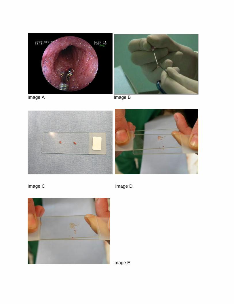
Image A Image B
Image C Image D
Image E

62
Staining the specimen: Romanowsky-type stains were utilized because of their rapidity
and easiness to use (BOX1). They are a combinations of basic and acidic dyes dis-
solved in methyl alcohol. These polychromatic stains impart the basophilic and eosino-
philic tinctorial properties observed on blood films.
The stained specimen is examined microscopically using the 10 or 20 mm objective for
staining quality and uniformity. When a 40 mm objective is to be used, a coverslip is
usually placed on the specimen. A temporary mount is made by placing a drop of im-
mersion oil on the specimen, followed by a coverslip. A permanent mount is made with
a commercially available coverslip mounting glue (e.g., EukittR, Calibrated Instruments).
BOX 1 Suggested Procedure for Staining Cytologic Sample Using Diff-Quik®Solutions
Fixative: 60 to 120 seconds
Solution 1: 30 to 60 seconds
Solution 2: 5 to 60 seconds†
Rinse under cold tap water: 15 seconds Examine staining adequacy using low power; eosinophilia or basophilia can be en-
hanced by returning to Solution 1 or Solution 2, respectively, followed by a rinse.
Air-dry and examine

CYTOLOGY
Description of different cytological features: Cytological smear were evaluated and the
following aspects were recorded for each sample:
¬small lymphocytes
¬plasma cells
¬eosinophils
¬immature lymphoid cells
¬membrane alterations of the lymphoid cells
¬atipical mitoses
¬presence of lympho glandular bodies
¬cytomorphologic anomalies in enterocytes
In the second part of the study we have blindly re-evaluated the same samples using a
reduced number of selected cytomorphological features. In particular, three cytological
features selected as recurrent in the first step of the study were identified independently
by two readers:

64
¬presence of lymphoid blast cells
¬fine structural change of the lymphoid blasts’s membrane
¬presence of lympho-glandular bodies
For each cytologic feature and reader, sensibility and specificity were evaluated. As ref-
erence the final diagnosis drawn by the specialist in gastroenterology who was the only
aware of all the results of different diagnostic tests (including histopathology), clinical
features, endoscopic aspect and follow up/response to therapy. In order to cpmpare the
results obtained by the different readers and the different techniques further evaluation
were then performed using the test of Cohen’s kappa coefficient for inter-rater agree-
ment. Predictive positive and negative values were also evaluated.

LARGE IMMATURE LYMPHOID CELLS
Medium-sized or large lymphocytes often compose 50% of the total cells in lymphomas.
An exception is the presentation of alimentary lymphoma in which reactive lymphocytes
represent the majority of the cell population. The population is often homogenous when
cell populations are mixed, including different cell sizes present such as small and large
lymphocytes, the diagnosis of lymphoma requires additional procedures. The cell size
based on comparison of the nucleus to the size of an erythrocyte.
• Small: 1-1.5 RBC
• Medium: 2-2.5 RBC
• Large: .>3 RBC
Immature lymphoid cells, intestine,dog. Lymphoma T. (Wright-Giemsa; HP oil)

66
LYMPHOGLANDULAR BODIES
Lymphoglandular bodies result from the rupture of lymphocytes and appear as small
platelet-sized basophilic cytoplasmic fragments. Although they may be seen in benign
lymph node conditions, a higher frequency is expected in lymphoma because of the
immaturity and fragility of these cells. Lysed nuclei may appear as a lacy, amorphous
eosinophilic material.
Lymphoglandular bodies, intestine,dog. Prominent basophilic round structures of
variable size indicate fragmentation of the cytoplasm. This appearance is often associ-
ated with lymphoma but may be found in other conditions having fragile cells. (Wright-
Giemsa; HP oil.)

NUCLEAR MEMBRANE
Morphologic appearance of the neoplastic cells has been used along with immunophe-
notype to further classify the lymphomas for prognostic value (Ponce et al.,2004).
Nuclei are generally described according to different aspects:
• Round: circular with no indentations
• Irregularly round: few indentations or convolutions
• Convoluted: several deep indentations
• Clefted: single deep indentation
• Central vs. eccentric placement
Irregual morphologic appearance of balt membrane, intestine,dog. Lymphoma T.
(Wright-Giemsa; HP oil)
68
HYSTOPATHOLOGY
Histopatologic evalutation: biopsy samples of ileum and jejunum were available from all
dogs. No gastric or large intestine biopsies were examined. The endoscopic tissues
consisted of mucosa and submucosa. For all cases, sufficient formalin-fixed, paraffin-
embedded samples were available for further testing. Serial sections from paraffin tis-
sue blocks from each of the 20 dogs were cut for routine HE staining.
The canine IBD activity index (CIBDAI) was used for evaluation of the severity of illness
(box2)

Box2: Histological Scoring System used to evaluate the degree of intestinal inflammation
Intestinal Histologic Scoring System
Feature Scored Score Description
Inflammation
Severity
0 None
1 Mild
2 Moderate
3 Severe
Inflammation
Extent
0 None
1 Mucosa
2 Mucosa and
Submucosa
3 Transmural
Crypt
Damage
0 None
1 1/3 of crypt damaged
2 2/3 of crypt damaged
3 Crypts lost, surface
epithelium present
4 Crypts and surface
epithelium lost
Percent
involvement
0 0%
1 1–25%
2 26–50%
3 51–75%
4 76–100%

70
IMMUNOHISTOCHEMISTRY
Serial sections were deparaffinized and rehydrated for routine immunohistochemistry
(IHC) to detect expression of CD3 (1:20 dilution; P. Moore, UC Davis, Davis, CA) in T
cells and CD79a (1:125 dilution; Clone HM57, Dako, Carpinteria, CA) in B cells. Serial
sections from paraffin tissue blocks from 14 dogs were cut for immunophenotyping
analysis (Fig 1-2).
Figure 1 Figure 2
Fig 1-2 Immunohistochemical staining of biopsy specimen taken from dog N°17. (1) Few neoplastic lymphocytes are stained by B cell marker, CD79a. (2) Many neo-plastic lymphocytes are positively stained by T cell marker, CD3.

10. STATISTICAL ANALYSIS
Diagnostic performances of the different techniques and the different observers were
calculated. Since, to our knowledge, no universally accepted gold standard has been
defined to differentiate IBD from intestinal lymphoma using endoscopic biopsies we de-
cided to blindly compare all the cytological and histological results with the final diagno-
sis drawn by the specialist in gastroenterology according to all the results of clinical ex-
amination, endoscopy, extemporaneous cytology, histopathology and confirmed with
follow-up and response to treatment. All the techniques were separately compared with
this clinical diagnosis in order to define their performance and reproducibility and to po-
tentially identify some features which could be used discriminate IBD from lymphoma.
Cohen's kappa coefficient is a statistical measure of inter-rater agreement or inter-
annotator agreement for qualitative (categorical) items. It is generally thought to be a
more robust measure than simple percent agreement calculation since κ takes into ac-
count the agreement occurring by chance. It measures the agreement between two
raters who each classify N items into C mutually exclusive categories. The first mention
of a kappa-like statistic is attributed to Galton (1892), see Smeeton (1985).
The equation for κ is:

72
where Pr (a) is the relative observed agreement among raters, and Pr (e) is the hy-
pothetical probability of chance agreement, using the observed data to calculate the
probabilities of each observer randomly saying each category. If the raters are in
complete agreement then κ = 1. If there is no agreement among the raters other
than what would be expected by chance (as defined by Pr(e)), κ = 0.
The positive predictive value, or precision rate is the proportion of positive test results
that are true positives. It is a critical measure for the performance of a diagnostic meth-
od, as it reflects the probability that a positive test reflects the underlying condition being
tested for. Its value does however depend on the prevalence of the outcome of interest,
which may be unknown for a particular target population.
Cohen’s Kappa Index
Test A
Yes No
Test B Yes 0
No 0
0 0 0
Accumulative concordance
Cohen’s Kappa
interpretation:
values < 0 as indicating no agreement and
0–0.20 as slight
0.21–0.40 as fair
0.41–0.60 as moderate
0.61–0.80 as substantial
0.81–1 as almost perfect agreement.
0.41-0.60 moderata 0.61-0.80 buona
0.81-1.00
eccel-
lente

The Positive Predictive Value is defined as:
The negative predictive value (NPV) is a summary statistic used to describe the perfor-
mance of a diagnostic testing procedure. It is defined as the proportion of subjects with
a negative test result who are correctly diagnosed. A high NPV for a given test means
that when the test yields a negative result, it is most likely correct in its assessment. In
the familiar context of medical testing, a high NPV means that the test only rarely mis-
classifies a sick subject as being healthy. Note that this says nothing about the tenden-
cy of the test to mistakenly classify a healthy subject as being sick.
The Negative Predictive Value is defined as:
The following diagram illustrates the relation positive predictive value, negative predic-
tive value, sensitivity, and specificity.

74
Condition
(as determined by "Gold
standard")
Condition
Positive
Condition
Negative
Test
Outcome
Test
Outcome
Positive
True Positive False Positive
(Type I error)
Positive predictive
value =
Σ True Positive
Σ Test Outcome Positive
Test
Outcome
Negative
False Negative
(Type II error) True Negative
Negative predictive val-
ue =
Σ True Negative
Σ Test Outcome Negative
Sensitivity =
Σ True Positive
Σ Condition Positive
Specificity =
Σ True Negative
Σ Condition Negative

11. RESULTS
Of the 20 dogs with intestinal lymphoma, the most common breeds were Boxers (3),
Beagles (3) and mongrels (2). Mean age in the lymphoma group was 8,2 years. The
clinical presentation of dogs with intestinal lymphoma in this study was similar to those
of dogs in previous reports, ranging from nonspecific symptoms such as anorexia and
lethargy to more specific gastrointestinal symptoms such as vomiting and hemorrhagic
diarrhea. In particular, the frequencies of clinical findings attributed to the intestinal tu-
mor were: vomiting 61%, weight loss 100% (fig3) and diarrhea 100% (fig 4), of which
72% was considered severe. Recent history revealed a progressive worsening of clini-
cal signs, not responding to symptomatic therapy or relapsing rapidly after an initial im-
provement. The mean time between the onset of clinical signs and the clinical presenta-
tion was 5,26 weeks, which is much shorter that the same time interval of dogs with
IBD: in our study, this time interval was longer than 3 months. The CBC of dogs with in-
testinal lymphoma showed mild to marked anemia in 88% of cases, leukocytosis with
left shift in 25% of cases. Hypoalbuminemia was a common abnormal biochemical find-
ing: 72% of the dogs with intestinal lymphoma had a low serum albumin concentration,
with most of them (84%) having less than 2 g/dL (reference interval, 3.1–4.1 g/dL).
All dogs with intestinal lymphoma were normocalcemic, when adjusted for concurrent
hypoalbubinemia; 30% of the dogs had hypocolesterolemia and 70% had hypocobala-
minemia.

76
Abdominal radiography showed non-specific findings whereas abdominal ultrasonogra-
phy showed an enteritis-like aspect (70%) with an increased thickness of the intestinal
wall and mesenteric lymphadenopathy (45%) (fig5). In 20% of patients, the ultraso-
nographic exam was normal.
The endoscopic examination showed a clobbed paving aspect of the mucosa in 50% of
the patients (fig6).
Fig.3 Fig4
Fig5 Fig6

11.1 Cytological Results
The present study included 71 dogs that suffered from gastrointestinal symptoms: 20
cases of enteropathy-type lymphoma and 52 cases of inflammatory enteropathy.
The cytological samples were independently revued and subjected to a blind study from
two clinical pathologists with different experience, although not expert in gastroenterolo-
gy. The casistic included 20 cases of enteropathy-type lymphoma and 52 cases of in-
flammatory enteropathy. Cytologic smears were evaluated, drawing a temptative diag-
nosis according to the cytomorphological aspect.

78
Table 1: Review from 2 pathologyst of 20 cases of enteropathy-type lymphoma
PATIENT A B FINAL DIAGNOSIS
Patient 1 ENTERITIS ENTERITIS LYMPHOMA
Patient 2 ENTERITIS LYMPHOMA LYMPHOMA
Patient 3 LYMPHOMA ENTERITIS LYMPHOMA
Patient 4 ENTERITIS LYMPHOMA LYMPHOMA
Patient 5 ENTERITIS LYMPHOMA LYMPHOMA
Patient 6 LYMPHOMA LYMPHOMA LYMPHOMA
Patient 7 LYMPHOMA LYMPHOMA LYMPHOMA
Patient 8 LYMPHOMA LYMPHOMA LYMPHOMA
Patient 9 ENTERITIS ENTERITIS LYMPHOMA
Patient 10 LYMPHOMA LYMPHOMA LYMPHOMA
Patient 11 ENTERITIS ENTERITIS LYMPHOMA
Patient 12 ENTERITIS LYMPHOMA LYMPHOMA
Patient 13 ENTERITIS LYMPHOMA LYMPHOMA
Patient 14 ENTERITIS ENTERITIS LYMPOMA
Patient 15 LYMPHOMA LYMPHOMA LYMPHOMA
Patient 16 LYMPHOMA LYMPHOMA LYMPHOMA
Patient 17 LYMPHOMA LYMPHOMA LYMPHOMA
Patient 18 ENTERITIS ENTERITIS LYMPHOMA
Patient 19 LYMPHOMA LYMPHOMA LYMPHOMA
Patient 20 LYMPHOMA ENTERITIS LYMPHOMA

During the first review the observer A and the observer B showed a sensitivity of 50
percent and 65 percent respectively. For both of them the specificity was 100 percent
(box1). The statistics included 20 cases of enteropathy-type lymphoma and 52 cases of
inflammatory enteropathy This means that no dogs with enteritis were identified as hav-
ing lymphoma.
(box1)
Sens.= VP/VP+FN
Spec.= VN/FP+VN
A (expert):
Se= 10/10+10= 50%
Sp= 52/0+52= 100%
B : Se=13/13+7=65%
Sp= 52/0+52= 100%
All the cytomorphological features identified were reviewed in order to define the most
relevant in lymphoma samples. According to the results of this first phase three main
features were identified as mainly associated to lymphoma.Thus, in the second part of
the study we have blindly re-evaluated the same samples using an interpretative al-
gorythm based on the following selected cytomorphological criteria: 1) presence of lym-
phoid blast cells, 2) membrane alterations of the lymphoiblasts, 3) presence of lympho-
glandular bodies (results are expressed in Table 2)
Test A
+ -
Test B + 10 3 13
- 0 59 59
10 62 72
K Cohen
0,845
EXCELLENT CONCORDANCE

80
Table 2: 2° review from 2 pathologyst of 20 cases of enteropathy-type lymphoma
PATIENT A B FINAL DIAGNOSIS
Patient 1 ENTERITIS ENTERITIS LYMPHOMA
Patient 2 LYMPHOMA LYMPHOMA LYMPHOMA
Patient 3 LYMPHOMA ENTERITIS LYMPHOMA
Patient 4 LYMPHOMA LYMPHOMA LYMPHOMA
Patient 5 LYMPHOMA LYMPHOMA LYMPHOMA
Patient 6 LYMPHOMA LYMPHOMA LYMPHOMA
Patient 7 LYMPHOMA LYMPHOMA LYMPHOMA
Patient 8 LYMPHOMA LYMPHOMA LYMPHOMA
Patient 9 LYMPHOMA LYMPHOMA LYMPHOMA
Patient 10 LYMPHOMA LYMPHOMA LYMPHOMA
Patient 11 ENTERITIS ENTERITIS LYMPHOMA
Patient 12 LYMPHOMA LYMPHOMA LYMPHOMA
Patient 13 LYMPHOMA LYMPHOMA LYMPHOMA
Patient 14 ENTERITIS LYMPHOMA LYMPOMA
Patient 15 LYMPHOMA LYMPHOMA LYMPHOMA
Patient 16 LYMPHOMA LYMPHOMA LYMPHOMA
Patient 17 LYMPHOMA LYMPHOMA LYMPHOMA
Patient 18 ENTERITIS ENTERITIS LYMPHOMA
Patient 19 LYMPHOMA LYMPHOMA LYMPHOMA
Patient 20 LYMPHOMA ENTERITIS LYMPHOMA

During the second review the observer A showed a sensitivity of 85 percent, while the
observer B of 80 percent. For both the specificity was 100% (box2). The statistics in-
cluded 20 cases of enteropathy-type lymphoma and 52 cases of inflammatory enteropa-
thy. This means that no dogs with enteritis were identified as having lymphoma.
(box2)
Sens.= VP/VP+FN
Spec.= VN/FP+VN
A:
Se= 17/17+3= 85%
Sp= 52/0+52= 100%
B: Se= 16/16+4= 80%
Sp= 52/0+52= 100%
If we consider each cytologic feature independently the results in patients with lympho-
ma were as follows:
Test A
+ -
Test B + 16 0 16
- 1
55 56
17 55 72
K Cohen
0,986
EXCELLENT CONCORDANCENCE

82
For the observer A:
immature lymphoid cells analysis
Sens.= VP/VP+FN
Spec.= VN/FP+VN
Se= 18/18+2= 90%
Sp= 50/2+50= 96%
membrane alterations of the lymphoid blasts
Sens.= VP/VP+FN
Spec.= VN/FP+VN
Se= 13/13+7= 65%
Sp= 45/7+45= 87%
lympho-glandular bodies
Sens.= VP/VP+FN
Spec.= VN/FP+VN
Se= 12/12+8= 60%
Sp= 44/8+44= 85%

Table 3: scoring of observer A in 20 cases of lymphoma, using the 3 main cyto-logical diagnostic features. observer A
PATIENT diagnosis blast membrane lg bodies
Patient 1 ENTERITIS NO NO NO
Patient 2 LYMPHOMA YES NO NO
Patient 3 LYMPHOMA YES YES NO
Patient 4 LYMPHOMA YES NO YES
Patient 5 LYMPHOMA YES YES YES
Patient 6 LYMPHOMA YES YES YES
Patient 7 LYMPHOMA YES YES NO
Patient 8 LYMPHOMA YES YES YES
Patient 9 LYMPHOMA YES NO YES
Patient 10 LYMPHOMA YES YES NO
Patient 11 ENTERITIS NO NO NO
Patient 12 LYMPHOMA YES YES YES
Patient 13 LYMPHOMA YES YES NO
Patient 14 ENTERITIS YES YES YES
Patient 15 LYMPHOMA YES YES NO
Patient 16 LYMPHOMA YES YES YES
Patient 17 LYMPHOMA YES YES YES
Patient 18 ENTERITIS YES NO YES
Patient 19 LYMPHOMA YES YES YES
Patient 20 LYMPHOMA YES NO YES
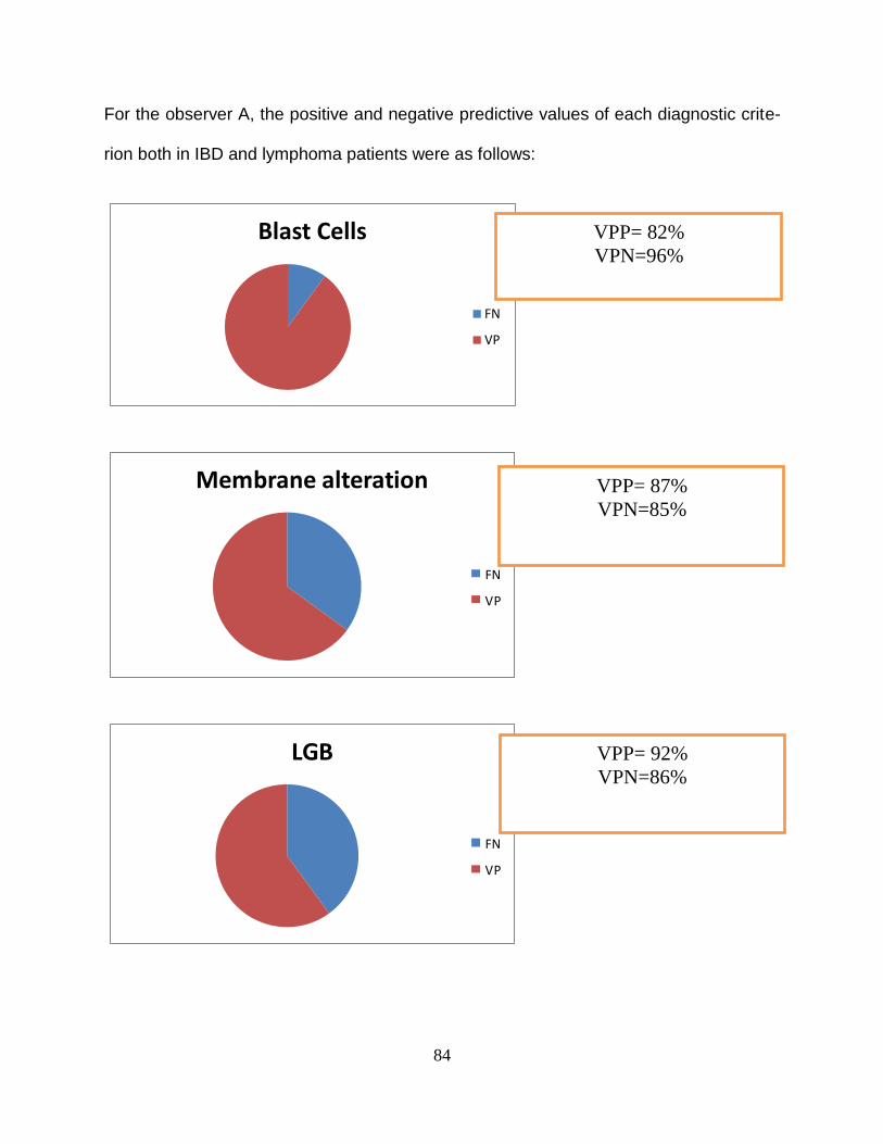
84
For the observer A, the positive and negative predictive values of each diagnostic crite-
rion both in IBD and lymphoma patients were as follows:
Blast Cells
FN
VP
Membrane alteration
FN
VP
LGB
FN
VP
VPP= 82%
VPN=96%
VPP= 87%
VPN=85%
VPP= 92%
VPN=86%

For observer B (expert) the results, in patients with IBD and lymphoma, were as follows:
immature lymphoid cells analysis
Sens.= VP/VP+FN
Spec.= VN/FP+VN
Se= 16/16+4= 80%
Sp= 50/4+50= 93%
membrane alterations of the lymphoid blasts
Sens.= VP/VP+FN
Spec.= VN/FP+VN
Se= 15/15+5= 75%
Sp= 47/5+47= 90%
lympho-glandular bodies
Sens.= VP/VP+FN
Spec.= VN/FP+VN
Se= 13/13+7= 65%
Sp= 45/7+45= 87%

86
Table 4: scoring of observer A in 20 cases of lymphoma, using the 3 main cyto-logical diagnostic features. observer B
PATIENT diagnosis blast membrane Clg
Patient 1 ENTERITIS NO NO NO
Patient 2 LYMPHOMA YES YES NO
Patient 3 ENTERITIS YES YES NO
Patient 4 LYMPHOMA YES NO YES
Patient 5 LYMPHOMA YES YES YES
Patient 6 LYMPHOMA NO YES YES
Patient 7 LYMPHOMA YES YES NO
Patient 8 LYMPHOMA YES YES YES
Patient 9 LYMPHOMA YES NO YES
Patient 10 LYMPHOMA YES YES NO
Patient 11 ENTERITIS NO NO NO
Patient 12 LYMPHOMA YES YES YES
Patient 13 LYMPHOMA YES YES NO
Patient 14 LYMPHOMA YES YES YES
Patient 15 LYMPHOMA YES YES YES
Patient 16 LYMPHOMA YES YES YES
Patient 17 LYMPHOMA YES YES YES
Patient 18 ENTERITIS NO YES YES
Patient 19 LYMPHOMA YES YES YES
Patient 20 ENTERITIS NO NO YES

For observer B, the positive and negative predictive values of each diagnostic criterion
both in IBD and lymphoma patients were as follows:
LGB
FN
VP
VPP= 100%
VPN=93%
VPP= 100%
VPN=91%
VPP= 100%
VPN=88%

88
The next tables show the disaggregate concordance index of three morphological crite-
ria between observers A and B:
Blast cells: Cohen’s Kappa
Test A
+ -
Test B + 16 0 16
- 2 2 4
18 2 20
Cohen’ K=0.615 GOOD
Membrane alteration : Cohen’s Kappa
Test A
+ -
Test B + 13 2 15
- 0 5 5
13 7 20
Cohen’s K=0.765 GOOD

LGB: Cohen’s Kappa
Test A
+ -
Test B
+ 11 2 13
- 1 6 7
12 8 20
Cohen’s K=0.681 GOOD (0,61-0,80)

90
11.2 Histological results
Fourteen histological samples of dogs with lymphoma were available for reviewing by a
reference pathologist (table 5). HE-stained sections were blindly analyzed by a
pathologist in parallel with immunohistochemistry preparations for CD3e and CD79a. I
evaluate the diagnostic accuracy of gold standard and histologic results for the diagno-
sis of lymphoma (79%).
Table 5: Patient with lymphoma avaiable for histological rview
PATIENT Gold Standard Histological Diagnosis
Patient1 LYMPHOMA LYMPHOMA
Patient2 LYMPHOMA LYMPHOMA
Patient3 LYMPHOMA LYMPHOMA
Patient5 LYMPHOMA LYMPHOMA
Patient6 LYMPHOMA ENTERITIS
Patient7 LYMPHOMA LYMPHOMA
Patient8 LYMPHOMA LYMPHOMA
Patient11 LYMPHOMA LYMPHOMA
Patient12 LYMPHOMA LYMPHOMA
Patient13 LYMPHOMA LYMPHOMA
Patient14 LYMPOMA LYMPHOMA
Patient16 LYMPHOMA LYMPHOMA
Patient17 LYMPHOMA ENTERITIS
Patient18 LYMPHOMA ENTERITIS
Accuracy = VP + VN/VP + VN + FN + FP = 79%
Based on histologic features and immunohistochemistry for CD3 and CD79a, a diagno-
sis of IBD, T-cell lymphoma, or B-cell lymphoma was made using published criteria (ta-
ble 6) and revealed that the majority of neoplastic lymphocytes were positive for CD3.

In our study, in 6 out of 8 dogs with alimentary lymphoma an immunophenotypic analy-
sis confirmed a T-cell origin of the disease.
Table 6
PATIENT Histological Diagnosis HIC
Patient1 LYMPHOMA LYMPHOMA(T)
Patient2 LYMPHOMA LYMPHOMA(T)
Patient3 LYMPHOMA ENTERITIS
Patient5 LYMPHOMA LYMPHOMA(T)
Patient6 ENTERITIS ENTERITIS
Patient7 LYMPHOMA LYMPHOMA(B)
Patient8 LYMPHOMA LYMPHOMA(B)
Patient11 LYMPHOMA LYMPHOMA(T)
Patient12 LYMPHOMA LYMPHOMA(T)
Patient13 LYMPHOMA ENTERITIS
Patient14 LYMPHOMA LYMPHOMA(T)
Patient17 ENTERITIS ENTERITIS
Patient18 ENTERITIS ENTERITIS

92
The agreement index calculated with Cohen’s kappa coefficient, between histology and
hic, was 0.649 (i.e., substantial).
Index «Kappa» ‘s di Cohen
Histology
+ -
HIC + 8 0 8
- 2 3 5
10 3 13
Kappa di Cohen
0,649

12. DISCUSSION Among the intestinal tumours of hematopoietic cell origin, lymphoma is the most com-
mon in dogs (Guillform WG and Strombeck DR, 1996) with primary intestinal lymphoma
being less common than the multicentric form (Head KW et al, 2002).
Alimentary lymphoma, common in cats, usually accounts for 7% of all canine lympho-
mas and 5-7% of all gastrointestinal neoplasms (Theilen, G. H. and Madewell, B. R.
1979). A recent report has indicated that the majority of canine intestinal lymphoma is of
T-cell origin. It has been divided in two types based on morphological characteristics of
the tumour cells: small to moderate-sized lymphoma showing epiteliotropic behaviour,
and moderate to large sized lymphoma with or without epitheliotropism (Coyle KA et al,
1996; Steimber H et al, 1995).
The former type has been classify as intestinal T-cell lymphoma according to the World
Health Organization (WHO) classification system (Valli VE et al, 2002). MALT lympho-
ma, a small, usually of B-cell origin tumor, leads to the formation of submucosal mass,
wehereas the epitheliotropic T-cell lymphoma (also known as intestinal T-cell lympho-
ma) is usually multifocal and/or diffuse throughout the submucosa and the lamina pro-
pria of the intestine, especially in the small intestine, with frequent superficial ulceration
and occasional transmural infiltration of the serosa (Willard, M. D et al, 2002). Lympho-
cytic- plasmacytic inflammation adjacent to or distant from the primary tumour can oc-
cur, and distinguishing between alimentary lymphoma and enteritis, especially lympho-
cytic- plasmacytic enteritis (LPE), can be difficult (Couto et al, 1989; Valli D.M. and
Young, K.M. 2007). Generally, the prognosis of the alimentary lymphoma is poor as

94
compared to that of the multicentric form, regardless of treatment (Rassnick, K. M. et al,
2009).
Currently, no clear consensus exists regarding sex predilection for canine lymphoma. In
some studies, the percentage of males affected with gastrointestinal lymphoma was
higher than that of females, while no difference was found in other studies (Keller ET et
al, 1993; Kiupel M et al, 1999). In our study, no significant sex difference was identified
although this finding should be considered with caution because of the low number of
cases examined.
In light of the clinical and symptomatic overlapping between IBD and enteropathy-type
lymphoma, patients are often submitted to endoscopic examination for a definitive diag-
nosis. During the first stage of the disease, the enteropathy-type lymphoma is principally
localized in the mucosa and the neoplastic infiltration is enough to cause an abnormal
digestive function with non-specific gastro-intestinal signs. The endoscopic examination
of the small bowel in the lymphoma group showed an aspect referred by gastroenterol-
ogists as cobblestone appearance of the intestinal mucosa (50 %): the intestinal muco-
sa looked friable, edematous, irregularly thickened and with a cobblestone appearance.
In dogs with this endoscopic aspect, the cytological examination alone has shown in our
study a good diagnostic accuracy in the differential diagnosis between lymphoma and
IBD.
In our study, the cytological samples were independently and blindly reviewed by two
clinical pathologists with different level of experience, although both of them had no
specific expertise in gastroenteric cytology. During the first review, the experienced
pathologist showed a lower sensitivity than the inexperienced one, whereas the specific-

ity was 100 percent for both. These results might be explained by the tendency of more
experienced pathologists to be more conservative in their judgement in order to avoid
false positive results. The agreement index calculated using the Cohen's kappa coeffi-
cient was 0.5 and it can be defined as a "mild" concordance.
In the second part of the cytology study we have blindly re-evaluated the same samples
using an interpretative algorythm based on a reduced number of some selected cyto-
morphological clues defined by the expert gastroenterologist. These criteria were: 1)
presence of lymphoid blast cells; 2) membrane alterations of the lymphoid blasts; 3)
presence of lympho-glandular bodies. During the second review the observer A (experi-
enced cytologist) showed a higher sensitivity compared with the observer B (inexperi-
enced cytologist). For both cytologists the specificity was again 100 percent. The
agreement index calculated with Cohen’s kappa coefficient was 0.986 (i.e., "excellent"
correlation).
The most sensitive and specific single cytomorphological criterion in differentiating en-
teric lymphoma from IBD was the presence of immature lymphoid cells (A: Se= 90%;
Sp= 96% B: Se= 80%; Sp= 93%), although combing all criteria provides the best accu-
racy. The concordance index calculated with Cohen’s kappa coefficient was good for all
three criteria. In this study, the group of dogs with intestinal lymphoma had a very short
median survival time (25 days) and the response to various therapeutic regimens was
poor. A possible explanation for the poor response to treatment could be related to the
immunophenotype of the lymphomas included in our study. Most cases of canine gas-
trointestinal lymphoma have been assumed to be of B-cell origin, likely based on the
fact that almost all human, non-Hodgkin’s lymphomas are of B-cell origin ( Aster J et al,

96
1999; Ozaki K.et al, 2006). However, it is now known that canine gastrointestinal lym-
phoma is more commonly of T-cell origin (Miura T et al, 2004; Carter RF et al,1986).
In our study, when immunophenotyping was performed, T-cell lymphoma was diag-
nosed in 71 percent of cases and B-cell lymphoma in 29 percent of cases. Because T-
cell lymphomas may have a more aggressive biological behaviour and are less respon-
sive to chemotherapeutic treatment, dogs with a gastrointestinal lymphoma may be less
responsive to chemotherapy (Cartel et al, 1986). In the present study, however lym-
phoma subtype seemed to have no influence on survival time, although this conclusion
must be interpreted with caution due to the low number of cases examined.
In conclusion, cytological examination of squash-prep samples could be very useful in
addressing infiltrating enteropahy, using the proposed diagnostic criteria. The most out-
standing result of our study, was the higher diagnostic accuracy of squash-prep cytolo-
gy compared with histology.
Histopathology and immunohistochemistry are generally considered useful in the differ-
ential diagnosis between IBD and lymphoma.
Seventy-five percent of the alimentary lymphomas in this study stained positively for the
CD3 marker, indicating a T-cell origin for these tumors in dogs, whereas only 25 percent
stained positively for the B-cell marker. This is opposite to the pattern seen in humans,
wherein a B-cell is the origin of nearly all non-Hodgkin’s lymphomas. Several studies on
humans have found the incidence of B-cell lymphoma to be from 50 up to 100 percent
when compared with T-cell lymphomas (Hansen PB et al 1998; Parodi AL et al, 1988;
Teske E et al, 1994; Vinzio S et al, 1998). Primary gastrointestinal T-cell lymphomas are
so rare in humans that several studies either do not address T versus B-cell as a prog-

nostic indicator, even though tumors were identified by the immunotype (Hansen PB et
al 1998; Parodi AL et al, 1988), or all the tumors in the study were of B-cell origin
(Whooley BPet al,1998).
In our findings however, although the evaluation of histopathologic results is not within
the aims of this study, the diagnostic performances of histopathology were not better
than those of cytology alone. In our view, this stresses the possible role of cytology for
diagnosing intestinal lymphoma and suggests that a wider panel of tests including cytol-
ogy, histology, immunophenotyping and possibly molecular biology techniques (PCR for
antigen receptor rearrangement or PARR) is mandatory to address the problems of a
differential diagnosis.
In our view, only an accurate evaluation of both clinical, imaging and laboratory results
by a specialist in gastroenterology may bring to a final diagnosis and appropriate treat-
ment and prognosis.

98
REFERENCES Albers TM, Alroy J, McDonnell JJ and Moore AS: A poorly differentiated gastric car-cinoid in a dog. J Vet Diagn Invest 10: 116-118, 1998
Allenspach K, Styner M, Doherr M. Validation of serologic markers of disease activity (pANCA and ASCA) in dogs with inflammatory bowel disease (abstr). J Vet Intern Med 2003;17:414 Appelbaum FR, Sale GE, Storb R Charrier K, Deeg HJ, Graham T, Wulff JC: Phenotyp-ing of canine lymphoma with monoclonal antibodies directed at cell surface antigens: classification, morphology, clinical presentation and response to chemotherapy, Hema-tol Oncol 2:151–168, 1984 Aresu L, Pregel P, Zanetti R, Caliari D, Biolatti B, Castagnaro M.; E-cadherin and β-catenin expression in canine colorectal adenocarcinoma, Res Vet Sci 89(3):409–414, 2010 Aster J, Kumar V. White cells, lymph nodes, spleen and thymus. In: Cotran RS, Kumar V, Collins T, Robbins SL, eds. RobbinsmPathologic Basis of Disease. 6th ed. Philadel-phia: Saunders, 1999;xv:1425 Baez JL, Hendrick MJ, Walker LM,Washabau RJ . Radiographic, ultrasonographic, and endoscopic findings in cats with inflammatory bowel disease of the stomach and small intestine: 33 cases (1990-1997). J Am Vet Med Assoc 1999;215:349-354 Baskin CR, Couto CG, Wittum TE. Factors influencing first remission and survival in 145 dogs with lymphoma: a retrospective study. J Am Anim Hosp Assoc 2000;36:404-409 Bennett PF, DeNicola DB, Bonney P, Glickman NW, Knapp DW.: Canine anal sac ade-nocarcinomas: clinical presentation and response to therapy, J Vet Intern Med 16:100–104, 2002 Berrocal A, Vos JH, van den Ingh TS,Molenbeek RF, van Sluijs FJ. : Canine perineal tumours J Vet Med Ser A 36:739–749, 1989 Birchard SJ, Couto CG, Johnson S: Nonlymphoid intestinal neoplasia in 32 dogs and 14 cats, J Am Anim Hosp Assoc 22:533–537, 1986
Blackwood L, German AJ, Stell AJ,O'Neill T. : Multicentric lymphoma in a dog after cy-
closporine therapy, J Small Anim Pract 45:259–262, 2004
Bonfanti U, Bussadori C, Zatelli A, De Lorenzi D, Masserdotti C, Bertazzolo W, Faver-zani S, Ghisleni G, Capobianco R, Caniatti M. Percutaneous fine-needle biopsy of deep thoracic and abdominal masses in dogs and cats. J Small Anim Pract 2004;45:191-198

Bonfanti U, Bertazzolo W, Bottero E,De Lorenzi D, Marconato L, Masserdotti C, Zatelli A, Zini E. . Diagnostic value of cytologic examination of gastrointestinal tract tumors in dogs and cats: 83 cases (2001-2004). J Am Vet Med Assoc 2006;229:1130-1133 Buddington RK. Structure and function of the dog and cat intestine. Recent advances in canine and feline nutritional research. Proceding of the 1996 Iams International Nutrition Symposium 61, Wilmington, OH, 1996 Carter RF, Valli VE, Lumsden JH: The cytology, histology and prevalence of cell types in canine lymphoma classified according to the National Cancer Institute Working For-mulation, Can J Vet Res 50:154–164, 1986 Carter RF, Valli VE, Lumsden JH. The cytology, histology and prevalence of cell types in canine lymphoma classified according to the National Cancer Institute Working For-mulation. Can J Vet Res 1986;50:154-164
Church EM, Mehlhaff CJ, Patnaik AK: Colorectal adenocarcinoma in dogs: 78 cases (1973-1984), J Am Vet Med Assoc 191(6):727–730, 1987
Clinical Outcomes of 30 Cases (1997-2004)of Canine Gastrointestinal Lymphoma
J Am Anim Hosp Assoc 2007;43:313-321. Joseph David Frank, DVM
Coyle KA, Steinberg H: Characterization of limphocytes in canine gastrointestinal lym-phoma. Vet Pathol 41: 141-146,2004 Couto CG, Rutgers HC, Sherding RG,Rojko J. : Gastrointestinal lymphoma in 20 dogs: a retrospective study, J Vet Intern Med 3:73–78, 1989 Correa SS, Mauldin GN, Mauldin GE, Mooney SC.: Serum Alpha 1-acid glycoprotein concentration in cats with lymphoma, J Am Anim Hosp Assoc 37:153–158, 2001 Crawshaw J, Berg J, Sardinas JC, Engler SJ, Rand WM, Ogilvie GK, Spodnick GJ, O'Keefe DA, Vail DM, Henderson RA. : Prognosis for dogs with nonlymphomatous, small intestinal tumors treated by surgical excision, J Am Anim Hosp Assoc 34:451–456, 1998 Cribb AE: Feline gastrointestinal adenocarcinoma: a review and retrospective study, Can Vet J 29:709–712, 1988 Day MJ: Immunophenotypic characterization of cutaneous lymphoid neoplasia in the dog and cat, J Comp Pathol 112:79–96, 1995 Dobson JM, Samuel S, Milstein H, Rogers K, Wood JL.: Canine neoplasia in the UK: estimates of incidence rates from a population of insured dogs J Small Anim Pract 43(6):240–246, 2002.

100
Dorn CR, Taylor DON, Schneider R, Hibbard HH, Klauber MR. Survey of animal neo-
plasms in Alameda and Contra Costa Counties, California. II. Cancer morbidity in dogs
and cats from Alameda County, J Natl Cancer Inst 40:307–318, 1968
Elwood CM, Garden OA. Gastrointestinal immunity in health and disease. Vet Clin North Am Small Anim Pract 1999;29:471-500
Flood-Knapik KE, Durham AC, Gregor TP, Sánchez MD, Durney ME, Sorenmo KU :
Clinical, histopathological, and immunohistochemical characterization of canine indolent
lymphoma, Vet Comp Oncol 2012
French RA, Seitz SE, Valli VE: Primary epitheliotropic alimentary T-cell lymphoma with
epatic involvement in a dog. Vet Pathol 33: 349-352, 1996
Fry MM, Vernau W, Pesavento PA, Brömel C, Moore PF.: Hepatosplenic lymphoma in
a dog, Vet Pathol 40:556–562, 2003
Frost D, Lasota J, Miettinen M: Gastrointestinal stromal tumors and leiomyomas in the dog: a histopathologic, immunohistochemical, and molecular genetic study of 50 cases, Vet Pathol 40:42– Fugiwara S, Yasunaga H, Doi K, Quantitative analysis of cytokine messenger RNAs in the duodenal mucosa in dogs with small intestinal inflammatory bowel disease (abstr). J Vet Intern Med 2002;16:32754, 2003 Fukushima K, Ohno K, Koshino-Goto Y, Uchida K, Nomura K, Takahashi M, Nakashima K, Fujino Y, Tsujimoto H. Sensitivity for the detection of a clonally rearranged antigen receptor gene in endoscopically obtained biopsy specimens from ca-nine alimentary lymphoma. J Vet Intern Sci 2009;71(12):1673-6. Gallagher RE, Yeap BY, Bi W, Livak KJ, Beaubier N, Rao S, Bloomfield CD, Appelbaum FR, Tallman MS, Slack JL, Willman CL: Quantitative real-time RT-PCR analysis of PML-RAR alpha mRNA in acute promyelocytic leukemia: Assessment of prognostic significance in adult patients from intergroup protocol 0129, Blood 101:2521–2528, 2003 Gaschen L: Ultrasonography of small intestinal inflammatory and neoplastic diseases in dogs and cats, Vet Clin North Am Small Anim Pract 41(2):329–344, 2011 German AJ, Bland PW, Hall EJ, Day MJ.: Expression of major histocompatibility com-plex class II antigens in the canine intestine. Vet Immunol Immunopathol 61:171, 1998 German AJ, Hall EJ, Moore PFRingler DJ, Newman W, Day MJ. : The distribution of lymphocytes expressing αβ andɣɗ T cell receptor, and expression of mucosal addressin cell adhesion molecule-1 in the canine intestine. J Comp Pathol 121:249,1999

German AJ, Hall EJ, Day MJ: Analysis of leucocytes subsets in the canine intestine. J Comp Pathol 120:129, 1999 German AJ, Helps CR, Hall EJ,Day MJ. . Cytokine mRNA expression in mucosal biop-sies from German shepherd dogs with small intestinal enteropathies. Dig Dis Sci 2000;45:7-17 German AJ, Hall EJ, Day MJ. Immune cell populations within the duodenal mucosa of dogs with enteropathies. J Vet Intern Med 2001;15:14-25 Giles R, Hildebrant P and Montgomery C: Carcinoid tumor in the small intestine of a dog. Vet Pathol 11: 340-349, 1974 Gillespie V, Baer K, Farrelly J,Craft D, Luong R. : Canine gastrointestinal stromal tu-mors: immunohistochemical expression of CD34 and examination of prognostic indica-tors including proliferation markers Ki67 and AgNOR, Vet Pathol 48:283, 2011 Gröne A, Werkmeister JR, Steinmeyer CL,Capen CC, Rosol TJ. : Parathyroid hormone-
related protein in normal and neoplastic canine tissues: immunohistochemical localiza-
tion and biochemical extraction, Vet Pathol 31:308–315, 1994
Guilford WG. Idiopathic inflammatory bowel diseases. In: Guilford WG, Center SA, Strombeck DR, eds. Strombeck’s Small Animal Gastroenterology. Philadelphia: WB Saunders, 1996:451-486
Guilford WG, Strombeck DR: Neoplasms of the gastrointestinal tract, APUD tumors,
endocrinopathies and gastrointestinal tract. In: Strombeck's Small Animal Gastroenter-
ology, ed. Guilford WG, Center SA,Strombeck DR, Williams DA, Meyer DJ, pp. 522–
525. WB Saunders,Philadelphia, PA, 1996Guilford WG, Center SA, Strmbeck DR, Wil-
liams DA, and Meyer DJ, pp.552-525. WB Saunders, Philadelphia, PA, 1996
Gunawardana SC, Jergens AE, Ahrens FA,Niyo Y. . Colonic nitrite and immunoglobulin G concentrations in dogs with inflammatory bowel disease. J Am Vet Med Assoc 1997;211:318-321.
Halliwell B, Gutteridge JM. The antioxidants of human extracellular fluids. Arch Biochem Biophys 1990;280:1-8.
Hause WR, Stevenson S, Meuten DJ: Pseudohyperparathyroidism associated with ad-
enocarcinomas of anal sac origin in four dogs, J Am Anim Hosp Assoc 17:373–379,
1981Head KW, Else RW; Dubielzig RR: Tumor of the alimentary tract. In: Tumor in Do-
mestic Animals, ed. Meuten DJ, pp. 401-481. Iowa State Press, Ames, IA, 2002

102
Hughes D. Fluid therapy with macromolecular plasma volume expanders. In: DiBartola SP, ed. Fluid Therapy in Small Animal Practice. 2nd ed. Philadelphia: WB Saunders, 2000;xii:611. Jacobs G, Collins-Kelly L, Lappin M,Tyler D. . Lymphocytic-plasmacytic enteritis in 24 dogs. J Vet Intern Med 1990;4:45-53 Jaffe ES: The 2008 WHO classification of lymphomas: implications for clinical practice
and translational research, Hematology Am Soc Hematol Educ Program 523–531, 2009
Jergens AE, Moore FM, Haynes JS, Miles KG . Idiopathic inflammatory bowel disease in dogs and cats: 84 cases (1987-1990). J Am Vet Med Assoc 1992;201:1603-1608 Jergens AE, Moore FM, Kaiser MS, Haynes JS, Kinyon JM. Morphometric evaluation of immunoglobulin A-containing and immunoglobulin G-containing cells and T cells in duo-denal mucosa from healthy dogs and from dogs with inflammatory bowel disease or nonspecific gastroenteritis. Am J Vet Res 1996;57:697-704 Jergens AE, Carpenter SL, Wannemuehler Y.Molecular detection of inducible nitric ox-ide synthase in canine inflammatory bowel disease (abstr). J Vet Intern Med 1998;12:205 Jergens AE. Inflammatory bowel disease: current perspectives. Vet Clin North Am Small Anim Pract 1999;29:501-521 Jergens AE, Gamet Y, Moore FM, Niyo Y, Tsao C, Smith B. . Colonic lymphocyte and plasma cell populations in canine lymphocytic-plasmacytic colitis: an immunohistochem-ical and morphometric study. Am J Vet Res 1999;60:515-520 Jergens AE, O'Connor AM, Kauffman LK, Grozdanic SD, Ackermann MR, Evans RB. Cytokine mRNA expression in intestinal biopsies of dogs with inflammatory bowel dis-ease (abstr). J Vet Intern Med 2003;17:413. Jergens AE, Schreiner CA, Frank DE, Niyo Y, Ahrens FE, Eckersall PD, Benson TJ, Evans R. A scoring index for disease activity in canine inflammatory bowel disease. J Vet Intern Med 2003;17:291-297. Juopperi TA, Cesta M, Tomlinson L Grindem CB.: Extensive cutaneous metastases in a dog with duodenal adenocarcinoma, Vet Clin Pathol 32:88–91, 2003
Kaneko,N., Yamamoto, Y., Wada,Y.,Shimokava Miyama, T.,Hiraoka,H.,Itamoto,K., Mi-
zuno et al.M.2009:application of polymerase reaction to analysis of antigen receptos re-
arrangements to support endoscopic diagnosis of canine lymphoma. J Vet Med Sci.
71:555-559

Kapatkin AS, Mullen HS, Matthiesen DT, Patnaik AK.: Leiomyosarcoma in dogs: 44
cases (1983-1988), J Am Vet Med Assoc 201(7):1077–1079, 1992.
Kararli TT: Comparision of the gastrointestinal anatomy, physiology and biochemistry of humans and commonly used laboratory animals. Biopharm Drugs Dispos 16:351, 1995 Keller ET, MacEwen EG, Rosenthal RC, Helfand SC, Fox LE. Evaluation of prognostic factors and sequential combination chemotherapy with doxorubicin for canine lympho-ma. J Vet Intern Med 1993;7:289-295 Kiupel M, Teske E, Bostock D. Prognostic factors for treated canine malignant lympho-ma. Vet Pathol 1999;36:292-300 Knottenbelt C, Mellor D, Nixon C, Thompson H, Argyle DJ.: Cohort study of COX-1 and COX-2 expression in canine rectal and bladder tumours, J Small Anim Pract 47(4):196–200, 2006 Kosovsky JE, Matthiesen DT, Patnaik AK: Small intestinal adenocarcinoma in cats: 32 cases (1978-1985), J Am Vet Med Assoc 192(2):233–235, 1988 LaRock RG, Ginn PE: Immunohistochemical staining characteristics of canine gastroin-testinal stromal tumors, Vet Pathol 34:303–311, 1997 Lennert K, Feller A: Histopathology of non-Hodgkin’s lymphomas (based on the Updat-ed Kiel Classification), ed 2, Berlin, 1990,Springer-Verlag Locher C, Tipold A, Welle M, Busato A, Zurbriggen A, Griot-Wenk ME. Quantitative as-sessment of mast cells and expression of IgE protein and mRNA for IgE and interleukin 4 in the gastrointestinal tract of healthy dogs and dogs with inflammatory bowel disease. Am J Vet Res 2001;62:211-216. Maas CPHJ, Haar G, van der Gaag I,Kirpensteijn J. : Reclassification of small intestinal and cecal smooth muscle tumors in 72 dogs: clinical, histologic, and immunohistochem-ical evaluation, Vet Surg 36(4):302–313, 2007 McEntee MF, Brenneman KA: Dysregulation of beta-catenin is common in canine spo-radic colorectal tumors, Vet Pathol 36:228– 236, 1999 McEntee MF, Cates JM, Neilsen N: Cyclooxygenase-2 expression in spontaneous in-testinal neoplasia of domestic dogs, Vet Pathol 39:428–436, 2002 Messinger JS, Windham WR, Ward CR: Ionized hypercalcemia in dogs: a retrospective study of 109 cases (1998-2003), J Vet Intern Med 23:514–519, 2009

104
Miettinen M, Majidi M, Lasota J: Pathology and diagnostic criteria of gastrointestinal stromal tumors (GISTs): a review, Eur J Cancer 38 Suppl 5:S39–S51, 2002
Miura T, Maruyama H, Sakai M, Takahashi T, Koie H, Yamaya Y, Shibuya H, Sato T,
Watari T, Tokuriki M, Hasegawa A.: Endoscopic findings on alimentary lymphoma in 7
dogs, J Vet Med Sci 66(5):577–580, 2004
Myers NC, Penninck DG: Ultrasonographic diagnosis of gastrointestinal smooth muscle tumors in the dog, Vet Radiol Ultrasound 35(5):391–397, 1994 Morrell CN, Volk MV and MankowskiJL:A carcinoid tumor in the gallbladder of a dog. Vet Pathol 39: 756-758, 2002. Modiano JF, Breen M, Burnett RC,Parker HG, Inusah S, Thomas R, Avery PR, Lind-blad-Toh K, Ostrander EA, Cutter GC, Avery AC. : Distinct B-cell and T-cell lymphopro-liferative disease prevalence among dog breeds indicates heritable risk, Cancer Res 65:5654–5661, 2005 Mukaratirwa S, de Witte E, van Ederen AM, Nederbragt H.: Tenascin expression in rela-tion to stromal tumour cells in canine gastrointestinal epithelial tumours, J Comp Path 129:137–146, 2003 Mukaratirwa S, Gruys E, Nederbragt H: Relationship between cell proliferation and tenascin-C expression in canine gastrointestinal tumours and normal mucosa, Res Vet Sci 76:133–138, 2004 Mukaratirwa S, van Ederen AM, Gruys E,Nederbragt H. : Versican and hyaluronan ex-pression in canine colonic adenomas and carcinomas: relation to malignancy and depth of tumour invasion, J Comp Path 131:259–270, 2004 Oberg K: Chemotherapy and biotherapy in the treatment of neuroendocrine tumors. Ann Oncol 12(S): 11S-114S, 2001. Ozaki K, Yamagami T, Nomura K, Narama I. T-cell lymphoma with eosinophilic infiltra-tion involving the intestinal tract in 11 dogs. Vet Pathol 2006 Ozaki K, Yamagami T, Nomura K, Narama I.: Mast cell tumors of the gastrointestinal tract in 39 dogs, Vet Pathol 39:557–564, 2002.
Paoloni MC, Penninck DG, Moore AS: Ultrasonographic and clinicopathologic findings
in 21 dogs with intestinal adenocarcinoma, Vet Radiol Ultrasound 43(6):562–567, 2002

Parodi AL, Dargent F, Crespeau F: Histological classifi- cation of canine malignant lym-
phoma. J Vet Med 35: 178–192, 1988
Patnaik AK, Liu SK, Johnson GF: Feline intestinal adenocarcinoma, Vet Pathol 13:1–10,
1976
Patnaik AK, Hurvitz AI, Johnson GF: Canine gastrointestinal neoplasms, Vet Pathol 14:547–555, 1977
Patnaik AK, Hurvitz AI, Johnson GF: Canine intestinal adenocarcinoma and carcinoid,
Vet Pathol 17:149–163, 1980.
Penninck DG, Smyers B, Webster CRL: Diagnostic value of ultrasonography in differen-tiating enteritis from intestinal neoplasia in dogs, Vet Radiol Ultrasound 44(5):570–575, 2003 Platz SJ, Breuer W, Pfleghaar S,Minkus G, Hermanns W. : Prognostic value of histo-pathological grading in canine extramedullary plasmacytomas, Vet Pathol 36:23–27, 1999 Ponce F, Magnol JP, Ledieu D,Marchal T, Turinelli V, Chalvet-Monfray K, Fournel-Fleury C : Prognostic significance of morphological subtypes in canine malignant lym-phomas during chemotherapy, Vet J 167:158–166, 2004 Prater MR, Flatland B, Newman SJ,Sponenberg DP, Chao J. : Diffuse annular fusiform adenocarcinoma in a dog, J Am Anim Hosp Assoc 36:169–173, 2000 Price GS, Page RL, Fischer BM, Levine JF, Gerig TM. Efficacy and toxicity of doxorubi-cin/cyclophosphamide maintenance therapy in dogs with multicentric lymphosarcoma. J Vet Intern Med 1991;5:259-262 Rappaport H, Winter WJ, Hicks EB: Follicular lymphoma: A re-evaluation of its position in the scheme of malignant lymphoma, based on a survey of 253 cases, Cancer 9:792–821, 1956 Rappaport H: Tumors of the hematopoietic system. In Atlas of tumor pathology, Section 3, Fasicle 8. Washington DC, 1966, Armed Forces Institute of Pathology Rassnick, K. M., Moore, A. S., Collister, K. E., Northrup, N. C., Kristal, O., Chretin, J. D. and Bailey, D. B. 2009. Efficacy of Combination Chemotherapy for Treatment of Gastro-intestinal Lymphoma in Dogs. J. Vet. Intern. Med. 1:1-6 Reimer ME, Leib MS, Reimer MS, Saunders GK, Johnston SA. : Rectal ganglioneuroma in a dog, J Am Anim Hosp Assoc 35:107–110, 1999

106
Restucci B, Martano M, De Vico G, Lo Muzio L, Maiolino P. : Expression of E-cadherin, beta-catenin and APC protein in canine colorectal tumours, Anticancer Res 29(8):2919–2925, 2009 Ridyard AE, Nuttal TJ, Else RW, Simpson JW, Miller HR.. Evaluation of Th1, Th2 and immunosuppressive cytokine mRNA expression within the colonic mucosa of dogs with idiopathic lymphocytic-plasmacytic colitis. Vet Immunol Immunopathol 2002;86:205-214 Rivers BJ, Walter PA, Feeney DA,Johnston GR. : Ultrasonographic features of intestinal adenocarcinoma in five cats, Vet Radiol 38(4):300–306, 1997 Rosol TJ, Capen CC: Biology of disease: Mechanisms of cancerinduced hypercalcemia, Lab Invest 67:680–702, 1992 Russell KN, Mehler SJ, Skorupski KA,Baez JL, Shofer FS, Goldschmidt MH. : Clinical and immunohistochemical differentiation of gastrointestinal stromal tumors from leiomy-osarcomas in dogs: 42 cases (1990-2003), J Am Vet Med Assoc 230(9):1329–1333, 2007 Ruslander DA, Gebhard DH, Tompkins MG, Grindem CB, Page RL. : Immunophenotyp-ic characterization of canine lymphoproliferative disorders, In Vivo 11:169–172, 1997. Sako T, Uchida E, Okamoto M , Yamamoto E, Kagawa Y, Yoshino T, Hirayama K, Taniyama H.: Immunohistochemical evaluation of a malignant intestinal carcinoid in a dog, Vet Pathol 40:212–215, 2003 Seiler RJ: Colorectal polyps of the dog: a clinicopathologic study of 17 cases, J Am Vet Med Assoc 174:72–75, 1979 Selting KA, Ogilvie GK, Lana SE,Fettman MJ, Mitchener KL, Hansen RA, Richardson KL, Walton JA, Scherk MA. : Serum Alpha 1-acid glycoprotein concentrations in healthy and tumor-bearing cats, J Vet Intern Med 14(5):503–506, 2000 Sherding RG. Diseases of the small bowel. In: Ettinger SJ, ed. Textbook of Veterinary Internal Medicine. 2nd ed. Philadelphia: WB Saunders, 1982:1278-1346 Shoieb AM, Hanshaw DM: Anal sac gland carcinoma in 64 cats in the United Kingdom
(1995-2007), Vet Pathol 46:677–683, 2009
Snipes RL, Snipes H: Quantitative investigation of the intestines in eight species of do-
mestic mammals. Mamm Biol 62:359, 1997
Sonea IM, Jergens AE, Sacco RE, Niyo Y, Merten E, Kauffman LK, Moore PF. Flow cy-tometric analysis of mucosal lymphocytes in dogs with inflammatory bowel disease (ab-str). J Vet Intern Med 2001;15:275

Stefanello D, Valenti P, Zini E,Comazzi S, Gelain ME, Roccabianca P, Avallone G, Ca-
niatti M, Marconato L. : Splenic marginal zone lymphoma in 5 dogs (2001-2008), J Vet
Intern Med 25:90–93, 2011;
Steimber H, Dubielzig RR, Thomson J, Dzata G: Primary gastrointestinal lymphosar-coma with epitheliotropism in three Shar-Pei and one boxer dog. Vet Pathol 32: 423-426, 1995 Stokes C, Waly N: Mucosal defence along the gastrointestinal tract of cats and dogs. Vet Res 37:281, 2006 Stonehewer J, Simpson JW, Else RW, Macintyre N. Evaluation of B and T lymphocytes and plasma cells in colonic mucosa from healthy dogs and dogs with inflammatory bowel disease. Res Vet Sci 1998;65:59-63 Strombeck DR. Chronic inflammatory bowel disease. In: Strombeck DR, ed. Small Ani-mal Gastroenterology. Davis, CA: Stonegate Publishing, 1979:240-261
Sueiro FA, Alessi AC, Vassallo J: Canine lymphomas: a morphological and immuno-
histochemical study of 55 cases, with observations on p53 immunoexpression, J Comp
Pathol 131:207– 213, 2004
Teske E, van Heerde P, Rutteman GR, Kurzman ID, Moore PF, MacEwen EG.: Prog-
nostic factors for treatment of malignant lymphoma in dogs, J Am Vet Med Assoc
205:1722–1728, 1994
Theilen, G. H. and Madewell, B. R. 1979. Leukemia-sarcoma disease complex. pp. 241-
252. In: Veterinary Cancer Medicine (Theilen, G. H. and Madewell, B. R. eds.), Lea and
Febiger, Philadelphia
Theilen, G. H. and Madewell, B. R. 1987. pp 392-407 In: Veterinary Cancer Medicine
2nd ed. (Theilen, G. H. and Madewell, B. R. eds.), Lea and Febiger, Philadelphia
Trevor PB, Saunders GK, Waldron DR,Leib MS. : Metastatic extramedullary plasmacy-toma of the colon and rectum in a dog, J Am Vet Med Assoc 203(3):406–409, 1993 Vaerman JP, Heremans JF: Distribution of varius immunoglobulin containing cells in canine lymphoid tissue. Immunology 17:627, 1969 Vail DM, MacEwen EG, Young KM. Canine lymphoma and lymphoid leukemias. In: Withrow SJ, MacEwen EG, eds. Small Animal Clinical Oncology. 3rd ed. Philadelphia: WB Saunders, 2001;xvii:718

108
Vail DM, Kisseberth WC, Obradovich JE,Moore FM, London CA, MacEwen EG, Ritter MA : Assessment of potential doubling time (Tpot), argyrophilic nucleolar organizer re-gions (AgNOR), and proliferating cell nuclear antigen (PCNA) as predictors of therapy response in canine non-Hodgkin’s lymphoma, Experim Hematol 24:807–815, 1996 Valerius KD, Powers BE, McPherron MA, Hutchison JM, Mann FA, Withrow SJ.: Ade-nomatous polyps and carcinoma in situ of the canine colon and rectum: 34 cases (1982-1994), J Am Anim Hosp Assoc 33:156–160, 1997 Vezzali E, Parodi AL, Marcato PS, Bettini G. Histopathological classification of 171 cases of canine and feline non-Hodgkin lymphoma according to the WHO, Vet Comp Oncol 8:38–49, 2009. Valli VE, Jacobs RM, Parodi AL,: Histological classification of hematopoietic tumors of domestic animals, Washington, DC, 2002, Armed Forces Institute of Pathology and The World Health Organization Valli VE, Vernau W, de Lorimier P,Graham PS, Moore PF. : Canine indolent nodular lymphoma, Vet Pathol 43:241–256, 2006 Valli VE, Myint MS, Barthel A, Bienzle D, Caswell J, Colbatzky F, Durham A, Ehrhart EJ, Johnson Y, Jones C, Kiupel M, Labelle P, Lester S, Miller M, Moore P,Moroff S, Roccabianca P, Ramos-Vara J, Ross A, Scase T, Tvedten H, Vernau W. : Classifica-tion of canine malignant lymphomas according to the World Health Organization criteria, Vet Pathol 48:198–211, 2011. Vezzali E, Parodi AL, Marcato PS, Bettini G. : Histopathological classification of 171 cases of canine and feline non-Hodgkin lymphoma according to the WHO, Vet Comp Oncol 8:38–49, 2009 Vinzio S, Goichot B, Krebs-Wurtz E, Herbrecht R, Imler M: Diffuse digestive tract B-cell lymphoma manifesting as exudative enteropathy. Presse Med 27:963–964, 1998 Weir EC, Norrdin RW, Matus RE, Brooks MB, Broadus AE, Mitnick M, Johnston SD, In-sogna KL. Endocrinology. 1988 Feb;122(2):602-8. Humoral hypercalcemia of malignan-cy in canine lymphosarcoma. Whooley BP, Bernik S, Sarkis AY, Wallack MK: Primary gastrointestinal non-Hodgkin’s lymphoma: increasingly AIDS-related. Am Surg 64:137–143, 1998 Willard MD, Leid RW: Nonuniform horizontal and vertical distributions of immunoglobu-lin A cells in canine intestine. Am J Vet Res 42: 1573, 1981

Willard, M. D., Jergens, A. E., Duncan, R. B., Leib, M. S., McCracken, M. D., DeNovo, R. C., Helman, R. G., Slater, M. R. and Harbison, J. L. 2002. Interobserver variation among histopathologic evaluations of intestinal tissue from dogs and cats. J. Am. Vet. Med. Assoc. 220: 1177-1182 Williams LE, Gliatto JM, Dodge RK, Johnson JL, Gamblin RM, Thamm DH, Lana SE, Szymkowski M, Moore AS; Veterinary Cooperative Oncology Group: Carcinoma of the apocrine glands of the anal sac in dogs: 113 cases (1985-1995), J Am Vet Med As-soc 223:825–831, 2003 Yamazaki J, Baba K, Goto-Koshino YSetoguchi-Mukai A, Fujino Y, Ohno K, Tsujimoto
H. : Quantitative assessment of minimal residual disease (MRD) in canine lymphoma by
using real-time polymerase chain reaction, Vet Immunol Immunopath 126:321–331,
2008

110
RINGRAZIAMENTI
Ringrazio il Dott. Enrico Bottero, collega ma prima di tutto amico.
E’ stato un piacere conoscerti e lavorare insieme a te.


![Somatostatin Analogs · K52.9 Noninfective gastroenteritis and colitis, unspecified K56.1 Intussusception K56.2 Volvulus K56.50 Intestinal adhesions [bands], unspecified as to partial](https://static.fdocuments.us/doc/165x107/5e99aa745ccca6756144b359/somatostatin-analogs-k529-noninfective-gastroenteritis-and-colitis-unspecified.jpg)

