Universidade do Minho Escola de Engenhariarepositorium.sdum.uminho.pt/bitstream/1822/8996/1/TESE...
151
Maria Teresa Gonçalves de Macedo Matamá Julho de 2008 Minho 2008 U Enzymatic Treatment of Acrylic and Cellulose Acetate Fibres This work was financially supported by Fundação para a Ciência e Tecnologia, by means of a PhD grant (SFRH / BD / 13423 / 2003), and by the project BioSYNTEX (G5RD-CT-2001-00560) from the European Union 5th Framework Program - GROWTH Universidade do Minho Escola de Engenharia Enzymatic Treatment of Acrylic and Cellulose Acetate Fibres Maria Teresa Gonçalves de Macedo Matamá
Transcript of Universidade do Minho Escola de Engenhariarepositorium.sdum.uminho.pt/bitstream/1822/8996/1/TESE...
Maria Teresa Gonçalves de Macedo Matamá
Julho de 2008Min
ho 2
008
U
Enzymatic Treatment of Acrylic andCellulose Acetate Fibres
This work was financially supported by Fundação para a Ciência e Tecnologia,
by means of a PhD grant (SFRH / BD / 13423 / 2003), and by the project
BioSYNTEX (G5RD-CT-2001-00560) from the European Union 5th Framework
Program - GROWTH
Universidade do MinhoEscola de Engenharia
En
zym
ati
c Tr
ea
tme
nt
of
Acr
ylic
an
d C
ell
ulo
se A
ceta
te F
ibre
sM
aria
Ter
esa
Gon
çalv
es d
e M
aced
o M
atam
á
Thesis for Doctoral degree in Textile Engineering - Textile Chemistry
Elaborated under the supervision ofProfessor Doutor Artur Cavaco-Paulo
Maria Teresa Gonçalves de Macedo Matamá
Julho de 2008
Enzymatic Treatment of Acrylic andCellulose Acetate Fibres
Universidade do MinhoEscola de Engenharia

É AUTORIZADA A REPRODUÇÃO PARCIAL DESTA TESE , APENAS PARA EFEITOS DE INVESTIGAÇÃO, MEDIANTE DECLARAÇÃO ESCRITA DO INTERESSADO, QUE A TAL SE COMPROMETE.
Maria Teresa Gonçalves de Macedo Matamá
Enzymatic Treatment of Acrylic and Cellulose Acetate Fibres
The research described in this thesis was performed
at the Textile Engineering Department and at the Biology
Department of University of Minho and was financially
supported by Fundação para a Ciência e Tecnologia, by
means of a PhD grant (SFRH / BD / 13423 / 2003), and by
the project BioSYNTEX (G5RD-CT-2001-00560) from the
European Union 5th Framework Program - GROWTH.
ii

Enzymatic Treatment of Acrylic and Cellulose Acetate Fibres
iii
AAGGRRAADDEECCIIMMEENNTTOOSS
Em primeiro lugar gostaria de expressar o meu sincero agradecimento ao meu supervisor Prof. Artur Cavaco-Paulo pela oportunidade de realizar este trabalho e do que com ele aprendi. Professor, agradeço a sua orientação, paciência e voto de confiança, principalmente nos momentos em que mais precisei. Agradeço a consideração que teve no aproveitamento da minha formação em BIO(química). Muito agradecida também pela possibilidade que me deu de participar em vários encontros científicos, de onde retirei proveito profissional e também o contacto com outros países e culturas, que de outro modo talvez não chegasse a conhecer.
Estou especialmente agradecida à Professora Margarida Casal por me
acolher no seu grupo, pela sua amabilidade, pela paciência com que lidou com as várias peripécias laborais, pelo encorajamento que me deu, pela valiosa supervisão do trabalho que desenvolvi no seu laboratório do Departamento de Biologia e pelas suas sugestões durante a elaboração desta dissertação.
Não posso deixar de referir todas as pessoas que estiveram a meu lado
em Azurém e me ajudaram ao longo de todo o trabalho. Queria agradecer à Carla Manuela e ao Alex, com quem partilhei as venturas e desventuras da cutinase, por toda a ajuda preciosa. Queria agradecer à geração “mais velha” Carla Joana, Zille e Tzanko pela ajuda e orientação. Um muito obrigada aos alunos Erasmus Aitor e Cristina sem os quais o meu trabalho teria sido mais difícil e incompleto. Obrigada Xana, Raquel e Andreia pela vossa disponibilidade e ajuda. Agradeço ao Carlos, à Ana, à Florentina, à Filipa, à Margarida, à Suyon, à Clarinda e a todos pela amizade, companheirismo, paciência e apoio.
Rita, estou-te muito grata pela tua valiosa orientação e sugestões, pela
tua energia contagiante e por tudo o que me ensinaste no laboratório de Biologia Molecular. Jorge, não existem palavras suficientes para expressar o meu agradecimento por toda a tua ajuda e colaboração, cavalheirismo incomparável e coração grande. Obrigada Raul e Isabel pela vossa disponibilidade inesgotável, amizade e apoio e por todo o auxílio que me deram ao longo da minha estadia em Braga. Quero ainda agradecer à Leonor, à Susana, ao Paulo, à Neide, ao Rui, à Sandra, à Magda, ao Carlos, ao Amaro, à Manuela, e a todos por transformarem os 100 km diários de viagem até Braga em dez e tornarem inesquecíveis os momentos que partilhei convosco.
Não posso deixar de agradecer ao Departamento de Engenharia Têxtil e
à Universidade do Minho sem os quais esta tese não teria tido lugar. Pedro, obrigada por fazeres parte da luz do meu dia, da serenidade do
meu sono, do som do meu riso, do calor do meu corpo e da energia necessária para realizar este trabalho. Pai e mãe, obrigada pelo vosso amor, carinho e confiança, sem vocês nada disto teria sido possível. Agradeço a toda a minha família e amigos que me apoiaram ao longo deste percurso.

Enzymatic Treatment of Acrylic and Cellulose Acetate Fibres
« One of the wonders of human nature is our ability to hope...
The great biologist Edward O. Wilson calls us "the future-seeking
species" and suggests that natural selection has made hopefulness a
unique human quality, "a necessary companion of intelligence".
Still more human, perhaps, is our capacity for acting on our
hopes. We not only dream, we strive to achieve the dreams we
imagine. Behind all human achievement, from the creative acts of
artists to the building of communities, from the making and trading of
goods to the work of nations, there is aspiration, resolve, and action. »
By William McDonough, a “thinking green” architect
2001
iv
Enzymatic Treatment of Acrylic and Cellulose Acetate Fibres
v
AABBSSTTRRAACCTT
The background theme of the present thesis is the multidisciplinary area of textile
biotechnology, which is of major importance for the textile industry and its sustainable
development. The work here described was devoted to the treatment with enzymes of two man-
made fibres - acrylic and cellulose acetate. The thesis is divided in several chapters being the
first one a general introduction. Biocatalysis is addressed, especially in the context of textile
industry and surface modification of polymers, followed by a general description on the
properties and applications of both fibres. The enzymes used throughout the work – nitrilase
(EC 3.5.5.1) and cutinase (EC 3.1.1.74), are briefly mentioned as well as the major methods to
manipulate and improve enzymes. The general aim of the work is the formation of reactive
and/or hydrophilic groups at the surface of acrylic and cellulose acetate fibres by enzymatic
hydrolysis of their pendent groups. As follows, the purpose is to preserve the desirable bulk
properties of the fibres acting only at the surface by using eco-friendly catalysts.
In chapter 2, the modification of the surface of acrylic fabric with a commercial nitrilase is
reported. The enzymatic conversion of nitrile groups into the carboxylic groups, on the fibre
surface, was monitored for 36 hours by the release of ammonia to the media and by the
improvement in the affinity of the treated fabric for a basic dye. The steady release of ammonia
along the enzymatic treatment showed that the adsorption of nitrilase to the acrylic led to an
increase in its operational stability, resembling the immobilization procedures used to stabilize
proteins. A maximum affinity for the basic dye was observed for a treatment period of 8 hours,
which corresponded to a relative K/S of 135% when the colouration of acrylic was performed at
70 ºC. A surface erosion phenomenon took place causing the “oscillatory” behaviour of the
amount of dye uptake with the time of treatment. Polyacrylic acid was determined in solution as
a non desirable, secondary product of the modification of acrylic with nitrilase. These results
showed that the outcome of nitrilase application is closely dependent on reaction parameters
like time, enzyme activity and media formulation.
The chapter 3 describes the modification of the comonomer vinyl acetate of the acrylic
used with two enzymes: cutinase from the fungus Fusarium solani pisi and a commercial
esterase (Texazym PES). The effect of acrylic solvents and stabilizing polyalcohols on cutinase
operational stability in solution was studied. The influence of these additives and mechanical
agitation on the enzymatic modification of acrylic fabric was also investigated. The hydroxyl
groups produced on the fibre surface reacted with the dye Remazol Brilliant Blue R, C.I. 61200,
increasing the colour of treated fabric. The best colour level was obtained with a high level of
mechanical agitation and with the addition of 1% (v/v) N,N-dimethylacetamide. Under these
conditions, the increase in the acrylic fabric colour depth was around 30% for cutinase and 25%
for Texazym, comparing to the respective controls. The crystallinity degree, determined by wide
angle X-ray scattering, was not significantly changed between control samples and samples

Enzymatic Treatment of Acrylic and Cellulose Acetate Fibres
treated with cutinase. The results showed, once more, that the success of the application of
enzymes, in this case cutinase and a commercial esterase, depends closely on the conditions in
which the treatment takes place.
Cutinase was also chosen to modify the surface of cellulose diacetate and triacetate
fibres. This work is reported in chapter 4. The enzymatic hydrolysis of acetyl groups on the fibre
surface was evaluated by the release of acetic acid and by the specific chemical colouration of
the fabrics with Remazol Brilliant Blue R. The treatment for 8 hours, at 30 ºC and pH 8, resulted
in an acetyl esterase activity of 0.010 U and 0.0072 U on cellulose diacetate and triacetate,
respectively. The colour levels for samples treated with cutinase for 24 hours increased 25% for
cellulose diacetate and 317% for cellulose triacetate, comparing to the controls. Cross-sections
of both fibres were analysed by fluorescence microscopy and the superficial action of cutinase
was confirmed. Comparing to other enzymes described in literature, cutinase is a catalyst to
consider for the superficial regeneration of cellulose hydrophilicity and reactivity on highly
substituted acetates.
For further improvement of cutinase activity on cellulose modified fibres, chimeric
cutinases were produced, by recombinant DNA technologies, and used to treat cellulose
acetate fabrics, as described in chapter 5. Two distinct carbohydrate-binding modules were
fused independently to the C-terminal of cutinase: the carbohydrate-binding module of
cellobiohydrolase I, from the fungus Trichoderma reesei, and the carbohydrate-binding module
of endoglucanase C, from the bacterium Cellulomonas fimi. Both chimeric cutinases had a more
efficient performance than the wild type enzyme, but the interaction of these bifunctional
enzymes with cellulose acetate needs to be further characterized for a better assessment of the
nature and yield of the observed modifications.
The chapter 6 is dedicated to the general discussion, final remarks and future
perspectives. In this thesis, evidences are presented showing that enzymes, more specifically,
nitrilase and cutinase are important tools for the acrylic and cellulose acetate surface
functionalization. This work also evidenced that this is only the first step towards the efficient
utilization of these resources that Nature provide us.
vi
Enzymatic Treatment of Acrylic and Cellulose Acetate Fibres
vii
RREESSUUMMOO
O tema de fundo deste trabalho é a área multidisciplinar da biotecnologia têxtil que tem
vindo a afirmar-se como uma área de grande importância para a indústria têxtil e para o seu
desenvolvimento sustentável. O trabalho aqui apresentado consistiu no tratamento com
enzimas de duas fibras, a acrílica e o acetato de celulose. A tese encontra-se dividida em
vários capítulos consistindo o primeiro numa introdução geral a diversos tópicos abordados
pelo trabalho. A biocatálise é referida, especialmente, no contexto da indústria têxtil e
modificação superficial de polímeros, seguida de uma descrição geral das propriedades e
aplicações das duas fibras. As enzimas utilizadas ao longo do trabalho – nitrilase (EC 3.5.5.1) e
cutinase (EC 3.1.1.74), são mencionadas de forma sucinta assim como os métodos principais
de manipulação da actividade enzimática. O objectivo geral do trabalho assentou no
desenvolvimento de metodologias não poluentes conducentes à formação de grupos
reactivos/hidrofílicos à superfície das fibras, via hidrólise dos grupos laterais dos respectivos
polímeros de forma a preservar as propriedades nucleares e desejáveis das fibras.
No capítulo 2 é reportada a modificação da superfície da fibra acrílica por uma nitrilase
comercial. A conversão enzimática dos grupos nitrilo em grupos carboxílicos à superfície da
fibra foi avaliada durante 36 horas através da libertação de amoníaco para a solução e através
do aumento da afinidade do tecido tratado para um corante básico. O aumento linear do
amoníaco, libertado durante o tratamento, mostrou que a adsorção da nitrilase à fibra acrílica
conduziu a um aumento da sua estabilidade operacional assemelhando-se aos procedimentos
de imobilização usados para estabilizar enzimas. Um valor máximo de K/S foi observado para
um período de incubação de 8 horas, o que corresponde a um K/S relativo de 135% quando se
fez uma coloração a 70 ºC. Teve lugar um fenómeno de erosão superficial que determinou o
comportamento oscilatório da quantidade de corante fixada com o tempo de tratamento. Foi
determinado ácido poliacrílico nas soluções de tratamento como produto secundário não
desejado da modificação da acrílica pela nitrilase. Estes resultados revelam que o efeito final
da aplicação da nitrilase no tratamento da acrílica está intimamente dependente dos
parâmetros da reacção como o tempo, a actividade da enzima e a composição do meio.
O capítulo 3 descreve a modificação do comonómero acetato de vinilo da fibra acrílica
usada pela acção da cutinase do fungo Fusarium solani pisi e de uma esterase comercial
(Texazym PES). Foi estudado o efeito de solventes da acrílica e de poli-álcoois na estabilidade
operacional da cutinase em solução, bem como o impacto desses aditivos e da agitação
mecânica na modificação enzimática do tecido de acrílica. Os grupos hidroxilo produzidos na
superfície da fibra reagiram com o corante Remazol Brilliant Blue R, C.I. 61200, aumentando a
cor do tecido tratado. O melhor nível de coloração foi obtido com elevada agitação mecânica e
com a adição de 1% (v/v) de N,N-dimetilacetamida. Sob estas condições, o aumento da
intensidade de cor, em relação aos controlos, foi de 30% para o tratamento com a cutinase e
Enzymatic Treatment of Acrylic and Cellulose Acetate Fibres
25% para a Texazym. O grau de cristalinidade determinado por difracção de raios-X não foi
alterado significativamente entre amostras controlo e amostras tratadas com cutinase. Uma vez
mais, os resultados mostraram que o sucesso da aplicação de enzimas, neste caso a cutinase
e uma esterase comercial, depende muito das condições em que se realiza o tratamento.
A cutinase foi escolhida para modificar também a superfície de fibras de diacetato e
triacetato de celulose, trabalho este que é abordado no capítulo 4. A hidrólise enzimática dos
grupos acetilo na superfície da fibra foi monitorizada pela libertação de ácido acético e pela
coloração específica dos tecidos com o corante Remazol Brilliant Blue R. O tratamento do
tecido durante 8 horas a 30 ºC e pH 8 resultou numa actividade acetil esterase de 0.010 U e
0.0072 U tendo como substrato a fibra de diacetato e triacetato de celulose, respectivamente.
Os níveis de cor das amostras tratadas com cutinase durante 24 horas aumentaram 25% para
o diacetato e 317% para o triacetato, comparando com os controlos. Foram analisadas secções
transversais de ambas as fibras, por microscopia de fluorescência, e confirmou-se a acção
superficial da cutinase. Por comparação com outras enzimas já descritas, a cutinase é um
catalizador a ter em consideração para a regeneração superficial da hidrofilicidade e
reactividade da celulose em acetatos com grau de substituição elevado.
Para melhorar a actividade da cutinase nas fibras modificadas de celulose foram
produzidas, através de tecnologias de ADN recombinante, cutinases quiméricas que foram,
posteriormente, usadas no tratamento dos tecidos de acetato de celulose descrito no capítulo
5. Dois módulos distintos de ligação a carbohidratos foram fundidos, independentemente, ao
terminal carboxílico da cutinase: o módulo da celobiohidrolase I, do fungo Trichoderma reesei e
o da endoglucanase C, da bactéria Cellulomonas fimi. Ambas cutinases quiméricas tiveram
uma performance mais eficiente que a cutinase nativa, mas a interacção destas enzimas
bifuncionais com os acetatos de celulose carece de mais estudos para melhor caracterizar a
natureza e extensão destas modificações.
O capítulo 6 é dedicado a uma discussão geral, observações finais e perspectivas
futuras. Nesta tese são apresentadas evidências que mostram que as enzimas, mais
concretamente, a nitrilase e a cutinase são ferramentas importantes para a funcionalização da
superfície das fibras acrílicas e de acetato de celulose. Com este trabalho também fica claro
que apenas se deu o primeiro passo num percurso que eventualmente nos conduzirá a um
aproveitamento eficiente destes recursos que a Natureza nos providencia.
viii
Enzymatic Treatment of Acrylic and Cellulose Acetate Fibres
ix
TTAABBLLEE OOFF CCOONNTTEENNTTSS
Agradecimentos .............................................................................................. iii Abstract ............................................................................................................. v Resumo ........................................................................................................... vii Table of Contents ............................................................................................ ix List of Abbreviations ...................................................................................... xii List of Figures ................................................................................................ xiv List of Tables .................................................................................................. xx Introduction ...................................................................................................... 1
1. Introduction ............................................................................................ 2 1.1 Biocatalysis ................................................................................... 2
1.1.1 Industrial enzymes ........................................................... 2 1.1.2 Biocatalysis in textile industry .......................................... 4 1.1.3 Biocatalysis in synthetic polymer surface modification ..... 6
1.2 Acrylic fibre ................................................................................... 9 1.2.1 General description .......................................................... 9 1.2.2 Structural characterization ............................................. 13 1.2.3 Acrylic fibre properties .................................................... 16 1.2.4 Products and applications .............................................. 18 1.2.5 Polyacrylonitrile as an enzymatic substrate ................... 19
1.3 Cellulose acetate fibres ............................................................... 21 1.3.1 General description ........................................................ 21 1.3.2 Structural characterization ............................................. 25 1.3.3 Cellulose acetate fibre properties ................................... 26 1.3.4 Products and applications .............................................. 28 1.3.5 Cellulose acetate as an enzymatic substrate ................. 30
1.4 Nitrilase (E.C. 3.5.5.1)................................................................. 31 1.4.1 General description, structure and function .................... 31 1.4.2 Applications .................................................................... 33
1.5 Cutinase (E.C. 3.1.1.74) ............................................................. 35 1.5.1 General description, structure and function .................... 35 1.5.2 Applications .................................................................... 38
1.6 Tailoring the properties of biocatalysts ....................................... 39 1.6.1 Protein and media engineering ...................................... 39 1.6.2 Fusion proteins with charbohydrate - binding modules and
applications ............................................................................... 41 1.6.3 Carbohydrate-binding module of cellobiohydrolase I from
Trichoderma reesei ................................................................... 43 1.6.4 Carbohydrate-binding module N1 of endoglucanase C
from Cellulomonas fimi .............................................................. 45 1.7 Aims of the work ......................................................................... 47
Enzymatic Treatment of Acrylic and Cellulose Acetate Fibres
Using a Nitrilase for the Surface Modification of Acrylic Fibres ................ 48 2. Using a nitrilase for the surface modification of acrylic fibres ............... 49
2.1 Materials and methods................................................................ 49 2.1.1 Reagents and enzymes ................................................. 49 2.1.2 Nitrilase activity assay .................................................... 49 2.1.3 pH and temperature profile ............................................ 50 2.1.4 Stability of nitrilase ......................................................... 50 2.1.5 Effect of additives in the enzymatic treatment of acrylic
fabric ......................................................................................... 50 2.1.6 Enzymatic treatment of acrylic fabric .............................. 51 2.1.7 Quantification of total protein concentration ................... 51 2.1.8 Determination of ammonia concentration ....................... 51 2.1.9 Determination of polyacrylic acid concentration ............. 52 2.1.10 Determination of fabric weight differences ................... 52 2.1.11 Acrylic fabric colouration with a basic dye .................... 52
2.2 Results and discussion ............................................................... 53 2.2.1 pH and temperature profile of nitrilase activity ............... 53 2.2.2 Stability of nitrilase ......................................................... 54 2.2.3 Effect of additives on the modification of acrylic surface
with nitrilase .............................................................................. 56 2.2.4 Studies of acrylic surface modification with nitrilase ....... 57
2.3 Concluding remarks .................................................................... 61 The Effect of Additives and Mechanical Agitation in Surface Modification of Acrylic Fibres by Esterases ...................................................................... 64
3. The effect of additives and mechanical agitation in surface modification of acrylic fibres by esterases ...................................................................... 65
3.1 Materials and methods .............................................................. 65 3.1.1 Reagents and enzymes ................................................. 65 3.1.2 Esterase activity assay ................................................... 65 3.1.3 Stability of cutinase ........................................................ 66 3.1.4 Enzymatic treatment of acrylic fabric .............................. 66 3.1.5 Quantification of total protein concentration ................... 67 3.1.6 Determination of acetic acid concentration in the bath
solutions .................................................................................... 67 3.1.7 Acrylic fabric colouration with a reactive dye .................. 68 3.1.8 Wide angle X-ray scattering ........................................... 68
3.2 Results and discussion .............................................................. 69 3.2.1 Operational stability of cutinase ..................................... 69 3.2.2 Enzymatic modification of acrylic surface ....................... 71 3.2.3 Wide angle X-ray scattering ........................................... 75
3.3 Concluding remarks ................................................................... 78 Surface Modification of Cellulose Acetate with cutinase ........................... 80
4. Surface modification of cellulose acetate with cutinase ....................... 81 4.1 Materials and methods .............................................................. 81
4.1.1 Reagents and enzymes ................................................. 81 4.1.2 Esterase activity assay ................................................... 81
x

Enzymatic Treatment of Acrylic and Cellulose Acetate Fibres
4.1.3 Treatment of cellulose di- and triacetate fabric with cutinase ..................................................................................... 81
4.1.4 Quantification of total protein concentration ................... 82 4.1.5 Determination of acetic acid concentration in the
treatment solutions .................................................................... 83 4.1.6 Cellulose acetate fabric colouration with a reactive dye . 83 4.1.7 Fluorescein Isothiocyanate labelling .............................. 83 4.1.8 Fluorescence microscopy .............................................. 83 4.1.9 Fourier transform infrared spectroscopy (FT-IR) ............ 84 4.1.10 Wide angle X-ray scattering ......................................... 84
4.2 Results and discussion .............................................................. 85 4.2.1 Effect of cutinase concentration on the modification of
cellulose di- and triacetate ........................................................ 85 4.2.2 Progress curves for the modification of cellulose di- and
triacetate fabrics and protein adsorption ................................... 86 4.2.3 Surface modification of cellulose di- and triacetate fabrics
with cutinase ............................................................................. 88 4.3 Concluding remarks ................................................................... 92
Production of Cutinase Fused to Carbohydrate-binding Modules for the Modification of Cellulose Acetate ................................................................. 94
5. Production of cutinase fused to carbohydrate-binding modules for the modification of cellulose acetate ................................................................ 95
5.1 Materials and methods .............................................................. 95 5.1.1 Reagents and enzymes ................................................. 95 5.1.2 Bacteria, plasmids and genes ........................................ 95 5.1.3 Plasmid construction ...................................................... 96 5.1.4 Expression and purification of chimeric cutinases .......... 97 5.1.5 Esterase activity assay ................................................... 98 5.1.6 Treatment of cellulose di- and triacetate fabric with
cutinase fused to carbohydrate-binding modules ...................... 99 5.1.7 Quantification of total protein concentration ................... 99 5.1.8 Cellulose acetate fabric colouration with a reactive dye . 99
5.2 Results and discussion ............................................................ 100 5.2.1 Cellulose di- and triacetate treatment with cutinase fused
to carbohydrate-binding modules ............................................ 100 5.3 Concluding remarks ................................................................. 103
General Discussion, Final Remarks and Future Perspectives ................. 104 6. General discussion, final remarks and future perspectives ................ 105
6.1 General discussion .................................................................. 105 6.2 Final remarks ........................................................................... 109 6.3 Future perspectives ................................................................. 110
References .................................................................................................... 111
xi
Enzymatic Treatment of Acrylic and Cellulose Acetate Fibres
xii
LLIISSTT OOFF AABBBBRREEVVIIAATTIIOONNSS
AXE II – acetyl xylan esterase II
C.I. – Colour Index
CBH I – cellobiohydrolase I from Trichoderma reesei
CBM – carbohydrate-binding module
CD – crystallinity degree CDA – cellulose diacetate
CenA – endoglucanase A of Cellulomonas fimi
CenC – endoglucanase C from Cellulomonas fimi
CTA – cellulose triacetate
DMA – N,N-dimethylacetamide
DMF – N,N-dimethylformamide
DNA – deoxyribonucleic acid
DRIFT – diffuse reflectance infrared Fourier transform spectroscopy
DS – degree of substitution or acetylation
EG – ethylene glycol
FITC – fluorescein isothiocyanate
FT-IR – Fourier transform infrared spectroscopy
IC – crystallinity index
IR – infrared
K/S – Kubelka-Munk relationship: K is the adsorption coefficient and S is
the scattering coefficient
LCD – liquid crystal display
nd – non detected
NMR – nuclear magnetic resonance
o.w.f. – of weight of fabric
PA – polyamide
PAA – polyacrylic acid
PAN – polyacrylonitrile
PBS – phosphate buffered saline
PCR – polymerase chain reaction
Enzymatic Treatment of Acrylic and Cellulose Acetate Fibres
PEG – polyethylene glycol
PET – poly(ethylene terephthalate)
PLA – polylactic acid
p-NP – p-nitrophenol
p-NPB – p-nitrophenyl butyrate
r – radius
rpm – rotations per minute
SDS-PAGE – sodium dodecyl sulfate polyacrylamide gel electrophoresis
SEM – scanning electron microscopy
Td – dyeing transition temperature
Tg – glass transition temperature
WAXS – Wide angle X-ray scattering
XRD – X-ray diffraction
xiii
Enzymatic Treatment of Acrylic and Cellulose Acetate Fibres
xiv
LLIISSTT OOFF FFIIGGUURREESS
Figure 1.1 – Biocatalysts used in industrial transformations (adapted from
Straathof et al., 2002) ............................................................................. 3
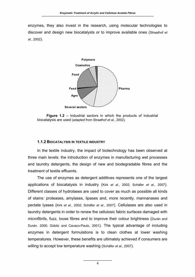
Figure 1.2 – Industrial sectors in which the products of industrial biocatalysis
are used (adapted from Straathof et al., 2002) ........................................ 4
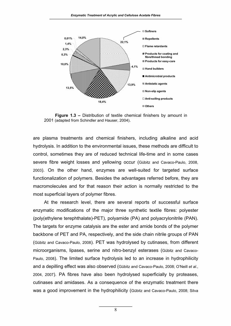
Figure 1.3 – Distribution of textile chemical finishers by amount in 2001
(adapted from Schindler and Hauser, 2004) ............................................ 8
Figure 1.4 – World acrylic production between 1990 and 2005 (from
www.yarnsandfibers .com/ir/report/acrylic_chain_report2006.html) ....... 10
Figure 1.5 – The most important world acrylic fibre producers in 2006 (from
www.yarnsandfibers.com/revamp_ir/report_fullstory.php3?id=401&p_typ
e=62&source_id=15&source=YarnsandFibers%20Paid&story_type=F&B
F=Special&reportshow=First)... ............................................................. 10
Figure 1.6 – The acrylic polymer structure and some examples of its common
comonomers .......................................................................................... 11
Figure 1.7 – Conventional manufacture process of acrylic fibres (adapted from
Frushour and Knorr, 1998) ..................................................................... 13
Figure 1.8 – A) Model of the oriented acrylic fibres morphology with emphasis
for the assumed irregular helical conformation of PAN polymer chain
(adapted from Frushour, 1995). B) Pictorial representation of hexagonally
packed chains of PAN (adapted from Bashir and Rastogi, 2005) .......... 15
Figure 1.9 – Possible chemical transformations on PAN surface catalysed by
nitrile metabolizing enzymes .................................................................. 20
Figure 1.10 – Chemical structure of the anhydroglucose repeating unit of
cellulose (from La Nieve, 2007) ............................................................. 21
Figure 1.11 – World production of cellulose diacetate (CDA) and triacetate
(CTA) fibres compared with the cellulose diacetate tow and total man-
made fibres (adapted from La Nieve, 2007) ........................................... 22
Enzymatic Treatment of Acrylic and Cellulose Acetate Fibres
Figure 1.12 – Schematic representation of the manufacturing line of cellulose
acetate fibres (from Collier and Tortora, 2001) ...................................... 23
Figure 1.13 – Structural models of CTA I and CTA II in two projections: A) CTA
I perpendicular to the ac plane, B) CTA I along the c axis (the chain axis),
C) CTA II perpendicular to the ac plane, D) CTA II along the c axis, E) unit cell dimensions of CTA I and F) unit cell dimensions of CTA II
(adapted from Zugenmaier, 2004) ......................................................... 26
Figure 1.14 – Main reactions occurring during biodegradation of cellulose
acetate ................................................................................................... 30
Figure 1.15 – Mechanism of the nitrile hydrolysis catalysed by nitrilase
(adapted from Banerjee et al., 2002) ..................................................... 33
Figure 1.16 – Schematic ribbon diagrams depicting the overall structure of N-
carbamyl-D-amino acid amidohydrolase from Agrobacterium sp. (Protein
Data Bank ID: 1erz). A) View of the dimer where each polypeptide has a
different colour and a labelled N- and C-terminal. B) View of one subunit
where the catalytic triad, composed by a cysteine, lysine and glutamic
acid, is shown as spheres ...................................................................... 33
Figure 1.17 – Mechanism of the carboxylic ester bond hydrolysis catalysed by
cutinase (adapted from Lau and Bruce, 1999) ....................................... 36
Figure 1.18 – A) Schematic ribbon diagrams depicting the overall structure of
cutinase from Fusarium solani pisi (Protein Data Bank ID: 1cex); the
secondary structural elements are represented in different colours (β-
strands are in yellow, α-helixes are in red and connecting loops are in
green; the N- and C-terminal are labelled; the two disulfide bridges and
catalytic residues are depicted in blue by the stick model. B) Closed view
of the catalytic triad composed by a serine, histidine and aspartic acid,
shown by the stick model; the two possible locations of the Ser120 side
chain are both represented .................................................................... 39
Figure 1.19 – Schematic representation of a cellulase based on the
cellobiohydrolase I from Trichoderma reesei (adapted from Hildén and
Johansson, 2004) .................................................................................. 41
xv

Enzymatic Treatment of Acrylic and Cellulose Acetate Fibres
Figure 1.20 – A) Primary sequence and secondary structures of the CBM of
CBH I from Trichoderma reesei; the two disulfide bridges are represented
by the sticks connecting the cysteine residues; the tyrosines involved in
the interaction with the ligand are in red. B) Ribbon schematic
representation of the overall 3D-structure (Protein Data Bank ID: 2cbh);
the two disulfide bridges and ligand-binding tyrosines are depicted by the
stick model. C) Binding of the flat surface of CBM to an oligosaccharide
of 6 glucose units (from Mattinen et al., 1997b) ..................................... 44
Figure 1.21 – A) Primary sequence and secondary structures of the CBMN1 of
CenC from Cellulomonas fimi; the disulfide bridge is represented by the
line connecting the cysteine residues; the tyrosines involved in the
interaction with the ligand are in red. B) Ribbon schematic representation
of the overall 3D-structure showing a ligand with 5 glucose units (Protein
Data Bank ID: 1gu3); the N- and C-terminal are labelled. C) Close up
view of the binding-site where the tyrosines involved are represented by
the stick model ....................................................................................... 46
Figure 2.1 – Total protein and nitrogen concentrations in the treatment
solutions. The acrylic samples were treated with 412 U of Cyanovacta
Lyase per gram of fabric, at pH 7.8 and 40 ºC ....................................... 58
Figure 2.2 – Relative K/S values for acrylic fabric treated with 412U of
Cyanovacta Lyase, at pH 7.8 and 40 ºC. Samples and controls were
competitively coloured at 70, 75, 80 and 90 ºC. Relative K/S was
calculated as control
enzyme
K/SK/S (%) .............................................................. 59
Figure 2.3 – Relative K/S for acrylic fabric coloured at 70 ºC and polyacrylic
acid concentration in the treatment solutions. The acrylic fibre was
treated with 412 U of Cyanovacta Lyase per gram of fabric, at pH 7.8 and
40 ºC ...................................................................................................... 60
Figure 2.4 – Surface erosion model for acrylic fibres treated with nitrilase at
alkaline pH (for simplification, the comonomer vinyl acetate is not
represented) .......................................................................................... 62
xvi
Enzymatic Treatment of Acrylic and Cellulose Acetate Fibres
Figure 3.1 – Total protein concentration in bath treatment solutions during the 3
hours incubation of acrylic with 9 U mL-1 of cutinase and different
amounts of DMA, (A) in absence and (B) in presence of stainless steel
discs ...................................................................................................... 72
Figure 3.2 – Percentage of increase in K/S for acrylic samples treated (A) with
cutinase (573 U per gram of fabric) and (B) with a commercial esterase
(223 U per gram of fabric). To increase the mechanical agitation,
stainless steel discs were added to all the assays plotted. Samples were
competitively coloured at 70 ºC and 80 ºC, and the relative increase in
K/S was calculated as control
controlenzyme
SKSKSK
/)//( − ..................................... 74
Figure 3.3 – X-ray diffraction pattern of control sample for the treatment
performed in the absence of additives and stainless steal discs. The XRD
data was analyzed by profile fitting of the scans with the Pearson VII
function, using the software WinFit! beta release 1.2.1, 1997. ............... 76
Figure 3.4 – SEM photographs of acrylic fabric (A) control and (B) cutinase
treated sample without stainless steel discs, and (C) control and (D)
cutinase treated sample treated with discs and 1% DMA. All images were
acquired under the same conditions with a total magnification of 400x, in
a scanning electronic microscope Leica S360 ....................................... 78
Figure 4.1 – Effect of cutinase concentration on the acetic acid release. The
CDA and CTA fabrics (2% w/v) were treated during 8 hours, at pH 8 and
30 ºC, with several concentrations of cutinase expressed as esterase
activity in U mL-1 .................................................................................... 85
Figure 4.2 – Progress curves for the formation of hydroxyl groups at the fibres
surface, measured as relative increase in K/S values, and total protein
adsorption for (A) CDA and (B) CTA. All the samples were treated with
cutinase (5000 U per gram of fabric) at pH 8 and 30 ºC. Samples and
controls were competitively coloured at 60 ºC. The relative increase in
K/S was calculated as control
controlenzyme
SKSKSK
/)//( − (%) and the relative
protein adsorption as h
th
PPP
0
0 )( − (%), where P is the total protein in
solution .................................................................................................. 87
xvii

Enzymatic Treatment of Acrylic and Cellulose Acetate Fibres
Figure 4.3 – DRIFT spectra showing the carbonyl group stretching band of (A)
CDA and (B) CDT controls and samples. The samples were treated with
cutinase (1250 U per gram of fabric), at pH 8 and 30 ºC, for 24 hours. . 89
Figure 4.4 – Epifluorescent photographs of cross-sections from (A) CDA and
(B) CTA samples. The samples were treated with 10 mg of FITC-
conjugated cutinase per gram of fabric, at pH 8 and 30 ºC, for 15 hours.
Both images were acquired with a total magnification of 1000x ............. 90
Figure 4.5 – SEM photographs of CDA (A) control and (B) treated sample, and
CTA (C) control and (D) treated sample. The samples were previously
treated with cutinase (5000 U per gram of fabric), at pH 8 and 30 ºC, for
18 hours. All images were acquired under the same conditions with a
total magnification of 3000x, in a scanning electronic microscope Leica
S360 ...................................................................................................... 91
Figure 4.6 – X-ray diffraction pattern obtained for (A) CDA sample and (B) CTA
sample, both treated with cutinase (1250 U per gram of fabric), at pH 8
and 30 ºC, for 24 hours. The XRD data was analyzed by profile fitting of
the scans with the Pseudo-Voigt peak function from the software
OriginPro 7.5 (Cerqueira et al., 2006) .................................................... 92
Figure 5.1 – Schematic representation of the recombinant wild-type cutinase from F. solani pisi (Araújo et al., 2007) and its new chimeric proteins with
the fungal carbohydrate-binding module of CBH I, from T. reesei, and the
bacterial carbohydrate-binding module N1 of CenC, from C. fimi. The
primary sequences of the linkers used are specified in the figure .......... 98
Figure 5.2 – Protein adsorption and relative increase in K/S values for the (A)
CDA and (B) CTA treated with cutinase and cutinase fused to CBMs
(initial concentration is expressed in units per gram of fabric). The
samples were incubated during 18 hours with cutinase, cutinase-CBMN1
(cut-N1), cutinase-PTboxCBMN1 (cut-PT-N1), cutinase-wtCBMT.reesei (cut-
wtCBM), and cutinase-sCBMT.reesei (cut-sCBM), at pH 8 and 30 ºC.
Samples and control were competitively coloured at 60 ºC. Relative
xviii
Enzymatic Treatment of Acrylic and Cellulose Acetate Fibres
protein adsorption was calculated as cutinasehcutinaseh
hh
PPPP
180
180
−− and relative K/S as
controlcutinase
controlcutinase chimeric
SKSKSKSK
////
−− . ............................................................................ 101
xix
Enzymatic Treatment of Acrylic and Cellulose Acetate Fibres
xx
LLIISSTT OOFF TTAABBLLEESS
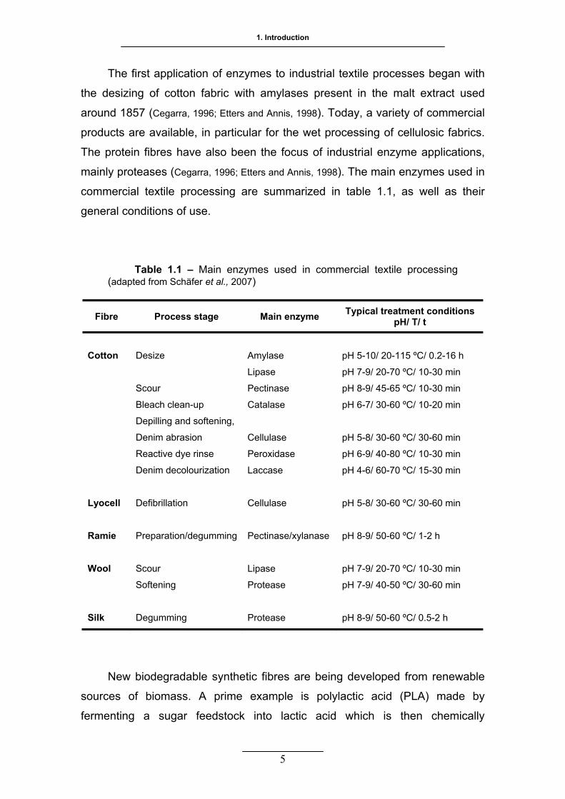
Table 1.1 – Main enzymes used in commercial textile processing (adapted from
Schäfer et al., 2007) ................................................................................ 5
Table 2.1 –Conditions for the colouration of acrylic fabrics with Basic Blue 9,
C.I. 52015, according to each enzymatic treatment performed .............. 53
Table 2.2 – Influence of organic solvents and polyalcohols on initial activity and
half-life time of Nitrilase, at 30 ºC and pH 7.8. An initial activity and a half-
life time of 100% correspond to 12.6 U mg-1 and 15 hours, respectively.
The half-life time was calculated as kt 2ln2
1 = , from the 1st order
exponential decay fit of data ( ) ................................................ 55 kxtt eaa −== .0
Table 2.3 – Effect of the additives on the enzymatic modification of acrylic
fabric. The acrylic samples were treated with 24 U of nitrilase per gram of
fabric, at pH 7.8 and 30 ºC, for 116 hours .............................................. 57
Table 3.1 – Influence of two organic solvents and several polyalcohols on
operational stability of cutinase. The cutinase was incubated at 35 ºC,
pH 8, in a water bath with agitation, under different media compositions.
The half-life time was calculated as kt 2ln2
1 = , in the cases of a 1st order
exponential decay of activity ( ) ............................................... 70 kxtt eaa −== .0
Table 3.2 – Crystallinity degrees (CD) for acrylic fabric samples treated for
three hours with 573 U of cutinase per gram of fabric, at pH 8 and 30 ºC.
The values were obtained according to the equation ( )%)010(
)300()010(
IIICD −
=
where I(010) and I(300) are the intensities of the peaks at 2θ 16.8º and
29.4º, respectively .................................................................................. 77
Table 4.1 – Hydrolysis of CDA and CTA fabrics by cutinase. The samples were
treated with 1250 U per gram of fabric, at pH 8 and 30 ºC, for 24 hours.
Samples and controls were competitively coloured at 50 ºC ................. 88
Enzymatic Treatment of Acrylic and Cellulose Acetate Fibres
xxi
Table 4.2 – Crystallinity indexes for CDA and CTA. The samples were treated
with cutinase (1250 U per gram of fabric), at pH 8 and 30 ºC, for 24
hours. The crystallinity index (IC) was determined according to the
equation )( ACCC AAAI += , where Ac is the total area of the crystalline
peaks and AA is the total area of the amorphous peaks ........................ 92
1
IINNTTRROODDUUCCTTIIOONN
The present thesis, entitled Enzymatic Treatment of Acrylic and
Cellulose Acetate fibres, connects topics from different areas of
science that will be the subject of a brief bibliographic revision
throughout this chapter.
The first topic to be addressed is biocatalysis which is the background
theme of this thesis and its importance will be emphasized in the
context of the textile industry and surface modification of synthetic
polymers. A general description of the two fibres used in this work,
acrylic and cellulose acetate, will be given as well as their most
common end-uses and reported biomodifications. The properties and
general applications of the two main enzymes used to modify the fibres
will be referred next. Finally, a summary of the methods used to
manipulate enzymes is made before a concise description of the two
carbohydrate-binding modules chosen to be fused with the C-terminal
of cutinase.
11

Enzymatic Treatment of Acrylic and Cellulose Acetate Fibres
2
11.. IINNTTRROODDUUCCTTIIOONN
11..11 BBIIOOCCAATTAALLYYSSIISS
1.1.1 INDUSTRIAL ENZYMES
Biocatalysis is present in some of the oldest transformations known to
humans: descriptions of various beer recipes were found in Sumerian writings
(Ball, 2001). More recently, in the beginning of the 19th century, acetic acid was
produced industrially from ethanol by an Acetobacter strain (Wandrey et al., 2000).
Studies of fermentation processes led to a big bang in the knowledge of life’s
chemistry. In the 19th century, Louis Pasteur came to the conclusion that the
fermentation of sugar to alcohol by yeast was catalyzed by a vital force
contained within the yeast cells called “ferments”, which were thought to
function only within living organisms. Wilhelm Kühne was the first to use the
term “enzyme” and, years later, Emil Fisher proposed the “Lock and Key Model”
to visualize the substrate and enzyme interaction (Cabral et al., 2003).
Enzymes are the subgroup of proteins that catalyse the chemistry of life,
transforming both macromolecular and small molecules; they are the focus of
present biocatalysis research (Walsh, 2001). The chiral nature of enzymes results
in a remarkable chemical precision seen as different types of selectivity:
substrate, stereo-, regio- and functional group selectivity (Rozzell, 1999). Most
enzymes operate at room temperature, under neutral aqueous conditions and in
the absence of functional group-protection (Koeller and Wong, 2001). Enzymes are
environmental friendly catalysts not just because they are biodegradable
themselves but also because of their mild operating conditions. They can result
in processes that generate fewer waste disposal problems and that require
lower energy input, leading to lower costs and lower emissions of greenhouse
gases to the environment (Rozzell, 1999). These qualities make enzymes
remarkable catalysts.
Nowadays, it is widely recognized that enzyme-catalysed chemical
transformations are convenient alternatives to traditional (non-biological)

1. Introduction
3
transformations and suitable solutions to difficult synthetic problems (Koeller and
Wong, 2001). The enzyme can be used as the sole catalyst in a reaction, in
combination with other enzymes, or with inorganic reagents. Besides, many
enzymes accept unnatural substrates, and genetic, pathway and medium
engineering can improve further their stability and specific activity as well as
modulate their substrate specificity (Koeller and Wong, 2001). Biocatalysis is
accomplished by either using isolated enzymes or using whole cells which are
more common in synthesis reactions that require cofactors (Schmid et al., 2001).
The majority of currently used industrial enzymes are hydrolases (figure 1.1)
from which proteases remain the dominant enzyme type, followed by amylases
and cellulases (Kirk et al., 2002).
Biocatalysis is a tool of increasing importance for industries which aim a
sustainable development. Successful industrial applications of enzymes are
growing rapidly over the past decade (Straathof et al., 2002). The major traditional
consumers of enzymes are the food, feed, agriculture, paper, leather and textile
industries (van Beilen and Li, 2002; Schäfer et al., 2007). However, the largest growth
seen, in terms of number of occurrences (figure 1.2), is in the application of
enzymes to the industrial chemical synthesis, especially in the pharma and agro
sectors (Straathof et al., 2002; Schmid et al., 2001). Another sector that is becoming
important includes the companies that provide enzymes. Besides producing the
Hydrolases
LyasesIsomerases
Transferases
Oxido-reductases
Oxidizing cells
Reducing cells
Figure 1.1 – Biocatalysts used in industrial transformations (adapted from Straathof et al., 2002).
Enzymatic Treatment of Acrylic and Cellulose Acetate Fibres
4
enzymes, they also invest in the research, using molecular technologies to
discover and design new biocatalysts or to improve available ones (Straathof et
al., 2002).
1.1.2 BIOCATALYSIS IN TEXTILE INDUSTRY
In the textile industry, the impact of biotechnology has been observed at
three main levels: the introduction of enzymes in manufacturing wet processes
and laundry detergents, the design of new and biodegradable fibres and the
treatment of textile effluents.
The use of enzymes as detergent additives represents one of the largest
applications of biocatalysis in industry (Kirk et al., 2002; Schäfer et al., 2007).
Different classes of hydrolases are used to cover as much as possible all kinds
of stains: proteases, amylases, lipases and, more recently, mannanases and
pectate lyases (Kirk et al., 2002; Schäfer et al., 2007). Cellulases are also used in
laundry detergents in order to renew the cellulosic fabric surfaces damaged with
microfibrils, fuzz, loose fibres and to improve their colour brightness (Durán and
Durán, 2000; Gübitz and Cavaco-Paulo, 2001). The typical advantage of including
enzymes in detergent formulations is to clean clothes at lower washing
temperatures. However, these benefits are ultimately achieved if consumers are
willing to accept low temperature washing (Schäfer et al., 2007).
Pharma
Several sectors
Agro
Feed
Food
Cosmetics
Polymers
Figure 1.2 – Industrial sectors in which the products of industrial biocatalysis are used (adapted from Straathof et al., 2002).
1. Introduction
The first application of enzymes to industrial textile processes began with
the desizing of cotton fabric with amylases present in the malt extract used
around 1857 (Cegarra, 1996; Etters and Annis, 1998). Today, a variety of commercial
products are available, in particular for the wet processing of cellulosic fabrics.
The protein fibres have also been the focus of industrial enzyme applications,
mainly proteases (Cegarra, 1996; Etters and Annis, 1998). The main enzymes used in
commercial textile processing are summarized in table 1.1, as well as their
general conditions of use.
Table 1.1 – Main enzymes used in commercial textile processing
(adapted from Schäfer et al., 2007)
Fibre Process stage Main enzyme Typical treatment conditions pH/ T/ t
Cotton Lyocell Ramie Wool Silk
Desize
Scour
Bleach clean-up
Depilling and softening,
Denim abrasion
Reactive dye rinse
Denim decolourization
Defibrillation
Preparation/degumming
Scour
Softening
Degumming
Amylase
Lipase
Pectinase
Catalase
Cellulase
Peroxidase
Laccase
Cellulase
Pectinase/xylanase
Lipase
Protease
Protease
pH 5-10/ 20-115 ºC/ 0.2-16 h
pH 7-9/ 20-70 ºC/ 10-30 min
pH 8-9/ 45-65 ºC/ 10-30 min
pH 6-7/ 30-60 ºC/ 10-20 min
pH 5-8/ 30-60 ºC/ 30-60 min
pH 6-9/ 40-80 ºC/ 10-30 min
pH 4-6/ 60-70 ºC/ 15-30 min
pH 5-8/ 30-60 ºC/ 30-60 min
pH 8-9/ 50-60 ºC/ 1-2 h
pH 7-9/ 20-70 ºC/ 10-30 min
pH 7-9/ 40-50 ºC/ 30-60 min
pH 8-9/ 50-60 ºC/ 0.5-2 h
New biodegradable synthetic fibres are being developed from renewable
sources of biomass. A prime example is polylactic acid (PLA) made by
fermenting a sugar feedstock into lactic acid which is then chemically
5
Enzymatic Treatment of Acrylic and Cellulose Acetate Fibres
transformed into a polymer fibre. PLA based materials have properties similar to
other synthetic fibres; they are durable, with a silky feel and may be blended
with wool or cotton. PLA was launched in 2003 as the first man-made fibre
derived from renewable resources under the commercial name Ingeo (Burke,
2008). Another commercial fibre named Sorona, made by DuPont, results from
the polymerization of 1,3-propanediol, derived from a fermenting process, and a
petrochemical-based monomer (Burke, 2008). New fibres are also being derived
from natural materials like chitin, collagen and alginate. These materials are
used for medical applications in wound dressings and investigated for drug-
releasing systems (Lu and Chen, 2004; Qin, 2008; Rinaudo, 2008).
The textile industry is under substantial environmental pressure.
Considering the volume discharged and effluent composition, the wastewater
generated by this industry is classified as one of the most polluting among all
industrial sectors (Vandevivere et al., 1998). Biocatalysis can be applied as a tool
for the textile effluents treatment. Possible bioremediation processes can be
divided in whole cell microbial systems and isolated enzymes (Gübitz and Cavaco-
Paulo, 2001; Ramalho, 2005; Soares, 2000). Biodegradation through activated sludge
is widely used in treatment plants but is still ineffective is decolorizing textile
effluents (Vandevivere et al., 1998). It is necessary to integrate other chemical and
physical technologies in order to increase the efficient treatment and reuse of
this kind of effluents. Several laboratory-scale investigations have illustrated the
potential of sequential anaerobic/aerobic biotreatment steps but a current use is
still missing (Vandevivere et al., 1998).
Although the widespread academic efforts in the textile biotechnology, the
actual application of such work is modest. Hopefully in a near future this reality
will be changed by both the academic and industrial communities.
1.1.3 BIOCATALYSIS IN SYNTHETIC POLYMER SURFACE MODIFICATION
In the preparation of wide-ranging synthetic polymer materials, sometimes
it is desirable for the properties at the surface to be different from the bulk
properties. Surfaces that promote cell adhesion, biocompatibility, hydrophobic/
hydrophilic character or chemical resistance are some examples of the
properties that can be changed (Hutchings et al., 2008).
6
1. Introduction
One important field where surface modification of polymers has a great
impact is the materials science. The biocompatibility can be accomplished by
immobilizing certain bioactive molecules on functionalized surfaces of the
materials to be used. The superficial immobilization of molecules is also
important for the manufacturing of specific analytical assays like microarrays
and biosensors. The immobilization of industrial enzymes into the surface of
solid supports is also a widespread process (Goddard and Hotchkiss, 2007).
Textiles are one of the major and oldest subjects for surface modifications.
Textile finishing is the final stage in the fabric manufacturing process and
includes all the processes that modify the surface of fibres to add useful
qualities to the fabric, ranging from interesting appearance and fashion aspects
to high performance properties for industrial needs (Schindler and Hauser, 2004).
The methods to accomplish distinct surface properties may involve the
application of a coating layer or chemical modification of the surface (Hutchings et
al., 2008). The modification of the surface can be performed by ionized gas
treatments, like plasma and Corona discharge, and UV irradiation (Goddard and
Hotchkiss, 2007). The wet chemical modification is the most classical and easy
approach for the functionalization of polymer surfaces, offering advantages for
porous materials, besides, it does not require very specialized equipment
(Goddard and Hotchkiss, 2007). In the textile industry both chemical and physical
methods are used, but chemical finishing has always been an important
component of textile processing and its importance is growing in recent years
with the trend to ‘high tech’ products (Schindler and Hauser, 2004). As the use of
high performance textiles grows, the need for chemical finishes to provide the
fabric properties required in these special applications has grown accordingly.
Nowadays, more than 20 different types of chemical finishers exist for both
natural or man-made fibres (figure 1.3) (Schindler and Hauser, 2004).
Textile materials made from synthetic fibres are, in general, uncomfortable
to wear because they are hydrophobic. This means that these materials can not
absorb the perspiration and the water vapour can not easily be transported
away from the body. The hydrophobic nature also leads to their characteristic
static cling and stain retention during laundering (Gübitz and Cavaco-Paulo, 2008).
There are several methods for the surface modification of synthetic polymers
with the purpose of increasing the hydrophilicity. The methods currently used
7
Enzymatic Treatment of Acrylic and Cellulose Acetate Fibres
8
are plasma treatments and chemical finishers, including alkaline and acid
hydrolysis. In addition to the environmental issues, these methods are difficult to
control, sometimes they are of reduced technical life-time and in some cases
severe fibre weight losses and yellowing occur (Gübitz and Cavaco-Paulo, 2008,
2003). On the other hand, enzymes are well-suited for targeted surface
functionalization of polymers. Besides the advantages referred before, they are
macromolecules and for that reason their action is normally restricted to the
most superficial layers of polymer fibres.
At the research level, there are several reports of successful surface
enzymatic modifications of the major three synthetic textile fibres: polyester
(poly(ethylene terephthalate)-PET), polyamide (PA) and polyacrylonitrile (PAN).
The targets for enzyme catalysis are the ester and amide bonds of the polymer
backbone of PET and PA, respectively, and the side chain nitrile groups of PAN
(Gübitz and Cavaco-Paulo, 2008). PET was hydrolysed by cutinases, from different
microorganisms, lipases, serine and nitro-benzyl esterases (Gübitz and Cavaco-
Paulo, 2008). The limited surface hydrolysis led to an increase in hydrophilicity
and a depilling effect was also observed (Gübitz and Cavaco-Paulo, 2008; O’Neill et al.,
2004, 2007). PA fibres have also been hydrolysed superficially by proteases,
cutinases and amidases. As a consequence of the enzymatic treatment there
was a good improvement in the hydrophilicity (Gübitz and Cavaco-Paulo, 2008; Silva
0,01%
13,5%
18,4%
13,9%
4,1%
22,1%14,0%
1,4%
2,3%
0,3%
10,0%
Softners
Repellents
Flame retardants
Products for coating andfibre/thread bondingProducts for easy-care
Hand builders
Antimicrobial products
Antistatic agents
Non-slip agents
Anti-soiling products
Others
Figure 1.3 – Distribution of textile chemical finishers by amount in 2001 (adapted from Schindler and Hauser, 2004).

1. Introduction
9
et al., 2005b, 2007). The enzymatic modification of PAN with nitrilases and nitrile
hydratases led to the formation of more hydrophilic carboxylic and amide
groups (section 1.2.5) (Gübitz and Cavaco-Paulo, 2008).
The broad substrate specificity and catalytic promiscuity, exhibited by
some enzymes, uncover a new range of possible biocatalytic transformations
(Bornscheuer and Kazlauskas, 2004). The ability of enzymes to use textile synthetic
fibres as substrates under mild conditions, which are known for their stability
and chemical inertia, is an evidence of the vast potential of these catalysts in
industrial processes.
11..22 AACCRRYYLL IICC FF IIBBRREE
1.2.1 GENERAL DESCRIPTION
Acrylic fibres are defined, according to the Federal Trade Commission of
United States, as manufactured fibres in which the fibre forming substance is
any long-chain synthetic polymer composed of at least 85% by weight of
acrylonitrile units (Guillen, 1987).
Although the acrylonitrile was synthesised by Moreau in 1893, only in the
1940s, suitable solvents were found that allowed the polyacrylonitrile
processing, for the reason that its polymer decomposes prior to melting (Frushour
and Knorr, 1998). The first appearance of acrylic fibres was in the year 1950,
when DuPont introduced them in the market under the commercial name
Orlon®. During the 1950s, the acrylic industry experienced an impressive growth
with at least 18 companies producing acrylic goods (Frushour and Knorr, 1998).
During the year 2005, global production of acrylic staple fibre reached
2 791 thousand tons and acrylic accounted for 8% of all chemical fibre
produced in the world (figure 1.4). The share has fallen dramatically from 15%
in early 1980s. During the period 2000-2005, the acrylic staple fibre production
increased at a rate of 1.25% per annum which was the slowest growth rate
among all chemical fibres production (www.yarnsandfibers.com/ir/report/acrylic_chain_
report2006.html). In the year of 2006, the acrylic fibre production decreased to
6.8% of the total chemical fibres. In terms of the contribution, Asia holds a major
share of 59.5% in global production, where China has emerged as the leading
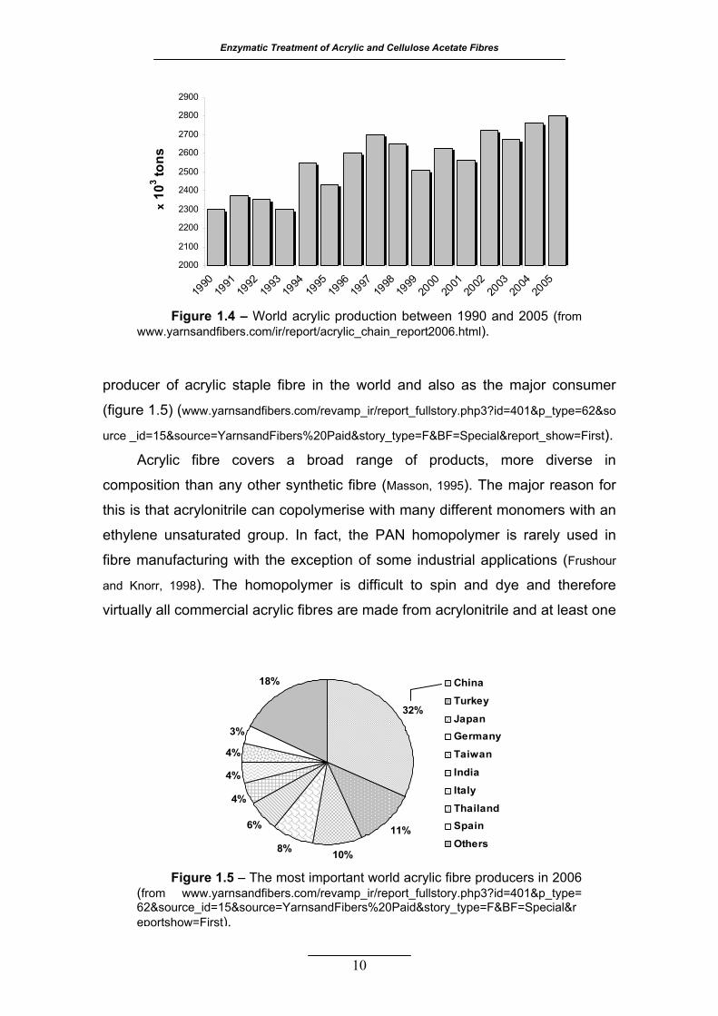
Enzymatic Treatment of Acrylic and Cellulose Acetate Fibres
10
producer of acrylic staple fibre in the world and also as the major consumer
(figure 1.5) (www.yarnsandfibers.com/revamp_ir/report_fullstory.php3?id=401&p_type=62&so
urce _id=15&source=YarnsandFibers%20Paid&story_type=F&BF=Special&report_show=First).
Acrylic fibre covers a broad range of products, more diverse in
composition than any other synthetic fibre (Masson, 1995). The major reason for
this is that acrylonitrile can copolymerise with many different monomers with an
ethylene unsaturated group. In fact, the PAN homopolymer is rarely used in
fibre manufacturing with the exception of some industrial applications (Frushour
and Knorr, 1998). The homopolymer is difficult to spin and dye and therefore
virtually all commercial acrylic fibres are made from acrylonitrile and at least one
Figure 1.4 – World acrylic production between 1990 and 2005 (from www.yarnsandfibers.com/ir/report/acrylic_chain_report2006.html).
2000
2100
2200
2300
2400
2500
2600
2700
2800
2900
1990
1991
1992
1993
1994
1995
1996
1997
1998
1999
2000
2001
2002
2003
2004
2005
x 10
3 tons
32%
11%
10%8%
6%
4%
4%
4%
3%
18% China
Turkey
JapanGermany
Taiwan
India
Italy
ThailandSpain
Others
Figure 1.5 – The most important world acrylic fibre producers in 2006 (from www.yarnsandfibers.com/revamp_ir/report_fullstory.php3?id=401&p_type= 62&source_id=15&source=YarnsandFibers%20Paid&story_type=F&BF=Special&reportshow=First).
1. Introduction
11
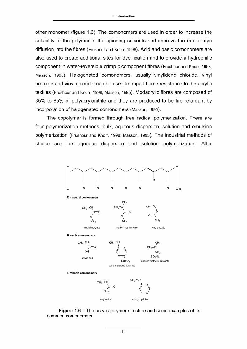
other monomer (figure 1.6). The comonomers are used in order to increase the
solubility of the polymer in the spinning solvents and improve the rate of dye
diffusion into the fibres (Frushour and Knorr, 1998). Acid and basic comonomers are
also used to create additional sites for dye fixation and to provide a hydrophilic
component in water-reversible crimp bicomponent fibres (Frushour and Knorr, 1998;
Masson, 1995). Halogenated comonomers, usually vinylidene chloride, vinyl
bromide and vinyl chloride, can be used to impart flame resistance to the acrylic
textiles (Frushour and Knorr, 1998; Masson, 1995). Modacrylic fibres are composed of
35% to 85% of polyacrylonitrile and they are produced to be fire retardant by
incorporation of halogenated comonomers (Masson, 1995).
The copolymer is formed through free radical polymerization. There are
four polymerization methods: bulk, aqueous dispersion, solution and emulsion
polymerization (Frushour and Knorr, 1998; Masson, 1995). The industrial methods of
choice are the aqueous dispersion and solution polymerization. After
R
N N N N N N n
R = neutral comonomers
CH2 CH
C O
O
CH3
CH2 C
C O
O
CH3
CH3
CH CH
O
C
CH3
O
methyl acrylate methyl methacrylate vinyl acetate
R = acid comonomers
R = basic comonomers
CH2 CH
C O
OH
acrylic acid
CH2 CH
sodium styrene sufonate
CH2 C
CH2
SO3Na
CH3
sodium methallyl sufonate
CH2 CH
C O
NH2
CH2 CH
N
acrylamide 4-vinyl pyridine
NaSO3
Figure 1.6 – The acrylic polymer structure and some examples of its common comonomers.

Enzymatic Treatment of Acrylic and Cellulose Acetate Fibres
precipitation the copolymer is dried and dissolved in an appropriate organic
solvent, mainly dimethylformamide (DMF) or dimethylacetamide (DMA), and
wet or dry spun (Capone, 1995; von Falkai, 1995).
After the spinning stage, the acrylic fibre properties are unsuitable for their
end-use; the fibres contain residual amounts of the organic solvent, the tenacity
and elastic modulus are low while the plastic elongation is high, the fibre lacks
the crimp needed to provide cohesion and bulk to the yarn (von Falkai, 1995).
Thus, the fibre is subjected to further treatments in order to develop the
desirable balance of processing and performance properties (Frushour and Knorr,
1998). During this pos-spinning stage, the fibre undergoes a series of steps
along a production line. The essential steps of washing, orientation drawing,
drying and relaxation are common to all acrylic fibre spinning processes, but the
sequences as well as a number of possible variations in the manufacture of
acrylic fibres are protected and the details are not available (Frushour and Knorr,
1998). The improvement of the acrylic fibre properties by manipulation of the
conditions of spinning and pos-spinning processes is an important issue and it
is the subject of several research groups (Bahrami et al., 2003; Wu et al., 2003; Chen
and Harrison, 2002). The figure 1.7 is a schematic representation that illustrates a
production line for acrylic fibres in the tow and staple forms.
Wet spinning Dry spinning
Drawing Washing
Finish application
Drying
Crimping
Relaxation
Figure 1.7 – Conventional manufacture process of acrylic fibres (adapted from Frushour and Knorr, 1998).
TowCutting Staple
12
1. Introduction
The nature of the comonomer in acrylic fibres affect the overall dyeability
and the classes of dyes that may be used (Needles, 1986). The cationic or basic
dyes are the most used in acrylics given the good fastness properties obtained.
The fixation occurs with the salt bridge between dye cations and anionic sites
(mainly sulphate from the initiator radicals). Acrylic can be dyed using disperse
dyes, in particular the smaller ones, for pastel and light shades where dyeing
uniformity may be hard to obtain with other dyes (Emsermann and Foppe, 1995).
Acrylics can be dyed by either batch or continuous processes, in the fibre, yarn,
fabric or garment form (Emsermann and Foppe, 1995).
1.2.2 STRUCTURAL CHARACTERIZATION
Textile fabrics are planar structures produced by interlacing or entangling
yarns or fibres in a particular manner (Needles, 1986). Textile yarns are made up
of fibres and each individual fibre is, in turn, made up from millions of individual
long molecular chains of discrete chemical composition (Needles, 1986). The
molecular structure of the long polymer chains determines the basic chemical
and physical properties of the fibre. Although special treatments and changes in
yarn and fabric production parameters can alter the fabric properties to some
degree, the basic properties are inherent to the structure of the polymer from
which the fibre is produced (Needles, 1986).
Most textile fibres have a morphology that can be described by the two-
phase model for semicrystalline polymers (Frushour, 1995). According to this
model, discrete crystalline domains of several hundred angstroms (Å) are mixed
with amorphous domains of similar size. The individual polymer chains have
lengths in the order of 1000 to 2000 Å, so a single chain can span two or more
crystalline domains and its assembly in the intercrystalline regions forms the
amorphous domains (Frushour, 1995). When a synthetic fibre is drawn, the
molecules, in most cases, orientate themselves in crystalline domains parallel to
the fibre axis. The degree of crystallinity is dependent on the total forces
available for chain interaction and the stereo-regularity of adjacent chains
(Needles, 1986; Stevens, 1990).
Commercial vinyl polymers are in general atactic (with no stereo-
regularity) and form amorphous glasses that have no long-range crystalline
13
Enzymatic Treatment of Acrylic and Cellulose Acetate Fibres
order (Frushour, 1995). Fibres can be melt-spun from these glassy polymers but
they can not be used for textile applications because of the lack of crystallinity
(Frushour, 1995). Crystallinity is a very important characteristic because it confers
good tensile properties (Frushour, 1995). PAN is atactic, but the fact that
functional synthetic fibres can be made from it suggests that some degree of
order must be present in PAN fibres. The other major factor that contributes to
the formation of a crystalline phase is the ability of interchain bonding. In fact,
some atactic polymers like PAN possess an unexpected high degree of order or
pseudocrystallinity due to a substitutent group capable of strong interactions
(Frushour, 1995).
The pendent group in PAN molecules is the nitrile group. The
distinguishing feature of the nitrile group is the large dipole moment, turning it
one of the most polar organic functional groups (Frushour, 1995). The interaction
between two nitrile groups can be either attractive or repulsive, depending upon
the spatial orientation of the nitriles, while the magnitude depends upon the
distance of separation (Frushour, 1995). In the PAN isolated chain, the potential
energy will be minimized by placing the adjacent nitrile groups as far as
possible, since they have parallel orientation between each other and this will
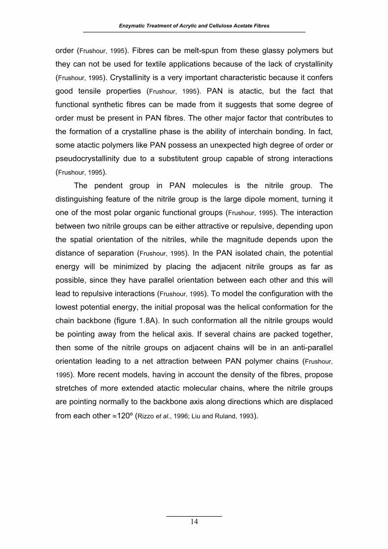
lead to repulsive interactions (Frushour, 1995). To model the configuration with the
lowest potential energy, the initial proposal was the helical conformation for the
chain backbone (figure 1.8A). In such conformation all the nitrile groups would
be pointing away from the helical axis. If several chains are packed together,
then some of the nitrile groups on adjacent chains will be in an anti-parallel
orientation leading to a net attraction between PAN polymer chains (Frushour,
1995). More recent models, having in account the density of the fibres, propose
stretches of more extended atactic molecular chains, where the nitrile groups
are pointing normally to the backbone axis along directions which are displaced
from each other ≈120º (Rizzo et al., 1996; Liu and Ruland, 1993).
14

1. Introduction
BA
Figure 1.8 – A) Model of the oriented acrylic fibres morphology with emphasis for the assumed irregular helical conformation of PAN polymer chain (adapted from Frushour, 1995). B) Pictorial representation of hexagonally packed chains of PAN (adapted from Bashir and Rastogi, 2005).
Bohn et al. (1961) reported the first detailed model of the PAN fibre
morphology using wide angle X-ray scattering (WAXS) and by studying the
thermal behaviour of polyacrylonitrile. The diffraction pattern was indexed to a
two-dimensional hexagonal lattice with an interchain distance of 6 Å, given by
the most intense reflection at 2θ of 17º (Bohn et al., 1961; Frushour, 1995). A single
polymer chain resembles a rod with a diameter of 6 Å where some of the nitrile
groups extend beyond the confines of the cylinder (figure 1.8B) (Bohn et al., 1961;
Frushour, 1995). It is believed that these protuberant nitrile groups are responsible
for the net attraction between adjacent PAN polymer chains and the ability of
this vinyl polymer to form fibres with structural order.
The differences in the order between the paracrystalline and amorphous
phase are much lower than in conventional crystalline polymers, not only
because there seems to be a lack of a true three-dimensional order in the
crystalline phase but also because the amorphous phase may be quite stiff and
extended due to intrachain dipole repulsions (Frushour, 1995). Despite the
investigation performed for more than 50 years, polyacrylonitrile is still a very
controversial polymer, because it belongs to an unusual type of material that
15
Enzymatic Treatment of Acrylic and Cellulose Acetate Fibres
cannot be obtained in proper crystalline or fully amorphous state (Bashir, 1994,
2001; Bashir and Rastogi, 2005).
The effect of the comonomers on the PAN morphology was studied by
several authors. In all cases, the addition of comonomers lowers the
crystallinity, here referred to the two-dimensional order (Frushour, 1995). This
decrease in the crystallinity of PAN copolymers affects the dynamic-mechanical
properties as well as the melting behaviour and glass transition temperature
(Frushour, 1982). Incorporation of comonomers, as methyl acrylate and vinyl
acetate, disrupts the laterally bonded structure in a way dependent on the
comonomer molar fraction and on the molar volume of its side chain (Frushour,
1995). The polymer becomes more soluble, making the preparation and storage
of spinning dopes easier. The resultant fibres are also more extensible, less
prone to fibrillation and more easily dyed, albeit a decrease in the hot-wet
strength and modulus (Frushour, 1995). These main advantages provided by the
comonomer incorporation are the reason for virtually all commercial acrylic
fibres being spun from PAN with 5% - 10% comonomer.
1.2.3 ACRYLIC FIBRE PROPERTIES
The wet spinning technology is the most used to produce acrylic fibres
(Lulay, 1995). This process leads to a round or bean shaped in contrast with the
dog-bone shaped fibre produced by the dry spinning method. The macro-
structure of the fibre formed in the spin bath influences the mechanical
properties of the final product. The values of stiffness predict softer yarn at a
comparable fibre denier for the dry spun fibres (Lulay, 1995). The dog-bone
shaped fibres also lead to a less dense and bulkier yarn, which in turn
contributes to increase the comfort of the final products allowing a faster
perspiration removal (Frushour and Knorr, 1998). The acrylic fibre densities may
vary from 1.14 to 1.19 g cm-3 and the filament denier can range from 1.2 to 15
(Collier and Tortora, 2001).
In spite of all the differences among the existing commercial fibres, the
general idea is that acrylics have moderate strength and elongation at break
(Needles, 1986). The standard breaking strength (tenacity) varies from 2.0 to 3.6 g
16

1. Introduction
per denier and the breaking elongation varies from 20% to 64% (Collier and
Tortora, 2001; Frushour and Knorr, 1998).
At 2% elongation the elastic recovery is 99% and at 5% it decreases to
50% - 95% (Needles, 1986). The acrylic fibre is, therefore, moderately stiff, but its
resilience ranges from good to excellent; the crimped fibres have good
compression and bending recovery (Collier and Tortora, 2001). Comparing to the
other two major synthetic fibres, acrylics have a lower breaking elongation and
considerably lower tenacity, which make them inappropriate for end-uses
requiring high strength (Frushour and Knorr, 1998).
Acrylic fibres are less moisture absorbent than cellulose acetate, cotton,
wool and rayon and more absorbent than nylon and polyester (Frushour and Knorr,
1998). The standard moisture regain varies from 1.0% to 2.5% and the fibres
swell ≈5% when saturated with water (Collier and Tortora, 2001; Frushour and Knorr,
1998). At room temperature, there is a slight decrease in the tenacity when the
acrylic fibres are wet, however when the temperature of water is raised above
the glass transition temperature, the reduction in the modulus becomes
significant (Frushour and Knorr, 1998). Raising the water temperature from 20 ºC to
95 ºC, the tenacity and the fibre modulus are reduced 65% and 98%,
respectively (Frushour and Knorr, 1998). The hot-wet strength of acrylic is very low,
compared to other man-made fibres like cellulose acetate, nylon and polyester,
and is attributed to the plasticizing effect of water and the lack of a true
crystalline phase (Frushour and Knorr, 1998). The acrylic fibres have low electrical
conductivity, even so they dissipate static charge more readily than most other
synthetic fibres because they are more hydrophilic (Frushour and Knorr, 1998).
The acrylic fibres have good colour and heat stability bellow 130 ºC
(Frushour and Knorr, 1998). In a dry state, the fibre decomposes before melting at
315 ºC. Acrylic fibres are moderately flammable; untreated acrylic fibres ignite,
burn and melt, leaving a hard black bead residue at the edge of the fabric as
other synthetic fibres (Collier and Tortora, 2001). The glass transition temperature
for the oriented dry commercial fibres is between 85 ºC and 95 ºC (Emsermann
and Foppe, 1995). Acrylic fibres can be heat-set under appropriate conditions
acquiring good dimensional stability (Collier and Tortora, 2001).
The chemical stability of acrylic is good; it is resistant to weak acids, weak
alkalis, organic solvents, oxidizing agents and dry cleaning solvents. These
17

Enzymatic Treatment of Acrylic and Cellulose Acetate Fibres
fibres are sensitive to strong bases and highly polar organic solvents (Frushour
and Knorr, 1998). Regarding environmental factors, acrylics show very good
resistance to sunlight and to all biological agents.
The acrylic fibres impart warm, natural-like aesthetics to most fabrics as
opposed to the cold, plastic handle of polyester and nylon fibres (Lulay, 1995).
According to a study performed to evaluate qualitatively the properties of
several fibres, consumers considered the acrylic as having moderate
performance on highly desirable properties like abrasion, wrinkle and pill
resistances, strength and wash-wear. Nylon and polyester were considered to
have a better performance for those properties. Therefore a continuous effort to
improve acrylic properties is being taken in order to compete with the other
synthetic fibres. Several methods were already tested, consisting in the
incorporation of comonomers, modification of the spinning process and/or
finishing treatments (Frushour and Knorr, 1998). Success has been achieved mostly
regarding the improvement on the pilling behaviour as well as in the
strengthening of the already good properties of acrylic fibres (Frushour and Knorr,
1998).
1.2.4 PRODUCTS AND APPLICATIONS
Acrylic fibres are commonly produced in the tow and staple forms and the
main use is the replacement of wool. Acrylic has many of the desirable wool
properties: warmth, softness of hand and generous bulk qualities. It is also less
costly, more resistant to abrasion, light, heat, chemical and biological attacks
and it has lower tendency to shrink than wool (Frushour and Knorr, 1998). Acrylic
fibres are fabricated into woven and specially knitted constructions in a variety
of textures and weights, according to the end-use. The major market sectors
are the apparel and home furnishing. The apparel uses are sweaters,
sleepwear, knit-accessories, fleece fabrics and hand-knitted garments (Collier
and Tortora, 2001). In the home furnishing sector, the main uses are blankets,
upholstery, draperies and carpets (Masson, 1995).
There are several product variants that constitute less than 25% of total
acrylic fibre sales. Acrylic fibres can be made in bicomponent varieties by
extruding two different types of acrylic polymer as one fibre. The differences in
18
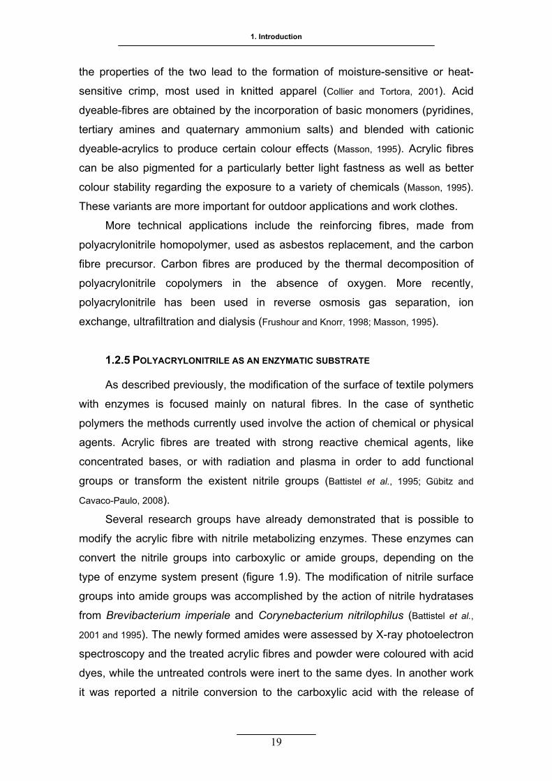
1. Introduction
the properties of the two lead to the formation of moisture-sensitive or heat-
sensitive crimp, most used in knitted apparel (Collier and Tortora, 2001). Acid
dyeable-fibres are obtained by the incorporation of basic monomers (pyridines,
tertiary amines and quaternary ammonium salts) and blended with cationic
dyeable-acrylics to produce certain colour effects (Masson, 1995). Acrylic fibres
can be also pigmented for a particularly better light fastness as well as better
colour stability regarding the exposure to a variety of chemicals (Masson, 1995).
These variants are more important for outdoor applications and work clothes.
More technical applications include the reinforcing fibres, made from
polyacrylonitrile homopolymer, used as asbestos replacement, and the carbon
fibre precursor. Carbon fibres are produced by the thermal decomposition of
polyacrylonitrile copolymers in the absence of oxygen. More recently,
polyacrylonitrile has been used in reverse osmosis gas separation, ion
exchange, ultrafiltration and dialysis (Frushour and Knorr, 1998; Masson, 1995).
1.2.5 POLYACRYLONITRILE AS AN ENZYMATIC SUBSTRATE
As described previously, the modification of the surface of textile polymers
with enzymes is focused mainly on natural fibres. In the case of synthetic
polymers the methods currently used involve the action of chemical or physical
agents. Acrylic fibres are treated with strong reactive chemical agents, like
concentrated bases, or with radiation and plasma in order to add functional
groups or transform the existent nitrile groups (Battistel et al., 1995; Gübitz and
Cavaco-Paulo, 2008).
Several research groups have already demonstrated that is possible to
modify the acrylic fibre with nitrile metabolizing enzymes. These enzymes can
convert the nitrile groups into carboxylic or amide groups, depending on the
type of enzyme system present (figure 1.9). The modification of nitrile surface
groups into amide groups was accomplished by the action of nitrile hydratases
from Brevibacterium imperiale and Corynebacterium nitrilophilus (Battistel et al.,
2001 and 1995). The newly formed amides were assessed by X-ray photoelectron
spectroscopy and the treated acrylic fibres and powder were coloured with acid
dyes, while the untreated controls were inert to the same dyes. In another work
it was reported a nitrile conversion to the carboxylic acid with the release of
19

Enzymatic Treatment of Acrylic and Cellulose Acetate Fibres
20
ammonia, on granular PAN as substrate, and to the amide on acrylic fibres
surface, with a nitrile hydratase and amidase from Rhodococcus rhodochrous
(Tauber et al., 2000). It was also reported that the molecular weight of PAN
influenced negatively the enzymatic action. A nitrile hydratase, from
Arthrobacter sp. Ecu 1101, was used to selectively transform the nitrile into
amide groups (Wang et al., 2004). The modified acrylic fibre became more
hydrophilic, antistatic and acid-dyeable. The authors concluded that the use of
non-ionic surfactants improved the enzyme activity while DMF did not. Another
microorganism, Agrobacterium tumefaciens, was found to grow on
polyacrylonitrile as a carbon source, converting it to polyacrylic acid (PAA) as
shown by solid state nuclear magnetic resonance (NMR) (Fischer-Colbrie et al.,
2006). The detected enzyme activities were nitrile hydratase and amidase,
although this amidase was, once more, less efficient in using acrylic fabric as a
substrate, like amidases from other species.
More recently, a membrane-bound nitrilase was isolated from Micrococcus
luteus BST20 (Fischer-Colbrie et al., 2007). Nitrile groups from both polymers and
fabrics of PAN were converted to carboxylic acid groups, with the associated
R
N N N N N N n
R
N N N N nOHO OH2N
Nitrilase (EC 3.5.5.1)
Nitrile hydratase (EC 4.2.1.84)H2O2 H2O
NH3
Amidase (EC 3.5.1.4)
H2ONH3
Figure 1.9 – Possible chemical transformations on PAN surface catalysed by nitrile metabolizing enzymes.
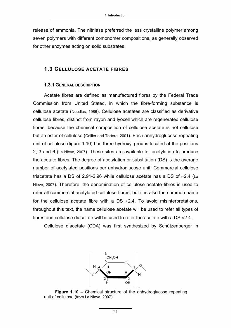
1. Introduction
21
release of ammonia. The nitrilase preferred the less crystalline polymer among
seven polymers with different comonomer compositions, as generally observed
for other enzymes acting on solid substrates.
11..33 CCEELLLLUULLOOSSEE AACCEETTAATTEE FF IIBBRREESS
1.3.1 GENERAL DESCRIPTION
Acetate fibres are defined as manufactured fibres by the Federal Trade
Commission from United Stated, in which the fibre-forming substance is
cellulose acetate (Needles, 1986). Cellulose acetates are classified as derivative
cellulose fibres, distinct from rayon and lyocell which are regenerated cellulose
fibres, because the chemical composition of cellulose acetate is not cellulose
but an ester of cellulose (Collier and Tortora, 2001). Each anhydroglucose repeating
unit of cellulose (figure 1.10) has three hydroxyl groups located at the positions
2, 3 and 6 (La Nieve, 2007). These sites are available for acetylation to produce
the acetate fibres. The degree of acetylation or substitution (DS) is the average
number of acetylated positions per anhydroglucose unit. Commercial cellulose
triacetate has a DS of 2.91-2.96 while cellulose acetate has a DS of ≈2.4 (La
Nieve, 2007). Therefore, the denomination of cellulose acetate fibres is used to
refer all commercial acetylated cellulose fibres, but it is also the common name
for the cellulose acetate fibre with a DS ≈2.4. To avoid misinterpretations,
throughout this text, the name cellulose acetate will be used to refer all types of
fibres and cellulose diacetate will be used to refer the acetate with a DS ≈2.4.
Cellulose diacetate (CDA) was first synthesized by Schützenberger in
Figure 1.10 – Chemical structure of the anhydroglucose repeating unit of cellulose (from La Nieve, 2007).
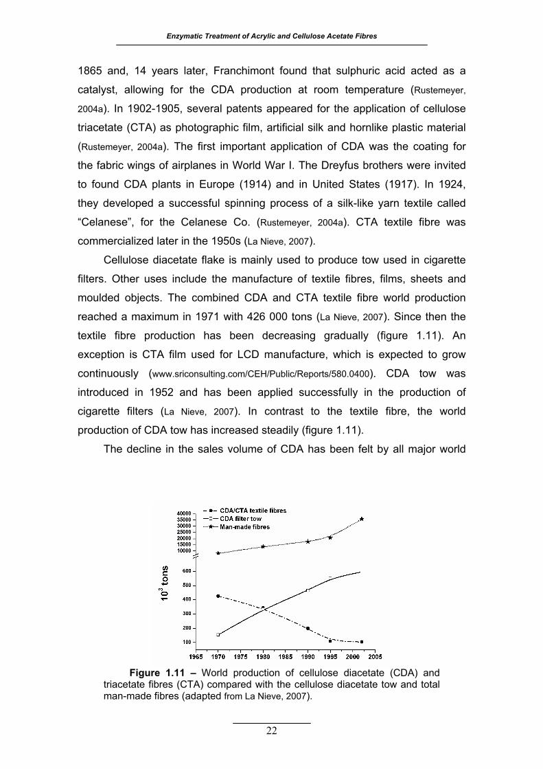
Enzymatic Treatment of Acrylic and Cellulose Acetate Fibres
22
1865 and, 14 years later, Franchimont found that sulphuric acid acted as a
catalyst, allowing for the CDA production at room temperature (Rustemeyer,
2004a). In 1902-1905, several patents appeared for the application of cellulose
triacetate (CTA) as photographic film, artificial silk and hornlike plastic material
(Rustemeyer, 2004a). The first important application of CDA was the coating for
the fabric wings of airplanes in World War I. The Dreyfus brothers were invited
to found CDA plants in Europe (1914) and in United States (1917). In 1924,
they developed a successful spinning process of a silk-like yarn textile called
“Celanese”, for the Celanese Co. (Rustemeyer, 2004a). CTA textile fibre was
commercialized later in the 1950s (La Nieve, 2007).
Cellulose diacetate flake is mainly used to produce tow used in cigarette
filters. Other uses include the manufacture of textile fibres, films, sheets and
moulded objects. The combined CDA and CTA textile fibre world production
reached a maximum in 1971 with 426 000 tons (La Nieve, 2007). Since then the
textile fibre production has been decreasing gradually (figure 1.11). An
exception is CTA film used for LCD manufacture, which is expected to grow
continuously (www.sriconsulting.com/CEH/Public/Reports/580.0400). CDA tow was
introduced in 1952 and has been applied successfully in the production of
cigarette filters (La Nieve, 2007). In contrast to the textile fibre, the world
production of CDA tow has increased steadily (figure 1.11).
The decline in the sales volume of CDA has been felt by all major world
Figure 1.11 – World production of cellulose diacetate (CDA) and triacetate fibres (CTA) compared with the cellulose diacetate tow and total man-made fibres (adapted from La Nieve, 2007).

1. Introduction
areas except China, Central Europe and Russia. In these developing countries
the trend in cigarettes consumption is increasing and it is expected that it will
offset the declining number of smokers in North America and Western Europe
(www.sriconsulting.com/CEH/Public/Reports/580.0400). Global market for CDA is highly
concentrated with major producers representing over 80% of global capacity:
Celanese Corp, Eastman Chemical Company, Daicel Chemical Industries,
Mitsubishi Rayon and Rhodia Acetow (www.industrialnewsupdate.com/news/manufac
turing/archives/2008 /04/cellulose_aceta.php).
Cellulose diacetate and triacetate are produced from high quality cellulose,
such as cotton linters and wood pulps, with an α-cellulose content above 95%
(Saka and Matsumura, 2004). The initial stages of both CDA and CTA formation are
the same. Purified and shredded cellulose pulp is pretreated to increase its
reactivity and to disperse it uniformly in the reaction media (La Nieve, 2007). The
most used commercial acetylation process is the acetic acid system where the
acetic acid serves as solvent for the cellulose acetylation and acetic anhydride
and sulphuric acid as catalysts (La Nieve, 2007). The esterification of cellulose is
Figure 1.12 – Schematic representation of the manufacturing line of cellulose acetate fibres (from Collier and Tortora, 2001).
23
Enzymatic Treatment of Acrylic and Cellulose Acetate Fibres
allowed for 7-8 hours till the CTA, also known as primary acetate, is formed
(Collier and Tortora, 2001). After this point the procedure differs depending on
which DS is being produced (figure 1.12).
To obtain the CTA, water is added to the acetylating media and CTA is
precipitated as a white flake, which is then collected, and dried (Collier and Tortora,
2001). To obtain the CDA, also known as secondary acetate, water is added in
an amount and for a period of time that allow partial hydrolysis of the primary
acetate. The CDA is then precipitated, collected, washed and dried (Collier and
Tortora, 2001). The spinning dope preparation is basically the same for both CDA
and CTA except for the solvent used (La Nieve, 2007). CDA is usually dissolved in
95% aqueous acetone while CTA is dissolved in methylene chlorine containing
5% to 15% of methanol. At the dope stage, titanium dioxide pigment can be
added if dull yarns are to be produced. Dope dying is possible for both fibres by
the addition of pigments or dyes soluble in the particular solvent system used
for the spinning dope (La Nieve, 2007). The CDA and CTA fibres are produced by
the dry-spinning technology. The fibres are lubricated and crimp is also
introduced for further processing. Besides the continuous filament, acetate
fibres are also produced in the tow and staple forms (La Nieve, 2007).
Cellulose diacetate and triacetate are not satisfactorily dyed by soluble
dyes; instead disperse dyes are the ones preferred for these fibres (La Nieve,
2007; Needles, 1986). Disperse dyes are insoluble or slightly soluble in hot
aqueous baths, so dispersing agents and carriers are currently used to enhance
the uniformity and penetration of the dye molecules into the fibres. In the case
of CDA, carriers are not necessary and the dyeing temperatures range between
70º and 80 ºC. CTA has a slower relative dyeing rate, the carriers are needed
as well as dyeing temperatures in the range of 96 ºC - 100 ºC (La Nieve, 2007;
Steinmann, 1998).
Although triacetate and diacetate are produced in a very similar way and
have similar structures, the small difference in the DS value provides significant
differences in the properties of these fibres (Needles, 1986).
24
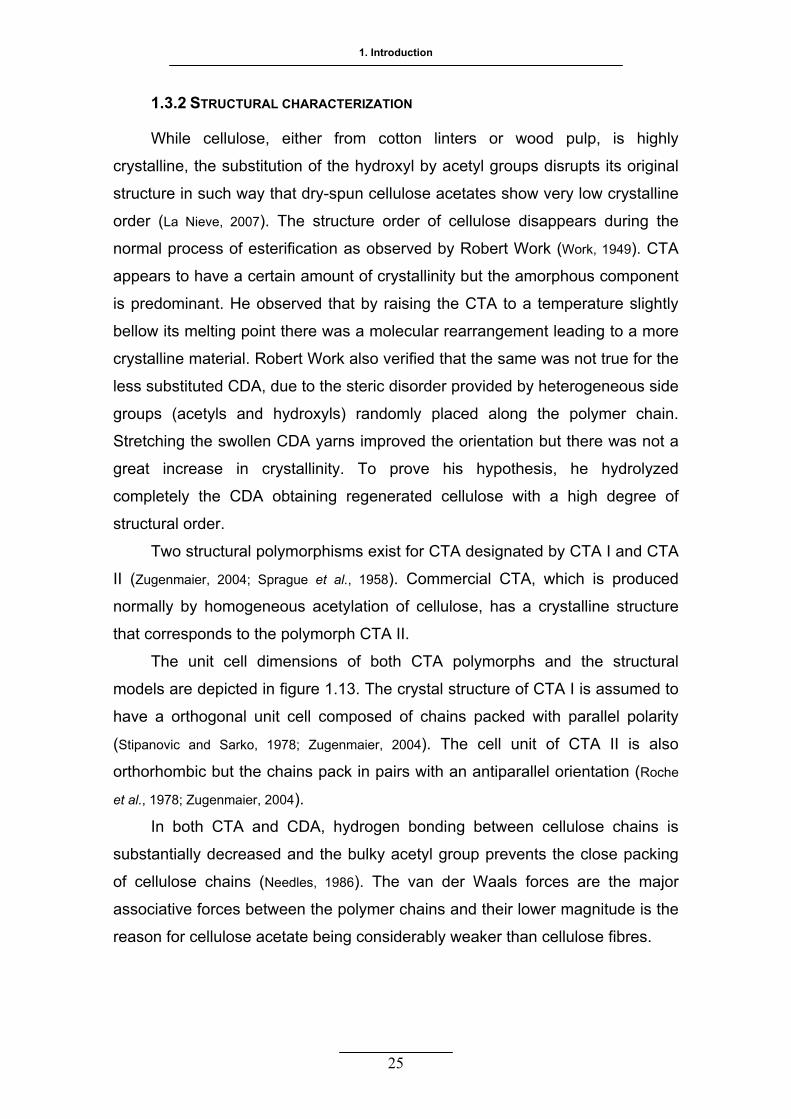
1. Introduction
1.3.2 STRUCTURAL CHARACTERIZATION
While cellulose, either from cotton linters or wood pulp, is highly
crystalline, the substitution of the hydroxyl by acetyl groups disrupts its original
structure in such way that dry-spun cellulose acetates show very low crystalline
order (La Nieve, 2007). The structure order of cellulose disappears during the
normal process of esterification as observed by Robert Work (Work, 1949). CTA
appears to have a certain amount of crystallinity but the amorphous component
is predominant. He observed that by raising the CTA to a temperature slightly
bellow its melting point there was a molecular rearrangement leading to a more
crystalline material. Robert Work also verified that the same was not true for the
less substituted CDA, due to the steric disorder provided by heterogeneous side
groups (acetyls and hydroxyls) randomly placed along the polymer chain.
Stretching the swollen CDA yarns improved the orientation but there was not a
great increase in crystallinity. To prove his hypothesis, he hydrolyzed
completely the CDA obtaining regenerated cellulose with a high degree of
structural order.
Two structural polymorphisms exist for CTA designated by CTA I and CTA
II (Zugenmaier, 2004; Sprague et al., 1958). Commercial CTA, which is produced
normally by homogeneous acetylation of cellulose, has a crystalline structure
that corresponds to the polymorph CTA II.
The unit cell dimensions of both CTA polymorphs and the structural
models are depicted in figure 1.13. The crystal structure of CTA I is assumed to
have a orthogonal unit cell composed of chains packed with parallel polarity
(Stipanovic and Sarko, 1978; Zugenmaier, 2004). The cell unit of CTA II is also
orthorhombic but the chains pack in pairs with an antiparallel orientation (Roche
et al., 1978; Zugenmaier, 2004).
In both CTA and CDA, hydrogen bonding between cellulose chains is
substantially decreased and the bulky acetyl group prevents the close packing
of cellulose chains (Needles, 1986). The van der Waals forces are the major
associative forces between the polymer chains and their lower magnitude is the
reason for cellulose acetate being considerably weaker than cellulose fibres.
25
Enzymatic Treatment of Acrylic and Cellulose Acetate Fibres
A D
B
C
The increase in crystallinity of CTA by heat treatment has beneficial effects
on the fibre and fabric properties, resulting in “ease-of-care” and “wash-and-
wear” characteristics (Steinmann, 1998).
1.3.3 CELLULOSE ACETATE FIBRE PROPERTIES
The fibre cross section of both CTA and CDA is irregular with as many as
5 to 6 lobes and the longitudinal views show striations along the fibre length
(Needles, 1986). Both fibres have white bright appearance and good lustre if not
dyed or treated with a delustering agent (Collier and Tortora, 2001). CDA and CTA
have densities of 1.30-1.35 g cm-3 and the filament denier ranges from 3 to 60
(Needles, 1986; Steinmann, 1998).
Figure 1.13 – Structural models of CTA I and CTA II in two projections: A) CTA I perpendicular to the ac plane, B) CTA I along the c axis (the chain axis), C) CTA II perpendicular to the ac plane, D) CTA II along the c axis, E) unit cell dimensions of CTA I and F) unit cell dimensions of CTA II (adapted from Zugenmaier, 2004).
a = 23.6 Å b = 6.3 Å
c = 10.4 Å E c = 10.5 Å F
b = 11.5 Å
a = 24.7 Å
26
1. Introduction
Both CDA and CTA have a very low strength, with standard tenacities of
1.2-1.4 g per denier and standard elongations at break of 25% - 45%, however,
for their end-uses, strength is not a primary consideration (Steinmann, 1998; Collier
and Tortora, 2001). At 2% elongation the fibres have recoveries ranging from 80%
to 99%, but above 5% elongation the recovery is very low, in particular for CDA.
CDA and CTA are moderately stiff fibres with good resilience on bending and
deformation.
The moisture regains for CDA and CTA is 6.5% and 3.5%, respectively
(Steinmann, 1998; La Nieve, 2007). The CDA has a moisture regain close to the
value 7% of natural cotton yarn, while, the CTA has a lower value but still higher
than the commercial synthetic fibres. Wetting the cellulose acetate fibres
decreases their tenacity to 0.8-1.0 g per denier and the elongation at break
increases more in the case of CDA (35% - 50%) than in the case of CTA (30% -
40%). CTA is more hydrophobic and the water has lower impact on the CTA
crystalline structure when compared to CDA. This imparts a better dimensional
stability to CTA during washing and drying cycles, whereas CDA may exhibit
relaxation shrinkage on laundering (Collier and Tortora, 2001). Both fibres have a
high electrical resistance and, as a consequence, static build-up is a problem
unless antistatic treatments are applied (Needles, 1986).
The CDA and CTA are good heat conductors and they are cool to the
touch (Needles, 1986). These fibres are thermoplastic so they soft and melt with
the application of heat. CDA is more heat sensitive than CTA, it softens in the
range of 190 ºC - 205 ºC and it melts at ≈260 ºC with discolouration (Steinmann,
1998; La Nieve, 2007). The softening temperature for heat-treated CTA is ≈240 ºC
and the melting point is ≈305 ºC with discoloration and decomposition. If ignited,
CDA and CTA burn with melting, leaving a small, hard, beadlike residue at the
edge of the burning area (Collier and Tortora, 2001).
The chemical stability of cellulose acetates is poor. They are attacked by a
number of organic solvents capable of dissolving esters, strong acids and
bases, which result in saponification of acetyl groups (La Nieve, 2007; Collier and
Tortora, 2001). CDA is more susceptible of being attacked by strong oxidizing
agents than CTA, however both can be bleached. They can be damaged by
microorganisms under certain conditions, but CTA is considerably more
27

Enzymatic Treatment of Acrylic and Cellulose Acetate Fibres
resistant to biological attack (Needles, 1986; Collier and Tortora, 2001). In general, the
CTA fibre is more resistant to sunlight exposure and aging, while CDA looses
strength upon extended exposure to sunlight. Neither CDA nor CTA are
resistant to abrasion (La Nieve, 2007; Collier and Tortora, 2001).
Acetate fibres are soft, cool, have silk-like aesthetics, good drape and they
can be easily blended with other fibres like silk, rayon, nylon, cotton and
polyester (Law, 2004). Their unique attributes remain desirable and they are
responsible for the survival of acetate production in the competitive market of
man-made fibres. In a study made in 1999 by the Institute of Environmental
Research from Kansas State University to determine the perceived difference
between acetate and polyester linings, a group of consumers felt that acetate
was more comfortable (less sticky, clammy and damp) and lighter in weight
than polyester (www.acetateworld.com). Another attribute that is gaining importance
is the fact that cellulose acetate fibres are environmental friendly compared to
the major synthetic fibres.
Some methods were developed to improve the strength, abrasion
resistance and dimension stability of acetate fibres, in particular of CDA
(Steinmann, 1998). One approach was to add to CDA spin dope polymer additives.
Several were tested but, unless their concentration was bellow 5%, the phase
compatibility was poor (Steinmann, 1998). To improve the compatibility, some
polymers were graft on CDA. In the case of acrylonitrile, the graft copolymer
increased the compatibility of PAN and cellulose acetate and the resulting fibres
possessed improved thermal and chemical stabilities (Steinmann, 1998). It was
also investigated the effect of crosslinking agents on cellulose diacetate, though
the improved properties were still not equal to those of heat-treated CTA
(Steinmann, 1998).
1.3.4 PRODUCTS AND APPLICATIONS
The properties of acetate fibres led to their largest use in women’s apparel
and apparel linings. The yarns are evenly distributed between woven and knit
constructions. Principal items of clothing are blouses, dresses, lingerie, robes
and linings for suits, skirts and pants. They are also used in household textiles
like draperies, bedcovers, curtains, etc (La Nieve, 2007; Collier and Tortora, 2001; Law,
28

1. Introduction
2004). Other textile applications include sportswear, medical bandages and tape
(Law, 2004).
Staple fibres and nonwovens are used in wound dressings, personal
hygiene, wipes and speciality papers (Law, 2004).
CDA is also used for the production of cigarette filters. The CDA crimped
tow presents several advantages like the ability to produce uniform and firm
filters, it selectively removes phenols, nitrosamines, quinolines and other
undesired smoke components, it is non toxic, tasteless, odourless and it is
biodegradable. These attributes account for the preference for filters made from
CDA which represents 90% of the consumed cigarettes in the world (Rustemeyer,
2004b).
Other non textile applications include solvent-cast diacetate film that is
produced by dissolution of CDA flake in acetone and the addition of certain
additives like plasticizers that impart the film attributes. The films are applied in
a variety of end-uses like adhesive tapes, labels, general packaging, photo
negative sleeves, and graphic arts, among others. They are known for their high
transparency and gloss, although they can be produced with irregular matt
surfaces (Law et al., 2004). The CTA film has unique properties exploited in its
application as photographic film, protective film for polarizing plates or optical
compensation film for liquid crystal displays (Sata et al., 2004). CDA is also used
to produce plastic products whose use implies direct and prolonged contact with
humans, like fashion accessories, tooth brushes, protective glasses, casino
chips, etc (Carrolo and Grospietro, 2004).
Cellulose acetate is extensively applied in the separation technology.
Acetate membranes are used in desalting (by reverse osmosis), hemodialysis,
drinking-water purification (by ultrafiltration) and filters for laboratory use.
Cellulose acetate is also used as stationary phase in chromatography
separations (Shibata, 2004).
The manufacture of products made from cellulose acetate and other
esters, has a promising future due to their renewable resource and properties.
Cellulose acetate is becoming the raw material of choice for a new generation
of high-tech products for the separation technology, the pharmaceutical
industry, the controlled release and biopolymer fields (Glasser, 2004).
29
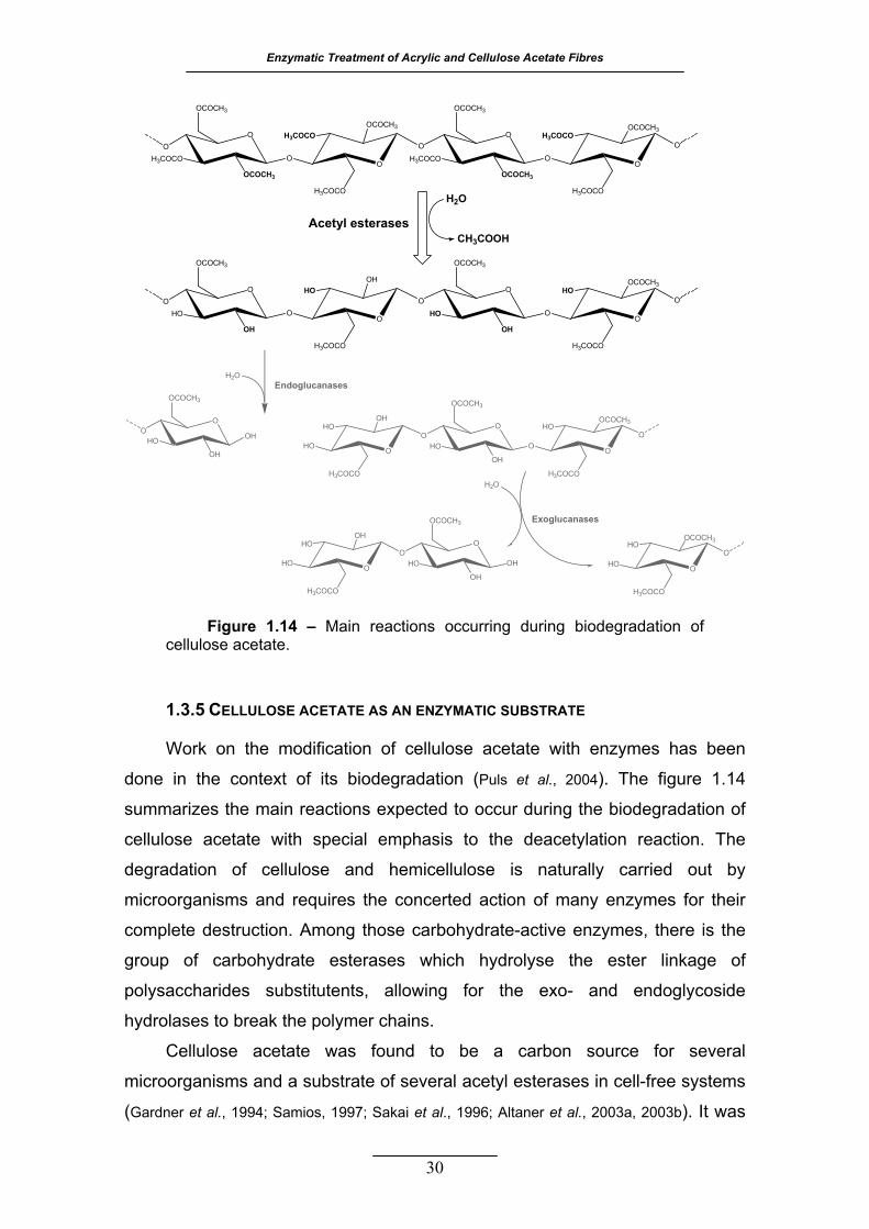
Enzymatic Treatment of Acrylic and Cellulose Acetate Fibres
30
1.3.5 CELLULOSE ACETATE AS AN ENZYMATIC SUBSTRATE
Work on the modification of cellulose acetate with enzymes has been
done in the context of its biodegradation (Puls et al., 2004). The figure 1.14
summarizes the main reactions expected to occur during the biodegradation of
cellulose acetate with special emphasis to the deacetylation reaction. The
degradation of cellulose and hemicellulose is naturally carried out by
microorganisms and requires the concerted action of many enzymes for their
complete destruction. Among those carbohydrate-active enzymes, there is the
group of carbohydrate esterases which hydrolyse the ester linkage of
polysaccharides substitutents, allowing for the exo- and endoglycoside
hydrolases to break the polymer chains.
Cellulose acetate was found to be a carbon source for several
microorganisms and a substrate of several acetyl esterases in cell-free systems
(Gardner et al., 1994; Samios, 1997; Sakai et al., 1996; Altaner et al., 2003a, 2003b). It was
OO
H3COCO O
OCOCH3
OCOCH3
O
H3COCO
OCOCH3H3COCO O
OH3COCO O
OCOCH3
OCOCH3
O
O
H3COCO
OCOCH3H3COCO
OO
HO HO
OCOCH3
OH O
H3COCO
OHHO O
OHO O
OCOCH3
OHO
O
H3COCO
OCOCH3HO
OO
HO O
OCOCH3
OHO
H3COCO
OHHO O
OHO O
OCOCH3
OHO
O
H3COCO
OCOCH3HO
H2O
CH3COOHAcetyl esterases
EndoglucanasesH2O
HO O
H3COCO
OHHO O
OHO OH
OCOCH3
OH
Exoglucanases
H2O
OH
HO O
O
H3COCO
OCOCH3HO
Figure 1.14 – Main reactions occurring during biodegradation of cellulose acetate.
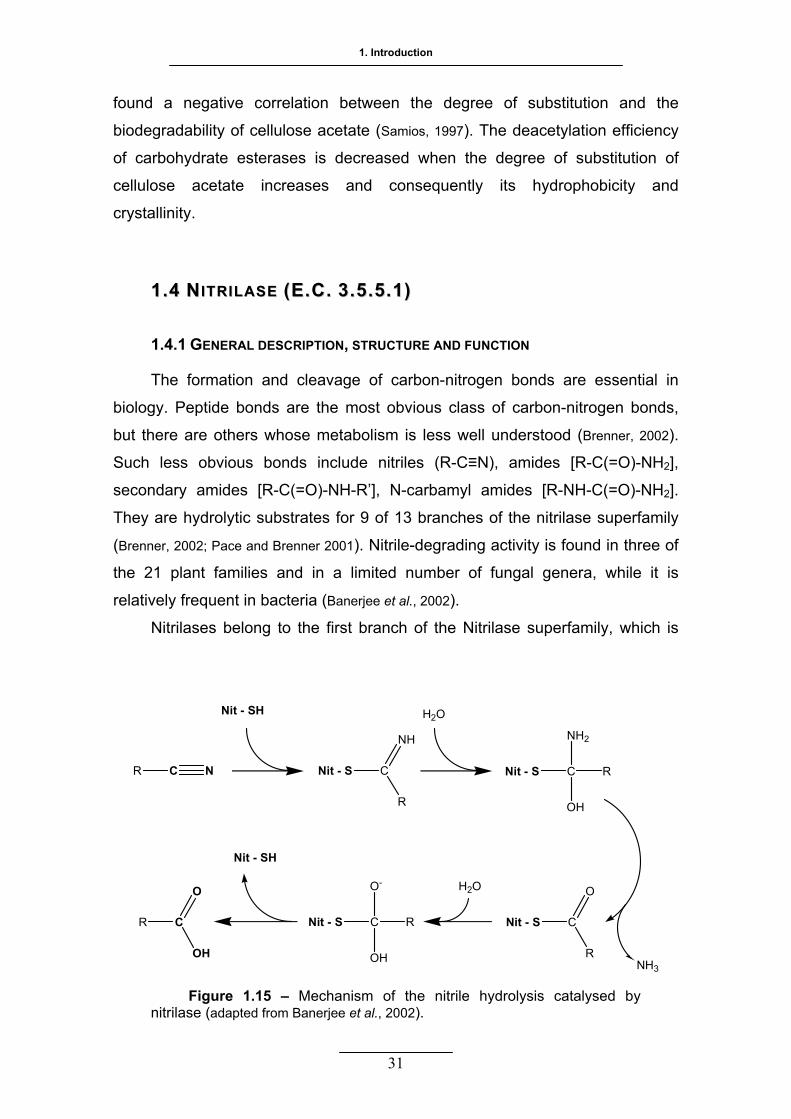
1. Introduction
31
found a negative correlation between the degree of substitution and the
biodegradability of cellulose acetate (Samios, 1997). The deacetylation efficiency
of carbohydrate esterases is decreased when the degree of substitution of
cellulose acetate increases and consequently its hydrophobicity and
crystallinity.
11..44 NNIITTRRIILLAASSEE ((EE..CC.. 33 ..55 ..55..11))
R C
1.4.1 GENERAL DESCRIPTION, STRUCTURE AND FUNCTION
The formation and cleavage of carbon-nitrogen bonds are essential in
biology. Peptide bonds are the most obvious class of carbon-nitrogen bonds,
but there are others whose metabolism is less well understood (Brenner, 2002).
Such less obvious bonds include nitriles (R-C≡N), amides [R-C(=O)-NH2],
secondary amides [R-C(=O)-NH-R’], N-carbamyl amides [R-NH-C(=O)-NH2].
They are hydrolytic substrates for 9 of 13 branches of the nitrilase superfamily
(Brenner, 2002; Pace and Brenner 2001). Nitrile-degrading activity is found in three of
the 21 plant families and in a limited number of fungal genera, while it is
relatively frequent in bacteria (Banerjee et al., 2002).
Nitrilases belong to the first branch of the Nitrilase superfamily, which is
Figure 1.15 – Mechanism of the nitrile hydrolysis catalysed by nitrilase (adapted from Banerjee et al., 2002).
N
Nit - SH
Nit - S C
NH
R
Nit - S C
NH2
R
OH
Nit - S C
O
R
Nit - S C
O-
R
OH
Nit - SH
R C
O
OH
H2O
NH3
H2O
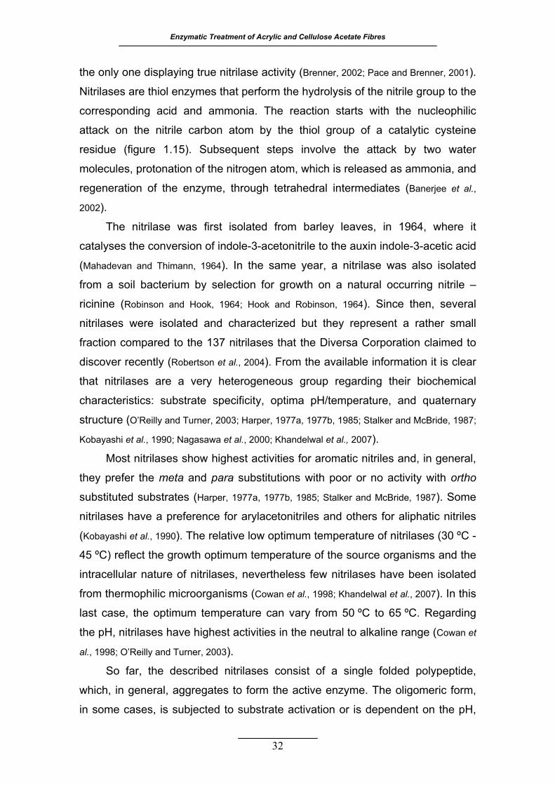
Enzymatic Treatment of Acrylic and Cellulose Acetate Fibres
the only one displaying true nitrilase activity (Brenner, 2002; Pace and Brenner, 2001).
Nitrilases are thiol enzymes that perform the hydrolysis of the nitrile group to the
corresponding acid and ammonia. The reaction starts with the nucleophilic
attack on the nitrile carbon atom by the thiol group of a catalytic cysteine
residue (figure 1.15). Subsequent steps involve the attack by two water
molecules, protonation of the nitrogen atom, which is released as ammonia, and
regeneration of the enzyme, through tetrahedral intermediates (Banerjee et al.,
2002).
The nitrilase was first isolated from barley leaves, in 1964, where it
catalyses the conversion of indole-3-acetonitrile to the auxin indole-3-acetic acid
(Mahadevan and Thimann, 1964). In the same year, a nitrilase was also isolated
from a soil bacterium by selection for growth on a natural occurring nitrile –
ricinine (Robinson and Hook, 1964; Hook and Robinson, 1964). Since then, several
nitrilases were isolated and characterized but they represent a rather small
fraction compared to the 137 nitrilases that the Diversa Corporation claimed to
discover recently (Robertson et al., 2004). From the available information it is clear
that nitrilases are a very heterogeneous group regarding their biochemical
characteristics: substrate specificity, optima pH/temperature, and quaternary
structure (O’Reilly and Turner, 2003; Harper, 1977a, 1977b, 1985; Stalker and McBride, 1987;
Kobayashi et al., 1990; Nagasawa et al., 2000; Khandelwal et al., 2007).
Most nitrilases show highest activities for aromatic nitriles and, in general,
they prefer the meta and para substitutions with poor or no activity with ortho
substituted substrates (Harper, 1977a, 1977b, 1985; Stalker and McBride, 1987). Some
nitrilases have a preference for arylacetonitriles and others for aliphatic nitriles
(Kobayashi et al., 1990). The relative low optimum temperature of nitrilases (30 ºC -
45 ºC) reflect the growth optimum temperature of the source organisms and the
intracellular nature of nitrilases, nevertheless few nitrilases have been isolated
from thermophilic microorganisms (Cowan et al., 1998; Khandelwal et al., 2007). In this
last case, the optimum temperature can vary from 50 ºC to 65 ºC. Regarding
the pH, nitrilases have highest activities in the neutral to alkaline range (Cowan et
al., 1998; O’Reilly and Turner, 2003).
So far, the described nitrilases consist of a single folded polypeptide,
which, in general, aggregates to form the active enzyme. The oligomeric form,
in some cases, is subjected to substrate activation or is dependent on the pH,
32

1. Introduction
temperature and enzyme concentration (Harper, 1977a; Nagasawa et al., 2000; O’Reilly
and Turner, 2003). The number of polypeptides in the aggregates can range from
6 to as much as 26 subunits with the addition of subunits taking place in pairs
(Sewell et al., 2003).
The crystal structures of three members of the nitrilase superfamily are
available (Kumaran et al., 2003; Nakai et al., 2000; Pace et al., 2000). Their domains
have similar topology and dimerization modes (Kumaran et al., 2003). Each subunit
is a compact αββα sandwich and it forms dimers with an eight layer αββα:αββα
structure (figure 1.16A). The subunit is well defined, globular and, in all the
three structures, the dimer interface is through interactions between α layers.
These dimers combine further to form tetramers in two of the three studied
proteins through contacts between the exposed edges of the β-layers (Nakai et
al., 2000; Pace et al., 2000). From the structures, mutagenesis and gene sequence
comparison, it is assumed that the entire superfamily uses a catalytic triad
consisting of conserved glutamic acid, lysine and cysteine residues (figure
1.16B) (Brenner, 2002; Robertson et al., 2004; Kumaran et al., 2003; Nakai et al., 2000; Pace
et al., 2000).
The enzymes of the nitrilase superfamily play diverse and important roles
in biology. The known functions include synthesis of signalling molecules,
vitamins and coenzymes metabolism, small molecules detoxification and protein
post-translation modifications (Brenner, 2002).
1.4.2 APPLICATIONS
Nitrilases have attracted the attention and interest in the past two decades
and they are been recognised as important “green” catalysts (Kaul et al., 2007;
Singh et al., 2006). The conversion of nitriles into carboxylic acids is a key organic
reaction in the synthesis of many intermediate compounds and final products.
The chemical hydrolysis of nitriles requires drastic conditions such as strong
bases or acids and/or elevated temperatures, besides, it originates undesirable
by-products and inorganic wastes (Zhu et al., 2007). The advantages presented by
these enzymes, besides the eco-friendly biotransformation is their inherent
enantio- and, in particular, regio-selectivity.
33
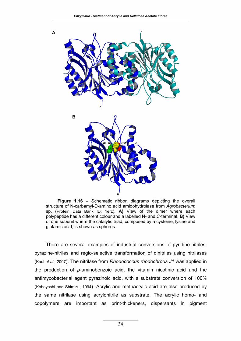
Enzymatic Treatment of Acrylic and Cellulose Acetate Fibres
34
There are several examples of industrial conversions of pyridine-nitriles,
pyrazine-nitriles and regio-selective transformation of dinitriles using nitrilases
(Kaul et al., 2007). The nitrilase from Rhodococcus rhodochrous J1 was applied in
the production of p-aminobenzoic acid, the vitamin nicotinic acid and the
antimycobacterial agent pyrazinoic acid, with a substrate conversion of 100%
(Kobayashi and Shimizu, 1994). Acrylic and methacrylic acid are also produced by
the same nitrilase using acrylonitrile as substrate. The acrylic homo- and
copolymers are important as print-thickeners, dispersants in pigment
Figure 1.16 – Schematic ribbon diagrams depicting the overall structure of N-carbamyl-D-amino acid amidohydrolase from Agrobacterium sp. (Protein Data Bank ID: 1erz). A) View of the dimer where each polypeptide has a different colour and a labelled N- and C-terminal. B) View of one subunit where the catalytic triad, composed by a cysteine, lysine and glutamic acid, is shown as spheres.
C
C
N
NA
N
CGlu 46
Lys126
Cys171
B
1. Introduction
35
suspensions, detergent builders, and disposable diapers, among others (Hughes
et al., 1998).
One of the most important industrial applications of nitrilase (from
Alcaligenes strains) is the transformation of racemic mandelonitrile to (R)-(-)-
mandelic acid. This compound is a key intermediate in the production of semi-
synthetic cephalosporins, penicillins, and it is also important in the synthetic
process of antitumor and antiobesity agents (Kaul et al., 2007).
Transgenic crop plants expressing the nitrilase gene from Klebsiella
ozaenae were obtained in order to survive to the herbicide bromoxynil which is
a photosynthesis inhibitor (Kobayashi and Shimizu, 1994). The nitrilase destroys the
herbicide by hydrolysis of its nitrile group.
Another important application of nitrilase or nitrilase-producing organisms
is the decontamination of acrylonitrile waste effluents and aqueous polymer
emulsions containing this highly toxic and carcinogenic compound (Battistel et al.,
1997; Wyatt and Knowles, 1995).
11..55 CCUUTTIINNAASSEE ((EE..CC.. 33..11..11..7744))
1.5.1 GENERAL DESCRIPTION, STRUCTURE AND FUNCTION
Cutinases are carboxylic ester hydrolases that degrade cutin which is the
structural component of the outer envelope (the cuticle) of higher plants (Purdy
and Kolattukudy, 1975a; Kolattukudy, 2002). Cutin is a natural polyester composed
mainly of C16 and C18 hydroxyl and epoxy fatty acids as monomers. The cuticle
constitutes an efficient barrier against desiccation and entry of pathogens in
plants (Kolattukudy, 2002). The enzymatic degradation of the cuticle, as a
consequence of the secretion of cutinases by fungi, was proved to be one of the
first steps in the infectious process of plants (Carvalho et al., 1998).
Cutinases have been purified from different sources, especially fungi but
also from pollen and bacteria (Carvalho et al., 1998). The first and most studied
cutinase is from the fungal pathogen of peas, Fusarium solani pisi (Purdy and
Kolattukudy, 1975a, 1975b). This is a small ellipsoid protein (≈22 KDa, 45x30x30 Å)
that bridges functional properties of lipases and esterases, because it
hydrolyses both soluble esters and emulsified triacylglycerols (Mannesse et al.,

Enzymatic Treatment of Acrylic and Cellulose Acetate Fibres
36
1995; Egmond and de Vlieg, 2000). Studies with triglyceride analogues revealed that
cutinase activity is very sensitive to the length and distribution of the acyl
chains, with highest activities observed for short chain lengths of three to five
carbons (Mannesse et al., 1995; Egmond and de Vlieg, 2000). The optimum pH for the
hydrolysis is around 8.5, depending on the particular substrate used, and the
maximum thermal stability is obtained for the pH range 6-9 (Petersen et al., 1998,
2001b).
Cutinase is a serine esterase that shares the basic catalytic features with
serine proteases (Köller and Kolattukudy, 1982). The essential feature is the
catalytic triad involving the hydroxyl group of a serine, the imidazole side chain
of a histidine and a carboxylic side chain of an acidic residue which, in cutinase,
is an aspartic residue (Martinez et al., 1994). The function of the serine is to
participate in a transacylation reaction with the substrate to form an acyl-
enzyme intermediate (figure 1.17). The intermediate is hydrolysed to release
the first product. The serine acyl ester reacts then with a water molecule to
O-
O
Enz
NN
Enz
O
Enz
HH
R1OR2
O
O-
O
Enz
N+N Enz
O
Enz
H
R1O
R2-O
H
O-
O
Enz
NN
Enz
H
Enz
O
R1O
O-
O
Enz
NN
Enz
H
Enz
OH
H2O
HO
R1
O
OHR2
Asp
HisSer
Substrate
Figure 1.17 – Mechanism of the carboxylic ester bond hydrolysis catalysed by cutinase (adapted from Lau and Bruce, 1999).

1. Introduction
release the fatty acid and to regenerate the catalytic hydroxyl group (Lau and
Bruce, 1999; Petersen et al., 2001a). The role of the imidazole ring is to act as a base
to remove the proton from the serine hydroxyl group in concert with the
nucleophilic attack of the serine oxygen on the carbonyl carbon of the substrate.
The aspartic residue seems to orient the imidazole ring properly and to stabilize
the local structure around the active site (Lau and Bruce, 1999).
The F. solani pisi cutinase belongs to the superfamily of α/β-hydrolases, to
which lipases also belong. Cutinases have a central slightly twisted β-sheet
consisting of five parallel strands covered by two α-helices on either side of the
sheet (figure 1.18A) (Longhi and Cambillau, 1999; Egmond and de Vlieg, 2000). The
nucleophilic serine is located in an extremely sharp turn between a β-chain and
an α-helice, named nucleophilic elbow. The amino acid sequence around the
catalytic serine (Gly-Tyr-Ser-Gln-Gly) matches the consensus sequence
commonly present in lipases (Gly-His/Tyr-Ser-X-Gly). The catalytic triad is
located at one edge of the ellipsoid protein (figure 1.18B), it is rather accessible
to the solvent, in contrast with lipases, and it is surrounded by two loops with
A
B
Asp 175
His 188 Ser 120
N
C
Figure 1.18 – A) Schematic ribbon diagrams depicting the overall structure of cutinase from Fusarium solani pisi (Protein Data Bank ID: 1cex); the secondary structural elements are represented in different colours (β-strands are in yellow, α-helixes are in red and connecting loops are in green; the N- and C-terminal are labelled; the two disulfide bridges and catalytic residues are depicted in blue by the stick model. B) Closed view of the catalytic triad composed by a serine, histidine and aspartic acid, shown by the stick model; the two possible locations of the Ser120 side chain are both represented.
37
Enzymatic Treatment of Acrylic and Cellulose Acetate Fibres
high mobility (Prompers et al., 1999; Longhi et al., 1997). These loops delimit the
catalytic site and the substrate binding cleft which is hydrophobic, reflecting the
lipolytic nature of cutinases (Egmond and de Vlieg, 2000).
The F. solani pisi cutinase is also classified as a member of the family 5 of
carbohydrate esterases (www.cazy.org/fam/CE5.html), sharing a very similar 3D-
structure with other two members with known structure: the acetyl xylan
esterase (E.C. 3.1.1.72) from Trichoderma reesei and the acetyl xylan esterase
II (AXE II) from Penicillium purpurogenum (Hakulinen et al., 2000; Ghosh et al., 2001).
Although they present very similar overall structures, the conformation of the
active site is different. These esterases have more exposed catalytic residues
than cutinase, reflecting their specificity towards nonlipidic polar substrates
(Ghosh et al., 2001). There is also an additional loop, delimiting the active site, in
the AXE II from P. purpurogenum, which is responsible for the preference for
short substrates. When this loop is deleted, AXE II is also able to hydrolyse long
chain fatty acidic esters, up to at least 14 carbons, resembling cutinase
specificity (Colombres et al., 2008).
1.5.2 APPLICATIONS
Cutinase is a very versatile enzyme: it can utilise a broad range of
substrates, it is rather stable and at low water activities cutinase catalyses the
reverse reaction – trans-esterification of fats and selective esterification of
alcohols (Carvalho et al., 1998). Both, hydrolytic and synthesis reactions have
potential use in food processing, beverages, perfume industries, pharmaceutical
industries, agriculture, chemical industries and others like pulp/paper, textile
and leather industries (Panda and Gowrishankar, 2005). Some of its potentialities are
already applied in industry, but many others are still at a research level. Some
cutinase preparations have been produced by Genencor and Unilever for
detergent and surfactant formulations (Carvalho et al., 1998). A lot of research is
devoted to the study and optimization of cutinase activity in non conventional
media (organic media, supercritical fluids and gas/solid systems) and in the
stabilization of the enzyme through immobilization, micro-encapsulation and
lyophilized preparations (Carvalho et al., 1999a).
38
1. Introduction
39
Another important application of cutinase is the degradation of plastics.
The synthetic polyester, polycaprolactone, was hydrolysed to soluble products
that were used by F. solani wild type strains as source of carbon and energy
(Murphy et al., 1996).
11..66 TTAAIILLOORRIINNGG TTHHEE PPRROOPPEERRTT IIEESS OOFF BBIIOOCCAATTAALLYYSSTTSS
1.6.1 PROTEIN AND MEDIA ENGINEERING
The Nature’s biodiversity provides us with a large collection of enzymes
well suited for supporting life, however, they may not always be well suited for
our technological interests (Arnold, 2001). Therefore, the impact of enzymes is
dependent on our ability to tailor their properties according to the demands of a
particular technological process (Arnold, 2001). Some important issues that often
need to be addressed are limited substrate range, limited stability to
temperature, pH and solvent, limited enantioselectivity and limited turnover
number (Powell et al., 2001). Although screening the biodiversity continues to be
an important approach to find better biocatalysts, the manipulation of the protein
molecule itself and/or the reaction media are gaining increasing importance.
Reaction media engineering is important to optimize and modulate
biocatalysts activity and stability. Biocatalysis in non-aqueous media offers
unique advantages compared to aqueous enzymology (Castro and Knubovets,
2003). There are drastic changes in the enantioselectivity of catalysed reactions,
reversal of the thermodynamic equilibrium of certain reactions, suppression of
water-dependent side-reactions, improved solubility of hydrophobic substrates
or products and resistance to bacterial contamination (Klibanov, 2001; Castro and
Knubovets, 2003). Homogeneous biocatalysis in water-organic mixtures is also
used and major changes in enzyme catalytic activity and initial rates have been
reported, depending on the type and amount of the cosolvent (Castro and
Knubovets, 2003). In several cases, at low concentration of the organic cosolvent
(between 5% and 25%) the initial rate and stability of the enzyme are
maximized regarding aqueous buffers (Castro and Knubovets, 2003).
The stability of enzymes in vitro is a very critical issue in biotechnology
and both protein and media engineering strategies are used to increase it.
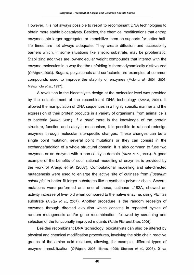
Enzymatic Treatment of Acrylic and Cellulose Acetate Fibres
However, it is not always possible to resort to recombinant DNA technologies to
obtain more stable biocatalysts. Besides, the chemical modifications that entrap
enzymes into larger aggregates or immobilize them on supports for better half-
life times are not always adequate. They create diffusion and accessibility
barriers which, in some situations like a solid substrate, may be problematic.
Stabilizing additives are low-molecular weight compounds that interact with the
enzyme molecules in a way that the unfolding is thermodynamically disfavoured
(Ó’Fágáin, 2003). Sugars, polyalcohols and surfactants are examples of common
compounds used to improve the stability of enzymes (Melo et al., 2001, 2003;
Matsumoto et al., 1997).
A revolution in the biocatalysts design at the molecular level was provided
by the establishment of the recombinant DNA technology (Arnold, 2001). It
allowed the manipulation of DNA sequences in a highly specific manner and the
expression of their protein products in a variety of organisms, from animal cells
to bacteria (Arnold, 2001). If a priori there is the knowledge of the protein
structure, function and catalytic mechanism, it is possible to rational redesign
enzymes through molecular site-specific changes. These changes can be a
single point mutation, several point mutations or they can consist in the
exchange/addition of a whole structural domain. It is also common to fuse two
enzymes or an enzyme with a non-catalytic domain (Nixon et al., 1998). A good
example of the benefits of such rational modelling of enzymes is provided by
the work of Araújo et al. (2007). Computational modelling and site-directed
mutagenesis were used to enlarge the active site of cutinase from Fusarium
solani pisi to better fit larger substrates like a synthetic polymer chain. Several
mutations were performed and one of these, cutinase L182A, showed an
activity increase of five-fold when compared to the native enzyme, using PET as
substrate (Araújo et al., 2007). Another procedure is the random redesign of
enzymes through directed evolution which consists in repeated cycles of
random mutagenesis and/or gene recombination, followed by screening and
selection of the functionally improved mutants (Rubin-Pitel and Zhao, 2006).
Besides recombinant DNA technology, biocatalysts can also be altered by
physical and chemical modification procedures, involving the side chain reactive
groups of the amino acid residues, allowing, for example, different types of
enzyme immobilization (Ó’Fágáin, 2003; Illanes, 1999; Sheldon et al., 2005). Silva
40
1. Introduction
41
(2005) modified a commercial serine protease by covalent coupling to a soluble-
insoluble polymer of high molecular weight. The enzyme conjugated to such a
carrier, in its soluble form, may be used as a catalyst to modify the scales of
wool fibres surface, with the purpose of providing resistance to felting and
shrinkage without significant loss in fibre resistance. Then, it can be recovered
via the insoluble state, presenting good operational and storage stabilities which
are quite interesting from an industrial point of view (Cavaco-Paulo and dos Santos
Martinho da Silva, 2003).
1.6.2 FUSION PROTEINS WITH CHARBOHYDRATE - BINDING MODULES AND
APPLICATIONS
In 1950, it was proposed that the initial stage of enzymatic degradation of
cellulose involved the action of two components: C1 was the non-hydrolytic
component and Cx was the catalytic component (Din et al., 1994). The C1 was
thought to be responsible for the nonhydrolytic disruption of cellulose, making
the substrate more accessible to the Cx (Shoseyov et al., 2006). Decades later, the
C1 component, named cellulose-binding domain, was isolated from the fungus
Trichoderma reesei and the bacterium Cellulomonas fimi by proteolytic
cleavage of the linker that connected this domain to the catalytic domain of
cellulose-hydrolytic enzymes (figure 1.19) (van Tilbeurgh et al., 1986; Langsford et al.,
1987; Tomme et al., 1988; Gilkes et al., 1988). The name cellulose-binding domain
evolved to carbohydrate-binding module (CBM) to reflect the diverse ligand
specificity of these non-catalytic polysaccharide-recognizing domains.
More than 500 putative sequences from bacteria, fungi, plants and
Figure 1.19 – Schematic representation of a cellulase based on the cellobiohydrolase I from Trichoderma reesei (adapted from Hildén and Johansson, 2004).
CH 2CH2
H2C
HN
CBM
Linker
Catalytic domain
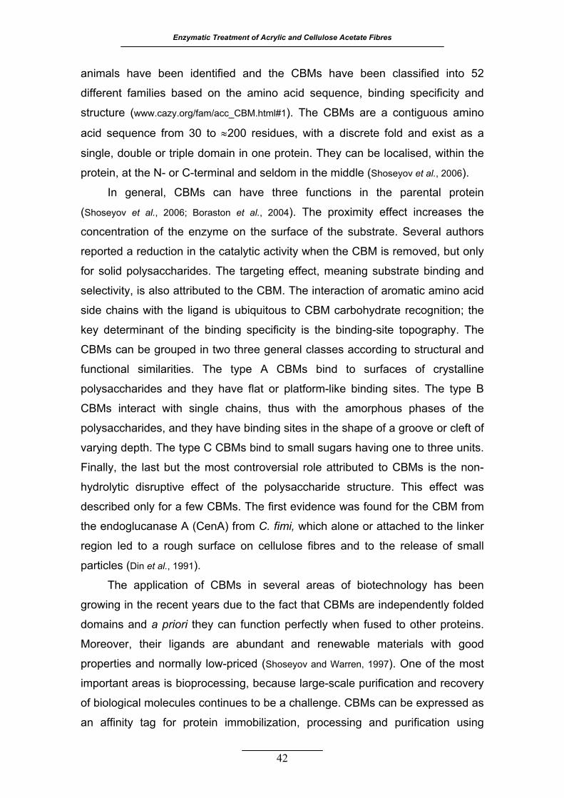
Enzymatic Treatment of Acrylic and Cellulose Acetate Fibres
animals have been identified and the CBMs have been classified into 52
different families based on the amino acid sequence, binding specificity and
structure (www.cazy.org/fam/acc_CBM.html#1). The CBMs are a contiguous amino
acid sequence from 30 to ≈200 residues, with a discrete fold and exist as a
single, double or triple domain in one protein. They can be localised, within the
protein, at the N- or C-terminal and seldom in the middle (Shoseyov et al., 2006).
In general, CBMs can have three functions in the parental protein
(Shoseyov et al., 2006; Boraston et al., 2004). The proximity effect increases the
concentration of the enzyme on the surface of the substrate. Several authors
reported a reduction in the catalytic activity when the CBM is removed, but only
for solid polysaccharides. The targeting effect, meaning substrate binding and
selectivity, is also attributed to the CBM. The interaction of aromatic amino acid
side chains with the ligand is ubiquitous to CBM carbohydrate recognition; the
key determinant of the binding specificity is the binding-site topography. The
CBMs can be grouped in two three general classes according to structural and
functional similarities. The type A CBMs bind to surfaces of crystalline
polysaccharides and they have flat or platform-like binding sites. The type B
CBMs interact with single chains, thus with the amorphous phases of the
polysaccharides, and they have binding sites in the shape of a groove or cleft of
varying depth. The type C CBMs bind to small sugars having one to three units.
Finally, the last but the most controversial role attributed to CBMs is the non-
hydrolytic disruptive effect of the polysaccharide structure. This effect was
described only for a few CBMs. The first evidence was found for the CBM from
the endoglucanase A (CenA) from C. fimi, which alone or attached to the linker
region led to a rough surface on cellulose fibres and to the release of small
particles (Din et al., 1991).
The application of CBMs in several areas of biotechnology has been
growing in the recent years due to the fact that CBMs are independently folded
domains and a priori they can function perfectly when fused to other proteins.
Moreover, their ligands are abundant and renewable materials with good
properties and normally low-priced (Shoseyov and Warren, 1997). One of the most
important areas is bioprocessing, because large-scale purification and recovery
of biological molecules continues to be a challenge. CBMs can be expressed as
an affinity tag for protein immobilization, processing and purification using
42

1. Introduction
cellulose as a matrix in many formats, from affinity chromatography to two
phase liquid separations (McCormick and Berg, 1997). Targeting of compounds to
polysaccharides that are present in many daily products, especially cellulose, is
also an important application of CBMs. This area includes oral care products,
denim stonewashing, the targeting of enzymes that do not possess natural
affinity to cotton, in laundry detergents, and other chemicals, like fragrances (von
der Osten et al., 2000a,b; Cavaco-Paulo, 1995; Kalum and Andersen, 2000; Berry et al., 2001;
Fuglsang and Tsuchiya, 2001). Other applications include whole cell immobilization,
enzyme immobilization and modification of polysaccharide fibres by non-
hydrolytic disruptive activity (Shoseyov et al., 2006; Pinto et al., 2004). The application
of CBMs is faraway from being exhausted and will be further expanded with the
increasing knowledge on these binding domains.
1.6.3 CARBOHYDRATE-BINDING MODULE OF CELLOBIOHYDROLASE I FROM
TRICHODERMA REESEI
In the 1980s, major steps were taken in the understanding of how
cellulolytic enzymes work. Many studies were conducted with the fungus T.
reesei and it was evident that the four cellulases, known at that time, shared a
common structural organization. They consisted in a catalytic domain (≈400-500
residues), a highly conserved terminal domain (≈40 residues), either at the C- or
N-terminal and a heavily O-glycosylated linker region (≈30 residues) connecting
the two domains (Fägerstam et al., 1984; Bhikahbhai and Pettersson, 1984; van Tilbeurgh et
al., 1986). The limited proteolysis of the cellobiohydrolase I from T. reesei (CBH I)
led to the first clue on the function of the conserved C-terminal domain (van
Tilbeurgh et al., 1986). The core protein was fully active against small, soluble
substrates, while the whole cellulase, with the C-terminal peptide was active
against Avicel (insoluble microcrystalline cellulose). The implication of the C-
terminal peptide of CBH I in the adsorption process and enhancement of
cellulase activity towards solid substrates was confirmed later on and it was
demonstrated that the C-terminal domain and the linker interacted directly with
insoluble cellulose independently of the presence of the core domain (Tomme et
al., 1988; Stahlberg et al., 1988). This cellulose-binding activity was eliminated by
43
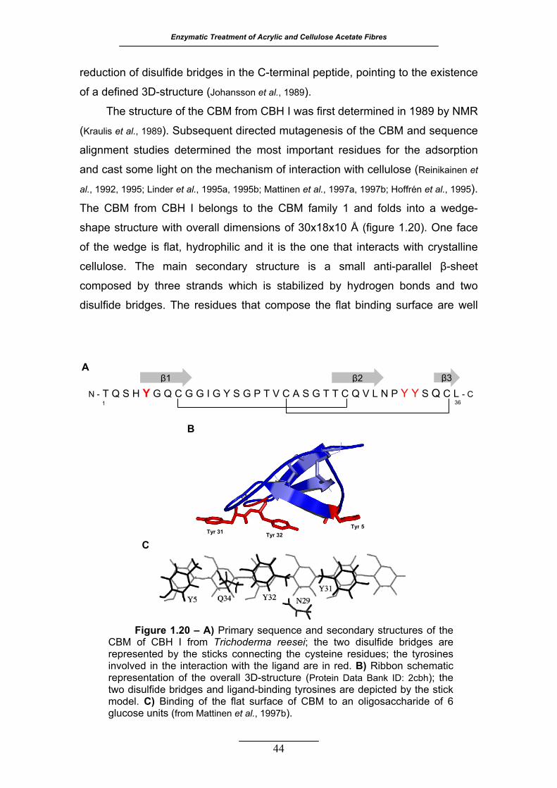
Enzymatic Treatment of Acrylic and Cellulose Acetate Fibres
reduction of disulfide bridges in the C-terminal peptide, pointing to the existence
of a defined 3D-structure (Johansson et al., 1989).
The structure of the CBM from CBH I was first determined in 1989 by NMR
(Kraulis et al., 1989). Subsequent directed mutagenesis of the CBM and sequence
alignment studies determined the most important residues for the adsorption
and cast some light on the mechanism of interaction with cellulose (Reinikainen et
al., 1992, 1995; Linder et al., 1995a, 1995b; Mattinen et al., 1997a, 1997b; Hoffrén et al., 1995).
The CBM from CBH I belongs to the CBM family 1 and folds into a wedge-
shape structure with overall dimensions of 30x18x10 Å (figure 1.20). One face
of the wedge is flat, hydrophilic and it is the one that interacts with crystalline
cellulose. The main secondary structure is a small anti-parallel β-sheet
composed by three strands which is stabilized by hydrogen bonds and two
disulfide bridges. The residues that compose the flat binding surface are well
A
36
N - T Q S H Y G Q C G G I G Y S G P T V C A S G T T C Q V L N P Y Y S Q C L - C
β3 β1 β2
1
B
Tyr 5Tyr 31 Tyr 32
C
Figure 1.20 – A) Primary sequence and secondary structures of the CBM of CBH I from Trichoderma reesei; the two disulfide bridges are represented by the sticks connecting the cysteine residues; the tyrosines involved in the interaction with the ligand are in red. B) Ribbon schematic representation of the overall 3D-structure (Protein Data Bank ID: 2cbh); the two disulfide bridges and ligand-binding tyrosines are depicted by the stick model. C) Binding of the flat surface of CBM to an oligosaccharide of 6 glucose units (from Mattinen et al., 1997b).
44
1. Introduction
conserved among fungal CBMs. In the CBM from CBH I, the three tyrosines
Tyr5, Tyr31 and Tyr32 are involved in the interaction with the glucose rings of
cellulose, mainly through van der Waals forces and their substitution leads to a
significant decrease in the binding affinity.
The construction of a bifunctional CBM, using the CBM from CBH II and
CBH I spaced by a small linker, showed that the connection affects the binding
of the individual CBMs (Linder et al., 1996). The double CBM exhibits higher
affinity to the solid substrate than the isolated CBMs. Another work
demonstrated the reversibility with a high exchange rate of the CBM from CBH I
which is crucial for the mobility of CBH I on the cellulose surface and its
continuous activity (Linder and Teeri, 1996).
1.6.4 CARBOHYDRATE-BINDING MODULE N1 OF ENDOGLUCANASE C FROM
CELLULOMONAS FIMI
The gene and its product, the endoglucanase C (CenC), from the
bacterium C. fimi, were first identified in 1989, after other cellulases from the
same organism had already been characterized (Moser et al., 1989). The complete
sequence of cenC gene and the characterization of the cellulase CenC were
accomplished later, revealing an unusual organization that did not resemble the
other cellulases from C. fimi found previously (Coutinho et al., 1991). The
identification of the cellulose-binding domain was possible by the dissection of
the gene and independent expression of the different sequences (Coutinho et al.,
1992). It was found that CenC has two CBMs (N1 and N2) in tandem at the N-
terminal, with distinct structures and affinities (Coutinho et al., 1993). The CBMN1
binds preferentially amorphous cellulose and can also bind Avicel weakly but
not crystalline cellulose (Tomme et al., 1996a).
The structure of the CBMN1 was first determined in 1996 by NMR (Johnson
et al., 1996a, 1996b). Subsequent directed mutagenesis and structure
determination of the CBMN1 in complex with an oligosaccharide by NMR and X-
ray crystallography have elucidated the interaction of this CBM with a single
chain of cellulose and revealed the most important residues (Kormos et al., 2000;
Johnson et al., 1999; Boraston et al., 2002). The CBMN1 belongs to the CBM family 4
and it folds into jelly-roll β-sandwich (figure 1.21). There are no α-helices, the
45
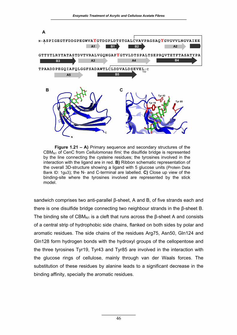
Enzymatic Treatment of Acrylic and Cellulose Acetate Fibres
46
sandwich comprises two anti-parallel β-sheet, A and B, of five strands each and
there is one disulfide bridge connecting two neighbour strands in the β-sheet B.
The binding site of CBMN1 is a cleft that runs across the β-sheet A and consists
of a central strip of hydrophobic side chains, flanked on both sides by polar and
aromatic residues. The side chains of the residues Arg75, Asn50, Gln124 and
Gln128 form hydrogen bonds with the hydroxyl groups of the cellopentose and
the three tyrosines Tyr19, Tyr43 and Tyr85 are involved in the interaction with
the glucose rings of cellulose, mainly through van der Waals forces. The
substitution of these residues by alanine leads to a significant decrease in the
binding affinity, specially the aromatic residues.
N
C
Tyr 43
Tyr 85 Tyr 19
Figure 1.21 – A) Primary sequence and secondary structures of the CBMN1 of CenC from Cellulomonas fimi; the disulfide bridge is represented by the line connecting the cysteine residues; the tyrosines involved in the interaction with the ligand are in red. B) Ribbon schematic representation of the overall 3D-structure showing a ligand with 5 glucose units (Protein Data Bank ID: 1gu3); the N- and C-terminal are labelled. C) Close up view of the binding-site where the tyrosines involved are represented by the stick model.
N–ASPIGEGTFDDGPEGWVAYGTDGPLDTSTGALCVAVPAGSAQYGVGVVLNGVAIEE GTTYTLRYTATASTDVTVRALVGQNGAPYGTVLDTSPALTSEPRQVTETFTASATYPA TPAADDPEGQIAFQLGGFSADAWTLCLDDVALDSEVEL-C
1
152
A1 B1 B2 A2
B3 A3 A4
A5
B4
B5
A
B C
1. Introduction
47
11..77 AAIIMMSS OOFF TTHHEE WWOORRKK
In the industrial processing of natural textile fibres, several enzyme
applications have been introduced. The same did not happen regarding the
processing of most man-made fibres, specially the synthetic ones. Conventional
modifications of such fibres require high amounts of energy and chemicals in
order to obtain the desired end-product properties.
The general purpose of the present work is the development of eco-
friendly methodologies to produce value added fibres with new surface
functionalities, through the application of enzymes for the specific and targeted
modification of the surface of acrylic and cellulose acetate fibres. More precise
objectives are the characterization of the fibre modifications by physicochemical
methods and the improvement of the enzymes performance through the
manipulation of the treatment media and recombinant DNA technologies.

48
UUSSIINNGG AA NNIITTRRIILLAASSEE FFOORR TTHHEE
SSUURRFFAACCEE MMOODDIIFFIICCAATTIIOONN OOFF
AACCRRYYLLIICC FFIIBBRREESS
he modification of nitrile surface groups into carboxylic groups by
the action of a commercial nitrilase, is reported in this chapter.
The enzymatic modification of acrylic fibres is a heterogeneous process
and, for that reason, requires longer treatments than one-phase
catalysis. The reaction media was manipulated with the purpose of
improving nitrilase efficiency. The nitrilase catalysis was further studied
without additives for a better understanding of the chemical
modifications that were occurring at the acrylic fibre surface.
The direct enzymatic modification of nitrile into carboxylic groups
constitutes a promising approach to make the acrylic fibre more
hydrophilic, enhancing the basic dye uptake of PAN treated fabrics and
producing surface reactive chemical sites that may be the target of
other finishing processes.
T
22
Enzymatic Treatment of Acrylic and Cellulose Acetate Fibres
49
22.. UUSSIINNGG AA NNIITTRRIILLAASSEE FFOORR TTHHEE SSUURRFFAACCEE MMOODDIIFFIICCAATTIIOONN OOFF
AACCRRYYLLIICC FFIIBBRREESS
22 ..11 MMAATTEERRIIAALLSS AANNDD MMEETTHHOODDSS
2.1.1 REAGENTS AND ENZYMES
The enzyme used in this work was a commercial nitrilase (EC 3.5.5.1),
Cyanovacta Lyase, supplied by Novacta Byosystems Ltd, Hatfield, Herts, UK.
The acrylic taffeta fabric used was produced from a copolymer of PAN and
7% vinyl acetate, with 82 g m-2 and 36/36 ends/picks per cm, supplied by
Fisipe, Lavradio, Portugal.
The cationic dye Basic Blue 9, C.I. 52015, was from Carlo Erba, Milan,
Italy. All other reagents were laboratory grade reagents from Sigma-Aldrich, St.
Louis, USA, unless stated otherwise. The buffer used throughout the work was
a phosphate buffer having a concentration of 50 mM and pH 7.8. The
exceptions are specified when necessary.
2.1.2 NITRILASE ACTIVITY ASSAY
Nitrilase activity was determined by the quantification of the ammonia
released into the reaction media, using benzonitrile as the substrate. The
reaction was started by the addition of 0.25 mL of a buffered nitrilase solution
containing 0.08 mg mL-1 of total protein to 0.25 mL of 19.4 mM benzonitrile. The
mixture was incubated for 1 to 10 min in a water bath at 30 ºC. The reaction
was stopped by adding 0.1 mL of 2 M hydrochloric acid. The ammonia was
quantified using an enzymatic kit from Sigma Diagnostics, St. Louis, USA (cat.
No 171-A). All the assays were performed in triplicate. One unit of nitrilase initial
activity was defined as one µmol of ammonia released per minute.

2. Using a Nitrilase for the Surface Modification of Acrylic Fibres
2.1.3 PH AND TEMPERATURE PROFILE
Nitrilase solutions with 0.08 mg ml-1 of total protein were incubated for 10
min at 30 ºC in 300 mM Britton-Robinson buffer of constant ionic strength. The
pH range tested was 4.5 to 10.6.
The effect of temperature was investigated within the range 20 ºC - 60 ºC.
Preparations of buffered nitrilase solution with 0.08 mg mL-1 total protein were
incubated for 10 minutes, at the tested temperatures.
The nitrilase activity was measured as described previously.
2.1.4 STABILITY OF NITRILASE
The operational stability of nitrilase was investigated in the presence of
two organic solvents and several polyalcohols. The buffered enzyme was
incubated at a final total protein concentration of 0.08 mg mL-1 in different media
compositions: 4%-15% v/v of DMA, 4%-15% DMF or 1 M of a polyalcohol. The
polyalcohols studied were glycerol, sorbitol and ethylene glycol (EG). The
enzyme was also incubated at the same final concentration with 4% v/v DMA
and 1 M sorbitol. For each assay, the final volume was 25 mL, kept in 50 mL
polypropylene containers. The control was identically prepared except that
buffer substituted the enzyme. The incubation took place in a water bath at
30 ºC, with an orbital agitation, until the drop in nitrilase activity was above 50%.
The enzyme activity was assayed at proper time intervals as previously
described.
2.1.5 EFFECT OF ADDITIVES IN THE ENZYMATIC TREATMENT OF ACRYLIC FABRIC
All samples of acrylic fabric used were washed prior to use in order to
remove possible impurities from manufacture and from human handling. The
washing was performed at 60 ºC with an aqueous solution of 1.0 g L-1 Lutensol
AT25, a non-ionic detergent (BASF, Ludwigshafen, Germany). After removing
the detergent, the fabric was washed with distilled water and allowed to air dry.
The treatment of acrylic fabric was carried out in stainless steel pots of
450 mL in capacity, housed in a laboratory scale machine Rotawash MKIII
(vertical agitation simulating European washing machines, from SDL
International Ltd.), operating at 30 ºC and 20 rpm, for 116 hours. The samples
50
Enzymatic Treatment of Acrylic and Cellulose Acetate Fibres
of acrylic fabric, having an average weight of 9 g, were incubated with 24 U of
nitrilase per gram of fabric, in a final volume of 360 mL of buffer. Three different
media were tested: no additives, 4% v/v DMA plus 1 M sorbitol and the control
without enzyme and without additives.
After the enzymatic treatment, all fabric samples were immediately
washed to remove the adsorbed protein. The samples were left under running
tap water for several minutes, then they were washed with 2 g L-1 sodium
carbonate and distilled water, for 60 min each, at 60 ºC.
2.1.6 ENZYMATIC TREATMENT OF ACRYLIC FABRIC
All samples of acrylic fabric used were previously washed, as already
described. The acrylic fabric was treated with 412 U of nitrilase per gram of
fabric in buffer, at 40 ºC. The acrylic fabric samples were treated in independent
containers, one piece of fabric and bath solution for each time determination, in
the Rotawash MKIII at 20 rpm. Samples of acrylic fabric with an average weight
of 2 g were incubated in a final volume of 100 mL of treatment solution for 0, 2,
4, 8, 24, and 36 hours.
After enzymatic treatment, all fabric samples were washed twice in 2 g L-1
sodium carbonate for 20 min, once in 4 g L-1 Lutensol AT25 for 20 min and
three times in distilled water for 25 min, in the Rotawash at 70 ºC. Before and in
between the changes of the washing solution, the samples were left under
running tap water.
2.1.7 QUANTIFICATION OF TOTAL PROTEIN CONCENTRATION
Total protein in solution was quantified following Bradford methodology,
using BSA as standard (Bradford, 1976). All samples were measured in triplicate.
2.1.8 DETERMINATION OF AMMONIA CONCENTRATION
Detection of ammonia in the reaction media of the first acrylic fabric
treatment (section 2.1.5) was performed using the enzymatic kit from Sigma
Diagnostics.
For the second nitrilase treatment (section 2.1.6), the Nessler method was
adapted and applied to quantify ammonia (Greenberg et al., 1992). The protein was
51
2. Using a Nitrilase for the Surface Modification of Acrylic Fibres
previously precipitated using trichloroacetic acid and the pH of supernatants
was neutralized with 250 mM sodium hydroxide. The standard solutions were
prepared using anhydrous ammonium chloride and were submitted to the same
protein precipitation procedure and pH adjustment. For the Nessler
determination, 0.5 mL of samples/standards were diluted with 4.5 mL of distilled
water in glass tubes and 10 µL of Rochelle salt (aqueous solution: 50% w/v
potassium sodium tartrate tetrahydrated) were added. After mixing well, 0.2 mL
of Nessler reagent was added to each tube and again mixed. The colour was
allowed to develop for 10 min and the absorbance was registered at 425 nm. All
the assays were performed at least in duplicate.
2.1.9 DETERMINATION OF POLYACRYLIC ACID CONCENTRATION
Detection of PAA in the reaction media was performed by an absorption-
colorimetric method using a kit from Hach Company, USA (cat. No. 22252-00
and No. 22257-00). The standards were prepared from aqueous solutions of
PAA (average molecular weight of 2000 g mol-1). All the assays were performed
in duplicate.
2.1.10 DETERMINATION OF FABRIC WEIGHT DIFFERENCES
The fabric weight differences were determined by the difference between
the weight of the dry fabric before and after the enzymatic treatment. The fabric
pieces were whipstitched with the same acrylic yarn before the treatment. After
the washing procedures, the samples were dried at 60 ºC for three hours in a
laboratory oven and they were then conditioned in a glass desiccator for 30 min.
The drying and desiccation were repeated until differences in the measures
were not significant (<5%).
2.1.11 ACRYLIC FABRIC COLOURATION WITH A BASIC DYE
After the enzymatic treatment, samples were competitively coloured in
duplicate with 1% o.w.f. (of weight of fabric) Basic Blue 9, C.I.52015. The
colouration was performed in a lab-scale dyeing machine (AHIBA Spectradye,
from Datacolor International) with a temperature gradient of 4 ºC min. For a
more detailed procedure, see table 2.1.
52
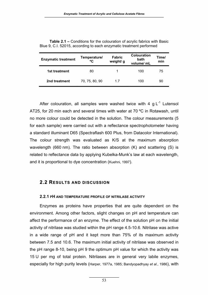
Enzymatic Treatment of Acrylic and Cellulose Acetate Fibres
53
Table 2.1 – Conditions for the colouration of acrylic fabrics with Basic
Blue 9, C.I. 52015, according to each enzymatic treatment performed
Enzymatic treatment Temperature/ ºC
Fabric weight/ g
Colouration bath
volume/ mL Time/ min
1st treatment 80 1 100 75
2nd treatment 70, 75, 80, 90 1.7 100 90
After colouration, all samples were washed twice with 4 g L-1 Lutensol
AT25, for 20 min each and several times with water at 70 ºC in Rotawash, until
no more colour could be detected in the solution. The colour measurements (5
for each sample) were carried out with a reflectance spectrophotometer having
a standard illuminant D65 (Spectraflash 600 Plus, from Datacolor International).
The colour strength was evaluated as K/S at the maximum absorption
wavelength (660 nm). The ratio between absorption (K) and scattering (S) is
related to reflectance data by applying Kubelka-Munk’s law at each wavelength,
and it is proportional to dye concentration (Kuehni, 1997).
22..22 RREESSUULLTTSS AANNDD DDIISSCCUUSSSSIIOONN
2.2.1 PH AND TEMPERATURE PROFILE OF NITRILASE ACTIVITY
Enzymes as proteins have properties that are quite dependent on the
environment. Among other factors, slight changes on pH and temperature can
affect the performance of an enzyme. The effect of the solution pH on the initial
activity of nitrilase was studied within the pH range 4.5-10.6. Nitrilase was active
in a wide range of pH and it kept more than 75% of its maximum activity
between 7.5 and 10.6. The maximum initial activity of nitrilase was observed in
the pH range 8-10, being pH 9 the optimum pH value for which the activity was
15 U per mg of total protein. Nitrilases are in general very labile enzymes,
especially for high purity levels (Harper, 1977a, 1985; Bandyopadhyay et al., 1986), with

2. Using a Nitrilase for the Surface Modification of Acrylic Fibres
optimum pH values ranging between 7.5 and 8 (Banerjee et al., 2002; O’Reilly and
Turner, 2003). The commercial enzyme used throughout this work is supplied as
cell preparation and according to the results obtained, this nitrilase has a
relative high optimum pH, resembling nitrilases from fungi and from the
bacterium Klebsiella ozaenae (Harper, 1977b; Stalker et al., 1988). According to the
suppliers, the enzyme solution should be maintained in the pH region 7-8, thus
the pH 7.8 was chosen to perform all the enzymatic treatments.
The temperature effect on nitrilase initial activity was studied between
20 ºC and 60 ºC. The maximum formation of ammonia occurred between 40 ºC
and 45 ºC. The highest value of activity was 33 U mg-1, obtained at 45 ºC.
Above this temperature, there was a sharp decrease in the nitrilase activity. In
the literature, the optimum temperature for mesophilic nitrilases varies
considerably but there is some agreement in considering this group of enzymes
very sensitive to this parameter (Cowan et al., 1998). The range of temperatures
for which enzymes are stable is often below the optimum value.
2.2.2 STABILITY OF NITRILASE
Operational stability, defined as the persistence of enzyme activity under
the conditions of use, was studied before applying the enzyme to acrylic fabric
treatment (Ó’Fágáin, 2003). The effect of the addition of two organic solvents and
polyalcohols on nitrilase activity was studied by comparing the half-life time, as
a measure of stability. The values of half-life time were calculated from first
order exponentional decay fitting of data, using OriginPro 7.5 SR0 (OriginLab
Corporation, Northampton, USA). The decay of nitrilase activity obeyed to this
exponential model of enzyme deactivation.
The addition of both organic solvents, DMA and DMF, to the enzyme
solution decreased the initial activity and the half-life time of nitrilase. For many
enzymes, deactivation in homogeneous water-organic solvent mixtures may be
due to the disruption of the quaternary structure, disruption of the protein
hydrophobic core and/or to the water stripping (Castro and Knubovets, 2003). This
effect was more pronounced as the organic cosolvent concentration increased.
The decrease in the half-life time is almost linear with the increase in the
organic solvent concentration (table 2.2). The decrease of nitrilase activity was
54

Enzymatic Treatment of Acrylic and Cellulose Acetate Fibres
55
faster and more pronounced when DMF was added. Therefore, DMA should be
used instead of DMF regarding the enzymatic treatments of acrylic fibre. The
investigated organic solvents were chosen because of their action on the fibre
structure. They are known solvents of PAN and they are commonly used in
industrial production of acrylic fibre (Burkinshaw, 1995; Capone, 1995). Their
plasticizer function disturbs the regular structure of the polymer, reducing the
magnitude of interchain bonding, which should aid the accessibility to the
enzyme, improving its action on the fibre.
Table 2.2 – Influence of organic solvents and polyalcohols on initial
activity and half-life time of Nitrilase, at 30 ºC and pH 7.8. An initial activity and a half-life time of 100% correspond to 12.6 U mg-1 and 15 hours, respectively. The half-life time was calculated as kt 2ln
21 =
kxtt eaa −== .0
, from the 1st
order exponential decay fit of data ( )
Assay conditions Initial activitya (%) Half-life timea (%)
Control No additives 100 100
DMF
4 % 87 30
10 % 78 19
15 % 59 9
DMA
4 % 84 90
10 % 89 55
15 % 87 19
Polyalcohols
Glycerol 122 101
Sorbitol 122 294
EG 107 160
4% DMA + 1 M sorbitol 105 249 a The standard deviations for the initial activity and half-life time values were
≤ 5% and ≤ 7%, respectively.
The initial activity of nitrilase increased in the presence of all the studied
polyalcohols, particularly, sorbitol and glycerol (table 2.2). It cannot be excluded
that this effect could be due to a better solubility of the substrate benzonitrile in
2. Using a Nitrilase for the Surface Modification of Acrylic Fibres
the media containing these compounds. The highest half-life time values were
obtained with the addition of sorbitol and EG. Sorbitol showed the best
performance as a nitrilase stabilizer for long-term treatments. The half-life time
increased almost three times (from 15 to 44 hours) in the presence of sorbitol. It
has been found that the addition of high concentrations (usually higher than
1 M) of some low molecular weight compounds in solution stabilizes the native
conformation of globular proteins (Xie and Timasheff, 1997; Timasheff, 1995).
A combined effect of the organic solvent and the stabilizer was studied in
order to check the possibility of a compromise between an increase in the
accessibility of the nitrilase to acrylic fibre and a good operational stability of the
enzyme. The least deleterious condition studied was 4% (v/v) of DMA. The
addition of 1 M of sorbitol to this medium proved to be beneficial (table 2.2). The
initial activity was slightly improved and the half-life time, although shorter than
the one obtained only in the presence of sorbitol, was still 249% of the half-life
time for the control (no additives). The half-life time of nitrilase without additives
increased from 15 to 38 hours in the presence of the additives. Therefore, these
conditions were chosen for an enzymatic treatment of acrylic fabric.
2.2.3 EFFECT OF ADDITIVES ON THE MODIFICATION OF ACRYLIC SURFACE WITH
NITRILASE
The hydrolysis of nitrile groups from the acrylic fibre should result in the
formation of carboxylic groups at the fibre surface and in the release of
ammonia to the treatment solution.
The formation of carboxylic groups at the fibre surface was evaluated by
colouring the fabric with a basic dye, which has a cationic group able to
establish ionic bonds with anionic groups on the fibre. If nitrilase is able to
modify some of the nitrile groups into carboxylic groups, at the surface, then
more dye can be absorbed into the fibre and this could be seen as an increase
in K/S. The colouration methodology was already reported and proved to be a
valuable and a very sensitive qualitative method (Silva et al., 2005a, 2005b).
For samples treated with nitrilase without additives, the K/S value
increased 156% at the dyeing temperature of 80 ºC (table 2.3). The dye uptake
was even more pronounced by the addition of sorbitol and DMA to the
56
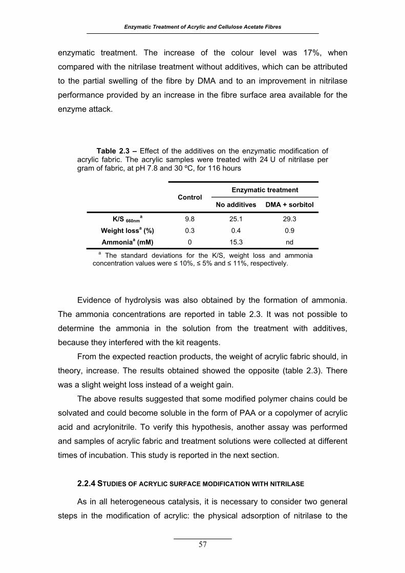
Enzymatic Treatment of Acrylic and Cellulose Acetate Fibres
enzymatic treatment. The increase of the colour level was 17%, when
compared with the nitrilase treatment without additives, which can be attributed
to the partial swelling of the fibre by DMA and to an improvement in nitrilase
performance provided by an increase in the fibre surface area available for the
enzyme attack.
Table 2.3 – Effect of the additives on the enzymatic modification of
acrylic fabric. The acrylic samples were treated with 24 U of nitrilase per gram of fabric, at pH 7.8 and 30 ºC, for 116 hours
Control
Enzymatic treatment
No additives DMA + sorbitol
K/S 660nma 9.8 25.1 29.3
Weight lossa (%) 0.3 0.4 0.9
Ammoniaa (mM) 0 15.3 nd a The standard deviations for the K/S, weight loss and ammonia
concentration values were ≤ 10%, ≤ 5% and ≤ 11%, respectively.
Evidence of hydrolysis was also obtained by the formation of ammonia.
The ammonia concentrations are reported in table 2.3. It was not possible to
determine the ammonia in the solution from the treatment with additives,
because they interfered with the kit reagents.
From the expected reaction products, the weight of acrylic fabric should, in
theory, increase. The results obtained showed the opposite (table 2.3). There
was a slight weight loss instead of a weight gain.
The above results suggested that some modified polymer chains could be
solvated and could become soluble in the form of PAA or a copolymer of acrylic
acid and acrylonitrile. To verify this hypothesis, another assay was performed
and samples of acrylic fabric and treatment solutions were collected at different
times of incubation. This study is reported in the next section.
2.2.4 STUDIES OF ACRYLIC SURFACE MODIFICATION WITH NITRILASE
As in all heterogeneous catalysis, it is necessary to consider two general
steps in the modification of acrylic: the physical adsorption of nitrilase to the
57

2. Using a Nitrilase for the Surface Modification of Acrylic Fibres
58
fibre surface and the formation of the enzyme/substrate complex and
consequent hydrolysis of the nitrile group.
The protein adsorption was indirectly calculated by a decrease in total
protein remaining in the treatment solution (figure 2.1). In the first 8 hours of
treatment, there was a sharp decrease in the protein content in the treatment
solution. Afterwards, the equilibrium around an average value was reached,
with some oscillations due to alternating adsorption and desorption phenomena
(Azevedo et al., 2000). The equilibrium value of protein concentration in the
solution was around 9 mg L-1, meaning a total protein adsorption of 80%, at
40 ºC.
There was no significant ammonia release before 8 hours of treatment
(figure 2.1). It is interesting to notice that the release of ammonia into treatment
solutions was coincident with the achievement of the adsorption equilibrium.
This lag phase is also seen in homogeneous catalysis of nitrilases whose
activity onset depends on the assembly of its oligomeric active form (Harper,
1977a). The ammonia release is a good indicator of successful nitrilase
catalysis, which should render an improvement of the acrylic dyeability. It also
shows that a certain amount of those 80% of total protein adsorption must be
nitrilase and that the enzyme is able to recognize the nitrile groups of acrylic
fabric as the substrate.
Figure 2.1 – Total protein and nitrogen concentrations in the treatment solutions. The acrylic samples were treated with 412 U of Cyanovacta Lyase per gram of fabric, at pH 7.8 and 40 ºC.
0
10
20
30
40
50
60
0 5 10 15 20 25 30 35 40
Time of treatment (h)
Tota
l pro
tein
(mg
L-1)
0
5
10
15
20
25
30
Total nitrogen (mg L
-1)
proteinnitrogen

Enzymatic Treatment of Acrylic and Cellulose Acetate Fibres
59
The linear release of ammonia also implies the absence of activity decay
during the 36 hours of treatment. Since its half-life time in solution was 15
hours, it is feasible to assume that the adsorption of the enzyme to acrylic led to
an increase in its stability, resembling the immobilization procedures employed
to stabilize proteins.
In the figure 2.2, the values of K/S are represented as a percentage of the
initial value. The relative K/S had a waving behaviour with incubation time. This
behaviour was attenuated with the increase in the colouration temperature, as
expected. Enzymes are large molecules and they will not penetrate inside the
tight structure of PAN fibres. Therefore, the chemical changes catalysed by
nitrilase are located at the surface. At lower temperatures, the adsorption of dye
occurs mostly at the fibre surface where the enzymatic catalysis takes place,
producing different K/S values between samples treated with enzyme and
controls. The significant raise in dye diffusion that occurs at or about the glass
transition temperature (Tg) is often referred as the dyeing transition temperature
(Td). Both transition temperatures, Tg and Td, indicate the onset of segmental
mobility of the polymer chains and the consequent increase of the free volume
within the polymer (Burkinshaw, 1995). Approaching these temperatures, the
colour depth increases sharply for both enzymatic treated and untreated
Figure 2.2 – Relative K/S values for acrylic fabric treated with 412 U of Cyanovacta Lyase, at pH 7.8 and 40 ºC. Samples and controls were competitively coloured at 70, 75, 80 and 90 ºC. Relative K/S was calculated as
control
enzyme
K/SK/S (%).
0
20
40
60
80
100
120
140
160
0 5 10 15 20 25 30 35 40
Time of treatment (h)
Rel
ativ
e K
/S ( λ
max
= 6
60 n
m) (
%)
70 ºC75 ºC80 ºC90 ºC

2. Using a Nitrilase for the Surface Modification of Acrylic Fibres
60
samples and camouflages the differences in K/S due to superficial changes.
This is the reason for the observed decrease in the amplitude of the oscillation
of relative K/S with increasing colouration temperatures (70 ºC to 80 ºC). At
90 ºC, which is above Td, the differences are absent.
The oscillation of relative K/S values with time of treatment could be
explained assuming that above a threshold conversion of nitrile groups into
carboxylic groups, the polymer chains would be more stable in solution and
would be detached from the surface of the fibre. The modification of CN into
COOH groups could create some instability in the arrangement of PAN chains
at the fibre surface, mainly due to steric hindrance and pH dependent charge
repulsion. It was possible to detect increasing concentrations of PAA in
treatment solutions (figure 2.3). The removal of carboxylic groups from the
surface of the fabric could be the cause of the lower points on the K/S curve.
When the PAA macromolecules leave the surface, the underneath PAN chains
are exposed to further nitrilase catalysis. This leads to an increasing number of
carboxylic groups, thus, to an increase of K/S, until the threshold value for chain
solubilization is again achieved (figure 2.4).
A maximum K/S value was observed for the treatment of 8 hours and it
corresponded to a relative K/S of 135% (for the lower colouration temperature).
Figure 2.3 – Relative K/S for acrylic fabric coloured at 70 ºC and polyacrylic acid concentration in the treatment solutions. The acrylic fibre was treated with 412 U of Cyanovacta Lyase per gram of fabric, at pH 7.8 and 40 ºC.
0
20
40
60
80
100
120
140
160
0 5 10 15 20 25 30 35 40
Time of treatment (h)
Rel
ativ
e K
/S ( λ
max
= 6
60 n
m) (
%)
0
10
20
30
40
50
60
70
80
Total PAA
(mg L
-1)
K/SPAA
Enzymatic Treatment of Acrylic and Cellulose Acetate Fibres
61
For longer incubation periods, the “wave amplitude” tends to decrease, as the
“length” tends to increase with time. This could be related to modifications in the
fibre surface, since there was not an activity decay of the enzyme. However, the
particular phenomenon that explains why this happens is still unclear.
The hypothetical model of “surface erosion” of acrylic fibre, here described
(figure 2.4), is very similar to the one applicable for synthetic polymers
biodegradation (Marten et al., 2003, 2005). The acrylic fibre surface is depleted
layer by layer, depending on factors such as micro-structural properties of PAN
copolymers, molar mass of the polymer chains, enzyme adsorption, nitrilase
deactivation, removal and dissolution of products.
22..33 CCOONNCCLLUUDDIINNGG RREEMMAARRKK SS
In spite of the increasing interest in the nitrilase enzymatic system and its
successful industrial applications using whole cells (Kobayashi and Shimizu, 1994;
Hughes et al., 1998), there is still much space to fulfil in the understanding and
improvement of the nitrile conversion by purified or semi-purified nitrilases. The
work reported evaluates the stabilization efficiency of various additives and the
influence of two organic cosolvents in the activity of a commercial nitrilase.
From the additives studied, the best stabilizing performance was induced by
sorbitol. Another important aspect is the fact that the organic solvent DMA, used
in acrylic fibre industry, revealed no significant loss in enzyme initial activity,
when used in small amounts.
It was also demonstrated in this study that a successful application of
nitrilase to the acrylic surface modification would be dependent on the control of
important factors like time and enzyme activity. The nitrilase action seems to
induce “surface erosion” of PAN fibres, by the dissolution of the modified
polymer molecules. This hypothesis is confirmed by the detection of PAA in the
treatment solutions and the increase of its concentration with the period of
incubation of acrylic fabric with nitrilase.
Further studies will be necessary to evaluate the impact of nitrilase activity
in the physical properties of the fabric and to assess if some amide groups were
also produced as by-products of nitrilase hydrolysis.
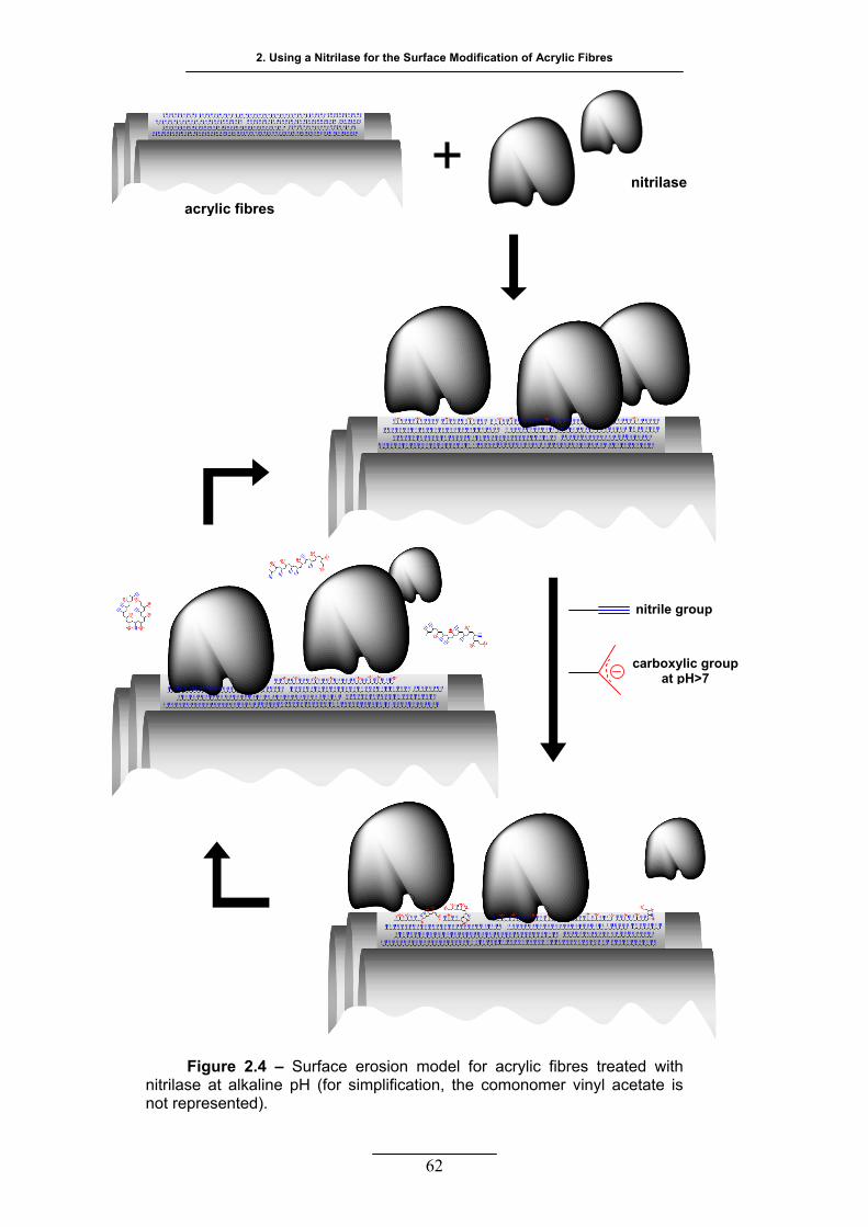
2. Using a Nitrilase for the Surface Modification of Acrylic Fibres
nitrilase
acrylic fibres
nitrile group
carboxylic group at pH>7
Figure 2.4 – Surface erosion model for acrylic fibres treated with nitrilase at alkaline pH (for simplification, the comonomer vinyl acetate is not represented).
62
Enzymatic Treatment of Acrylic and Cellulose Acetate Fibres
From the above considerations, it could be suggested that the stabilized
nitrilase has potential in textile industry for the modification of PAN fibres. The
wet spinning would be the most adequate industrial process for the introduction
of such enzymatic treatment, though it would demand a very fast enzymatic
modification (a few minutes). The accessibility of the fibre would be greater than
that of the woven fabric used in this study. If the application of the enzyme
occurred when the fibre is still in a gel state, a considerable reduction in the
time needed to maximize the enzymatic conversion into carboxylic groups could
occur. In addition, after coagulation, there are other possible stages for the
application of an enzymatic treatment, during which several finishing processes
normally take place. The nitrilase stability would not be so compromised by the
amount of solvent present as in earlier stages. In any case, a careful study is
needed to evaluate the feasibility of such application, but the work here reported
gives encouraging perspectives.
63

64
TTHH EE EEFF FF EE CC TT OO FF AADD DD II TT II VV EE SS AA NN DD
MMEE CC HH AA NN II CC AA LL AAGG II TT AA TT II OO NN II NN SSUU RR FF AA CC EE
MMOO DD II FF II CC AA TT II OO NN OO FF AACC RR YY LL II CC FF II BB RR EE SS BB YY
EESS TT EE RR AA SS EE SS
previous work reported that cutinase could be used to modify
the acrylic surface. After a long enzymatic treatment, the
hydrolysis of ester bonds from the comonomer vinyl acetate produced
acetic acid and hydroxyl groups on the surface of the fibre that could be
detected by reactive colouration (Silva et al., 2005a).
The major purpose of this work described in this chapter was to reduce
the treatment time of acrylic fabric with cutinase and to improve its
catalysis efficiency by means of reaction media manipulation. In order
to do that, the influence of known acrylic solvents and the influence of
known stabilizers on cutinase operational stability were investigated.
The impact of mechanical agitation and additives on vinyl acetate
enzymatic hydrolysis by cutinase and by a commercial esterase was
also studied.
A
33
Enzymatic Treatment of Acrylic and Cellulose Acetate Fibres
65
33.. TTHHEE EEFFFFEECCTT OOFF AADDDDIITTIIVVEESS AANNDD MMEECCHHAANNIICCAALL AAGGIITTAATTIIOONN
IINN SSUURRFFAACCEE MMOODDIIFFIICCAATTIIOONN OOFF AACCRRYYLLIICC FFIIBBRREESS BBYY EESSTTEERRAASSEESS
33..11 MMAATTEERRIIAALLSS AANNDD MMEETTHHOODDSS
3.1.1 REAGENTS AND ENZYMES
The enzymes used in this work were a cutinase (EC 3.1.1.74), from
Fusarium solani pisi, and a commercial esterase Texazym PES, from inoTEX
Ltd, Dvur Kralove nad Labem, Czech Republic. The recombinant wild type
cutinase was over-expressed in Saccharomyces cerevisiae SU50 strain and
supplied as culture medium, with 50% to 70% of purification degree in respect
to total protein (Silva et al., 2005a; Calado et al., 2002). It was a generous gift from
Centro de Engenharia Biológica, Instituto Superior Técnico, Lisbon, Portugal.
The acrylic taffeta fabric used was produced from a copolymer of PAN and
7% vinyl acetate, with 82 g m-2 and 36/36 ends/picks per cm, supplied by
Fisipe, Lavradio Portugal.
The reactive dye Remazol Brilliant Blue R, C.I. 61200, was acquired from
Sigma. All other reagents were laboratory grade reagents also from Sigma-
Aldrich, St. Louis, USA, unless stated otherwise. The buffer used throughout
this work was a phosphate buffer having a concentration of 50 mM and pH 8.
3.1.2 ESTERASE ACTIVITY ASSAY
Esterase activity was determined following the product release (p-
nitrophenol, p-NP) through the increase in the absorbance at 400 nm. The
activity assay conditions for cutinase were described previously (Silva et al.,
2005b; Shirai and Jackson, 1982). The esterase activity of Texazym PES was also
determined by using p-nitrophenyl butyrate (p-NPB) as substrate, but using
slightly different conditions that were optimized for this enzyme. The enzymatic
reaction was started with the addition of 0.1 mL of 10 mg L-1 Texazym PES to a
final volume of 2 mL of phosphate buffer, containing 75 µM p-NPB and 5% v/v
3. The Effect of Additives and Mechanical Agitation in Surface Modification of Acrylic Fibres by Cutinase and Esterase
ethanol. The mixture was incubated for 1.5 min in a water bath, at 35 ºC. The
hydrolysis was stopped by the addition of 2 mL of acetone. All the assays were
performed in triplicate. Standard solutions of p-NP were used to obtain the
calibration curve. One unit of esterase initial activity was defined as one µmol of
p-NP released per minute.
3.1.3 STABILITY OF CUTINASE
The operational stability of cutinase was investigated in the presence of
two organic solvents and several polyalcohols. The enzyme was incubated at a
final concentration 1 mg L-1 in phosphate buffer containing 0.05% w/v sodium
azide. The concentrations of DMA or DMF used in this study were 7.5%, 15%,
25% and 50% v/v. The final concentration of glycerol, sorbitol, xylitol and EG
used was 1 M. For each assay, the control was identically prepared except that
buffer substituted the enzyme. The incubation took place in a water bath at
35 ºC with orbital agitation until the drop in esterase activity was above 50%.
3.1.4 ENZYMATIC TREATMENT OF ACRYLIC FABRIC
All samples of acrylic fabric used were previously washed to remove
impurities. The washing consisted in several steps, all performed in a laboratory
scale machine, the Rotawash MKIII (vertical agitation simulating European
washing machines, from SDL International Ltd.), at 60 ºC and 20 rpm. The
fabric was washed twice for 30 min with 0.1 g L-1 Lutensol AT25 (non-ionic
detergent, BASF, Ludwigshafen, Germany) and left for 10 min under running
tap water. Then, the fabric was washed once for 30 min with 2 g L-1 sodium
carbonate and left for another 10 min under running tap water. Finally, the fabric
was washed three times in distilled water for 20 min each and was left to dry at
room temperature.
Two sets of experiments were carried out for each enzyme taking into
account the degree of mechanical agitation. For both sets, the treatment of
acrylic fabric was performed in stainless steel pots of 450 mL capacity in
Rotawash machine, in the case of cutinase, and in Washtec-P 05/99A (vertical
agitation simulating European washing machines, from Roaches International
Ltd.), in the case of Texazym. All the treatments were performed in phosphate
66
Enzymatic Treatment of Acrylic and Cellulose Acetate Fibres
buffer at 30 ºC and 20 rpm, for three hours. To increase the mechanical
agitation, stainless steel discs (19.2 g, 32 x 3 mm) were added to the reaction
mixture.
In the cutinase treatment, samples of acrylic fabric, having an average
weight of 1.57 g, were incubated with 9 U mL-1 of cutinase, in a final volume of
100 mL. Five different media were tested: no additives, 1% and 15% v/v of
DMA, 1 M glycerol and 1% DMA + 1 M glycerol. In a second set of assays,
these conditions were repeated and 4 stainless steel discs were added to each
assay.
In the treatment with Texazym PES, samples of acrylic fabric, having an
average weight of 3.05 g, were incubated with 3.4 U mL-1 of the enzyme, in a
final volume of 200 mL. The reaction media were the same as described for
cutinase. The number of discs added was raised to 9 per assay. For each
reaction media, a control was run in parallel in which buffer substituted the
enzyme.
After enzymatic treatment, all fabric samples were washed twice in 2 g L-1
sodium carbonate for 30 min, once in 0.25 g L-1 Lutensol AT25 for 30 min and
three times in distilled water for 15 min, in the Rotawash or Washtec-P at 70 ºC.
3.1.5 QUANTIFICATION OF TOTAL PROTEIN CONCENTRATION
Total protein in solution was quantified following Bradford methodology
(Bradford, 1976), using BSA as standard. All samples were measured in triplicate.
3.1.6 DETERMINATION OF ACETIC ACID CONCENTRATION IN THE BATH
SOLUTIONS
Detection of acetic acid in reaction media was performed with a kit from
Boehringer Mannhaim R-Biopharm (cat. No.10148261035), Darmstadt,
Germany. Protein was previously precipitated using perchloric acid according to
the manufacture instructions. The samples pH was neutralized using 1 M
potassium hydroxide and the subsequent salts were removed by centrifugation.
67

3. The Effect of Additives and Mechanical Agitation in Surface Modification of Acrylic Fibres by Cutinase and Esterase
68
3.1.7 ACRYLIC FABRIC COLOURATION WITH A REACTIVE DYE
After enzymatic treatment, samples were competitively coloured in
duplicate with 2% o.w.f. Remazol Brilliant Blue R. The colouration bath
consisted in a 10 g L-1 sodium carbonate solution. The colouration was
performed at 70 ºC and 80 ºC, in a lab dyeing machine (AHIBA Spectradye,
from Datacolor International), for 90 min at 20 rpm. The total fabric average
weight per colouration assay was 2.87 g and 2.18 g for samples treated with
cutinase and Texazym PES, respectively.
After colouration, all samples were washed once with 0.25 g L-1 Lutensol
AT25 and several times with water at 70 ºC in Rotawash, until no more dye
could be detected in the solution. The colour measurements (5 for each sample)
were carried out with a reflectance spectrophotometer having a standard
illuminant D65 (Spectraflash 600 Plus, from Datacolor International). The colour
strength was evaluated as K/S at maximum absorption wavelength (590 nm).
The ratio between absorption (K) and scattering (S) is related to reflectance
data by applying Kubelka-Munk’s law at each wavelength, and it is proportional
to dye concentration (Kuehni, 1997).
3.1.8 WIDE ANGLE X-RAY SCATTERING
The X-ray diffraction (XRD) patterns were obtained for the acrylic fabric
samples treated with cutinase without and with stainless steel discs (the later for
the samples treated in the presence of the additives). The WAXS experiments
were undertaken in a Philips PW1710 apparatus, using Cu Kα radiation and
operating at a 40 KV voltage and 30 mA current. The patterns were
continuously recorded in the diffraction angular range 2θ from 5º to 40º, with a
step size of 0.02º at 0.6º min-1.
The degrees of crystallinity were obtained according to the method of El-
Zaher (El-Zaher, 2001).
Enzymatic Treatment of Acrylic and Cellulose Acetate Fibres
69
33..22 RREESSUULLTTSS AANNDD DDIISSCCUUSSSSIIOONN
3.2.1 OPERATIONAL STABILITY OF CUTINASE
From the biotechnological point of view, both storage and operational
stabilities greatly influence the usefulness of enzyme-based products (Ó’Fágáin,
2003). Operational stability, defined as the persistence of enzyme activity under
the conditions of use, was studied for cutinase before applying the enzyme to
acrylic fabric treatment. The media conditions, such as buffer, pH and
temperature were chosen based on preliminary studies performed in the
laboratory, using the esterase activity determination methodology described
earlier. The conditions chosen, which maximize the hydrolysis of p-NPB by
cutinase, were phosphate buffer at pH 8 and temperatures comprised between
30 ºC and 40 ºC. These preliminary results were in agreement with those
described in literature (Petersen et al., 1998, 2001b; Melo et al., 1997). Under the
reported conditions, the specific activities of cutinase and Texazym PES were
253 ± 51 and 11 ± 1 U mg-1, respectively.
The half-life times obtained for cutinase, incubated under different
conditions, are shown in table 3.1, as a measure of operational stability. The
values of half-life time were calculated whenever the experimental data was
adequately fitted with a first order exponential decay, using OriginPro 7.5
(Origin Lab Corporation, USA). This was the case for the assays where
cutinase was incubated with 7.5% and 15% of both organic solvents and with
EG and PEG. The half-life times for cutinase incubated with glycerol, sorbitol,
xylitol, and both organic solvents at 25% and 50% (v/v) were obtained from the
second order exponential decay fitting data. Many protein deactivation models
are non first order (Ó’Fágáin, 2003; Aymard and Belarbi, 2000; Baptista et al., 2000 and
2003; Melo et al., 2001). Cutinase has already been described as displaying non-
first order exponential decay for acid values of pH (Melo et al., 1997; Baptista et al.,
2003).
The values attained suggest that cutinase tolerates well the two organic
solvents tested if their concentration remains bellow 15% (v/v). The stability of
cutinase was improved with 15% DMA, being the half-life time 3.5 times higher
than that of the control. Considering organic solvent contents above 15%, the
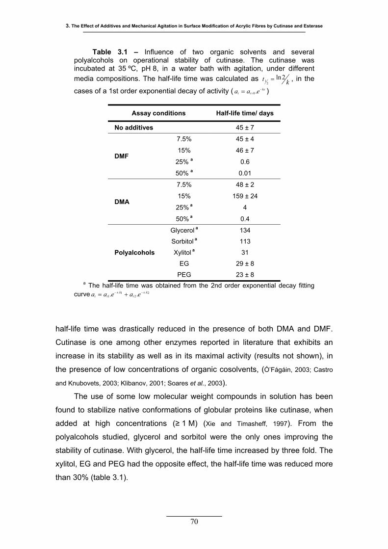
3. The Effect of Additives and Mechanical Agitation in Surface Modification of Acrylic Fibres by Cutinase and Esterase
70
half-life time was drastically reduced in the presence of both DMA and DMF.
Cutinase is one among other enzymes reported in literature that exhibits an
increase in its stability as well as in its maximal activity (results not shown), in
the presence of low concentrations of organic cosolvents, (Ó’Fágáin, 2003; Castro
and Knubovets, 2003; Klibanov, 2001; Soares et al., 2003).
The use of some low molecular weight compounds in solution has been
found to stabilize native conformations of globular proteins like cutinase, when
added at high concentrations (≥ 1 M) (Xie and Timasheff, 1997). From the
polyalcohols studied, glycerol and sorbitol were the only ones improving the
stability of cutinase. With glycerol, the half-life time increased by three fold. The
xylitol, EG and PEG had the opposite effect, the half-life time was reduced more
than 30% (table 3.1).
Table 3.1 – Influence of two organic solvents and several polyalcohols on operational stability of cutinase. The cutinase was incubated at 35 ºC, pH 8, in a water bath with agitation, under different media compositions. The half-life time was calculated as kt 2ln
21 =
kxtt eaa −== .0
, in the
cases of a 1st order exponential decay of activity ( )
Assay conditions Half-life time/ days
No additives 45 ± 7
DMF
7.5% 45 ± 4
15% 46 ± 7
25% a 0.6
50% a 0.01
DMA
7.5% 48 ± 2
15% 159 ± 24
25% a 4
50% a 0.4
Polyalcohols
Glycerol a 134
Sorbitol a 113
Xylitol a 31
EG 29 ± 8
PEG 23 ± 8 a The half-life time was obtained from the 2nd order exponential decay fitting
curve 2.2
1.1 .. kx
tkx
tt eaeaa −− +=
Enzymatic Treatment of Acrylic and Cellulose Acetate Fibres
3.2.2 ENZYMATIC MODIFICATION OF ACRYLIC SURFACE
The modification of a solid substrate, like acrylic fibres, with enzymes
constitutes a heterogeneous biocatalysis, since enzyme and substrate are in
different phases. Therefore, it is necessary to consider two general steps in
order to make the catalysed reaction occur: the physical adsorption of
cutinase/Texazym to acrylic fibre surface and the formation of the
enzyme/substrate complex and consequent hydrolysis of the ester bond
between vinyl acetate and the backbone of the polymer chain.
During the treatment, aliquots were taken at different time intervals to
follow the protein adsorption. In figure 3.1, the total protein is plotted for the
cutinase treatment with different amounts of DMA, under low and high
mechanical agitation. These conditions were chosen as an example. It is clear
from the results that there is no significant protein adsorption under the
conditions tested, taking into account the experimental error. When the discs
are present, the greater mechanical agitation did not affect the adsorption
behaviour of this enzyme into acrylic fibre. In addition, it was not possible to see
a clear effect of the organic solvent on protein adsorption. The lack of significant
adsorption was also verified in the acrylic treatment with Texazym PES for both
degrees of mechanical agitation.
These results were not surprising since polyacrylonitrile polymers are polar
materials and they are known low-protein-adsorbing polymers, often used to
produce inert membranes and supports for bioprocessing technologies (Belfort
and Zydney, 2003).
In theory, if the hydrolysis of vinyl acetate happens on the surface of the
fibre, according to the ‘electrostatic catapult’ model (Petersen et al., 2001a, 2001b)
at pH 8, cutinase should release the product – acetate anion. It was not
possible to detect acetic acid in the aliquots collected during the treatment of
acrylic with both enzymes. The detection limit, according to the kit suppliers, is
1.5 mg L-1 acetic acid. The average acrylic weight used was 15.5 g L-1 of
treatment solution. Thus, it means that less than 0.14% of the total available
vinyl acetate was hydrolysed (in average, 7% w/w of 15.5 g L-1). According to
the model proposed by Warner et al., they are organized in fibres of strong
laterally bound chains (Frushour and Knorr, 1998). Assuming that the enzyme
71
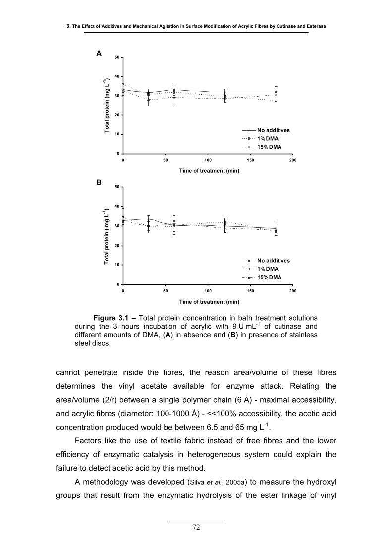
3. The Effect of Additives and Mechanical Agitation in Surface Modification of Acrylic Fibres by Cutinase and Esterase
72
cannot penetrate inside the fibres, the reason area/volume of these fibres
determines the vinyl acetate available for enzyme attack. Relating the
area/volume (2/r) between a single polymer chain (6 Å) - maximal accessibility,
and acrylic fibres (diameter: 100-1000 Å) - <<100% accessibility, the acetic acid
concentration produced would be between 6.5 and 65 mg L-1.
Factors like the use of textile fabric instead of free fibres and the lower
efficiency of enzymatic catalysis in heterogeneous system could explain the
failure to detect acetic acid by this method.
A methodology was developed (Silva et al., 2005a) to measure the hydroxyl
groups that result from the enzymatic hydrolysis of the ester linkage of vinyl
Figure 3.1 – Total protein concentration in bath treatment solutions during the 3 hours incubation of acrylic with 9 U mL-1 of cutinase and different amounts of DMA, (A) in absence and (B) in presence of stainless steel discs.
0
10
20
30
40
50
0 50 100 150 200
Time of treatment (min)
Tota
l pro
tein
(mg
L-1)
No additives1%DMA15%DMA
0
10
20
30
40
50
0 50 100 150 200
Time of treatment (min)
Tota
l pro
tein
( m
g L-1
)
No additives1%DMA15%DMA
B
A

Enzymatic Treatment of Acrylic and Cellulose Acetate Fibres
acetate and that remain attached to the polymer backbone. The basic principle
is the specific reaction between a vinylsulphonic group from a reactive dye, in
this case Remazol Brilliant Blue R, and the hydroxyl group at the fibre surface.
The specificity of the method is increased by the fact that this kind of reactive
dye was designed for cellulose fibre dyeing, thus it has low affinity for synthetic
fibres. The sensitivity is also very high due to the large molar absortivities of dye
molecules.
A higher level of mechanical agitation was of crucial importance in the
acrylic fabric treatment with both enzymes. Without the addition of stainless
steel discs, it was not possible to measure any difference in K/S between
treated samples and controls. The relative increase in K/S, obtained for the set
of experiments where the discs were used, is represented in figure 3.2. Treating
acrylic fabric, either with cutinase or with a commercial esterase, only led to the
formation of hydroxyl groups when the stainless steel discs were introduced into
the incubation vial. The experiments were performed in lab machines
reproducing the vertical agitation of European washing machines, where the
mechanical work involves the fibre-fibre and fibre-metal friction as well beating
effects. The introduction of metal discs increases the beating effects and the
fibre-metal friction (Vasconcelos and Cavaco-Paulo, 2006). The finishing and washing
effects produced by cellulases also depend highly on strong mechanical work
delivered to cotton fabrics (Cavaco-Paulo, 1998; Azevedo et al., 2000). Heterogeneous
catalysis implies adsorption of the enzyme which is in itself a complex process
depending, among other issues, on transport toward the surface by convection
and diffusion. Accordingly, it is likely that an increase in mechanical agitation is
helpful for the outcome of acrylic biotransformation, increasing the accessibility.
In addition, in a comparable way to cellulose, the increased friction has a micro-
pilling effect on the acrylic fabric, therefore, increasing the surface area
available for enzymatic attack.
DMA is a known solvent of PAN and it is commonly used in acrylic fibre
industrial production. Its plasticizer function disturbs the regular structure of the
polymer, reducing the magnitude of inter-chain bonding, which should aid the
penetration of the enzyme, improving its action on the fibre. The acrylic fabric
sample treated, both with cutinase and with Texazym PES, in the presence of
15% DMA did not show any difference in respect to controls (figure 3.2). The
73
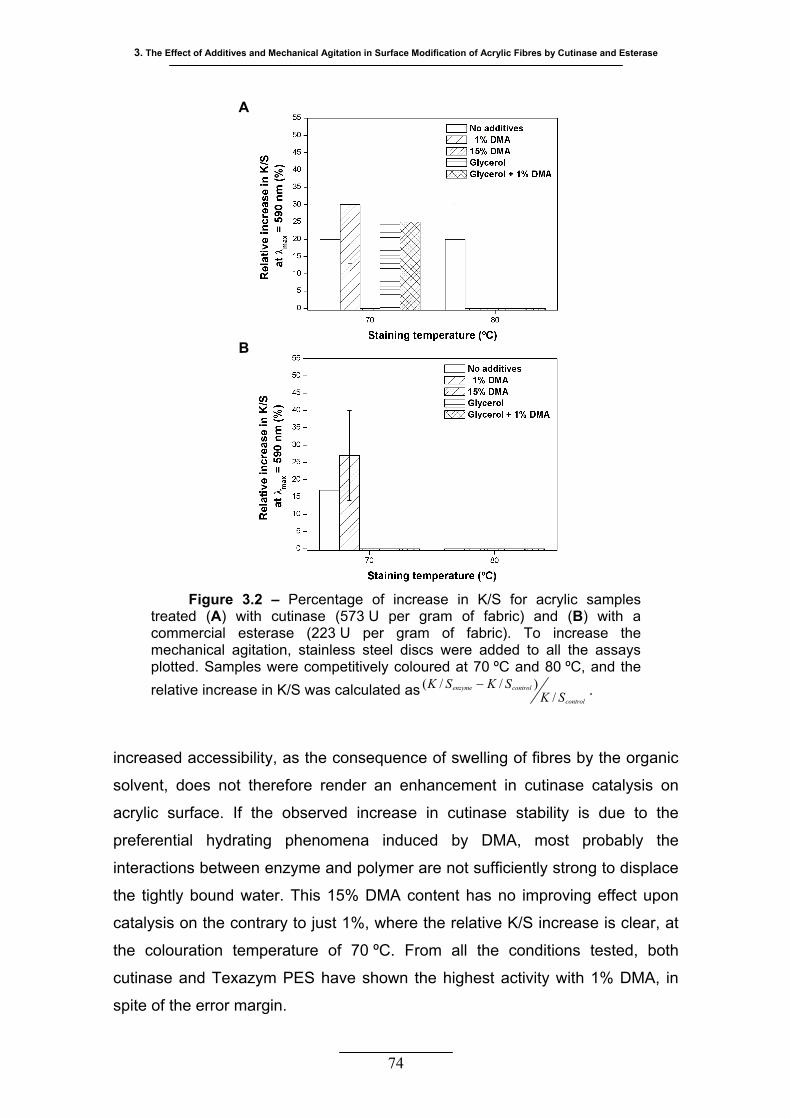
3. The Effect of Additives and Mechanical Agitation in Surface Modification of Acrylic Fibres by Cutinase and Esterase
74
increased accessibility, as the consequence of swelling of fibres by the organic
solvent, does not therefore render an enhancement in cutinase catalysis on
acrylic surface. If the observed increase in cutinase stability is due to the
preferential hydrating phenomena induced by DMA, most probably the
interactions between enzyme and polymer are not sufficiently strong to displace
the tightly bound water. This 15% DMA content has no improving effect upon
catalysis on the contrary to just 1%, where the relative K/S increase is clear, at
the colouration temperature of 70 ºC. From all the conditions tested, both
cutinase and Texazym PES have shown the highest activity with 1% DMA, in
spite of the error margin.
B
A
Figure 3.2 – Percentage of increase in K/S for acrylic samples treated (A) with cutinase (573 U per gram of fabric) and (B) with a commercial esterase (223 U per gram of fabric). To increase the mechanical agitation, stainless steel discs were added to all the assays plotted. Samples were competitively coloured at 70 ºC and 80 ºC, and the relative increase in K/S was calculated as
control
controlenzyme
SKSKSK
/)//( − .
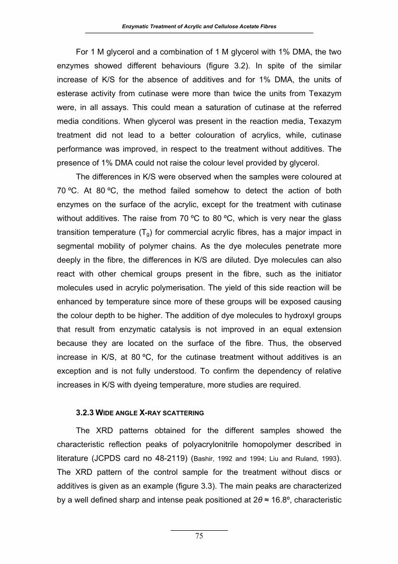
Enzymatic Treatment of Acrylic and Cellulose Acetate Fibres
For 1 M glycerol and a combination of 1 M glycerol with 1% DMA, the two
enzymes showed different behaviours (figure 3.2). In spite of the similar
increase of K/S for the absence of additives and for 1% DMA, the units of
esterase activity from cutinase were more than twice the units from Texazym
were, in all assays. This could mean a saturation of cutinase at the referred
media conditions. When glycerol was present in the reaction media, Texazym
treatment did not lead to a better colouration of acrylics, while, cutinase
performance was improved, in respect to the treatment without additives. The
presence of 1% DMA could not raise the colour level provided by glycerol.
The differences in K/S were observed when the samples were coloured at
70 ºC. At 80 ºC, the method failed somehow to detect the action of both
enzymes on the surface of the acrylic, except for the treatment with cutinase
without additives. The raise from 70 ºC to 80 ºC, which is very near the glass
transition temperature (Tg) for commercial acrylic fibres, has a major impact in
segmental mobility of polymer chains. As the dye molecules penetrate more
deeply in the fibre, the differences in K/S are diluted. Dye molecules can also
react with other chemical groups present in the fibre, such as the initiator
molecules used in acrylic polymerisation. The yield of this side reaction will be
enhanced by temperature since more of these groups will be exposed causing
the colour depth to be higher. The addition of dye molecules to hydroxyl groups
that result from enzymatic catalysis is not improved in an equal extension
because they are located on the surface of the fibre. Thus, the observed
increase in K/S, at 80 ºC, for the cutinase treatment without additives is an
exception and is not fully understood. To confirm the dependency of relative
increases in K/S with dyeing temperature, more studies are required.
3.2.3 WIDE ANGLE X-RAY SCATTERING
The XRD patterns obtained for the different samples showed the
characteristic reflection peaks of polyacrylonitrile homopolymer described in
literature (JCPDS card no 48-2119) (Bashir, 1992 and 1994; Liu and Ruland, 1993).
The XRD pattern of the control sample for the treatment without discs or
additives is given as an example (figure 3.3). The main peaks are characterized
by a well defined sharp and intense peak positioned at 2θ ≈ 16.8º, characteristic
75

3. The Effect of Additives and Mechanical Agitation in Surface Modification of Acrylic Fibres by Cutinase and Esterase
76
of (010) plane, and a second one, less intense at 2θ ≈ 29.4º, from the (300)
plane. A third diffuse peak was also observed in all samples, located at
2θ ≈ 26.5º, attributed to (210) plane. This peak was found to be a very broad
and diffuse one, commonly associated to amorphous phases. In order to obtain
an accurate fitting profile, it was always necessary to consider a fourth
significantly diffuse peak, at an angular position 2θ ≈ 16.8º, which is in fact very
similar to that of the previously mentioned peak. Although unusual, similar
behaviour was also found by Causin et al. (2005). This peak appearance might
result from the introduction of the comonomer vinyl acetate, which probably
induces some defects in the lateral packaging of polymer chains and thus being
responsible for this second “amorphous type” phase. In a systematic study of
homologous series of amorphous polymers, Miller and Boyer (1984a, 1984b)
found in the XRD patterns two types of amorphous halos. The position of the
first type corresponded to the close packaging of atoms (2θ ≥18º) and its
position was dependent on the size of the pendent group, while the second type
Figure 3.3 – X-ray diffraction ttern of ntrol sample for the treatment performed in the absence of additives and stainless steal discs. The XRD data was analyzed by profile fitting of the scans with the Pearson VII function, using the software WinFit! beta release 1.2.1, 1997.
pa co
Stefan Krumm, Geologisches Institut Erlangen, Schloßgarten 5, D 91054 Erlangen
5.332 7.832 10.332 12.832 15.332 17.832 20.332 22.832 25.332 27.832 30.332 32.832
638
1138
1638
2138
2638
3138
3638
4138
2θ
Inte
nsity
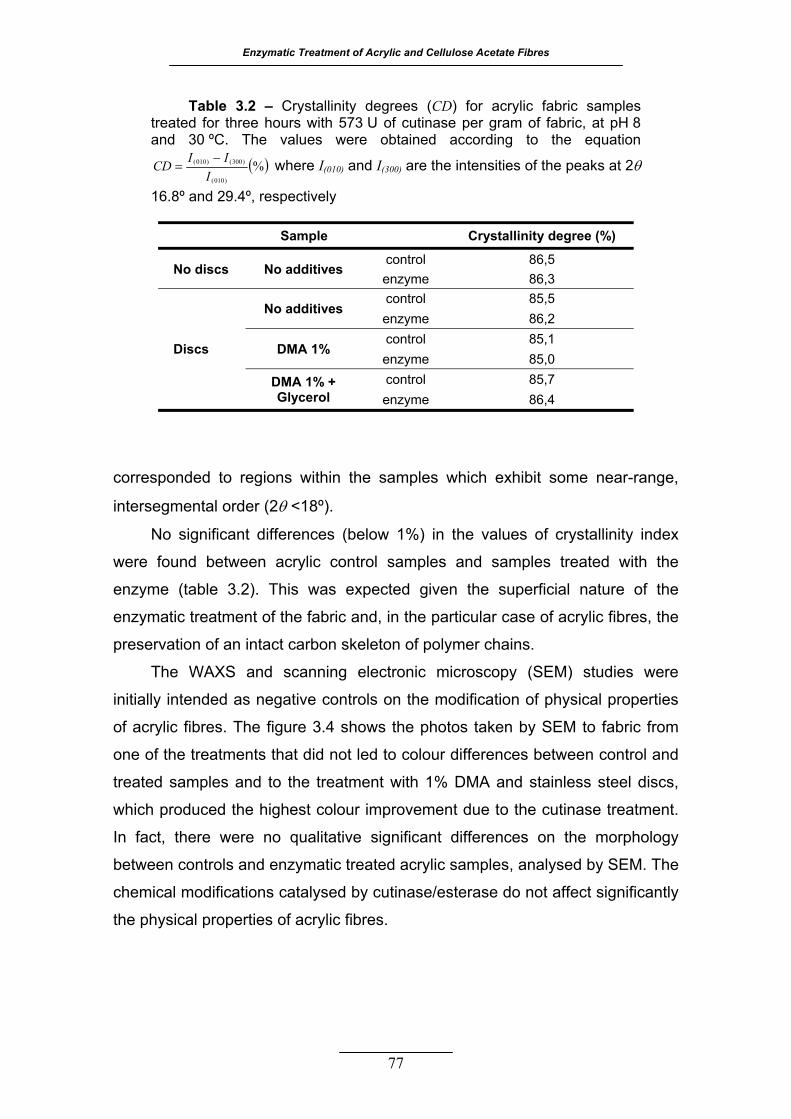
Enzymatic Treatment of Acrylic and Cellulose Acetate Fibres
77
corresponded to regions within the samples which exhibit some near-range,
intersegmental order (2θ <18º).
No significant differences (below 1%) in the values of crystallinity index
were found between acrylic control samples and samples treated with the
enzyme (table 3.2). This was expected given the superficial nature of the
enzymatic treatment of the fabric and, in the particular case of acrylic fibres, the
preservation of an intact carbon skeleton of polymer chains.
The WAXS and scanning electronic microscopy (SEM) studies were
initially intended as negative controls on the modification of physical properties
of acrylic fibres. The figure 3.4 shows the photos taken by SEM to fabric from
one of the treatments that did not led to colour differences between control and
treated samples and to the treatment with 1% DMA and stainless steel discs,
which produced the highest colour improvement due to the cutinase treatment.
In fact, there were no qualitative significant differences on the morphology
between controls and enzymatic treated acrylic samples, analysed by SEM. The
chemical modifications catalysed by cutinase/esterase do not affect significantly
the physical properties of acrylic fibres.
Table 3.2 – Crystallinity degrees (CD) for acrylic fabric samples treated for three hours with 573 U of cutinase per gram of fabric, at pH 8 and 30 ºC. The values were obtained according to the equation
( )%)010(
)300()010(
IIICD −
= where I(010) and I(300) are the intensities of the peaks at 2θ
16.8º and 29.4º, respectively
Sample Crystallinity degree (%)
No discs No additives control 86,5 enzyme 86,3
Discs
No additives control 85,5 enzyme 86,2
DMA 1% control 85,1 enzyme 85,0
DMA 1% + Glycerol
control 85,7 enzyme 86,4
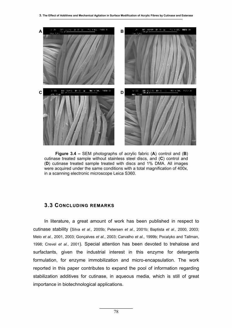
3. The Effect of Additives and Mechanical Agitation in Surface Modification of Acrylic Fibres by Cutinase and Esterase
78
33..33 CCOONNCCLLUUDDIINNGG RREEMMAARRKK SS
In literature, a great amount of work has been published in respect to
cutinase stability (Silva et al., 2005b; Petersen et al., 2001b; Baptista et al., 2000, 2003;
Melo et al., 2001, 2003; Gonçalves et al., 2003; Carvalho et al., 1999b; Pocalyko and Tallman,
1998; Crevel et al., 2001). Special attention has been devoted to trehalose and
surfactants, given the industrial interest in this enzyme for detergents
formulation, for enzyme immobilization and micro-encapsulation. The work
reported in this paper contributes to expand the pool of information regarding
stabilization additives for cutinase, in aqueous media, which is still of great
importance in biotechnological applications.
A B
C D
Figure 3.4 – SEM photographs of acrylic fabric (A) control and (B) cutinase treated sample without stainless steel discs, and (C) control and (D) cutinase treated sample treated with discs and 1% DMA. All images were acquired under the same conditions with a total magnification of 400x, in a scanning electronic microscope Leica S360.
Enzymatic Treatment of Acrylic and Cellulose Acetate Fibres
The catalytic efficiency of both enzymes studied on acrylic surface
modification, although not considerably significant, was proved to be enhanced
by DMA at low concentrations, a solvent of PAN fibres commonly used in its
spinning process. Besides, the reduction in incubation time with cutinase from
90 hours (Silva et al., 2005a) to just three hours, due to a higher mechanical
agitation, represents a great advance in the application of these enzymes to
acrylic surface modification, especially in wet spinning lines of production and at
stages of a more “exposed and accessible” surface of the acrylic fibre. It is
expected that the impact of esterase activity on fibre surface modification will be
greater when used in acrylic or other textile fibres with higher contents in
hydrolysable ester monomers in their compositions.
79
80
SSUURRFFAACCEE MMOODDIIFFIICCAATTIIOONN OOFF
CCEELLLLUULLOOSSEE AACCEETTAATTEE WWIITTHH
CCUUTTIINNAASSEE
ork on the modification of cellulose acetate with enzymes has
been mostly done in the context of its biodegradation (Puls et
al., 2004). This chapter describes the hydrolysis of acetate surface
groups of CDA and CTA fabrics using Fusarium solani pisi cutinase.
The preference for hydrophobic substrates, as well as the versatility in
respect to soluble and insoluble substrates makes cutinase an
attractive esterase for highly substituted cellulose acetates.
The hydrolysis of surface acetyl groups from CDA and CTA with a
cutinase constitutes a promising approach for the partial regeneration
of cellulose reactivity and hydrophilicity in these fibres, here
demonstrated by the enhanced reactive dye uptake of treated fabrics.
W
44
Enzymatic Treatment of Acrylic and Cellulose Acetate Fibres
81
44.. SSUURRFFAACCEE MMOODDIIFFIICCAATTIIOONN OOFF CCEELLLLUULLOOSSEE AACCEETTAATTEE WWIITTHH
CCUUTTIINNAASSEE
44..11 MMAATTEERRIIAALLSS AANNDD MMEETTHHOODDSS
4.1.1 REAGENTS AND ENZYMES
The cellulose diacetate and triacetate plain woven fabrics used were
kindly supplied by Mitsubishi Rayon Co. Ltd., Tokyo, Japan. The CDA fabric has
41/27 ends/picks per cm and 64 g m-2. The CTA fabric has 45/31 ends/picks per
cm and 98 g m-2.
The cutinase (EC 3.1.1.74) from Fusarium solani pisi used in this work
was expressed and purified as previously reported by Araújo et al. (2007).
The reactive dye Remazol Brilliant Blue R, C.I. 61200, was from Sigma. All
other reagents were laboratory grade reagents also from Sigma-Aldrich, St.
Louis, USA, unless stated otherwise. The buffer used throughout this work was
a phosphate buffer having a concentration of 50 mM and pH 8.
4.1.2 ESTERASE ACTIVITY ASSAY
Esterase activity was determined following the product release (p-NP)
continuously through the increase in the absorbance at 400 nm at 30 ºC. The
assay conditions for the determination of cutinase activity were described
previously (section 3.1.2). All the assays were performed at least in triplicate.
Standard solutions of p-NP were used to obtain the calibration curve. One unit
of esterase activity was defined as one µmol of p-NP released per minute.
4.1.3 TREATMENT OF CELLULOSE DI- AND TRIACETATE FABRIC WITH CUTINASE
All samples of cellulose acetate fabric were washed prior to use in order to
remove possible impurities from manufacture and from human handling.
Washing was performed at 35 ºC and 20 rpm, in stainless steel pots of 450 mL
in capacity and housed in a laboratory scale machine, the Rotawash MKIII
4. Surface Modification of Cellulose Acetate with Cutinase
(vertical agitation simulating European washing machines, from SDL
International Ltd.). The fabric was washed twice for 30 min in 40 mg L-1 Lutensol
AT25 (non-ionic detergent, BASF, Ludwigshafen, Germany), then rinsed four
times with distilled water for 30 min each and left to dry at room temperature.
Several sets of experiments were carried out taking into account the
amounts of enzyme, fabric and time of incubation. For all experiments, the
treatment of cellulose acetate fabric was performed in phosphate buffer with
vertical agitation, in the Rotawash machine operating at 30 ºC and 20 rpm. To
evaluate the effect of enzyme concentration, samples of CDA and CTA fabric,
with an average weight of 0.1 g, were incubated in duplicate for 8 hours with 0,
25, 50, 75 and 100 U mL-1 of cutinase, in a total volume of 5 mL. To obtain a
progress curve, samples of CDA and CTA fabric, with an average weight of
0.1 g, were treated with 50 U mL-1 of cutinase, in a final volume of 10 mL for
different periods of time. For each sample a control was run in parallel in which
the buffer substituted the corresponding volume of enzyme. In another
treatment, the average weight of both type of fabric was increased to 0.5 g and
the incubation extended to 24 hours. The initial activity of cutinase was
25 U mL-1 in a final volume of 25 mL. For each sample a control was run in
parallel without the enzyme.
After enzymatic treatment, all fabric samples were washed at 35 ºC, in the
Rotawash machine, to remove the adsorbed protein, according to the order:
250 mg L-1 Lutensol AT25 for 30 min, 70% ethanol for 20 min, 15% isopropanol
for 15 min, three steps of increasing concentrations of sodium chloride for
10 min each, three steps in distilled water for 20 min each. Between the
detergent/alcohol and alcohol/salt steps the fabric was rinsed under running
cold tap water for 5 min.
4.1.4 QUANTIFICATION OF TOTAL PROTEIN CONCENTRATION
Total protein in solution was quantified following Bradford methodology
(Bradford, 1976) using BSA as standard. All samples were measured at least
twice.
82

Enzymatic Treatment of Acrylic and Cellulose Acetate Fibres
4.1.5 DETERMINATION OF ACETIC ACID CONCENTRATION IN THE TREATMENT
SOLUTIONS
Detection of acetic acid in the reaction media was performed with the
acetic acid UV test from Roche (Darmstadt, Germany) as described previously
(section 3.1.6).
4.1.6 CELLULOSE ACETATE FABRIC COLOURATION WITH A REACTIVE DYE
After enzymatic treatment, samples were competitively coloured in
phosphate buffer with 2% o.w.f. Remazol Brilliant Blue R, in duplicate. The
colouration was performed at 50 ºC or 60 ºC, for 90 min at 20 rpm, in sealed
stainless steel beakers of 140 mL in capacity and housed on a lab-scale dyeing
machine (AHIBA Spectradye, from Datacolor International).
After colouration, all samples were washed once with 0.25 g L-1 Lutensol
AT25 and several times with distilled water in Rotawash, until no more dye
could be detected in the solution. The washing temperature was 5 ºC higher
than the colouration temperature.
The colour measurements (5 for each sample) were carried out according
to the procedure described previously (section 3.1.7).
4.1.7 FLUORESCEIN ISOTHIOCYANATE LABELLING
Enzymes were incubated with fluorescein isothiocyanate - FITC (33:1 w/w)
in 0.5 M sodium carbonate buffer pH 9.5, for one hour at room temperature. The
unconjugated FITC was removed with HiTrap Desalting 5 mL columns (GE
Healthcare Bio-Sciences Europe GmbH, Munich, Germany) while the carbonate
buffer was exchanged by the phosphate buffer.
4.1.8 FLUORESCENCE MICROSCOPY
Thin strips of CDA and CTA fabric samples were embedded in an epoxy
resin (Epofix kit, Struers, Copenhagen, Denmark) and cross sections were cut
with 20-25 µm thickness. The samples were observed under a Leica
Microsystems DM5000 B epifluorescence microscope equipped with a 100 W
Hg lamp and an appropriate filter setting. Digital images were acquired with
83

4. Surface Modification of Cellulose Acetate with Cutinase
84
Leica DFC350 FX digital Camera and Leica Microsystems LAS AF software,
version 2.0 (Leica Microsystems GmbH, Wetzlar, Germany).
4.1.9 FOURIER TRANSFORM INFRARED SPECTROSCOPY (FT-IR)
The diffuse reflectance (DRIFT) technique was used to collect the infrared
spectra of CDA and CTA fabric samples treated during 24 hours with cutinase
and respective controls. The spectra were recorded in a Michelson FT-IR
spectrometer MB100 (Bomem, Inc., Quebec, Canada) with a DRIFT accessory.
The fabric pieces were placed and hold on top of the sample cup, previously
filled with potassium bromide powder that was used to collect the background.
All the spectra were obtained under a nitrogen atmosphere in the range
cm-1 at 8 cm-1 resolution and as the ratio of 32 scans to the same 800־4000
number of background scans. The spectra were acquired in Kubelka-Munk units
and baseline corrections were made using Bomem Grams/32R software,
version 4.04.
4.1.10 WIDE ANGLE X-RAY SCATTERING
The XRD patterns were obtained for the CDA and CTA fabric samples
treated during 24 hours with cutinase and respective controls. The WAXS
experiments were undertaken in a Philips PW1710 apparatus, using Cu Kα
radiation and operating at a 40 KV voltage and 30 mA current. The patterns
were continuously recorded in the diffraction angular range 2θ from 4º to 40º,
with a step size of 0.02º at 0.6º min-1. The non linear fitting of the diffraction
patterns was performed using the Pseudo-Voigt peak function from
OriginPro 7.5 (Origin Lab Corporation, USA) considering the CTA structural
polymorphism II (Cerqueira et al., 2006). The peaks that were considered
crystalline were at the diffraction angles 2θ: 10º, 17º and 21º for CDA, and 8º,
10º, 13º, 17º, 21º and 23º for CTA (Chen et al., 2002; Hindeleh and Johnson, 1972).
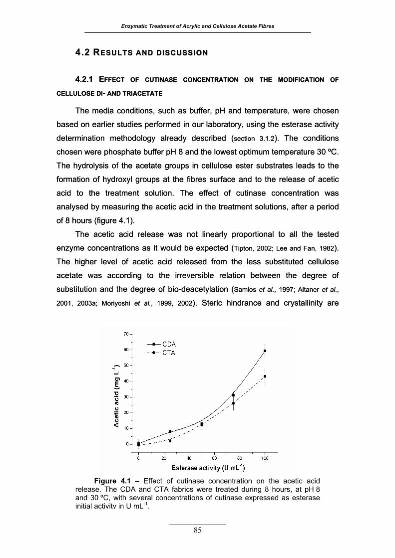
Enzymatic Treatment of Acrylic and Cellulose Acetate Fibres
85
44..22 RREESSUULLTTSS AANNDD DDIISSCCUUSSSSIIOONN U
4.2.1 EFFECT OF CUTINASE CONCENTRATION ON THE MODIFICATION OF
CELLULOSE DI- AND TRIACETATE 4.2.1 EFFECT OF CUTINASE CONCENTRATION ON THE MODIFICATION OF
CELLULOSE DI- AND TRIACETATE
The media conditions, such as buffer, pH and temperature, were chosen
based on earlier studies performed in our laboratory, using the esterase activity
determination methodology already described (section 3.1.2). The conditions
chosen were phosphate buffer pH 8 and the lowest optimum temperature 30 ºC.
The hydrolysis of the acetate groups in cellulose ester substrates leads to the
formation of hydroxyl groups at the fibres surface and to the release of acetic
acid to the treatment solution. The effect of cutinase concentration was
analysed by measuring the acetic acid in the treatment solutions, after a period
of 8 hours (figure 4.1).
The acetic acid release was not linearly proportional to all the tested
enzyme concentrations as it would be expected (Tipton, 2002; Lee and Fan, 1982).
The higher level of acetic acid released from the less substituted cellulose
acetate was according to the irreversible relation between the degree of
substitution and the degree of bio-deacetylation (Samios et al., 1997; Altaner et al.,
2001, 2003a; Moriyoshi et al., 1999, 2002). Steric hindrance and crystallinity are
ULLTTSS AANNDD DDIISSCCUUSSSSIIOONN
Figure 4.1 – Effect of cutinase concentration on the acetic acid release. The CDA and CTA fabrics were treated during 8 hours, at pH 8 and 30 ºC, with several concentrations of cutinase expressed as esterase initial activity in U mL-1.
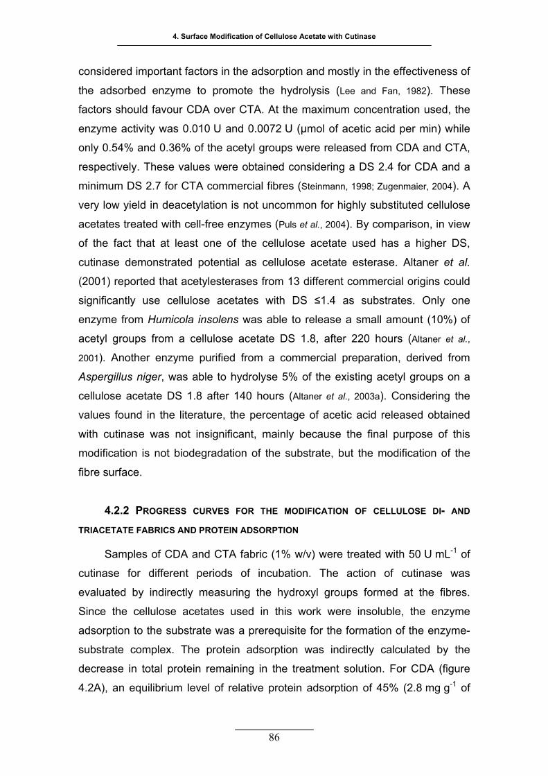
4. Surface Modification of Cellulose Acetate with Cutinase
86
considered important factors in the adsorption and mostly in the effectiveness of
the adsorbed enzyme to promote the hydrolysis (Lee and Fan, 1982). These
factors should favour CDA over CTA. At the maximum concentration used, the
enzyme activity was 0.010 U and 0.0072 U (µmol of acetic acid per min) while
only 0.54% and 0.36% of the acetyl groups were released from CDA and CTA,
respectively. These values were obtained considering a DS 2.4 for CDA and a
minimum DS 2.7 for CTA commercial fibres (Steinmann, 1998; Zugenmaier, 2004). A
very low yield in deacetylation is not uncommon for highly substituted cellulose
acetates treated with cell-free enzymes (Puls et al., 2004). By comparison, in view
of the fact that at least one of the cellulose acetate used has a higher DS,
cutinase demonstrated potential as cellulose acetate esterase. Altaner et al.
(2001) reported that acetylesterases from 13 different commercial origins could
significantly use cellulose acetates with DS ≤1.4 as substrates. Only one
enzyme from Humicola insolens was able to release a small amount (10%) of
acetyl groups from a cellulose acetate DS 1.8, after 220 hours (Altaner et al.,
2001). Another enzyme purified from a commercial preparation, derived from
Aspergillus niger, was able to hydrolyse 5% of the existing acetyl groups on a
cellulose acetate DS 1.8 after 140 hours (Altaner et al., 2003a). Considering the
values found in the literature, the percentage of acetic acid released obtained
with cutinase was not insignificant, mainly because the final purpose of this
modification is not biodegradation of the substrate, but the modification of the
fibre surface.
IFICATION OF CELLULOSE DI- AND
TRIACETATE FABRICS AND PROTEIN ADSORPTION
Samples of CDA and CTA fabric (1% w/v) were treated with 50 U mL-1 of
cutinase for different periods of incubation. The action of cutinase was
evaluated by indirectly measuring the hydroxyl groups formed at the fibres.
Since the cellulose acetates used in this work were insoluble, the enzyme
adsorption to the substrate was a prerequisite for the formation of the enzyme-
substrate complex. The protein adsorption was indirectly calculated by the
decrease in total protein remaining in the treatment solution. For CDA (figure
4.2A), an equilibrium level of relative protein adsorption of 45% (2.8 mg g-1 of
4.2.2 PROGRESS CURVES FOR THE MOD
Enzymatic Treatment of Acrylic and Cellulose Acetate Fibres
87
protein per fabric weight) was reached. For CTA (figure 4.2B), the equilibrium
level of protein adsorption was higher, with 57% of relative protein adsorption
(3.5 mg g-1). The hydrophobic character of the substrate should not be a
problem for cutinase adsorption since this enzyme is a lipolytic enzyme and its
natural substrate, cutin, is hydrophobic (Egmond and Vlieg, 2000; Mannesse et al.,
1995; Kolattukudy, 2002).
The formation of hydroxyl groups at the fibre surface was evaluated by
colouring the fabric with a cotton reactive dye. If the cutinase is able to
Figure 4.2 – Progress curves for the formation of hydroxyl groups at the fibres surface, measured as relative increase in K/S values, and total protein adsorption for (A) CDA and (B) CTA. All the samples were treated with cutinase (5000 U per gram of fabric) at pH 8 and 30 ºC. Samples and controls were competitively coloured at 60 ºC. The relative increase in K/S was calculated as
control
controlenzyme
SKSKSK
/)//( − (%) and the relative protein
adsorption as h
th
PPP
0
0 )( − (%), where P is the total protein in solution.
B
A
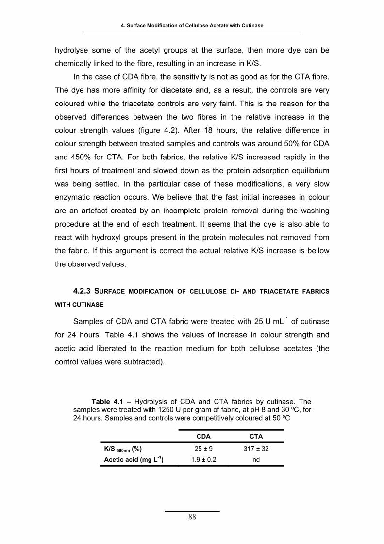
4. Surface Modification of Cellulose Acetate with Cutinase
88
hydrolyse some of the acetyl groups at the surface, then more dye can be
chemically linked to the fibre, resulting in an increase in K/S.
In the case of CDA fibre, the sensitivity is not as good as for the CTA fibre.
The dye has more affinity for diacetate and, as a result, the controls are very
coloured while the triacetate controls are very faint. This is the reason for the
observed differences between the two fibres in the relative increase in the
colour strength values (figure 4.2). After 18 hours, the relative difference in
colour strength between treated samples and controls was around 50% for CDA
and 450% for CTA. For both fabrics, the relative K/S increased rapidly in the
first hours of treatment and slowed down as the protein adsorption equilibrium
was being settled. In the particular case of these modifications, a very slow
enzymatic reaction occurs. We believe that the fast initial increases in colour
are an artefact created by an incomplete protein removal during the washing
procedure at the end of each treatment. It seems that the dye is also able to
react with hydroxyl groups present in the protein molecules not removed from
the fabric. If this argument is correct the actual relative K/S increase is bellow
the observed values.
4.2.3 RICS
WITH
ase
for 24 hours. Table 4.1 shows the values of increase in colour strength and
acetic ac o the reac
control values were subtracted).
SURFACE MODIFICATION OF CELLULOSE DI- AND TRIACETATE FAB
CUTINASE
Samples of CDA and CTA fabric were treated with 25 U mL-1 of cutin
id liberated t tion medium for both cellulose acetates (the
Table 4.1 – Hydrolysis of CDA and CTA fabrics by cutinase. The
samples were treated with 1250 U per gram of fabric, at pH 8 and 30 ºC, for 24 hours. Samples and controls were competitively coloured at 50 ºC
CDA CTA
K/S 590nm (%) 25 ± 9 317 ± 32
Acetic acid (mg L-1) 1.9 ± 0.2 nd
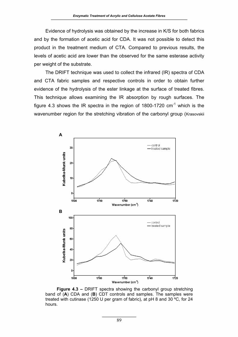
Enzymatic Treatment of Acrylic and Cellulose Acetate Fibres
89
Evidence of hydrolysis was obtained by the increase in K/S for both fabrics
and by the formation of acetic acid for CDA. It was not possible to detect this
product in the treatment medium of CTA. Compared to previous results, the
levels of acetic acid are lower than the observed for the same esterase activity
per weight of the substrate.
gh surfaces. The
figure
The DRIFT technique was used to collect the infrared (IR) spectra of CDA
and CTA fabric samples and respective controls in order to obtain further
evidence of the hydrolysis of the ester linkage at the surface of treated fibres.
This technique allows examining the IR absorption by rou
4.3 shows the IR spectra in the region of 1800-1720 cm-1 which is the
wavenumber region for the stretching vibration of the carbonyl group (Krasovskii
A
B
Figure 4.3 – DRIFT spectra showing the carbonyl group stretching A) CDA and (B) CDT controls and samples. The samples were
eated with cutinase (1250 U per gram of fabric), at pH 8 and 30 ºC, for 24 hours.
band of (tr
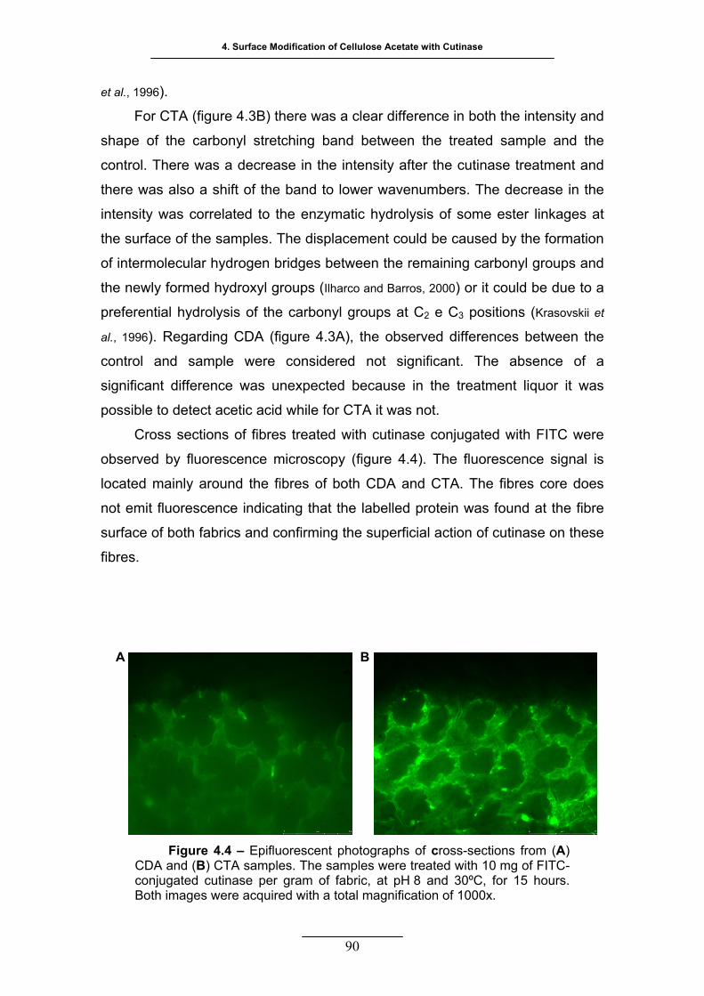
4. Surface Modification of Cellulose Acetate with Cutinase
90
et al., 1996)
and
shape of the carbonyl stretching band between the treated sample and the
control. There was a decrease in the intensity after the cutinase treatment and
there was also d to lo enumbers. The decrease in the
intensity was correlated to the enzymatic hydrolysis of some ester linkages at
the surface of the samples. The displacement could be caused by the formation
of int
considered not significant. The absence of a
significant difference was unexpected because in the treatment liquor it was
possible to detect acetic acid while for CTA it was not.
Cross sections of fibres treated with cutinase conjugated with FITC were
observed by fluorescence microscopy (figure 4.4). The fluorescence signal is
located mainly around the fibres of both CDA and CTA. The fibres core does
not emit fluorescence indicating that the labelled protein was found at the fibre
surface of both fabrics and confirming the superficial action of cutinase on these
fibres.
Figure 4.4 – Epifluorescent photographs of cross-sections from (A) CDA and (B) CTA samples. The samples were treated with 10 mg of FITC-conjugated cutinase per gram of fabric, at pH 8 and 30ºC, for 15 hours. Both images were acquired with a total magnification of 1000x.
.
For CTA (figure 4.3B) there was a clear difference in both the intensity
a shift of the ban wer wav
ermolecular hydrogen bridges between the remaining carbonyl groups and
the newly formed hydroxyl groups (Ilharco and Barros, 2000) or it could be due to a
preferential hydrolysis of the carbonyl groups at C2 e C3 positions (Krasovskii et
al., 1996). Regarding CDA (figure 4.3A), the observed differences between the
control and sample were
A B
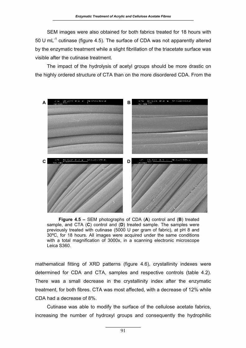
Enzymatic Treatment of Acrylic and Cellulose Acetate Fibres
91
SEM images were also obtained for both fabrics treated for 18 hours with
50 U mL-1 cutinase (figure 4.5). The surface of CDA was not apparently altered
by the enzymatic treatment while a slight fibrillation of the triacetate surface was
visible after the cutinase treatment.
The impact of the hydrolysis of acetyl groups should be more drastic on
the highly ordered structure of CTA than on the more disordered CDA. From the
mathematical fitting of XRD patterns (figure 4.6), crystallinity indexes were
determined for CDA and CTA, samples and respective controls (table 4.2).
There was a small decrease in the crystallinity index after the enzymatic
treatment, for both fibres. CTA was most affected, with a decrease of 12% while
CDA had a
rics,
increasing the number of hydroxyl groups and consequently the hydrophilic
decrease of 8%.
Cutinase was able to modify the surface of the cellulose acetate fab
BA
DC
Figure 4.5 – SEM photographs of CDA (A) control and (B) treated sample, and CTA (C) control and (D) treated sample. The samples were previously treated with cutinase (5000 U per gram of fabric), at pH 8 and 30ºC, for 18 hours. All images were acquired under the same conditions with a total magnification of 3000x, in a scanning electronic microscope Leica S360.
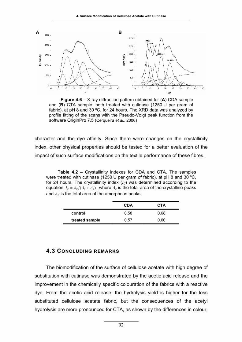
4. Surface Modification of Cellulose Acetate with Cutinase
92
character and the dye affinity. Since there were changes on the crystallinity
index, other physical properties should be tested for a better evaluation of the
impact of such surface modifications on the textile performance of these fibres.
Table 4.2 – Crystallinity indexes for CDA and CTA. The samples
were treated with cutinase (1250 U per gram of fabric), at pH 8 and 30 ºC, for 24 hours. The crystallinity index (IC) was determined according to the equation )( ACCC AAAI += , where Ac is the total area of the crystalline peaks and AA is the total area of the amorphous peaks
CDA CTA
control 0.58 0.68
treated sample 0.57 0.60
44..33 CCOONNCCLLUUDDIINNGG RREEMMAARRKK SS
The biomodification of the surface of cellulose acetate with high degree of
substitution with cutinase was demonstrated by the acetic acid release and the
improvement in the chemically specific colouration of the fabrics with a reactive
dye. From less
subst etyl
hydro ences in colour,
the acetic acid release, the hydrolysis yield is higher for the
ituted cellulose acetate fabric, but the consequences of the ac
lysis are more pronounced for CTA, as shown by the differ
A B
Figure 4.6 – X-ray diffraction pattern obtained for (A) CDA sample and (B) CTA sample, both treated with cutinase (1250 U per gram of fabric), at pH 8 and 30 ºC, for 24 hours. The XRD data was analyzed by profile fitting of the scans with the Pseudo-Voigt peak function from the software OriginPro 7.5 (Cerqueira et al., 2006)

Enzymatic Treatment of Acrylic and Cellulose Acetate Fibres
93
morp
d of the physical, rather than chemical,
modi
hology of the fibres surface and crystallinity between controls and treated
samples. Further studies will be necessary to evaluate the impact of cutinase
activity in the physical properties of the fabrics and to assess the contribution of
the incomplete protein removal an
fications on the differences seen upon enzymatic treatment.

94
PPRROODDUUCCTTIIOONN OOFF CCUUTTIINNAASSEE FFUUSSEEDD
TTOO CCAARRBBOOHHYYDDRRAATTEE--BBIINNDDIINNGG
MMOODDUULLEESS FFOORR TTHHEE MMOODDIIFF IICCAATTIIOONN
OOFF CCEELLLLUULLOOSSEE AACCEETTAATTEE
he enzymatic modification of highly substituted cellulose acetate
fibres is a hetero us process, fore, an attempt was
to increase cut efficiency towards this substrate by
her carboh -active enzy with modular nature.
Two different carbohydrate-binding modules (CBMs) were fused to the
C-terminal of cutinase. The CBMs act synergistically with the catalytic
domains by increasing the effective enzyme concentration at the
for some CBMs, by physical disruption of the
solid substrate. Two types of CBM were chosen on the basis of ligand
T
geneo there
made inase
mimicking ot ydrate mes
substrate surface and,
affinity, since the two cellulose acetate fibres used in this work are
structurally different from cellulose (the native ligand) and different
between themselves, presenting two different overall crystallinities. The production of fusion cutinases with new functionalities is here
described and a comparison with cutinase regarding its efficiency for
CDA and CTA modification is presented.
55
Enzymatic Treatment of Acrylic and Cellulose Acetate Fibres
95
55.. PPRROODDUUCCTTIIOONN OOFF CCUUTTIINNAASSEE FFUUSSEEDD TTOO CCAARRBBOOHHYYDDRRAATTEE--
BBIINNDDIINNGG MMOODDUULLEESS FFOORR TTHHEE MMOODDIIFFIICCAATTIIOONN OOFF CCEELLLLUULLOOSSEE
AACCEETTAATTEE
55 ..11 MMAATTEERRIIAALLSS AANNDD MMEETTHHOODDSS
5.1.1 REAGENTS AND ENZYMES
The cellulose diacetate and triacetate plain woven fabrics used were
kindly supplied by Mitsubishi Rayon Co. Ltd., Tokyo, Japan. Their
characteristics were described in a previous section (section 4.1.1).
All reagents were laboratory grade reagents from Sigma-Aldrich, St. Louis,
USA, unless stated otherwise. The buffer used for the enzymatic treatment of
both fabrics was a phosphate buffer having a concentration of 50 mM and pH 8.
Other buffers are specified in the text when necessary.
The cutinase (EC 3.1.1.74) from F. solani pisi was expressed and purified
as previously reported by Araújo et al. (2007).
Restriction enzymes were purchased from MBI Fermentas (Vilnius,
Lithuania) and from Roche Diagnostics GmbH (Penzberg, Germany).
AccuzymeTM DNA polymerase was purchased from Bioline GmbH
(Luckenwalde, Germany) and recombinant Taq DNA polymerase was
purchased from MBI Fermentas. T4 DNA ligase was purchased from Roche
Diagnostics GmbH (Penzberg, Germany).
5.1.2 BACTERIA, PLASMIDS AND GENES
The bacterial hosts used for cloning and expression of cutinase fusion
genes were the Escherichia coli strain XL1-Blue and strain BL21 (DE3),
respectively. The plasmid pGEM®-T Easy (Promega Corporation, Madison,
USA) was used to clone and sequence the products of the polymerase chain
reaction (PCR). The plasmid pCWT (pET25b(+) carrying native cutinase gene
5. Production of Cutinase Fused to Cellulose-binding Domains for the Modification of Cellulose Acetate
96
from F. solan
CBMs at the
The DN
wtCBMCBHI, w
USA), as we
sCBMCBHI. Th
fimi CenC w
i pisi, (Araújo et al., 2007) was used to insert the genes coding for the
3’ end f the cutinase gene and to express the fusion proteins.
A cod I,
as syn ssouri City,
ll as, th ,
e plas e CBMN1 from C.
as kind provided by Professor Anthony Warren (Department of
Microbiology, University of British Columbia, Vancouver, Canada) (Johnson et al.,
1996a).
5.1.3 PLASMID CONSTRUCTION
Standard techniques were used for all the DNA manipulations. The
wtCBMCBHI and sCBMCBHI were amplified by PCR, using the primers supplied by
Epoch Biolabs, and cloned directly into pGEM®-T Easy. Transformants were
selected and the gene sequences were confirmed by DNA sequencing,
following the method of Sanger (Sanger et al., 1977). The constructs
pGEM::wtCBMCBHI and pGEM::sCBMCBHI were digested with SacI and SalI, the
DNA fragments were extracted and purified from agarose gels and cloned into
the SacI/SalI restricted and dephosphorilated pCWT, resulting in the final
pCWT::wtCBMCBHI and pCWT::sCBMCBHI vectors. The CBMN1 sequence was
PCR-amplified fro
ataagaatgcggccgct
accgctcgagctcgacct
bold). The PCR pro
was selected and co
was restricted with N
from agarose gel an
pCWT, resulting in the final pCWT::CBM
PTbox of C. fimi CenA
overlapping primers (in
bold): PTbox for (5
o
ing the wild type linker and a CBM of T. reesei CBH
thesized and purchased from Epoch Biolabs (Mi
e DNA fragment coding for a smaller linker and the CBM
mid pTugN1 containing the gene coding th
ly
m pTug, with the primers CBM N1 for (5’-
agcccgatcggggagggaacgt) and CBM N1 rev (5’-
cggagtcgagcgc) containing the NotI and XhoI sites (in
duct was cloned into pGEM®-T Easy and a positive clone
nfirmed by DNA sequencing. The construct pGEM::CBMN1
otI and XhoI, the DNA fragment was extracted and purified
d cloned into the NotI/XhoI restricted and dephosphorilated
N1 construct. The DNA coding the linker
(Shen et al., 1991) was obtained by PCR amplification of two
(underlined sequence) containing the SalI and NotI sites
’-ctcgagctcagtcgacccgacgccaaccccgacgcctacaactccgactccg
acgccgaccccgactc)
tgttggagtcggggtcggcgtcggagtcggagttg
and PTbox rev (5’-gagggactgcgtcgcggccgcggtaggggtcgg
). The amplification by PCR consisted in

Enzymatic Treatment of Acrylic and Cellulose Acetate Fibres
97
30 cycles of 20 s at 94 ºC and 20 s at 72 ºC for the Accuzyme extension. The
PCR product was cloned into pGEM®-T Easy and a positive clone was selected
and c
and cloned into the SalI/NotI restricted and dephosphorilated pCWT::CBMN1,
e final pCWT::PTbox::CBMN1 vector.
5.1.4 EXPRESSION AND PURIFICATION OF CHIMERIC CUTINASES
The constructs pCWT::wtCBMCBHI, pCWT::sCBMCBHI, pCWT::CBMN1 and
pCWT blished in E. coli strain XL1-Blue. Medium-
scale purifications of plasmid DNA were made and used to transform the E. coli
strain BL21(DE3). Clones harbouring the constructs were grown, at 15 ºC and
200 rpm, in 2.5 L Luria-Broth medium supplemented with 100 µg mL
d with
0.7 mM isopropyl-1-thio-β-D-galactopyranoside, and further incubated for 16
hours at 15 ºC. The cells were harvested by centrifugation at 4 ºC (7500 xg,
onfirmed by DNA sequencing. The plasmid pGEM::PTbox was restricted
with SalI and NotI, the DNA fragment was extracted and purified from agarose
resulting in th
::PTbox::CBMN1 were first esta
-1 ampicillin
until an absorbance A of 0.3-0.5 was reached. Cells were induce600 nm
10 min), washed with phosphate buffered saline (PBS) pH
80 ºC. The ultrasonic disruption of the bacterial cells was accomplished on ice
7.4 and frozen at -
with a 25.4
16000 x
7.6, 500 mM
sodium chloride, 25 with
10 column volumes of binding buffer followed by buffers with 50 and 100 mM
imida
mm probe in an Ultrasonic Processor VCX-400 watt (Cole-Parmer
Instrument Company, Illinois, USA). The lysate was centrifuged for 30 min at
g and 4 ºC. The supernatant was collected, pH was adjusted to 7.6 and
imidazole was added to a final concentration of 25 mM. Protein purification was
performed with the affinity chromatography system HiTrap Chelating HP (GE
Healthcare Bio-Sciences Europe GmbH, Munich, Germany) coupled to a
peristaltic pump. The 5 mL column was loaded with 100 mM nickel(II) and
equilibrated with the binding buffer (20 mM phosphate buffer pH
mM imidazole). The samples were loaded and washed
zole. The fusion proteins (figure 5.1) were eluted with 550 mM imidazole
buffer.
The fractions obtained were monitored by SDS-PAGE with Coomassie
Brilliant Blue staining. The elution buffer was changed to 50 mM phosphate
buffer pH 8 with HiTrap Desalting 5 mL columns (GE Healthcare Bio-Sciences
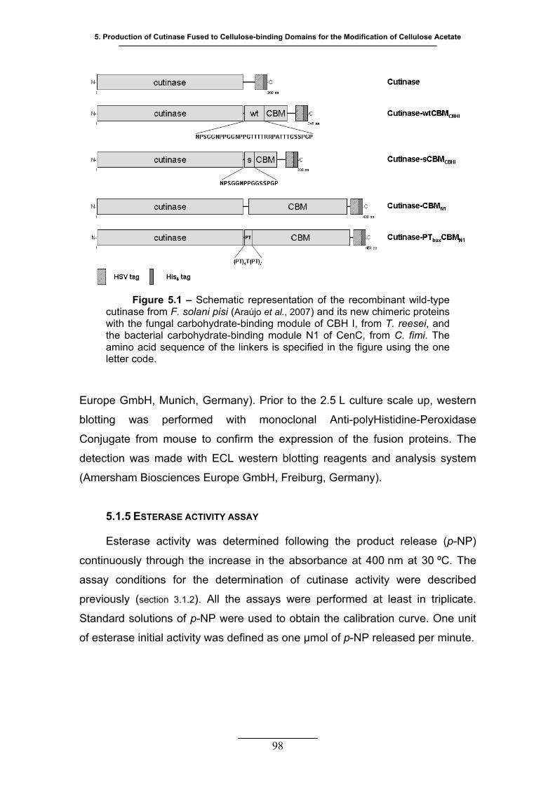
5. Production of Cutinase Fused to Cellulose-binding Domains for the Modification of Cellulose Acetate
Europe GmbH, Munich, Germany). Prior to the 2.5 L culture scale up, western
blotting was performed with monoclonal Anti-polyHistidine-Peroxidase
Conjugate from mouse to confirm the expression of the fusion proteins. The
detection was made with ECL western blotting reagents and analysis system
(Amersham Biosciences Europe GmbH, Freiburg, Germany).
5.1.5 ESTERASE ACTIVITY ASSAY
Esterase activity was determined following the product release (p-NP)
continuously through the increase in the absorbance at 400 nm at 30 ºC. The
assay conditions for the determination of cutinase activity were described
previously (section 3.1.2). All the assays were performed at least in triplicate.
Standard solutions of p-NP were used to obtain the calibration curve. One unit
of esterase initial activity was defined as one µmol of p-NP released per minute.
Figure 5.1 – Schematic representation of the recombinant wild-type cutinase from F. solani pisi (Araújo et al., 2007) and its new chimeric proteins with the fungal carbohydrate-binding module of CBH I, from T. reesei, and the bacterial carbohydrate-binding module N1 of CenC, from C. fimi. The amino acid sequence of the linkers is specified in the figure using the one letter code.
98

Enzymatic Treatment of Acrylic and Cellulose Acetate Fibres
99
5.1.6 TREATMENT OF CELLULOSE DI- AND TRIACETATE FABRIC WITH CUTINASE
FUSED TO CARBOHYDRATE-BINDING MODULES
All samples of cellulose acetate fabric were washed prior to use in order to
remove possible impurities from manufacture and from human handling, as
described previously (section 4.1.3).
Cellulose acetate fabric samples with an average weight of 0.1 g were
incubated with 100 U mL-1 of cutinase and cutinase-CBMN1, 50 U mL-1 of
cutin of cutinase-
sCBM
section 4.1.6).
ase-PTboxCBMN1 and cutinase-wtCBMT.reesei, 25 U mL-1
T.reesei in 10 mL of phosphate buffer with 0.01% sodium azide, under
continuous vertical agitation at 30 ºC and 20 rpm, for 18 hours. A control for
both types of fabric was run in parallel in which the buffer substituted the
enzyme.
After enzymatic treatment, all fabric samples were washed at 35 ºC, in the
Rotawash machine, to remove the adsorbed protein, according to the procedure
described previously (section 4.1.3).
5.1.7 QUANTIFICATION OF TOTAL PROTEIN CONCENTRATION
Total protein in solution was quantified following Bradford methodology
(Bradford, 1976) using BSA as standard. All samples were measured at least
twice.
5.1.8 CELLULOSE ACETATE FABRIC COLOURATION WITH A REACTIVE DYE
After enzymatic treatment, samples were competitively coloured in
phosphate buffer with 2% o.w.f. Remazol Brilliant Blue R, C.I. 61200, in
duplicate. The colouration was performed at 60 ºC, for 90 min at 20 rpm, in
sealed stainless steel beakers of 140 mL in capacity and housed on a lab-scale
dyeing machine (AHIBA Spectradye, from Datacolor International).
After the colouration, all samples were washed and the colour was
measured as described previously (
5. Production of Cutinase Fused to Cellulose-binding Domains for the Modification of Cellulose Acetate
100
55..22 RREESSUULLTTSS AANNDD DDIISSCCUUSSSSIIOONN
5.2.1 CELLULOSE DI- AND TRIACETATE TREATMENT WITH CUTINASE FUSED TO
CARBOHYDRATE-BINDING MODULES
For further improvement of cutinase catalysis, several fusion proteins with
known and well characterized CBMs were produced. The inclusion of spacers
between the cutinase and the CBMs was performed in three of the fusion
proteins. The importance of these spacers was studied by several authors
mainly through deletion studies. It was demonstrated that linker peptides,
connecting the catalytic domains of carbohydrate-active enzymes and the
CBMs, are necessary for the synergistic activity between the two domains
(Srisodsuk et al. ed
in the was
also ose
was sed
to CBMCBHI, by removing from the wild-type linker a sequence of residues that
constitute possible sites for O-glycosylation. Since E. coli does not possess the
he CenA from C.
fimi ( hen et al., 1991). This type of PT linker is also naturally glycosylated, but
when it is not, the conformations of catalytic domain and CBM are preserved,
since
be due to non-hydrolytic disruption of cellulose acetate fibres, in particular, of
CDA for which this phenomenon was most visible. This mechanical disruption
was already described for cellulose and cotton in the presence of CenA, Cex
, 1993; Shen et al., 1991). The wild-type linker of CBHI was includ
fusion protein with the CBM from the same enzyme. A smaller linker
used to connect cutinase to the fungal CBM (figure 5.1). The initial purp
to increase the levels of expression in E. coli of the soluble cutinase fu
machinery necessary for this post-translation eukaryotic modification, removing
those residues could promote correct folding of the fusion protein. The
expression levels were very low for soluble cutinase-wtCBMCBHI and were not
significantly improved in the case of cutinase-sCBMCBHI. The bacterial linker
used was the proline-threonine box (PT)4T(PT)7 present on t
S
only a partial increase in the linker flexibility seems to occur (Poon et al.,
2007).
Protein quantification after the cellulose acetate treatment with cutinase-
CBMN1 and cutinase-PTboxCBMN1 was unviable due to the turbidity of solutions.
This turbidity happened only for the referred assays, where protein adsorption
might be underestimated. The turbidity could be precipitated protein or it could

Enzymatic Treatment of Acrylic and Cellulose Acetate Fibres
101
and i
/S between chimeric proteins and
cutin
tate, independently of the CBM type. The
solated CBMs (Din et al., 1991, 1994; Cavaco-Paulo et al., 1999). Comparing the
amount of protein adsorbed and relative K
ase, there was a clear difference between the two cellulose acetates
studied (figure 5.2). The fusion of cutinase to the CBMs had a more pronounced
effect for the less substituted ace
A
B
Figure 5.2 – Protein adsorption and relative increase in K/S values for the (A) CDA and (B) CTA treated with cutinase and cutinase fused to CBMs (initial concen is expressed in units per gram of fabric). The samples were incu ing 18 hours with cutinase, cutinase-CBMN1 (cut-N1), cutinase-PTbox (cut-PT-N1), cutinase-wtCBMT.reesei (cut-wtCBM), and cutinase-sCBM -sCBM), at pH 8 and 30 ºC. Samples and control were comp ured at 60 ºC. Relative protein adsorption was calculated as
trationbated dCBMN1
T.reeseietitively colo
ur
(cut
cutinaseh18cutinaseh
h
PPP
0
0
− and relative K/S as hP18−
controlcutinase
controlcutinase chimeric
SKSKSKSK
////
−− .

5. Production of Cutinase Fused to Cellulose-binding Domains for the Modification of Cellulose Acetate
102
steric triacetate fibre given its higher DS
than in the diacetate fibre. The backbone is more fully ornamented with the
acety
CBM should be more impaired on this fibre
surfa
N1 (Tomme et al., 1996b). Other reason could be the difference in
size
constrains should be stronger in the
l groups and consequently the interactions necessary for the recognition of
the anhydroglucose units by the
ce regarding the diacetate fibre. Due to the fact that different initial
amounts of protein were used, is not possible to compare directly the protein
adsorption behaviour of the several constructs. But it is possible to see for this
particular treatment that there was no obvious relation between the colour
differences and the amount of protein adsorbed.
Taking into account the different esterase activities used, the cutinase-
wtCBMCBHI and cutinase-sCBMCBHI seem the most efficient catalysts under the
treatment conditions used. For CDA, the relative K/S was improved 3.8 and 2.6
fold by cutinase-wtCBMCBHI and cutinase-sCBMCBHI, respectively, regarding
cutinase alone. For treated CTA, the relative increase in K/S was not different
between cutinase alone and fused to the fungal CBMs, but the initial esterase
activity of cutinase was higher (figure 5.2). The differences in relative K/S were
also improved with the fusion of the bacterial CBM to cutinase. For CDA,
cutinase-CBMN1 improved the relative K/S by 1.8 fold, the same as cutinase-
PTboxCBMN1.
The treatment was performed at pH 8 which was the optimum pH for the
cutinase. The optimum pH for binding of most CBMs corresponds to the
optimum pH for the catalytic domain of the respective carbohydrate-active
enzyme and it is in the range of acidic to neutral. The better performance on
cellulose acetate fibres of the fungal CBM could be explained by the affinity of
CBMCBHI to insoluble ligands being relatively more insensitive to pH than the
affinity of CBM
of both CBMs. The activity of cutinase could be more constrained by the
bigger bacterial CBM than by the smaller fungal CBM. Indeed, using half the
esterase activity in the treatment with cutinase-PTboxCBMN1, the increase in K/S
obtained was in the same range of that with cutinase-CBMN1, for both fabrics
(figure 5.2). Further studies, aiming at a better characterization of the action of
chimeric cutinases on the surface modification of cellulose acetates, would
contribute to clarify these issues.
Enzymatic Treatment of Acrylic and Cellulose Acetate Fibres
103
55..33 CCOONNCCLLUUDDIINNGG RREEMMAARRKK SS
reesei, provided strong evidences of being an interesting strategy to pursuit.
Future work is needed to improve the recombinant production of modular
cutinases and to study in detail their affinities toward the cellulose acetates.
From the above considerations, it could be suggested that the cutinase
has potential in textile industry for the surface modification and consequently on
the “bicomponent yarns/fibres” production of cellulose acetate.
The design of hybrid enzymes mimics the strategies that Nature uses to
evolve and it is a powerful tool in biotechnology. The production and application
of the cutinase fused to CBMs, especially to the fungal CBM of CBHI of T.

104
GGEENNEERRAALL DDIISSCCUUSSSSIIOONN,,
FFIINNAALL RREEMMAARRKKSS AANNDD
FFUUTTUURREE PPEERRSSPPEECCTTIIVVEESS 66
Enzymatic Treatment of Acrylic and Cellulose Acetate Fibres
105
66.. GGEENNEERRAALL DDIISSCCUUSSSSIIOONN,, FFIINNAALL RREEMMAARRKKSS AANNDD FFUUTTUURREE
PPEERRSSPPEECCTTIIVVEESS
66..11 GGEENNEERRAALL DDIISSCCUUSSSSIIOONN
me
appli
-made fibres, in
particular the synthetic fibres, the scenario is by far different. Nature did not
design efficient ways to use and integrate them as part of ecosystems and they
are not natural substrates for enzymes. Besides, due to their chemical structure
man-made fibres require high amounts of energy and chemicals for their
modification in order to achieve the desired properties. Taking into account that
they have a market share of more than 50% (Aizenshtein et al., 2003), it is
imperative to address the environmental and safety issues brought by their
processing and disposal. Finding eco-sustainable alternatives to traditional
practices for man-made fibres in an extremely competitive market, such as the
textiles, is a hard challenge. The first and critical step is to find, to develop and
to improve methodologies for the enzymatic treatment of a given fibre (based on
the target chemical bounds of the polymer molecules). However it may not be
enough. The manipulation of the structure of enzymes through recombinant
DNA technologies may be crucial to the competitiveness of a bioprocess by
improving the ability of an enzyme to recognize and use the synthetic fibre as a
substrate.
The work presented in this thesis was focused on the application of
enzymes to specifically modify two man-made fibres – acrylic and cellulose
acetate, producing new functional groups on their surfaces. The enzymatic
modification of both acrylic and cellulose acetate fibres was targeted to the
pendent groups of the backbone chain of their polymers, which were the nitrile
and acetyl groups of acrylic, and the acetyl groups of cellulose acetate.
The textile industry presents well succeed examples of enzy
cations on the processing of natural fibres. For cotton, enzymes can be
used almost along all steps of its wet processing, nearly replacing conventional
textile practices like the stone washing of denim. Regarding man
6. General Discussion, Final Remarks and Future Perspectives
106
Chapter
treatment of
carboxylic gr
substrate wit
hours, at 40
of the enzym
any substrate
2 rep rts the employment of a commercial nitrilase on the
acryli rile into
oups. I re as a
h a ste release o monia to the reaction media, during 36
ºC. Firs ved stability
e under operational conditions. When incubated in solution, without
, at 30 en the
acrylic fabric samples were present in the system, there was an adsorption of
80% of total protein and the nitrilase adsorbed did not loose activity during the
36 hours of treatment, since it was not observed a slowing down on the release
of ammonia. Second, it can be inferred that the adsorption that takes place is a
dynamic process. Either an alternating adsorption and desorption take place, or
nitrilase has the ability to move over the acrylic surface like other processive
enzymes. Otherwise the release of ammonia would eventually stop and a
plateau would be established with time.
Looking to the other and most important product of the biocatalysed
reaction – the carboxylic groups at the fibre surface, the results were somehow
unforeseen. The treatment of acrylic with nitrilase presented an interesting case
study due to the fact that while ammonia was steadily produced the same was
not verified for the acidic groups. The increase in K/S of treated acrylic fabric
samples was not regular along the 36 hours of treatment. Instead, it was
observed a succession of colour strength values higher and lower than the
controls, which created an oscillating pattern. The hypothesis was that in some
way the groups could leave the surface of the fabric. This was confirmed when
the polyacrylic acid was determined in the treatment solutions. The important
consequence of this finding is that the nitrilase biomodification of acrylic needs
to be properly controlled in order to maximize the concentration of the surface
acidic groups.
In order to achieve a proper control over the acrylic biomodification is
important to understand better how nitrilase acts on such substrate. Is the
enzyme very sensitive to the crystallinity degree of the fibre? How does
crystallinity affect the release of polyacrylic acid? How the molecular size of the
polymer is related to the released amount of polyacrylic acid? Is there any
difference in treating the fibre before and after orientation? It would be
o
cs, leading to the superficial conversion of nit
t was found that nitrilase was able to use acrylic fib
ady f am
t important outcome of this work was the impro
ºC, nitrilase lost half of its activity after 15 hours. Wh

Enzymatic Treatment of Acrylic and Cellulose Acetate Fibres
107
interesting to treat polymers with different compositions, crystallinities and
mole
more complete picture of the modifications catalysed by
nitrilase and to minimize the treatment time.
The acrylic samples, non treated and treated for 4 and 24 hours, were
evalu ant difference between the degrees of
crystallinity of samples was observed (less than 1%). In a previous work, where
alkali
increased the surface area available for enzymatic hydrolysis by a
micro
a good
starting point for the application of biocatalysts.
cular size distributions, as well as, from different stages of the manufacture
process to have a
ated by WAXS, but no signific
ne and enzymatic treatments of synthetic fibres were compared, the
nitrilase did not affect significantly the breaking strength of the acrylic fabric
(Silva, 2002). Once the main chain of each molecule of acrylonitrile copolymer is
not chemically altered and because enzymatic hydrolysis is in general
superficial, major consequences of such modifications on the bulk properties of
these fibres are not expected.
The acrylonitrile represents 93% of the total monomer composition of the
acrylic used in this work. The remaining 7% is vinyl acetate which constitutes a
possible substrate for another class of enzymes – esterases. Chapter 3
describes the experiments performed with an esterase that has the natural
ability to hydrolyse both soluble and insoluble substrates – the cutinase from
Fusarium solani pisi. The enzyme was very stable and active but the
modifications were faint, since only 7% of the surface groups were possible
substrates for cutinase. The protein adsorption was practically absent, in
contrast to the prior observed nitrilase adsorption, indicating that significant
unspecific interactions were not occurring, but also indicating lack of specific
binding sites for cutinase. The introduction of stainless steel discs in the
reaction media allowed the evaluation of changes, in terms of colour, on the
fibre treated for short periods. Besides increasing the mass transfer, the discs
may have
pilling effect. Nevertheless, the observation that small amounts of a
common industrial acrylic solvent (DMA) improved the activity of the enzyme
lead to the hypothesis that the accessibility of this substrate can be higher in
early stages of its wet-spinning manufacture. As long as the stability of the
biocatalyst is not to much compromised, the industrial tanks after coagulation,
where the washing and orientation of the acrylic fibre begin, may be
6. General Discussion, Final Remarks and Future Perspectives
108
In a similar way, cutinase was used to hydrolyse the acetyl groups
esterified to the main chains of cellulose acetate (chapter 4). Commercial CDA
and CTA fabrics, having different degrees of substitution and, consequently,
different degrees of crystallinity and hydrophilic character, were tested. From
the formation of the soluble product (acetic acid), during 8 hours, it was possible
to conclude that cutinase, like most enzymes, slightly preferred the less
crystalline and more hydrophilic substrate, the CDA. When the solid product of
the enzymatic reaction (hydroxyl groups) was evaluated through colouration
and DRIFT, there was an apparent opposite preference. This result could be
due to the hydroxyl groups that are already present at the CDA fibre comparing
to their almost absence at CTA fibre. The CDA controls had a much higher
capacity to fix the cotton reactive dye than the CTA controls; the K/S values for
CDA were around 8 times higher than the ones obtained for CTA. This meant a
lower sensitivity of the colouration methodology and maybe a lower sensitivity of
DRIFT for the differences in the concentration of hydroxyl groups between
contr
fibres. In terms of increase in the hydroxyl groups at the fibres
surfa
ols and cutinase treated samples of CDA fabric. The differences between
the K/S and the intensity/shape of the carbonyl stretching band of controls and
treated samples of CTA were clearer than the differences obtained for CDA.
The chapter 5 describes the production of cutinases with new
functionalities by recombinant DNA technologies and the study of their
efficiency for CDA and CTA modification in comparison with the native enzyme.
Since the two cellulose acetate fibres used are structurally different from
cellulose (the native ligand) and different between themselves, presenting
different overall crystallinities, two different CBMs were fused to the C-terminal
of cutinase. The idea was to mimic other carbohydrate-active enzymes which
also have CBMs that improve their activity. In terms of protein adsorption, the
purpose was attained: the fusion with both CBMs increased the affinity of native
cutinase for the
ce the results of the colouration were preliminary but there are strong
evidences that the chimeric cutinases are more efficient than the native
cutinase.
The adsorption of cutinase to the cellulose acetate fibres was higher than
the one observed for acrylic, obviously reflecting a higher concentration of
recognizable substrates at the fibre surface. However, because the washing
Enzymatic Treatment of Acrylic and Cellulose Acetate Fibres
109
procedure after the enzymatic treatments was milder, in terms of pH and
temperature, and because the cellulose acetate is less polar and more
hydrophobic than the acrylic taffeta used, it is not possible to exclude unspecific
binding of cutinase to the cellulose acetates. It will be necessary to have
controls like null mutated or inhibited cutinases for a better assessment of this
phenomenon.
66..22 FFIINNAALL RREEMMAARRKKSS
quate biocatalyst for the modification of nitrile
The general conclusions that can be drawn from the work here described
are:
• nitrilase is an ade
groups of polyacrylonitrile copolymers; using a single enzyme,
(which is an advantage regarding the enzymatic systems of nitrile
hydratase and amidase, reported in the literature) at mild conditions
of pH and temperature, acid groups were formed at the fibre
surface;
• cellulose acetate and triacetate can have more reactive surfaces
using a lipolytic enzyme - cutinase, which is a new and important
finding; the surface ‘regeneration’ of cellulose composition can
impart these yarns a bicomponent character that would be very
interesting for the sportswear field;
• the affinity of cutinase for cellulose acetates can be improved by
adding CBMs, resembling natural carbohydrate esterases.
The application of enzymes to the surface modification of acrylic and
cellulose acetate fibres, and the resultant new functionalities may lead to eco-
friendly finishing steps of these textile materials compared to the traditional
ones, saving on water, energy and reducing the amounts of hazardous
compounds.
In spite of these findings, many questions remain to be answered which
give space for future work.
6. General Discussion, Final Remarks and Future Perspectives
110
66..33 FFUUTTUURREE PPEERRSSPPEECCTT IIVVEESS
tyl
ester
In order to transform the biomodifications here reported into cost
competitive industrial processes, a considerable amount of work will be needed.
The modification of acrylic by nitrilase needs to be carefully studied in
order to maximize the formation of acid groups on its surface, in terms of time
and without compromising too much the nitrilase activity. The production of a
know nitrilase that can be redesigned to use more efficiently acrylic as a
substrate is, beyond doubt, needed. It would be interesting to look for naturally
more stable nitrilases, like the ones found in extremophile microorganisms
(Cowan et al., 1998; Mueller et al., 2006; Khandelwal et al., 2007), because the presence
of high temperatures and organic solvents during the wet-spinning process of
acrylic fibres can have deleterious effects on the enzymatic activity.
A better biochemical characterization of cutinase fusion proteins is needed
in addition to their interaction with the cellulose acetates. The optimization of the
expression system also is needed for higher yields of protein production which
is essential for the economical point of view.
It also would be interesting to compare cutinase with natural ace
ases in the hydrolysis of cellulose acetates surface, like the acetyl esterase
II from Trichoderma reesei and Penicillium purpurogenum (Margolles-Clark et al.,
1996; Hakulinen et al., 2000; Ghosh et al., 2001; Colombres et al., 2008). These enzymes
have acetyl esters as natural substrates and their structures are very similar to
cutinase.
Finally it would be necessary to study the feasibility of upgrading the
optimized processes to an industrial scale and the application of such modified
fibres for the production of speciality-properties textile products.
111
111
RREEFFEERREENNCCEESS
77
Enzymatic Treatment of Acrylic and Cellulose Acetate Fibres
112
77.. RREEFFEERREENNCCEESS
Aizen
Araúj
challenges for biocatalyst design. Nature
rption, and activities on cotton fabrics of full length and core domains of EGV
Ball P (2001). Biocatalysis: synthesis methods that exploit enzymatic activities. Nature 409:
225.
Bandyopadhyay AK, Nagasawa T, Asano Y, Fujishiro K, Tani Y, Yamada H (1986). Purification and characterization of benzonitrilases from Arthrobacter sp. Strain J-1.
Appl Environ Microb 51: 302-306.
Banerjee A, Sharma R, Banerjee UC (2002). The nitrile-degrading enzymes: current status
and future prospects. Appl Microbiol Biot 60: 33-44.
Baptista RP, Cabral JMS, Melo EP (2000). Trehalose delays the reversible but not the
irreversible thermal denaturation of cutinase. Biotechnol Bioeng 70: 699-703.
shtein EM, Efremov VN, Shnaider R (2003). Retrospective analysis of the status of
textile stock in 2001. Fibre Chem 35: 1-11.
Altaner C, Saake B, Puls J (2001). Mode of action of acetylesterases associated with
endoglucanases towards water-soluble and – insoluble cellulose acetates. Cellulose 8:
259-265.
Altaner C, Saake B, Puls J (2003a). Specificity of an Aspergillus niger esterase deacetylating
cellulose acetate. Cellulose 10: 85-95.
Altaner C, Saake B, Tenkanen M, Eyzaguirre J, Faulds CB, Biely P, Viikari L, Siika-aho M, Puls J (2003b). Regioselective deacetylation of cellulose acetates by acetyl xylan
esterases from different CE-families. J Biotechnol 105: 95-104.
o R, Silva C, O’Neill A, Micaelo N, Gübitz G, Soares CM, Casal M, Cavaco-Paulo A (2007). Tailoring cutinase activity towards polyethylene terephthalate and polyamide 6.6
fibers. J Biotechnol 128: 849-857.
Arnold FH (2001). Combinatorial and computational
409: 253-257.
Aymard C, Belarbi A (2000). Kinetics of thermal deactivation of enzymes: a simple three
parameters phenomenological model can describe the decay of enzyme activity,
irrespectively of the mechanism. Enzyme Microb Tech 27: 612-618.
Azevedo H, Bishop D, Cavaco-Paulo A (2000). Effects of agitation level on the adsorption,
deso
(Humicola insolens) and CenA (Cellulomonas fimi). Enzyme Microb Tech 27: 325-329.
Bahrami SH, Bajaj P, Sen K (2003). Effect of coagulation conditions on properties of
poly(acrylonitrile-carboxylic acid) fibers. J Appl Polym Sci 89: 1825-1837.
Enzymatic Treatment of Acrylic and Cellulose Acetate Fibres
113
Baptista RP, Cdeactiv
Bashir Z (19924304-43
Bashir Z (1994solvent
1128.
hen LY, Paixão A, Cabral JMS, Melo EP (2003). A novel pathway to enzyme
ation: the cutinase model. Biotechnol Bioeng 82: 851-857.
). Therm reversible gelation and plasticization of polyacrylonitrile. Polymer 33:
13.
). Co-cry ray diffraction behaviour of
-containing and solvent-free polyacrylonitrile. J Polym Sci Pol Phys 32: 1115-
Bashir Z (2001). The h al mesophase in atactic polyacrylonitrile: a new interpretation of
the phase transitions in the polymer. J Macromol Sci B 40: 41-67.
Bashir Z, Rastogi S (2 5). The explanation of the increase in slope at the Tg in the plot of d-
spacing versus temperature in polyacrylonitrile. J Macromol Sci B 44: 55-78.
Battistel E, Francalan i F, Marinetti M, Morra M (1995). Modified materials based on
polyacrylonitrile and process for their production. European Patent EP0662540A1.
Battistel E, Bernardi A, Maestri P (1997). Enzymatic decontamination of aqueous polymer
emulsions containing acrylonitrile. Biotechnol Lett 19: 131-134.
Battistel E, Morra M, M rinetti M (2001). Enzymatic surface modification of acrylonitrile fibers.
Appl Surf Sci 177: 32-41.
Belfort G, Zydney AL (2003). Interaction of Proteins with polymeric synthetic membranes. In:
Surfactant Science Series/110 - Biopolymers at Interfaces, Malmsten M. (Ed.), Marcel
De 671-709.
Berry MJ, Davis PJ, Gidley MJ (2001). Conjugated polysaccharide fabric detergent and
conditioning pro s. United States Patent 6225462.
Bhikhabhai R, Petters n LG (1984). The cellulolytic enzymes of Trichoderma reesei as a
system of homologous proteins. FEBS Lett 167: 301-308.
Bohn CR, Schaefgen J , Statton WO (1961). Laterally ordered polymers: polyacrylonitrile and
poly(vinyl trifluor acetate). J Polym Sci 55: 531-549.
Boraston AB, Nurizzo D, Notenboom V, Ducros V, Rose DR, Kilburn DG, Davies GJ (2002). Differential oligosaccharide recognition by evolutionarily-related β-1,4 and β-1,3
glucan-binding m . J Mol Biol 319: 1143-1156.
Boraston AB, Bolam DN, Gilbert HJ, Davies GJ (2004). Carbohydrate-binding modules: fine-
tuning polysaccharide recognition. Biochem J 382: 769-781.
Bornscheuer UT, Kazlauskas RJ (2004). Catalytic promiscuity in biocatalysis: using old
enzymes to form w bonds and follow new pathways. Angew Chem Int Edit 43: 6032-
6040.
o
stallization of solvents with polymers: the X-
exagon
00
c
a
kker, Inc., Ne York, ppw
duct
so
Ro
odules
ne
Enzymatic Treatment of Acrylic and Cellulose Acetate Fibres
114
Bradford MM (1976). A rapid and sensitive method for the quantification of microgram
quantities of protein utilizing the principle of protein-dye binding. Anal Biochem 72: 248-
Brenner C (2002). Catalysis in the nitrilase superfamily. Curr Opin Struc Biol 12: 775-782.
Cabral es-Barros MR, Gama M (2003). Engenharia enzimatica. Lidel – edições
Calado (2002). Optimisation of culture conditions
Capone ology and applications,
Carrolod applications, Rustemeyer P (Ed.), Wiley-VCH,
Carvalho CML, Aires-Barros MR, Cabral JMS (1998). Cutinase structure, function and
Carvalh
by infrared spectroscopy. Forensic Sci Int 151: 125-131.
Guimarães.
254.
Burke M (2008). Green couture - Synthetic fibres are back in fashion after an ecological
makeover. Chemical World, 5: 58-61.
Burkinshaw SM (1995). Chemical Principles of synthetic fibre dyeing. Blackie Academic &
Professional, UK, pp 157-193.
JMS, Airtécnicas, Lda, Lisboa, pp 1-3.
CRC, Taipa MA, Cabral JSM, Fonseca LPand characterisation of cutinase produced by recombinant Saccharomyces cerevisiae.
Enzyme Microb Tech 31: 161-170.
GJ (1995). Wet-spinning technology. In: Acrylic fiber techn
Masson JC (Ed.), Marcel Dekker, Inc, New York, pp 69-103.
P, Grospietro B (2004). Plastic materials. In: Macromol Symp – special issue:
Cellulose acetates: properties an
Weinheim, pp 335-351.
biocatalytic applications. Electron J Biotechnol 1: 1-14.
o CML, Aires-Barros MR, Cabral JMS, (1999a). Cutinase: from molecular level to
bioprocess development. Biotechnol Bioeng 66: 17-34.
Carvalho CML, Cabral JMS, Aires-Barros MR (1999b). Cutinase stability in AOT reversed
micelles: system optimization using the factorial design methodology. Enzyme Microb
Tech 24: 569-576.
Castro GR, Knubovets T (2003). Homogeneous biocatalysis in organic solvents and water-
organic mixtures. Crit Rev Biotechnol 23: 195-231.
Causin V, Marega C, Schiavone S, Marigo A (2005). A quantitative differentiation method for
acrylic fibers
Cavaco-Paulo A (1995). Influência da agitação mecânica e da composição enzimática no
tratamento do algodão com celulases. PhD Dissertation, Textile Engineering
Department of University of Minho,
Cavaco-Paulo A (1998). Mechanism of cellulase action in textile processes. Carbohyd Polym
37: 273-277.
Enzymatic Treatment of Acrylic and Cellulose Acetate Fibres
115
Cavaco-Paulo A, Morgado J, Andreaus J, Kilburn D (1999). Interactions of cotton with CBM
peptides. Enzyme Microb Tech 25: 639-643.
Cavaco-Paulo A, dos Santos Marinho da Silva CJ (2003). Treatment of animal hair fibers
with modified p
o
di
roteases. International Patent WO 03/097927 A1.
Cerqu GR, Assunção RMN (2006). A new value for the heat of fusion of a
Chen J on fibre precursor via
369-
Collier w Jersey, 576 p.
rre J (2008). An eleven amino acid residue deletion expands
Coutin RAJ, Miller RC Jr (1991). Nucleotide sequence
ol Microbiol 5: 1221-1233.
nd sephadex is mediated by
inal repeats. Mol Microbiol 6: 1243-1252.
Coutin r RC Jr (1993). The nature of the
ng domain affects the activities of a bacterial endoglucanase on different
Cowan ah Q (1998). Biochemistry and
y of mesophilic and thermophilic nitrile metabolizing enzymes.
Crev W, Berendsen HJC, Pepermans HAM (2001). DSC studies of Fusarium
Cegarra J (1996). The state of the art in textile biotechnology. J Soc Dyers Colour 112: 326-
329.
ira DAe , Filh
h
the N-term
hcellulose-bin
Biotechnolog
el LD, Meijberg
perfect crystal of cellulose acetate. Polym Bull 56: 475-484.
C, Harrison IR (2002). Modification of polyacrylonitrile (PAN) carb
post-spinning plasticization and stretching in dimethylformamide (DMF). Carbon 40: 25-
45.
Chen Y, Xiong X, Yang G, Zhang L, Lei S, Liang H (2002). Characterization of regenerated
cellulose membranes hydrolyzed from cellulose acetate. Chinese J Polym Sci 20:
375.
BJ, Tortora PG (2001). Understanding textiles. Prentice Hall, Ne
Colombres M, Garate JA, Lagos CF, Araya-Secchi R, Norambuena P, Quiroz S, Larrondo L, Pérez-Acle T, Eyzaguithe substrate specificity of acetyl xylan esterase II (AXE II) from Penicillium
purpurogenum. J Comput Aid Mol Des 22: 19-28.
o JB, Moser B, Kilburn DG, Warrenof the endoglucanase C gene (cenC) of Cellulomonas fimi, its high-level expression in
Escherichia coli, and characterization of its products. M
Coutinho JB, Gilkes NR, Warren RAJ, Kilburn DG, Miller RC Jr (1992). The binding of
Cellulomonas fimi endoglucanase C (CenC) to cellulose a
o JB, Gilkes NR, Warren RAJ, Kilburn DG, Mille
forms of cellulose. FEMS Microbiol Lett 113: 211-217.
D, Cramp R, Pereira R, Graham D, Almataw
Extremophiles 2: 207-216.
solani pisi cutinase: consequences for stability in the presence of surfactants. Biophys
Chem 92: 65-75.
Enzymatic Treatment of Acrylic and Cellulose Acetate Fibres
116
Din N, Gilkes NR, Tekant B, Miller RC, Warren RAJ, Kilburn DG (1991). Non-hydrolytic
disruption of cellulose fibres by the binding domain of bacterial cellulase. Nat Biotechnol
9: 1096-1099.
ed:
Durán ns in the textile industry. Rev Prog Coloration 30:
Egmon m solani pisi cutinase. Biochimie 82: 1015-1021.
chnol 40: 689-702.
Etters J e use: a developing technology. Am Dyest Rep 87:
Fägers structure of a 1,4-β-glucan
Fischer M, Heumann S, Puolakka A, Wirth A, Cavaco-Paulo A,
ransfor 24: 419-425.
trile hydrolysing enzymes from
Frusho
(Ed.), Marcel Dekker, Inc, New
Frusho chnology
Fuglsa omains (CBDS) for oral care products.
Gardne CM, Komarek R, Dorschel D, Boggs C, White AW (1994). Compostability of Cellulose Acetate Films. J Appl Polym Sci 52: 1477-1488.
Din N, Damude HG, Gilkes NR, Miller RC, Warren RAJ, Kilburn DG (1994). C1-Cx revisit
intramolecular synergism in cellulose. Proc Natl Acad Sci USA 91: 11383-11387.
N, Durán M (2000). Enzyme applicatio
41-44.
d MR, de Vlieg J (2000). Fusariu
El-Zaher NA (2001). Study of the effect of ultraviolet radiation on some physical properties of
Dralon fabric. Polym – Plast Te
Emsermann H, Foppe R (1995). Dyeing processes. In: Acrylic fiber technology and
applications, Masson JC (Ed.), Marcel Dekker, Inc, New York, pp 285-312.
N, Annis PA (1998). Textile enzym
18-23.
tam LG, Pettersson LG, Engström JA (1984). The primary
cellobiohydrolase from the fungus Trichoderma reesei QM 9414. FEBS Lett 167: 309-
315.
-Colbrie G, Herrmann Gübitz GM (2006). Surface modification of polyacrylonitrile with nitrile hydratase and
amidase from Agrobacterium tumefaciens. Biocatal Biot
Fischer-Colbrie G, Matama T, Heumann S, Martinkova L, Cavaco-Paulo A, Gübitz GM (2007). Surface hydrolysis of polyacrylonitrile with ni
Micrococcus luteus BTS20. J Biotechnol 129: 62-68.
ur BG (1982). Water as a melting point depressant for acrylic polymers. Polymer Bull 7:
1-8.
Frushour BG (1995). Acrylic polymer characterization in the solid state and in solution. In:
Acrylic fiber technology and applications, Masson JC
York, pp 197-257.
ur BG, Knorr RS (1998). Acrylic fibres. In: International Fiber Science and Te
Series/15 - Handbook of Fibre Chemistry, Lewin M, Pearce EM (Ed.), Marcel Dekker,
Inc., New York, pp 869-1070.
ng CC, Tsuchiya R (2001). Cellulose binding d
United States Patent 6264925.
r RM, Buchanan
Enzymatic Treatment of Acrylic and Cellulose Acetate Fibres
117
Ghosh D, Sawicki M, Lala P, Erman M, Pangborn W, Eyzaguirre J, Gutiérrez R, Jörnvall H, Thiel DJ (2001). Multiple conformations of catalytic serine and histidine in acetylxylan
Gilkes cision of the cellulose
on catalysis. J Biol Chem 263: 10401-10407.
tions, Rustemeyer P (Ed.),
Wiley-VCH, Weinheim, pp 371-394.
Goddarctive compounds. Prog Polym Sci 32: 698-725.
b Tech 32: 868-879.
water
and wastewater American Public Health Association, Washington DC, pp 4.75- 4.80.
Gübitz
Gübitz for reliable enzymes: enzymatic
Gübitz
Hakulin ensional structure of the catalytic
Harper Enzymology of C-N cleavage by
Harper 167: 685-692.
Hildén L, Johansson G (2004). Recent developments on cellulases and carbohydrate-binding
modules with cellulose affinity. Biotechnol Lett 26: 1683-1693.
esterase at 0.90 Å. J Biol Chem 276: 11159-11166.
NR, Warren AJ, Miller Jr. RC, Kilburn DG (1988). Precise ex
binding domains of two Cellulomonas fimi cellulases by homologous protease and the
effect
Glasser WG (2004). Prospects for future applications of cellulose acetate. In: Macromol Symp
– special issue: Cellulose acetates: properties and applica
d JM, Hotchkiss JH (2007). Polymer surface modification for the attachment of
bioa
Gonçalves AMD, Aires-Barros MR, Cabral JMS (2003). Interaction of an anionic surfactant
with a recombinant cutinase from Fusarium solani pisi: a spectroscopic study. Enzyme
Micro
Greenberg AE, Clesceri LS, Eaton AD (1992). Standard methods for the examination of
G, Cavaco-Paulo A (2001). Biotechnology in the textile industry – perspectives for the
new millennium. J Biotechnol 89: 89-90.
G, Cavaco-Paulo A (2003). New substrates
modification of polymers. Curr Opin Biotech 14: 577-582.
G, Cavaco-Paulo A (2008). Enzymes go big: surface hydrolysis and functionalisation of
synthetic polymers. Trends Biotechnol 26: 32-38.
Guillen JG (1987). Fibras Acrílicas. Universidade Politécnica da Catalunha, LLibreria Costa
Terrassa, 191 p.
en N, Tenkanen M, Rouvinen J (2000). Three-dim
core of acetylxylan esterase from Trichoderma reesei: insights into the deacetylation
mechanism. J Struct Biol 132: 180-190.
DB (1977a). Microbial metabolism of aromatic nitriles.
Nocardia sp. (Rhodochrous group) N.C.I.B. 11216. Biochem J 165: 309-319.
DB (1977b). Fungal degradation of aromatic nitriles. Enzymology of C-N cleavage by
Fusarium solani. Biochem J
Harper DB (1985). Characterization of a nitrilase from Nocardia sp. (Rhodochrous group)
N.C.I.B. 11215, using p-hidroxylbenzonitrile as sole carbon source. Int J Biochem 17:
677-683.

Enzymatic Treatment of Acrylic and Cellulose Acetate Fibres
118
Hindeleh AM, Johnson DJ (1972). Peak resolution and X-ray crystallinity determination in
heat-treated cellulose triacetate. Polymer 13: 27-32.
AM, Teeri TTHoffrén , Teleman O (1995). Molecular dynamics simulation of fungal cellulose-
3-4267.
alysts
Hutchin N, Ansari I (2008). Modifying and
Ilharco lose acetate
Illanes A (1999). Stability of biocatalysts. Electron J Biotechnol 2: 1-9.
43: 389-393.
95-13906.
14381-1394.
m Cellulomonas fimi β-1,4-glucanase CenC bind nitroxide spin-labelled
Kalum
Kaul P ee UC (2007). Cross-linked amorphous nitrilase aggregates for
Khande thermophilic isolate. J Chem Technol Biot 82: 646-651.
binding domains: differences in molecular rigidity but a preserved cellulose binding
surface. Protein Eng 8: 443-450.
Hook RH, Robinson WG (1964). Ricinine nitrilase: II. Purification and properties. J Biol Chem
239: 426
Hughes J, Armitage YC, Symes KC (1998). Application of whole cell rhodococcal biocat
in acrylic polymer manufacture. Anton van Lee 74: 107-118.
gs LR, Narrianen AP, Thompson RL, Clarke managing the surface properties of polymers. Polym Int 57: 163-170.
LM, Barros RB (2000). Aggregation of pseudoisocyanine iodide in cellu
films: structural characterization by FTIR. Langmuir 16: 9331-9337.
Johansson G, Stahlberg J, Lindeberg G, Engström A, Pettersson G (1989). Isolated fungal
cellulase terminal domains and a synthetic minimum analogue bind to cellulose. FEBS
Lett 2
Johnson PE, Tomme P, Joshi MD, McIntosh LP (1996a). Interaction of soluble
cellooligosaccharides with the N-terminal cellulose-binding domain of Cellulomonas fimi
CenC. 2. NMR and Ultraviolet Absorption Spectroscopy. Biochemistry 35: 138
Johnson PE, Joshi MD, Tomme P, Kilburn DG, McIntosh LP (1996b). Structure of the N-
terminal cellulose-binding domain of Cellulomonas fimi CenC determined by Nuclear
Magnetic Resonance Spectroscopy. Biochemistry 35:
Johnson PE, Brun E, MacKenzie LF, Withers SG, McIntosh LP (1999). The cellulose-binding
domains fro
cellooligosaccharides in multiple orientations. J Mol Biol 287: 609-625.
L, Adersen BK (2000). Enzymatic treatment of denim. United States Patent 6146428.
, Stolz A, Banerjenantioselective nitrile hydrolysis. Adv Synth Catal 349: 2167-2176.
lwal AK, Nigam VK, Choudhury B, Mohan MK, Ghosh P (2007). Optimization of
nitrilase production from a new
Kirk O., Borchert TV, Fuglsang CC (2002). Industrial enzyme applications. Curr Opin Biotech
13: 345-351.
Klibanov AM (2001). Improving enzymes by using them in organic solvents. Nature 409: 241-
246.
Enzymatic Treatment of Acrylic and Cellulose Acetate Fibres
119
Kobayashi M, Yanaka N, Nagasawa T, Yamada H, (1990). Purification and characterization of
a novel nitrilase of Rhodococcus rhodochrous K22 that acts on aliphatic nitriles. J
Bacteriol 172: 4807-4815.
Kobayashi M, Shimizu S (1994). Versatile nitrilases: nitrile-hydrolysing enzymes. FEMS
Microbiol Lett 120: 217-224.
KM, Wong C-H (2001). Enzymes for chemical synthKoeller esis. Nature 409: 232-240.
Köller W, Kolattukudy PE (1982). Mechanism of action of cutinase: chemical modification of
Kormos J, Johnson PE, Brun E, Tomme P, McIntosh LP, Haynes CA, Kilburn DG (2000).
Krasovskii AN, Plodistyi AB, Polyakov DN (1996). IR study of distribution of primary and
olution structure of the C-terminal domain of
aling. Biochemistry 28: 7241-
Kuehni principles. John Wiley & Sons, Inc.
Kumara orthy S, Gerchman SE, Kycia H, Studier FW, Swaminathan S
La Niev 774-810.
functional
Lau EY eaking the Asp-His hydrogen bond of the
Law PW, Longdon A, Willins GG (2004). Solvent cast diacetate film. In: Macromol Symp –
Kolattukudy PE (2002). Cutin from plants. In: Polyesters I: biological systems and
biotechnological production, Doi Y, Steinbüchel A (Ed.), Wiley-VCH, Weinheim, pp 1-8.
the catalytic triad characteristic for serine hydrolases. Biochemistry 21: 3083-3090.
Binding site analysis of cellulose binding domain CBDN1 from endoglucanase C of
Cellulomonas fimi by site-directed mutagenesis. Biochemistry 39: 8844-8852.
secondary functional groups in highly substituted cellulose acetates, acetomaleates,
and acetophthalates. Russ J Appl Chem 69: 1048-1054.
Kraulis PJ, Clore M, Nilges M, Jones A, Pettersson G, Knowles J, Gronenborn AM (1989). Determination of the three-dimensional s
cellobiohydrolase I from Trichoderma reesei. A study using nuclear magnetic resonance
and hybrid distance geometry-dynamical simulated anne
7257.
RG (1997). Color: an introduction to practice and
New York, pp 109-119.
n D, Eswaramo(2003). Crystal structure of a putative CN hydrolase from yeast. Proteins 52: 283-291.
e HL (2007). Cellulose acetate and triacetate fibers. In: Handbook of fiber chemistry,
Lewin M (Ed.), CRC Press, New York, pp
Langsford ML, Gilkes NR, Singh B, Moser B, Miller RC Jr, Warren RAJ, Kilburn DG (1987). Glycosylation of bacterial cellulases prevents proteolytic cleavage between
domains. FEBS Lett 225: 163-167.
, Bruice TC (1999). Consequences of br
catalytic triad: effects on the structure and dynamics of the serine esterase cutinase.
Biophys J 77: 85-98.
special issue: Cellulose acetates: properties and applications, Rustemeyer P (Ed.),
Wiley-VCH, Weinheim, pp 81-166.

Enzymatic Treatment of Acrylic and Cellulose Acetate Fibres
120
Law RC (2004). Cellulose acetate in textile applications. In: Macromol Symp – special issue:
Cellulose acetates: properties and applications, Rustemeyer P (Ed.), Wiley-VCH,
Lee Y-Hng 24: 2383-2406.
wo fungal cellulose-binding domains is dominated by a single amino
Linder rakenberg T, Reinikainen
. Protein Sci 4:
1056-1064.
Linder a double cellulose-
binding domain. Synergistic high affinity binding to crystalline cellulose. J Biol Chem
cad Sci USA 93: 12251-12255.
Longhi S, Czjzek M, Lamzin V, Nicolas A, Cambillau C (1997). Atomic resolution (1.0 Å)
Longhi S, Cambillau C (1999). Structure-activity of cutinase, a small lipolytic enzyme. Biochim
Lu Y, C for drug delivery.
Adv Drug Deliver Rev 56: 1621-1633.
Lulay A (1995). Apparel end-uses. In: Acrylic fiber technology and applications, Masson JC
Mahadevan S, Thimann KV (1964). Nitrilase: II. Substrate specificity and possible mode of
Mannesse MLM, Cox RC, Koops BC, Verheij HM, de Haas GH, Egmond MR, van der
alogues. Effect of acyl chain length and position in the substrate molecule
Weinheim, pp 255-265.
, Fan LT (1982). Kinetic studies of enzymatic hydrolysis of insoluble cellulose: analysis
of the initial rates. Biotechnol Bioe
Linder M, Lindeberg G, Reinikainen T, Teeri TT, Pettersson G (1995a). The difference in
affinity between t
acid substitution. FEBS Lett 372: 96-98.
M, Mattinen ML, Kontteli M, Lindeberg G, Stahlberg J, DT, Pettersson G, Annila A (1995b). Identification of functionally important amino acids
in the cellulose-binding domain of Trichoderma reesei cellobiohydrolase I
M, Salavuori I, Ruohonen L, Teeri TT (1996). Characterization of
271: 21268-21272.
Linder M, Teeri TT (1996). The cellulose-binding domain of the major cellobiohydrolase of
Trichoderma reesei exhibits true reversibility and a high exchange rate on crystalline
cellulose. Proc Natl A
Liu XD, Ruland W (1993). X-ray studies on the structure of polyacrylonitrile fibers.
Macromolecules 26: 3030-3036.
crystal structure of Fusarium solani cutinase: steriochemical analysis. J Mol Biol 268:
779-799.
Biophys Acta 1441: 185-196.
hen SC (2004). Micro and nano-fabrication of biodegradable polymers
(Ed.), Marcel Dekker, Inc, New York, pp 313-339.
action. Arch Biochem Biophys 107: 62-68.
Hijden HTWM, de Vlieg J (1995). Cutinase from Fusarium solani pisi hydrolyzing
triglyceride an
on activity and enantioselectivity. Biochemistry 34: 6400-6407.

Enzymatic Treatment of Acrylic and Cellulose Acetate Fibres
121
Margolles-Clark E, Tenkanen M, Söderlund H, Penttilä M (1996). Acetyl xylan esterase from
Trichoderma reesei contains an active-site serine residue and a cellulose-binding
domain. Eur J Biochem 237: 553-560.
l esters and aliphatic polyesters. Polym Degrad Stabil 80:
33:
83-89.
Masson a
Mattine indeberg G, Reinikainen T,
Mattine
McCorm , Berg J (1997). Purification and S-Tag detection of CBD fusion proteins.
Melo E sta SMB, Cabral JMS (1997). Thermal unfolding of proteins at
Melo E d
Melo E tinase stability in aqueous solution
Miller local order. J Polym Sci Pol Phys 22: 2021-2041.
Moriyoshi K, Ohmoto T, Ohe T, Sakai K (1999). Purification and characterization of an
Marten E, Müller RJ, Deckwer WD (2003). Studies on the enzymatic hydrolysis of polyesters:
I. Low molecular mass mode
485-501.
Marten E, Müller RJ, Deckwer WD (2005). Studies on the enzymatic hydrolysis of polyesters:
II. Aliphatic-aromatic copolyesters. Polym Degrad Stabil 88: 371-381.
Martinez C, Nicolas A, van Tilbeurgh H, Egloff MP, Cudrey C, Verger R, Cambillau C (1994). Cutinase, a lipolytic enzyme with a preformed oxyanion hole. Biochemistry
JC (1995). Product variants. In: Acrylic fiber technology nd applications, Masson JC
(Ed.), Marcel Dekker, Inc, New York, pp 167-196.
Matsumoto M, Kida K, Kondo K (1997). Effects of polyols and organic solvents on
thermostability of lipase. J Chem Technol Biot 70: 188-192.
n ML, Kontteli M, Kerovuo J, Linder M, Annila A, LDrakenberg T (1997a). Three-dimensional structures of the three engineered cellulose-
binding domains of cellobiohydrolase I from Trichoderma reesei. Protein Sci 6: 294-303.
n ML, Linder M, Teleman A, Annila A (1997b). Interaction between cellohexaose and
cellulose-binding domains from Trichoderma reesei cellulases. FEBS Lett 407: 291-296.
ick MinNovations 7: 12-15.
P, Aires-Barros MR, Cohigh pH range studied by UV absorbance. J Biochem Bioph Meth 34: 45-59.
P, Faria TQ, Martins LO, Gonçalves AM, Cabral JMS (2001). Cutinase unfolding an
stabilization by trehalose and mannosylglycerate. Proteins 42: 542-552.
P, Baptista RP, Cabral JMS (2003). Improving cu
and in reverse micelles by media engineering. J Mol Catal B-Enzym 22: 299-306.
RL, Boyer RF (1984a). X-ray scattering patterns from amorphous acrylate and
methacrylate polymers: evidence of
Miller RL, Boyer RF (1984b). Regularities in X-ray scattering patterns from amorphous
polymers. J Polym Sci Pol Phys 22: 2043-2050.
esterase involved in cellulose acetate degradation by Neisseria sicca SB. Biosci Biotech
Bioch 63: 1708-1713.

Enzymatic Treatment of Acrylic and Cellulose Acetate Fibres
122
Moriyoshi K, Ohmoto T, Ohe T, Sakai K (2002). Purification and characterization of an endo-
1,4-β-glucanase from Neisseria sicca SB that hydrolyses β-1,4 linkages in cellulose
acetate. Biosci Biotech Bioch 66: 508-515.
nas fimi, cloning the gene, and
Muelleraracterization of a thermoactive nitrilase from
Murphy
Nagasa r M, Nakamura T, Iwahara H, Yoshida T, Gekko K (2000). Nitrilase of
Nakai T ashita E, Yamamoto M, Kumasaka T, Ueki T, Nanba H, Ikenaka
Ó’Fágáin C (2003). Enzyme stabilization - recent experimental progress. Enzyme Microb Tech
O’Neill . Monitoring biotransformations in polyesters. Biocatal
O’Neill aulo A (2007). Effect of the agitation on the
Pace H , structure and function.
Genome Biol 2: 1-9.
Moser B, Gilkes NR, Kilburn DG, Warren RAJ, Miller RC Jr (1989). Purification and
characterization of endoglucanase C of Cellulomo
analysis of in vivo transcripts of the gene. Appl Environ Microb 55: 2480-2487.
P, Egorova K, Vorgias CE, Boutou E, Trauthwein H, Verseck S, Antranikian G (2006). Cloning, overexpression, and ch
hyperthermophilic archaeon Pyrococcus abyssi. Protein Expres Purif 47: 672-681.
CA, Cameron JA, Huang SJ, Vinopal RT (1996). Fusarium polycaprolactone
depolymerase is cutinase. Appl Environ Microb 62: 456-460.
wa T, WieseRhodococcus rhodochrous J1: conversion into the active form by subunit association.
Eur J Biochem 267: 138-144.
, Hosegawa T, YamY, Takahashi S, Sato M, Tsukihara T (2000). Crystal structure of N-carbamyl-D-amino
acid amidohydrolase with a novel catalytic framework common to amidohydrolases.
Structure 8: 729-738.
Needles HL (1986). Textile fibers, dyes, finishes and processes – a concise guide. Noyes
Publications, New Jersey, 227 p.
Nixon AE, Ostermeier M, Benkovic SJ (1998). Hybrid enzymes: manipulating enzyme design.
Trends Biotechnol 16: 258-264.
33: 137-149.
A, Cavaco-Paulo A (2004)Biotransfor 22: 353-356.
A, Araújo R, Casal M, Gübitz G, Cavaco-Padsorption and hydrolytic efficiency of cutinases on polyethylene terephthalate fibres.
Enzyme Microb Tech 40: 1801-1805.
O’Reilly C, Turner PD (2003). The nitrilase family of CN hydrolysing enzymes – a comparative
study. J Appl Microbiol 95: 1161-1174.
Pace HC, Hodawadekar SC, Draganescu A, Huang J, Bieganowski P, Pekarsky Y, Croce CM, Brenner C (2000). Crystal structure of the worm NitFhit Rosetta Stone protein
reveals a Nit tetramer binding two Fhit dimers. Curr Biol 10: 907-917.
C, Brenner C (2001). The nitrilase superfamily: classification

Enzymatic Treatment of Acrylic and Cellulose Acetate Fibres
123
Panda T, Gowrishankar BS (2005). Production and applications of esterases. Appl Microbiol
Biot 67: 160-169.
n MTN, Petersen EI, Fojan P, NorPeterse onha M, Madsen RG, Petersen SB (2001a).
Peterse nson PH, Fojan P, Petersen EI, Petersen MTN, Hansen S, Ishak RJ,
Peterse al stability of the
Pinto Rion to cellulose. Langmuir 20: 1409-1413.
651.
fimi xylanase Cex through
Powell
Prompe
nate inhibitors. Biochemistry 38: 5982-5994.
s
Purdy R ns. Purification,
i.
Biochemistry 14: 2832-2840.
Qin Y duction processes and applications in
wound management. Polym Int 57: 171-180.
Ramalhtion, Textile Engineering Department of University of Minho,
Guimarães.
Engineering the pH-optimum of a triglyceride lipase: from predictions based on
electrostatic computations to experimental results. J Biotechnol 87: 225-254.
n SB, JoHough E (1998). Protein engineering the surface of enzymes. J Biotechnol 66: 11-26.
n SB, Fojan P, Petersen EI, Petersen MTN (2001b). The therm
Fusarium solani pisi cutinase as a function of pH. J Biomed Biotechnol 1: 62-69.
, Moreira S, Mota M, Gama M (2004). Studies on the cellulose-binding domains
adsorpt
Pocalyko DJ, Tallman M (1998). Effects of amphipaths on the activity and stability of Fusarium
solani pisi cutinase. Enzyme Microb Tech 22: 647-
Poon DKY, Withers SG, McIntosh LP (2007). Direct demonstration of the flexibility of the
glycosylated proline-threonine linker in the Cellulomonas
NMR spectroscopic analysis. J Biol Chem 282: 2091-2100.
KA, Ramer SW, del Cardayré SB, Stemmer WPC, Tobin MB, Longchamp PF, Huisman GW (2001). Directed evolution in biocatalysis. Angew Chem Int Edit 40: 3948-
3959.
rs JJ, van Noorloos B, Mannesse MLM, Groenewegen A, Egmond MR, Verheij HM, Hilbers CW, Pepermans HAM (1999). NMR studies of Fusarium solani pisi cutinase in
complex with phospho
Puls J, Altaner C, Saake B (2004). Degradation and modification of cellulose acetates by
biological systems. In: Macromol Symp – special issue: Cellulose acetates: propertie
and applications, Rustemeyer P (Ed.), Wiley-VCH, Weinheim, pp 81-166.
E, Kolattukudy PE (1975a). Hydrolysis of plant cuticle by plant pathoge
amino acid composition, and molecular weight of two isozymes of cutinase and
nonspecific esterase from Fusarium solani f. pisi. Biochemistry 14: 2824-2831.
Purdy RE, Kolattukudy PE, (1975b). Hydrolysis of plant cuticle by plant pathogens. Properties
of cutinase I, cutinase II and nonspecific esterase isolated from Fusarium solani pis
(2008). Alginate fibres: an overview of the pro
o PA (2005). Degradation of dyes with microorganisms : studies with ascomycete
yeasts. PhD Disserta

Enzymatic Treatment of Acrylic and Cellulose Acetate Fibres
124
Reinikainen T, Ruohonen L, Nevanen T, Laaksonen L, Kraulis P, Jones TA, Knowles JKC, Teeri TT (1992). Investigation of the function of mutated cellulose-binding domains of
Trichoderma reesei cellobiohydrolase I. Proteins 14: 475-482.
Rizzo P rder
tz PL, Burk MJ, Short JM (2004). Exploring nitrilase
Robins
Roche eulle M, Marchessault RH, Sundararajan P (1978). Three-
Rozzell catalysis: myths and realities. Biorgan Med Chem 7:
Rubin-P ces in biocatalysis by directed enzyme evolution.
Rustem a). History of CA and evolution of the markets. In: Macromol Symp –
1-6.
llulose acetates: properties and applications,
Sakai K
Reinikainen T, Teleman O, Teeri TT (1995). Effects of pH and ionic strength on the adsorption
and activity of native and mutated cellobiohydrolase I from Trichoderma reesei. Proteins
22: 392-403.
Rinaudo M (2008). Main properties and current applications of some polysaccharides as
biomaterials. Polym Int 57: 397-430.
, Auriemma F, Guerra G, Petraccone V, Corradini P (1996). Conformational diso
in the pseudohexagonal form of atactic polyacrylonitrile. Macromolecules 29: 8852-
8861.
Robertson DE, Chaplin JA, DeSantis G, Podar M, Madden M, Chi E, Richardson T, Milan A, Miller M, Weiner DP, Wong K, McQuaid, Farwell B, Preston LA, Tan X, Snead MA, Keller M, Mathur E, Kresequence space for enantioselective catalysis. Appl Environ Microb 70: 2429-2436.
on WG, Hook RH (1964). Ricinine nitrilase: I. Reaction product and substrate
specificity. J Biol Chem 239: 4257-4262.
E, Chanzy H, Bouddimensional crystalline structure of cellulose triacetate II. Macromolecules 11: 86-94.
JD (1999). Commercial scale bio
2253-2261.
itel SB, Zhao H (2006). Recent advan
Comb Chem High T Scr 9: 247-257.
eyer P (2004special issue: Cellulose acetates: properties and applications, Rustemeyer P (Ed.),
Wiley-VCH, Weinheim, pp
Rustemeyer P (2004b). CA tow for cigarette filters. In: Macromol Symp – special issue:
Cellulose acetates: properties and applications, Rustemeyer P (Ed.), Wiley-VCH,
Weinheim, pp 293-322.
Saka S, Matsumura H (2004). Wood pulp manufacturing and quality characteristics. In:
Macromol Symp – special issue: Ce
Rustemeyer P (Ed.), Wiley-VCH, Weinheim, pp 37-48.
, Yamauchi T, Nakasu F, Ohe T (1996). Biodegradation of cellulose acetate by
Neisseria sicca. Biosci Biotech Biochem 60: 1617-1622.
Enzymatic Treatment of Acrylic and Cellulose Acetate Fibres
125
Samios E, Dart RK, Dawkins JV (1997). Preparation, characterization and biodegradation
studies on cellulose acetates with varying degrees of substitution. Polymer 38: 3045-
Sanger
d
Schäfe ack F,
7). Industrial enzymes. Adv Biochem
1.
Schind iles. Woodhead Publishing Ltd,
, Abington, 206 p.
Schmid
e 11: 1413-1422.
and catalytic
activit Chem 266: 11335-11340.
Shibata
Wein 353-369.
Shirai K
on of recombinant proteins. inNovations 7:
1-3.
Shoseylications. Microbiol Mol Biol R 70: 283-295.
3054.
F, Nicklen S, Coulson AR (1977). DNA sequencing with chain-terminating inhibitors.
Proc Natl Acad Sci USA 74: 5463-5467
Sata H, Murayama M, Shimamoto S (2004). Properties and applications of cellulose triacetate
film. In: Macromol Symp – special issue: Cellulose acetates: properties an
applications, Rustemeyer P (Ed.), Wiley-VCH, Weinheim, pp 323-333.
r T, Borchert TW, Nielsen VS, Skagerlind P, Gibson K, Wenger K, HatzNilsson LD, Salmon S, Pedersen S, Heldt-Hansen HP, Poulsen PB, Lund H, Oxenbøll KM, Wu GF, Pedersen HH, Xu H (200Eng Biot 105: 59-13
Abington Hall
Stru
y. J Biol
heim, pp
ler WD, Hauser PJ (2004). Chemical finishing of text
A, Dordidick JS, Hauer B, Kiener A, Wubbolts M, Witholt B (2001). Industrial
Biocatalysis today and tomorrow. Nature 409: 258-268.
Sewell BT, Berman MN, Meyers PR, Jandhyala D, Benedik MJ (2003). The cyanide
degrading nitrilase from Pseudomonas stutzeri AK61 is a two fold symmetric, 14-subunit
spiral. ctur
p
Sheldon RA, Schoevaart R, van Langen M (2005). Cross-linked enzyme aggregates (CLEAs):
a novel and versatile method for enzyme immobilization (a review). Biocatal Biotransfor
23: 141-147.
Shen H, Schmuck M, Pilz I, Gilkes NR, Kilburn DG, Miller RC Jr, Warren AJ (1991). Deletion of the linker connecting the catalytic and cellulose-binding domains of
endoglucanase A (CenA) of Cellulomonas fimi alters its conformation
T (2004). Cellulose acetate in separation technology. In: Macromol Symp – special
issue: Cellulose acetates: properties and applications, Rustemeyer P (Ed.), Wiley-VCH,
, Jackson RL (1982). Lipoprotein lipase-catalyzed hydrolysis of p-nitrophenyl butyrate.
J Biol Chem 257: 1253-1258.
Shoseyov O, Warren RAJ (1997). Cellulose-binding domains – a novel fusion technology for
efficient, low cost purification and immobilizati
ov O, Shani Z, Levy I (2006). Carbohydrate binding modules: biochemical properties
and novel ap
Enzymatic Treatment of Acrylic and Cellulose Acetate Fibres
126
Silva CMP (2002). Tratamento alcalino e enzimático de fibras sintéticas. Master Dissertation,
Textile Engineering Department of University of Minho, Guimarães.
M, Carneiro F, O’Neill A, Fonseca LP, Cabral JSM, GübitSilva C z GM, Cavaco-Paulo A
Silva C Gübitz GM, Cavaco-Paulo A (2005b). Influence of organic solvents on
Tech 40: 1678-1685.
Singh R
ics simulation studies. Biophys J 84: 1628-
Soares is.
Spragu L, Noether HD (1958). Factors influencing the crystal structure of cellulose
Srisods Teeri TT (1993). Role of the interdomain linker
Stahlbe 8). A binding-site-deficient, catalytic active, core
Stalker 7). Cloning and expression in Escherichia coli of a Klebsiella
Stalker operties of a nitrilase specific for
(2005a). Cutinase – a new tool for biomodification of synthetic fibers. J Polym Sci Pol
Chem 43: 2448-2450.
M, Matamá T,cutinase stability and accessibility to polyamide fibres. J Polym Sci Pol Chem 43: 2749-
2753.
Silva CM, Araújo R, Casal M, Gübitz GM, Cavaco-Paulo A (2007). Influence of mechanical
agitation on cutinases and protease activity towards polyamide substrates. Enzyme
Microb
Silva CJSM (2005). Enzymatic Treatment of Wool with Modified Proteases. PhD Dissertation,
Textile Engineering Department of University of Minho, Guimarães.
, Sharma R, Tewari N, Geetanjali, Rawat DS (2006). Nitrilase and its application as a
‘green’ catalyst. Chem Biodevers 3:1279-1287.
Soares CM, Teixeira VH, Baptista AM (2003). Protein structure and dynamics in nonaqueous
solvents: insights from molecular dynam
1641.
GMB (2000). Aplicação de sistemas enzimáticos à degradação de corantes têxte
PhD Dissertation, Textile Engineering Department of University of Minho, Guimarães.
e BS, Riley Jtriacetate. Text Res J 28: 275-287.
uk M, Reinikainen T, Penttilä M, peptide of Trichoderma reesei Cellobiohydrolase I in its interaction with crystalline
cellulose. J Biol Chem 268: 20756-20761.
rg J, Johansson G, Pettersson G (198protein of endoglucanase III from the culture filtrate of Trichoderma reesei. Eur J
Biochem 173: 179-183.
DM, McBride KE (198ozaenae plasmid-borne gene encoding a nitrilase specific for the herbicide bromoxynil.
J Bacteriol 169: 955-960.
DM, Malyj LD, McBride KE (1988). Purification and pr
the herbicide Bromoxynil and corresponding nucleotide sequence analysis of the bxn
gene. J Biol Chem 263: 6310-6314.
Enzymatic Treatment of Acrylic and Cellulose Acetate Fibres
127
Steinmann HW (1998). Cellulose acetate and triacetate fibres. In: International Fiber Science
and Technology Series/15 - Handbook of Fibre Chemistry, Lewin M, Pearce EM (Ed.),
Marcel Dekker, Inc., New York, pp 803-868.
Straath chemicals by
Tauber
: 1634-1638.
folding: theory and practice, Shirley BA (Ed.), Human
Tipton studies. In: Enzyme assays,
Tommees of cellulolytic system of Trichoderma reesei QM
Tomme ilkes NR, Kilburn DG, Warren RAJ (1996a). Characterization of CenC,
Tommeellulomonas fimi CenC. 1. Binding specificity
van Be
van Tilbeurgh H, Tomme P, Claeyssens M, Bhikhabhai R, Pettersson G (1986). Limited
Vandevivere PC, Bianchi R, Verstraete W (1998). Treatment and reuse of wastewater from
Vasconcelos A, Cavaco-Paulo A (2006). Enzymatic removal of cellulose from cotton/polyester
Stevens MP (1990). Polymer chemistry - an introduction. Oxford University Press, New York,
633 p.
Stipanovic AJ, Sarko A (1978). Molecular and crystal structure of cellulose triacetate I: a
parallel chain structure. Polymer 19: 3-8.
of AJJ, Panke S, Schmid A (2002). The production of fine
biotransformations. Curr Opin Biotech 13: 548-556.
MM, Cavaco-Paulo A, Robra H, Gübitz GM (2000). Nitrile hydratase and amidase
from Rhodococcus rhodochrous hydrolyze acrylic fibers and granular polyacrylonitriles.
Appl Environ Microb 66
Timasheff SN (1995). Solvent Stabilization of Protein Structure. In: Methods in Molecular
Biology - Protein Stability and
Press Inc., Totowa, NJ, pp 253-269.
KF (2002). Principles of enzyme assay and kinetic
Eisenthal R, Danson MJ (Ed.), Oxford University Press, New York, pp 1-48.
P, van Tilbeurgh H, Pettersson G, van Damme J, Vandekerckhove J, Knowles J, Teeri T, Claeyssens M (1988). Studi
9414: analysis of domain function in two cellobiohydrolases by limited proteolysis. Eur J
Biochem 170: 575-581.
P, Kwan E, Gan enzyme from Cellulomonas fimi with both endo- and exoglucanase activities. J
Bacteriol 178: 4216-4223.
P, Creagh L, Kilburn DG, Haynes CA (1996b). Interaction of polysaccharides with the
N-terminal cellulose-binding domain of C
and calorimetric analysis. Biochemistry 35: 13885-13894.
ilen JB, Li Z (2002). Enzyme technology: an overview. Curr Opin Biotech 13: 338-344.
proteolysis of the cellobiohydrolase I from Trichoderma reesei. FEBS Lett 204: 223-227.
textile wet-processing industry: review of emerging technologies. J Chem Technol Biot
72: 289-302.
blends. Cellulose 13: 611-618.
Enzymatic Treatment of Acrylic and Cellulose Acetate Fibres
128
von der Osten C, Cherry JR, Bjornvad MF, Vind J, Rasmussen MD (2000a). Process for
removal or bleaching of soiling or stains from cellulosic fabrics. United States Patent
von debric with an enzyme hybrid. United States Patent 6017751.
Walsh . Enabling the chemistry of life. Nature 409: 226-231.
Wang N, Xu Y, Lu DN (2004). Enzymatic surface modification of acrylic fibre. AATCC Rev 4:
Work RW (1949). The effect of variations in degree of structural order and some physical
Wu Z, Zhang A, Percec S, Jin S, Jing AJ, Ge JJ, Cheng SZD (2003). Structure and
Wyatt JM, Knowles CJ (1995). Microbial degradation of acrylonitrile waste effluents: the
denatured than for the native protein. Protein Sci
Zhu D,rboxylic acids of pharmaceutical importance.
Zugenmcial issue: Cellulose acetates: properties and applications,
6015783.
r Osten C, Bjornvad ME, Vind J, Rasmussen MD (2000b). Process and composition
for desizing cellulosic fa
von Falkai B (1995). Dry-spinning technology. In: Acrylic fiber technology and applications,
Masson JC (Ed.), Marcel Dekker, Inc, New York, pp 105-165.
C (2001)
Wandrey C, Liese A, Kihumbu D (2000). Industrial Biocatalysis: past, present, and future. Org
Process Res Dev 4: 286-290.
28-30.
properties of cellulose and cellulose acetate yarns. Text Res J 19: 381-393.
properties of melt-spun high acrylonitrile copolymer fibers via continuous zone-drawing
and zone-annealing processes. Thermochim Acta 396: 87-96.
degradation of effluents and condensates from the manufacture of acrylonitrile. Int
Biodeter Biodegr 35: 227-248.
Xie G, Timasheff SN (1997). Mechanism of the stabilization of ribonuclease A by sorbitol:
preferential hydration is greater for the
6: 211-221.
Mukherjee C, Biehl ER, Hua L (2007). Nitrilase-catalysed selective hydrolysis of
dinitriles and green access to the cyanoca
Adv Synth Catal 349: 1667-1670.
aier P (2004). Characterization and physical properties of cellulose acetates. In:
Macromol Symp – spe
Rustemeyer P (Ed.), Wiley-VCH, Weinheim, pp 81-166.