Units of plasticity in bacterial genomes: new insight from the
21
RESEARCH ARTICLE Open Access Units of plasticity in bacterial genomes: new insight from the comparative genomics of two bacteria interacting with invertebrates, Photorhabdus and Xenorhabdus Jean-Claude Ogier 1,2 , Alexandra Calteau 3 , Steve Forst 4 , Heidi Goodrich-Blair 5 , David Roche 3 , Zoé Rouy 3 , Garret Suen 6 , Robert Zumbihl 1,2 , Alain Givaudan 1,2 , Patrick Tailliez 1,2 , Claudine Médigue 3 , Sophie Gaudriault 1,2* Abstract Background: Flexible genomes facilitate bacterial evolution and are classically organized into polymorphic strain- specific segments called regions of genomic plasticity (RGPs). Using a new web tool, RGPFinder, we investigated plasticity units in bacterial genomes, by exhaustive description of the RGPs in two Photorhabdus and two Xenorhabdus strains, belonging to the Enterobacteriaceae and interacting with invertebrates (insects and nematodes). Results: RGPs account for about 60% of the genome in each of the four genomes studied. We classified RGPs into genomic islands (GIs), prophages and two new classes of RGP without the features of classical mobile genetic elements (MGEs) but harboring genes encoding enzymes catalyzing DNA recombination (RGP mob ), or with no remarkable feature (RGP none ). These new classes accounted for most of the RGPs and are probably hypervariable regions, ancient MGEs with degraded mobilization machinery or non canonical MGEs for which the mobility mechanism has yet to be described. We provide evidence that not only the GIs and the prophages, but also RGP mob and RGP none , have a mosaic structure consisting of modules. A module is a block of genes, 0.5 to 60 kb in length, displaying a conserved genomic organization among the different Enterobacteriaceae. Modules are functional units involved in host/environment interactions (22-31%), metabolism (22-27%), intracellular or intercellular DNA mobility (13-30%), drug resistance (4-5%) and antibiotic synthesis (3-6%). Finally, in silico comparisons and PCR multiplex analysis indicated that these modules served as plasticity units within the bacterial genome during genome speciation and as deletion units in clonal variants of Photorhabdus. Conclusions: This led us to consider the modules, rather than the entire RGP, as the true unit of plasticity in bacterial genomes, during both short-term and long-term genome evolution. Background The portion of the bacterial genome common to all strains in a defined set of species and required for basic cellular functions is known as the core genome. The genes variably present between individual strains consti- tute the flexible genome [1-3]. The estimate of the core and the flexible genomes not only depend on the phylo- genetic depth of the group considered, the number of genomes available for comparison but also on the meth- odology used [3]. Some genes of the flexible genome may play a role in adaptation to special growth condi- tions, such as those involved in the colonization of new ecological niches, symbiosis, host-cell interaction, and pathogenicity [1,2]. The plasticity of the flexible genome contributes to bacterial genome evolution [2-4]. The flexible genome is organized principally into poly- morphic strain-specific DNA segments that are missing in at least one of the genomes analyzed. These segments are named regions of genomic plasticity (RGPs) without any assumption about the evolutionary origin or genetic * Correspondence: [email protected] 1 INRA, UMR 1133, Laboratoire EMIP, Place Eugène Bataillon, F-34095 Montpellier, France Full list of author information is available at the end of the article Ogier et al. BMC Genomics 2010, 11:568 http://www.biomedcentral.com/1471-2164/11/568 © 2010 Ogier et al; licensee BioMed Central Ltd. This is an Open Access article distributed under the terms of the Creative Commons Attribution License (http://creativecommons.org/licenses/by/2.0), which permits unrestricted use, distribution, and reproduction in any medium, provided the original work is properly cited.
Transcript of Units of plasticity in bacterial genomes: new insight from the

RESEARCH ARTICLE Open Access
Units of plasticity in bacterial genomes: newinsight from the comparative genomics of twobacteria interacting with invertebrates,Photorhabdus and XenorhabdusJean-Claude Ogier1,2, Alexandra Calteau3, Steve Forst4, Heidi Goodrich-Blair5, David Roche3, Zoé Rouy3,Garret Suen6, Robert Zumbihl1,2, Alain Givaudan1,2, Patrick Tailliez1,2, Claudine Médigue3, Sophie Gaudriault1,2*
Abstract
Background: Flexible genomes facilitate bacterial evolution and are classically organized into polymorphic strain-specific segments called regions of genomic plasticity (RGPs). Using a new web tool, RGPFinder, we investigatedplasticity units in bacterial genomes, by exhaustive description of the RGPs in two Photorhabdus and twoXenorhabdus strains, belonging to the Enterobacteriaceae and interacting with invertebrates (insects andnematodes).
Results: RGPs account for about 60% of the genome in each of the four genomes studied. We classified RGPs intogenomic islands (GIs), prophages and two new classes of RGP without the features of classical mobile geneticelements (MGEs) but harboring genes encoding enzymes catalyzing DNA recombination (RGPmob), or with noremarkable feature (RGPnone). These new classes accounted for most of the RGPs and are probably hypervariableregions, ancient MGEs with degraded mobilization machinery or non canonical MGEs for which the mobilitymechanism has yet to be described. We provide evidence that not only the GIs and the prophages, but alsoRGPmob and RGPnone, have a mosaic structure consisting of modules. A module is a block of genes, 0.5 to 60 kb inlength, displaying a conserved genomic organization among the different Enterobacteriaceae. Modules arefunctional units involved in host/environment interactions (22-31%), metabolism (22-27%), intracellular orintercellular DNA mobility (13-30%), drug resistance (4-5%) and antibiotic synthesis (3-6%). Finally, in silicocomparisons and PCR multiplex analysis indicated that these modules served as plasticity units within the bacterialgenome during genome speciation and as deletion units in clonal variants of Photorhabdus.
Conclusions: This led us to consider the modules, rather than the entire RGP, as the true unit of plasticity inbacterial genomes, during both short-term and long-term genome evolution.
BackgroundThe portion of the bacterial genome common to allstrains in a defined set of species and required for basiccellular functions is known as the core genome. Thegenes variably present between individual strains consti-tute the flexible genome [1-3]. The estimate of the coreand the flexible genomes not only depend on the phylo-genetic depth of the group considered, the number of
genomes available for comparison but also on the meth-odology used [3]. Some genes of the flexible genomemay play a role in adaptation to special growth condi-tions, such as those involved in the colonization of newecological niches, symbiosis, host-cell interaction, andpathogenicity [1,2]. The plasticity of the flexible genomecontributes to bacterial genome evolution [2-4].The flexible genome is organized principally into poly-
morphic strain-specific DNA segments that are missingin at least one of the genomes analyzed. These segmentsare named regions of genomic plasticity (RGPs) withoutany assumption about the evolutionary origin or genetic
* Correspondence: [email protected], UMR 1133, Laboratoire EMIP, Place Eugène Bataillon, F-34095Montpellier, FranceFull list of author information is available at the end of the article
Ogier et al. BMC Genomics 2010, 11:568http://www.biomedcentral.com/1471-2164/11/568
© 2010 Ogier et al; licensee BioMed Central Ltd. This is an Open Access article distributed under the terms of the Creative CommonsAttribution License (http://creativecommons.org/licenses/by/2.0), which permits unrestricted use, distribution, and reproduction inany medium, provided the original work is properly cited.

basis of these variable chromosomal segments [5]. Thisterminology covers two classes: hypervariable segmentsthat are likely to be the result of deletions of particularDNA regions in one or more strains, and the mobilegenetic elements (MGEs).The plasticity of MGEs depends on three kinds of
molecular events, the duplications, inversions and dele-tions, mediated by transposases and site-specific recom-binases whose genes are located either on core genomeor on the RGPs themselves [4,6]. The MGEs may beexcised from one location and reintegrated elsewhere inthe genome or may undergo replicative transpositionbefore integration of a new copy of the element else-where in the genome (intracellular mobility). Finally,some MGEs may undergo horizontal genetic transfer(HGT) by natural transformation, transduction or welldeveloped and efficient conjugation mechanisms (inter-cellular mobility) [4].The MGE class covers some well characterized ele-
ments. Plasmids are stable self-replicating MGEs [4].Some of them may be transferred in other prokaryoticcells by conjugation. Prophages, the integrated form oftemperate bacteriophages, are MGEs that undergo inter-cellular DNA mobility via transduction [4]. Non replica-tive MGEs are integrated into the host chromosome andencode at least one enzyme involved in their own exci-sion and integration; these MGEs constitute a large,diverse family [7,8]. They are referred to as (i) transpo-sable elements, if they do not undergo HGT, (ii) geno-mic islands (GI) if they present features of HGT (phageand/or plasmid-derived sequences, transfer genes, inte-grases, insertion sequences (IS), G+C content and codonusage bias) but do not encode genes involved in trans-fer, (iii) integrative mobilizable elements when theyrequire “helper” elements for mobilization and (iv) inte-grative conjugative elements (ICEs) when they encodetheir own complete mobility machinery, generally a type4 secretion system (T4SS) [8]. However, it is often diffi-cult to apply this nomenclature, because MGEs are gen-erally described on the basis of in silico analysis inlarge-scale prokaryotic genome sequencing programs.Thus, the effective excision, intracellular or intercellularmobility and subsequent reintegration via site-specificrecombination of MGEs have been demonstrated inonly a few cases [9-14]. For these reasons, in the courseof genomic projects, in the absence of experimentaldata, the term “GI” is generally used for putative mobi-lizable MGEs without the organization typical of pro-phages [1,2,4,15].MGEs are potent agents of bacterial genome evolution
[4,16]. This property results from both the plasticity ofMGE and intra-MGE recombination. Indeed, someMGEs are organized into an array of MGE sub-seg-ments, known as modules [7,17,18]. This mosaic
organization is the product of the combination of a lim-ited number of constitutive modules [17]: intracellularmobility modules (recombination and replication func-tions), intercellular mobility modules (transformation,phage propagation and conjugative transfer) and stabilitymodules. The stability modules are responsible for themaintenance of the MGE in the host cell and encodefunctions such as poison/antidote systems [19] and anti-biotic resistance functions [20,21]. Recombinationbetween MGEs has been studied in a few cases. Dele-tions and tandem accretions of modules generate hybridMGEs [22-24]. The bacterial recA gene or the recombi-nation systems of the MGEs themselves may mediatethe generation of hybrid MGEs [25].We investigated the plasticity of the flexible genome,
by addressing three questions: what are the respectiveroles of MGEs and hypervariable segments within theflexible genome? Are all RGPs, like MGEs, composed ofmodules? Do all modules undergo accretion? Weaddressed these questions by studying the flexible gen-omes of Photorhabdus and Xenorhabdus. Photorhabdusand Xenorhabdus are closely related Enterobacteriaceae[26], both of which are appropriate for genomic evolu-tion studies because of their particular lifestyle [27,28].Photorhabdus and Xenorhabdus live in monoxenic cul-tures within the gut of the soil nematodes, Heterorhab-ditis and Steinernema, respectively. These nematodesinfect insect larvae, releasing the bacteria into the hemo-lymph of the insect. The nematode and the bacteria killthe insect and convert the cadaver into a source of foodfor nematode growth and development. After severalrounds of reproduction, the bacteria recolonize thenematodes, which then emerge from the insect cadaverinto the soil, to search for a new host [29-31]. This life-style, including obligatory, cyclic pathogenic and mutua-listic interactions with invertebrate hosts, restricts theecological niches colonized by Photorhabus and Xenor-habdus. This biological constraint may favor clonalityamong bacteria and intrachromosomal rearrangementswithin the genome. Moreover, Photorhabdus asymbio-tica has been recovered in clinical isolates from humanwounds, in both North America and Australia [32,33].The emergence of pathogenicity in humans is also con-sistent with a potential for genomic exchange withenvironmental bacteria.Genomic plasticity has been studied to different
extents in the two genera. Whole-genome analysis hasjust begun for Xenorhabdus [34], whereas full genomesequences have been published for two Photorhabdusstrains, revealing the presence of a large number ofphage remnants, IS, transposases, repeat elements andoverrepresented families of paralogs, consistent with ahigh level of potential plasticity in these genomes[35,36]. These features are indicative of a general
Ogier et al. BMC Genomics 2010, 11:568http://www.biomedcentral.com/1471-2164/11/568
Page 2 of 21

process of genome evolution, as repeatedly observed inhost-restricted lineages from many phylogenetic groups[37]. One study described genomic deletion and amplifi-cation events in Photorhabdus clonal variants obtainedin laboratory conditions [38]. These genomic changesare cryptic, but are always found within the Enterobac-teriaceae flexible genome. Finally, some studies havecharacterized a few Photorhabdus and XenorhabdusRGPs by in silico analysis [35,36,39-43] or by microarrayhybridization [38,44].In this study, we carried out an exhaustive description
of RGPs in the genomes of three entomopathogenicstrains isolated from nematodes: Xenorhabdus nemato-phila ATCC19061 [34], Xenorhabdus bovienii SS-2004[34] and Photorhabdus luminescens TT01 [35], and astrain isolated from humans: Photorhabdus asymbioticaATCC43949 [36]. For the identification of hypervariableregions, recent MGEs, ancient MGEs and non canonicalMGEs with unknown mobility mechanisms, we used anew Web tool, RGPFinder, which identifies both syntenyruptures in the core genome and classical intrinsic andextrinsic MGE features (Roche, D., unpublished data).We then described the fine modular structure of RGPsand showed that (i) each module is a functional unit, (ii)modules have diverse functions, (iii) modules shape theflexible genomes of the various strains studied and, (iv)some modules are deletion units. Overall, our datastrongly suggest that modules are the functional inte-grated systems serving as the real unit of plasticitywithin RGPs.
Results and DiscussionIdentification of regions of genomic plasticity (RGPs)The size of the flexible genome depends on the metho-dology used, the depth of phylogenetic comparison andthe number of genomes compared [3]. Methods basedon genomic comparison, detection of composition biasand search of mobility genes are the most performingtools for the flexible genome characterization [45].Some methods such as IslandViewer and MobileHome-Finder are dedicated to predict genomic islands (GIs)with high stringency. Our goal is the identification ofthe regions of genomic plasticity (RGPs), which coversnot only GIs but also rearrangement events without anyassumption about the evolutionary origin or geneticbasis of these variable chromosomal segments. For thisreason, we developed a new Web tool, RGPFinder,which combines comparison and composition basedapproaches (Roche et al., unpublished data). Further-more, RGPFinder is specifically designed to identifyregions absent from at least one genome inside thecomparison genome set. We applied this tool on thegenomes of Photorhabdus luminescens TT01 (Pl), Photo-rhabdus asymbiotica ATCC43949 (Pa), Xenorhabdus
nematophila ATCC19061 (Xn) and Xenorhabdus bovie-nii SS-2004 (Xb) strains. We compared the results ofIslandViewer and RGPFinder on our four genomes (datanot shown). RGPFinder carries out a larger descriptionof the flexible genome (more and larger predictedregions) than IslandViewer.We applied the RGPFinder method to different bacter-
ial genome sets (Figure 1.A and Table 1). The first setincluded strains from the same genus ("Genus” set: Xnversus Xb and Pl versus Pa). The second set includedthe four Photorhabdus and Xenorhabdus strains ("Photo+ Xeno” set). The third set, the “Entero” set, includedPhotorhabdus and Xenorhabus strains together withother closely related strains from the Enterobacteriaceae(Additional file 1), including the mammalian pathogenSalmonella enterica subsp. enterica Typhi CT18, themammalian pathogen Yersinia pestis CO92, which alsointeracts with insects, the plant pathogen, Erwinia caro-tovora subsp. atroseptica SCRI1043, and a commensalstrain, Escherichia coli K12. The proportion of the gen-ome accounted for by RGPs increased with the size ofthe bacterial genome set: 26-36%, 46-58% and 60-69%for the Genus, Photo + Xeno and Entero sets (Table 1).However, the number of RGPs was similar in the differ-ent bacterial genome sets. The nucleus of each RGP wasconserved, but was larger when the “Entero” set wasused. The characterization of flexible genomes throughthe comparison of closely related strains can be used toidentify RGPs that have recently been acquired or modi-fied, but this technique may result in RGPs generated bymore ancient HGT or rearrangements being incorrectlyconsidered part of the core genome[5]. As our objectivewas the exhaustive characterization of flexible genomeplasticity and identification of as many HGT and rear-rangement events as possible, we chose to work on thelargest comparison set, the Entero set.After manual inspection of the predicted RGPs (see
Methods), we obtained a list of 96, 92, 83 and 71 RGPssensu lato for Pl, Pa, Xn and Xb, respectively (Table 1and see RGPs listed in Additional File 2; these lists arethe references used throughout this work.). The RGPswere between 5 kb and 316 kb in length, and more than50% were less than 20 kb long (Additional File 3). Nointegral RGP was found to be conserved in all four gen-omes. The flexible genome of the Photorhabdus andXenorhabdus genera accounted for 52.6 to 61.5% of theentire genome (Table 1). In other studies in conditionssimilar to those used here, the flexible genome has beenfound to cover: i) 1 to 10% of the genome when serovaror clinical isolates are compared [5,46]; ii) 10 to 40% ofthe genome when strains are compared [47,48], iii) 25to 60% of the genome when species are compared[49-51] and iv) 50 to 69% of the genome when generaare compared [52]. Thus, the sizes of the flexible
Ogier et al. BMC Genomics 2010, 11:568http://www.biomedcentral.com/1471-2164/11/568
Page 3 of 21

RGP sensu lato
UnconservedGenomic Region
Pa
Xn
Xb
Yp
St
Eca
Eco
Entero set
Reference genome : Pl
A.
B.
Search for characteristics of foreign DNA acquired by horizontal genetic transfer
Prophage (P) Genomic island (GI) RGPmob RGP none
One feature among the following items :• tRNA• integrase• atypical GC content
Prophinderanalysis
Mobilitygenes
No feature of foreign origin
RGP sensu stricto
Figure 1 Strategy for RGP identification and classification in the Photorhabdus luminescens TT01 (Pl), Photorhabdus asymbioticaATCC43949 (Pa), Xenorhabdus nematophila ATCC19061 (Xn) and Xenorhabdus bovienii SS-2004 (Xb) genomes. A. Schematicrepresentation of the procedure used by RGPFinder to identify synteny ruptures in the core genome (http://www.genoscope.cns.fr/agc/mage;Roche et al., unpublished data). In the example shown, the reference genome, the P. luminescens TT01 (Pl) genome, is compared with a bacterialgenome set, the Entero set, composed of the P. asymbiotica ATCC43949 (Pa), X. nematophila ATCC19061 (Xn), X. bovienii SS-2004 (Xb), Yersiniapestis CO92 (Yp), Salmonella enterica subsp. enterica Typhi CT18 (St), Erwinia carotovora subsp. atroseptica SCRI1043 (Eca) and E. coli K12 (Eco)genomes. A region of genomic plasticity (RGP) sensu lato is the sum of overlapping subregions missing from at least one of the genomes in thebacterial genome set. RGPs have a minimal size of 5 kb. B. Classification of the RGPs sensu lato as a function of the genetic features identified byRGPFinder and Prophinder http://aclame.ulb.ac.be/Tools/Prophinder/.
Ogier et al. BMC Genomics 2010, 11:568http://www.biomedcentral.com/1471-2164/11/568
Page 4 of 21

genomes of Photorhabdus and Xenorhabdus withinEnterobacteriaceae were consistent with the findings ofother studies.The flexible genome was found to be larger in Photo-
rhabdus than in Xenorhabdus genus. This finding isconsistent with previous genomic analyses highlightingthe importance of genome plasticity in Photorhabdusgenomes, at both the species [36,40,44] and clonal [38]levels.
Classification of RGPsWe classified the RGPs into different classes accordingto their genetic features (Figure 1.B and Table 2). First,we used the Prophinder tool [53] to identify prophages(P) (Table 2): Pl, Pa, Xn and Xb have five, eight, six andseven predicted prophages, respectively. P0_PL, P78_PL,P0_PA, P30_PA, P67_XB and P22bis_XN are P2-relatedphages. This is consistent with previous studies in thePhotorhabdus and Xenorhabdus genera in which P2-phage tail structures were identified [54-58].RGPs showing at least one of the typical features of
MGEs acquired by HGT (insertion near a tRNA gene,an integrase-coding gene or a G+C content differentfrom that of the core genome) and that are not pro-phages were named GIs. No ICE class was created since
no T4SS loci was identified in the four studied genomes.GI85_PL and GI25_PA, in the Pl and Pa genomes,respectively, were found to harbor type three secretionsystem (T3SS) loci similar to those of Yersinia pestisand Pseudomonas aeruginosa [35,36,40,59]. Many patho-genic Gram-negative bacteria encode T3SSs of the Ysctype [60]. In Pl, this T3SS is involved in bacterial adap-tation to the insect host, as it prevents the uptake ofbacteria by the immunity organs of Locusta migratoria[61]. As previously described [30,59], no such loci arefound in Xenorhabdus genomes. The GI27_PL of Pl isanother cluster potentially involved in interactionsbetween bacteria and host, as it harbors a homolog ofthe Yersinia adhesion pathogenicity island (YAPI) [43].The YAPI encodes a type IV pilus, which contributes topathogenicity in Yersinia pseudotuberculosis serotypeO:1, but also includes genes encoding proteins involvedin general metabolism, a gene cluster for a restriction-modification system and a large number of mobilegenetic elements [62]. This YAPI cluster was detectedonly on the Pl chromosome. Finally, GI63_XN from Xnis a gene cluster potentially involved in nematode inter-action. It harbors the nil locus, enabling X. nematophilastrains to colonize their nematode host, S. carpocapsae,specifically [42]. It also encodes putative peptide
Table 1 Number and size of regions of genomic plasticity (RGPs) in the P. luminescens TT01, P. asymbioticaATCC43949, X. nematophila ATCC19061 and X. bovienii SS-2004 genomes, according to the set of bacterial genomesused to search for synteny ruptures in the core genome
Genome Number of predicted RGPs (% of the whole genome), when compared with the
size (bp) “Genus” set1* “Photo + Xeno” set2* “Entero” set3* “Entero” set after cleaning**
P. luminescens TT01 5688987 111 (32.8%) 122 (58.2%) 113 (69%) 96 (61.5%)
P. asymbiotica ATCC43949 5064808 85 (25.68%) 118 (56.4%) 107 (67.4%) 92 (59.1%)
X. nematophila ATCC19061 4432590 96 (36.2%) 95 (49.5%) 97 (62.9%) 83 (54.1%)
X. bovienii SS-2004 4225498 81 (33.1%) 80 (45.8%) 88 (59.6%) 71 (52.6%)
* RGPs before manual inspection
** RGPs after manual inspection (see Materials and Methods)1P. luminescens TT01 and P. asymbiotica ATCC 43949 for Photorhabdus strains; X. bovienii SS-2004 and X. nematophila ATCC19061 for Xenorhabdus strains.2P. luminescens TT01, P. asymbiotica ATCC 43949, X. bovienii SS-2004, X. nematophila ATCC190613P. luminescens TT01, P. asymbiotica ATCC43949, X. nematophila ATCC19061, X. bovienii SS-2004, Yersinia pestis CO92 (accession number NC_003143); Salmonellaenterica subsp. enterica Typhi (accession number NC_003198), Erwinia carotovora SCRI1043 (accession number NC_004547) and E. coli K12 (accession numberNC_000913).
Table 2 Classification of RGPs in the P. luminescens TT01, P. asymbiotica ATCC43949, X. nematophila ATCC19061 andX. bovienii SS-2004 genomes as a function of their genetic composition (the proportion of the modules belonging toa given class is indicated in parentheses)
Typical MGE RGP sensu stricto
Prophages Genomic islands RGPmob1 RGPnone
2
P. luminescens TT01 5 (5%) 38 (39,5%) 23 (24%) 30 (31%)
P. asymbiotica ATCC 43949 8 (8,5%) 33 (36%) 10 (11%) 41 (45%)
X. nematophila ATCC19061 6 (7%) 32 (38,5%) 27 (32%) 18 (22%)
X. bovienii SS-2004 7 (10%) 23 (32%) 21 (30%) 20 (28%)1RGPs that are not prophages, GIs but encode potential DNA recombination enzymes (resolvase, invertase and excisionase)2RGPs that are not prophages, GIs or RGPmob
Ogier et al. BMC Genomics 2010, 11:568http://www.biomedcentral.com/1471-2164/11/568
Page 5 of 21

synthetases, which may be involved in antibiotic produc-tion, thereby facilitating the eviction of competing bac-teria during the association of nematodes and bacteriabefore the nematodes leave the insect cadaver. TheGI63_XN is specific to Xn.The other predicted regions were named RGPs sensu
stricto. Several of these RGPs contained ISs and genesencoding enzymes catalyzing DNA recombination, suchas resolvase, invertase and excisionase. We named theseRGPs RGPmob. RGPs with no remarkable features werenamed RGPnone. This last group of RGPs probably con-sists of hypervariable regions with intracellular mobilitymediated by chromosomal rearrangements, such as dele-tion, duplication or inversion. RGPmob and RGPnone mayalso be ancient mobilizable MGEs with degraded mobi-lity machinery. The membership to multiple RGP classesof Photorhabdus virulence cassettes (PVCs) and thetoxin complex (Tc) loci is consistent with this hypoth-esis. PVCs are phage-like elements flanked by variableputative or identified toxin genes [63-65]). They arefound in Photorhabdus genomes, but not in Xenorhab-dus genomes. Yang and coworkers speculated that PVCsmight encode phage-like structures, acting as syringes todeliver toxins to the interior of eukaryotic cells [64]. Sixof the eight previously described PVC families [36],belong to the prophage and GI classes (PVCcif:GI52_PA, GI60_PL, PVClopT of Pl: GI56_PL, PVCpnfof Pa: GI81_PA, PVClmt of Pa: GI57_PA, PVCphx of Pland its tandem repeat: P41_PL). The remaining threefamilies were classified as RGPmob, despite their putativeancestral origin from phages (PVClopT of Pa:RGP54_PA, PVCphx of PA: RGP66_PA). The Tc loci ofPhotorhabdus and Xenorhabdus encoded families ofinsecticidal toxins active by ingestion [35,36,39,41].These loci are conserved in several other entomopatho-genic bacteria (Serratia entomophila, Pseudomonas ento-mophila) and bacteria associated with insects (Yersiniaspp., Pseudomonas syringae) [43,63,66,67], strongly sug-gesting that HGT of Tc loci has occurred between soilbacteria from different genera known to interact withinsects. Some Tc loci are embedded in GIs (GI94_PL,GI27_PL, GI22_PL, GI23_PA, GI91_PA, GI50_XN,GI56_XN, GI23_XB), whereas others are found inRGPnone (RGP14_PL, RGP58_PL, RGP47_XN,RGP14_XB, RGP30_XB). In these two examples, thegenomic erosion of HGT features may lead to a loss ofinformation in some RGPs. This process has classicallybeen reported for nucleotide usage: with increasing timesince the HGT event, codon usage in the inserted DNAis gradually modified to match the background of therecipient genome [68]. RGPmob and RGPnone may alsobe MGE that can be mobilizable in trans by otherMGEs or non canonical MGEs with mobility mechan-isms that have yet to be described. Our classification
thus opens up promising new lines of research that maylead to the identification of new classes of MGE.From the viewpoint of whole-genome evolution, our
classification highlights the unusual position of the Pagenome. The proportions of RGPmob and RGPnone arefairly similar in the Pl, Xb and Xn genomes, whereas thePa genome contains a much higher proportion ofRGPnone (Table 2). This difference probably results fromdifferences in genome evolution and/or plasticitybetween Pa, which was recovered from a human patientin North America [32], and the other three strains,which were isolated from nematodes. The evolutionaryimplications of the higher proportion of RGPnone in Paremain unclear, but a systematic functional analysis ofRGPnone specific to Pa would complement virulencemapping techniques [29], thereby contributing to theidentification of new MGEs involved in the emergenceof human pathogens.
RGPs in the core genome architectureWe mapped the different classes of RGPs on schematiccircular maps, to visualize their chromosomal distribu-tion (Additional File 4). GIs, prophages and RGPs sensustricto were found to be evenly distributed throughoutthe four genomes. Replication, by its inherent asymme-try, shapes the global structure of the prokaryotic chro-mosome, and some regions of the chromosome aremuch less accessible for internal recombination thanothers [15]. However, GI, prophages and RGPs sensustricto were equally likely to be located in the region ofthe origin of replication, the replication terminationregion or other regions. This permissiveness probablyresults from compensatory lateral transfers and/orrecombination events, preventing dramatic effects ongene order or the large-scale organization of the genome[15].We searched for RGP location sites by identifying the
genes flanking them on the right and left (AdditionalFile 2). We then looked for RGP location sites conservedwithin the core genome. We identified 108 hotspotsconserved in at least three of the studied genomes,including 14 sites located close to a tRNA gene (tRNA-site) and 94 sites located close to a protein-encodinggene (coding sequence or CDS-site). By definition,tRNA attachment sites are associated with GIs and pro-phages, whereas coding sequence sites may be associatedwith all classes of RGPs (Table 3). Therefore, CDS-sitesare potential recombination hot spots for hypervariableregions or integration hot spots for MGE or ancientmobilizable MGE. CDS-sites have been little described.However, a recent comparative genomic study in Escher-ichiacoli showed that most gene acquisitions and losses(83%) are adjacent to CDS-sites rather than tRNA-sites[69]. Similarly to tRNA-sites, primary nucleotide
Ogier et al. BMC Genomics 2010, 11:568http://www.biomedcentral.com/1471-2164/11/568
Page 6 of 21

Table 3 RGP sites (genes flanking RGPs on the right or left) conserved in the P. luminescens TT01 (Pl), P. asymbioticaATCC43949 (Pa), X. nematophila ATCC19061 (Xn) and X. bovienii strain SS-2004 (Xb) genomes.
location sites * Xn Xb Pl Pa
yjeS_tRNA-gly GI10 GI7 GI103 core genome
ampH_tRNA-leu GI89 GI10 GI100 P95
yjdC_tRNA-phe P75 GI23 GI94 GI23
folD_tRNA-arg P75 P22bis GI95 GI25
rpoD_tRNA-met P88 GI29 GI88 P86
ylaC_tRNA-ans GI36 GI29 inside GI44 inside GI64
ghrA_tRNA-ser GI45 GI33 GI49 GI60
tRNA gene integration site pgsA_tRNAleu_tRNAcys GI56 P46 GI47 GI61
yjeM_tRNA-pro GI69 GI52 GI71 P39
mltC_tRNAphe GI69 GI64 GI27 GI81
gltX_tRNA-val_2tRNAlys_ 3tRNA-val GI71bis GI59 GI33 GI35
vacJ_tRNA-arg GI71bis inside GI55 GI76 inside GI35
rsmC_tRNA-leu GI76 GI67 GI95 GI13
yccK_tRNA-ser P43 GI16 GI44 GI64
aroH not present RGP43 RGP64 RGP48
asmA/hisL RGP32 core genome RGP38 RGP68
cheZ RGP35 RGP30 RGP45 RGP63
cpxA/cysE GI2 RGP83 GI110 GI104
cpxP RGP1 RGP82 GI109 P103
cspE RGP28bis GI61 RGP31 RGP62
deaD core genome RGP8 RGP101 RGP96
dnaB/zur RGP84 P77 RGP96 RGP94
dnaJ RGP17 RGP27 RGP17 inside RGP15
dnaQ GI78 core genome GI26 GI22
dxs GI22 GI25 core genome RGP82
ecfL P88 GI76 GI88 P86
eno GI20 RGP71 RGP25 inside GI21
Protein encoding gene integration site exbD RGP79 RGP26 RGP87 RGP84
fbaA RGP65 RGP58 GI27 GI23
fis GI20 RGP73 RGP92 RGP89
flgL GP37 G I31 RGP46 GP62
flgN G I38 GP30 GP45 GP63
flhD G I36 G I29 G I44 GI64
fliR GI37 RGP48 RGP46 RGP62
TfrdA/yhhQ RGP15 core genome RGP93 RGP90
gcvP RGP25 core genome GI81 GI27
glnG RGP95 core genome RGP5 RGP5
glnS RGP29 inside RGP22 GI32 RGP73
glpD RGP96bis RGP2 GI3bis GI3
gntX (yhgH) RGP97 RGP3 RGP4 RGP4
gyrB/glmS RGP0 RGP0 P0 P0
hrpA RGP49bis RGP38 RGP52 GI57
kdpE RGP29 RGP22 GI33 RGP73
leuB RGP22bis GI65 RGP85bis core genome
lysS P26 GI64 P80 GI28
malM GI91 core genome GI12 GI10
mdtA core genome RGP48 GI68 RGP43
mdtC/icd RGP60 GI49 GI69 GI42
mltD RGP77 GI57 RGP25 GI21
mobB GI91 RGP81 core genome RGP8
Ogier et al. BMC Genomics 2010, 11:568http://www.biomedcentral.com/1471-2164/11/568
Page 7 of 21

Table 3: RGP sites (genes flanking RGPs on the right or left) conserved in the P. luminescens TT01 (Pl), P. asymbioticaATCC43949 (Pa), X. nematophila ATCC19061 (Xn) and X. bovienii strain SS-2004 (Xb) genomes. (Continued)
nagC/ubiG core genome GI60 core genome GI75
nhaA GI79 RGP27 RGP17 RGP15
nth/yciH GI50 core genome GI56 RGP54
pbgE RGP40 RGP43 RGP64 RGP48
pckA RGP100 RGP1 RGP3 RGP3bis
pgpA RGP19 GI25 core genome RGP82
phoU RGP95 RGP4 RGP5 RGP5
pntB RGP49 GI37 RGP53 inside GI57
ppx core genome P32 GI68 RGP43
prlC RGP96 RGP2 GI3bis GI3
prmB RGP71 RGP55 RGP75 GI35
pta GI63 RGP53 GI73 RGP37
purC core genome P46 RGP67 RGP45
purK core genome GI59 RGP86 RGP24
putP GI36 P32 GI47 GI61
rpmG RGP4 core genome RGP111 RGP105
rpoC RGP94 core genome GI12 GI10
rpoH RGP16 RGP14 RGP92 RGP89
rpoS RGP81 core genome RGP22 RGP19
rrmA/guaA RGP42 RGP44 RGP66 core genome
sbcC RGP17 RGP26 RGP87 RGP84
smpB RGP72 core genome P80 GI28
sprT GI22 RGP71 GI85 GI25
thiH RGP12 RGP79 RGP13 bis RGP11
thrS GI39 core genome RGP65 RGP47
tldD RGP19 core genome RGP90 RGP88
tpiA GI5 RGP82 GI109 P103
trmE RGP100 P85 RGP112 RGP106
typA RGP99 RGP5 core genome RGP6
tyrB core genome GI76 GI95 RGP93
ung/srmB RGP73 core genome GI79 GI29
uvrY GI57 RGP47 RGP48 inside GI61
valS RGP90 RGP9 RGP101 RGP96bis
xseA P43 RGP45 RGP67 RGP45
yafK RGP66 GI63 RGP28 RGP79
ybhL/folE GI31 RGP20 GI37 RGP69
yccR RGP35 GI16bis core genome GI64
ychM inside GI45 GI33 GI49 GI60
yfaE RGP65 P15 RGP72 RGP38
yfiF RGP28bis GI61 RGP31 core genome
ygdE RGP86 GI10 core genome RGP16
yheS RGP93 core genome RGP11 GI9
yhgF RGP96 RGP3 RGP4 inside RGP4
yicC GI5 P85 RGP112 RGP106
yjeP core genome RGP81 RGP104 RGP98
ynfK core genome GI37 RGP54 GI57
*location sites are listed when they are conserved in at least three of the genomes panel.
When the two RGP flanking genes are conserved, the gene pair is indicated as location site. If note, each flanking gene is considered as potential location site.
Ogier et al. BMC Genomics 2010, 11:568http://www.biomedcentral.com/1471-2164/11/568
Page 8 of 21

sequences (e.g. repeat sequences) or secondary struc-tures close to the CDS-sites may be targeted as integra-tion hot spots. Alternatively, the function of theneighboring gene may be required for intracellularmobility. Indeed, in our study, the genes encoded atCDS-sites were frequently found to be involved in DNAand RNA metabolism or often encoding transferases(Table 3). They may therefore act as cofactors in theexcision/integration process.Very different RGPs were found to be located in the
same integration hot spot, in the different Photorhabdusand Xenorhabdus genomes. For example, we comparedthe RGPs located within the trmE CDS-site (Figure 2).In the Salmonella enterica Typhimurium genome, trmEis the insertion point for the SG1 genomic island, whichincludes antibiotic resistance genes and metabolic genes
[70,71]. We found no homolog of SGI1 within the trmEsites of the Photorhabdus and Xenorhabdus genomes orelsewhere in the complete genome sequences for thesegenera. In Photorhabdus strains, RGP112_PL andRGP106_PA are located in the trmE site. These twoRGPs are very similar, differing only in the presence ofa large central inversion and additional flanking blocksof genes in Pl. Unlike the Photorhabdus genomes, theXenorhabdus genomes had different RGPs at the trmEsite, RGP100_XN and P85_XB. Some P85_XB geneblocks were found to be located in another region of theX. nematophila genome, between the mrcA and cpxPintegration sites (RGP1_XN), highlighting the modularstructure of the RGP (see below).In addition to the trmE CDS-site, a number of other
integration hotspots are conserved in other bacteria.
environment interaction genestransposase geneunknown function genescore genome geneintegrase gene
metabolism genesphage remnant genesdrug resistance genes
ST
RGP100_XN
P85_XB
RGP106_PA
trmERGP112_PL
trmE
trmE
trmE
trmE
5 kb
Figure 2 Example of the trmE integration hotspot and its different contents in the Salmonella enterica typhimurium (ST),P. luminescens TT01 (Pl), P. asymbiotica ATCC43949 (Pa), X. nematophila ATCC19061 (Xn) and X. bovienii SS-2004 (Xb) genomes. Theboxes above and below the axis represent ORFs in the forward and reverse orientations, respectively. The ORF coding for trmE is highlighted ingray at the end of each RGP. The putative functions of ORFs within RGPs are indicated by specific colors (see the legend below the figure).Identical subregions are linked by dotted lines.
Ogier et al. BMC Genomics 2010, 11:568http://www.biomedcentral.com/1471-2164/11/568
Page 9 of 21

The CS54 genomic island of Salmonella enterica sero-type Typhimurium [72] and the 14 kb genomic island ofE. coli CFT073 [73] are both flanked by the xseA inte-gration hotspot. The rpoS gene is considered to be arecombination hotspot within the Enterobacteriaceae[74]. Integration hotspots are also conserved in moredistantly related bacteria. flgL and fbaA are the integra-tion sites of the flagellin glycosylation island of Pseudo-monas aeruginosa [75] and the ICESt1 and ICESt3 ofStreptococcus thermophilus [76], respectively. The trop-ism of MGEs for particular integration hotspots in phy-logenetically unrelated taxa highlights the probableancestral role of such sites in integration andrecombination.
Modules are the functional units within RGPsWe have shown that no single RGP is conservedbetween the four genomes. Indeed, the RGPs identifiedwere either unique or shared a limited number of subre-gions. An exhaustive in silico analysis provided evidencefor the structuring of each RGP (P, GI, RGPmob andRCPnone) into several subregions or modules. Thesemodules are blocks of genes 0.5 to 60 kb in length, witha conserved gene order (synteny) in at least two gen-omes of the Entero set or specific to the strain (seeMaterials and Methods).The modular structure of RGPs sensu lato is illu-
strated by the organization of RGP99_XN in Xn (Figure3.A). This RGP is located between two genes of the coregenome, typA and treC. RGP99_XN can be brokendown into four modules. Two of these modules,RGP99_XN_b and RGP99_XN_c, have a similar organi-zation to gene blocks present in Xb, Pa and Pl. Theother two modules, RGP99_XN_a and RGP99_XN_d,are specific to the Xn strain (Figure 3.B). Each moduleis characterized by a specific G+C content. Furthermore,most of the genes within a given module are involved insimilar functions: the RGP99_XN_b module encodes aputative type six secretion system (T6SS; [77]), theRGP99_XN_c module encodes the XhlA hemolysin,which has been implicated in virulence in insects [78],and the RGP99_XN_d module encodes proteins thatmay be involved in DNA recombination.For each genome, a list of modules is given in Addi-
tional File 5 and the module to which each gene belongsis identified in the gene file accessible on PhotoScopeand XenorhabduScope (see Materials and Methods). Wedistinguished eight functional classes of modules: 1)metabolic modules, consisting of genes involved in pri-mary metabolism, metabolite transport and cell compo-nent biosynthesis; 2) drug resistance modules; 3)antibiotic synthesis modules encoding bacteriocins, nonribosomal peptide synthetase, polyketide synthases; 4)phage modules, 5) recombination modules encoding
enzymes involved in DNA recombination, such as trans-posase, invertase, excisionase; 6) environment interac-tion modules encoding proteins or proteinaceousstructures involved in interactions with the environment(iron uptake, adhesion to surfaces etc.), 7) host-interac-tion modules encoding virulence or symbiosis factorsessential for interaction with the insect or nematodehost; 8) modules of unknown function.We calculated the proportion of modules belonging to
each functional class in the four genomes studied (Fig-ure 4). As previously reported [17], RGPs consist ofrecombination (4 to 16%) and phage (9 to 14%) mod-ules, each of which mediate intracellular and intercellu-lar DNA mobilization in Eubacteria. Surprisingly,recombination modules were found to be more frequentin the Xenorhabdus genomes (13.5% and 16.5%, in Xband Xn, respectively) than in the Photorhabdus genomes(4% and 11% in Pa and Pl, respectively). This differencein recombination module content suggests a differencein recombination strategy between the two genera, withgreater recombination activity in the Xenorhabdus gen-omes. This hypothesis is supported by the different pat-terns of GC skew between the Photorhabdus andXenorhabdus genomes. Indeed, whereas the Photorhab-dus genome GC skews were classical, with two majorshifts, one near the origin and the other near the repli-cation termination site, the Xenorhabdus genome GCskews were inverted at several points over the chromo-some (Additional File 4). These data were confirmed byoptical mapping [34] and pulsed-field gel electrophoresis(Ogier and Gaudriault, unpublished data). Such unusualGC skew patterns have already been described in a fewgenomes [79-82] and were interpreted as the result ofrecent chromosomal rearrangements, which are com-monly observed in vivo [79].Antibiotic modules (3 to 6%) and drug resistance
modules (4 to 5%) constitute another canonical moduleclass [17,20]. The Photorhabdus and Xenorhabdus flex-ible genomes contain numerous genes and operonsencoding antibiotic molecules and secondary metabo-lites, such as non-ribosomal peptides, polyketides andbacterocins. These products are prime candidates forHGT, because they provide a gain-of-function pheno-type and are not essential to the microbial cell [2].Furthermore, one of the major challenges for entomo-pathogenic nematode symbiosis is the maintenance of amonoxenic infection in an insect cadaver in the soil.Flexibility, resulting in the renewal of antimicrobial fac-tors, is therefore likely to be determinant for Photorhab-dus and Xenorhabdus.In addition to the most common building blocks
found in MGEs, we identified modules more specifi-cally involved in metabolism, environment/host inter-action and unknown functions. Metabolic modules
Ogier et al. BMC Genomics 2010, 11:568http://www.biomedcentral.com/1471-2164/11/568
Page 10 of 21

constituted the most frequently represented class,regardless of the strain considered (22-27%). This find-ing highlights the contribution of the acquisition ofadditional metabolic traits to adaptability and competi-tiveness under certain circumstances, such as duringthe colonization of a new niche or rapidly changinggrowth conditions [2]. Interactions between host andenvironment also account for a large proportion of
modules (22-31%). This may reflect the complex lifestyle of these bacteria, which interact with two inverte-brate hosts, an insect and a nematode. No knownfunction could be attributed to 13 to 18% of the mod-ules, and the hypothetical proteins encoded by thegenes of these modules are good candidates for identi-fying new genes playing a role in the particular lifestyleof these bacteria.
RGP99_XN
RGP9_PL
RGP8_PA
A.
GI7_XB
B.
10 kb
host interaction
drug resistance
metabolism
antibiotic synthesis recombination
environment interaction
unknown function
core genome
conserverved modules between genomes
30
40
50
60
typA treC
GC%
yhjD yhiR
yhjJ mobB
yhjH yjeS
T6SS xhlA/B
a b c d
Figure 3 Schematic diagram of the modular structure of RGP99_XN in the X. nematophila ATCC19061 (Xn) genome and of someRGP99_XN module counterparts in the P. luminescens TT01 (Pl), P. asymbiotica ATCC43949 (Pa), X. bovienii SS-2004 (Xb) genomes. A.RGP99_XN can be divided into four subregions or modules, which are distinguished by letters a, b, c, d. G + C is indicated above the modules.B. The T6SS (RGP99_XN_b) and XhlA/B (RGP99_XN_c) module counterparts in P. luminescens TT01 (RGP9_PL), P. asymbiotica ATCC43949(RGP8_PA) and X. bovienii Sj- 2004 (GI7_XB). The boxes represent modules, corresponding to blocks of genes specific to the strain or blocks ofsyntenic genes, (i.e. with a conserved genomic organization in at least two genomes of the Entero set). The putative biological functions ofmodules are defined by the specific colors of the box (see the legend below the figure). Yellow squares indicate modules conserved in at leasttwo genomes of the Entero set. Others are modules specific to the strain. The thin vertical black arrows indicate RGP integration sites. Theconserved modules in the four genomes are linked by dotted lines. Modules are conserved if they have more than 80% of syntenic genes incommon (syntenic genes between two genomes are colocalized genes that shared at least 30% of identity on 80% of the shortest sequence byBlastP). For example, the two modules RGP99_XN_b (T6SS) and RGP99_XN_c (xhlAB) are conserved between the four genomes. RGP99_XN_b iscomposed of 16 genes potentially encoding a type six secretion system (T6SS). The 16 genes display colocalized orthologous genes in XB, PAand PL (identity between orthologs of XN and XB modules: 75% to 97%; identity between orthologs of XN, PL and PA modules: 58% to 91%).RGP99_XN_c is composed of 3 genes, two of them encode an hemolysin belonging to the two partner secretion system family and displaycolocalized orthologs in XB (73 to 90% of identity), PA (52 to 75% of identity) and PL (51 to 77% of identity).
Ogier et al. BMC Genomics 2010, 11:568http://www.biomedcentral.com/1471-2164/11/568
Page 11 of 21

The modular structure of some MGEs has alreadybeen reported in previous studies. This structure con-sists principally of intracellular mobility modules, inter-cellular mobility modules and antibiotic resistancemodules [17,20]. Furthermore, it has been describedprincipally for phages, transposons, ICEs and GIs[24,69,83-89]. By measuring features describing the rateof change of DNA in the locus of enterocyte and efface-ment of Escherichia coli, Castillo and coworkers alsoconcluded that the GI had a mosaic structure and iden-tified subregions, rather than the whole GI, as the trueunits of selection [90].In this study, we show that (i) the modular structure
relates not only to prophages and GIs but also toRGPmob and RGPnone, (ii) module functions cover abroad range of functions involved in adaptation to thebacterial environment. RGPs sensu lato are thereforepolyfunctional, and the functional unit of the RGPs isthe module. This RGP organization is observed in the
Enterobacteriaceae family, but probably also throughoutthe prokaryotic kingdom.
Modules are the plasticity units of RGPs duringlong-term genome evolutionAs discussed above, no entire RGP is conserved betweenthe four genomes analyzed in this study and RGPs arecomposed of functional units, the modules. We investi-gated whether the mobility of these modules within andbetween cells could have contributed to shaping theRGPs in the different strains or species of a taxon. Wecompared the distribution of modules throughout thegenome and their organization, between the four gen-omes studied.We first analyzed the distribution of modules within
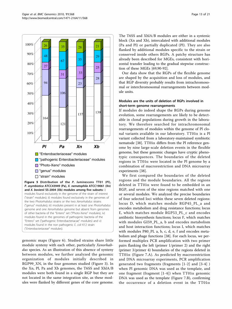
the Entero set initially used for RGP characterization(Figure 5). We defined five classes of module as a func-tion of their conservation within a taxonomic group: i)modules found exclusively in the genome of the strainof interest ("strain” modules); ii) modules found exclu-sively in the genomes of the two Photorhabdus strainsor the two Xenorhabdus strains ("genus” modules); iii)modules present in at least one Photorhabdus genomeand one Xenorhabdus genome but absent from the gen-omes of other bacteria of the “Entero” set ("Photo-Xeno” modules), iv) modules found in genomes ofpathogenic bacteria of the “Entero” set ("pathogens”modules) and v) modules found in the non pathogenicbacterium E. coli K12 strain ("Enterobacteriaceae” mod-ules). The modules belonging to “Photo-Xeno”, “Entero-bacteriaceae” and “pathogens” groups were evenlydistributed in the four strains, together accounting forbetween 30 and 40% of the modules (Figure 5). Therewere relatively few “Photo-Xeno"-specific modules (7%to 13%). Therefore, despite having very similar lifestyles(entomopathogenic bacteria living in symbiosis withnematodes) and being closely related phylogenetically,the flexible genomes of Xenorhabdus and Photorhabduswere different, suggesting different mechanisms of mole-cular adaptation to the environment and hosts of thesetwo genera. Most of the modules in these four genomeswere of the “strain” and “genus” types, these two typesof module together accounting for about two thirds ofthe modules. However, the Photorhabdus genus hadsimilar proportions of “strain” and “genus” modules,whereas the Xenorhabdus genus had a higher proportionof “strain” modules (54 to 57%) than of “genus” modules(9% to 11%). Thus, the flexible genome of Xenorhabdus,unlike that of Photorhabdus, is mostly strain-specific.This may reflect the closer phylogenetic relationshipbetween P. luminescens and P. asymbiotica thanbetween X. nematophila and X. bovienii [26].We then assessed module synteny between the four
strains studied, by aligning conserved modules on linear
20%
40%
60%
80%
100%
Pl Pa Xn Xb
metabolismrecombinationdrug resistancehost interactions
phages componentantibiotic synthesisenvironment interactionsunknown
Figure 4 Module function distribution in the flexible genomesof Photorhabdus luminescens TT01 (Pl), Photorhabdusasymbiotica ATCC43949 (Pa), Xenorhabdus nematophilaATCC19061 (Xn) and Xenorhabdus bovienii SS-2004 (Xb). Eightmodule functions were defined (see the legend below the figure).
Ogier et al. BMC Genomics 2010, 11:568http://www.biomedcentral.com/1471-2164/11/568
Page 12 of 21
genomic maps (Figure 6). Studied strains share littlemodule synteny with each other, particularly Xenorhab-dus species. As an illustration of this absence of syntenybetween modules, we further analyzed the genomicorganization of modules initially described inRGP99_XN, in the four genomes studied (Figure 3). Inthe Xn, Pl, Pa and Xb genomes, the T6SS and XhlA/Bmodules were both found in a single RGP but they arenot located in the same integration site, as these mod-ules were flanked by different genes of the core genome.
The T6SS and XhlA/B modules are either in a syntenicblock (Xn and Xb), intercalated with additional modules(Pa and Pl) or partially duplicated (Pl). They are alsoflanked by additional modules specific to the strain orconserved inside others RGPs. A patchy structure hasalready been described for MGEs, consistent with hori-zontal transfer leading to the gradual stepwise construc-tion of these MGEs [69,90-92].Our data show that the RGPs of the flexible genome
are shaped by the acquisition and loss of modules, andthat RGP diversity probably results from intrachromoso-mal or interchromosomal rearrangements between mod-ule units.
Modules are the units of deletion of RGPs involved inshort-term genome rearrangementsIf modules do indeed shape the RGPs during genomeevolution, some rearrangements are likely to be detect-able in clonal populations during growth in the labora-tory. We therefore searched for intrachromosomalrearrangements of modules within the genome of Pl clo-nal variants available in our laboratory. TT01a is a Plvariant collected from a laboratory-maintained symbioticnematode [38]. TT01a differs from the Pl reference gen-ome by nine large-scale deletion events in the flexiblegenome, but these genomic changes have cryptic pheno-typic consequences. The boundaries of the deletedregions in TT01a were located in the Pl genome by acombination of macrorestriction and DNA microarrayexperiments [38].We first compared the boundaries of the deleted
regions and the module boundaries. All the regionsdeleted in TT01a were found to be embedded in anRGP, and seven of the nine regions matched with oneor several modules. We analyzed the precise boundariesof four selected loci within these seven deleted regions:locus D, which matches module RGP45_PL_a andencodes metabolism and drug resistance functions; locusE, which matches module RGP53_PL_c and encodesantibiotic biosynthesis functions; locus F, which matcheswith modules GI59_PL_a, b and encodes metabolismand host interaction functions; locus I, which matcheswith modules P80_PL a, b, c, d, e, f and encodes meta-bolism and phage functions [38]. For each locus, we per-formed multiplex PCR amplification with two primerpairs flanking the left (primer 1/primer 2) and the right(primer 3/primer 4) boundaries of the regions deleted inTT01a (Figure 7.A). As predicted by macrorestrictionand DNA microarray experiments, PCR amplificationgenerated two fragments (fragments [1-2] and [3-4] )when Pl genomic DNA was used as the template, andone fragment (fragment [1-4]) when TT01a genomicDNA was used as the template (Figure 7.B), confirmingthe occurrence of a deletion event in the TT01a
164
94
27
48
26
121
106
31
67
35
137
23
33
35
28
139
28
25
29
21
10%
20%
30%
40%
50%
60%
70%
80%
90%
100%
Pl Pa Xn Xb
"Enterobacteriaceae" modules
"pathogenic Enterobacteriaceae" modules
"Photo-Xeno" modules
"genus" modules
"strain" modules
Figure 5 Distribution of the P. luminescens TT01 (Pl),P. asymbiotica ATCC43949 (Pa), X. nematophila ATCC19061 (Xn)and X. bovienii SS-2004 (Xb) modules among five subsets: i)modules found exclusively in the genome of the strain of interest("strain” modules); ii) modules found exclusively in the genomes ofthe two Photorhabdus strains or the two Xenorhabdus strains("genus” modules); iii) modules present in at least one Photorhabdusgenome and one Xenorhabdus genome but absent from genomesof other bacteria of the “Entero” set ("Photo-Xeno” modules), iv)modules found in the genomes of pathogenic bacteria of the“Entero” set ("pathogenic Enterobacteriacae” modules) and v)modules found in the non pathogenic E. coli K12 strain(”Enterobacteriaceae“ modules).
Ogier et al. BMC Genomics 2010, 11:568http://www.biomedcentral.com/1471-2164/11/568
Page 13 of 21

genome. We mapped the deletion boundaries more pre-cisely, with the goal of obtaining an exact picture of theresult of the deletion making it possible to predict theintrachromosomal rearrangements occurring at theseloci, by sequencing the four fragments [1-4] obtainedfrom the TT01a genome.For all four fragments, the sequence data confirmed
the predicted deletion boundaries: the D, E, F, I locusboundaries exactly matched the module boundariesidentified by in silico analysis, validating our modulari-zation procedure. Moreover, we distinguished twoclasses of deletion patterns potentially matching atleast three deletion scenarios (Figure 8). At loci F andI, which are embedded in GIs, the whole module orblock of modules was found to be missing in theTT01a genome, suggesting that a single block deletionevent led to the Pl/TT01a transition in this part of thegenome. In both cases, a gene encoding an enzymeinvolved in DNA recombination (transposase for locusF and integrase for locus I) is located at the internalborder of the locus and is therefore a good candidatefor involvement in the rearrangement. By contrast, atloci D and E, which are embedded in RGPs sensustricto, only subregions of modules are missing in theTT01a genome, consistent with the occurrence ofmore complex rearrangement events in these genomicareas during the Pl/TT01a transition. Locus E in theTT01a genome consists of five fragmented remnantsof the initial locus E. Genomic reduction has probablyoccurred in several stages at locus E, although theabsence of mobile elements or genes encoding
recombination functions makes it difficult to determinethe genomic rearrangement scenario. Locus D in theTT01a genome displays a shuffling pattern at threelocations: a 105 bp nucleotide sequence at the leftinternal border; a region composed of two Enterobac-teriaceae repeat intergenic consensus (ERIC)sequences, a transposase-encoding gene and a nucleo-tidic sequence of 154 bp at the right internal border; aregion composed of the truncated plu1870 andplu1872 genes and the plu1871 gene interrupted by atransposase gene in the middle part. As the elementswithin these shuffled subregions are probably remnantsof molecular actors involved in the rearrangements oflocus D, we analyzed them further. ERIC elements areminiature (127 bp) non autonomous mobile elementsin Enterobacteriaceae genomes [93,94]. The Pl genomeis particularly rich in such repeats [35]. The two trans-posase genes encoded a transposase of the IS928family. The 105 and 154 bp sequences are palindromicnucleotide sequences consisting of fragments dispersedin the Pl genome. They display no sequence similarityto each other or to ERIC and the two transposasegenes. The inserted transposases and ERIC sequencesclearly played a role in the plasticity of locus D, butthe origin and role of the exogenous 105 bp and 154bp nucleotide sequences remain unknown.In conclusion, whatever the molecular mechanism
involved in these deletion scenarios, in the case of clonalgenomic plasticity, the modules may be deleted over atimescale corresponding to growth in the laboratory andmay be considered units of deletion within RGPs.
Xn
Pa
Xb
Pl
Figure 6 Schematic representation of the genomic organization (synteny) of “genus” and “Photo-Xeno” modules conserved betweenP. luminescens TT01 (Pl), P. asymbiotica ATCC43949 (Pa), X. nematophila ATCC19061 (Xn) and X. bovienii SS-2004 (Xb). Each genome isrepresented by a horizontal line, with the RGPs sensu lato represented as green boxes. Purple lines indicate the conservation of modulesbetween two adjacent chromosomes(the representation is dependent on chromosome order on the figure, i.e. a module conserved betweenX. bovienii and P. luminescens will not be drawn).
Ogier et al. BMC Genomics 2010, 11:568http://www.biomedcentral.com/1471-2164/11/568
Page 14 of 21

ConclusionsThe data presented here participate to a better vision ofthe bacterial flexible genome organization. The charac-terization of RGPs by the RGPFinder method showedthe flexible genome to be much broader than the sum
of GIs and prophage elements. Additional elements –RGPmob and RGPnone elements – lacking classical mobi-lity features may be hypervariable regions that undergodeletions, ancient mobile elements with a degradedmobilization machinery, MGE that can be mobilizablein trans by other MGEs or non canonical MGE forwhich the mobility mechanism has yet to be described.Furthermore, we provide evidence that not only GIs andprophages, but all RGPs sensu lato have a mosaic struc-ture composed of modules that are both functional andplasticity units.The application of comparative genome sequencing to
experimental evolution studies provides us with anopportunity to study the link between genome dynamicsand adaptive evolution. Nevertheless, such studies aregenerally carried out on bacterial populations evolvingin a synthetic broth culture, and they mostly identifypoint mutations [95,96]. Here, by carrying out compara-tive genomics studies on variants obtained from theirhost in the laboratory, we showed experimentally thatthe same modules undergo genomic rearrangementsduring genome speciation and short-term genomic rear-rangements. This work improves our understanding ofthe process responsible for bacterial genome diversifica-tion and evolution.Obtaining of these data were made possible by the use
of the Photorhabdus and Xenorhabdus genera for ourcomparative genomic study. Indeed, the life cycle ofthese genera is restricted to two successive ecologicalniches. We argue that this unusual pattern of selectivepressure is responsible for an alternation of genomicshuffling: HGT in the insect cadaver, which constitutesan abundant nutrient resource potentially shared withmany other microorganisms and intrachromosomalrearrangements of recently acquired modules in the bac-terial monoxenic culture within the nematode gut, asobserved in the Pl variant isolated from a laboratory-maintained symbiotic nematode. We therefore suggestthat Photorhabdus and Xenorhabdus are suitable newbacterial models for studies of the evolution of bacterialgenomes.Finally, the data obtained in this study contribute to
our understanding of the fluid nature of genomesthroughout the kingdoms of life. According to J. A. Sha-piro, prokaryotic and eukaryotic cells are genetic engi-neers and mobile elements are “natural geneticengineering systems” that facilitate the evolutionaryrewriting of genomic information [97]. Shapiro’s hypoth-esis is that repeated evolutionary challenges haveselected systems that (i) reduce the size of the genomicsearch space and (ii) maximize the chance of success byusing combinatorial processes based on basal functionalcomponents [97]. We argue that the modules describedhere are entirely consistent with this vision, as these
Figure 7 Boundaries of deleted regions in the genome of theP. luminescens TT01a variant match with the moduleboundaries defined in the P. luminescens TT01 (Pl) genome.A. Schematic diagram of the strategy used for multiplex PCRamplification. The yellow box indicates the deleted region. Blueboxes represent the flanking regions. Red horizontal arrows indicatethe location of the primers. The primer mixture (P1, P2, P3 and P4)was designed to amplify two fragments from the Pl genome andonly one fragment if the region is effectively deleted, as predictedfor the P. luminescens TT01a variant genome. B. Agarose gelelectrophoresis of the PCR products generated by amplifying thegenomic DNA of Pl (lanes 2, 5, 9, 12), of the TT01a variant (lanes 3,6, 10, 13) or of water (lanes 4, 7, 11, 14) with primers P1, P2, P3 andP4, designed as indicated in (A) for loci D (lanes 2-4), E (lanes 5-7), F(lanes 9-11) and I (lanes 12-14). Loci D, E, F and I are four regions ofthe Pl genome identified as missing in the TT01a variant genome(see text for details). [1-2], [3-4] and [1-4]. indicate bands of sizescompatible with amplification of the regions between the P1 andP2 primers, the P3 and P4 primers and the P1 and P4 primers,respectively. Lanes 1, 8, 15: molecular markers. The sizes offragments are indicated in kb to the left of the gel.
Ogier et al. BMC Genomics 2010, 11:568http://www.biomedcentral.com/1471-2164/11/568
Page 15 of 21

Figure 8 Schematic genetic map of the F, I, E and D loci in the Pl genome and their counterparts in the TT01a variant genome. Theboxes above and below the axis represent ORFs in the forward and reverse orientations, respectively. The putative functions of the ORFs withinRGPs are indicated by specific colors (see legend Figure 4). Mobility genes, repeat sequences or specific features are marked with motifsdescribed below the figure. The modules in loci D, E, F and I are indicated, below the axis, by black horizontal double arrows.
Ogier et al. BMC Genomics 2010, 11:568http://www.biomedcentral.com/1471-2164/11/568
Page 16 of 21

functional units recombined at a limited number of hot-spots shaping and delimiting the flexible genome.
MethodsBacterial strains and genome sequencesPhotorhabdus luminescens subspecies laumondii strainTT01 is a symbiont of the nematode Heterorhabditisbacteriophora isolated in Trinidad and Tobago [35,98].The genome of strain TT01 consists of a single circularchromosome of 5,688,987 bp (accession numberNC_005126). Photorhabdus asymbiotica subspeciesasymbiotica strain ATCC43949 is a North Americanclinical isolate. This strain was isolated in 1977 from afemale patient with endocarditis, in Maryland, USA[32,99]. The genome of strain ATCC43949 consists of asingle circular chromosome of 5,064,808 bp and a29,732 bp plasmid [36] (accession numbers NC_012962and NC_012961, respectively). Xenorhabdus nemato-phila ATCC19061, the type strain of the species, is asymbiont of the nematode Steinernema carpocapsae,isolated from Georgia, USA [100]. The genome of strainATCC19061 comprises a single circular chromosome of4,432,590 bp and a 155,327 bp plasmid [34] (accessionnumbers FN667742 and FN667743, respectively). Xenor-habdus bovienii SS-2004 is a symbiont of the nematodeSteinernema jollieti sp. isolated from a woodland in theMissouri valley, USA, in 1999 [101]. The genome ofstrain SS-2004 comprises a single circular chromosomeof 4,225,498 bp [34] (accession number FN667741). Thefour genomes were input into the PhotoScope andXenorhabduScope databases http://www.genoscope.cns.fr/agc/mage.
RGP identificationRegions of genomic plasticity (RGPs) were sought in theP. luminescens TT01, P. asymbiotica ATCC43949, X.nematophila ATCC19061 and X. bovienii SS-2004 gen-omes, with the RGPfinder web tool implemented in theMaGe annotation platform (http://www.genoscope.cns.fr/agc/mage; Roche et al., unpublished data). Briefly,RGPFinder searches for breaks in synteny between areference genome and the genomes of a set of relatedbacteria – the bacterial genome set (Figure 1.A). A RGPsensu lato is the sum of overlapping subregions missingin at least one of the bacterial genomes of the compari-son set. RGPs have a minimal size of 5 kb. Thisexcludes the isolated insertion sequences of the RGPs,but favors regions with several genes of potential func-tional interest in the bacterial biology. This definitiondoes not involve any underlying assumption about theevolutionary origin or genetic basis of these variablechromosomal segments. RGPFinder also provides infor-mation about composition abnormalities (GC% devia-tion, codon adaptation index) and about the features
flanking the RGPs, such as tRNA, IS, integrase (int) andgenetic elements involved in DNA mobility (mob),which are common characteristics of foreign DNAacquired by horizontal genetic transfer. The resultsobtained with this web tool include those for AlienHunter [102], a method detecting atypical sequences (i.e., sequences potentially acquired by horizontal genetictransfer) through the analysis of composition bias.Predicted RGPs were then manually inspected, to
eliminate false-positive results. Indeed, point mutationsmay lead RGPFinder to identify a region as an RGPwhen it actually belongs to the core genome. Finally, theboundaries of the RGP were homogenized between thecompared genomes and potential insertion sites weredefined. The genomes used in the bacterial genome setwere those of P. luminescens TT01, P. asymbioticaATCC43949, X. nematophila ATCC19061, X. bovieniiSS-2004, Yersinia pestis CO92 (accession number003143); Salmonella enterica subsp. enterica TyphiCT18 (accession number NC_003198), Erwinia caroto-vora subsp. atroseptica SCRI1043 (accession numberNC_004547) and E. coli K12 (accession numberNC_000913). Finally, Prophinder was used to detect pro-phages among the RGP sensu lato [53], http://aclame.ulb.ac.be/Tools/Prophinder/.
Definition and distribution of modulesThe MaGe web interface [103] was used to divide RGPsmanually into subregions corresponding to blocks ofgenes specific to the strain or blocks of syntenic genes(i.e. genes with a conserved genomic organization in atleast two genomes of the Entero set). These subregions,which often contain genes of similar biological function,were named “modules”. The distribution of modulesamong the “Enterobacteriaceae” genome set was ana-lyzed manually: a module was considered present (orpartially present) in a genome if it had more than 80%(25%) of syntenic orthologous genes (orthologous genesshared at least 30% of identity on 80% of the shortestsequence by BlastP) with the module of the referencegenome. The module was otherwise considered to beabsent. Descriptions of the modules and their distribu-tions are available from PhotoScope https://www.geno-scope.cns.fr/agc/mage/wwwpkgdb/Login/log.php?pid=13and XenorhabduScope https://www.genoscope.cns.fr/agc/mage/wwwpkgdb/Login/log.php?pid=24, by openingthe Genomic Object Editor of a gene and consulting the“Module” results.
Multiplex PCR procedure and sequencingGenomic DNA was extracted as previously described[44] and stored at 4°C. Primers flanking the right (pri-mers P1/P2) and left (primers P3/P4) module borderswere designed with Primer 3 http://frodo.wi.mit.edu/
Ogier et al. BMC Genomics 2010, 11:568http://www.biomedcentral.com/1471-2164/11/568
Page 17 of 21

primer3/. Primer sequences are listed in Additional File6. Multiplex PCR with the four primers (P1, P2, P3, P4)was performed with a Bio-Rad thermocycler (Bio-Rad,Marne La Vallée, France). Fragments with a predictedsize smaller than 3 kb were amplified with InvitrogenTaq polymerase (Invitrogen, France), according to themanufacturer’s protocol. Fragments with a predictedsize greater than 3 kb were amplified with the HerculaseEnhanced DNA polymerase (Stratagene, AmsterdamZuidoost, Pays Bas) in accordance with the manufac-turer’s recommendations. Samples of reaction mixtureswere analyzed by electrophoresis in an agarose gel. Thefragments amplified by PCR [P3-P4] were purified fromthe gel with the high purity purification kit from Roche(Roche Diagnostic, France) and sequenced with the PCRprimers described in Additional File 6, via a chromo-some walking process, by Macrogen (South Korea).
Additional material
Additional File 1: Phylogenetic tree for the Enterobacteriaceaederived from a distance analysis of 16S rRNA gene sequences. Thegenomes used in this study belong to species indicated in red(Photorhabdus and Xenorhabdus) and blue (other Enterobacteriaceae).Vibrio cholerae (Vibrionaceae) was used as an outgroup. The GenBankaccession numbers of the sequences are shown in brackets. Bootstrapvalues of more than 50% are indicated at the nodes. The bar indicates1% sequence divergence. A figure showing a phylogenetic tree for theEnterobacteriaceae used in this study.
Additional File 2: List of regions of genomic plasticity (RGPs) in theP. luminescens TT01 (Pl), P. asymbiotica ATCC43949 (Pa), X.nematophila ATCC19061 (Xn) and X. bovienii SS- 2004 (Xb)genomes. A table listing the RGPs.
Additional File 3: Distribution of RGP sizes in the Photorhabdusluminescens TT01 (Pl), Photorhabdus asymbiotica ATCC43949 (Pa),Xenorhabdus nematophila ATCC19061 (Xn) and Xenorhabdusbovienii SS-2004 genomes (Xb). A figure showing the distribution ofRGP size.
Additional File 4: Schematic diagram of the distribution of RGPssensu lato on the circular chromosomes of P. luminescens TT01 (Pl),P. asymbiotica ATCC43949 (Pa), X. nematophila ATCC19061 (Xn) andX. bovienii SS-2004 (Xb). Successive circles from inside to outside: GCskew; GC deviation (with values exceeding +/- 2 standard deviationsindicated in red). Distribution of the different RGP types: GIs (orange),Phages (green) and RGPmob and RGPnone (yellow). A figure showingschematic diagrams of the distribution of RGPs.
Additional File 5: List of modules in the P. luminescens TT01 (Pl), P.asymbiotica ATCC43949 (Pa), X. nematophila ATCC19061 (Xn) and X.bovienii SS-2004 (Xb) genomes and their distribution in the Yersiniapestis CO92 (Yp), Salmonella enterica subsp. enterica Typhi CT18(St), Erwinia carotovora subsp. atroseptica SCRI1043 (Eca) and E. coliK12 (Eco) genomes. A table listing the modules.
Additional File 6: Primers used in the study. A table listing theprimers used in this study.
List of abbreviationsEco: Escherichia coli K12; Eca: Erwinia carotovora subsp. atroseptica SCRI1043;GI: genomic island; HGT: horizontal genetic transfer; ICE: integrativeconjugative element; int: integrase; IS: insertion sequence; MGE: mobilegenetic element; P: prophage; Pa:, Photorhabdus asymbiotica ATCC43949; Pl:
Photorhabdus luminescens TT01; PVC: Photorhabdus virulence cassette; RGP :region of genomic plasticity; St : Salmonella enterica subsp. enterica TyphiCT18; T3SS : type three secretion system; T6SS: type six secretion system; Tc:toxin complex; Xn: Xenorhabdus nematophila ATCC19061; Xb: Xenorhabdusbovienii SS-2004; YAPI: Yersinia adhesion pathogenicity island; Yp: Yersiniapestis CO92
AcknowledgementsWe thank Christine Laroui for technical assistance. We thank theXenorhabdus genome consortium for access to the Xenorhabdus genomes,R. Ffrench-constant and N. Waterfield for access to the Photorhabdusasymbiotica genomes before public access. We thank Eric Duchaud forcritical reading of parts of the manuscript. This study received financialsupport from the Institut National de la Recherche Agronomique (grant SPE2007_1133_03), the Agence Nationale de la Recherche (ANR PFTVMicroScope) and the GIS IBiSA.
Author details1INRA, UMR 1133, Laboratoire EMIP, Place Eugène Bataillon, F-34095Montpellier, France. 2Université Montpellier 2, UMR 1133, Laboratoire EMIP,Place Eugène Bataillon, F-34095 Montpellier, France. 3CEA, Genoscope &CNRS-UMR 8030, Laboratoire d’Analyse Bioinformatique en Génomique etMétabolisme, Evry cedex F-91006, France. 4Department of BiologicalSciences, University of Wisconsin, Milwaukee, WI 53201, USA. 5Department ofBacteriology, University of Wisconsin, Madison, WI 53706 USA. 6Great LakesBioenergy Research Center, University of Wisconsin, Madison, WI 53706 USA.
Authors’ contributionsJ-CO, AG, CM, PT, SG designed the project. SF, HG-B, GS provided genomicdata. JCO carried out in silico description of the RGPs, their modularizationand the PCR experiments. AC, DR, ZR contributed in computational analysis.J-CO, AC, RZ, AG, PT, CM, SG analyzed the data. J-CO and SG wrote thepaper with contributions from AG. All authors read and approved the finalmanuscript.
Received: 19 March 2010 Accepted: 15 October 2010Published: 15 October 2010
References1. Hacker J, Carniel E: Ecological fitness, genomic islands and bacterial
pathogenicity - A Darwinian view of the evolution of microbes. EMBOReports 2001, 2(5):376-381.
2. Dobrindt U, Hochhut B, Hentschel U, Hacker J: Genomic islands inpathogenic and environmental microorganisms. Nature ReviewsMicrobiology 2004, 2(5):414-424.
3. Abby S, Daubin V: Comparative genomics and the evolution ofprokaryotes. Trends in Microbiology 2007, 15(3):135-141.
4. Frost LS, Leplae R, Summers AO, Toussaint A: Mobile genetic elements:The agents of open source evolution. Nature Reviews Microbiology 2005,3(9):722-732.
5. Mathee K, Narasimhan G, Valdes C, Qiu X, Matewish JM, Koehrsen M,Rokas A, Yandava CN, Engels R, Zeng E, et al: Dynamics of Pseudomonasaeruginosa genome evolution. Proceedings of the National Academy ofSciences of the United States of America 2008, 105(8):3100-3105.
6. Rocha EPC: Order and disorder in bacterial genomes. Current Opinion inMicrobiology 2004, 7(5):519-527.
7. Burrus V, Pavlovic G, Decaris B, Guedon G: Conjugative transposons: thetip of the iceberg. Molecular Microbiology 2002, 46(3):601-610.
8. Roberts AP, Chandler M, Courvalin P, Guédon G, Mullany P, Pembroke T,Rood JI, Smith CJ, Summers AO, Tsuda M, et al: Revised nomenclature fortransposable genetic elements. Plasmid 2008, 60(3):167-173.
9. Beaber JW, Hochhut B, Waldor MK: SOS response promotes horizontaldissemination of antibiotic resistance genes. Nature 2004,427(6969):72-74.
10. Qiu XY, Gurkar AU, Lory S: Interstrain transfer of the large pathogenicityisland (PAPI-1) of Pseudomonas aeruginosa. Proceedings of the NationalAcademy of Sciences of the United States of America 2006,103(52):19830-19835.
11. Ramsay JP, Sullivan JT, Stuart GS, Lamont IL, Ronson CW: Excision andtransfer of the Mesorhizobium loti R7A symbiosis island requires an
Ogier et al. BMC Genomics 2010, 11:568http://www.biomedcentral.com/1471-2164/11/568
Page 18 of 21

integrase IntS, a novel recombination directionality factor RdfS, and aputative relaxase RlxS. Molecular Microbiology 2006, 62(3):723-734.
12. Maiques E, Ubeda C, Tormo MA, Ferrer MD, Lasa I, Novick RP, Penades JR:Role of staphylococcal phage and SaPI integrase in intra- andinterspecies SaPI transfer. Journal of Bacteriology 2007, 189(15):5608-5616.
13. Juhas M, Crook DW, Dimopoulou ID, Lunter G, Harding RM, Ferguson DJ,Hood DW: Novel type IV secretion system involved in propagation ofgenomic islands. Journal of Bacteriology 2007, 189(3):761-771.
14. Ravatn R, Zehnder AJB, van der Meer JR: Low-frequency horizontaltransfer of an element containing the chlorocatechol degradation genesfrom Pseudomonas sp. strain B13 to Pseudomonas putida F1 and toindigenous bacteria in laboratoryscale activated-sludge microcosms.Applied and Environmental Microbiology 1998, 64(6):2126-2132.
15. Rocha EPC: The Organization of the Bacterial Genome. Annual Review ofGenetics 2008, 42:211-233.
16. Canchaya C, Fournous G, Brussow H: The impact of prophages onbacterial chromosomes. Molecular Microbiology 2004, 53(1):9-18.
17. Toussaint A, Merlin C: Mobile elements as a combination of functionalmodules. Plasmid 2002, 47(1):26-35.
18. Hendrix RW: Bacteriophage genomics. Current Opinion in Microbiology2003, 6(5):506-511.
19. Engelberg-Kulka H, Glaser G: Addiction modules and programmed celldeath and antideath in bacterial cultures. Annual Review of Microbiology1999, 53:43-70.
20. Mazel D: Integrons: agents of bacterial evolution. Nature ReviewsMicrobiology 2006, 4(8):608-620.
21. Boucher Y, Labbate M, Koenig JE, Stokes HW: Integrons: mobilizableplatforms that promote genetic diversity in bacteria. Trends inMicrobiology 2007, 15(7):301-309.
22. Pavlovic G, Burrus V, Gintz B, Decaris B, Guedon G: Evolution of genomicislands by deletion and tandem accretion by site-specific recombination:ICESt1-related elements from Streptococcus thermophilus. Microbiology2004, 150:759-774.
23. Burrus V, Waldor MK: Shaping bacterial genomes with integrative andconjugative elements. Research in Microbiology 2004, 155(5):376-386.
24. Brochet M, Rusniok C, Couve E, Dramsi S, Poyarts C, Trieu-Cuot P, Kunst F,Glaser P: Shaping a bacterial genome by large chromosomalreplacements, the evolutionary history of Streptococcus agalactiae.Proceedings of the National Academy of Sciences of the United States ofAmerica 2008, 105(41):15961-15966.
25. Garriss G, Waldor MK, Burrus V: Mobile Antibiotic Resistance EncodingElements Promote Their Own Diversity. Plos Genetics 2009, 5(12):e1000775.
26. Tailliez P, Laroui C, Ginibre N, Paule A, Pages S, Boemare N: Phylogeny ofPhotorhabdus and Xenorhabdus based on universally conserved protein-coding sequences and implications for the taxonomy of these twogenera. Proposal of new taxa: X. vietnamensis sp. nov., P. luminescenssubsp. caribbeanensis subsp. nov., P. luminescens subsp. hainanensissubsp. nov., P. temperata subsp. khanii subsp. nov., P. temperata subsp.tasmaniensis subsp. nov., and the reclassification of P. luminescens subsp.thracensis as P. temperata subsp. thracensis. International journal ofsystematic and evolutionary microbiology 2009.
27. Boemare N: Interactions between the partners of the entomopathogenicbacterium nematode complexes, Steinernema-Xenorhabdus andHeterorhabditis- Photorhabdus. Nematology 2002, 4:601-603.
28. Forst S, Dowds B, Boemare N, Stackebrandt E: Xenorhabdus andPhotorhabdus spp.: Bugs that kill bugs. Annual Review of Microbiology1997, 51:47-72.
29. Waterfield NR, Sanchez-Contreras M, Eleftherianos I, Dowling A, Wilkinson P,Parkhilll J, Thomson N, Reynolds SE, Bode HB, Dorus S, et al: RapidVirulence Annotation (RVA): Identification of virulence factors using abacterial genome library and multiple invertebrate hosts. Proceedings ofthe National Academy of Sciences of the United States of America 2008,105(41):15967-15972.
30. Goodrich-Blair H, Clarke DJ: Mutualism and pathogenesis in Xenorhabdusand Photorhabdus: two roads to the same destination. MolecularMicrobiology 2007, 64(2):260-268.
31. Richards GR, Goodrich-Blair H: Masters of conquest and pillage:Xenorhabdus nematophila global regulators control transitions fromvirulence to nutrient acquisition. Cellular Microbiology 2009,11(7):1025-1033.
32. Farmer JJ, Jorgensen JH, Grimont PAD, Akhurst RJ, Poinar GO, Ageron E,Pierce GV, Smith JA, Carter GP, Wilson KL, et al: Xenorhabdus luminescens(DNA Hybridization Group-5) from Human Clinical Specimens. Journal ofClinical Microbiology 1989, 27(7):1594-1600.
33. Gerrard J, Waterfield N, Vohra R, ffrench-Constant R: Human infection withPhotorhabdus asymbiotica: an emerging bacterial pathogen. Microbes andInfection 2004, 6(2):229-237.
34. Latreille P, Norton S, Goldman BS, Henkhaus J, Miller N, Barbazuk B,Bode HB, Darby C, Du ZJ, Forst S, et al: Optical mapping as a routine toolfor bacterial genome sequence finishing. BMC Genomics 2007, 8:321.
35. Duchaud E, Rusniok C, Frangeul L, Buchrieser C, Givaudan A, Taourit S,Bocs S, Boursaux-Eude C, Chandler M, Charles JF, et al: The genomesequence of the entomopathogenic bacterium Photorhabdusluminescens. Nature Biotechnology 2003, 21(11):1307-1313.
36. Wilkinson P, Waterfield NR, Crossman L, Corton C, Sanchez-Contreras M,Vlisidou I, Barron A, Bignell A, Clark L, Ormond D, et al: Comparativegenomics of the emerging human pathogen Photorhabdus asymbioticawith the insect pathogen Photorhabdus luminescens. BMC Genomics 2009,10:302.
37. Moran NA, Plague GR: Genornic changes following host restriction inbacteria. Current Opinion in Genetics & Development 2004, 14(6):627-633.
38. Gaudriault S, Pages S, Lanois A, Laroui C, Teyssier C, Jumas-Bilak E,Givaudan A: Plastic architecture of bacterial genome revealed bycomparative genomics of Photorhabdus variants. Genome Biology 2008,9(7):R117.
39. Waterfield NR, Bowen DJ, Fetherston JD, Perry RD, ffrench-Constant RH: Thetc genes of Photorhabdus: a growing family. Trends in Microbiology 2001,9(4):185-191.
40. Waterfield NR, Daborn PJ, ffrench-Constant RH: Genomic islands inPhotorhabdus. Trends in Microbiology 2002, 10(12):541-545.
41. Sergeant M, Baxter L, Jarrett P, Shaw E, Ousley M, Winstanley C,Morgan JAW: Identification, typing, and insecticidal activity ofXenorhabdus isolates from entomopathogenic nematodes in UnitedKingdom soil and characterization of the xpt toxin loci. Applied andEnvironmental Microbiology 2006, 72(9):5895-5907.
42. Cowles CE, Goodrich-Blair H: The Xenorhabdus nematophila nilABC genesconfer the ability of Xenorhabdus spp. to colonize Steinernemacarpocapsae nematodes. Journal of Bacteriology 2008, 190(12):4121-4128.
43. Heermann R, Fuchs TM: Comparative analysis of the Photorhabdusluminescens and the Yersinia enterocolitica genomes: uncoveringcandidate genes involved in insect pathogenicity. BMC Genomics 2008,9:40.
44. Gaudriault S, Duchaud E, Lanois A, Canoy AS, Bourot S, DeRose R, Kunst F,Boemare N, Givaudan A: Whole-genome comparison betweenPhotorhabdus strains to identify genomic regions involved in thespecificity of nematode interaction. Journal of Bacteriology 2006,188(2):809-814.
45. Langille MGI, Hsiao WWL, Brinkman FSL: Detecting genomic islands usingbioinformatics approaches. Nature Reviews Microbiology 2010, 8(5):372-382.
46. Thomson NR, Clayton DJ, Windhorst D, Vernikos G, Davidson S, Churcher C,Quail MA, Stevens M, Jones MA, Watson M, et al: Comparative genomeanalysis of Salmonella Enteritidis PT4 and Salmonella Gallinarum 287/91provides insights into evolutionary and host adaptation pathways.Genome Research 2008, 18(10):1624-1637.
47. Cazalet C, Rusniok C, Bruggemann H, Zidane N, Magnier A, Ma L, Tichit M,Jarraud S, Bouchier C, Vandenesch F, et al: Evidence in the Legionellapneumophila genome for exploitation of host cell functions and highgenome plasticity. Nature Genetics 2004, 36(11):1165-1173.
48. Silby MW, Cerdeno-Tarraga AM, Vernikos GS, Giddens SR, Jackson RW,Preston GM, Zhang XX, Moon CD, Gehrig SM, Godfrey SAC, et al: Genomicand genetic analyses of diversity and plant interactions of Pseudomonasfluorescens. Genome Biology 2009, 10(5):R51.
49. Amadou C, Pascal G, Mangenot S, Glew M, Bontemps C, Capela D,Carrere S, Cruveiller S, Dossat C, Lajus A, et al: Genome sequence of thebeta-rhizobium Cupriavidus taiwanensis and comparative genomics ofrhizobia. Genome Research 2008, 18(9):1472-1483.
50. Lima WC, Van Sluys MA, Menck CFM: Non-gamma-proteobacteria geneislands contribute to the Xanthomonas genome. Omics-a Journal ofIntegrative Biology 2005, 9(2):160-172.
Ogier et al. BMC Genomics 2010, 11:568http://www.biomedcentral.com/1471-2164/11/568
Page 19 of 21

51. Lefebure T, Stanhope MJ: Evolution of the core and pan-genome ofStrepococcus: positive selection, recombination, and genomecomposition. Genome Biology 2007, 8(5):R71.
52. Gu JY, Wang YF, Lilburn T: A Comparative Genomics, Network-BasedApproach to Understanding Virulence in Vibrio cholerae. Journal ofBacteriology 2009, 191(20):6262-6272.
53. Lima-Mendez G, Van Helden J, Toussaint A, Leplae R: Prophinder: acomputational tool for prophage prediction in prokaryotic genomes.Bioinformatics 2008, 24(6):863-865.
54. Boemare NE, Boyergiglio MH, Thaler JO, Akhurst RJ, Brehelin M: Lysogenyand Bacteriocinogeny in Xenorhabdus nematophilus and OtherXenorhabdus spp. Applied and Environmental Microbiology 1992,58(9):3032-3037.
55. Baghdiguian S, Boyergiglio MH, Thaler JO, Bonnot G, Boemare N:Bacteriocinogenesis in Cells of Xenorhabdus nematophilus andPhotorhabdus luminescens - Enterobacteriaceae Associated withEntomopathogenic Nematodes. Biology of the Cell 1993, 79(2):177-185.
56. Thaler JO, Baghdiguian S, Boemare N: Purification and Characterization ofXenorhabdicin, a Phage Tail-Like Bacteriocin, from the Lysogenic StrainF1 of Xenorhabdus nematophilus. Applied and Environmental Microbiology1995, 61(5):2049-2052.
57. Bondi M, Messi P, Sabia C, Contri MB, Manicardi G: Antimicrobialproperties and morphological characteristics of two Photorhabdusluminescens strains. Microbiologica 1999, 22(2):117-127.
58. Gaudriault S, Thaler JO, Duchaud E, Kunst F, Boemare N, Givaudan A:Identification of a P2-related prophage remnant locus of Photorhabdusluminescens encoding an R-type phage tail-like particle. FEMSMicrobiology Letters 2004, 233(2):223-231.
59. Brugirard-Ricaud K, Givaudan A, Parkhill J, Boemare N, Kunst F, Zumbihl R,Duchaud E: Variation in the effectors of the type III secretion systemamong Photorhabdus species as revealed by genomic analysis. Journal ofBacteriology 2004, 186(13):4376-4381.
60. Cornelis GR: The type III secretion injectisome. Nature Reviews Microbiology2006, 4(11):811-825.
61. Brugirard-Ricaud K, Duchaud E, Givaudan A, Girard PA, Kunst F, Boemare N,Brehelin M, Zumbihl R: Site-specific antiphagocytic function of thePhotorhabdus luminescens type III secretion system during insectcolonization. Cellular Microbiology 2005, 7(3):363-371.
62. Collyn F, Billault A, Mullet C, Simonet M, Marceau M: YAPI, a new Yersiniapseudotuberculosis pathogenicity island. Infection and Immunity 2004,72(8):4784-4790.
63. Hurst MR, Glare TR, Jackson TA, Ronson CW: Plasmid-located pathogenicitydeterminants of Serratia entomophila, the causal agent of amber diseaseof grass grub, show similarity to the insecticidal toxins of Photorhabdusluminescens. Journal of Bacteriology 2000, 182(18):5127-5138.
64. Yang G, Dowling AJ, Gerike U, ffrench-Constant RH, Waterfield NR:Photorhabdus virulence cassettes confer injectable insecticidal activityagainst the wax moth. Journal of Bacteriology 2006, 188(6):2254-2261.
65. Jubelin G, Chavez CV, Taieb F, Banfield MJ, Samba-Louaka A, Nobe R,Nougayrede JP, Zumbihl R, Givaudan A, Escoubas JM, et al: Cycle inhibitingfactors (CIFs) are a growing family of functional cyclomodulins presentin invertebrate and mammal bacterial pathogens. PLoS One 2009, 4(3):e4855.
66. Buell CR, Joardar V, Lindeberg M, Selengut J, Paulsen IT, Gwinn ML,Dodson RJ, Deboy RT, Durkin AS, Kolonay JF, et al: The complete genomesequence of the Arabidopsis and tomato pathogen Pseudomonassyringae pv. tomato DC3000. Proceedings of the National Academy ofSciences of the United States of America 2003, 100(18):10181-10186.
67. Vodovar N, Vallenet D, Cruveiller S, Rouy Z, Barbe V, Acosta C, Cattolico L,Jubin C, Lajus A, Segurens B, et al: Complete genome sequence of theentomopathogenic and metabolically versatile soil bacteriumPseudomonas entomophila. Nature Biotechnology 2006, 24(6):673-679.
68. Mann S, Chen YP: Bacterial genomic G + C composition-elicitingenvironmental adaptation. Genomics 2010, 95(1):7-15.
69. Touchon M, Hoede C, Tenaillon O, Barbe V, Baeriswyl S, Bidet P, Bingen E,Bonacorsi S, Bouchier C, Bouvet O, et al: Organised genome dynamics inthe Escherichia coli species results in highly diverse adaptive paths. PLoSGenetics 2009, 5(1):e1000344.
70. Doublet B, Golding GR, Mulvey MR, Cloeckaert A: Potential integrationsites of the Salmonella genomic island 1 in Proteus mirabilis and otherbacteria. Journal of Antimicrobial Chemotherapy 2007, 59(4):801-803.
71. Doublet B, Golding G, Mulvey M, Cloeckaert A, Fairhead C: SecondaryChromosomal Attachment Site and Tandem Integration of theMobilizable Salmonella Genomic Island 1. PLoS ONE 2008, 3(4):e2060.
72. Kingsley RA, Humphries AD, Weening EH, Zoete MRd, Winter S,Papaconstantinopoulou A, Dougan G, umler AJB: Molecular andPhenotypic Analysis of the CS54 Island of Salmonella enterica SerotypeTyphimurium: Identification of Intestinal Colonization and PersistenceDeterminants. Infection and Immunity 2003, 71(2):629-640.
73. Schouler C, Koffmann F, Amory C, Leroy-Sétrin S, Moulin-Schouleur M:Genomic subtraction for the identification of putative new virulencefactors of an avian pathogenic Escherichia coli strain of O2 serogroup.Microbiology 2004, 150(Pt 9):2973-2984.
74. Martinez-Garcia E, Tormo A, Navarro-Lloréns JM: Polymorphism in the yclC-rpoS region of enterobacteria. Current Microbiology 2003, 46(5):365-370.
75. Arora SK, Bangera M, Lory S, Ramphal R: A genomic island inPseudomonas aeruginosa carries the determinants of flagellinglycosylation. Proceedings of the National Academy of Sciences of the UnitedStates of America 2001, 98(16):9342-9347.
76. Bellanger X, Roberts AP, Morel C, Choulet F, Pavlovic G, Mullany P,Decaris B, Guédon G: Conjugative transfer of the integrative conjugativeelements ICESt1 and ICESt3 from Streptococcus thermophilus. Journal ofBacteriology 2009, 191(8):2764-2775.
77. Shrivastava S, Mande SS: Identification and Functional Characterization ofGene Components of Type VI Secretion System in Bacterial Genomes.Plos One 2008, 3(8):e2955.
78. Cowles KN, Goodrich-Blair H: Expression and activity of a Xenorhabdusnematophila haemolysin required for full virulence towards Manducasexta insects. Cellular Microbiology 2005, 7(2):209-219.
79. Parkhill J, Wren BW, Thomson NR, Titball RW, Holden MTG, Prentice MB,Sebaihia M, James KD, Churcher C, Mungall KL, et al: Genome sequence ofYersinia pestis, the causative agent of plague. Nature 2001,413(6855):523-527.
80. Wu M, Sun LV, Vamathevan J, Riegler M, Deboy R, Brownlie JC, McGraw EA,Martin W, Esser C, Ahmadinejad N, et al: Phylogenomics of thereproductive parasite Wolbachia pipientis wMel: a streamlined genomeoverrun by mobile genetic elements. PLoS Biology 2004, 2(3):E69.
81. Klasson L, Andersson SGE: Strong asymmetric mutation bias inendosymbiont genomes coincide with loss of genes for replicationrestart pathways. Molecular Biology and Evolution 2006, 23(5):1031-1039.
82. Bentley SD, Corton C, Brown SE, Barron A, Clark L, Doggett J, Harris B,Ormond D, Quail MA, May G, et al: Genome of the actinomycete plantpathogen Clavibacter michiganensis subsp sepedonicus suggests recentniche adaptation. Journal of Bacteriology 2008, 190(6):2150-2160.
83. Lescat M, Calteau A, Hoede C, Barbe V, Touchon M, Rocha E, Tenaillon O,Medigue C, Johnson JR, Denamur E: A Module Located at a ChromosomalIntegration Hot Spot Is Responsible for the Multidrug Resistance of aReference Strain from Escherichia coli Clonal Group A. AntimicrobialAgents and Chemotherapy 2009, 53(6):2283-2288.
84. Lloyd AL, Henderson TA, Vigil PD, Mobley HL: Genomic Islands ofUropathogenic Escherichia coli Contribute to Virulence. Journal ofBacteriology 2009, 191(11):3469-3481.
85. Tumapa S, Holden MTG, Vesaratchavest M, Wuthiekanun V,Limmathurotsakul D, Chierakul W, Feil EJ, Currie BJ, Day NPJ, Nierman WC,et al: Burkholderia pseudomallei genome plasticity associated withgenomic island variation. BMC Genomics 2008, 9:190.
86. He X, Ou HY, Yu Q, Zhou X, Wu J, Liang J, Zhang W, Rajakumar K, Deng Z:Analysis of a genomic island housing genes for DNA S-modificationsystem in Streptomyces lividans 66 and its counterparts in other distantlyrelated bacteria. Molecular Microbiology 2007, 65(4):1034-1048.
87. Konczy P, Ziebell K, Mascarenhas M, Choi A, Michaud C, Kropinski AM,Whittam TS, Wickham M, Finlay B, Karmali MA: Genomic O island 122,locus for enterocyte effacement, and the evolution of virulentverocytotoxin-producing Escherichia coli. Journal of Bacteriology 2008,190(17):5832-5840.
88. Chouikha I, Germon P, Bree A, Gilot P, Moulin-Schouleur M, Schouler C: AselC-associated genomic island of the extraintestinal avian pathogenicEscherichia coli strain BEN2908 is involved in carbohydrate uptake andvirulence. Journal of Bacteriology 2006, 188(3):977-987.
89. Hacker J, Kaper JB: Pathogenicity islands and the evolution of microbes.Annual Review of Microbiology 2000, 54:641-679.
Ogier et al. BMC Genomics 2010, 11:568http://www.biomedcentral.com/1471-2164/11/568
Page 20 of 21

90. Castillo A, Eguiarte LE, Souza V: A genomic population genetics analysisof the pathogenic enterocyte effacement island in Escherichia coli: Thesearch for the unit of selection. Proceedings of the National Academy ofSciences of the United States of America 2005, 102(5):1542-1547.
91. Sandner L, Eguiarte LE, Navarro A, Cravioto A, Souza V: The elements ofthe focus of enterocyte effacement in human and wild mammal isolatesof Escherichia coli: evolution by assemblage or disruption? Microbiology2001, 147:3149-3158.
92. Van Houdt R, Monchy S, Leys N, Mergeay M: New mobile geneticelements in Cupriavidus metallidurans CH34, their possible roles andoccurrence in other bacteria. Antonie Van Leeuwenhoek InternationalJournal of General and Molecular Microbiology 2009, 96(2):205-226.
93. Delihas N: Small mobile sequences in bacteria display diverse structure/function motifs. Molecular Microbiology 2008, 67(3):475-481.
94. De Gregorio E, Silvestro G, Petrillo M, Carlomagno MS, Di Nocera PP:Enterobacterial repetitive intergenic consensus sequence repeats inyersiniae: genomic organization and functional properties. Journal ofBacteriology 2005, 187(23):7945-7954.
95. Herring CD, Raghunathan A, Honisch C, Patel T, Applebee MK, Joyce AR,Albert TJ, Blattner FR, van den Boom D, Cantor CR, et al: Comparativegenome sequencing of Escherichia coli allows observation of bacterialevolution on a laboratory timescale. Nature Genetics 2006,38(12):1406-1412.
96. Barrick JE, Yu DS, Yoon SH, Jeong H, Oh TK, Schneider D, Lenski RE, Kim JF:Genome evolution and adaptation in a long-term experiment withEscherichia coli. Nature 2009, 461(7268):1243-U1274.
97. Shapiro JA: Letting Escherichia coli Teach Me About GenomeEngineering. Genetics 2009, 183(4):1205-1214.
98. Fischer-Le Saux M, Viallard V, Brunel B, Normand P, Boemare NE: Polyphasicclassification of the genus Photorhabdus and proposal of new taxa: P-luminescens subsp luminescens subsp nov., P-luminescens subsp akhurstiisubsp nov., P-luminescens subsp laumondii subsp nov., P.temperata spnov., P-temperata subsp temperata subsp nov and P-asymbiotica sp nov.International Journal of Systematic Bacteriology 1999, 49:1645-1656.
99. Akhurst RJ, Boemare NE, Janssen PH, Peel MM, Alfredson DA, Beard CE:Taxonomy of Australian clinical isolates of the genus Photorhabdus andproposal of Photorhabdus asymbiotica subsp asymbiotica subsp nov andP-asymbiotica subsp australis subsp nov. International Journal of Systematicand Evolutionary Microbiology 2004, 54:1301-1310.
100. Poinar GO Jr, Thomas GM: A new bacterium, Achromobacter nematophilussp. nov. (Achromobacteriaceae: Eubacteriales), associated with anematode. International Bulletin of Bacteriological Nomenclature andTaxonomy 1965, 15:249-252.
101. Spiridonov SE, Krasomil-Osterfeld K, Moens M: Steinernema jollieti sp n.(Rhabditida: Steinernematidae), a new entomopathogenic nematodefrom the American midwest. Russian Journal of Nematology 2004,12(1):85-95.
102. Vernikos GS, Parkhill J: Interpolated variable order motifs for identificationof horizontally acquired DNA: revisiting the Salmonella pathogenicityislands. Bioinformatics 2006, 22(18):2196-2203.
103. Vallenet D, Labarre L, Rouy Z, Barbe V, Bocs S, Cruveiller S, Lajus A, Pascal G,Scarpelli C, Medigue C: MaGe: a microbial genome annotation systemsupported by synteny results. Nucleic Acids Research 2006, 34(1):53-65.
doi:10.1186/1471-2164-11-568Cite this article as: Ogier et al.: Units of plasticity in bacterial genomes:new insight from the comparative genomics of two bacteria interactingwith invertebrates, Photorhabdus and Xenorhabdus. BMC Genomics 201011:568. Submit your next manuscript to BioMed Central
and take full advantage of:
• Convenient online submission
• Thorough peer review
• No space constraints or color figure charges
• Immediate publication on acceptance
• Inclusion in PubMed, CAS, Scopus and Google Scholar
• Research which is freely available for redistribution
Submit your manuscript at www.biomedcentral.com/submit
Ogier et al. BMC Genomics 2010, 11:568http://www.biomedcentral.com/1471-2164/11/568
Page 21 of 21


![Comparison of Xenorhabdus bovienii bacterial strain ... · bacterial genomes [5, 31, 32] that were isolated from 6 different Steinernema spp. nematode hosts (Table 1), focusing on](https://static.fdocuments.us/doc/165x107/600f75812d65012e6c59f4ad/comparison-of-xenorhabdus-bovienii-bacterial-strain-bacterial-genomes-5-31.jpg)