Unit Map: Follow Along in your packet WHAT ARE YOU LEARNING? Describe tissue types and their...
77
-
Upload
imogene-bond -
Category
Documents
-
view
216 -
download
1
Transcript of Unit Map: Follow Along in your packet WHAT ARE YOU LEARNING? Describe tissue types and their...


Unit Map: Follow Along in your packet
WHAT ARE YOU LEARNING?Describe tissue types and their
functionsUnderstand and Explain tissue purpose

Know Understand Do!
Know Tissue Types Tissue
Locations Tissue
Anatomy
Understando Tissue purposes
in the bodyo Tissue
propertieso Tissue
development
Do Outline tissue
functions Represent
tissue types Explain tissue
importance in animal health

Key Learning: Tissue Types and Functions
Unit EQ: Why are tissues critical to animal health?
Concept : Nerve
Lesson EQ:
Why is nerve tissue damage a serious injury?
Vocab
CNS, PNS
Concept : Connective/ Muscle
Lesson EQ: How does connective function?
VocabTendon, Ligament
Concept :Epithelial
Lesson EQ:Where is epithelial tissue located?
VocabIntegument
Keratin

Essential Question
Where is epithelial tissue located?
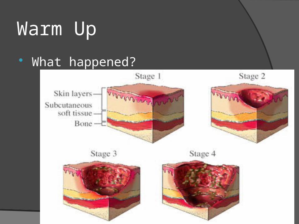
Warm Up
What happened?

Epithelial Tissue
Collections of cells packed together into layers
Line body surfaces , openings, and tubesIntestinal, reproductive, and urinary tractsBlood vessels

Epithelial Functions
Protection from outside environmentExpulsion and movement of material
within lumen tubes Secretory Absorption of specific material Conservation Sensory input
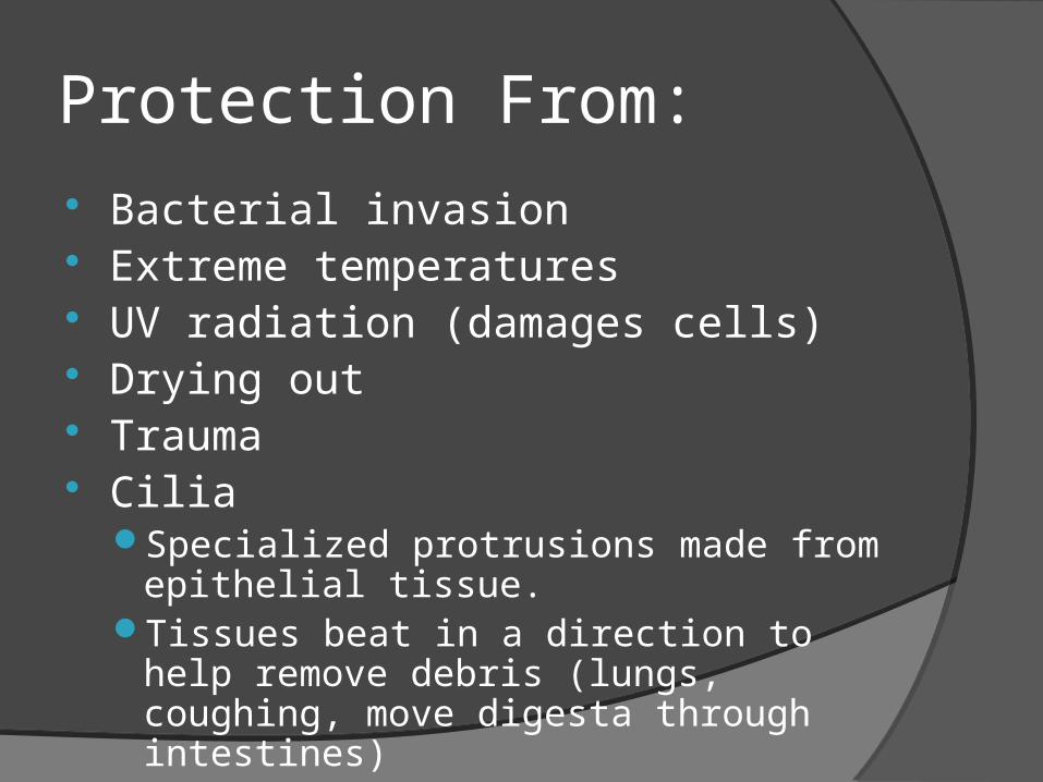
Protection From: Bacterial invasion Extreme temperatures UV radiation (damages cells) Drying out Trauma Cilia
Specialized protrusions made from epithelial tissue.
Tissues beat in a direction to help remove debris (lungs, coughing, move digesta through intestines)

Secretory Tears : protect eye
tissue Saliva : begin digestion,
moistens food Mucus: in airways, trap
particles Mammary System:
secrete milk (only in MAMMALS)

Absorption
Highly Selective! Minerals
Intestines, lungs, and kidneys Oxygen/CO2 exchange
Blood vessels Extracellular Fluid
Is exchanged etc

Conservation
ECF within cells is conserved

Sensory
Sensory InputRetina: essential in transmitting visual
input to CNS (central nervous system)Tongue: taste, touch and temperatureNoseEars

Histology: Epithelium
Histology: study of cell structure in plants and animals
ALL epithelial tissue have:Basement membrane: collection of
fibers that tie epithelial to the underlying connective tissue
No direct blood supply (underlying connective tissue supplies everything)
Diffusion of nutrients and waste
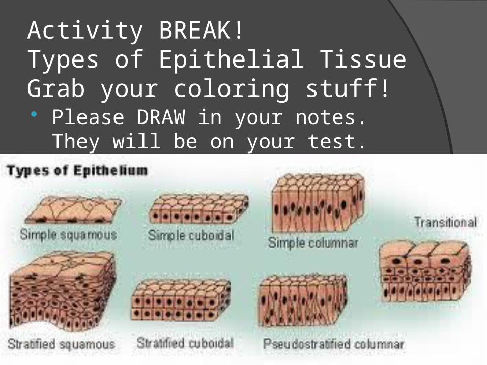
Activity BREAK! Types of Epithelial TissueGrab your coloring stuff! Please DRAW in your notes. They
will be on your test.

Epithelial Tissue Categorized by:
Layers○ Simple: One layer○ Stratified: multiple layers○ Transitional: multiple layers that can change
shapeShape○ Squamous: flat○ Cuboidal: Cube○ Columnar: column
All terms are combined ( Layer + Shape) to appropriately describe epithelium
What type if Where?
Simple squamous (1 layer and flat)Found where there is a need for
exchange. Very fragile. In protected areas. Large surface area for transfer○ Line blood vessels: transfer of waste,
fluids, nutrients, gases○ Air sacs in the respiratory system:
carbon dioxide and oxygen
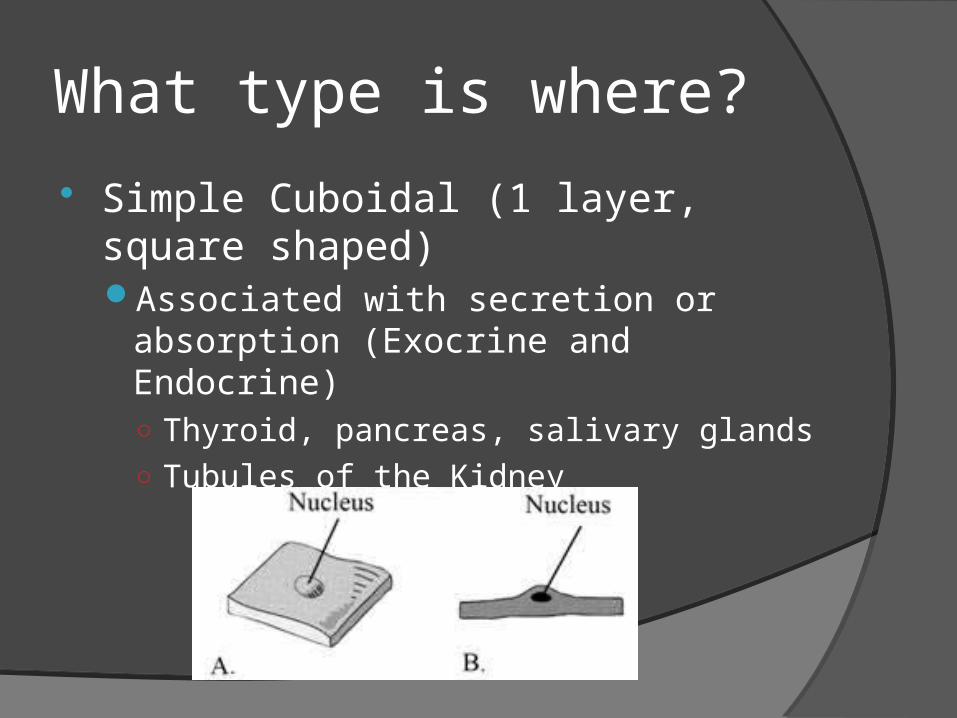
What type is where?
Simple Cuboidal (1 layer, square shaped)Associated with secretion or
absorption (Exocrine and Endocrine) ○ Thyroid, pancreas, salivary glands○ Tubules of the Kidney

Endo / Exo Explained Endocrine Cells
Secrete hormones directly in to the blood stream for transport ○ Thyroid, adrenal, pituitary
oExocrine oHave ducts to transport their secretions to impact more local areas. Hormones do not enter blood stream
oSweat and salivary glands
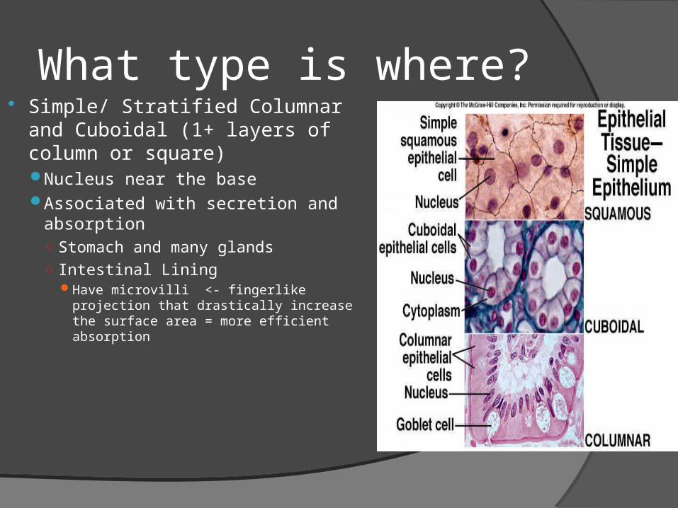
What type is where? Simple/ Stratified Columnar
and Cuboidal (1+ layers of column or square)Nucleus near the baseAssociated with secretion and
absorption○ Stomach and many glands○ Intestinal Lining
Have microvilli <- fingerlike projection that drastically increase the surface area = more efficient absorption
What type is where? Stratified Squamous (many layers, flat)
Found in areas of mechanical stress. Thickness determined by amount of stress to the area (back vs bottom of foot)○ Rectum, Vagina, esophagus, mouth○ SKIN!! = Integumentary System
Originate from a basal layer○ Appear more cuboidal
Continually divide and move outward ○ Loose cytoplasm and nucleus becoming flatter
and fluff off

Review Time!
Epithelial Tissue review worksheet


Essential Question
How does connective tissue function?

Warm Up

Consists of two basic elements:
Cellsand
Extra-cellular matrix

General Information
Types and functions:Loose connective tissue: fat, areolar –
insulation, protectionDense Connective: Ligaments and
tendons – binding and supportCartilage – protection, supportBone - support Blood- transportation
Common Characteristics
All Connective tissue share the following characteristics, despite the variety.Develop from the same embryonic
tissueComposed of many different types of
cells within the tissue.Contains extracellular matrixGreat variation in the amount of blood
supply (vascularization)
Extracellular Matrix Extracellular matrix is the substance that
connective cells are embedded within. Made of nonliving material called ground
substance.Can be liquid, gel-like, semi-solid or hard.Texture depends on amount of cell adhesion
proteins: proteoglycans which trap water forming a gel. More proteins, more solid ground substance.
Contains fibers that provide great strength, or flexibility, or both.
Extracellular matrix allows Conn. Tissue to bear weight, mechanical abuse – protects!!
Fibers Fibers are embedded in ground
substance to add strength/flexibility.
Collagen: stronger than steel of the same size. STRENGTH!
Elastic: allows tissue to stretch and recoil. FLEXIBLE!
Reticular: delicate, forms nets that support free cells.
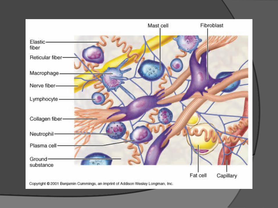
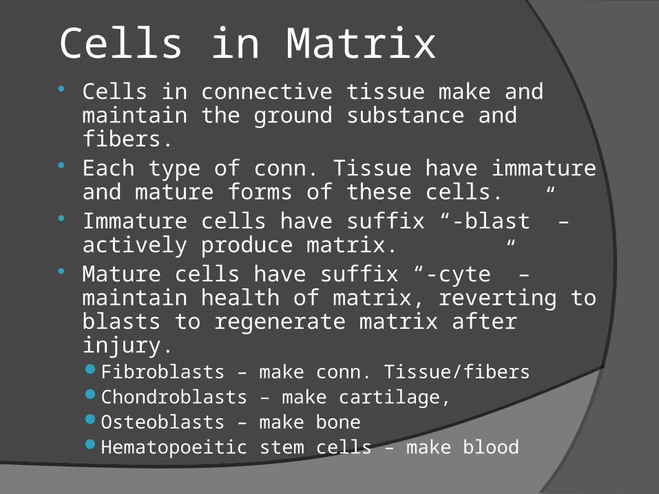
Cells in Matrix Cells in connective tissue make and
maintain the ground substance and fibers.
Each type of conn. Tissue have immature and mature forms of these cells.
Immature cells have suffix “-blast” – actively produce matrix.
Mature cells have suffix “-cyte” – maintain health of matrix, reverting to blasts to regenerate matrix after injury.Fibroblasts – make conn. Tissue/fibersChondroblasts – make cartilage,Osteoblasts – make boneHematopoeitic stem cells – make blood
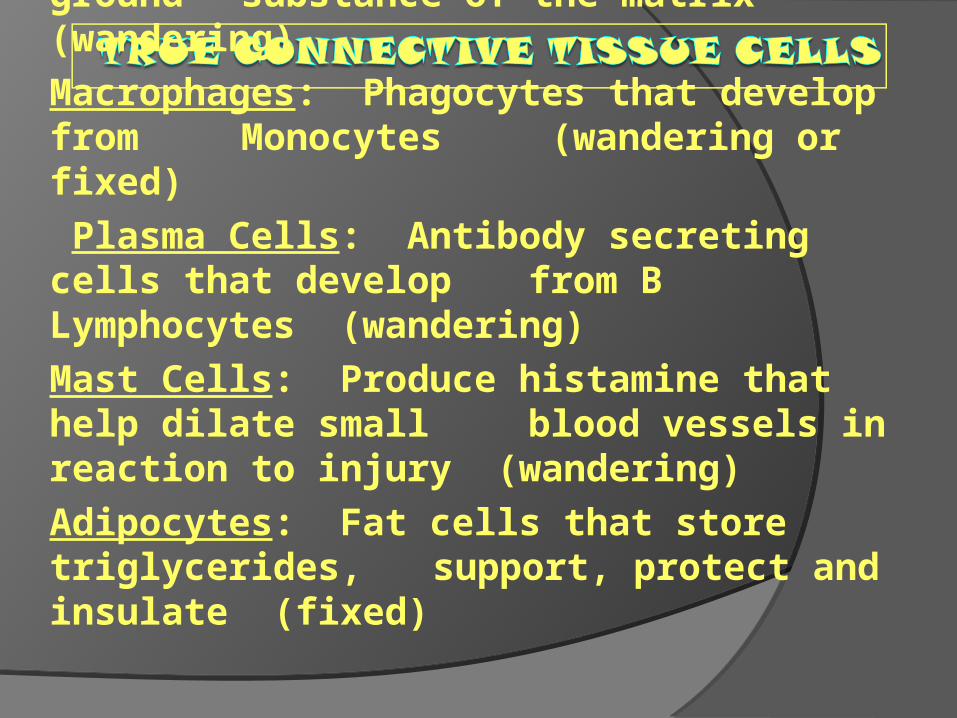
Fibroblasts: Secrete both fibers and ground substance of the matrix (wandering)Macrophages: Phagocytes that develop from Monocytes (wandering or fixed) Plasma Cells: Antibody secreting cells that develop from B Lymphocytes (wandering)Mast Cells: Produce histamine that help dilate small blood vessels in reaction to injury (wandering)Adipocytes: Fat cells that store triglycerides, support, protect and insulate (fixed)

Collagen Fibers: Large fibers made of the protein collagen and are typically the most abundant fibers. Promote tissue flexibility.Elastic Fibers: Intermediate fibers made of the protein elastin. Branching fibers that allow for stretch and recoilReticular Fibers: Small delicate, branched fibers that have same chemical composition of collagen. Forms structural framework for organs such as spleen and lymph nodes.

TYPES OF CONNECTIVE TISSUE
1. True Connective Tissuea. Loose Connective Tissueb. Dense Connective Tissue
2. Supportive Connective Tissuea. Cartilageb. Bone
5. Liquid Connective Tissuea. Blood

Loose Connective Tissue Areolar – most widely distributed
type.Serves as “packing material”
cushioning organs, subcutaneous, attaches skin to muscle.
Contains all three fibersGelatinousVery loosely packed, lots of liquidSwells during inflammation (edema)

Loose Continued
Adipose = Fat!Loosely packed with little matrix90% of tissue is adipocytesMost of each cell is a fat dropletNutrient storage, cushioning.Richly vascularizedDevelops within areolar
tissue/subcutaneous○ 18% of an average person’s weight○ 50% of chubby person

Loose Continued
ReticularOnly contains reticular fibers.VERY DELICATE – a fine net of fibers.Supports free blood cells.Found in lymph nodes, bone marrow.REALLY TINY!
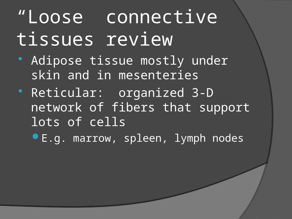
“Loose” connective tissues review Adipose tissue mostly under skin
and in mesenteries Reticular: organized 3-D network
of fibers that support lots of cellsE.g. marrow, spleen, lymph nodes

“Dense” Connective tissues Irregular
Thick fibers running in many planesE.g. dermis, fibrous capsules around organs
RegularAligned parallel fibersResists tensionE.g. tendon, ligaments, aponeurosesSometimes with elastic fibers (e.g.
ligamentum nuchae)

Cartilage Characteristics
Resists tension and pressureTough and flexibleNo nerves or blood vesselsLots of collagen and elastic fibers, high
content of proteoglycans.80% waterChondroblasts make matrix until end of
human adolescence.Mature Chondrocytes found in cavities
called lacunae (pit)
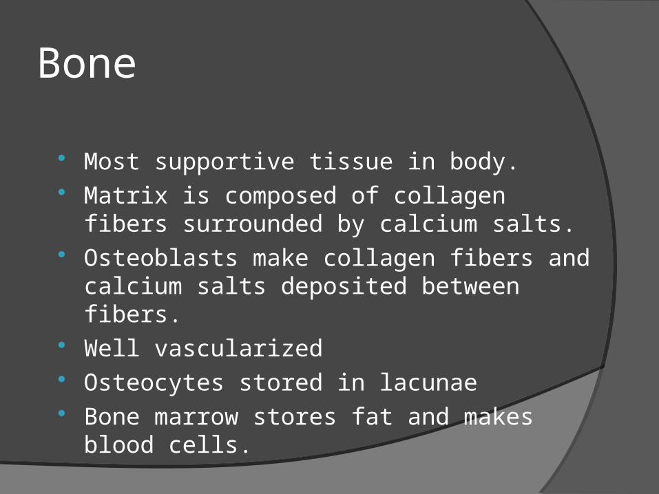
Bone
Most supportive tissue in body. Matrix is composed of collagen fibers
surrounded by calcium salts. Osteoblasts make collagen fibers and
calcium salts deposited between fibers. Well vascularized Osteocytes stored in lacunae Bone marrow stores fat and makes
blood cells.

Connective Tissue Review Connective tissue review
worksheet

Essential Question

Warm up

Functions
Movement Posture maintenance Heat generation (3/4 of energy
produced by ATP escapes as heat) Stabilization of joints Protection of some internal organs

Muscle Types
Muscle TypesSkeletal: striated, voluntaryCardiac: only in heart, striated,
involuntarySmooth/Visceral: walls of organs,
not striated, involuntary

Muscle types

Properties of Muscle
Excitability: capacity of muscle to respond to a stimulus
Contractility: ability of a muscle to shorten and generate pulling force
Extensibility: muscle can be stretched back to its original length
Elasticity: ability of muscle to recoil to original resting length after stretched
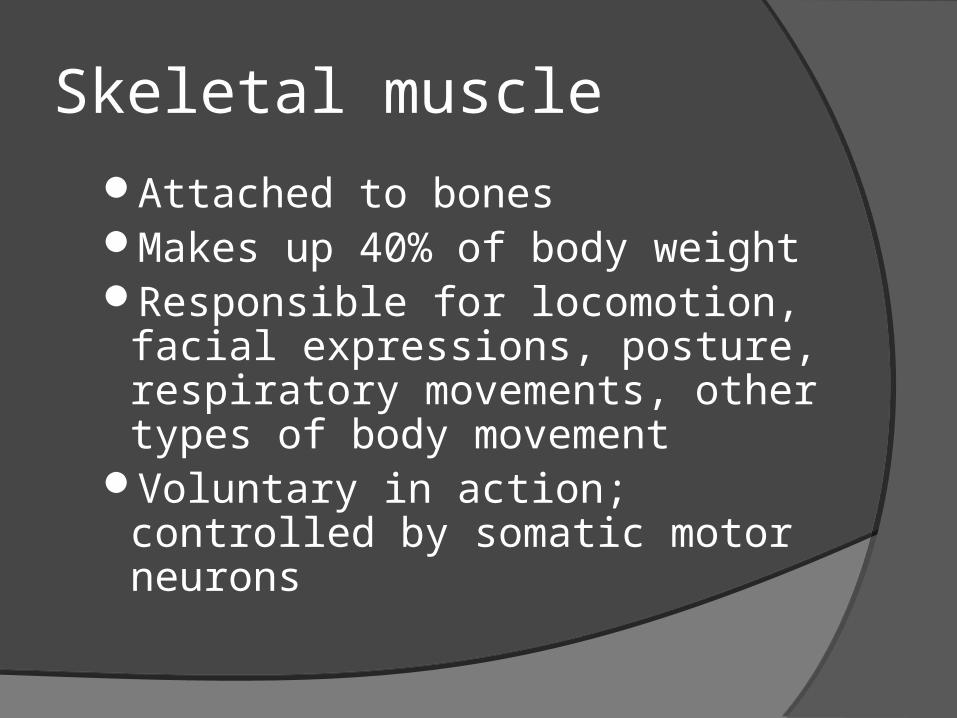
Skeletal muscleAttached to bonesMakes up 40% of body weightResponsible for locomotion,
facial expressions, posture, respiratory movements, other types of body movement
Voluntary in action; controlled by somatic motor neurons

Basic Features of a Skeletal Muscle Muscle
attachmentsMost skeletal
muscles run from one bone to another
One bone will move – other bone remains fixed○ Origin – less
movable attach- ment
○ Insertion – more movable attach- ment

Smooth MuscleIn the walls of hollow organs, blood
vessels, eye, glands, uterus, skinSome functions: propel urine, mix
food in digestive tract, dilating/constricting pupils, regulating blood flow,
In some locations, autorhythmicControlled involuntarily by
endocrine and autonomic nervous systems

Smooth Muscle
Cells are not striated Fibers smaller than those in
skeletal muscle Spindle-shaped; single, central
nucleus More actin than myosin No sarcomeres
Not arranged as symmetrically as in skeletal muscle, thus NO striations.
Caveolae: indentations in sarcolemma; May act like T tubules
Dense bodies instead of Z disks Have noncontractile intermediate
filaments

Smooth Muscle
Figure 9.24
• Grouped into sheets in walls of hollow organs• Longitudinal layer – muscle fibers run parallel to organ’s long axis• Circular layer – muscle fibers run around circumference of the organ
• Both layers participate in peristalsis

Cardiac muscle
Heart: major source of movement of blood
AutorhythmicControlled involuntarily by endocrine and autonomic nervous systems

Cardiac Muscle Found only in heart where it forms a thick layer
called the myocardium Striated fibers that branch Each cell usually has one centrally-located nucleus Fibers joined by intercalated disks
IDs are composites of desmosomes and gap junctions Allow excitation in one fiber to spread quickly to adjoining
fibers Under control of the ANS (involuntary) and
endocrine system (hormones) Some cells are autorhythmic
Fibers spontaneously contract (aka Pacemaker cells)

Cardiac Muscle Tissue
Figure 10.10a

Neuromuscular Junction Region where the motor neuron stimulates the
muscle fiber The neuromuscular junction is formed by :
1. End of motor neuron axon (axon terminal)○ Terminals have small membranous sacs (synaptic
vesicles) that contain the neurotransmitter acetylcholine (ACh)
2. The motor end plate of a muscle• A specific part of the sarcolemma that contains
ACh receptors Though exceedingly close, axonal ends and
muscle fibers are always separated by a space called the synaptic cleft

Neuromuscular Junction
Figure 9.7 (a-c)

Muscle Quiz!
Fill in the graphic organizer below:Muscle Type of Movement Striated or
Unstriated
Heart
Intestines
Stomach Wall
Bicep
Ham String
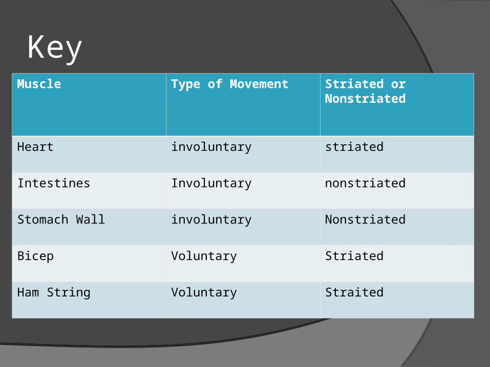
KeyMuscle Type of Movement Striated or
Nonstriated
Heart involuntary striated
Intestines Involuntary nonstriated
Stomach Wall involuntary Nonstriated
Bicep Voluntary Striated
Ham String Voluntary Straited


Essential Question
Why is nerve tissue damage a serious injury?

Warm Up

Over view
Allow for communication between and among parts of the body
Found in:Brain+ spinal chord= CNS Everything else = PNS

Nerve tissue = Neurons Neurons have
Dendrites- impulses received from other cells at synapses are transmitted to the cell body
Nucleus- “brain” Neuron soma- Cell Body
contains nucleus Axon- conducts electrical
impulses away from the neuron's soma

Nerve Attachments
Synapses- connection between 2 neurons
On TissuesMuscle

Nerve Impulse
Impulse occurs as a flow of ions passes through the cell membrane
Resting neuron: Sodium ions actively transported into
ECF (OUT). Potassium pumped into cytoplasm (IN)
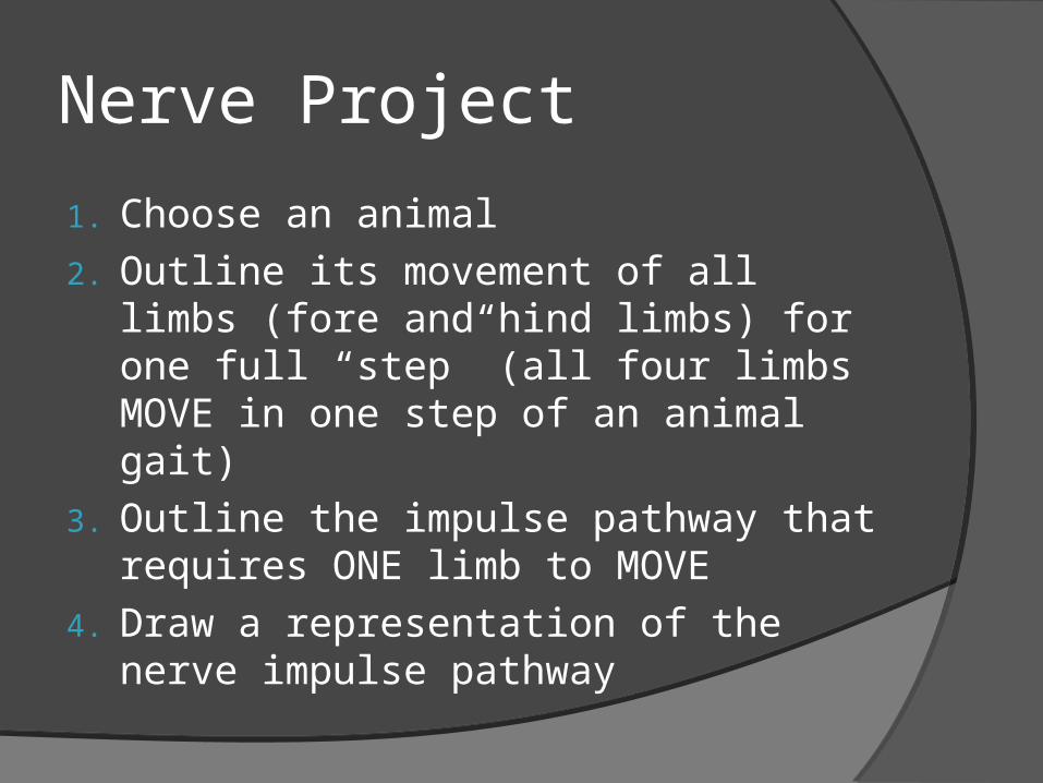
Nerve Project
1. Choose an animal2. Outline its movement of all limbs
(fore and hind limbs) for one full “step” (all four limbs MOVE in one step of an animal gait)
3. Outline the impulse pathway that requires ONE limb to MOVE
4. Draw a representation of the nerve impulse pathway
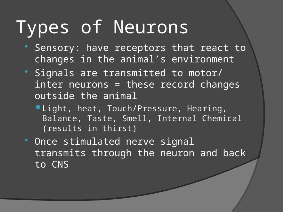
Types of Neurons Sensory: have receptors that react to
changes in the animal’s environment Signals are transmitted to motor/ inter
neurons = these record changes outside the animalLight, heat, Touch/Pressure, Hearing,
Balance, Taste, Smell, Internal Chemical (results in thirst)
Once stimulated nerve signal transmits through the neuron and back to CNS
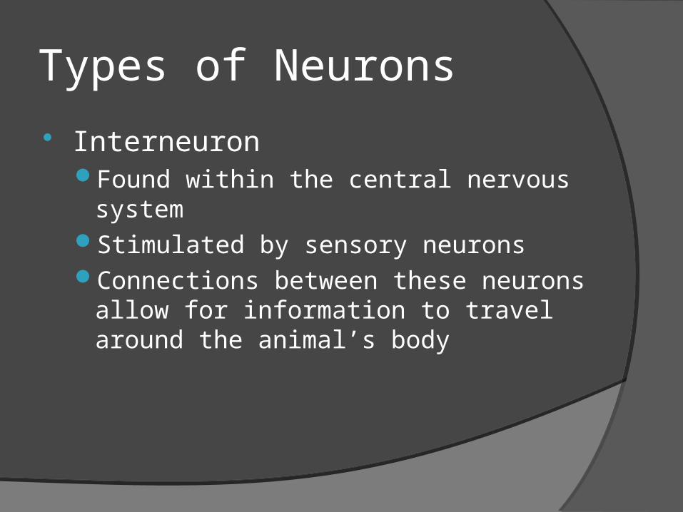
Types of Neurons
InterneuronFound within the central nervous
system Stimulated by sensory neurons Connections between these neurons
allow for information to travel around the animal’s body
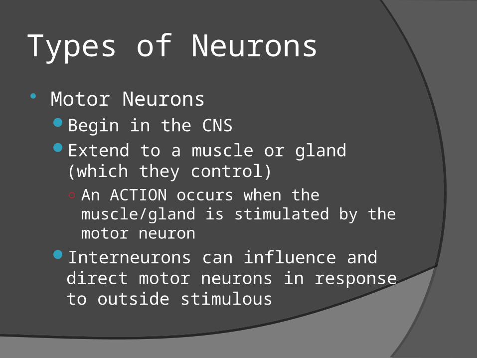
Types of Neurons
Motor NeuronsBegin in the CNS Extend to a muscle or gland (which
they control)○ An ACTION occurs when the
muscle/gland is stimulated by the motor neuron
Interneurons can influence and direct motor neurons in response to outside stimulous
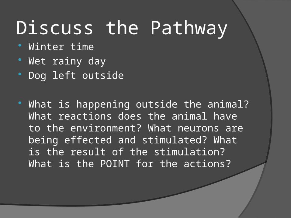
Discuss the Pathway Winter time Wet rainy day Dog left outside
What is happening outside the animal? What reactions does the animal have to the environment? What neurons are being effected and stimulated? What is the result of the stimulation? What is the POINT for the actions?
Clinical Practice Read in Chapter 2
the section on Clinical practice
Give a summary of: Foot and Mouth Disease Common Companion
Animal Medicine occurrences
Monday-Morning Disease (no not the kind YOU get!)
Horner’s Syndrome
Summary includes:What?Where does it
happen?Why does is happen?What is the effect on
the animal?How is it treated? How is it prevented?SUMMARIZE the
information given to you in this section

Test Review Define: Integument Keratin Tendon Ligament CNS PNS Epithelial Connective Nerve Basement membrane Fibrous Mast Cell
Answer:1. What are the types of
epithelial tissue. Draw them
2. What are the types of connective tissue and what do they do?
3. What are the types of loose connective tissue?
4. What are the main types of muscles? What type of movement do they have? Where are they found in the body?
5. What are the 3 types of neuron receptors? What does each type of
receptor perform?