
Unit IV: Coordination Impulse Transmission Ch. 11 – pgs 373-389 Ch. 14 – pgs 471-487.
23
Unit IV: Coordination Impulse Transmission Ch. 11 – pgs 373-389 Ch. 14 – pgs 471-487
-
Upload
stanley-erik-howard -
Category
Documents
-
view
218 -
download
5
Transcript of Unit IV: Coordination Impulse Transmission Ch. 11 – pgs 373-389 Ch. 14 – pgs 471-487.
Unit IV: CoordinationImpulse Transmission
Ch. 11 – pgs 373-389
Ch. 14 – pgs 471-487
Review
1. What is the CSF and what are the three functions it provides?
2. Which way is caudal?
3. Which is not a region the spinal nerves are named for? a.) cervical, b.) thoracic, c.) pelvic, d.) lumbar, e.) sacral
4. How does the location of white and gray matter differ in the spinal cord compared to the cerebrum?
5. The formation of the blood-brain barrier is stimulated by which glial cells?
6. _____ are folds that increase surface.
7. By increasing surface area, they allow for more ___________.
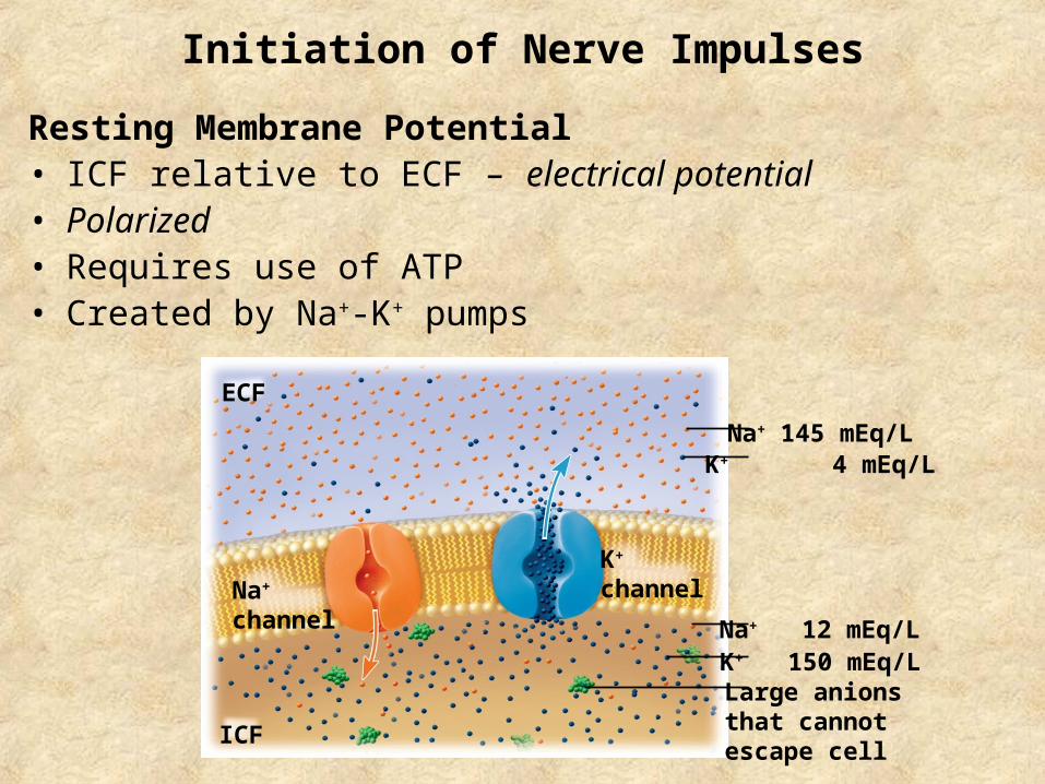
Initiation of Nerve Impulses
Resting Membrane Potential• ICF relative to ECF – electrical potential• Polarized• Requires use of ATP• Created by Na+-K+ pumps
ECF
ICF
Na+
channel
K+
channel
Na+ 145 mEq/LK+ 4 mEq/L
Na+ 12 mEq/LK+ 150 mEq/LLarge anionsthat cannotescape cell
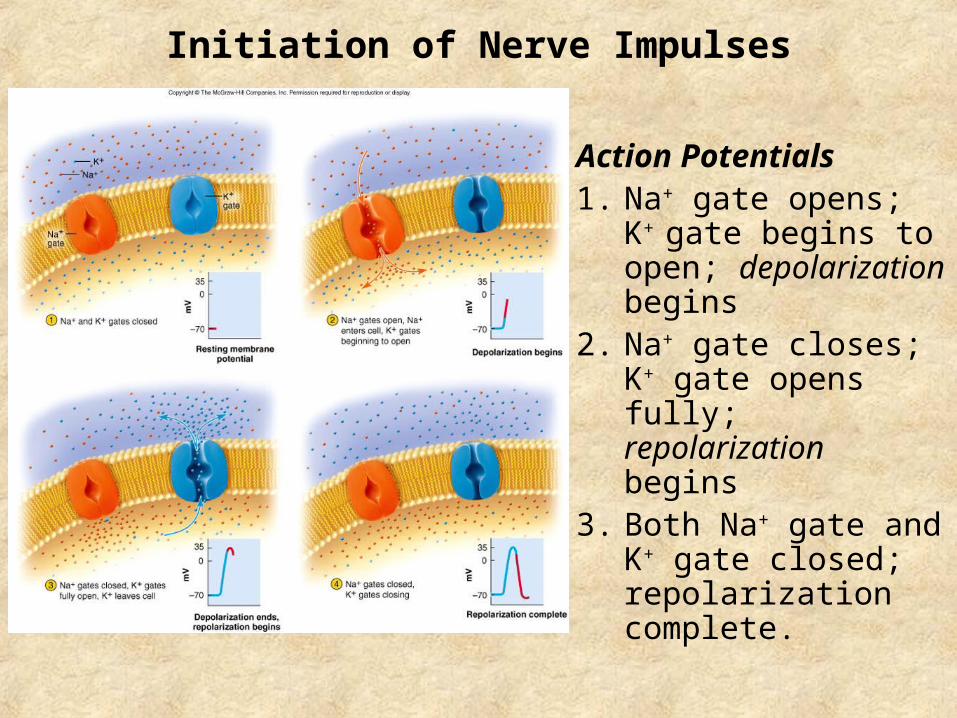
Initiation of Nerve Impulses
Action Potentials1. Na+ gate opens; K+ gate
begins to open; depolarization begins
2. Na+ gate closes; K+ gate opens fully; repolarization begins
3. Both Na+ gate and K+ gate closed; repolarization complete.
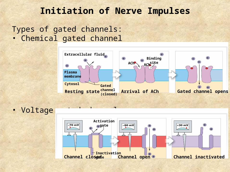
Extracellular fluid
Plasmamembrane
Cytosol Gatedchannel(closed)
Resting state Arrival of ACh
Bindingsite
Gated channel opens
AChACh
Initiation of Nerve Impulses
Types of gated channels:• Chemical gated channel
• Voltage gated channel
Channel inactivatedChannel closed Channel openInactivationgate
Activationgate
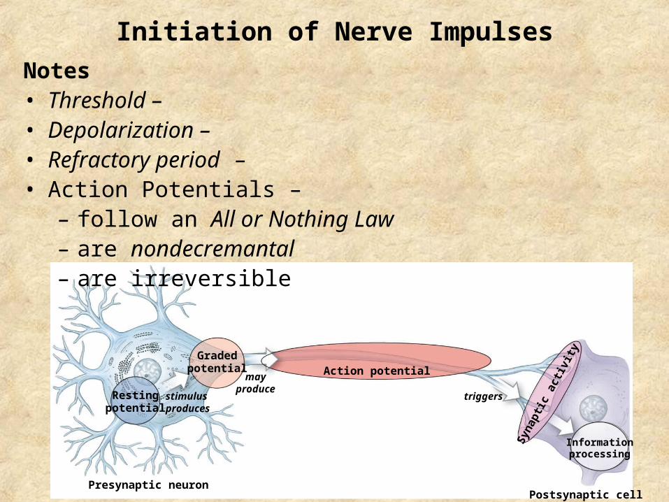
Restingpotential
Gradedpotential
Presynaptic neuron
stimulusproduces
mayproduce
Action potential
Postsynaptic cell
triggers
Informationprocessing
Syna
ptic
act
ivity
Initiation of Nerve Impulses
Notes• Threshold – • Depolarization – • Refractory period – • Action Potentials –
– follow an All or Nothing Law– are nondecremantal– are irreversible
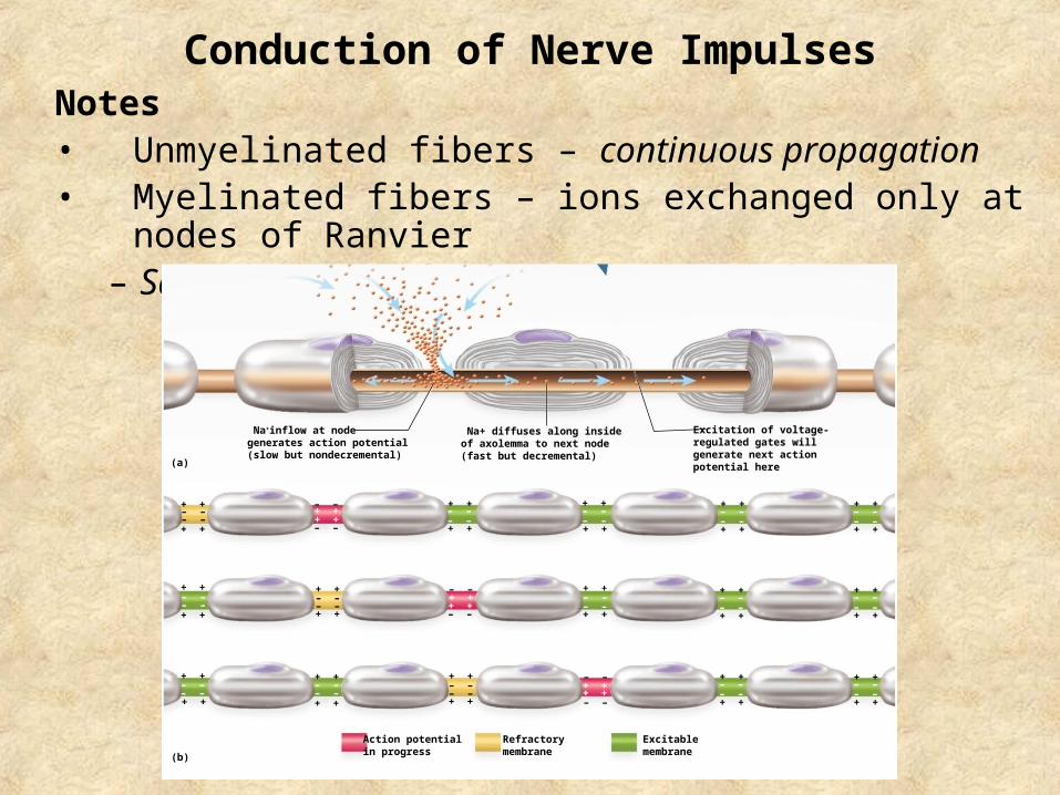
Conduction of Nerve ImpulsesNotes• Unmyelinated fibers – continuous propagation• Myelinated fibers – ions exchanged only at nodes of Ranvier
– Saltatory propagation
+ ++ +
+ +
+ +
+ +
+ +
+ ++ +
+ +
+ +
+ +
+ +
+ ++ +
+ +
+ +
+ +
+ +
– –– –
– –– –
– –– –
– –– –
– –– –
+ +
+ +
– –– –
+ +
+ +
– –– –
– –– –
+ +
+ +
– –– –
– –
– –
– –
– –
– –
– –
(a)
(b)
Na+inflow at nodegenerates action potential(slow but nondecremental)
Na+ diffuses along insideof axolemma to next node(fast but decremental)
Excitation of voltage-regulated gates willgenerate next actionpotential here
+ +
+ +
– –– –
+ +
+ +
– –– –
+ +
+ +
– –– –
+ +
+ +
– –– –
+ +
+ +
– –– –
+ +
+ +
– –– –
Action potentialin progress
Refractorymembrane
Excitablemembrane
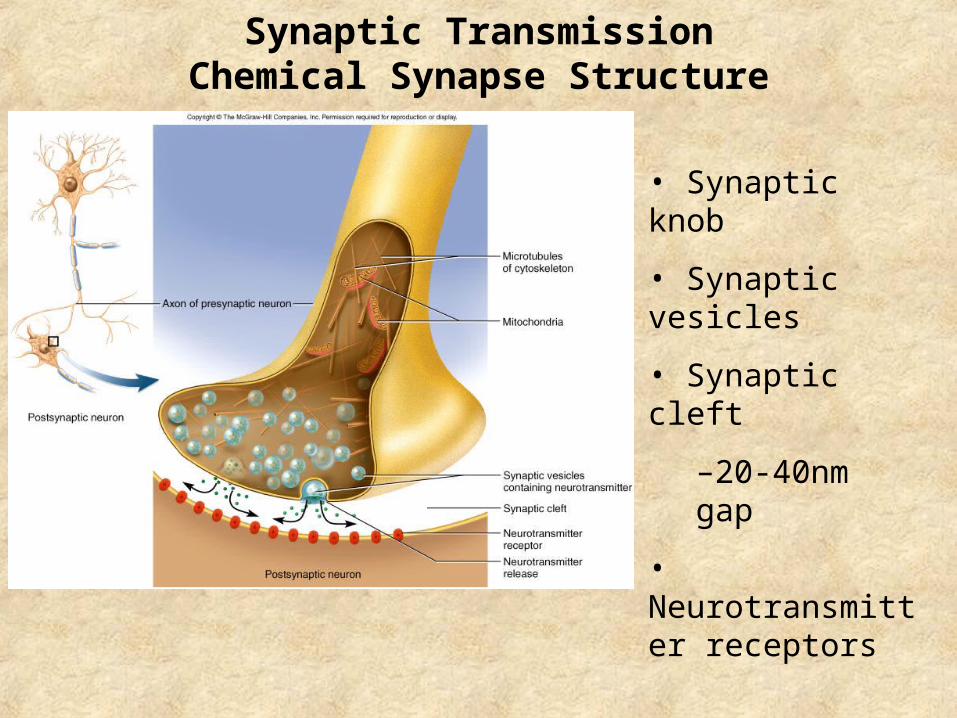
Synaptic TransmissionChemical Synapse Structure
• Synaptic knob
• Synaptic vesicles
• Synaptic cleft
–20-40nm gap
• Neurotransmitter receptors
Synaptic Transmission
Synaptic delay (0.5 msec)
Classes of Neurotransmitters
– Excitatory, inhibitory, neuromuscular junction
3 kinds of synapses:
• Excitatory cholinergic synapse = ACh
• Inhibitory GABA-ergic synapse = GABA
• Excitatory adrenergic synapse = NE
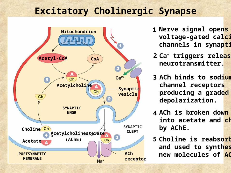
Excitatory Cholinergic Synapse
Mitochondrion
AcetylcholineSynapticvesicle
SYNAPTICKNOB
SYNAPTICCLEFT
POSTSYNAPTICMEMBRANE
Choline
Acetate
Acetylcholinesterase(AChE)
AChreceptor
CoAAcetyl-CoA
Nerve signal opens voltage-gated calcium channels in synaptic knob
Ca+ triggers release of neurotransmitter.
ACh binds to sodium channel receptors producing a graded depolarization.
ACh is broken down into acetate and choline by AChE.
Choline is reabsorbed and used to synthesize new molecules of ACh.
1
2
3
4
5
Inhibitory GABA-ergic Synapse
• Nerve signal triggers release of neurotransmitters
• Receptors trigger opening of Cl- channels
• Postsynaptic neuron now less likely to reach threshold
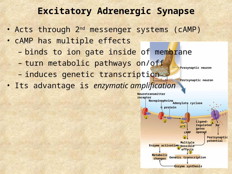
Excitatory Adrenergic Synapse
cAMP
Enzyme activation
Genetic transcription
Enzyme synthesis
Na+
Adenylate cyclase
G protein–
+
–
+
–
+
Postsynaptic neuron
Presynaptic neuron
Norepinephrine
2 3
4
5
1
6
7
Neurotransmitterreceptor
Ligand-regulatedgatesopened
Multiplepossibleeffects
Metabolicchanges
ATP
Postsynapticpotential
• Acts through 2nd messenger systems (cAMP)
• cAMP has multiple effects
– binds to ion gate inside of membrane
– turn metabolic pathways on/off
– induces genetic transcription
• Its advantage is enzymatic amplification
Cessation of the Signal
1. Stop signal in presynaptic neuron
2. Mechanisms to “turn off” the signal
– diffusion of neurotransmitter into ECF
– synaptic knob reabsorbs neurotransmitters
– degradation of neurotransmitters in synaptic cleft
• Acetylcholinesterase
• Adrenalate cyclase
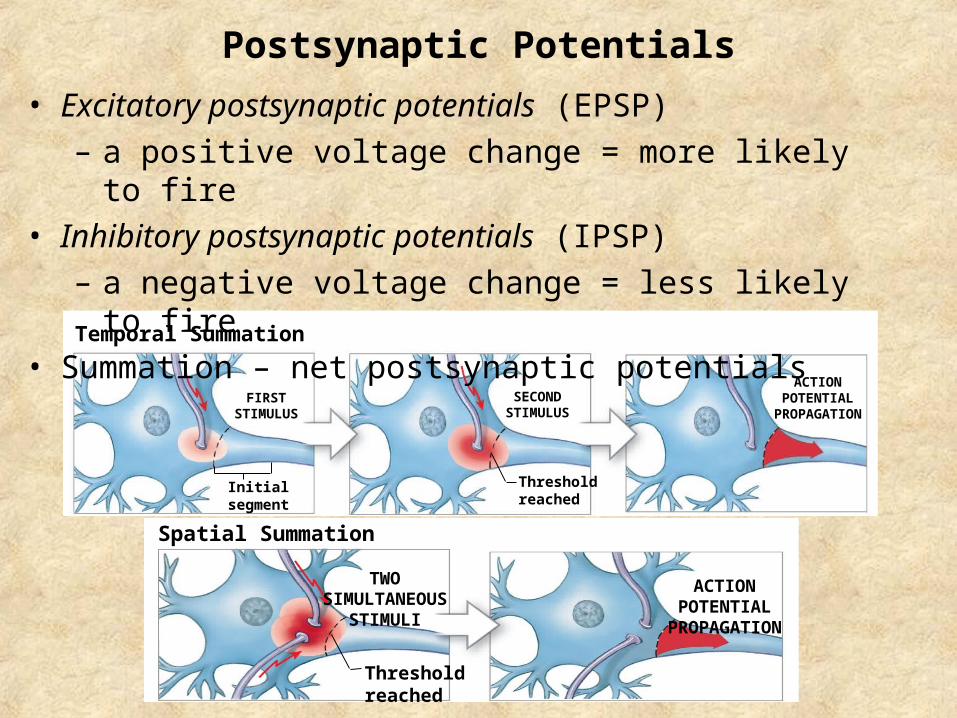
Temporal Summation
Initialsegment
Thresholdreached
ACTIONPOTENTIAL
PROPAGATIONFIRST
STIMULUSSECOND
STIMULUS
Postsynaptic Potentials
• Excitatory postsynaptic potentials (EPSP)
– a positive voltage change = more likely to fire
• Inhibitory postsynaptic potentials (IPSP)
– a negative voltage change = less likely to fire
• Summation – net postsynaptic potentials
Spatial Summation
TWOSIMULTANEOUS
STIMULI
ACTIONPOTENTIAL
PROPAGATION
Thresholdreached
Peripheral Nervous System
• Consists of spinal and cranial nerves
• Sensory (afferent) Division
–Somatic Sensory Division
–Visceral Sensory Division
• Motor (efferent) Division
–Somatic Motor Division
–Visceral Motor Division (Autonomic Nervous System)
• Sympathetic Division
• Parasympathetic Division
• Enteric Division
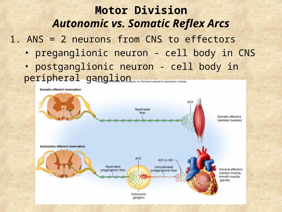
Motor DivisionAutonomic vs. Somatic Reflex Arcs
1. ANS = 2 neurons from CNS to effectors
• preganglionic neuron - cell body in CNS
• postganglionic neuron - cell body in peripheral ganglion
Motor DivisionAutonomic vs. Somatic Reflex Arcs
2. Autonomic effectors function in absence of stimulation
3. Autonomic stimulation can be excitatory or inhibitory
4. Autonomic control is usually involuntary
5. Autonomic effectors are smooth and cardiac muscle, glands
6. Autonomic integrative center in hypothalamus
Autonomic Nervous SystemSympathetic Division
1. Origin in thoracolumbar region
2. Short preganglionic fibers, long postganglionic fibers
3. Synapses at paravertebral ganglia
4. 17 postganglionic neurons for every preganglionic neuron
5. Mass activation
6. “Fight or Flight”
Associated with adrenal glands
• Stimulate the release of neurotransmitters
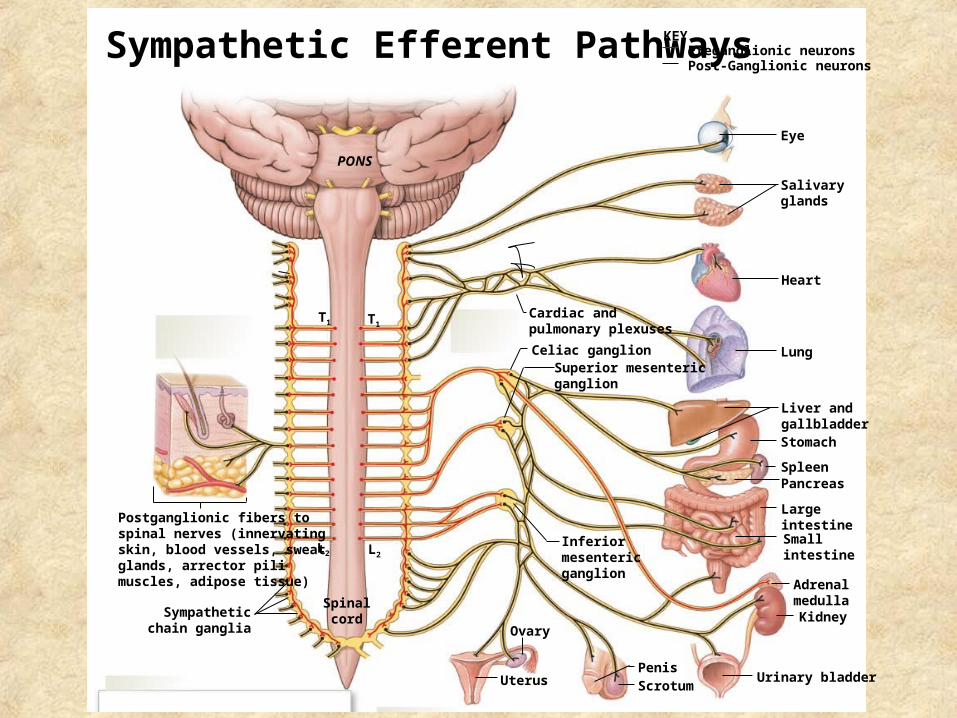
PONS
Eye
Salivaryglands
Heart
Lung
Liver andgallbladderStomach
SpleenPancreas
LargeintestineSmallintestine
AdrenalmedullaKidney
Urinary bladderScrotumPenis
Uterus
Ovary
Inferiormesentericganglion
Superior mesentericganglion
Celiac ganglion
Cardiac andpulmonary plexuses
T1T1
L2L2
Spinalcord
Postganglionic fibers tospinal nerves (innervatingskin, blood vessels, sweatglands, arrector pilimuscles, adipose tissue)
Sympatheticchain ganglia
Preganglionic neuronsKEY
Post-Ganglionic neuronsSympathetic Efferent Pathways
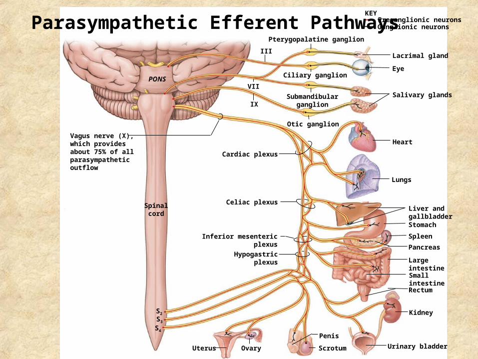
Autonomic Nervous SystemParasympathetic Division
1. Origin in craniosacral region
2. Long preganglionic fibers, short postganglionic fibers
3. Synapses at terminal ganglion
4. 2 postganglionic neurons for every preganglionic neuron
5. Specific and local effects
6. “Rest and Digest”
Spinalcord
S2
S3
S4
Uterus Ovary
Penis
Scrotum
Liver andgallbladderStomach
Spleen
Pancreas
LargeintestineSmallintestine
Kidney
Urinary bladder
Rectum
Eye
Salivary glands
Heart
Lungs
Hypogastricplexus
Inferior mesentericplexus
Celiac plexus
Cardiac plexus
Vagus nerve (X),which providesabout 75% of all parasympathetic outflow
PONS
Otic ganglion
Submandibularganglion
Ciliary ganglion
Pterygopalatine ganglion
III
VII
IX
Lacrimal gland
Preganglionic neuronsKEY
Ganglionic neuronsParasympathetic Efferent Pathways
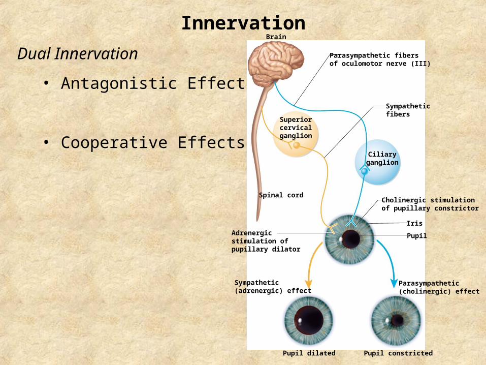
Innervation
Dual Innervation
• Antagonistic Effect
• Cooperative Effects
Brain
Spinal cord
Iris
Pupil
Pupil dilated Pupil constricted
Parasympathetic fibersof oculomotor nerve (III)
Sympatheticfibers
Ciliaryganglion
Superiorcervicalganglion
Cholinergic stimulationof pupillary constrictor
Parasympathetic(cholinergic) effect
Sympathetic(adrenergic) effect
Adrenergicstimulation ofpupillary dilator
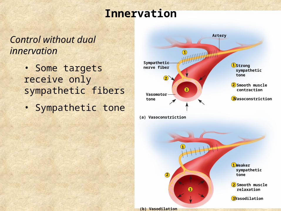
Innervation
Control without dual innervation
• Some targets receive only sympathetic fibers
• Sympathetic tone
Artery
1
1
2
3
3
1
2
3
2
2
3
1
Strongsympathetictone
Smooth musclecontraction
Vasoconstriction
Weakersympathetictone
Smooth musclerelaxation
Vasodilation
Sympatheticnerve fiber
Vasomotortone
(a) Vasoconstriction
(b) Vasodilation