Homeostasis BIO.A.4.2.1. homeostasis Maintaining a stable internal environment.
Upload
arlene-bennettCategory
view
215download
1
Unit III: HomeostasisDefense Against Invasion
Chapter 21

HemostasisPlatelet Plug Formation
• broken vessel exposes collagen
• platelet pseudopods
– contract and draw walls of vessel together platelet plug
• degranulation
• serotonin (vasoconstrictor)
• ADP attracts and degranulates more platelets
• thromboxane A2 (an eicosanoid)

HemostasisCoagulation
• “Clotting” – conversion of plasma protein fibrinogen into insoluble fibrin
threads to form framework of clot
• Extrinsic mechanism – factors released by damaged tissues
• Intrinsic mechanism – factors found in blood (platelet degranulation)
• Procoagulants (clotting factors) Table 18.8
– activate one factor and it will activate the next to form a reaction cascade

Inactive
Inactive
Inactive
Extrinsic mechanism Intrinsic mechanism
Factor V
Factor XII Platelets
Fibrin
Thrombin
Factor VII
Inactive
Ca2+
Damagedperivascular
tissues
Thromboplastin(factor III)
Factor VIII(active)
Ca2+, PF3
Factor IX(active)
Factor XI(active)
Factor X(active)
Factor IIIFactor VCa2+
PF3
Prothrombinactivator
Prothrombin(factor II)
Fibrinogen(factor I)
Fibrinpolymer
Factor XIIICa2+
Coagulation Pathways
• Extrinsic mechanism– initiated by Factor III– fewer steps– 15 seconds formation
• Intrinsic mechanism– initiated by factor XII– cascade to factor XI to
IX to VIII to X– 3-6 minutes formation
• Calcium required for either pathway

Fibrin
Thrombin
Rea
ctio
n c
asca
de
(tim
e)
Prothrombinactivator
FactorXII
FactorXI
FactorIX
FactorVIII
FactorX
Fate of Blood Clots
• Reaction Cascade
• Clot retraction occurs within 30 minutes
• growth factor secreted by platelets
• Fibrinolysis (dissolution of a clot)
– Factor XII initiates small cascade of reactions kallikrein – Plasminogen plasmin, a fibrin-dissolving enzyme (clot buster)

Prevention of Inappropriate Clotting
• Platelet repulsion
• Thrombin dilution
– by rapidly flowing blood
• Natural anticoagulants
– heparin (from basophils and mast cells) interferes with formation of prothrombin activator
– antithrombin (from liver) deactivates thrombin before it can act on fibrinogen

Hemophilia
• Genetic lack of any clotting factor
• Sex-linked recessive (on X chromosome)
– hemophilia A missing factor VIII (83% of cases)
– hemophilia B missing factor IX (15% of cases)
note: hemophilia C missing factor XI (autosomal)
• Physical exertion causes bleeding
– hematomas
– transfusion of plasma or purified clotting factors

Coagulation Disorders
• Thrombosis - abnormal clotting in unbroken vessel– most likely to occur in leg veins of inactive people
• Embolism - clot traveling in a vessel− pulmonary embolism - clot may break free, travel from veins
to lungs
• Infarction may occur if clot blocks blood supply to an organ (MI or stroke)– 650,000 Americans die annually of thromboembolism

Clot Prevention in Patients•Salts, heparin•Vitamin K
–Needed for synthesis of clotting factors–Coumarin
•Aspirin–Suppresses formation of thromboxane A2
•Medicinal leeches
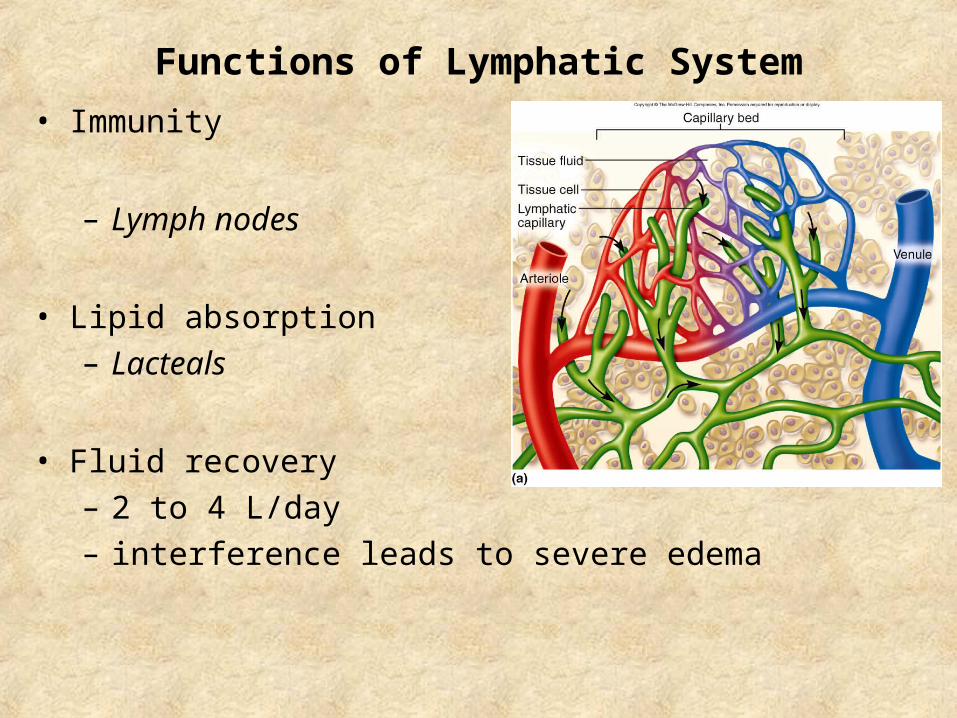
Functions of Lymphatic System
• Immunity
– Lymph nodes
• Lipid absorption
– Lacteals
• Fluid recovery
– 2 to 4 L/day
– interference leads to severe edema

Lymphatic Vessels
•Lymph
•Lymphatic capillaries
•Bud from veins
–Tunica interna, tunica media, tunica externa
–valves

Route of Lymph Flow
• Tissue fluid Lymphatic capillaries • Collecting vessels (lymph nodes) • 6 Lymphatic trunks • 2 Collecting ducts :
– right lymphatic duct R subclavian vein
– thoracic duct - begins as a prominent sac in abdomen called the cisterna chyli; empties into L subclavian vein

The Fluid Cycle

Lymphatic Cells
• Natural killer (NK) cells
– responsible for immune surveillance
• T lymphocytes (T-cells)
– mature in thymus
• B lymphocytes (B-cells)
– differentiation into plasma cells antibodies
• Antigen Presenting Cells (APCs)
– macrophages (from monocytes)
– dendritic cells (in epidermis, mucous membranes and lymphatic organs)
– reticular cells (also contribute to stroma of lymph organs)

Lymphatic Organs
• Primary lymphatic organs
– site where T and B lymphocytes become immunocompetent
– red bone marrow and thymus
• Secondary lymphatic organs
– immunocompetent lymphocytes populate these tissues
– lymph nodes, tonsils, and spleen
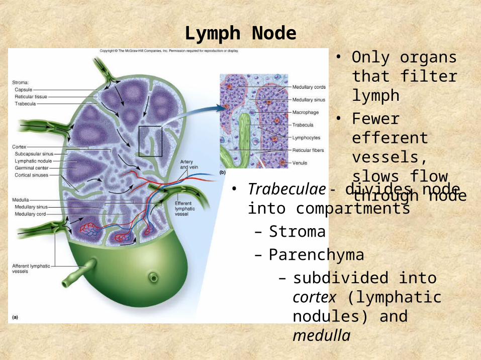
Lymph Node• Only organs that
filter lymph
• Fewer efferent vessels, slows flow through node
• Trabeculae - divides node into compartments
– Stroma
– Parenchyma
– subdivided into cortex (lymphatic nodules) and medulla

Lymph Node Diseases
• Lymphadenitis
– swollen, painful node
• Lymphoma (Metastatic cancer)
– swollen, firm and usually painless

Tonsil
• Tonsillar crypts and encounter lymphocytes
• 3 sets:
– Pharyngeal tonsil (adenoids)
– Palatine tonsils
– Lingual tonsils

Spleen
• Parenchyma tissues:– red pulp:
– white pulp:
• Functions– blood production in fetus– blood reservoir– RBC disposal (“graveyard”)– Stabilize blood volume

Thymus
• Both lymphatic and endocrine
– Maturation of T-cells and secretes hormones
• Most active in childhood (under age 14)
– If removed – no immunity
– Replaced by fibrous and fatty tissue

• Structure similar to lymph nodes
• Reticular epithelial cells
– Blood-thymus barrier
• isolates developing T lymphocytes from foreign antigens
– secretes hormones (thymopoietin, thymulin and thymosins)
Thymus
Lobule
Cortex
Medulla
Trabecula
Trabecula
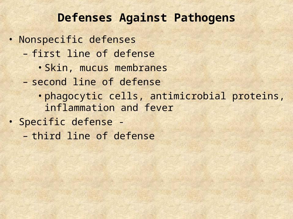
Defenses Against Pathogens
• Nonspecific defenses
– first line of defense
• Skin, mucus membranes
– second line of defense
• phagocytic cells, antimicrobial proteins, inflammation and fever
• Specific defense -
– third line of defense
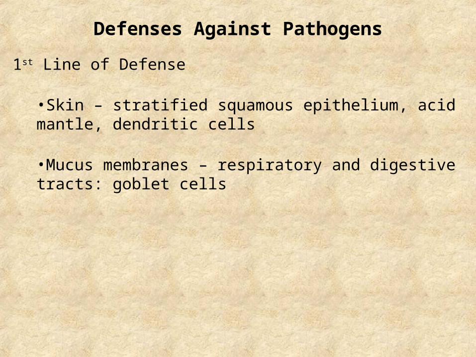
Defenses Against Pathogens
1st Line of Defense
•Skin – stratified squamous epithelium, acid mantle, dendritic cells
•Mucus membranes – respiratory and digestive tracts: goblet cells

Phagocytic Cells
2nd and 3rd Line of Defense
•Leukocytes and Macrophages
–Neutrophils – respiratory burst
–Eosinophils – kill parasites; limits histamine; promote basophils
–Basophils – secrete histamine and heparin
–Lymphocytes – 80% T-cells, 15% B-cells, 5% NK-cells
–Monocytes - transform into macrophages

Specific vs. Nonspecific Immunity
Nonspecific Immunity
• Immune surveillance, inflammation, fever
3rd Line of Defense: Immune System
• Specificity and memory
• Cellular immunity: cell-mediated (T cells)
• Humoral immunity: antibody mediated (B cells)– Comparison table: Table 21.5, p.850

Passive and Active Immunity
• Natural active immunity (produces memory cells)
– result of infection or natural exposure to antigen
• Artificial active immunity (produces memory cells)
– result of vaccination
• Natural passive immunity (through placenta, milk)
– temporary, fetus acquires antibodies from mother
• Artificial passive immunity (snakebite, rabies, tetanus)
– temporary, injection of immune serum (antibodies)

The Three “R”s of Immunity
Cellular Immunity
•Recognition
–Antigen presentation
–T-cell activation
•React (attack)
–Helper T-cells - attract neutrophils, natural killer cells, and macrophages, stimulate T and B-cell mitosis and maturation
–Cytotoxic T-cells – “lethal hit” of cytotoxic chemicals
•Remember
–T-cell recall response

The Three “R”s of Immunity
Humoral Immunity
•Recognition
–Receptors for one antigen on a B-cell
–Helper T-cell binds to complex
–B-cells differentiate into plasma cells
•React (attack)
–Neutralization, Complement fixation, Agglutination, Precipitation
•Remember
–Primary response

Notes on Immunity
• Memory lasts longer in Cellular Immunity than Humoral
• Both processes of immunity occur simultaneously and in conjunction with inflammation
• HIV attacks helper T-cells knocks out the central coordinating role in both processes

Test III
•Lecture•Chapters 24, 18, and 21
•Lab practical•Identification of slides: organ, cells, regions•Lab manual questions