Unit 4 - s37af0fcd02709117.jimcontent.com · Unit 4: The circulatory and excretory systems 1. The...
13
Unit 4: The circulatory and excretory systems 1. The internal medium 2. The circulatory system 2.1. The blood 2.2. The heart 2.3. The blood vessels 3. The lymphatic system 4. The excretory system 4.1. The urinary system 4.2. Functions of the kidney 4.3. The sweat glands a. Name some functions of the blood? b. How do veins and arteries differ? c. What is the lymph? d. How does the body rid of waste products? Think and answer? UNIT OBJECTIVES In this unit you will learn: The anatomy of the circulatory and excretory systems To the processes of pumping the blood by the heart and its circulation throughout the body. To distinguish between the structures of blood vessels and their functions. To distinguish among the different waste substances and their production To appreciate the importance of healthy habits related to circulatory and excretory systems. The main diseases of the circulatory and excretory systems.
Transcript of Unit 4 - s37af0fcd02709117.jimcontent.com · Unit 4: The circulatory and excretory systems 1. The...

Unit 4: The circulatory and excretory systems
1. The internal medium
2. The circulatory system
2.1. The blood
2.2. The heart
2.3. The blood vessels
3. The lymphatic system
4. The excretory system
4.1. The urinary system
4.2. Functions of the kidney
4.3. The sweat glands
a. Name some functions of the blood?
b. How do veins and arteries differ?
c. What is the lymph?
d. How does the body rid of waste products?
Think and answer?
UNIT OBJECTIVES
In this unit you will learn:
The anatomy of the circulatory and excretory systems
To the processes of pumping the blood by the heart and its circulation
throughout the body.
To distinguish between the structures of blood vessels and their functions.
To distinguish among the different waste substances and their production
To appreciate the importance of healthy habits related to circulatory and
excretory systems.
The main diseases of the circulatory and excretory systems.

UNIT 4: The circulatory and excretory systems Biology and Geology (3rd
ESO)
1. The internal medium
The internal medium is the set of liquids which forms the surrounding environment of cells in
multicellular organisms.
In humans these liquids are mainly blood, lymph and interstitial plasma (extracellular liquids)
- Interstitial plasma. It is a liquid which are among cells. It comes from blood plasma that exits
from capillaries.
- Blood. It is the transport medium of substances which flows within blood vessels.
- Lymph. It is a liquid formed from the interstitial plasma that flows within lymphatic vessels.
The lymphatic system ends into circulatory system.
The optimal operation of cells depends on the maintenance of constant conditions of interstitial plasma
that surround them. These conditions include temperature, chemical composition, pH, etc. The optimal
situation is called homeostasis. To keep homeostasis, blood and lymph renew constantly the
interstitial plasma.
ACTIVITIES
1.1. Listen and identify what component of the internal medium is described:
a. Interstitial plasma b. Blood c. Lymph
Blood vessel (blood)
Lymphatic vessel
(Lymph)
Extracellular medium (Interstitial plasma)

UNIT 4: The circulatory and excretory systems Biology and Geology (3rd
ESO)
2. The circulatory system
The function of the circulatory system is to distribute the nutrients of food and the oxygen throughout
the whole organism and to collect the waste substances produced by cellular metabolism and to bring
them to the excretory organs.
The circulatory system is formed by:
- A circulating liquid: the blood
- An impulse pump: the heart
- A distribution system: the blood vessels
2.1. Blood
Blood is a viscous, red and salad liquid which flows within blood vessels. An adult human has about 5
litres of blood, although it depends on weight, height, age and sex.
a. Functions of blood
- Transportation: The blood carries many different substances around the body. These include:
- Nutrients (glucose, amino acids, vitamins, minerals and oxygen)
- Waste products (water, urea and carbon dioxide)
- Hormones
- Maintaining homeostasis:
- Immunity and defence: White blood cells fight infection and platelets help to repair damage
and clot the blood
- Distribution of heat and force.
b. Composition of blood
Blood is a liquid tissue. It is formed by a liquid part called plasma in which are immersed the
cellular components: blood cells.

UNIT 4: The circulatory and excretory systems Biology and Geology (3rd
ESO)
- Plasma (55 % blood volume). It is a yellowish liquid formed by water (90 %), immersed
substances (proteins, lipids, glucose) and dissolved substances (ions and gases).
- Blood cells (45 % blood volume). There are three types of cellular components:
- Red blood cells (erythrocytes) They are little and biconcave cells (disc-shape) without nucleus.
They are very elastic and deformable.
They are rich in a protein called haemoglobin. Haemoglobin contains iron and its function is
to transport oxygen (It oxidizes when blood pass through alveoli).
A healthy person has about 5.106 erythrocytes / mm3 of blood.
- White blood cells (leukocytes) They are true cells, with nucleus (with variable shape).
They are the least numerous cellular blood components (5000-9000 leukocytes / mm3 of blood).
There are two groups of leukocytes, but all participate in defending of the organism:
- Granulocytes (with granules in the cytoplasm) Neutrophils, Eosinophils and Basophiles.
- Agranulocytes (without granules in the cytoplasm) They are the most important ones.
- Lymphocytes produce antibodies.
- Monocytes (macrophages) digest microorganisms.
- Platelets (thrombocytes) They are not true cells, but fragments of cells.
There are between 150.000 and 300.000 thrombocytes / mm3 of blood.
Their function is blood coagulation (clotting)
ACTIVITIES
2.1. Make a double entry chart which allows you to compare the characteristics of the
cellular components of blood: cellular characteristics (shape, size, contain, etc),
amount, types (if there would be any) and function.
2.2. Are red blood cells true cells? Why?
2.3. People who live at high altitude have more erythrocytes. Why?
2.4. Listen and indicate what type of blood cell is described:
a. Erythrocytes b. Leukocytes c. Thrombocytes
2.5. The following pictures represent the steps in the formation of a clot. Listen, identify and
put them in the correct order:

UNIT 4: The circulatory and excretory systems Biology and Geology (3rd
ESO)
2.2. Heart
The heart is a muscular organ about the size of a fist. It is located in the middle of thoracic cavity,
slightly left side and between the lungs. Its function is to pump the blood to the whole body.
The heart wall is formed by three layers:
- Pericardium: It is the most external layer. It is a connective tissue layer that joins to the heart:
- Nerves that innerve the heart (nodal tissue, responsible for the impulse that makes the heart beats)
- Blood vessels that irrigate the heart (coronary artery and vein)
- Myocardium: It is in the middle. It is a thick layer of cardiac muscle tissue.
- Endocardium: It is the most internal layer. It is a connective tissue layer that lines the inner
cavities of the heart and made up the valves.
The heart is divided into two parts, left and right, by the interventricular septum. These two parts
are not communicated.
Each part of the heart is divided into two chambers:
- Atrium: It is the upper cavity. It is smaller and its walls are thin and elastic.
- Ventricle: It is the lower cavity. It is bigger and its walls are thick and strong.
The two chambers of the same side are communicated by a valve, which allows the pass of blood from
atrium to ventricle but not in inverse way.
- Bicuspid (mitral) Valve: It is in the left side. It is formed by two languets.
- Tricuspid Valve: It is in the right side. It is formed by three languets.
The veins arrive to atria, which bring blood back to the heart:
- Pulmonary Veins (2 come from the left lung and 2 from the right lung) arrive to the left atrium.
- Inferior Vena Cava (brings blood from the inferior part of body) and Superior Vena Cava
(brings blood from the superior part of body) arrive to the right atrium.
Arteries exit from ventricles and carry blood to the lungs and the rest of body:
- Pulmonary Artery exits from right ventricle and later divides into two branches, one for each lung.
- Aorta Artery exits from left ventricle and carries blood to all the body.
The blood exiting from ventricles is controlled by Sigmoid Valves located at the beginning of arteries.
These valves prevent that blood goes back to ventricles.
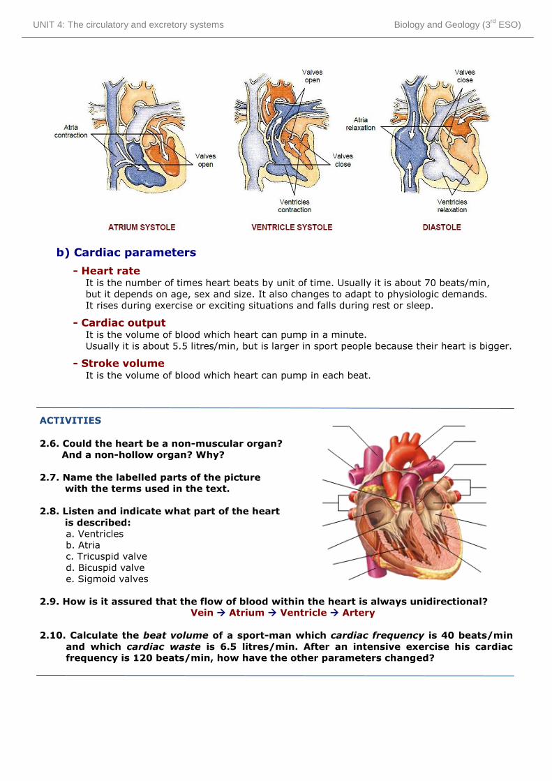
a) Cardiac cycle (heartbeat)
The cardiac cycle is the set of phases which heart pass through in each beat.
1st) Atrium systole (atrium contraction)
- Atria, full of blood, contract.
- Mitral and tricuspid valves open (because atrium pressure is bigger than ventricle pressure)
- Blood pass to ventricle
- Mitral and tricuspid valves close (because atrium pressure is smaller than ventricle pressure)
2nd) Ventricle systole (ventricle contraction)
- Ventricles, full of blood, contract.
- Sigmoid valves open (because ventricle pressure is bigger than artery pressure)
- Blood pass to arteries
- Sigmoid valves close (because ventricle pressure is smaller than artery pressure)
3rd) General diastole (heart relaxation)
- Atria and ventricles, empty of blood, relax.
- Blood pass to atria from the veins by suction.
UNIT 4: The circulatory and excretory systems Biology and Geology (3rd
ESO)
b) Cardiac parameters
- Heart rate It is the number of times heart beats by unit of time. Usually it is about 70 beats/min,
but it depends on age, sex and size. It also changes to adapt to physiologic demands.
It rises during exercise or exciting situations and falls during rest or sleep.
- Cardiac output It is the volume of blood which heart can pump in a minute.
Usually it is about 5.5 litres/min, but is larger in sport people because their heart is bigger.
- Stroke volume It is the volume of blood which heart can pump in each beat.
ACTIVITIES
2.6. Could the heart be a non-muscular organ?
And a non-hollow organ? Why?
2.7. Name the labelled parts of the picture
with the terms used in the text.
2.8. Listen and indicate what part of the heart
is described:
a. Ventricles
b. Atria
c. Tricuspid valve
d. Bicuspid valve
e. Sigmoid valves
2.9. How is it assured that the flow of blood within the heart is always unidirectional? Vein Atrium Ventricle Artery
2.10. Calculate the beat volume of a sport-man which cardiac frequency is 40 beats/min
and which cardiac waste is 6.5 litres/min. After an intensive exercise his cardiac
frequency is 120 beats/min, how have the other parameters changed?
UNIT 4: The circulatory and excretory systems Biology and Geology (3rd
ESO)
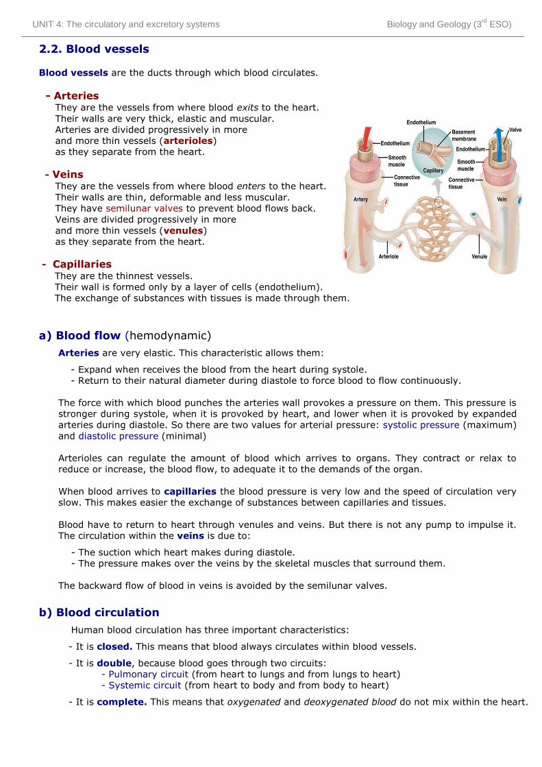
2.2. Blood vessels
Blood vessels are the ducts through which blood circulates.
- Arteries They are the vessels from where blood exits to the heart.
Their walls are very thick, elastic and muscular.
Arteries are divided progressively in more
and more thin vessels (arterioles)
as they separate from the heart.
- Veins They are the vessels from where blood enters to the heart.
Their walls are thin, deformable and less muscular.
They have semilunar valves to prevent blood flows back.
Veins are divided progressively in more
and more thin vessels (venules)
as they separate from the heart.
- Capillaries They are the thinnest vessels.
Their wall is formed only by a layer of cells (endothelium).
The exchange of substances with tissues is made through them.
a) Blood flow (hemodynamic)
Arteries are very elastic. This characteristic allows them:
- Expand when receives the blood from the heart during systole.
- Return to their natural diameter during diastole to force blood to flow continuously.
The force with which blood punches the arteries wall provokes a pressure on them. This pressure is
stronger during systole, when it is provoked by heart, and lower when it is provoked by expanded
arteries during diastole. So there are two values for arterial pressure: systolic pressure (maximum)
and diastolic pressure (minimal)
Arterioles can regulate the amount of blood which arrives to organs. They contract or relax to
reduce or increase, the blood flow, to adequate it to the demands of the organ.
When blood arrives to capillaries the blood pressure is very low and the speed of circulation very
slow. This makes easier the exchange of substances between capillaries and tissues.
Blood have to return to heart through venules and veins. But there is not any pump to impulse it.
The circulation within the veins is due to:
- The suction which heart makes during diastole.
- The pressure makes over the veins by the skeletal muscles that surround them.
The backward flow of blood in veins is avoided by the semilunar valves.
b) Blood circulation
Human blood circulation has three important characteristics:
- It is closed. This means that blood always circulates within blood vessels.
- It is double, because blood goes through two circuits:
- Pulmonary circuit (from heart to lungs and from lungs to heart)
- Systemic circuit (from heart to body and from body to heart)
- It is complete. This means that oxygenated and deoxygenated blood do not mix within the heart.

UNIT 4: The circulatory and excretory systems Biology and Geology (3rd
ESO)
- Pulmonary circulation
It is also known as minor circulation because the
circuit that blood follows is shorter and only
involves lungs.
Deoxygenated blood exits from the heart by
Pulmonary artery to the lungs where it is
recharged of oxygen and then the oxygenated
blood returns to the heart by Pulmonary veins.
- Systemic circulation
It is also known as major or general circulation
because the circuit is longer and involves all
body organs.
Oxygenated blood exits from the heart by Aorta
artery to be distributed by the tissues. Cells take
the oxygen and the deoxygenated blood returns
to the heart by Cava veins.
When deoxygenated blood arrives to the heart goes through its right side, meanwhile the
oxygenated blood goes through its left side. The interventricular septum avoid that they mix.
Each organ has an artery that provides it of nutrients and oxygen and a vein that collects the
waste substances and carbon dioxide. These artery and vein are called like the organ (ej:
gastric artery and gastric vein). Every one of these arteries and veins are branches of the Aorta
artery and of one of the Cava veins.
ACTIVITIES
2.11. Why people that pass long time standing up, has usually their feet swollen?
2.12. The exchange of substances is made in capillaries but not in arteries or veins, why?
2.13. Indicate why:
a. The ventricular wall is thicker and stronger than the atrial wall
b. The left ventricular wall is thicker and stronger than the right one.
c. Sometimes it is used the expressions: “left heart” and “right heart”
2.14. How can we know if we have cut a vein or an artery?
2.15. Is it the arterial blood always oxygenated blood and the venous blood,
deoxygenated blood? Why?
2.16. Listen and indicate what type of blood vessel is described:
a. Arteries b. Veins c. Capillaries d. Arterioles e. Venules
UNIT 4: The circulatory and excretory systems Biology and Geology (3rd
ESO)
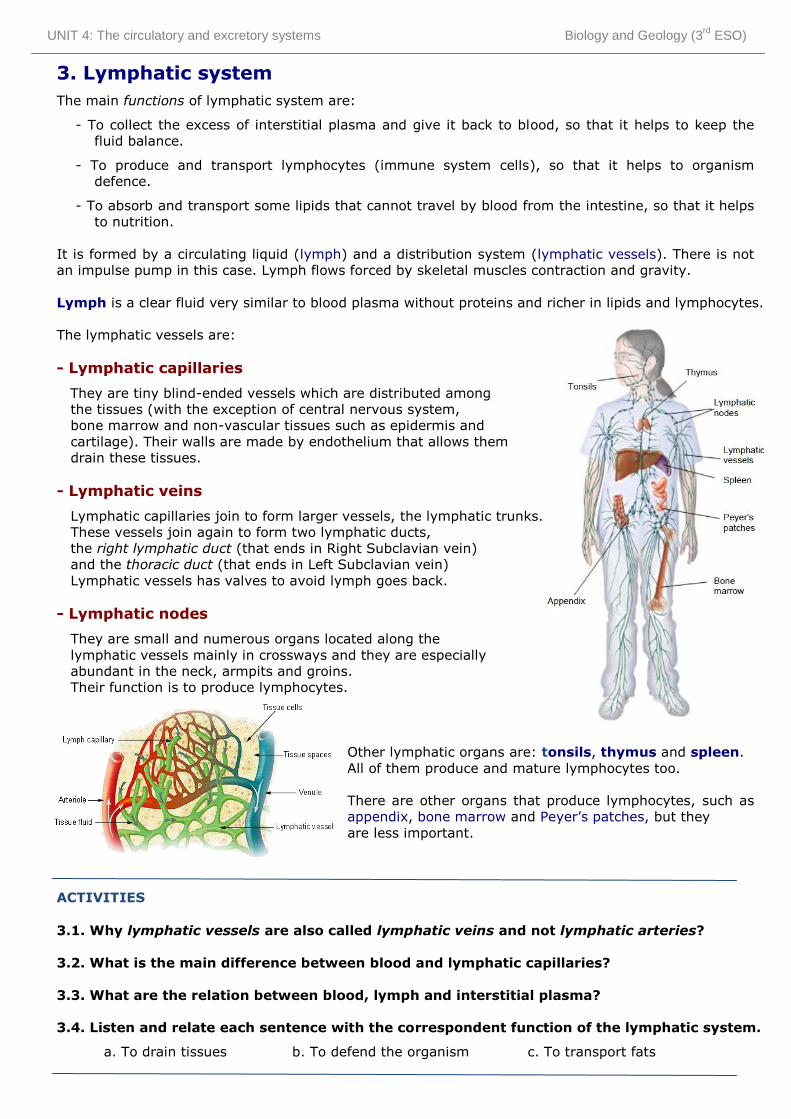
3. Lymphatic system
The main functions of lymphatic system are:
- To collect the excess of interstitial plasma and give it back to blood, so that it helps to keep the
fluid balance.
- To produce and transport lymphocytes (immune system cells), so that it helps to organism
defence.
- To absorb and transport some lipids that cannot travel by blood from the intestine, so that it helps
to nutrition.
It is formed by a circulating liquid (lymph) and a distribution system (lymphatic vessels). There is not
an impulse pump in this case. Lymph flows forced by skeletal muscles contraction and gravity.
Lymph is a clear fluid very similar to blood plasma without proteins and richer in lipids and lymphocytes.
The lymphatic vessels are:
- Lymphatic capillaries
They are tiny blind-ended vessels which are distributed among
the tissues (with the exception of central nervous system,
bone marrow and non-vascular tissues such as epidermis and
cartilage). Their walls are made by endothelium that allows them
drain these tissues.
- Lymphatic veins
Lymphatic capillaries join to form larger vessels, the lymphatic trunks.
These vessels join again to form two lymphatic ducts,
the right lymphatic duct (that ends in Right Subclavian vein)
and the thoracic duct (that ends in Left Subclavian vein)
Lymphatic vessels has valves to avoid lymph goes back.
- Lymphatic nodes
They are small and numerous organs located along the
lymphatic vessels mainly in crossways and they are especially
abundant in the neck, armpits and groins.
Their function is to produce lymphocytes.
Other lymphatic organs are: tonsils, thymus and spleen.
All of them produce and mature lymphocytes too.
There are other organs that produce lymphocytes, such as
appendix, bone marrow and Peyer’s patches, but they
are less important.
ACTIVITIES
3.1. Why lymphatic vessels are also called lymphatic veins and not lymphatic arteries?
3.2. What is the main difference between blood and lymphatic capillaries?
3.3. What are the relation between blood, lymph and interstitial plasma?
3.4. Listen and relate each sentence with the correspondent function of the lymphatic system.
a. To drain tissues b. To defend the organism c. To transport fats

UNIT 4: The circulatory and excretory systems Biology and Geology (3rd
ESO)
4. Excretory system
In addition to the CO2 produced during cellular respiration, the metabolic activity of our cells produce
other many waste substances that we have to expulse out of our bodies.
First these waste substances pass from cells to blood and then, blood carries them to the excretory
organs which eliminate them.
Excretion is the process of removing waste substances produced by vital activity from blood.
The excretory system is formed essentially by the urinary system and the sweat glands, although
other organs and organs systems share this function with them. These other organs are:
- Lungs (Respiratory system): They eliminate the CO2 produced in cellular respiration.
- Liver (Digestive system): It expulses bile pigments (rests of erythrocytes that cannot be
recycled) and toxins ingested (e.g. medicines, alcohol, etc).
- Large intestine (Digestive system): It makes and expulses faeces which are non-digested
rests of food mixed with excretion products of the liver.
Anyway, the most toxic waste substances produced by our bodies came from the destruction of proteins
and nucleic acids. They are respectively urea and uric acid and they have to be eliminated by specific
organs: kidneys and sweat glands.
4.1. The sweat glands
Sweat glands produce a secretion called sweat, which contains urea, uric acid, ions and water.
They are distributed all over your skin.
Sweat is a liquid secretion very similar to urine,
but it has more water and less waste products.
The main function of sweat is to keep the body temperature.
When it is too high, the sweat glands produce sweat.
Sweat, which is very rich in water, evaporates reducing
the temperature of your body.
Other secondary functions of sweat are:
- To excrete waste products (urea and uric acid)
- To play a role in sexual attraction.

UNIT 4: The circulatory and excretory systems Biology and Geology (3rd
ESO)
4.2. Urinary system
The urinary system is formed by kidneys and urinary tracts.
a) Urinary tracts
They are the ducts which collect and lead to
the urine out of body.
- Ureters: They are two thin and long
tubes which connect the renal pelvis
to the urinary bladder.
- Urinary bladder: It is a muscular
and elastic bag which stores urine.
- Urethra: It is the conduct which
connects the bladder to the
exterior. It has a sphincter that
controls the exit of urine. In men the
urethra is shared with the reproductive
system and cross along the penis.
By contrast, in women it is a shorter
independent tube.
Kidneys produce urine continuously.
This urine is stored into urinary bladder
until it is full. Miction is the act to evacuate
this urine from the body.
b) Kidneys:
They are two bean-shaped organs about 11 cm long, 5 cm wide and 3 cm thick. They are located
both sides of spinal column in lumbar region into abdominal cavity. The right one is slightly lower
due to the fact that the liver is above it.
In a transversal cut, we can distinguish:
- Renal cortex: It is the outer portion.
It has a granular shape.
- Renal medulla: It is the middle layer.
It has a fibrous shape and is divided into
portions called malpighian pyramids
(or renal pyramids)
- Renal pelvis: It is the internal cavity
of kidney which connects with the ureter.
Each kidney is surrounded by a layer of connective
tissue called renal capsule and by a layer of adipose
tissue that protects it from shock.
On the top of kidneys are located the adrenal
capsules which are endocrine organs. They really do
not form part of kidneys, only are attached to them.
Each kidney is irrigated by a renal artery and a renal vein which are branches of aorta artery and
inferior vena cava respectively.

UNIT 4: The circulatory and excretory systems Biology and Geology (3rd
ESO)
Nephron is the basic anatomical and physiological unit of the kidney.
Each kidney is formed by about a million of nephrons. A nephron is a blind-ended tubule with a
complex structure:
- Renal corpuscle
- Bowman’s capsule: Cup-shaped
structure with double wall in which
central cavity is located the renal
glomerulus.
- Renal glomerulus: Capillary network.
- Renal tubule
- Proximal convoluted tubule
- Loop of Henle
- Distal convoluted tubule
Each nephron connects with a collecting duct
which collects the urine produced by many
nephrons and carries it to the renal pelvis.
Blood arrives to glomerulus from an afferent
arteriole and exits from it through an efferent
arteriole. Arterioles can keep the blood flows to
high pressure. This help in filtration of blood.
Surrounding the renal tubule there is a dense
capillary web called vasa recta.
ACTIVITIES
4.1. What are the waste substances produced by our organism?
Indicate the origin and the way of excretion of each one.
4.2. Why do babies need nappies?
4.3. What could happen if we would not have urinary bladder?
4.4. Listen and indicate which part of the urinary system is described:
a. Kidneys b. Ureters c. Urinary bladder d. Uretha
4.5. Listen and complete the text:
The …………………….
This is the …………………… part of the kidney. It is formed by the …………………………….., which
surrounds the …………………….............…., an accumulation of ................................. from the ...............................
The Bowman's capsule extends into the …………………………., a tube surrounded by capillaries that leads into the ………………………….
Collecting ducts empty .................... into the ..................................

UNIT 4: The circulatory and excretory systems Biology and Geology (3rd
ESO)
4.3. Functions of kidney
a) Urine formation
It is the main function of kidneys. Urine is formed by three processes:
1st) Glomerular filtration
Blood is under high pressure in the glomerulus.
Blood plasma (except for plasma proteins) moves
from glomerulus capillaries into the Bowman’s capsule.
This fluid is called primary urine. It is
a clear liquid which contains many useful
substances (amino acids, glucose, ions, etc.)
and a large amount of water.
2nd) Tubular reabsorption
As the filtrated liquid moves along the renal tubule
most of the water and the other useful molecules
are reabsorbed.
These substances go through the tubule wall and
pass to the blood capillaries that surround it.
As a result the primary urine is progressively more
and more concentrate.
3rd) Tubular secretion
At the same time that reabsorption takes place,
the cells of the renal tubule secrete some toxic
substances which are added to the urine.
Once the fluid moves into the collecting duct,
it is called final urine. In the collecting duct,
additional water is removed from the urine,
concentrating more the wastes.
Urine is a liquid waste product which contains urea, uric acid, ions and water.
Urea and uric acid are very toxic and have to be excreted dissolved, so that it is absolutely
necessary that urine contains some water. Anyway, the most part of water is reabsorbed. For each
millilitre of urine, kidneys filtrate 125 ml of blood plasma.
b) Maintenance of homeostasis
Kidneys have other important functions that help in the maintenance of homeostasis, such as:
- Regulate the blood plasma volume (the water that it contains) and, as a result, the blood pressure.
- Control the concentration of plasma ions: sodium (Na+), potassium (K+), chloride (Cl-),
and nitrogenous wastes (especially urea).
- Contribute to keep the pH of the plasma (concentration of H+ ions)
ACTIVITIES
4.6. If the glomerular filtration speed is 125 ml/min, what volume of blood plasma is filtrated
and then, reabsorbed daily? Calculate the urine volume formed during a day.
4.7. If you eat a very salad meal, will you urinate more or less than usually? Why?
4.8. Listen and identify the phase of the urine formation that is described:
a. Glomerular filtration b. Tubular reabsorption c. Tubular secretion