13 Malignant Lesions of the Central and Posterior Skull Base
r e v b r a s o r t o p . 2 0 1 7;5 2(4):463–472
S
R
Uf
RRa
b
c
a
A
R
A
A
K
K
K
J
S
M
P
T
J
I
P
o
M
R
h2u
OCIEDADE BRASILEIRA DEORTOPEDIA E TRAUMATOLOGIA
www.rbo.org .br
eview Article
nderstanding posterior meniscal roots lesions:rom basic science to treatment�
aphael Serra Cruza,b,∗, Marcio Balbinotti Ferraria,b, Leonardo Metsavahtb,obert F. LaPradea,c
Steadman Philippon Research Institute, Vail, United StatesInstituto Brasil de Tecnologias da Saúde, Rio de Janeiro, RJ, BrazilThe Steadman Clinic, Vail, United States
r t i c l e i n f o
rticle history:
eceived 30 June 2016
ccepted 14 July 2016
vailable online 26 July 2017
eywords:
nee injuries
nee/anatomy & histology
oint instability
urgical procedures, operative
enisci
a b s t r a c t
The variability of symptoms and the fact that they are not easily recognized in imaging stud-
ies make the diagnosis and treatment of posterior meniscal roots lesions a challenging task
to the orthopedist. In recent years, a more precise understanding of the anatomy and biome-
chanical impair of the knee joint in these cases has enabled great advances in therapeutic
approaches. Well-documented studies have shown that the repair of these lesions presents
superior functional and clinical improvement when compared with meniscectomy. How-
ever, the progression of degenerative joint changes in the long-term still exhibits conflicting
results.© 2016 Sociedade Brasileira de Ortopedia e Traumatologia. Published by Elsevier Editora
Ltda. This is an open access article under the CC BY-NC-ND license (http://
creativecommons.org/licenses/by-nc-nd/4.0/).
Compreendendo as lesões das raízes posteriores dos meniscos: daciência básica ao tratamento
alavras-chave:
raumatismos do joelho
r e s u m o
A variabilidade da sintomatologia e o fato de não serem facilmente reconhecidas nos exames
de imagem tornam o diagnóstico e o tratamento das lesões das raízes posteriores dos
oelho/anatomia & histologia meniscos tarefas desafiadoras para o ortopedista. Nos últimos anos, uma compreensão nstabilidade articularrocedimentos cirúrgicos
peratórios
eniscos
mais precisa da anatomia e do comprometimento biomecânico da articulacão do joelho
nessas lesões têm possibilitado grandes avancos nas abordagens terapêuticas. Estudos bem
� Study conducted in a partnership of the Instituto Brasil de Tecnologias da Saúde, Rio de Janeiro, RJ, Brazil, with the Steadman-Philipponesearch Institute, Vail, United States.∗ Corresponding author.
E-mails: [email protected], [email protected] (R.S. Cruz).ttp://dx.doi.org/10.1016/j.rboe.2017.07.005255-4971/© 2016 Sociedade Brasileira de Ortopedia e Traumatologia. Published by Elsevier Editora Ltda. This is an open access articlender the CC BY-NC-ND license (http://creativecommons.org/licenses/by-nc-nd/4.0/).
464 r e v b r a s o r t o p . 2 0 1 7;5 2(4):463–472
documentados demonstram que o reparo dessas lesões oferece uma melhoria clínica e
funcional superior à meniscectomia. Entretanto, os resultados da progressão das alteracões
degenerativas articulares em longo prazo ainda são conflitantes.
© 2016 Sociedade Brasileira de Ortopedia e Traumatologia. Publicado por Elsevier
Editora Ltda. Este e um artigo Open Access sob uma licenca CC BY-NC-ND (http://
Introduction
In the past, lesions of the meniscal roots were underdiagnosedand often unrecognized, but now they are better understoodand have been biomechanically proven to be a source ofoverload in the knee joint.1–6 They are possibly related to early-onset osteoarthritis.1,7–9 The first description of a meniscalroot lesion in the literature was made by Pagnani et al.,10 whoin 1991 described the process of extrusion of the medial menis-cus in a football player. Historically, this type of lesion wastreated by partial or total meniscectomy, which, despite itsgood short-term subjective results, presented a higher risk oflong-term joint degeneration.11
The menisci play important roles in the knee; their integrityis essential for the proper functioning of the joint.1,12,13 Inaddition to sharing the loads and reducing the joint con-tact pressure by increasing the contact surface between thefemur and tibia,12 other functions assigned to the menisciare proprioception,14 stabilization,15 lubrication,16 and nutri-tion of the joint cartilage.17 The meniscal roots are essentialfor maintaining the meniscal ability to convert axial loadsinto circumferential tension.1,5 Biomechanical studies showthat lesions in these structures are comparable to a completemeniscectomy.1
As lesions of the posterior root of the meniscus are the mostfrequently reported in the literature,18,19 this study aimedto provide the surgeon with a comprehensive review of thisimportant condition in order to facilitate its understanding,diagnosis, and treatment.
Anatomy and composition
Knowledge of the anatomy of meniscal roots is important notonly to allow a precise repair in cases of injury, but also toprevent iatrogenic damage during procedures close to theirlocation, such as reconstruction of the anterior cruciate lig-ament (ACL) or intramedullary tibial nailing. Meniscal rootsare defined as the insertion of the meniscal horns into thetibial plateau, and extend to a distance of 0.9 mm from theattachment site.20 Respecting some characteristics, the rootsof the meniscus are basically formed by a dense fiber core,surrounded by additional fibers.1,21,22 Histologically, meniscalroots have a structure similar to a typical enthesis, comprisedof four zones: meniscus fibers, non-calcified fibrocartilage,fibrocartilage, and calcified bone.23
In addition to the qualitative anatomy, it is important thatthe surgeon is familiar with the quantitative descriptions ofrelevant surgical landmarks when performing arthroscopicprocedures.
creativecommons.org/licenses/by-nc-nd/4.0/).
Medial meniscus posterior root (MMPR)
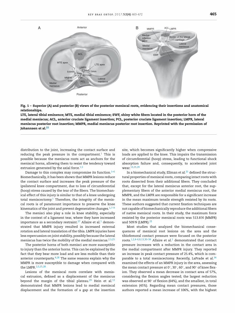
The additional fibers of the MMPR were first described byAnderson et al.24 as shiny white fibers (SWF). These fibersincrease the attachment area of the medial meniscus tothe posterior portion of the plateau. In a quantitative study,Johannsen et al.22 demonstrated that the area of the denserMMPR fiber insertion was on average 30.4 mm2, correspondingto a 6-mm diameter tunnel to reproduce its native attach-ment area. When SWFs were included in the measurement,the insertion area increased to 77.7 mm2. In the same study,the authors determined the distances from the center ofthe MMPR to the main arthroscopic frames. The MMPR waslocated 9.6 mm posterior and 0.7 mm lateral to the apex ofthe medial tibial eminence (the most reproducible landmark);3.5 mm lateral to the inflection point of the medial tibialplateau articular cartilage; and 8.2 mm anterior to the mostsuperior tibial attachment of the posterior cruciate ligament(PCL; Fig. 1).
Lateral meniscus posterior root (LMPR)
The attachment area of the main fibers of the LMPR measures39.2 mm2, corresponding to a 7-mm diameter tunnel to repro-duce its original attachment area.22 This area does not includethe additional fibers, which extend to the lateral edge of themedial tibial eminence. Some authors have reported an LMPRarea of 115 mm2.25 This discrepancy in measurement may berelated to the inclusion of the additional fibers as part of theroot. Johannsen et al.22 demonstrated that the center of LMPRis located 4.2 mm medial and 1.5 mm posterior to the lateraltibial eminence; 4.3 mm medial to articular cartilage marginof the lateral tibial plateau; 12.7 mm anterior to the superioraspect of the PCL tibial insertion; 10.1 mm posterior to theposteromedial corner of the anterior root attachment of thelateral meniscus; and 10.8 mm posterior to the posteromedialACL band.22
An important consideration regarding the LMPR anatomy isthe presence of the meniscofemoral ligaments (MFLs), whichattach the LMPR to the medial femoral condyle.26 Theseligaments play an important role in stabilizing the lateralmeniscus and preventing or reducing extrusion in cases ofinjury to this root.26
Biomechanics
Approximately 50–70% of the load transmitted throughthe knee is supported by the medial and lateral menisci,respectively.12 The menisci are able to convert axial load intocircumferential (hoop) stress; they aid in a uniform weight
r e v b r a s o r t o p . 2 0 1 7;5 2(4):463–472 465
Anterio rA B
ACL
MTE apexLTE apex
Medialmeniscus
Medialinflexionpoint
MMPR ACL LMPR
Lateralmeniscus
Border of the lateral cartilage
SWF
PCL
9.6mm
MMPRSWF PCL
Lateralmeniscus
Posterio r
Medial meniscus
LMPR4.2mm
8.2mm
1.5mm0.7mm
10.1mm
Fig. 1 – Superior (A) and posterior (B) views of the posterior meniscal roots, evidencing their insertions and anatomicalrelationships.LTE, lateral tibial eminence; MTE, medial tibial eminence; SWF, shiny white fibers located in the posterior horn of themedial meniscus; ACL, anterior cruciate ligament insertion; PCL, posterior cruciate ligament insertion; LMPR, lateralmeniscus posterior root insertion; MMPR, medial meniscus posterior root insertion. Reprinted with the permission ofJohannsen et al.22
drpme
Bti(itck
iisrlm
tfaMt
cbdd
istribution to the joint, increasing the contact surface andeducing the peak pressure in the compartment.1 This isossible because the meniscus roots act as anchors for theeniscal horns, allowing them to resist the tendency toward
xtrusion generated by the axial force.1,5
Damage to this complex may compromise its function.1–6
iomechanically, it has been shown that MMPR lesions reducehe contact surface and increases the peak pressure of thepsilateral knee compartment, due to loss of circumferentialhoop) stress caused by the tear of the fibers. The biomechan-cal effect of this injury is similar to that of a knee undergoingotal meniscectomy.1 Therefore, the integrity of the menis-al roots is of paramount importance to preserve the kneeinematics of the joint and prevent degenerative changes.1,7–9
The menisci also play a role in knee stability, especiallyn the context of a ligament tear, where they have increasedmportance as a secondary restraint.27 Allaire et al.1 demon-trated that MMPR injury resulted in increased externalotation and lateral translation of the tibia. LMPR injuries haveess repercussion on joint stability, possibly because the lateral
eniscus has twice the mobility of the medial meniscus.2,5,25
The posterior horns of both menisci are more susceptibleo injury than the anterior horns. This can be explained by theact that they bear more load and are less mobile than theirnterior counterparts.4,28 The same reasons explain why theMPR is more susceptible to damage when compared with
he LMPR.2,5,25,28
Lesions of the meniscal roots correlate with menis-al extrusion, defined as a displacement of the meniscus
eyond the margin of the tibial plateau.29 Hein et al.13emonstrated that MMPR lesions lead to medial meniscalisplacement and the formation of a gap at the insertion
site, which becomes significantly higher when compressiveloads are applied to the knee. This impairs the transmissionof circumferential (hoop) stress, leading to functional shockabsorption failure and, consequently, to accelerated jointwear.13,25,29
In a biomechanical study, Ellman et al.21 defined the struc-tural properties of meniscal roots, comparing intact roots withroots dissected from their additional fibers. They concludedthat, except for the lateral meniscus anterior root, the sup-plementary fibers of the anterior medial meniscus root, theMMPR, and the LMPR are responsible for a significant increasein the mean maximum tensile strength resisted by its roots.These authors suggested that current fixation techniques arenot capable of biomechanically reproduce the additional fibersof native meniscal roots. In their study, the maximum forceresisted by the posterior meniscal roots was 513.8 N (MMPR)and 509 N (LMPR).25
Most studies that analyzed the biomechanical conse-quences of meniscal root lesions on the area and thetibiofemoral contact pressure were focused on the posteriorroots.1,2,4–6,8,13,30–34 Allaire et al.1 demonstrated that contactpressure increases with a reduction in the contact area inthe medial compartment after MMPR injury. They reportedan increase in peak contact pressure of 25.4%, which is com-parable to a total meniscectomy. Recently, LaPrade et al.34
examined the effects of an MMPR injury in the area, assessingthe mean contact pressure at 0◦, 30◦, 60◦, and 90◦ of knee flex-ion. They observed a mean decrease in contact area of 57%,considering the flexion angles tested; the largest reduction
was observed at 90◦ of flexion (64%), and the smallest, in totalextension (45%). Regarding mean contact pressures, thoseauthors reported a mean increase of 106%, with the highest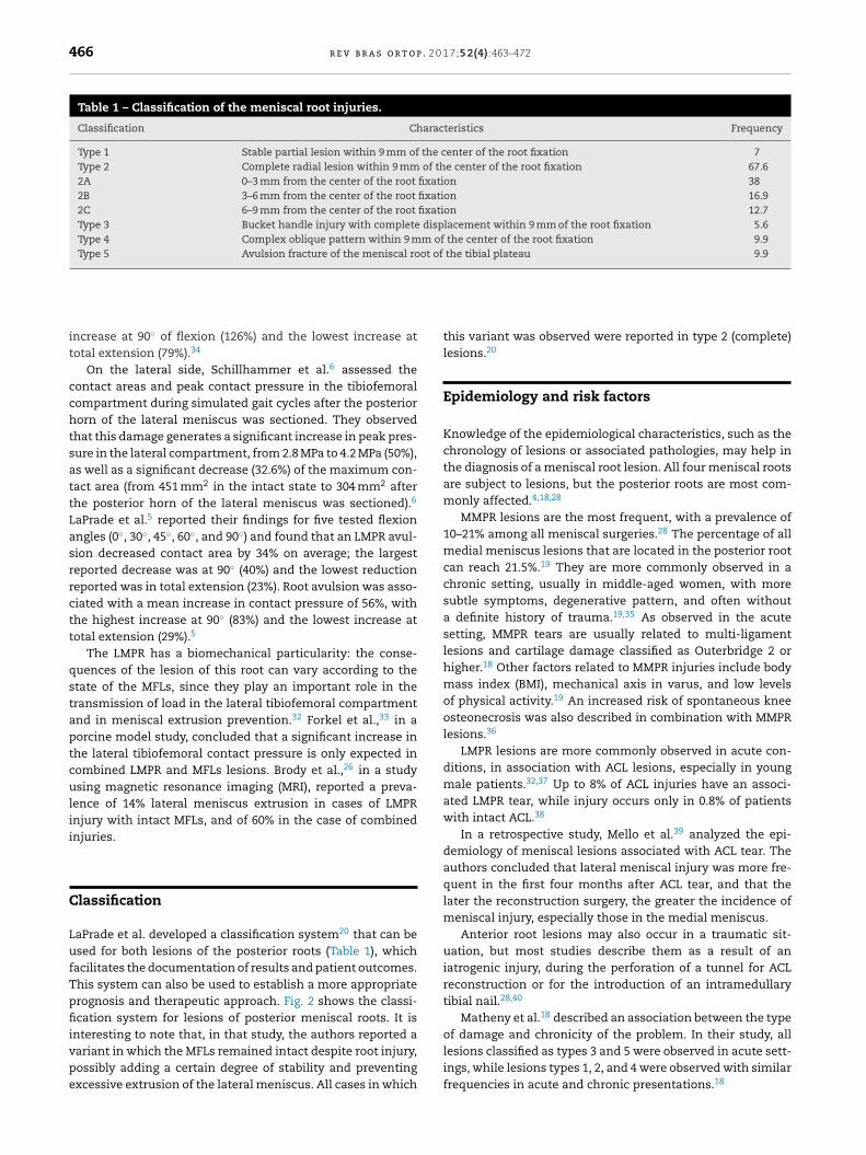
466 r e v b r a s o r t o p . 2 0 1 7;5 2(4):463–472
Table 1 – Classification of the meniscal root injuries.
Classification Characteristics Frequency
Type 1 Stable partial lesion within 9 mm of the center of the root fixation 7Type 2 Complete radial lesion within 9 mm of the center of the root fixation 67.62A 0–3 mm from the center of the root fixation 382B 3–6 mm from the center of the root fixation 16.92C 6–9 mm from the center of the root fixation 12.7Type 3 Bucket handle injury with complete displacement within 9 mm of the root fixation 5.6Type 4 Complex oblique pattern within 9 mm of the center of the root fixation 9.9Type 5 Avulsion fracture of the meniscal root of the tibial plateau 9.9
increase at 90◦ of flexion (126%) and the lowest increase attotal extension (79%).34
On the lateral side, Schillhammer et al.6 assessed thecontact areas and peak contact pressure in the tibiofemoralcompartment during simulated gait cycles after the posteriorhorn of the lateral meniscus was sectioned. They observedthat this damage generates a significant increase in peak pres-sure in the lateral compartment, from 2.8 MPa to 4.2 MPa (50%),as well as a significant decrease (32.6%) of the maximum con-tact area (from 451 mm2 in the intact state to 304 mm2 afterthe posterior horn of the lateral meniscus was sectioned).6
LaPrade et al.5 reported their findings for five tested flexionangles (0◦, 30◦, 45◦, 60◦, and 90◦) and found that an LMPR avul-sion decreased contact area by 34% on average; the largestreported decrease was at 90◦ (40%) and the lowest reductionreported was in total extension (23%). Root avulsion was asso-ciated with a mean increase in contact pressure of 56%, withthe highest increase at 90◦ (83%) and the lowest increase attotal extension (29%).5
The LMPR has a biomechanical particularity: the conse-quences of the lesion of this root can vary according to thestate of the MFLs, since they play an important role in thetransmission of load in the lateral tibiofemoral compartmentand in meniscal extrusion prevention.32 Forkel et al.,33 in aporcine model study, concluded that a significant increase inthe lateral tibiofemoral contact pressure is only expected incombined LMPR and MFLs lesions. Brody et al.,26 in a studyusing magnetic resonance imaging (MRI), reported a preva-lence of 14% lateral meniscus extrusion in cases of LMPRinjury with intact MFLs, and of 60% in the case of combinedinjuries.
Classification
LaPrade et al. developed a classification system20 that can beused for both lesions of the posterior roots (Table 1), whichfacilitates the documentation of results and patient outcomes.This system can also be used to establish a more appropriateprognosis and therapeutic approach. Fig. 2 shows the classi-fication system for lesions of posterior meniscal roots. It is
interesting to note that, in that study, the authors reported avariant in which the MFLs remained intact despite root injury,possibly adding a certain degree of stability and preventingexcessive extrusion of the lateral meniscus. All cases in whichthis variant was observed were reported in type 2 (complete)lesions.20
Epidemiology and risk factors
Knowledge of the epidemiological characteristics, such as thechronology of lesions or associated pathologies, may help inthe diagnosis of a meniscal root lesion. All four meniscal rootsare subject to lesions, but the posterior roots are most com-monly affected.4,18,28
MMPR lesions are the most frequent, with a prevalence of10–21% among all meniscal surgeries.28 The percentage of allmedial meniscus lesions that are located in the posterior rootcan reach 21.5%.19 They are more commonly observed in achronic setting, usually in middle-aged women, with moresubtle symptoms, degenerative pattern, and often withouta definite history of trauma.19,35 As observed in the acutesetting, MMPR tears are usually related to multi-ligamentlesions and cartilage damage classified as Outerbridge 2 orhigher.18 Other factors related to MMPR injuries include bodymass index (BMI), mechanical axis in varus, and low levelsof physical activity.19 An increased risk of spontaneous kneeosteonecrosis was also described in combination with MMPRlesions.36
LMPR lesions are more commonly observed in acute con-ditions, in association with ACL lesions, especially in youngmale patients.32,37 Up to 8% of ACL injuries have an associ-ated LMPR tear, while injury occurs only in 0.8% of patientswith intact ACL.38
In a retrospective study, Mello et al.39 analyzed the epi-demiology of meniscal lesions associated with ACL tear. Theauthors concluded that lateral meniscal injury was more fre-quent in the first four months after ACL tear, and that thelater the reconstruction surgery, the greater the incidence ofmeniscal injury, especially those in the medial meniscus.
Anterior root lesions may also occur in a traumatic sit-uation, but most studies describe them as a result of aniatrogenic injury, during the perforation of a tunnel for ACLreconstruction or for the introduction of an intramedullarytibial nail.28,40
Matheny et al.18 described an association between the type
of damage and chronicity of the problem. In their study, alllesions classified as types 3 and 5 were observed in acute sett-ings, while lesions types 1, 2, and 4 were observed with similarfrequencies in acute and chronic presentations.18
r e v b r a s o r t o p . 2 0 1 7;5 2(4):463–472 467
Type 1
Type 4 Type 5
Type 2 Type 3
FC
FCFC
FCFC
Partialroot lesion Complete
root lesion
Oblique root lesion
Bucket-handleinjury
Avulsionfracture
Fig. 2 – Arthroscopic simulation and illustration of the different types of meniscal root lesions classified based onmorphology: stable partial root lesion (type 1); complete radial lesion within 9 mm from the posterior fixation in the plateau(type 2); bucket-handle injury with complete root detachment (type 3); longitudinal or complex oblique lesion with total rootdetachment (type 4); and avulsion fracture of the meniscal root (type 5).FC, femoral condyle.Original photograph and partial reproduction of image, with the permission of LaPrade et al.20
atl
D
Aasa
C
Me(erwiopbi
Behavioral characteristics related to Eastern cultures, suchs kneeling and squatting, may also be associated with pos-erior root lesions41 but this aspect is not well defined in theiterature.28
iagnosis
s the symptomatology of meniscal root lesions is highly vari-ble, diagnosis can be challenging, requiring a high degree ofuspicion as well as knowledge of the associated risk factorsnd commonly related lesions.
linical evaluation
MPR injuries are not necessarily associated with a traumaticvent. Approximately 70% of patients report a routine eventsuch as squatting), while others do not report any specificvent.25,42 The most common symptoms of posterior meniscaloot tears are posterior knee pain and pain in the articular line,hich are nonspecific.32 Mechanical symptoms, such as block-
ng, are less common. Joint effusion is observed in only 14.3%
f the cases, and McMurray test is positive in only 57.1% ofatients.43 Seil et al.44 described a test to detect MMPR lesionsased on meniscal extrusion. The maneuver consists in apply-ng a stress in varus, with the knee in full extension, while
palpating the anteromedial joint line. It is considered positivewhen the meniscus extrusion can be palpated and disappearswhen the knee is brought back into normal alignment.
Imaging
Magnetic resonance imaging is the most indicated noninva-sive examination.4,25,38 Nonetheless, some authors postulatethat the only way to confirm a posterior meniscal root injuryis through arthroscopy.32 Up to one-third of adjacent radialMMPR lesions cannot be observed on MRI.9,25 The ability ofMRI to detect a posterior meniscal root tear depends on theimage quality and the ability of the radiologist.25 Generally,T2-weighted sequences are more suitable to assess meniscalroot injury.25 However, a recently-released protocol using fatsuppression (FS 3 D VISTA) has shown better performance inthe diagnosis of these lesions.28
Normally, posterior meniscal roots can be visualized intwo consecutive coronal images as a fibrocartilage band thatanchors the posterior horn of the meniscus to the tibialplateau.25 Ideally, coronal, sagittal, and axial images shouldbe assessed.38
In cases of posterior meniscal root injury, the two mostcommonly observed radiological signs in MRI studies aremeniscal extrusion and the ghost sign,4,25,29 which can be seenin Fig. 3.
468 r e v b r a s o r t o p . 2 0 1 7;5 2(4):463–472
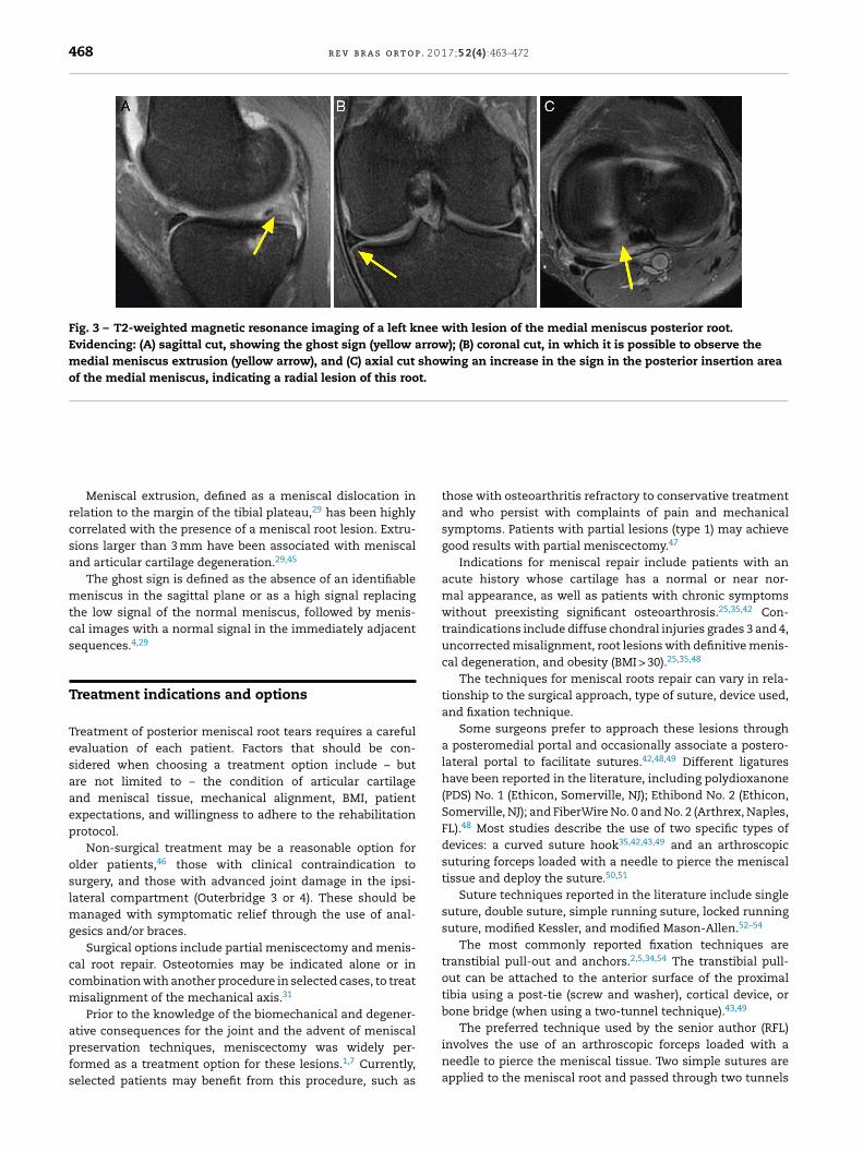
Fig. 3 – T2-weighted magnetic resonance imaging of a left knee with lesion of the medial meniscus posterior root.Evidencing: (A) sagittal cut, showing the ghost sign (yellow arrow); (B) coronal cut, in which it is possible to observe themedial meniscus extrusion (yellow arrow), and (C) axial cut showing an increase in the sign in the posterior insertion areaof the medial meniscus, indicating a radial lesion of this root.
Meniscal extrusion, defined as a meniscal dislocation inrelation to the margin of the tibial plateau,29 has been highlycorrelated with the presence of a meniscal root lesion. Extru-sions larger than 3 mm have been associated with meniscaland articular cartilage degeneration.29,45
The ghost sign is defined as the absence of an identifiablemeniscus in the sagittal plane or as a high signal replacingthe low signal of the normal meniscus, followed by menis-cal images with a normal signal in the immediately adjacentsequences.4,29
Treatment indications and options
Treatment of posterior meniscal root tears requires a carefulevaluation of each patient. Factors that should be con-sidered when choosing a treatment option include – butare not limited to – the condition of articular cartilageand meniscal tissue, mechanical alignment, BMI, patientexpectations, and willingness to adhere to the rehabilitationprotocol.
Non-surgical treatment may be a reasonable option forolder patients,46 those with clinical contraindication tosurgery, and those with advanced joint damage in the ipsi-lateral compartment (Outerbridge 3 or 4). These should bemanaged with symptomatic relief through the use of anal-gesics and/or braces.
Surgical options include partial meniscectomy and menis-cal root repair. Osteotomies may be indicated alone or incombination with another procedure in selected cases, to treatmisalignment of the mechanical axis.31
Prior to the knowledge of the biomechanical and degener-
ative consequences for the joint and the advent of meniscalpreservation techniques, meniscectomy was widely per-formed as a treatment option for these lesions.1,7 Currently,selected patients may benefit from this procedure, such asthose with osteoarthritis refractory to conservative treatmentand who persist with complaints of pain and mechanicalsymptoms. Patients with partial lesions (type 1) may achievegood results with partial meniscectomy.47
Indications for meniscal repair include patients with anacute history whose cartilage has a normal or near nor-mal appearance, as well as patients with chronic symptomswithout preexisting significant osteoarthrosis.25,35,42 Con-traindications include diffuse chondral injuries grades 3 and 4,uncorrected misalignment, root lesions with definitive menis-cal degeneration, and obesity (BMI > 30).25,35,48
The techniques for meniscal roots repair can vary in rela-tionship to the surgical approach, type of suture, device used,and fixation technique.
Some surgeons prefer to approach these lesions througha posteromedial portal and occasionally associate a postero-lateral portal to facilitate sutures.42,48,49 Different ligatureshave been reported in the literature, including polydioxanone(PDS) No. 1 (Ethicon, Somerville, NJ); Ethibond No. 2 (Ethicon,Somerville, NJ); and FiberWire No. 0 and No. 2 (Arthrex, Naples,FL).48 Most studies describe the use of two specific types ofdevices: a curved suture hook35,42,43,49 and an arthroscopicsuturing forceps loaded with a needle to pierce the meniscaltissue and deploy the suture.50,51
Suture techniques reported in the literature include singlesuture, double suture, simple running suture, locked runningsuture, modified Kessler, and modified Mason-Allen.52–54
The most commonly reported fixation techniques aretranstibial pull-out and anchors.2,5,34,54 The transtibial pull-out can be attached to the anterior surface of the proximaltibia using a post-tie (screw and washer), cortical device, orbone bridge (when using a two-tunnel technique).43,49
The preferred technique used by the senior author (RFL)involves the use of an arthroscopic forceps loaded with aneedle to pierce the meniscal tissue. Two simple sutures areapplied to the meniscal root and passed through two tunnels

r e v b r a s o r t o p . 2 0 1 7
Radial lesion 3 mm from the root
Meniscal root remnanttissue
Suture tunnels
Cortical fixationdevice
Fig. 4 – Technique for fixation of lesion of the posteriormeniscal root through a transtibial suture fixated to abutton on the anteromedial aspect of the tibia. Ananatomical positioning of the fixation is necessary torestore the circumferential hoop stress of the meniscus.Reprinted with the permission of Padalecki et al.2
ii
P
Ttaac
P
Bbaabjatla
L
Fa
n the proximal tibia. Attachment to the anterior tibial surfaces made with a cortical device (Fig. 4).
ostoperative rehabilitation
he rehabilitation protocol used by the senior author (RFL)akes into account the anatomy, biomechanics, outcomes,nd clinical judgment; it is divided into different phasesnd its progression is assessed according to the followingriteria.
hase 1
ased on surgical repair and range of motion (ROM). Weightearing is not allowed during the first six weeks, in order tovoid stress in the meniscus. Passive movement is immedi-tely performed, limited to 90◦ flexion for two weeks, followedy complete recovery of passive ROM. The patellofemoraloint, the quadriceps, and the patellar tendon are also immedi-tely mobilized, to prevent scar tissue adhesions. To progresso phase 2, a full ROM (when compared with the contralateralimb) should be achieved, as well as minimal joint effusion,nd normal knee temperature.
evel 2
rom the seventh week onwards, pre-gait activities are initi-ted. Weight bearing of 25% of total body weight is allowed,
;5 2(4):463–472 469
and larger loads are initiated according to patient tolerance.The patient should feel comfortable walking for at least 25 minto progress to phase 3.
Phase 3
Exercises based on a greater number of repetitions and shortrest periods are conducted to promote muscular endurance.Exercises with both legs progress to single leg exercises. Squatsare progressed up to 70◦, according to tolerance. At week 12,stationary bike, freestyle swimming, and treadmill walking areallowed.
Phase 4
The quadriceps is strengthened to achieve at least 80% of theforce of the unaffected limb. Multiplanar exercises are initi-ated to allow neuromuscular control.
Phase 5
This phase varies according to patient demand. At week 22,patients are encouraged to return to running, agility, andchange of direction activities. Total return to sports is autho-rized on a case-by-case status, based on the patient’s ability.55
Assessment of outcomes
The comparison of clinical outcomes between the differenttechniques used to treat meniscal root lesions has conflictingresults, as most publications present small samples, differ-ent inclusion criteria, lack of control group, and a low level ofevidence.
Partial meniscectomy
Widely used in the past as the primary form of treatment ofthese lesions, the results of meniscectomy in the literatureare conflicting. Ozkoc et al.9 analyzed 70 partial meniscec-tomies in a sample in which approximately 80% of patientswere aged over 50 years and were obese. The Lysholm score56
improved from 53 to 67 points. However, despite patientsatisfaction, the control radiographs evidenced arthrosisprogression.9
In the evaluation of 46 cases of partial meniscectomy witha minimum follow-up of five years, Han et al.8 found radio-graphic signs of degenerative alterations in 16 patients, andonly 56% reported improvement in pain, despite the fact thatthe Lysholm score had improved significantly.
Repair
Lee et al.43 analyzed the short-term clinical and radio-graphic results of 21 MMPR lesions treated using transtibial
pull-out technique, and observed a significant improvementin both the Lysholm score and the HSS scale. Progressionof the degenerative alterations was observed in only oneof the knees; in all ten knees in which an arthroscopic
p . 2 0
r
1
1
1
1
1
1
1
1
1
1
470 r e v b r a s o r t o
revision was made, complete healing of the lesion wasobserved.43
Regarding the cure rates of the pull-out technique, Choet al.49 found four completely healed lesions and eight par-tially healed injuries in 13 knees submitted to arthroscopicrevision. The Lysholm score increased from 34.7 to 75.6, andthe HSS score improved from 33.5 to 82.2.49
Chung et al.30 conducted a meta-analysis to assess theclinical outcomes of MMPR injury repair and observed a signif-icant improvement on the Lysholm scale; however, meniscalextrusion did not improve, and osteoarthrosis progression wasinevitable.
In a recent systematic review, all seven studies – including172 patients treated with transtibial pull-out – demon-strated functional improvement after the procedure. Amongthose who underwent an arthroscopic review, 48% presentedcomplete healing, 42% partial healing, and 10% treatmentfailure. Radiological aggravation of at least one stage in theKellgren–Lawrence classification was observed in 16% of the76 patients evaluated for osteoarthrosis progression.48
When comparing the fixation of meniscal root throughanchors vs. the pull-out technique, Kim et al.42 found no sig-nificant differences in the IKDC,57,58 Lysholm, and HSS scores,as well as in the degree of articular degeneration between thetwo groups.
Comparison between partial meniscectomy and repairtechniques
The highest Lysholm and IKDC scores and lower rates of pro-gression of degenerative changes were observed in patientswith MMPR lesions treated with transtibial pull-out type whencompared with those who underwent partial meniscectomy.35
Similar results were demonstrated by Chung et al.31: of 20patients who underwent meniscectomy, seven required totalknee arthroplasty, which was not required in the 37 patientstreated with root fixation.
Final considerations
Studies suggest that patients undergoing posterior meniscalroot repair, whether using anchors or the transtibial pull-outtechnique, present functional and clinical benefits when com-pared with those who underwent meniscectomy. However,more studies with a high level of evidence are needed to estab-lish the relationship between the use of these techniques andthe rate of progression to osteoarthritis in the long term.
Conflicts of interest
Dr. Robert F. LaPrade is a technical consultant for Arthrex,Össur, and Smith & Nephew. The other authors declare noconflicts of interest.
e f e r e n c e s
1. Allaire R, Muriuki M, Gilbertson L, Harner CD.Biomechanical consequences of a tear of the posterior
2
1 7;5 2(4):463–472
root of the medial meniscus. Similar to totalmeniscectomy. J Bone Jt Surg Am. 2008;90(9):1922–31.
2. Padalecki JR, Jansson KS, Smith SD, Dornan GJ, Pierce CM,Wijdicks CA, et al. Biomechanical consequences of acomplete radial tear adjacent to the medial meniscusposterior root attachment site: in situ pull-out repairrestores derangement of joint mechanics. Am J SportsMed. 2014;42(3):699–707.
3. Marzo JM, Gurske-DePerio J. Effects of medial meniscusposterior horn avulsion and repair on tibiofemoralcontact area and peak contact pressure with clinicalimplications. Am J Sports Med. 2009;37(1):124–9.
4. Papalia R, Vasta S, Franceschi F, D’Adamio S, Maffulli N,Denaro V. Meniscal root tears: from basic science toultimate surgery. Br Med Bull. 2013;106:91–115.
5. LaPrade CM, Jansson KS, Dornan G, Smith SD, WijdicksCA, LaPrade RF. Altered tibiofemoral contact mechanicsdue to lateral meniscus posterior horn root avulsions andradial tears can be restored with in situ pull-out suturerepairs. J Bone Jt Surg Am. 2014;96(6):471–9.
6. Schillhammer CK, Werner FW, Scuderi MG, Cannizzaro JP.Repair of lateral meniscus posterior horn detachmentlesions: a biomechanical evaluation. Am J Sports Med.2012;40(11):2604–9.
7. McDermott ID, Amis AA. The consequences ofmeniscectomy. J Bone Jt Surg Br. 2006;88(12):1549–56.
8. Han SB, Shetty GM, Lee DH, Chae DJ, Seo SS, Wang KH,et al. Unfavorable results of partial meniscectomy forcomplete posterior medial meniscus root tear with earlyosteoarthritis: a 5- to 8-year follow-up study. Arthroscopy.2010;26(10):1326–32.
9. Ozkoc G, Circi E, Gonc U, Irgit K, Pourbagher A, TandoganRN. Radial tears in the root of the posterior horn of themedial meniscus. Knee Surg Sports Traumatol Arthrosc.2008;16(9):849–54.
0. Pagnani MJ, Cooper DE, Warren RF. Extrusion of themedial meniscus. Arthroscopy. 1991;7(3):297–300.
1. Hede A, Larsen E, Sandberg H. The long term outcome ofopen total and partial meniscectomy related to thequantity and site of the meniscus removed. Int Orthop.1992;16(2):122–5.
2. Seedhom BB, Dowson D, Proceedings Wright V. Functionsof the menisci. A preliminary study. Ann Rheum Dis.1974;33(1):111.
3. Hein CN, Deperio JG, Ehrensberger MT, Marzo JM. Effectsof medial meniscal posterior horn avulsion and repair onmeniscal displacement. Knee. 2011;18(3):189–92.
4. Zimny ML, Albright DJ, Dabezies E. Mechanoreceptors inthe human medial meniscus. Acta Anat.1988;133(1):35–40.
5. Markolf KL, Mensch JS, Amstutz HC. Stiffness and laxityof the knee – the contributions of the supportingstructures. A quantitative in vitro study. J Bone Jt SurgAm. 1976;58(5):583–94.
6. Mac CM. The movements of bones and joints; thesynovial fluid and its assistants. J Bone Jt Surg Br.1950;32-b(2):244–52.
7. Renstrom P, Johnson RJ. Anatomy and biomechanics ofthe menisci. Clin Sports Med. 1990;9(3):523–38.
8. Matheny LM, Ockuly AC, Steadman JR, LaPrade RF.Posterior meniscus root tears: associated pathologies toassist as diagnostic tools. Knee Surg Sports TraumatolArthrosc. 2015;23(10):3127–31.
9. Hwang BY, Kim SJ, Lee SW, Lee HE, Lee CK, Hunter DJ,et al. Risk factors for medial meniscus posterior root tear.
Am J Sports Med. 2012;40(7):1606–10.0. LaPrade CM, James EW, Cram TR, Feagin JA, EngebretsenL, LaPrade RF. Meniscal root tears: a classification system

0 1 7
2
2
2
2
2
2
2
2
2
3
3
3
3
3
3
3
3
3
3
4
4
4
4
4
4
4
4
4
4
5
5
5
5
5
5
r e v b r a s o r t o p . 2
based on tear morphology. Am J Sports Med.2015;43(2):363–9.
1. Ellman MB, LaPrade CM, Smith SD, Rasmussen MT,Engebretsen L, Wijdicks CA, et al. Structural properties ofthe meniscal roots. Am J Sports Med. 2014;42(8):1881–7.
2. Johannsen AM, Civitarese DM, Padalecki JR, GoldsmithMT, Wijdicks CA, LaPrade RF. Qualitative and quantitativeanatomic analysis of the posterior root attachments ofthe medial and lateral menisci. Am J Sports Med.2012;40(10):2342–7.
3. Messner K, Gao J. The menisci of the knee joint.Anatomical and functional characteristics, and arationale for clinical treatment. J Anat. 1998;193 Pt2:161–78.
4. Anderson CJ, Ziegler CG, Wijdicks CA, Engebretsen L,LaPrade RF. Arthroscopically pertinent anatomy of theanterolateral and posteromedial bundles of the posteriorcruciate ligament. J Bone Jt Surg Am. 2012;94(21):1936–45.
5. Bhatia S, LaPrade CM, Ellman MB, LaPrade RF. Meniscalroot tears: significance, diagnosis, and treatment. Am JSports Med. 2014;42(12):3016–30.
6. Brody JM, Lin HM, Hulstyn MJ, Tung GA. Lateral meniscusroot tear and meniscus extrusion with anterior cruciateligament tear. Radiology. 2006;239(3):805–10.
7. Shybut TB, Vega CE, Haddad J, Alexander JW, Gold JE,Noble PC, et al. Effect of lateral meniscal root tear on thestability of the anterior cruciate ligament-deficient knee.Am J Sports Med. 2015;43(4):905–11.
8. Bonasia DE, Pellegrino P, D’Amelio A, Cottino U, Rossi R.Meniscal root tear repair: why, when and how? OrthopRev. 2015;7(2):5792.
9. Lerer DB, Umans HR, Hu MX, Jones MH. The role ofmeniscal root pathology and radial meniscal tear inmedial meniscal extrusion. Skelet Radiol.2004;33(10):569–74.
0. Chung KS, Ha JK, Ra HJ, Kim JG. A meta-analysis ofclinical and radiographic outcomes of posterior hornmedial meniscus root repairs. Knee Surg SportsTraumatol Arthrosc. 2016;24(5):1455–68.
1. Chung KS, Ha JK, Yeom CH, Ra HJ, Jang HS, Choi SH, et al.Comparison of clinical and radiologic results betweenpartial meniscectomy and refixation of medial meniscusposterior root tears: a minimum 5-year follow-up.Arthroscopy. 2015;31(10):1941–50.
2. Feucht MJ, Salzmann GM, Bode G, Pestka JM, Kuhle J,Sudkamp NP, et al. Posterior root tears of the lateralmeniscus. Knee Surg Sports Traumatol Arthrosc.2015;23(1):119–25.
3. Forkel P, Herbort M, Schulze M, Rosenbaum D, Kirstein L,Raschke M, et al. Biomechanical consequences of aposterior root tear of the lateral meniscus: stabilizingeffect of the meniscofemoral ligament. Arch OrthopTrauma Surg. 2013;133(5):621–6.
4. LaPrade CM, Foad A, Smith SD, Turnbull TL, Dornan GJ,Engebretsen L, et al. Biomechanical consequences of anonanatomic posterior medial meniscal root repair. Am JSports Med. 2015;43(4):912–20.
5. Kim SB, Ha JK, Lee SW, Kim DW, Shim JC, Kim JG, et al.Medial meniscus root tear refixation: comparison ofclinical, radiologic, and arthroscopic findings with medialmeniscectomy. Arthroscopy. 2011;27(3):346–54.
6. Robertson DD, Armfield DR, Towers JD, Irrgang JJ, MaloneyWJ, Harner CD. Meniscal root injury and spontaneousosteonecrosis of the knee: an observation. J Bone Jt Surg
Br. 2009;91(2):190–5.7. Anderson L, Watts M, Shapter O, Logan M, Risebury M,Duffy D, et al. Repair of radial tears and posterior horn
;5 2(4):463–472 471
detachments of the lateral meniscus: minimum 2-yearfollow-up. Arthroscopy. 2010;26(12):1625–32.
8. De Smet AA, Blankenbaker DG, Kijowski R, Graf BK,Shinki K. MR diagnosis of posterior root tears of thelateral meniscus using arthroscopy as the referencestandard. Am J Roentgenol. 2009;192(2):480–6.
9. Mello WAJ, Penteado PCF, Marchetto A, Fatarelli IFC,Rodrigues RL, Cerqueira PH. História das lesões meniscaisna reconstrucão do ligamento cruzado anterior. Rev BrasOrtop. 1999;34(11):569–74.
0. Ellman MB, James EW, LaPrade CM, LaPrade RF. Anteriormeniscus root avulsion following intramedullary nailingfor a tibial shaft fracture. Knee Surg Sports TraumatolArthrosc. 2015;23(4):1188–91.
1. Bin SI, Kim JM, Shin SJ. Radial tears of the posterior hornof the medial meniscus. Arthroscopy. 2004;20(4):373–8.
2. Kim JH, Chung JH, Lee DH, Lee YS, Kim JR, Ryu KJ.Arthroscopic suture anchor repair versus pullout suturerepair in posterior root tear of the medial meniscus: aprospective comparison study. Arthroscopy.2011;27(12):1644–53.
3. Lee JH, Lim YJ, Kim KB, Kim KH, Song JH. Arthroscopicpullout suture repair of posterior root tear of the medialmeniscus: radiographic and clinical results with a 2-yearfollow-up. Arthroscopy. 2009;25(9):951–8.
4. Seil R, Duck K, Pape D. A clinical sign to detect rootavulsions of the posterior horn of the medial meniscus.Knee Surg Sports Traumatol Arthrosc. 2011;19(12):2072–5.
5. Costa CR, Morrison WB, Carrino JA. Medial meniscusextrusion on knee MRI: is extent associated with severityof degeneration or type of tear? Am J Roentgenol.2004;183(1):17–23.
6. Camanho GL. Dor aguda no joelho do paciente idoso. RevBras Ortop. 2008;43(9):361–6.
7. Mordecai SC, Al-Hadithy N, Ware HE, Gupte CM.Treatment of meniscal tears: an evidence basedapproach. World J Orthop. 2014;5(3):233–41.
8. Feucht MJ, Kuhle J, Bode G, Mehl J, Schmal H, SudkampNP, et al. Arthroscopic transtibial pullout repair forposterior medial meniscus root tears: a systematic reviewof clinical, radiographic, and second-look arthroscopicresults. Arthroscopy. 2015;31(9):1808–16.
9. Cho JH, Song JG. Second-look arthroscopic assessmentand clinical results of modified pull-out suture forposterior root tear of the medial meniscus. Knee SurgRelat Res. 2014;26(2):106–13.
0. Anz AW, Branch EA, Saliman JD. Biomechanicalcomparison of arthroscopic repair constructs for meniscalroot tears. Am J Sports Med. 2014;42(11):2699–706.
1. Blackman AJ, Stuart MJ, Levy BA, McCarthy MA, Krych AJ.Arthroscopic meniscal root repair using a ceterixnovostitch suture passer. Arthrosc Tech. 2014;3(5):e643–6.
2. Mitchell R, Pitts R, Kim YM, Matava MJ. Medial meniscalroot avulsion: a biomechanical comparison of 4 differentrepair constructs. Arthroscopy. 2016;32(1):111–9.
3. Kopf S, Colvin AC, Muriuki M, Zhang X, Harner CD.Meniscal root suturing techniques: implications for rootfixation. Am J Sports Med. 2011;39(10):2141–6.
4. Feucht MJ, Grande E, Brunhuber J, Burgkart R, Imhoff AB,Braun S. Biomechanical evaluation of different suturetechniques for arthroscopic transtibial pull-out repair ofposterior medial meniscus root tears. Am J Sports Med.2013;41(12):2784–90.
5. Mueller BT, Moulton SG, O’Brien L, LaPrade RF.
Rehabilitation following meniscal root repair: a clinicalcommentary. J Orthop Sports Phys Ther.2016;46(2):104–13.
p . 2 0
5
5
58. Metsavaht L, Leporace G, Sposito MM, Riberto M, BatistaLA. Qual o melhor questionário para avaliar os aspectos
472 r e v b r a s o r t o
6. Peccin MS, Ciconelli R, Cohen M. Questionário específicopara sintomas do joelho Lysholm Knee Scoring Scale –Traducão e validacão para a língua portuguesa. ActaOrtop Bras. 2006;14(5):268–72.
7. Metsavaht L, Leporace G, Riberto M, Sposito MM, DelCastillo LN, Oliveira LP, et al. Translation andcross-cultural adaptation of the lower extremityfunctional scale into a Brazilian Portuguese version and
1 7;5 2(4):463–472
validation on patients with knee injuries. J Orthop SportsPhys Ther. 2012;42(11):932–9.
físicos de pacientes com osteoartrite do joelho napopulacão brasileira? Rev Bras Ortop. 2011;43(3):256–61.


![Superior labral anterior posterior lesions of the shoulder ...€¦ · Funk et al[11] reported a rate of 83% among professional rugby players with SLAP lesions following direct contact](https://static.fdocuments.us/doc/165x107/6009827cf9e1ab2cad772445/superior-labral-anterior-posterior-lesions-of-the-shoulder-funk-et-al11-reported.jpg)