Understanding and Managing Cell Culture Contamination
24
John Ryan, Ph.D. Corning Incorporated Life Sciences Acton, MA 01720 Table of Contents Introduction . . . . . . . . . . . . . . . . . . . . . . . . . . . .1 What Are the Major Cell Culture Contaminants? . . . . . . . . . . . . . . . . . . . . . . . . . . 2 What Are the Sources of Biological Contaminants? . . . . . . . . . . . . . . . . . . . . . . . . . 8 How Can Cell Culture Contamination Be Controlled? . . . . . . . . . . . . . . . . . . . . . . . . 11 A Final Warning . . . . . . . . . . . . . . . . . . . . . . . 20 Acknowledgements . . . . . . . . . . . . . . . . . . . . . . 21 References . . . . . . . . . . . . . . . . . . . . . . . . . . . . 21 Cell Culture Protocols and Technical Articles . . . . . . . . . . . . . . . . . . . . . . .22 Introduction No cell culture problem is as universal as that of culture loss due to contamination. All cell culture laboratories and cell culture workers have experi- enced it. Culture contaminants may be biological or chemical, seen or unseen, destructive or seem- ingly benign, but in all cases they adversely affect both the use of your cell cultures and the quality of your research. Contamination problems can be divided into three classes: ◗ Minor annoyances — when up to several plates or flasks are occasionally lost to contamination; ◗ Serious problems — when contamination frequency increases or entire experiments or cell cultures are lost; ◗ Major catastrophes — contaminants are discovered that call into doubt the validity of your past or current work. Understanding and Managing Cell Culture Contamination Technical Bulletin Life Sciences
Transcript of Understanding and Managing Cell Culture Contamination

John Ryan, Ph.D.Corning IncorporatedLife SciencesActon, MA 01720
Table of ContentsIntroduction . . . . . . . . . . . . . . . . . . . . . . . . . . . .1
What Are the Major Cell Culture Contaminants? . . . . . . . . . . . . . . . . . . . . . . . . . . 2
What Are the Sources of BiologicalContaminants? . . . . . . . . . . . . . . . . . . . . . . . . . 8
How Can Cell Culture Contamination Be Controlled? . . . . . . . . . . . . . . . . . . . . . . . . 11
A Final Warning . . . . . . . . . . . . . . . . . . . . . . . 20
Acknowledgements . . . . . . . . . . . . . . . . . . . . . . 21
References . . . . . . . . . . . . . . . . . . . . . . . . . . . . 21
Cell Culture Protocols and Technical Articles . . . . . . . . . . . . . . . . . . . . . . .22
IntroductionNo cell culture problem is as universal as that ofculture loss due to contamination. All cell culturelaboratories and cell culture workers have experi-enced it. Culture contaminants may be biologicalor chemical, seen or unseen, destructive or seem-ingly benign, but in all cases they adversely affectboth the use of your cell cultures and the qualityof your research. Contamination problems can bedivided into three classes:
◗ Minor annoyances — when up to several plates or flasks are occasionally lost to contamination;
◗ Serious problems — when contaminationfrequency increases or entire experiments or cell cultures are lost;
◗ Major catastrophes — contaminants are discoveredthat call into doubt the validity of your past orcurrent work.
Understanding and ManagingCell Culture ContaminationTechnical Bulletin
Life Sciences

Table 1. Some Consequences ofContamination◗ Loss of time, money, and effort◗ Adverse effects on the cultures◗ Inaccurate or erroneous experimental results◗ Loss of valuable products◗ Personal embarrassment
The most obvious consequence of cellculture contamination is the loss of yourtime, money (for cells, culture vessels,media and sera) and effort spent develop-ing cultures and setting up experiments.However, the less obvious consequencesare often more serious (Table 1). Firstthere are the adverse effects on culturessuffering from undetected chemical orbiological contaminants. These hidden(cryptic) contaminants can achieve highdensities altering the growth and charac-teristics of the cultures. Worse yet are thepotentially inaccurate or erroneous resultsobtained by unknowingly working withthese cryptically contaminated cultures.Products, such as vaccines, drugs ormonoclonal antibodies, manufactured bythese cultures will probably be useless.For some researchers the most seriousconsequence of contamination is sufferingthe embarrassment and damage to theirreputation that results when they notifycollaborators or journals that their exper-imental results are faulty and must beretracted due to contaminants in theircultures.
Preventing all cell culture contaminationhas long been the dream of manyresearchers, but it is an impractical, if notimpossible, dream. Contamination cannotbe totally eliminated, but it can be managedto reduce both its frequency of occurrence andthe seriousness of its consequences. The goalof this bulletin is to review the nature ofcell culture contamination and the prob-lems it causes, and then to explore someof the key concepts and practical strate-gies for managing contamination to pre-vent the loss of valuable cultures andexperiments.
What Are the Major Cell Culture Contaminants? A cell culture contaminant can be definedas some element in the culture systemthat is undesirable because of its possibleadverse effects on either the system or itsuse. These elements can be divided intotwo main categories: chemical contami-nants and biological contaminants.
Chemical Contamination Chemical contamination is best describedas the presence of any nonliving substancethat results in undesirable effects on theculture system. To define further is diffi-cult; even essential nutrients become toxicat high enough concentrations. Nor istoxicity the only concern since hormonesand other growth factors found in serumcan cause changes that, while not nec-essarily harmful to cultures, may beunwanted by researchers using thesystem. (Reviewed in references 1-3.)
MediaThe majority of chemical contaminantsare found in cell culture media and comeeither from the reagents and water usedto make them, or the additives, such assera, used to supplement them. Reagentsshould always be of the highest qualityand purity and must be properly stored toprevent deterioration. Ideally, they shouldbe either certified for cell culture use bytheir manufacturer or evaluated by theresearcher before use. Mistakes in mediapreparation protocols, reading reagentbottle labels, or weighing reagents areother common sources of chemicalcontamination.
Sera Sera used in media have long been asource of both biological and chemicalcontaminants. Due to cell culture-basedscreening programs currently used bygood sera manufacturers, it is unusual tofind a lot of fetal bovine sera that is toxicto a majority of cell cultures. However, itis common to find substantial variationsin the growth promoting abilities ofdifferent lots of sera for particular cell
2
culture systems, especially for culturesthat have specialized or differentiatedcharacteristics. Uncontrollable lot-to-lotvariation in hormone and growth factorconcentrations makes this probleminevitable; careful testing of sera beforepurchase, or switching to serum-freemedia can avoid these problems.
Table 2. Types and Sources of Potential Chemical Contaminants◗ Metal ions, endotoxins, and other impurities
in media, sera, and water◗ Plasticizers in plastic tubing and storage
bottles◗ Free radicals generated in media by the
photoactivation of tryptophan, riboflavin or HEPES exposed to fluorescent light
◗ Deposits on glassware, pipettes, instrumentsetc., left by disinfectants or detergents,antiscaling compounds in autoclave water,residues from aluminum foil or paper
◗ Residues from germicides or pesticides usedto disinfect incubators, equipment, and labs
◗ Impurities in gases used in CO2 incubators
Remember also that serum proteins havethe ability to bind substantial quantitiesof chemical contaminants, especiallyheavy metals, that may have entered theculture system from other sources, ren-dering them less toxic. As a result, switch-ing from serum-containing medium to aserum-free system can unmask these toxicchemical contaminants, exposing the cellsto their adverse effects.
Water The water used for making media andwashing glassware is a frequent source of chemical contamination and requiresspecial care to ensure its quality. Tradi-tionally, double or triple glass distillationwas considered to be the best source ofhigh quality water for cell culture mediaand solutions. Newer purification systemscombining reverse osmosis, ion exchangeand ultrafiltration are capable of removingtrace metals, dissolved organic compoundsand endotoxins and are increasingly pop-ular. However, these systems must beproperly maintained and serviced to ensurecontinued water quality. Because of its
aggressive solvent characteristics, highlypurified water can leach potentially toxicmetal ions from glassware or metal pipes,and plasticizers from plastic storage vesselsor tubing. These contaminants can thenend up in media or deposited on storagevessels and pipettes during washing andrinsing. Water used to generate steam inautoclaves may contain additives to reducescale buildup in pipes; these potentiallytoxic additives can also end up onglassware.
Endotoxins Endotoxins, the lipopolysaccaride-containing by-products of gram negativebacteria, are another source of chemicalcontaminants in cell culture systems. Endo-toxins are commonly found in water, seraand some culture additives (especially thosemanufactured using microbial fermenta-tion) and can be readily quantified usingthe Limulus Amebocyte Lysate assay (LAL).
These highly biologically reactive mole-cules have major influences in vivo onhumoral and cellular systems. Studies ofendotoxins using in vitro systems haveshown that they may affect the growth or performance of cultures and are asignificant source of experimental vari-ability (Reviewed in references 6 and 39).Furthermore, since the use of cell cultureproduced therapeutics, such as hybrido-mas and vaccines, are compromised byhigh endotoxin levels, efforts must bemade to keep endotoxin levels in culturesystems as low as possible.
In the past, sera have been a major sourceof endotoxins in cell cultures. As improvedendotoxin assays (LAL) led to an increasedawareness of the potential cell cultureproblems associated with endotoxins,most manufacturers have significantlyreduced levels in sera by handling the rawproducts under aseptic conditions. Poorlymaintained water systems, especially sys-tems using ion exchange resins, can harborsignificant levels of endotoxin-producingbacteria and may need to be tested ifendotoxin problems are suspected ordiscovered in the cultures.
3

4
Storage Vessels Media stored in glass or plastic bottlesthat have previously contained solutionsof heavy metals or organic compounds,such as electron microscopy stains,solvents and pesticides, can be anothersource of contamination. The contami-nants can be adsorbed onto the surface of the bottle or its cap (or absorbed intothe bottle if plastic) during storage of theoriginal solution. If during the washingprocess they are only partially removed,then once in contact with culture mediathey may slowly leach back into solution.Residues from chemicals used to disinfectglassware, detergents used in washing, or some aluminum foils and wrappingpapers for autoclaving or dry heat steri-lization can also leave potentially toxicdeposits on pipettes, storage bottles andinstruments.
Fluorescent Lights An important but often overlookedsource of chemical contamination resultsfrom the exposure of media containingHEPES (N-[2-hydroxylethyl] piperazine-N'-[2-ethanesulfonic acid]) — an organicbuffer commonly used to supplementbicarbonate-based buffers), riboflavin ortryptophan to normal fluorescent light-ing. These media components can bephotoactivated producing hydrogen per-oxide and free radicals that are toxic tocells; the longer the exposure the greaterthe toxicity (4,5). Short term exposure of media to room or hood lighting whenfeeding cultures is usually not a signifi-cant problem; but leaving media on labbenches for extended periods, storingmedia in walk-in cold rooms with thelights on, or using refrigerators with glassdoors where fluorescent light exposure is more extensive, will lead to a gradualdeterioration in the quality of the media.
Incubators The incubator, often considered a majorsource of biological contamination, canalso be a source of chemical contamination.The gas mixtures (usually containing car-bon dioxide to help regulate media pH)perfused through some incubators maycontain toxic impurities, especially oils orother gases such as carbon monoxide, thatmay have been previously used in the
same storage cylinder or tank. This prob-lem is very rare in medical grade gases,but more common in the less expensiveindustrial grade gas mixtures. Care mustalso be taken when installing new cylin-ders to make sure the correct gas cylinderis used. Other potential chemical contam-inants are the toxic, volatile residues leftbehind after cleaning and disinfectingincubators. Disinfectant odors should notbe detectable in a freshly cleaned incuba-tor when it is placed back into use.
Keep in mind that chemical contaminantstend to be additive in cell culture; smallamounts contributed from several differ-ent sources that are individually nontoxic,when combined together in medium, mayend up overloading the detoxificationcapabilities of the cell culture resulting in toxicity-induced stress effects or evenculture loss.
Biological Contamination Biological contaminants can be subdivid-ed into two groups based on the difficultyof detecting them in cultures:
◗ those that are usually easy to detect —bacteria, molds and yeast;
◗ those that are more difficult to detect,and as a result potentially more seriousculture problems, — viruses, protozoa,insects, mycoplasmas and other celllines.
For a comprehensive review, see references7 and 8.
Ultimately, it is the length of time that aculture contaminant escapes detection thatwill determine the extent of damage it creates in a laboratory or research project.
Bacteria, Molds, and Yeasts Bacteria, molds and yeasts are found vir-tually everywhere and are able to quicklycolonize and flourish in the rich and rela-tively undefended environment providedby cell cultures. Because of their size andfast growth rates, these microbes are themost commonly encountered cell culturecontaminants. In the absence of antibi-otics, microbes can usually be readilydetected in a culture within a few days ofbecoming contaminated, either by directmicroscopic observation. (See Figures 1and 2.) or by the effects they have on the
Figure 1. Photomicrograph of a low level yeast infection in a liver cell line (PLHC-1, ATCC # CRL-2406). Budding yeastcells can been seen in severalareas (arrows). At this lowlevel of contamination, nomedium turbidity would beseen; however, in the absenceof antibiotics, the culturemedium will probably become turbid within a day.
Figure 2. Photomicrograph ofa small fungal colony growingin a cell culture. At this point,this colony would still beinvisible to direct visualobservation. If this culturewas subcultured at this point,all of the cultures or experi-ments set up from it would soon be lost to fungalcontamination.
➞
➞
5
culture (pH shifts, turbidity, and celldestruction). However, when antibioticsare routinely used in culture, resistantorganisms may develop into slow grow-ing, low level infections that are very dif-ficult to detect by direct visual observa-tion. Similar detection problems canoccur with naturally slow growing organ-isms or very small or intracellular bacteriathat are difficult to see during routinemicroscopic culture observation. Thesecryptic contaminants may persist indefi-nitely in cultures causing subtle but sig-nificant alterations in their behavior. Bythe time these cryptic contaminants arediscovered, many experiments and cul-tures may have been compromised.
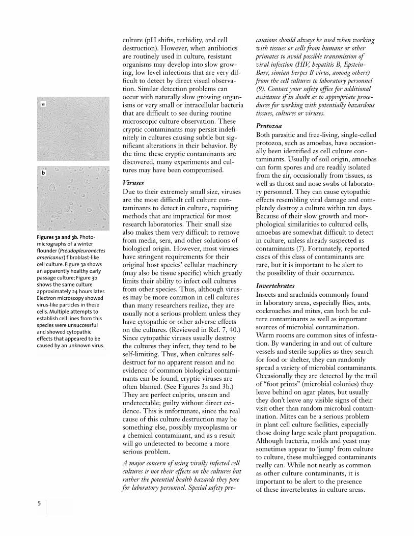
Viruses Due to their extremely small size, virusesare the most difficult cell culture con-taminants to detect in culture, requiringmethods that are impractical for mostresearch laboratories. Their small sizealso makes them very difficult to removefrom media, sera, and other solutions ofbiological origin. However, most viruseshave stringent requirements for theiroriginal host species’ cellular machinery(may also be tissue specific) which greatlylimits their ability to infect cell culturesfrom other species. Thus, although virus-es may be more common in cell culturesthan many researchers realize, they areusually not a serious problem unless theyhave cytopathic or other adverse effectson the cultures. (Reviewed in Ref. 7, 40.)Since cytopathic viruses usually destroythe cultures they infect, they tend to beself-limiting. Thus, when cultures self-destruct for no apparent reason and noevidence of common biological contami-nants can be found, cryptic viruses areoften blamed. (See Figures 3a and 3b.)They are perfect culprits, unseen andundetectable; guilty without direct evi-dence. This is unfortunate, since the realcause of this culture destruction may besomething else, possibly mycoplasma or a chemical contaminant, and as a resultwill go undetected to become a moreserious problem.
A major concern of using virally infected cellcultures is not their effects on the cultures butrather the potential health hazards they posefor laboratory personnel. Special safety pre-
cautions should always be used when workingwith tissues or cells from humans or otherprimates to avoid possible transmission ofviral infection (HIV, hepatitis B, Epstein-Barr, simian herpes B virus, among others)from the cell cultures to laboratory personnel(9). Contact your safety office for additionalassistance if in doubt as to appropriate proce-dures for working with potentially hazardoustissues, cultures or viruses.
Protozoa Both parasitic and free-living, single-celledprotozoa, such as amoebas, have occasion-ally been identified as cell culture con-taminants. Usually of soil origin, amoebascan form spores and are readily isolatedfrom the air, occasionally from tissues, aswell as throat and nose swabs of laborato-ry personnel. They can cause cytopathiceffects resembling viral damage and com-pletely destroy a culture within ten days.Because of their slow growth and mor-phological similarities to cultured cells,amoebas are somewhat difficult to detectin culture, unless already suspected ascontaminants (7). Fortunately, reportedcases of this class of contaminants arerare, but it is important to be alert to the possibility of their occurrence.
Invertebrates Insects and arachnids commonly found in laboratory areas, especially flies, ants,cockroaches and mites, can both be cul-ture contaminants as well as importantsources of microbial contamination.Warm rooms are common sites of infesta-tion. By wandering in and out of culturevessels and sterile supplies as they searchfor food or shelter, they can randomlyspread a variety of microbial contaminants.Occasionally they are detected by the trailof “foot prints” (microbial colonies) theyleave behind on agar plates, but usuallythey don’t leave any visible signs of theirvisit other than random microbial contam-ination. Mites can be a serious problem in plant cell culture facilities, especiallythose doing large scale plant propagation.Although bacteria, molds and yeast maysometimes appear to ‘jump’ from cultureto culture, these multilegged contaminantsreally can. While not nearly as commonas other culture contaminants, it isimportant to be alert to the presence of these invertebrates in culture areas.
Figures 3a and 3b. Photo-micrographs of a winterflounder (Pseudopleuronectesamericanus) fibroblast-like cell culture. Figure 3a showsan apparently healthy earlypassage culture; Figure 3bshows the same cultureapproximately 24 hours later.Electron microscopy showedvirus-like particles in thesecells. Multiple attempts toestablish cell lines from thisspecies were unsuccessful and showed cytopathiceffects that appeared to becaused by an unknown virus.
a
b

Mycoplasmas Mycoplasmas were first detected in cellcultures by Robinson and coworkers in1956. They were attempting to study theeffects of PPLO (pleuropneumonialikeorganisms — the original name for myco-plasma) on HeLa cells when they discov-ered that the control HeLa cultures werealready contaminated by PPLO (10). Inaddition, they discovered that the othercell lines currently in use in their labora-tory were also infected with mycoplasma,a common characteristic of mycoplasmacontamination. Based on mycoplasma testingdone by the FDA, ATCC, and two major cellculture testing companies, at least 11 to 15%of the cell cultures in the United States arecurrently infected by mycoplasmas (Table 3).Since many of these cultures were fromlaboratories that test routinely for myco-plasma, the actual rates are probably higherin the many laboratories that do not testat all (11-13). In Europe, mycoplasmacontamination levels were found to beeven higher: over 25% of 1949 cell cul-tures from the Netherlands and 37% of327 cultures from former Czechoslovakiawere positive (14). The Czechoslovakiastudy had an interesting, but typical find-ing: 100% of the cultures from labs withoutmycoplasma testing programs were contami-nated, but only 2% of the cultures from labsthat tested regularly. Other countries maybe worse: 65% of the cultures in Argentinaand 80% in Japan were reported to becontaminated by mycoplasma in otherstudies (11).
Unfortunately, mycoplasmas are not rela-tively benign culture contaminants buthave the ability to alter their host cul-ture’s cell function, growth, metabolism,morphology, attachment, membranes,virus propagation and yield, interferon
induction and yield, cause chromosomalaberrations and damage, and cytopathiceffects including plaque formation (12).Thus, the validity of any research doneusing these unknowingly infected culturesis questionable at best. (See references 11,12, and 15-18 for good overviews of thisvery serious mycoplasma contaminationproblem.)
What gives mycoplasmas this ability toreadily infect so many cultures? Threebasic characteristics: a) these simple, bac-teria-like microbes are the smallest self-replicating organism known (0.3 to 0.8µm in diameter), b) they lack a cell wall,and c) they are fastidious in their growthrequirements. Their small size and lack ofa cell wall allow mycoplasmas to grow tovery high densities in cell culture (107 to109 colony forming units/mL are com-mon) often without any visible signs ofcontamination — no turbidity, pHchanges or even cytopathic effects. (SeeFigures 4a and 4b.) Even careful micro-scopic observation of live cell culturescannot detect their presence. These sametwo characteristics also make mycoplas-mas, like viruses, very difficult to com-pletely remove from sera by membranefiltration. In addition, their fastidiousgrowth requirements (unfortunately,easily provided for by cell cultures) makethem very difficult to grow and detectusing standard microbiological cultivationmethods. Thus, these three simple char-acteristics, combined with their ability toalter virtually every cellular function andparameter, make mycoplasmas the mostserious, widespread, and devastating c-ulture contaminants.
Mycoplasmas have been described as the“crabgrass” of cell cultures, but this is too
6
Table 3. Mycoplasma Contamination of Cell CulturesNumber of Cultures Tested Number PositiveFood and Drug Administration (FDA) (1970’s to 1990’s) (11)
20,000 cultures tested over 3000 (15%)Bionique Testing Laboratories (several years prior to 1993) (41)
11,000 cultures tested 1218 (11.1%)Microbiological Associates (1985 to 1993) (13)
2,863 cultures tested 370 (12.9%)American Type Culture Collection (ATCC) (1989 to 1994) (42)
5362 752 (14%)
Figures 4a and 4b. Thesescanning electron micro-graphs show 3T6 cells (ATCC #CCL-96) with (4b) and without(4a) mycoplasma infections.The level of contamination ofthese cells by the mycoplasmashown here is typical of con-taminated cells. Examinationof this contaminated cultureby phase contrast microscopydid not show any evidence ofcontamination; nor did themedium show any turbidity.
a
b

benign a description for what are the mostsignificant and widespread cell culturecontaminants in the world. Unfortunately,even with the advances in detectionmethods (discussed in detail later) myc-oplasma infection rates (Table 3) have notchanged noticeably since they were firstdetected in cell cultures. Aggressivemanagement against mycoplasma con-tamination must be the central focus forany cell culture laboratory contaminationor quality control program (16).
Cross-Contamination by Other Cell Cultures With the advent of improved karyotypingmethods in the late 1950’s, it soon becameapparent that some cell lines were cross-contaminated by cells of other species (7).In 1967, isoenzyme analysis was used toshow that 20 commonly used human celllines were intraspecies contaminated byHeLa cells (19,20). Contaminated is actu-ally a misnomer since in fact 100% of theoriginal cells had been replaced by theHeLa contaminant. Unfortunately, thescientific community was slow to respondto this very serious problem. Tests doneat one research center on 246 cell linesover an 18 month period prior to 1976showed that nearly 30% were incorrectlydesignated: 14% were the wrong speciesand 25% of the human cell lines wereHeLa cells (21). A 1981 survey of culturesshowed over 60 cell lines that were actu-ally HeLa cells, 16 other human cell linescontaminated by non-HeLa human celllines, and 12 cases of interspecies con-tamination (See Table 4). Nor is theproblem limited to contamination byHeLa cells. The advent of DNA analysis
has shown that cells from a variety ofsources have contaminated many othercell lines (42).
The seriousness of cross-contamination,while not as common as microbial con-tamination, cannot be overstated. Thevalidity of experimental results fromcultures having inter- or intraspeciescontamination is, at the very least, ques-tionable. Furthermore, their use can leadto the embarrassment of having to retractpublished results. Whenever the invadingcell is better adapted to the culture con-ditions and thus faster growing than theoriginal cells, it will almost always com-pletely replace them. Because of the out-ward physical similarities of different celllines and the wide morphological varia-tions that can be caused by the cultureenvironment, it is impossible to rely onlyon microscopic observation to screen forcross-contamination of cultures. Simpleaccidents are one of the most commonmeans by which other cell lines gainentry into cultures and will be discussedseparately in the next section.
Remember, the seriousness of any culture contaminant is usually directly proportional to the difficulty of detecting it; those that goundetected the longest have the most seriousconsequences. Cultures containing nonlethal(but not harmless), cryptic chemical orbiological contaminants are sometimesused in research for months or even yearsbefore being uncovered; during this timethe quality and validity of all researchdone with those cultures is compromised,as is the reputation of the researchersusing them.
7
Table 4. Some HeLa Contaminated Cell LinesDetroit 6 (CCL-3) Conjunctiva (CCL-20.2)*Minnesota-EE (CCL-4) AV3 (CCL-21)*L132 (CCL 5)* HEp-2 (CCL-23)*Intestine 407 (CCL-6)* J-111 (CCL-24)Chang Liver (CCL-13) WISH (CCL-25)*KB (CCL-17)* Giardia Heart (CCL-27)Detroit 98 (CCL-18) Wilm’s Tumor (CCL-31)NCTC 2544 (CCL-19) FL (CCL- 62)*
CCL# is the ATCC catalog designation. All except CCL-20.2, CCL-31 and CCL-62 were shown to be HeLa byGartler in 1968 (20). Those marked with an asterisk can be found in the Cell Biology Collection on the ATCC web site (www.atcc.org) where they are clearly marked as HeLa contaminants.

What Are the Sources ofBiological Contaminants? To reduce the frequency of biologicalcontamination, it is important to knownot only the nature and identity of thecontaminants but also where they comefrom and how they gain entry into cul-tures. This section will detail some of the most common sources of biologicalcontaminants (3).
Nonsterile Supplies, Media andSolutions Unintentional use of nonsterile supplies,media or solutions during routine cellculture procedures is a major source ofbiological contaminants. These productsmay be contaminated as a result ofimproper sterilization or storage, or may become contaminated during use.
Glassware, including storage bottles and pipettes, is usually sterilized by auto-claving or dry heat sterilization. Seriouscontamination outbreaks are frequentlytraced to improper maintenance or oper-ation of sterilization autoclaves and ovens.Packing too much into an autoclave ordry heat oven will cause uneven heating,resulting in pockets of nonsterile supplies.Using too short a sterilization cycle, espe-cially for autoclaving volumes of liquidsgreater than 500 mL per vessel or solutionscontaining solids or viscous materials,such as agar or starches, is a commonmistake. The size, mass, nature and vol-ume of the materials to be sterilized mustalways be considered and the cycle timeappropriately adjusted to achieve sterility(23). Then, once achieved, sterility mustbe maintained by properly storing thesupplies and solutions in a dust- andinsect-free area to prevent recontamina-tion. Care must also be taken to avoidcondensation on bottles of solutions storedin refrigerators and cold rooms. Of course,
good aseptic technique is also required tomaintain the sterility of properly sterilizedsupplies and solutions once they are in use.
Plastic disposable cell culture vessels,pipettes, centrifuge tubes, etc. are usuallysterilized by their manufacturer using ahigh intensity gamma or electron beamradiation source after they are sealed intheir packaging. This is a very reliableprocess, however care must be taken whenopening and resealing the packaging toavoid contaminating the products within.
Most media, sera and other animal-derived biologicals are not heat steriliz-able and require membrane filtration(Sometimes radiation is also used.) toremove biological contaminants. Productsfilter sterilized in your laboratory shouldalways be tested for sterility before use(discussed in detail later); commerciallyproduced sterile products are tested bythe manufacturer before being sold. Whilefiltration through 0.2 µm membranes isvery effective in removing most biologicalcontaminants, it cannot guarantee thecomplete removal of viruses and myco-plasmas, especially in sera (16,18,24). In an excellent review of the rates andsources of mycoplasma contamination(25), Barile and coworkers reported that104 out of 395 lots (26%) of commercialfetal bovine sera tested were contaminat-ed by mycoplasma. They concluded inthe early 1970’s that animal sera wereamong the major sources of cell culturecontamination by mycoplasma. Many seramanufacturers responded to this problemover the next decade by improving bothfiltration and testing procedures; theycurrently use serial filtration through at least three filter membranes rated at 0.1 µm or smaller to remove myco-plasmas. This approach has been verysuccessful at reducing the problem ofmycoplasma in sera and other animal-
8
Table 5. How Do Biological Contaminants Enter Cultures?◗ Contact with nonsterile supplies, media, or solutions◗ Particulate or aerosol fallout during culture manipulation, transportation, or incubation◗ Swimming, crawling, or growing into culture vessels◗ Accidents and mistakes

derived products (16). While these prod-ucts are no longer a major source ofmycoplasma contamination, they muststill be considered as potential sources tobe evaluated whenever mycoplasmas aredetected in cultures.
Airborne Particles and Aerosols In most laboratories, the greatest sourcesof microbial contamination are airborneparticles and aerosols generated duringculture manipulations. The microbialladen particles are relatively large (gen-erally 4 to 28 µm in diameter) and settleat a rate of approximately one foot perminute in still air. As a result, the air in a sealed, draft-free room or laboratory(no people, open windows or doors, airhandling units, air conditioners, etc.) isvirtually free of biological contaminants.However as soon as people enter theroom, particles that have settled out willbe easily resuspended. In addition certainequipment and activities can generatelarge amounts of microbial laden par-ticulates and aerosols: pipetting devices,vacuum pumps and aspirators, centrifuges,blenders, sonicators, and heat sourcessuch as radiators, ovens, refrigerators and freezers. Animal care facilities andthe animals they house are especiallyserious particle and aerosol generators,and should always be kept as far from the culture area as possible.
McGarrity used a cell culture that wasintentionally infected with mycoplasma as a model to study how mycoplasmas arespread in a laminar flow hood during rou-tine subculturing procedures (26). (Thisreference is especially recommended for abetter understanding of how mycoplasmacan be spread in a lab.) Following tryp-sinization of the infected culture in alaminar flow hood, live mycoplasma wereisolated from the technician, the outsideof the flask, a hemocytometer, the pipet-tor, and the outside of the pipette discardpan. Live mycoplasma could even be suc-cessfully recovered from the surface of thelaminar flow hood four to six days later! Aclean culture, that was subcultured once a week in the same hood following thework with the contaminated cells, testedpositive for mycoplasma after only 6 weeks.
It is easy to understand from this studyhow the entry of a single mycoplasmainfected culture into a laboratory canquickly lead to the infection of all theother cultures in the laboratory. Thisexplains the frequent finding that if oneculture in a laboratory is mycoplasmacontaminated then usually most if not all of the other cultures will be as well.Currently, the major source of mycoplasmacontamination is infected cultures acquiredfrom other research laboratories or commercialsuppliers.
Another major source of particulates andaerosols are laboratory personnel. Streetclothes and dirty lab coats are dust mag-nets. Placing a dust-laden sleeve into alaminar flow hood generates a cloud ofdust particles that can easily fall into andcontaminate cultures during routine pro-cessing. Talking and sneezing can gener-ate significant amounts of aerosols thathave been shown to contain mycoplasma(26). Mouth pipetting is both a source ofmycoplasma contamination and a hazardto personnel and must not be permittedunder any circumstances. Dry, flaky skinis another source of contamination ladenparticles; this common condition isaggravated by the frequent hand washingrequired in the laboratory; even the lotionsdesigned to moisten dry skin have occa-sionally been found to be contaminated.Some laboratory personnel shed yeast-containing particles for several days fol-lowing bread making or beer brewing athome. Attempts by these individuals atcell culturing during this period haveroutinely ended in failure due to yeastcontamination.
Incubators, especially those maintained athigh humidity levels, can be a significantsource of biological contamination in thelaboratory. Dirty water reservoirs, andshelves or culture vessels soiled by spilledmedia, allow the growth of spore-gener-ating fungi. The fans used in many incu-bators to circulate the air and preventtemperature stratification can then spreadthese spores and other particulates. Someincubators humidify incoming gases bybubbling them through the water reser-voirs at the bottom of the incubator; the
9

aerosols generated by this will quicklyspread any contaminants in the water.
While laminar hoods and incubators are the major sites where biological con-tamination occurs, transporting culturesbetween these two sites also providesopportunities for contamination. Mostcell culture laboratories try very hard tokeep their incubators and laminar flowareas clean, but sometimes they overlookthe potential sources of contaminationfound in less clean laboratory areas trans-versed going from one location to theother. Rooms containing open windows,air conditioners, microbiology andmolecular biology work areas, and theother major particle generators discussedabove, add to the potential hazards ofmoving cultures around the laboratory.This problem increases both with thedistance traveled and when the culturevessels are unsealed.
Swimming, Growing, and Crawling into Cultures Unsealed culture plates and dishes, aswell as flasks with loose caps to allow gasexchange, provide another common wayfor contaminants to enter cultures. It isvery easy for the space between the topand bottom sidewalls of a dish, or a flaskand its cap to become wet by capillaryaction with medium or condensation.This thin film of liquid then provides a liquid bridge or highway for micro-organisms to either swim or grow intothe culture vessel.
Even without any detectable film, fungi,as well as other microorganisms, cangrow on the outside of culture vessels(Figures 5a and 5b); eventually theirhyphae grow right up the side wall of thedish or past the cap into the neck of theflask. This is more often observed in longterm cultures (a month or more) main-tained in the same unsealed culture ves-sel. Small insects and other invertebratescan also make temporary visits into un-sealed cultures, especially dishes andplates, leaving behind (unless they fall in and drown) only the contaminantscarried on their feet.
Accidents Accidents are often overlooked as a sig-nificant source of cell culture problems.An accident is defined as “an undesirableor unfortunate happening, unintentional-ly caused and usually resulting in harm,injury, damage or loss” (Webster’sEncyclopedic Unabridged Dictionary,1989). Cell culture-related accidents areone of the leading causes of cross-con-tamination by other cell cultures. Thefollowing actual cases demonstrate howrelatively simple accidents can result inserious cross-contamination problems:
◗ A technician retrieved a vial labeledWI-38 from a liquid nitrogen freezerthinking it contained the widely useddiploid human cell line. Once inculture, it was immediately discoveredto be a plant cell line derived from acommon strain of tobacco calledWisconsin 38, also designated WI-38.
◗ Two separate research laboratories,both attempting to develop cell linesfrom primary cultures, shared a walk-inincubator. One lab used the acronymsHL-1, HL-2, etc. to identify the pri-mary cultures they derived from humanlung. The other lab worked with culturesderived from human liver, but they too(unknowingly) used the identical codingsystem. It wasn’t long before a culturemix up occurred between the twolaboratories.
Fortunately, both of the above accidentalcross-contamination cases, althoughserious, were caught before they causedcatastrophic problems. But how manytimes have similar accidents occurred andnot been caught? Based on continuingreports in the literature (7,8,19-22) manyresearchers have not been lucky enoughto identify their mistakes.
The information presented above isdesigned to provide you with an increasedawareness and understanding of the natureof biological and chemical contamination,and its serious consequences. Theremaining sections will cover some basicideas, techniques and strategies for active-ly detecting and combating cell culturecontamination in your own laboratory.
10
Figures 5a and 5b.Photomicrographs of con-taminants growing on theoutside surfaces of culturevessels. Eventually, theseorganisms may grow into the culture.
a
b

How Can Cell CultureContamination Be Controlled? Cell cultures can be managed to reduceboth the frequency and seriousness ofculture-related problems, especially con-tamination. Lack of basic culture manage-ment procedures, especially in larger lab-oratories, frequently leads to long termproblems, making contamination morelikely for everyone. One solution is toactively manage your cultures to reduceproblems and if necessary set up a pro-gram for use in your laboratory (27,28).This program should be designed to meetthe needs of your specific working condi-tions and be based on the nature of yourpast cell culture problems; it can be verysimple and informal, or more structuredif required.
The first step in managing cultures is todetermine the extent and nature of theculture losses in your lab. Everyone in thelaboratory should keep an accurate recordfor a month or more of all problems, nomatter how minor or insignificant, thatresult in the loss of any cultures. Theseproblems may not only be contaminationrelated but can also be from other causessuch as incubator or equipment failures.Next, review the problems as a group todetermine their nature, seriousness andfrequency. The group’s findings may besurprising: what were thought to be indi-vidual and minor random occurrences ofcontamination often turn out to have apattern and be more extensive than anyindividual realized. This problem sharingis often a painful process, but rememberthe goal is not to place blame but toappreciate the extent and nature of theproblems confronting the laboratory. Acritical part of this process is understand-ing the seriousness and actual costs ofculture loss; placing a dollar value onthese losses is often required before thefull extent of the losses can be appreciat-ed. It is very important for everyone inthe laboratory to know the answers to the following questions:
1. How much time, money and effort have been invested in your cultures and experiments?
2. What are the consequences of their loss?
3. How expensive or difficult will it be to replace them?
Once the nature and consequences of theproblems in the laboratory are betterunderstood, the need for a managementsystem, if necessary, can be determined.Basic problem solving tools (2) can beused to help identify the source of prob-lems; changes to minimize or prevent theproblems from reoccurring can then beimplemented.
The following suggestions, concepts andstrategies, combined with basic manage-ment techniques, can be used to reduceand control contamination (Table 6).These may require modification to fityour own needs and situation.
Use Good Aseptic Techniques Aseptic technique is designed to provide a barrier between microorganisms in theenvironment, and your cultures and ster-ile supplies, yet permit you to work withthem. There are many successful tech-niques for achieving and maintainingaseptic cell cultures; ultimately, yourtechnique is “good” if it routinely pro-tects both you and your cultures fromcontamination. Teaching aseptic tech-nique is beyond the scope of this guide;the goal here is to review some of itsbasic tenets and present some suggestionsfor improving it. The reader is referred to Freshney (3) for a basic introduction to this very important area.
Table 6. Steps for ReducingContamination Problems◗ Use good aseptic techniques◗ Reduce accidents◗ Keep the laboratory clean◗ Routinely monitor for contamination◗ Use frozen cell repository strategically◗ Use antibiotics sparingly if at all
The first step in developing sound,rational aseptic techniques is a solidunderstanding of both the nature andpotential sources of biological contamina-tion. This is reviewed in the beginning ofthis bulletin and covered in many of thereferences.
11

The second step, based on the nature ofyour work, is to determine the level ofrisk or danger to yourself and other labo-ratory personnel and then design yourculture techniques accordingly. This isespecially true when working with cul-tures that are virally contaminated orderived from human and other primatesources. Ensure that all laboratory per-sonnel have been trained in the safehandling and disposal of any potentiallyhazardous cultures and materials; refer to your facility’s safety office for anynecessary assistance or guidance (9).
Next, based on the potential costs andconsequences if the cultures are lost,determine how rigorous your techniquemust be, and what degree of redundancyif any, is required. Very valuable or irre-placeable cultures can be carried by twoor more workers using media from differ-ent sources and separate incubators toreduce the chance of their simultaneousloss (27,28). Evaluate whether workersneed to be gloved, gowned and masked toreduce the potential for contamination.
The nature of your working environmentand any problems it may present mustalso be considered in choosing appropri-ate aseptic techniques. Certified laminarflow hoods and safety cabinets are recom-mended for use whenever possible. Someof the aseptic techniques taught in intro-ductory microbiology classes for use onthe open bench, such as flaming, whilepopular, are not appropriate or necessaryin laminar flow hoods (16). Hood manu-facturers recommend against the use ofBunsen burners and other sources offlames in hoods; they disrupt the movingcurtain of filtered air and the resultingturbulence can increase the probability of contamination by microbial ladenaerosols and particles generated duringroutine culture manipulation.
The following suggestions are recom-mended to reduce the probability ofcontamination:
◗ Make it more difficult for micro-organisms to gain entry by using sealedculture vessels whenever possible,especially for long term cultures. Themultiple well plates can be sealed withlabeling tape or placed in sealable bags,
35 and 60 mm dishes can be placedinside 150 or 245 mm dishes. Usevented cap flasks (See Figure 6) when-ever possible. These have hydrophobicfilter membranes that allow sterile gasexchange but prevent the passage ofmicroorganisms or liquids.
◗ Avoid pouring media from cell cultureflasks or sterile bottles by using 50 or 100 mL pipettes to transfer largervolumes. Using a disposable aspiratortube and vacuum pump is an economi-cal way to quickly and safely removemedium from cultures. A drop ofmedium remaining on the vessel’sthreads after pouring can form a liquidbridge when the cap is replaced pro-viding a means of entry for bacteria,yeasts and molds. If pouring cannot beavoided, carefully remove any traces ofmedia from the neck of the vessel with a sterile gauze or alcohol pad.
◗ Always carry unsealed cultures in traysor boxes to minimize contact with air-borne contaminants. Square 245 mmdishes are excellent carriers for 384 and96 well plates as well as for 35mm and60 mm dishes.
◗ Do not use the hood as a storage area.Storing unnecessary boxes, bottles, cansetc. in the hood, besides adding to thebioburden, disrupts the airflow patterns.
◗ Never mouth pipette. Besides the riskof injury to laboratory personnel, mouthpipetting has been implicated as thelikely source of human mycoplasmaspecies (M. orale and M. salivarium)often found in cell cultures (15).
12
Figure 6. Vented cap flasks greatly reduce theopportunities for contamination in culturesystems requiring gas exchange.
Gases
Biologicalcontaminants
Medium

◗ Use clean lab coats or other protectiveclothing to protect against sheddingcontaminants from skin or clothes.Their use should be restricted to thecell culture area to avoid exposure todirt and dust from other areas.
◗ Work with only one cell line at a timein the hood, and always use separatebottles of media, solutions, etc. for each cell line to avoid possible cross-contamination. Use disinfectant to wipe down the hood’s work surfacesbetween cell lines.
◗ Use antibiotic-free media for all routineculture work; this is a very importantconcept and will be discussed in detailbelow.
◗ Whenever possible, package sterilesolutions, such as trypsin, L-glutamineand antibiotics, in small volumes (i.e.,stored in 15 mL tubes) to reduce thenumber of times each tube must beentered and thus reduce the probabilityof contamination.
◗ Leave laminar flow hoods running 24hours a day. Only turn them off whenthey will not be used for extendedperiods.
Reduce Opportunities for Accidents Accidents usually involve people, andreducing them must take into considera-tion both human nature and stress. Basedon personal experience, accidents are farmore likely on: a) Friday afternoons, b)the day before a vacation begins, c) withnew employees, or d) when people arestressed, overworked or rushed. The fol-lowing suggestions can help reduce theconfusion and misunderstanding thatcause many accidents to happen in thelaboratory.
◗ Be very careful when labeling solutions,cultures, etc. Always clearly indicate ifsolutions or other supplies have beensterilized. Reduce misunderstandings incrowded or busy labs by using a colorcoding system: assign each worker theirown color for labeling tape and markingpen inks.
◗ Be very careful with the use and choiceof acronyms. Everyone in the laboratoryshould understand and agree to theirmeaning.
◗ Whenever possible use standardizedrecordkeeping forms; this simplifiestheir use and makes it more likely thatgood records will be kept.
◗ Use written protocols and formulationsheets when preparing media andsolutions, listing the reagents used, lotnumbers, weights, volumes, pH and anyspecial treatments that were done.These will both reduce the potential forerrors as well as provide a valuable aidin tracking down the cause of problems.
Clean Up the Work Area andSurrounding EnvironmentReducing the amount of airborne par-ticulates and aerosols in the laboratory,especially around the incubator and thelaminar flow hood, will reduce the amountof contamination. Routinely wipe floorsand work surfaces to keep down dust.Incubators, especially those that maintainhigh humidity levels, require periodiccleaning and disinfecting. Often over-looked but important sources of contami-nants are the water baths used to thaw seraand warm media. Dirty water baths notonly coat bottles with a layer of heavilycontaminated water right before they areplaced under the hood, but the waterdripping from bottles generates heavilycontaminated aerosols which can end upon lab coats and hands. Water baths shouldbe emptied and cleaned on a regularbasis, well before odor or visible turbiditydevelops. Pipette disposal trays and buck-ets, and other waste containers provide a source of potentially heavily contami-nated materials in close proximity to thelaminar flow hood and are a potentialmycoplasma source (26). Waste containersshould be emptied daily and the wastesdisposed of safely. Autoclaving of anywastes that have been in contact withcells is recommended.
The cooling coils on refrigerators andfreezers are a major source of microbialladen airborne particulates that are oftenoverlooked in otherwise very clean labo-ratories. These should be vacuumed atleast yearly; besides removing a significantsource of contamination, regular vacuum-ing will extend the life of the cooling unitsand allow them to run more efficiently.
13

Some laboratories may also need toconsider a pest management program toreduce the presence of mice, ants, cock-roaches and other multilegged creaturesthat can be sources of contamination.Potted plants, although attractive, canprovide a home for these creatures andshould not be kept in the culture vicinity.Care must be taken when using pesticidesas part of a pest management program toprevent accidentally chemically contami-nating the cultures in the laboratory.
Sterility Testing The best strategy for reducing contami-nation is to be proactive by routinelymonitoring supplies, media and solutions,work areas and, most importantly, cellcultures for contaminants before they areused in critical applications and experi-ments. The key to developing a realisticcontamination monitoring program is tokeep it as simple as possible so that peo-ple use it, yet ensure that it can get thejob done. Unfortunately there are no easysolutions: no single microbiological medi-um can detect all types of biological con-taminants, and practical testing methodsoften miss low levels of contaminants. Theprocess of detection is made even moredifficult by the presence of antibiotics.The techniques and concepts presentedbelow offer some practical approaches for monitoring contamination that can be readily adapted to meet the needs ofmost cell culture laboratories.
All autoclaves and dry heat ovens used to sterilize glassware, solutions and othersupplies must be regularly maintained,and personnel properly trained in theirloading and operation. Thermometersand chart recorders should be tested andcalibrated periodically to ensure theiraccuracy. Inexpensive (when compared to the cost of a single autoclave failure)autoclave thermometers, spore test stripsand capsules, or other testing devices canbe placed inside autoclaves or into bottlesof solutions or other packaged suppliesduring every run, or as necessary, toensure proper loading and operation.
Samples of all in-house filter-sterilizedsolutions should be tested for sterilityeach time they are prepared and the solu-
tions not used until testing is complete.Standard microbiological testing methodsfor bacteria, yeasts and fungi usuallyrequire placing samples for testing intoseveral different broths (trypticase soy,thioglycolate and Sabouraud broths, forexample) and semisolid media (brain-heart infusion, blood agar), and thenincubating them at both 30° and 37°C for at least two weeks (29).
Cell culture media, especially unopenedbottles of media that are outdated or nolonger used in the lab (as long as they donot contain any antibiotics) can provide a very rich, readily available and usefulsubstitute for standard microbiologicalmedia. A small amount of serum (3 to 5% — again outdated or unwanted seracan be used) should be added to promotegrowth. The medium can be dispensed in 10 mL amounts into sterile 16 mm by125 mm glass or plastic screw cap culturetubes or clear 15 mL plastic centrifugetubes and be stored at 4°C until needed.The sterility of either filtered solutions orcultures and products suspected of beingcontaminated can be routinely and easilychecked by placing a small sample intoeach of two tubes and incubating one at30° and the other at 37°C for at least two weeks.
This sterility test media substitute is alsovery useful for evaluating the amount orsource of particulate contamination in anarea, near a piece of equipment or by atechnique. Hoods, and especially incuba-tors, are frequently blamed by laboratorypersonnel as the source of their contami-nation problems as in: “my cultures keepgetting contaminated because somethingis wrong with the hood” (or incubator).Until these areas are screened and elimi-nated as the source of the problem, thereal problem, often simply aseptic tech-nique, can not be dealt with effectively.These suspected problem areas can bescreened by dispensing the test mediuminto 96 well culture plates or 100 mmculture dishes (use agar-gelled media forthe dishes). The vessels are then opened(with unopened vessels as controls) for 30to 60 minutes at several locations withinthe test site prior to being sealed andincubated. Cultures can be initially
14

checked for contamination after two tothree days although slow growing con-taminants may take two weeks or longerto appear. The rate of contamination(number of colonies or contaminatedwells/vessel or unit area/unit time) canthen be calculated and analyzed. Besidesgiving an accurate level of the bioburdenin that area, microscopic observation ofthe contaminants in the liquid test mediaalso allows their morphological compari-son with the microorganisms found caus-ing problems in the cell cultures. Pastexperience with this approach has shownit is a very useful tool when teachingaseptic technique as it clearly demon-strates that the air in a room, or eveninside a humidified incubator is usuallynot a major source of contamination in a well maintained laboratory. It is also auseful tool in tracking down mysteriouscontamination outbreaks.
Detecting Mycoplasma in Cultures No monitoring program is completeunless it can effectively detect contami-nated cultures, especially those infectedby mycoplasma. Unfortunately mycoplas-ma detection is not simple, and becauseof this, and a lack of awareness, many cellculture users simply don’t bother to test.(As many as 50%, see survey results pre-sented in Table 7.) As a result, it is esti-mated that at least 15% of all cell culturesin the United States are contaminatedwith mycoplasma. Because of these outra-geously high levels of contamination andthe proven ease with which mycoplasmascan be spread from contaminated cultures(26), it is very important to quarantine allcultures coming into the laboratory untilthey have been tested for mycoplasma.This is especially true of gifts of cell linesfrom other labs; often these “gifts” endup infecting your cultures.
There are two basic testing methods formycoplasma: direct culture in media, orindirect tests that measure specific char-acteristics of mycoplasma (16). Directculture is the most effective and sensitivemethod for detecting mycoplasma, but itis also the most difficult and time con-suming. It requires several carefully testedliquid and semisolid media and controlledenvironmental conditions (See reference 30
for detailed protocols), and must be runwith live mycoplasma controls. Addition-ally, although direct culture is the mostsensitive method, it is the slowest (requir-ing up to 28 days) and it may not reliablydetect some fastidious strains of myco-plasma, making it less than 100% effec-tive. Budget permitting, direct culturetesting is best contracted to an outsidetesting facility for two reasons: first, giventhe ease with which mycoplasma canspread in the laboratory, bringing livemycoplasma into a cell culture facility forthe required controls is not recommend-ed; second to do it well, direct testingrequires a serious effort and commitmentof resources better spent in doing cellculture. These tests are commerciallyavailable at a reasonable cost from severalcell culture testing companies. (Visitwww.atcc.org or www.bionique.com foradditional information on mycoplasmatesting services.)
There are a wide variety of indirect testmethods available for mycoplasma detec-tion, including PCR-based kits, DNAfluorochrome staining, autoradiography,ELISA, immunofluorescence and specificbiochemical assays. These tests are fasterthan direct culture, all are commerciallyavailable in kit form, and they can detectthe fastidious, difficult to cultivate strainsthat are occasionally missed by direct cul-ture. However they lack the sensitivity of direct culture, requiring much higherlevels of contamination for detection. Asa result, they have more frequent falsenegatives than direct culture methods,potentially leaving researchers who relysolely on a single indirect test with a falsesense of security. (Reviewed in references11, 12, and 18.)
The most widely used and recommendedindirect test is DNA fluorochrome stain-ing. (See reference 31 or the Corningweb site for detailed protocols.) This easyand relatively fast procedure stains DNAusing a fluorescent dye. When stainedand fixed cells are examined under a UVmicroscope equipped with the proper fil-ter package, DNA fluoresces brightly.(See Figure 7a and 7b.) Not only will thistest detect mycoplasma but as an addedbenefit it will also detect any other
15

microbial contaminants. This stainingmethod can be combined with an indi-cator cell line to increase its sensitivity.Interpreting results is not always easy,especially with hybridoma cultures; suit-able positive and negative control slidesshould always be used to help interpretstaining results. These positive and nega-tive mycoplasma control slides are com-mercially available; since they have alreadybeen fixed, they are safe to use in thelaboratory.
The best overall testing approach is acombination of both methods: directculture can provide very high sensitivitywhile DNA fluorochrome staining candetect any fastidious mycoplasma that thedirect culture misses. Both the FDA andUSDA requires this approach for cellculture derived products, such as mono-clonal antibodies, vaccines and drugs, andthe cells required to produce them. Ifresources do not permit the combinedapproach, then the DNA fluorochromestaining procedure using an indicator cellline, combined with one other indirecttest method should provide a minimumlevel of security.
Detecting Other BiologicalContaminants in Cultures The traditional microbiological mediadescribed earlier for testing the sterilityof solutions can be adapted for testingcultures for bacteria, yeasts and fungi(29). However, the direct culture testsand the indirect DNA fluorochrome testfor mycoplasma, although not designedfor this purpose, will also detect mostbacteria, yeasts and fungi, including intra-cellular forms, reducing the need for thetraditional tests. Special culture proceduresare also available for detecting suspectedprotozoan contaminants in culture.(Details can be found in reference 32.)
There are several other important qualitycontrol tests that should be used to bothidentify and characterize the cell culturesused in your research. Besides the seriousand widespread problem of cross-contam-ination by other cell lines described earli-er, cells are also continually evolving inculture: important characteristics can belost, mutations can occur, or chromo-
somes can undergo rearrangements orchanges in number. Monitoring thesechanges is important because altered cellcultures can have a significant impact onthe reproducibility of your research.(Reviewed in reference 33.) The follow-ing characterization methods are recom-mended for monitoring cell cultures;refer to the cited references for details.Most laboratories should incorporate atleast one of these methods as part of their monitoring program:
◗ Karyotyping, a relatively simple method used to determine the modalchromosome number and presence ofany unique marker chromosomes (34).
◗ Electrophoresis and isoenzyme analysisto generate a protein ‘fingerprint’ thatcan be used to determine species or for future comparisons (33).
◗ Immunological or biochemicaltechniques to detect markers that areunique to the tissue, cell line or thespecies from which it is derived (33).
◗ DNA fingerprinting, a relatively newtechnique but one that is becomingincreasingly useful, can be used todetect both intra- and interspeciescontamination (35).
The results from these characterizationtests can serve as an important baselineagainst which any future changes can becompared.
Recommendations for a Testing Program The cell culture testing program youchoose should be the best you can afford,as it is the cornerstone of your research.An inadequate program (or worse, noprogram at all) provides a false sense ofsecurity and can eventually lead to failurecompromising the validity of your research.The following steps are recommended forsetting up a sound, yet practical culturemonitoring program:
Test all current in-house cell lines usingthe methods described above to ensurethey are free from mycoplasma and othermicrobial contaminants, and to check theiridentity. Then incorporate these testedcultures into your cell repository and relyonly on them for all future experiments.
16
Figures 7a and 7b. Photomicro-graphs (1000x) of VERO cellsstained with Hoechst 33258dye. DNA-containing nucleiand mycoplasma stain brightlyunder ultraviolet light allowingthe clean culture (7a) to beeasily distinguished from the infected culture (7b).(Photomicrographs courtesy of Bionique Testing Laboratories, Inc.
a
b

Quarantine and then test all incomingcell lines and any cultures currentlystored in your cell repository that werenot tested when they were frozen.
Test all cell lines that are in continuoususe at least every three to four monthsand any time they behave suspiciously.Better yet, save time, money and effort byperiodically discarding these cultures andreplacing them with cultures from yourtested cell repository. (This strategy willbe discussed in detail later in the sectionon using a cell repository.)
New lots of sera should be evaluated forany critical applications before widespreaduse. The simplest test method is to usethe new serum in an indicator cell culturefor several weeks and then test the culturefor mycoplasma contamination usingDNA staining or other suitable test.
Detecting Chemical Contaminants Determining that a chemical contaminantis the cause of a cell culture problem isusually much more difficult than withbiological contaminants because it is sohard to detect. Often the first signs thatsomething is wrong are widespread alter-ations in the growth, behavior or mor-phology of the cultures in the laboratory;however, it may take weeks before thesechanges are noticed. Once noticed, thecause is frequently misconstrued to be ofbiological origin; only after extensive andunsuccessful testing for the usual micro-bial suspects does attention focus on the possibility it might be a chemicalcontaminant.
Begin the problem solving process byidentifying all changes that have occurredin the lab in the weeks prior to the prob-lem being noticed, especially in equip-ment, solutions, media and supplies, thatmay be related to the problem. Goodrecord keeping is essential for this processto be successful. Bring together laboratorypersonnel to brainstorm for all of thepossible causes and then select the bestpossibilities for evaluation. Simple com-parison experiments can then be done toeliminate each possibility as the source ofthe problem; media, solutions, sera andother products to use as controls in the
testing can be obtained from other labs orsources. The best way to avoid chemicalcontamination is to test all new lots ofreagents, media and especially sera, andtest the water purity at least yearly usingthe most sensitive culture assay available.
Strategic Use of a Frozen Cell Repository A cryogenic cell repository is commonlyused in laboratories to reduce the need to carry large numbers of cultures and to provide replacements for cultures lostto contamination or accidents. Freezingcultures also stops biological time forthem, preventing them from acquiringthe altered characteristics that can nor-mally occur in actively growing cells as a result of environmental or age relatedchanges. However, a cell repository isonly a reliable resource if the cultures it contains have been properly tested,labeled and stored. (Reviewed inreference 36.)
Equally important, a cell repository can alsobe used strategically to convert continuouslycarried cultures into a series of short-termcultures, thereby greatly reducing both theamount of quality control testing requiredand potential problems from cryptic contami-nants. When cultures are continuouslycarried for long periods in the laboratorythey should be tested for contaminants at least every three to four months (moreoften for critical applications). If they arenot tested regularly, then when a crypticcontaminant, such as a mycoplasma oranother cell line, is finally uncovered, it is impossible to determine how long ithas been in the culture and how muchresearch has been invalidated by its pres-ence. In addition, if the contaminant ismycoplasma, it is likely to have spread bythen to other cultures. However, regulartesting, although very important to ensurethe integrity of your cultures, can requireconsiderable effort, especially in labora-tories using multiple cell lines. Ratherthan test cultures several times a year, it is easier to simply discard them everythree months replacing them from therepository with cultures from the samelot or batch that have been previouslytested to ensure their integrity.
17

Tested stocks should be set up in the cell repository for each culture that isroutinely used in your laboratory. Thecultures should be grown for at least twoweeks in antibiotic-free media, thenthoroughly tested to check their viability,ensure they are free of contamination,and confirm their identity and presenceof any important characteristics. Testingshould be done both immediately beforeand after freezing; however, if you don’tmind assuming some added risk, testingcan be left until after freezing. The freezerstock should always be prepared frompooled cultures and contain enough vials,assuming a consumption rate of five vialsper year (or higher based on your experi-ence), to last the planned lifetime of anyresearch projects involving them. A betteralternative may be to first develop a seedor master stock (10 to 20 vials is usuallysufficient, depending on your envisionedneeds), and then from that develop a work-ing stock (approximately 20 vials). Whenthe original working stock is depleted, itis replaced by using a vial from the seedstock to develop a new working stock.Assuming a consumption rate of five vialsper year, each working stock will be goodfor four years, with the seed stock lastingfor 40 to 80 years. Hopefully, this will belong enough to finish a research project!This approach reduces the amount ofroutine testing to practical levels sinceonly newly introduced cultures will requiretesting. Equally important, discardingcultures after growing them for threemonths also destroys any undiscoveredbiological contaminants that may havegained access to the cultures, limiting both their damage to the integrity of theresearch and their spread to other cultures.
Strategic Use of Antibiotics When used intelligently, antibiotics are auseful tool in cell culture, but they can bevery dangerous when overused or usedincorrectly. Experienced cell culture usershave recommended for many years thatantibiotics never be used routinely inculture media (3,7,12,17,18,26,27). In amajor study, Barile found that 72% ofcultures grown continuously in antibioticswere contaminated by mycoplasma, but
only 7% grown without antibiotics werecontaminated, a ten-fold difference (37).Similar results are common: workers whoroutinely and continuously use antibioticsin their media tend to have higher conta-mination problems, including mycoplasma,than workers who don’t. Over reliance onantibiotics leads to poor aseptic technique.It also leads to increased antibiotic resist-ance among common culture contami-nants. In an ongoing study (41) of theantibiotic sensitivity of culture-derivedmycoplasmas, 80% were resistant to gen-tamycin, 98% to erythromycin, and 73%to kanamycin, all commonly used anti-biotics widely claimed to be effectiveagainst mycoplasmas. Mycoplasmas also showed resistance to the antibioticsrecommended and sold specifically forcleaning up mycoplasma infected cultures:15% were resistant to ciprofloxacin, 28%to lincomycin, and 21% to tylosin.
Why does the routine use of antibioticslead to higher rates of mycoplasma con-tamination? Everyone generates andsheds a relatively constant flow of parti-cles, consisting of fibers, aerosols anddroplets, as they work in the laboratory.These particles can have a mixture ofbacteria, yeast, fungi, and even myco-plasmas bound to them. If one of thesecontamination-laden particles enters anantibiotic-free culture, the chances arethat at least one of the contaminants willproduce a highly visible infection within24 to 48 hours. As a result the contami-nant is quickly detected and the culturediscarded. It is very unlikely that particlesshed by laboratory personnel would everconsist of just difficult to detect contami-nants, such as mycoplasmas, that couldenter cultures and not cause visible signsof contamination. However, if the culturecontains antibiotics, there is a chance thatthe antibiotics will prevent the growth of the usually more easily detected con-taminants but allow mycoplasma or othercryptic contaminants to grow undetected.As a result, instead of being discarded, the cryptically infected culture remains in use, is utilized in experiments, andbecomes a potential source of seriouscontamination for the other cultures inthe laboratory.
18

Antibiotics should never be used as a substi-tute for good aseptic technique, however theycan be used strategically to reduce the loss ofcritical experiments and cultures. The key isto use them only for short term applica-tions: for the first week or two of primarycultures, during the initial productionstages of hybridomas, for experiments ingeneral where the cultures will be termi-nated in the end. Whatever their use, the antibiotics ultimately chosen shouldbe proven effective, noncytotoxic andstable (37).
Curing Contaminated Cultures Autoclaving is the preferred method fordealing with contaminated cultures — italways works and is guaranteed to keepthe infection from spreading to othercultures. However, occasionally contami-nation will be found in a valuable culturethat cannot be replaced and attempts willbe made to save it. This is a task thatshould not be undertaken lightly as itusually entails considerable effort andfrequently turns out to be unsuccessful.In addition, cultures can lose importantcharacteristics as a result of the clean upprocedure. If the contaminant is a fungusor yeast, success is unlikely since anti-fungal agents, such as amphotericin B(Fungizone) and Nystatin, will not killthese organisms, but only prevent theirgrowth. Many bacterial culture con-taminants come from human or animalsources and are likely to have developedresistance to most commonly used cellculture antibiotics.
Most clean up attempts, however, areusually made against mycoplasma infectedcultures. Treating with antibiotics is themost widely used approach, but as dis-cussed earlier, cell culture mycoplasmastrains are often resistant to some of theantibiotics specifically recommended forcleaning up mycoplasma infected cul-tures. Furthermore, the more attemptsmade at cleaning up contaminated cul-tures with these antibiotics the more like-ly resistant mycoplasma strains will devel-op. Other approaches, usually combiningthe use of antibiotics with specific antis-era or other chemical treatments, can beused as well. (Reviewed in references 11,16, and 37.) However, none of these
methods are 100% successful and cleanup should only be tried as a last resort. Aword of caution: often these treatmentsreduce the level of contamination belowthat which can be detected by indirectmethods such as DNA staining or PCR.As a result, clean up attempts often appearsuccessful for the first month or more fol-lowing treatment because the low level ofsurviving mycoplasmas can escape detec-tion. But eventually the few remainingundetected mycoplasmas recover leadingto more serious problems. Budget per-mitting, there is at least one commerciallyavailable mycoplasma clean up service for contaminated cultures, it is relativelyexpensive but usually successful (17).
Table 7. Contamination Survey Resultsa
A. Do you consider microbial contamina-tion (bacteria, yeast, fungi, mycoplasmas) ofyour cultures to currently be a problem?
50% Yes, minor 8% Yes, serious
33% No 9% Not sure
B. How often is it a problem?67% 1-5 times/year20% 6-10 times/year12% More than 10 times/year
C. Have you ever encountered myco-plasma contamination in any of yourcultures?
9% Yes, once 14% Yes, several times33% Never 44% Maybe, not sure
D. Do you currently test your culturesfor mycoplasma?
50% No 32% Yes, occasionally18% Yes, an average of 4 times/year
E. Do you use antibiotics in your culturemedium?
65% Yes, usually7% Yes, short term only
17% Occasionally11% Never
aCombined summary of three surveys (130 respondents)conducted at Corning seminars in Baltimore, Bostonand St. Louis in 1990.
19

A Final Warning In the United States alone, losses due tocell culture contamination, especially bymycoplasma, cost cell culture users mil-lions of dollars annually; this is moneythat could otherwise be used for additionalresearch. Unfortunately this serious prob-lem does not appear to be getting anybetter. As shown by the survey results in Table 7 (page 19), contamination is aproblem for most cell culture workers. At least 23% of respondents have experi-enced mycoplasma contamination of theircultures, but an additional 44% suspectedmycoplasma contamination but were notsure. The reason for their uncertainty isclarified by the response to the next ques-tion: 50% of all respondents do not testfor mycoplasma, as a result they areunaware of the status of their cultures.The answer to the last question pointsout one important reason for widespreadcontamination problems – the over use of antibiotics. With 65% of respondentsusing antibiotics on a regular basis, thecontinued frequent occurrence of crypticcontaminants, especially mycoplasmas, is likely.
Because of the very serious nature ofmycoplasma contamination and its wide-spread distribution, it is important tosummarize the major sources of myco-plasma contamination and review thebasic steps for preventing it from happen-ing in your laboratory. Currently, the
number one source of mycoplasma con-tamination is other infected cell lines; it isessential to quarantine all cultures broughtinto the laboratory until they have beenscreened for mycoplasma contamination,and to use only tested cultures in research.The second common source is the cellculturist; good aseptic technique combinedwith the strategic use of a tested cellrepository and limited use of antibioticswill greatly reduce the opportunities forcontamination via this route. The lastimportant source of mycoplasma is seraand other biologicals that are sterilized byfiltration; buy only from sources that havea good reputation and that use currentlyacceptable filtration (0.1 µm or smaller)and testing procedures.
Cell culture contamination will never betotally eliminated, but through a betterunderstanding of the nature of contami-nation and the implementation of somebasic concepts it can be better controlledand its damage greatly reduced. Theinformation in this bulletin has beencompiled to provide you with the foun-dation (Figure 8) upon which you canbuild a contamination management pro-gram designed to fit your own needs. Foradditional assistance in these areas, pleasevisit www.corning.com/lifesciences, or call Corning Incorporated TechnicalInformation Center at 1.800.492.1110.International customers please call978.635.2200.
20
Good Aseptic Technique
Understanding theNature of
Contamination
Good Housekeepingby Everyone
ContaminationMonitoring
Program
Strategic Use of Antibiotics
Strategic Use of the Cell Repository
Figure 8. Key building blocks for successfully managing cell culture contamination

Acknowledgements The author would like to thank DanielLundin and coworkers at BioniqueTesting Laboratories, Inc. for helpfuldiscussions, photomicrographs and accessto data on the problems and incidence of mycoplasma contamination.
References 1. General Procedures for the Cell Culture
Laboratory. Corning, Inc. TechnicalPublication TC-CGW-4A (1987).
2. Ryan, J. A. General Guide for Identifyingand Correcting Common Cell CultureGrowth and Attachment Problems.Corning, Inc. Technical Bulletin (Availableon the Corning Life Sciences web site atwww.corning.com/lifesciences.)
3. Freshney, R. I. Culture of Animal Cells: AManual of Basic Technique. (Alan R. Liss,New York, 1981) Chapters 5, 10 and 17.
4. Wang, R. J. Effect of Room FluorescentLight on the Deterioration of TissueCulture Medium. In Vitro 12: 19-22(1976).
5. MacMichael, G. J. The Adverse Effects of UV and Short Wavelength VisibleRadiation on Tissue Culture. AmericanBiotechnology Laboratory: July/August(1986).
6. Case-Gould, M. J. Endotoxin in VertebrateCell Culture: Its Measurement and Signi-ficance. in Uses and Standardization ofVertebrate Cell Lines, (Tissue CultureAssociation, Gaithersburg, MD, 1984)125-136.
7. Fogh, J., Holmgren, N. B. and Ludovici,P. P. A Review of Cell Culture Contami-nations. In Vitro 7(1): 26-41 (1971).
8. Contaminants in Tissue Culture. J. Fogh,ed. (Academic Press, New York, 1973).
9. Barkley, W. E. Safety Considerations inthe Cell Culture Laboratory. in Methodsin Enzymology: Cell Culture, Vol. 58. W.B. Jacoby and I. H. Pastan, eds. (AcademicPress, New York, 1979) 36-44.
10. Robinson, L. B., Wichelhausen, R. H. andRoizman, B. Contamination of HumanCell Cultures by PleuropneumonialikeOrganisms. Science 124:1147-1148(1956).
11. Rottem, S. and Barile, M. F. Beware ofMycoplasmas. Trends in Biotechnology11:143-150 (1993).
12. Lincoln, K. L., and Gabridge, M. G. Cell Culture Contamination: Sources,Consequences, Prevention and Elimina-tion. in Methods in Cell Biology, Vol. 57Academic Press, New York, 1998) 49-65.
13. Luzak, J., Pawar, S. A., Knower, S. A.,Cox, M. S., Dubose Jr., J. and Harbell, J. W. Trends in the Incidence andDistribution of MycoplasmaContamination Detected in Cell Linesand Their Products (Presented at theWorld Congress on Cell and TissueCulture, June, 1993).
14. McGarrity, G. J. Working Group on CellCulture Mycoplasmas. Annual report tothe International Research Program inComparative Mycoplasmology(International Organization ofMycoplasmology, 1988).
15. Barile, M. Mycoplasma Contamination of Cell Cultures: a Status Report. in Cellculture and Its Applications. (AcademicPress, New York, 1977) 291-334.
16. Lincoln, C. K., and Lundin, D. J. Myco-plasma Detection and Control. UnitedStates Federation for Culture CollectionNewsletter 20 (4):1-3 (1990).
17. Gabridge, M. G., and Lundin, D. J. CellCulture User’s Guide to MycoplasmaDetection and Control. Bionique TestingLaboratories, Saranac Lake, NY (1989).
18. McGarrity, G. J., Sarama, J. and Vanaman,V. Cell Culture Techniques. ASM News51:170-183 (1985).
19. Gartler, S. M. Genetic Markers as Tracersin Cell Culture. National Cancer InstituteMonograph 26: 167-195 (1967).
20. Gartler, S. M. Apparent HeLa Cell Con-tamination of Human Heteroploid CellLines. Nature 217:750-751 (1968).
21. Stulburg, C. S., Peterson, Jr., W. D., andSimpson, W. F. Identification of Cells inCulture. Amer. J. Hematology 1:237-242(1976).
22. Nelson-Rees, W. A., Daniels, D. W. andFlandermeyer, R. R. Cross-Contaminationof Cells in Culture. Science 212:446-452(1981).
23. Bader, F. G. Sterilization: Prevention ofContamination. in Manual of IndustrialMicrobiology and Biotechnology, A. L.Demain and N. A. Solomon, eds.(American Society of Microbiology,Washington DC, 1986) pg. 345-362.
24. Roche, K. L. and Levy, R. V. Methodsused to Validate Microporous Membranesfor the Removal of Mycoplasma. BioPharm,April:22-33, (1992).
25. Barile, M. F., Hopps, H. E., Grabowski,M. W., Riggs, D. B. and DelGiudice, R. A. The Identification and Sources ofMycoplasmas Isolated from ContaminatedCell Cultures. Ann. NY Acad. Sci.225:251-264 (1973).
21

26. McGarrity, G. J. Spread and Control ofMycoplasmal Infection of Cell Cultures.In Vitro 12:643-647 (1976).
27. Wolf, K. and Quimby, M. C. Towards aPractical Fail-Safe System of ManagingPoikilothermic Vertebrate Cell Lines inCulture. In Vitro 8(4):316-321 (1973).
28. Wolf, K. Laboratory Management of CellCultures. in Methods in Enzymology: CellCulture, Vol. 58, W. B. Jacoby and I. H.Pasten, eds. (Academic Press, New York,1979) 116-119.
29. McGarrity, G J. Detection of Contami-nation. in Methods in Enzymology: CellCulture, Vol. 58, W. B. Jacoby and I. H.Pasten, eds. (Academic Press, New York,1979) 18-29.
30. Barile, M. and McGarrity, G. J. Isolationof Mycoplasmas from Cell Cultures byAgar and Broth Techniques. in Methods in Mycoplasmology, Vol. 2, J. G. Tully andS. Razin, eds. (Academic Press, New York,1983) 159-165.
31. McGarrity G. J., Steiner, T. and Vanaman,V. Detection of Mycoplasmal Infection of Cell Cultures by DNA FluorochromeStaining. in Methods in Mycoplasmology,Vol. 2, J. G. Tully and S. Razin, eds.(Academic Press, New York, 1983) 183-190.
32. Dilworth, S, Hay, R. J., and Daggett, P. M. Procedures in Use at the ATCC forDetection of Protozoan Contaminants inCultured Cells. Procedure 16655, TCAManual 5 (3):1107-1110 (1979).
33. Peterson, Jr, W. D., Simpson, W. F. andHukku, B. Cell Culture Characterization:Monitoring for Cell Identification. inMethods in Enzymology: Cell Culture,Vol. 58, W. B. Jacoby and I. H. Pasten,eds. (Academic Press, New York, 1979)164-177.
34. Worton, R. G. and Duff C. Karyotyping.in Methods in Enzymology: Cell Culture,Vol. 58, W. B. Jacoby and I. H. Pasten,eds. (Academic Press, New York, 1979)322-345.
35. Reid, Y. A., and Lou, X. The use of PCR-amplified hypervariable regions for theidentification and characterization ofhuman cell lines. In Vitro, 29A:120A(1993)
36. Ryan, J. A. General Guide for Cryo-genically Storing Animal Cell Cultures.Corning, Inc. Technical Bulletin (Availableon the Corning Life Sciences web site atwww.corning.com/lifesciences.)
37. Barile, M. F. Mycoplasmal Contaminationof Cell Cultures: Mycoplasma-Virus-CellCulture Interactions. in Contamination in Tissue Culture, J. Fogh, ed. (AcademicPress, Inc., New York, 1973) 131-171.
38. Perlman, D. Use of Antibiotics in CellCulture Media. in Methods in Enzymology:Cell Culture, Vol. 58, W. B. Jacoby and I. H. Pasten, eds. (Academic Press, New York, 1979) 110-116.
39. Ryan, J. A. Endotoxins and Cell Culture.Corning, Inc. Technical Bulletin (Availableon the Corning Life Sciences web site atwww.corning.com/lifesciences.)
40. Weiss, R. A. Why Cell Biologists ShouldBe Aware of Genetically TransmittedViruses. in National Cancer InstituteMonograph 48:183-190 (1978).
41. Lundin, D. J. and Lincoln, C. K. Myco-plasmal Contamination of Cell Cultureswithin the Clinical Diagnostic Laboratory.Amer. Clin. Lab. April (1994)
42. Hay, R. J., Reid, Y. A., Miranda, M. G.Chen, T. C. Current Techniques for Cell Line Authentication. BiotechnologyInternational 143-147.
Cell Culture Protocols andTechnical ArticlesFor additional cell culture protocols andtechnical articles, please visit the CorningTechnical web site at www.corning.com/lifesciences. The following are some ofthe technical articles that can be foundthere:
Introduction to Animal Cell CultureThis 10-page Corning Technical Guide isdesigned to serve as a basic introductionto animal cell culture. It is appropriate forboth laboratory workers who are usingthis tool for the first time or for thosewho interact with cell culture researchersand who want a better understanding ofthe key concepts and terminology in thisinteresting and rapidly growing field.Topics covered include:
◗ What is animal cell and tissue culture?◗ How are cell cultures obtained?◗ What are cultured cells like?◗ What are the problems faced by cultured
cells?◗ How to decide if cultured cells are
“happy”?◗ What is cell culture used for?
22

Identifying and Correcting CommonCell Growth and Attachment Problems This Corning Technical Guide reviewssome of the common and not so commoncell growth and attachment problems thatare often very difficult to identify andeliminate. Topics covered include:
◗ Identifying the causes of unusual growthpatterns due to technique or incubatorproblems.
◗ Problems with culture media, includingHEPES and fluorescent light-inducedproblems.
◗ Applying cell culture problem solvingstrategies.
General Guide for CryogenicallyStoring Animal Cell CulturesThis 8-page Corning Technical Guideexamines both the theoretical and practicalaspects of cryogenic preservation andreviews key strategies for managing a cell repository. Special attention will be given to:
◗ Understanding and controlling thefreezing process
◗ Selecting cryogenic storage systems◗ Record keeping◗ Quality control procedures to
minimize culture loss
Endotoxins and Cell CultureThis 8-page Corning Technical Guidediscusses endotoxins and their effects oncell cultures. Special attention will begiven to:
◗ What are endotoxins? ◗ Sources of endotoxins in cell culture ◗ Endotoxin effect on in vitro cell growth
and function
Helpful Hints to Manage Edge Effectsof Cultured Cells for High ThroughputScreeningThis technical note is a compendium oftechniques, collected from Corning CellCulture facilities and customers, to reducethe occurrence of irregular patterns of celladhesion or “edge effect” in microplates.
Analysis of CHO Cell Requirements for Assay Miniaturization in HighThroughput ScreeningThis technical note analyzes the cultureconditions for optimal growth of Chinesehamster ovary (CHO) cells for assayminiaturization in high-throughputscreening.
Mycoplasma Detection Using DNA StainingThis 3-page protocol provides a detailedprocedure for using Hoechst stain #33258to stain the DNA in cell monolayers fordetecting mycoplasmas or other prokary-otic organisms.
Roller Bottles Selection and Use GuideThis 9-page guide describes the character-istics of all the glass and plastic Corningroller bottles and offers tips on solvingcell growth and attachment problems that can occur in roller bottles.
Transwell® Permeable SupportsSelection and Use GuideSelecting the right Transwell inserts foryour research application does not haveto be difficult. This 11-page guidedescribes all of the importance physicaland performance characteristics of theCorning Transwell permeable supportproducts and offers tips on using them.
Transwell and Snapwell™ Bibliography This bibliography lists several hundredreferences, organized by research applica-tion, citing the use of Corning Transwellpermeable supports in cell cultureresearch.
23

Corning and Transwell are registered trademarks of Corning Incorporated, Corning, NY.Snapwell is a trademark of Corning Incorporated, Corning, NY.Corning Incorporated, One Riverfront Plaza, Corning, NY 14831-0001
Corning IncorporatedLife Sciences45 Nagog ParkActon, MA 01720t 800.492.1110t 978.635.2200f 978.635.2476
www.corning.com/lifesciences
Worldwide Support Offices
A S I AAustraliat 61 2-9416-0492f 61 2-9416-0493Chinat 86 21-6361-0826f 86 21-6361-0827Hong Kongt 852-2807-2723f 852-2807-2152
Indiat 91 11 341 3440f 91 11 341 1520Japant 81 (0) 3-3586 1996/1997f 81 (0) 3-3586 1291/1292Koreat 82 2-796-9500f 82 2-796-9300Singaporet 65 6733-6511f 65 6861-7310Taiwant 886 2-2716-0338f 886 2-2716-0339
E U R O P EFrancet 0800 916 882f 0800 918 636Germanyt 0800 101 1153f 0800 101 2427The Netherlandst 31 (0) 20 659 60 51f 31 (0) 20 659 76 73United Kingdomt 0800 376 8660f 0800 279 1117
L A T I N A M E R I C ABrasilt (55-11) 3089-7419f (55-11) 3167-0700Mexicot (52-81) 8158-8400f (52-81) 8313-8589
© 2
002
Cor
ning
Inc
orpo
rate
d
Pri
nted
in U
SA11
/02
KP
5M
CL
S-A
N-0
20 R
ev. 1