Unconventional Transport Routes of Soluble and Membrane...
23
warwick.ac.uk/lib-publications Original citation: Pompa, Andrea, De Marchis, Francesca, Pallotta, Maria Teresa, Benitez-Alfonso, Yoselin, Jones, Alexandra M., Schipper, Kerstin, Moreau, Kevin, Žárský, Viktor, Di Sansebastiano, Gian Pietro and Bellucci, Michele (2017) Unconventional transport routes of soluble and membrane proteins and their role in developmental biology. International Journal of Molecular Sciences, 18 (4). 703. Permanent WRAP URL: http://wrap.warwick.ac.uk/89142 Copyright and reuse: The Warwick Research Archive Portal (WRAP) makes this work of researchers of the University of Warwick available open access under the following conditions. This article is made available under the Creative Commons Attribution 4.0 International license (CC BY 4.0) and may be reused according to the conditions of the license. For more details see: http://creativecommons.org/licenses/by/4.0/ A note on versions: The version presented in WRAP is the published version, or, version of record, and may be cited as it appears here. For more information, please contact the WRAP Team at: [email protected]
Transcript of Unconventional Transport Routes of Soluble and Membrane...
warwick.ac.uk/lib-publications
Original citation: Pompa, Andrea, De Marchis, Francesca, Pallotta, Maria Teresa, Benitez-Alfonso, Yoselin, Jones, Alexandra M., Schipper, Kerstin, Moreau, Kevin, Žárský, Viktor, Di Sansebastiano, Gian Pietro and Bellucci, Michele (2017) Unconventional transport routes of soluble and membrane proteins and their role in developmental biology. International Journal of Molecular Sciences, 18 (4). 703. Permanent WRAP URL: http://wrap.warwick.ac.uk/89142 Copyright and reuse: The Warwick Research Archive Portal (WRAP) makes this work of researchers of the University of Warwick available open access under the following conditions. This article is made available under the Creative Commons Attribution 4.0 International license (CC BY 4.0) and may be reused according to the conditions of the license. For more details see: http://creativecommons.org/licenses/by/4.0/ A note on versions: The version presented in WRAP is the published version, or, version of record, and may be cited as it appears here. For more information, please contact the WRAP Team at: [email protected]
International Journal of
Molecular Sciences
Conference Report
Unconventional Transport Routes of Soluble andMembrane Proteins and Their Role inDevelopmental Biology
Andrea Pompa 1, Francesca De Marchis 1, Maria Teresa Pallotta 2, Yoselin Benitez-Alfonso 3,Alexandra Jones 4, Kerstin Schipper 5, Kevin Moreau 6, Viktor Žárský 7,8,Gian Pietro Di Sansebastiano 9 and Michele Bellucci 1,*
1 Institute of Biosciences and Bioresources—Research Division of Perugia, National Research Council (CNR),via della Madonna Alta 130, Perugia 06128, Italy; [email protected] (A.P.);[email protected] (F.D.M.)
2 Department of Experimental Medicine, University of Perugia, Perugia 06132, Italy; [email protected] Centre for Plant Science, School of Biology, University of Leeds, Leeds LS2 9JT, UK;
[email protected] School of Life Sciences, University of Warwick, Coventry CV4 7AL, UK; [email protected] Institute for Microbiology, Heinrich Heine University Düsseldorf, Düsseldorf 40225, Germany;
[email protected] Clinical Biochemistry, Institute of Metabolic Science, University of Cambridge, Cambridge CB2 1TN, UK;
[email protected] Department of Experimental Plant Biology, Faculty of Science, Charles University, Prague 2 12844,
Czech Republic; [email protected] Institute of Experimental Botany, v.v.i., the Czech Academy of Sciences, Prague 6 16502, Czech Republic9 Department of Biological and Environmental Sciences and Technologies (DISTEBA), University of Salento,
S.P. 6, Lecce 73100, Italy; [email protected]* Correspondence: [email protected]; Tel.: +39-075-501-4864
Academic Editor: Masatoshi MakiReceived: 15 February 2017; Accepted: 23 March 2017; Published: 25 March 2017
Abstract: Many proteins and cargoes in eukaryotic cells are secreted through the conventionalsecretory pathway that brings proteins and membranes from the endoplasmic reticulum to the plasmamembrane, passing through various cell compartments, and then the extracellular space. The recentidentification of an increasing number of leaderless secreted proteins bypassing the Golgi apparatusunveiled the existence of alternative protein secretion pathways. Moreover, other unconventionalroutes for secretion of soluble or transmembrane proteins with initial endoplasmic reticulumlocalization were identified. Furthermore, other proteins normally functioning in conventionalmembrane traffic or in the biogenesis of unique plant/fungi organelles or in plasmodesmatatransport seem to be involved in unconventional secretory pathways. These alternative pathways arefunctionally related to biotic stress and development, and are becoming more and more importantin cell biology studies in yeast, mammalian cells and in plants. The city of Lecce hosted specialistsworking on mammals, plants and microorganisms for the inaugural meeting on “UnconventionalProtein and Membrane Traffic” (UPMT) during 4–7 October 2016. The main aim of the meeting wasto include the highest number of topics, summarized in this report, related to the unconventionaltransport routes of protein and membranes.
Keywords: protein secretion; intercellular channels; exosomes; autophagy; trafficking mechanisms;leaderless proteins; unconventional secretion
Int. J. Mol. Sci. 2017, 18, 703; doi:10.3390/ijms18040703 www.mdpi.com/journal/ijms

Int. J. Mol. Sci. 2017, 18, 703 2 of 22
1. Introduction
The definition of the conventional secretory pathway arose from studies realized during the1960s and 1970s (reviewed in [1]). It is a eukaryotic metabolic transport pathway that brings proteinsharboring a N-terminal signal peptide, which mediates protein translocation in the lumen or themembrane of the endoplasmic reticulum (ER), from the ER to the Golgi apparatus, subsequentlyto the trans-Golgi network and then to the plasma membrane (PM), where proteins are releasedinto the extracellular space. Multiple rounds of sequential budding and fusion of vesicular carriersmediate protein secretion among compartments [2]. Membrane proteins, for example integral PMproteins, are also delivered to their target membrane through this secretory pathway, referred to asconventional protein secretion by some authors [3]. The conventional secretory pathway includes alsothe traffic of proteins to the vacuole/lysosome, since these proteins move through the ER and Golgiapparatus before being segregated in the trans-Golgi network from the other two protein destinations;PM and endosomes [1,3]. The role of the conventional secretory pathway in the life of an organism isfundamental because it regulates many physiological processes like growth, defense, hormone release,cell homeostasis, and reproduction among others.
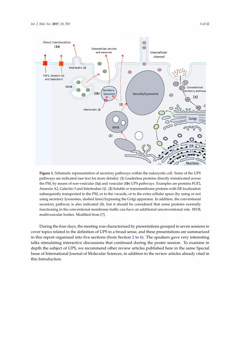
Recently, the identification of an increasing number of secreted signal peptide-lacking proteins,also called leaderless secretory proteins, revealed the existence of unconventional protein secretion(UPS) pathways where these proteins bypass intermediate compartments involved in secretionor exocytosis, such as the Golgi apparatus [4]. Current studies are increasing the number ofproteins known to traffic through the UPS pathways which can be broadly categorized as following:(i) leaderless proteins directly secreted and translocated across the PM, by means of vesicularand non-vesicular UPS pathways; (ii) soluble or transmembrane proteins with ER localizationsubsequently transported to the PM, or to the vacuole, or to the extra cellular space bypassingthe Golgi apparatus; (iii) proteins normally functioning in the conventional membrane traffic withan additional unconventional role; and (iv) proteins involved in unusual or unexplored intra- andintercellular pathways and organelle biogenesis (Figure 1). Some scientists underlined that alsointercellular channels represent a route for the transport of proteins and other macromolecules, largelyindependent of conventional secretory pathway, and therefore can represent another type of UPSpathway (Figure 2). Because of the large number of UPS pathways, Devis and colleagues stated that“one is tempted to speculate that what is termed unconventional protein secretion may not be thatunconventional after all” [5]. It is therefore clear that UPS is becoming more and more important incell biology studies, which were initially carried out by yeast and mammalian cell biologists [6,7], but,recently, insights from plant biologists have contributed to this topic [5]. Trafficking of proteins andmembranes with an unconventional role is related to human pathologies as well as to plant biotic stressand development. Thus, combining expertise and research efforts from different fields to develop anopen comparative approach to tackle these subjects is timely. The meeting on “Unconventional Proteinand Membrane Traffic” (UPMT) held in Lecce during 4–7 October 2016 tried to address this topic andgenerate an open discussion among specialists working on mammals, plants and microorganisms.
Int. J. Mol. Sci. 2017, 18, 703 3 of 22
Int. J. Mol. Sci. 2017, 18, 703 3 of 21
Figure 1. Schematic representation of secretory pathways within the eukaryotic cell. Some of the UPS pathways are indicated (see text for more details): (1) Leaderless proteins directly translocated across the PM, by means of non-vesicular (1a) and vesicular (1b) UPS pathways. Examples are proteins FGF2, Annexin A2, Galectin-3 and Interleukin-1β. (2) Soluble or transmembrane proteins with ER localization subsequently transported to the PM, or to the vacuole, or to the extra cellular space (by using or not using secretory lysosomes, dashed lines) bypassing the Golgi apparatus. In addition, the conventional secretory pathway is also indicated (3), but it should be considered that some proteins normally functioning in the conventional membrane traffic can have an additional unconventional role. MVB, multivesicular bodies. Modified from [7].
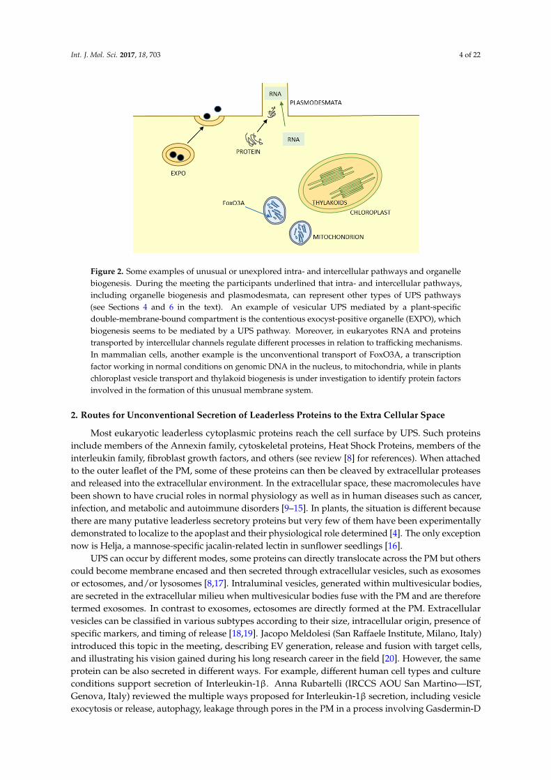
Figure 2. Some examples of unusual or unexplored intra- and intercellular pathways and organelle biogenesis. During the meeting the participants underlined that intra- and intercellular pathways, including organelle biogenesis and plasmodesmata, can represent other types of UPS pathways (see Sections 4 and 6 in the text). An example of vesicular UPS mediated by a plant-specific
Figure 1. Schematic representation of secretory pathways within the eukaryotic cell. Some of the UPSpathways are indicated (see text for more details): (1) Leaderless proteins directly translocated acrossthe PM, by means of non-vesicular (1a) and vesicular (1b) UPS pathways. Examples are proteins FGF2,Annexin A2, Galectin-3 and Interleukin-1β. (2) Soluble or transmembrane proteins with ER localizationsubsequently transported to the PM, or to the vacuole, or to the extra cellular space (by using or notusing secretory lysosomes, dashed lines) bypassing the Golgi apparatus. In addition, the conventionalsecretory pathway is also indicated (3), but it should be considered that some proteins normallyfunctioning in the conventional membrane traffic can have an additional unconventional role. MVB,multivesicular bodies. Modified from [7].
During the four days, the meeting was characterized by presentations grouped in seven sessions tocover topics related to the definition of UPS in a broad sense, and these presentations are summarizedin this report organized into five sections (from Section 2 to 6). The speakers gave very interestingtalks stimulating interactive discussions that continued during the poster session. To examine indepth the subject of UPS, we recommend other review articles published here in the same SpecialIssue of International Journal of Molecular Sciences, in addition to the review articles already cited inthis Introduction.
Int. J. Mol. Sci. 2017, 18, 703 4 of 22
Int. J. Mol. Sci. 2017, 18, 703 3 of 21
Figure 1. Schematic representation of secretory pathways within the eukaryotic cell. Some of the UPS pathways are indicated (see text for more details): (1) Leaderless proteins directly translocated across the PM, by means of non-vesicular (1a) and vesicular (1b) UPS pathways. Examples are proteins FGF2, Annexin A2, Galectin-3 and Interleukin-1β. (2) Soluble or transmembrane proteins with ER localization subsequently transported to the PM, or to the vacuole, or to the extra cellular space (by using or not using secretory lysosomes, dashed lines) bypassing the Golgi apparatus. In addition, the conventional secretory pathway is also indicated (3), but it should be considered that some proteins normally functioning in the conventional membrane traffic can have an additional unconventional role. MVB, multivesicular bodies. Modified from [7].
Figure 2. Some examples of unusual or unexplored intra- and intercellular pathways and organelle biogenesis. During the meeting the participants underlined that intra- and intercellular pathways, including organelle biogenesis and plasmodesmata, can represent other types of UPS pathways (see Sections 4 and 6 in the text). An example of vesicular UPS mediated by a plant-specific
Figure 2. Some examples of unusual or unexplored intra- and intercellular pathways and organellebiogenesis. During the meeting the participants underlined that intra- and intercellular pathways,including organelle biogenesis and plasmodesmata, can represent other types of UPS pathways(see Sections 4 and 6 in the text). An example of vesicular UPS mediated by a plant-specificdouble-membrane-bound compartment is the contentious exocyst-positive organelle (EXPO), whichbiogenesis seems to be mediated by a UPS pathway. Moreover, in eukaryotes RNA and proteinstransported by intercellular channels regulate different processes in relation to trafficking mechanisms.In mammalian cells, another example is the unconventional transport of FoxO3A, a transcriptionfactor working in normal conditions on genomic DNA in the nucleus, to mitochondria, while in plantschloroplast vesicle transport and thylakoid biogenesis is under investigation to identify protein factorsinvolved in the formation of this unusual membrane system.
2. Routes for Unconventional Secretion of Leaderless Proteins to the Extra Cellular Space
Most eukaryotic leaderless cytoplasmic proteins reach the cell surface by UPS. Such proteinsinclude members of the Annexin family, cytoskeletal proteins, Heat Shock Proteins, members of theinterleukin family, fibroblast growth factors, and others (see review [8] for references). When attachedto the outer leaflet of the PM, some of these proteins can then be cleaved by extracellular proteasesand released into the extracellular environment. In the extracellular space, these macromolecules havebeen shown to have crucial roles in normal physiology as well as in human diseases such as cancer,infection, and metabolic and autoimmune disorders [9–15]. In plants, the situation is different becausethere are many putative leaderless secretory proteins but very few of them have been experimentallydemonstrated to localize to the apoplast and their physiological role determined [4]. The only exceptionnow is Helja, a mannose-specific jacalin-related lectin in sunflower seedlings [16].
UPS can occur by different modes, some proteins can directly translocate across the PM but otherscould become membrane encased and then secreted through extracellular vesicles, such as exosomesor ectosomes, and/or lysosomes [8,17]. Intraluminal vesicles, generated within multivesicular bodies,are secreted in the extracellular milieu when multivesicular bodies fuse with the PM and are thereforetermed exosomes. In contrast to exosomes, ectosomes are directly formed at the PM. Extracellularvesicles can be classified in various subtypes according to their size, intracellular origin, presence ofspecific markers, and timing of release [18,19]. Jacopo Meldolesi (San Raffaele Institute, Milano, Italy)introduced this topic in the meeting, describing EV generation, release and fusion with target cells,and illustrating his vision gained during his long research career in the field [20]. However, the sameprotein can be also secreted in different ways. For example, different human cell types and cultureconditions support secretion of Interleukin-1β. Anna Rubartelli (IRCCS AOU San Martino—IST,Genova, Italy) reviewed the multiple ways proposed for Interleukin-1β secretion, including vesicleexocytosis or release, autophagy, leakage through pores in the PM in a process involving Gasdermin-D
Int. J. Mol. Sci. 2017, 18, 703 5 of 22
and pyroptosis, with a special focus on the role of redox response and on different inflammatoryresponses [21–23].
2.1. Pore Formation at the PM
One of the direct UPS translocation mechanisms required the formation of a pore at thePM, as summarized by the group of Walter Nickel (Heidelberg University, Heidelberg, Germany).They discussed unpublished data on the direct transport of the cytoplasmic protein fibroblastgrowth factor 2 across the PM. This transport, as reported by Nickel (Figure 3), requiresfibroblast growth factor 2 recruitment to the PM along with its Tec kinase dependent tyrosinephosphorylation, phosphoinositide PI(4,5)P2-dependent oligomerization and membrane poreformation, and extracellular trapping by cell surface heparin sulfate proteoglycans mediating fibroblastgrowth factor 2 translocation into the extracellular space [24–26]. An elegant approach to visualizethe translocation of fibroblast growth factor 2-GFP across the PM using live cells imaging was alsopresented by Eleni Dimou (Nickel group) based on the utilization of anti-GFP nanobodies that shouldconsiderably improve the understanding of the spatio-temporal coordination and the molecularmechanism of fibroblast growth factor 2 secretion. The importance of fibroblast growth factor 2oligomerization during the translocation process was further developed in a separate talk by SebastianUnger (Nickel group) showing the requirement of two surface cysteines that are uniquely presentin fibroblast growth factor 2, but not in other fibroblast growth factor family members carryingsignal peptides. Cell surface proteoglycans seems to be important for secretion of the cytoskeletalprotein tau, as described also for fibroblast growth factor 2. Taxiarchis Katsinelos, from ThomasJahn’s group (DKFZ Heidelberg University, Heidelberg, Germany), talked about his work on thesecretion and the trans-cellular propagation of Alzheimer’s disease associated tau protein [27]. A majorhallmark of Alzheimer’s disease is the progressive accumulation of the microtubule-associated tauprotein. However, the mechanisms for tau secretion are poorly understood. Using an inducible cellculture system to analyze tau secretion, they could correlate the phosphorylation status of tau with itsaggregation propensity and secretion efficiency. The secreted tau was mainly found as free solubleprotein and was not vesicle associated.
Int. J. Mol. Sci. 2017, 18, 703 5 of 21
cytoskeletal protein tau, as described also for fibroblast growth factor 2. Taxiarchis Katsinelos, from Thomas Jahn’s group (DKFZ Heidelberg University, Heidelberg, Germany), talked about his work on the secretion and the trans-cellular propagation of Alzheimer’s disease associated tau protein [27]. A major hallmark of Alzheimer’s disease is the progressive accumulation of the microtubule-associated tau protein. However, the mechanisms for tau secretion are poorly understood. Using an inducible cell culture system to analyze tau secretion, they could correlate the phosphorylation status of tau with its aggregation propensity and secretion efficiency. The secreted tau was mainly found as free soluble protein and was not vesicle associated.
Another likely example of direct translocation by pore formation was showed by Kevin Moreau (University of Cambridge, Cambridge, UK) regarding the transport to the cell surface of Annexin A2 and Galectin-3. Cell surface Annexin A2 acts as a plasminogen receptor able to stimulate fibrinolysis and cell migration through the extracellular matrix proteolysis while cell surface Galectin-3 serves as a cell-to-cell and cell-to-matrix adhesion/interaction molecule [8]. He presented data identifying key regulators of Galectin-3 localization to the cell surface using a genome-wide screen based on CRISPR-Cas9 technology. Moreover, using a combination of biochemical, biophysical and cell biological approaches, he showed that the translocation of Annexin A2 across membranes requires phospholipid binding and lipid reorganization. Interestingly, Pablo Pelegrin (IMIB-Arrixaca, Murcia, Spain) identified the P2X7 receptor in regulating Annexins secretion during macrophage polarization [28] and discussed this in the context of Annexins translocation across membranes. The purinergic receptor P2X7R once activated promotes the assembly of the NLRP3 inflammasome and the unconventional release of the pro-inflammatory cytokines Interleukin-1β and Interleukin-18 from macrophages [28]. In this study, he characterized P2X7R secretome and found the release of novel cell-related proteins.
Members of the interleukin family are a case study for this kind of UPS. Interleukin-1β is an essential mediator of inflammation after infection and injury. This cytokine is produced as an inactive precursor, which must be cleaved to its active form via a molecular complex called the inflammasome. Interleukin-1β is released from the cell via an unknown process. The group of David Brough (with the presentations of Jack Rivers-Auty, Michael Daniels, and Catherine Diamond, University of Manchester, Manchester, UK) discussed its recent publication about the release of Interleukin-1β via a mechanism that requires membrane permeabilization [29]. The mechanism of membrane permeabilization required for Interleukin-1β release seems to be distinct from the formation of membrane pores required for other types of unconventional protein secretion that do not result in cell death but still needs further characterization. Michael Daniels also talked about the unusual nuclear localization of Interleukin-1α due to a nuclear localization sequence at the N-terminus that is important for its function. He further discussed this observation in the context of evolution.
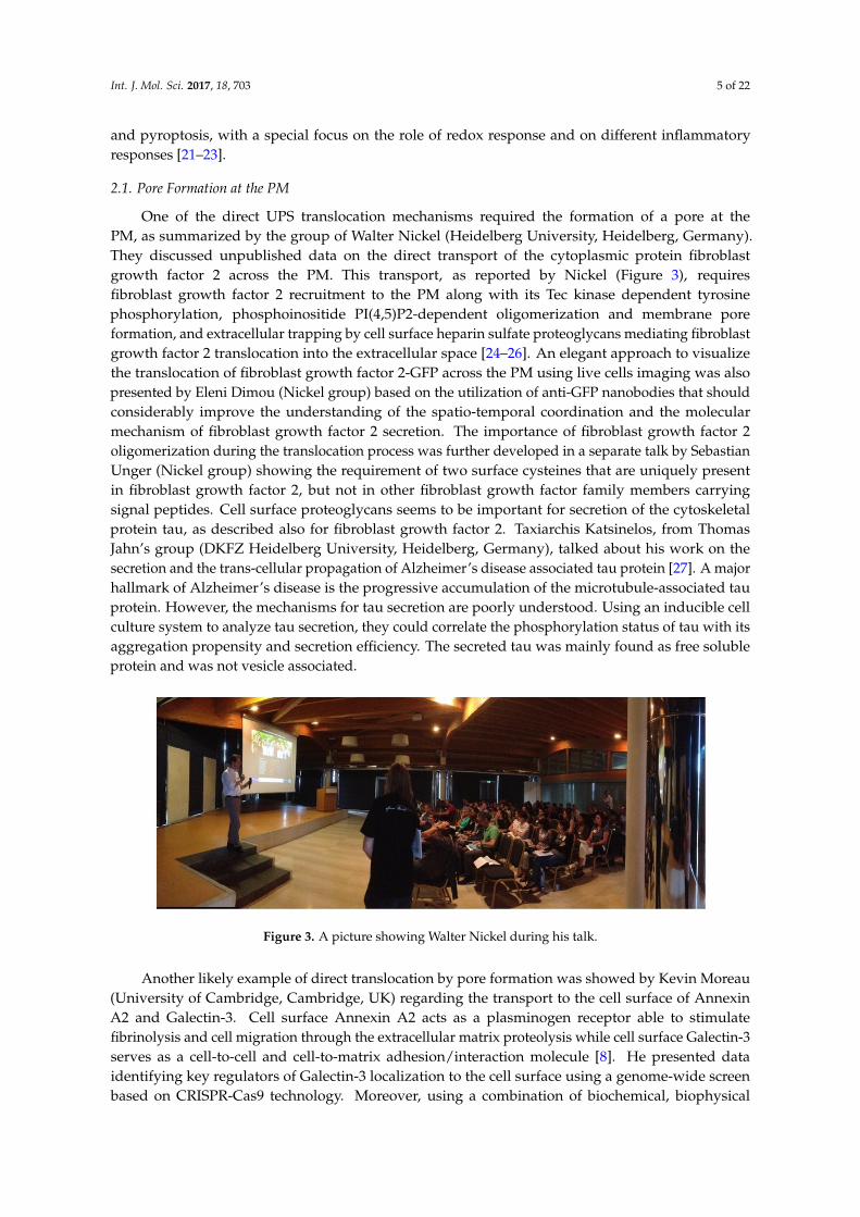
Figure 3. A picture showing Walter Nickel during his talk.
Figure 3. A picture showing Walter Nickel during his talk.
Another likely example of direct translocation by pore formation was showed by Kevin Moreau(University of Cambridge, Cambridge, UK) regarding the transport to the cell surface of AnnexinA2 and Galectin-3. Cell surface Annexin A2 acts as a plasminogen receptor able to stimulatefibrinolysis and cell migration through the extracellular matrix proteolysis while cell surface Galectin-3serves as a cell-to-cell and cell-to-matrix adhesion/interaction molecule [8]. He presented dataidentifying key regulators of Galectin-3 localization to the cell surface using a genome-wide screenbased on CRISPR-Cas9 technology. Moreover, using a combination of biochemical, biophysical
Int. J. Mol. Sci. 2017, 18, 703 6 of 22
and cell biological approaches, he showed that the translocation of Annexin A2 across membranesrequires phospholipid binding and lipid reorganization. Interestingly, Pablo Pelegrin (IMIB-Arrixaca,Murcia, Spain) identified the P2X7 receptor in regulating Annexins secretion during macrophagepolarization [28] and discussed this in the context of Annexins translocation across membranes.The purinergic receptor P2X7R once activated promotes the assembly of the NLRP3 inflammasomeand the unconventional release of the pro-inflammatory cytokines Interleukin-1β and Interleukin-18from macrophages [28]. In this study, he characterized P2X7R secretome and found the release of novelcell-related proteins.
Members of the interleukin family are a case study for this kind of UPS. Interleukin-1β is anessential mediator of inflammation after infection and injury. This cytokine is produced as an inactiveprecursor, which must be cleaved to its active form via a molecular complex called the inflammasome.Interleukin-1β is released from the cell via an unknown process. The group of David Brough (with thepresentations of Jack Rivers-Auty, Michael Daniels, and Catherine Diamond, University of Manchester,Manchester, UK) discussed its recent publication about the release of Interleukin-1β via a mechanismthat requires membrane permeabilization [29]. The mechanism of membrane permeabilization requiredfor Interleukin-1β release seems to be distinct from the formation of membrane pores required forother types of unconventional protein secretion that do not result in cell death but still needs furthercharacterization. Michael Daniels also talked about the unusual nuclear localization of Interleukin-1αdue to a nuclear localization sequence at the N-terminus that is important for its function. He furtherdiscussed this observation in the context of evolution.
2.2. Different Types of Secretory Vesicles for Leaderless Proteins Translocation
Interleukin-1β can be also be secreted by a vesicular mean of transport. In human monocytes,Interleukin-1β secretion is mediated by secretory lysosomes, induced by exogenous ATP, once moreunderlining the complex crosstalk among different secretory mechanisms [22]. However, othervesicular pathways seem to be involved in UPS. Claudia Verderio (Institute of Neuroscience, CNR,Milano, Italy) stressed the importance of extracellular vesicles as carriers of signals. Microglia-derivedextracellular vesicles store inflammatory signals and propagate an inflammatory response to otherglial cells, enhancing spontaneous and evoked excitatory transmission [30–32]. The meeting was anoccasion to discuss the molecular mechanism supporting propagation of synaptic dysfunctions amongconnected neurons through extracellular vesicles. A deeper knowledge on their behavior can have atremendous impact on medical research because microglia-derived extracellular vesicles concentrationin the cerebrospinal fluid correlates with classical markers of neurodegeneration and Alzheimer’sdisease [33,34]. However, the formation of multivesicular bodies releasing exosomes is far from beingfully elucidated.
Autophagic vesicles could be also used for unconventional protein secretion from the cell.For example, William Jackson (University of Maryland, Baltimore, MD, USA) described howPoliovirus 1, a Picornavirus responsible for human disease, uses autophagic vesicles to promote virusreplication and exit from the cell [35]. In particular, the maturation and acidification of autophagicvesicles into amphisomes by fusion with the lysosome benefits Poliovirus 1 by promoting maturationof the viral particle. Jackson demonstrated that functionally eliminating specific proteins of a complexthat mediates vesicle fusion (SNARE complex), Poliovirus 1 and other picornaviruses might enhanceunconventional autophagic secretion to promote viral exit from cells.
A novel compartment, formed by a tubulovesicular structure surrounded by a cup-shapedmembrane, named compartment for unconventional protein secretion (CUPS), is induced in yeast bynutrient starvation triggering secretion of the signal sequence-lacking Acb1 protein. The biogenesis ofthe compartment for unconventional protein secretion requires several proteins, including an ER exitsite/Golgi resident protein, Grh1. Amy Curwin (Centre for Genomic Regulation, Barcelona, Spain)from the Malhotra lab reported that the major endosomal sorting complex required for transport-IIIsubunit Snf7 localizes transiently to this novel compartment, and promotes direct engulfment of
Int. J. Mol. Sci. 2017, 18, 703 7 of 22
preexisting Grh1 containing vesicles and tubules into a saccule to generate a mature compartmentfor unconventional protein secretion containing Acb1 [36,37]. The Malhotra group suggest that thisnovel multivesicular compartment is the stable secretory form releasing Acb1-containing exosome-likevesicles into the extracellular space where they lyse to release Acb1.
2.3. Other Examples of Unconventional Leaderless Proteins Secretion
Kerstin Schipper (Heinrich Heine University, Düsseldorf, Germany) presented new data on thechitinase Cts1 from the fungus Ustilago maydis that lacks a classical N-terminal secretion signal butis secreted in the fragmentation zone between mother and daughter cells during cytokinesis [38].Her team has established an ingenious reporter system to test if secreted proteins pass through theER [39]. The bacterial enzyme β-glucuronidase (GUS) is widely used as a reporter of gene activityas it can catalyze the conversion of the colorless substrate 5-bromo-4-chloro-3-indolyl glucuronideto a blue product. GUS contains a eukaryotic N-glycosylation signal, but—crucially—glycosylationlargely inactivates the enzyme. Stock et al. realized that this feature could be harnessed to prove that asecreted protein fused to GUS does indeed bypass the ER [40]. Schipper discussed the first results oftheir genetic screen to identify proteins essential to the unconventional secretion of Cts1, before makinga passionate case for the development of U. maydis for protein production. Indeed, inappropriateN-glycosylation can be a problem in the heterologous production of pharmaceutical proteins. Cts1can act as a carrier to avoid unwanted ER processing whilst retaining the advantages of proteinsecretion for facilitated downstream processing. Another leaderless mammalian protein, indoleamine2,3-dioxygenase 1, seems to be secreted by UPS, even if the exact unconventional mechanism is stillunknown. Maria Teresa Pallotta (University of Perugia, Perugia, Italy) presented unpublished dataregarding this protein, an enzyme that exerts regulatory functions in autoimmune and inflammatorysettings [41]. She demonstrated that a specific extracellular milieu can promote different subcellularlocalizations and also extracellular secretion of the indoleamine 2,3-dioxygenase 1 enzyme.
It is clear from the above paragraphs that the UPS pathways of leaderless proteins to the extracellular space are important for the crucial roles of these proteins in normal physiology as well as inhuman diseases, but also in innate immune response. The majority of examples come from mammalianand yeast biology; therefore, to further characterize these pathways, more research efforts are neededin other disciplines like for example plant biology.
3. Routes for Unconventional Secretion of Soluble or Transmembrane Proteins with ERLocalization Subsequently Transported to the PM, to the Vacuole, or to the Extra Cellular Space
UPS can take also place by a transportation mode in which both soluble and membrane boundproteins initially enter the conventional secretory pathway whether through ER translocation asthey possess ER-targeting signals or through ER association because they are associated with thecytosolic side of ER membranes. Then, these proteins could be subsequently transported to the cellsurface or to the vacuole bypassing the Golgi apparatus. There are some examples of animal proteinstransported through this kind of pathway [6], but much more examples exist in plants of solubleor membrane proteins, or proteins aggregated into specialized ER vesicles, which are transportedfrom the ER to the vacuole bypassing the Golgi apparatus (reviewed in [42]). Moreover, recent datasuggest that newly formed plant lytic vacuoles arise directly from the ER, even though the Golgiapparatus contributes during vacuole formation with the delivery of soluble cargoes and proteinsfor the tonoplast, the vacuolar membrane [43]. Autophagy is also one possible source for doublemembrane compartments bypassing the Golgi apparatus in transporting specific cargos directly fromER to the lysosome/vacuole or to the extracellular space/cell wall. It is becoming more and moreapparent that autophagy-related endomembrane transport processes are an essential part of basalcellular machinery, but the mode of unconventional secretion that is dependent on autophagy is lessclear in mechanistic terms, as well as its intricate interrelations with other processes (including lateendosome/multivesicular bodies biogenesis).
Int. J. Mol. Sci. 2017, 18, 703 8 of 22
3.1. Involvement of Autophagy in Unconventional Secretion
Mature autophagosomes, the autophagy organelle, are double-membrane vesicles decorated withthe autophagy-related (ATG) protein ATG8 on both surfaces. Autophagosomes, moving along thecytoskeletal network, are transported to the tonoplast with which they fuse to deliver autophagic bodiesfor enzymatic recycling, or to the PM for secretion. These autophagy pathways can be also used in thecell for other purposes. For example, Alexandra Boeske, from Dieter Willbold’s lab (Heinrich HeineUniversity, Düsseldorf, Germany), discussed the function of ATG8 homologs GABARAP proteins inHIV-1 Nef protein pathogenesis and its unconventional secretion as a follow-up inquiry into previouslyidentified HIV-1 Nef interactors. To get the signal peptide lacking HIV-1 Nef protein out of the cell itneeds at least one of GABARAP paralogs to be active, forming a complex with HIV-1 Nef in perinuclearregion from where it is secreted to the extracellular space via PM localized patches. Interestingly,also GABARAP non-binding mutant HIV-1 Nef was detected within extracellular vesicles indicatinga parallel mechanism of HIV-1 Nef GABARAP dependent export, however not dependent on thedirect GABARP–HIV-1 Nef interaction. Interestingly, it was recently shown that, in transfected cells,a proportion of Nef localized to ER membranes where Nef physically interacted with the ER chaperonecalnexin [44].
3.2. Direct Delivery from the ER to the Vacuole
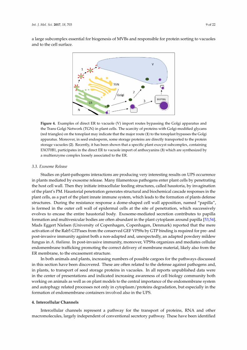
A new perspective about direct ER to vacuole import (Figure 4) was opened by Viktor Žárský(Charles University and Institute of Experimental Botany ASCR, Prague, Czech Republic). He focusedon the observation that a specific plant exocyst subcomplex, containing EXO70B1 [45], participatesin the direct ER to vacuole import of anthocyanins. Similar pathway seems to be exploited bysome tonoplast proteins. The quantity of proteins with Golgi-modified glycans on the tonoplastis very limited. Animal lysosomes share many characteristics with vacuoles, but the percentageof N-glycoproteome of the rat lysosomal and plasma membranes is much higher than that of theplant tonoplast, and is very similar to that of the Arabidopsis thaliana PM [46]. Emanuela Pedrazzini(Institute of Agricultural Biology and Biotechnology, CNR, Milano, Italy) suggested that this scarcityof glycoproteins may indicate that the major route to the tonoplast bypasses the Golgi apparatus and,indeed, and increasing number of publications support this hypothesis [46–49]. The use of a plantexperimental system to investigate the UPS mechanisms may also benefit research in the animal fieldbecause in animal cells traffic that bypasses the Golgi is evident only during ER stress and autophagyinduction [50]. For example, many investigations on lysosome/vacuole traffic started from the studyof sorting receptors. Despite the differences in binding mechanisms of cargo to receptors, the signalsinvolved are very similar in human, yeast and plants. These trafficking signals are often located in theC-terminal tails of sorting receptors with dileucine and tyrosine based motifs. Carine de Marcos Lousa(Beckett University, Leeds, UK) described the identification of a specific plant vacuolar sorting receptors(VSR) isoform that follows an alternative route to the vacuole [51]. Interestingly, understanding thepossible variability in lysosome/vacuole traffic can shed light on lysosomal sorting diseases, a groupof metabolic disorders resulting from lysosomal dysfunction caused by deficiencies in enzyme activityor by trafficking defects. Another interesting experimental system to study protein traffic is theendomembrane system of plant endosperm tissue. Several different storage compartments complicateprotein traffic and several different routes were described depending on cell type, developmental stageand environment. Seed storage proteins reach their final destination by two main routes: they travelthrough ER and Golgi to protein storage vacuoles or accumulate in ER-derived protein bodies. The firstroute involves post-Golgi multivesicular bodies, but some storage proteins are directly transportedto the protein storage vacuoles bypassing the Golgi. It was shown by Verena Ibl (University ofNatural Resources and Life Sciences, Vienna, Austria) that the Hordeum vulgare Vacuolar ProteinSorting 24 (HvVPS24) has a putative protein sorting role in seed storage protein trafficking in barleyendosperm [52]. HvVPS24 is a component of endosomal sorting complex required for transport-III,
Int. J. Mol. Sci. 2017, 18, 703 9 of 22
a large subcomplex essential for biogenesis of MVBs and responsible for protein sorting to vacuolesand to the cell surface.
Int. J. Mol. Sci. 2017, 18, 703 8 of 21
exploited by some tonoplast proteins. The quantity of proteins with Golgi-modified glycans on the tonoplast is very limited. Animal lysosomes share many characteristics with vacuoles, but the percentage of N-glycoproteome of the rat lysosomal and plasma membranes is much higher than that of the plant tonoplast, and is very similar to that of the Arabidopsis thaliana PM [46]. Emanuela Pedrazzini (Institute of Agricultural Biology and Biotechnology, CNR, Milano, Italy) suggested that this scarcity of glycoproteins may indicate that the major route to the tonoplast bypasses the Golgi apparatus and, indeed, and increasing number of publications support this hypothesis [46–49]. The use of a plant experimental system to investigate the UPS mechanisms may also benefit research in the animal field because in animal cells traffic that bypasses the Golgi is evident only during ER stress and autophagy induction [50]. For example, many investigations on lysosome/vacuole traffic started from the study of sorting receptors. Despite the differences in binding mechanisms of cargo to receptors, the signals involved are very similar in human, yeast and plants. These trafficking signals are often located in the C-terminal tails of sorting receptors with dileucine and tyrosine based motifs. Carine de Marcos Lousa (Beckett University, Leeds, UK) described the identification of a specific plant vacuolar sorting receptors (VSR) isoform that follows an alternative route to the vacuole [51]. Interestingly, understanding the possible variability in lysosome/vacuole traffic can shed light on lysosomal sorting diseases, a group of metabolic disorders resulting from lysosomal dysfunction caused by deficiencies in enzyme activity or by trafficking defects. Another interesting experimental system to study protein traffic is the endomembrane system of plant endosperm tissue. Several different storage compartments complicate protein traffic and several different routes were described depending on cell type, developmental stage and environment. Seed storage proteins reach their final destination by two main routes: they travel through ER and Golgi to protein storage vacuoles or accumulate in ER-derived protein bodies. The first route involves post-Golgi multivesicular bodies, but some storage proteins are directly transported to the protein storage vacuoles bypassing the Golgi. It was shown by Verena Ibl (University of Natural Resources and Life Sciences, Vienna, Austria) that the Hordeum vulgare Vacuolar Protein Sorting 24 (HvVPS24) has a putative protein sorting role in seed storage protein trafficking in barley endosperm [52]. HvVPS24 is a component of endosomal sorting complex required for transport-III, a large subcomplex essential for biogenesis of MVBs and responsible for protein sorting to vacuoles and to the cell surface.
Figure 4. Examples of direct ER to vacuole (V) import routes bypassing the Golgi apparatus and the Trans Golgi Network (TGN) in plant cells. The scarcity of proteins with Golgi-modified glycans (red triangles) on the tonoplast may indicate that the major route (1) to the tonoplast bypasses the Golgi apparatus. Moreover, in seed endosperm, some storage proteins are directly transported to the protein storage vacuoles (2). Recently, it has been shown that a specific plant exocyst subcomplex, containing EXO70B1, participates in the direct ER to vacuole import of anthocyanins (3) which are synthesized by a multienzyme complex loosely associated to the ER.
Figure 4. Examples of direct ER to vacuole (V) import routes bypassing the Golgi apparatus andthe Trans Golgi Network (TGN) in plant cells. The scarcity of proteins with Golgi-modified glycans(red triangles) on the tonoplast may indicate that the major route (1) to the tonoplast bypasses the Golgiapparatus. Moreover, in seed endosperm, some storage proteins are directly transported to the proteinstorage vacuoles (2). Recently, it has been shown that a specific plant exocyst subcomplex, containingEXO70B1, participates in the direct ER to vacuole import of anthocyanins (3) which are synthesized bya multienzyme complex loosely associated to the ER.
3.3. Exosome Release
Studies on plant-pathogens interactions are producing very interesting results on UPS occurrencein plants mediated by exosome release. Many filamentous pathogens enter plant cells by penetratingthe host cell wall. Then they initiate intracellular feeding structures, called haustoria, by invaginationof the plant’s PM. Haustorial penetration generates structural and biochemical cascade responses in theplant cells, as a part of the plant innate immune system, which leads to the formation of plants defensestructures. During the resistance response a dome-shaped cell wall apposition, named “papilla”,is formed in the outer cell wall of epidermal cells at the site of penetration, which successivelyevolves to encase the entire haustorial body. Exosome-mediated secretion contributes to papillaformation and multivesicular bodies are often abundant in the plant cytoplasm around papilla [53,54].Mads Eggert Nielsen (University of Copenhagen, Copenhagen, Denmark) reported that the mereactivation of the Rab5 GTPases from the conserved GEF VPS9a by GTP binding is required for pre- andpost-invasive immunity against both a non-adapted and, unexpectedly, an adapted powdery mildewfungus in A. thaliana. In post-invasive immunity, moreover, VPS9a organizes and mediates cellularendomembrane trafficking promoting the correct delivery of membrane material, likely also from theER membrane, to the encasement structure.
In both animals and plants, increasing numbers of possible cargoes for the pathways discussedin this section have been discovered. These are often related to the defense against pathogens and,in plants, to transport of seed storage proteins in vacuoles. In all reports unpublished data werein the center of presentations and indicated increasing awareness of cell biology community bothworking on animals as well as on plant models to the central importance of the endomembrane systemand autophagy related processes not only in cytoplasm/proteins degradation, but especially in theformation of endomembrane containers involved also in the UPS.
4. Intercellular Channels
Intercellular channels represent a pathway for the transport of proteins, RNA and othermacromolecules, largely independent of conventional secretory pathway. These have been identified
Int. J. Mol. Sci. 2017, 18, 703 10 of 22
in both plants and animals where they function in development, response to abiotic stress conditionsand diseases. In plants, intercellular channels known as plasmodesmata are inserted in cell walldomains enriched in the polysaccharide callose providing membrane and cytoplasmic continuityfor symplastic molecular transport [55,56]. An appressed ER structure (named the desmotubule)traverses the channels but these ER connections do not appear fully functional in intercellular transport.Symplastic communication is restricted by callose accumulation and this pathway plays a role inmeristem development, lateral organ formation, bud dormancy, vascular transport and in regulatingthe spreading of viruses and other pathogens. In animals, intercellular channels named tunnelingnanotubes are the main route for long distance macromolecular transport in vitro and in developingembryos. Tunneling nanotubes are long and thin (50–200 nm) membranous protrusions rich in F-actinthat appear to transfer cellular components over long distances [57]. As plasmodesmata, tunnelingnanotubes play a key role in pathogenesis, enabling movement between cells of viruses, bacteria,and of infectious prion and prion-like proteins.
An entire focus session, chaired by Yoselin Benitez-Alfonso (University of Leeds, Leeds, UK), wasdedicated to this topic during the meeting. Emmanuelle Bayer (University of Bordeaux/CNRS,Bordeaux, France) focused on plasmodesmata specialized membrane organization. Electrontomography micrographs revealed details on plasmodesmata ultrastructure as never seen before.Their work shows that within the pores, ER-PM junctions undergo substantial remodeling duringcell differentiation and tissue growth that vary from direct membrane contact to intermembranegap of about 10 nm spanned by spokes. They showed that in newly divided cells, plasmodesmatadisplayed almost non-existent space between the desmotubule and the plasmodesmata suggestingthe absence of cytoplasmic sleeve. Intriguingly, transport of macromolecules was still effective acrossthis type of connections suggesting that there is no simple correlation between ER-PM spacing andthe extent of cell-to-cell connectivity. Their work questioned the function of membrane contactswithin plasmodesmata.
Chris Hawes (Oxford Brook University, Oxford, UK) presented data on the role of reticulons incell plates and plasmodesmata formation, suggesting that specific members of the reticulon family(RTN3 and RTN6) target mature plasmodesmata and developing cell plate [58]. Studies on BY2 cellsindicated that RTN3 and RTN6 are likely involved in the generation of desmotubules during primaryplasmodesmata formation, consistent with overexpression phenotypes showing their capacity toconvert ER sheet into tubules. Using RTN for proteomic studies by co-immunoprecipitation, otherER-desmotubule proteins expressed at ER-PM contact sites were identified raising questions on therole of these membrane anchors in cell and plant development [59].
The role of the ER-actin transport network in virus movement through plasmodesmata waspresented by Manfred Heinlein (Institut de Biologie Moléculaire des Plantes, CNRS, Strasbourg,France). Tobamoviruses, such as the Tobacco mosaic virus, are transported in the form of viralreplication complexes that target plasmodesmata by means of its movement protein [60]. The formation,and intracellular and intercellular trafficking of these complexes relies on the cortical microtubuleand ER-actin networks. The formation and anchorage of viral replication complexes occurs at specificcortical platforms where microtubules intersect with the ER membrane (cortical microtubule-associatedER sites) and provide access to microtubule- and actin-motor mediated trafficking required for therecruitment of membranes and host factors. In newly infected cells, some early virus replicationcomplexes detach from their cortical microtubule-associated ER sites and target plasmodesmata,whereas other viral replication complexes remain at their attachment sites and grow to become virusfactories that amplify the virus. The Heinlein Lab recently showed that the formation and traffickingof the viral replication complexes and the spread of infection between cells depend on specificmyosin motors [61]. Whereas class XI myosins function in viral replication complexes formationand trafficking along the ER membrane between cortical microtubule-associated ER sites towardsplasmodesmata, class VIII support a specific step in viral replication complexes transport from the ERinto the plasmodesmata. In the absence of class VIII myosin activity the movement protein accumulates
Int. J. Mol. Sci. 2017, 18, 703 11 of 22
in the PM, thus implying a role of the PM during virus movement. The cortical microtubule-associatedER sites are associated with stable ER suggesting their potential relationship to ER-PM contact sites.Moreover, the cortical microtubule-associated ER sites -associated viral replication complexes have arole in dsRNA production, which is known to trigger antiviral RNA silencing and also acts as an elicitorof Pattern-Triggered Immunity [62]. Thus, cortical microtubule-associated ER sites are proposed torepresent important subcellular sites of combat between the replicating virus and its host.
Yka Helariutta (Sainsbury Lab University of Cambridge, Cambridge, UK) presented new resultson his research on symplastic interfaces and their role in phloem transport. Sieve pores connect theenucleated sieve elements and together with plasmodesmata connections in companion cells theyform the route for vascular phloem communication. The work in Helariutta lab carefully dissects themechanisms underlying the formation of sieve pores and the regulation of plasmodesmata aiming toidentify opportunities to engineer long distance signaling. Characterization of proteins, such as theputative choline transporter CHER1 [63], involved in the transition from plasmodesmata to maturesieve pores is combined with genetic analysis of plasmodesmata regulation. Identification of mutantsactivated in callose biosynthesis (via callose synthase 3, CALS3) indicates the importance of thispathway not only for plasmodesmata but also in vascular transport [64]. Callose accumulation in sieveelements, inhibit periclinal divisions in the vascular tissue as restricts the movement of transcriptionfactors (such as DOF2) that promote procambial cell division. Research on callose roles in modifyingcell walls was also presented.
Yoselin Benitez-Alfonso elaborated on plasmodesmata role in development and disease responseand presented new evidence that point to plasmodesmata as regulator of beneficial symbioticinteractions. Using proteins involved in the degradation of callose, symplastic transport betweenepidermal and cortical tissues was enhanced in the model legume Medicago truncatula. As a result,root infection and nodulation, after inoculation with the symbiotic nitrogen-fixing bacteria rhizobia,were significantly improved. Her research indicates that callose deposition at plasmodesmata cellwalls is significantly downregulated soon after inoculation, concomitant with enhanced symplasticcommunication. Inducing callose degradation prior inoculation facilitates the establishment of thesymbiotic interaction. The identity of the symplastic factors involved and how they regulate rootdevelopment and response to pathogenic and non-pathogenic microbes are still unknown.
Chiara Zurzolo (Institut Pasteur, Paris, France) talked about the formation, regulation and functionof very different type of intercellular channels: tunneling nanotubes in human cells. Different fromplasmodesmata in plants, tunneling nanotubes can connect very distant cells but they still transport bigmacromolecular proteins including GFP. Similarities with filopodia structures, precursors for dendriticspines in neurons [65], suggest a common origin, similar regulatory mechanisms and function but is thissupported? She showed that, although both tunneling nanotubes and filopodia require actin for theirformation, different remodeling complexes are involved. Specifically, the CDC42/IRSp53/VASP actinregulatory network which promotes filopodia, inhibit tunneling nanotubes function and, conversely,the actin regulatory protein epidermal growth factor receptor pathway 8 (Eps8) which restricts filopodiaextension, increases tunneling nanotubes formation. Fluorescent-tagged versions of these proteinswere ectopically expressed in neuronal cells and their effects on tunneling nanotubes number andfunction in vesicle transfer from a “donor” to an “acceptor” population of cells were addressed.Finally, she showed data demonstrating a role for tunneling nanotubes in the intercellular spreadingof prion-like aggregated proteins α-synuclein and tau respectively involved in the pathogenesis ofParkinson’s and Alzheimer’s diseases [66,67].
In summary, talks on this focus session contributed to the understanding of the processesunderlying the formation, regulation and function of intercellular channels in plants and animals.Moreover, a platform for communication between researchers in tunneling nanotubes andplasmodesmata was established aiming to create common knowledge on these intriguing structuresand their roles in UPS pathways.
Int. J. Mol. Sci. 2017, 18, 703 12 of 22
5. Unconventional Role of Proteins Normally Functioning in Conventional Protein andMembrane Traffic
During the meeting, it became apparent that proteins involved in conventional protein andmembrane secretion can also play additional unconventional roles, and it was reported in several talksthat uncharacterized processes, from exocyst tethering complex formation to the role of interferingSNARE or that of the RAB proteins, can lead to the formation of membranous structures involved inUPS. In the following subsections, are all the different cases belonging to Section 5 discussed in thefour-day meeting in Lecce.
5.1. Exocyst
The exocyst tethering complex is very recently and surprisingly implicated, not only in exocytosis,but also autophagy in both animals and plants [45–68]. Previous indication of exocyst engagement inthe autophagy initiation in animals and multiplicity of EXO70 paralogs in land plants both imply apossible role for the exocyst complex as a coordinator contributing to the endomembrane dynamics.
Indeed, Tamara Pecenková (Charles University and Institute of Experimental Botany ASCR,Prague, Czech Republic) discussed the possible role of the plant exocyst complex on transport indefense against pathogens attacks. In plants, the exocyst tethering complex has been shown toparticipate in numerous events as cell wall differentiation and maturation, cell plate formation, plasmamembrane recycling, autophagy and plant defense [45,69]. This great versatility is probably becausesingle copy genes encode just two of the eight subunits of the exocyst complex in A. thaliana, whereasthere are two or more genes for all the other exocyst components and up to 23 gene members for theEXO70 subunit. Therefore, different exocyst complexes could correspond to the great diversification ofendomembrane structure and function in plants. The work of Pecenková and colleagues is focalizedon the EXO70B1 and B2, which are highly homologous. Both subunits are involved in defense againstpathogens and the EXO70B1 is also implicated in autophagy-related transport to vacuole. Based onpreliminary results from microscopic observations they concluded that the role for EXO70Bs could beto support the unconventional cargo transportation and secretion. Rosana Sanchez-Lopez (UniversidadNacional Autonoma de Mexico, Mexico City, Mexico) provided another example of the importanceof endomembrane dynamics in biotic interactions by documenting the importance of GNOM relatedARF guanine-nucleotide exchange factor (GEF) in nodulation induced in Vicia faba by Rhizobacteria.
It would be interesting to test the role of each single component of the exocyst complex with thesuite of chemical inhibitors of membrane trafficking discussed by Glenn Hicks (University of California,Riverside, CA, USA). Chemical genomics—the use of small molecule inhibitors combined withgenetic screening or reporter systems—is well-established as a tool for investigating endomembranetrafficking in plants. This is particularly true for A. thaliana, where a wide variety of reporter lines andcharacterized cargo proteins are available [70]. He presented new data on endosidin 2 demonstratingthat this inhibitor targets a component of the exocyst complex, EXO70A1, to inhibit exocytosis [71].
5.2. SNAP29
To explain homo- and heterotypic fusions among large compartments, it will probably benecessary to investigate in more detail the unconventional function of important regulatory proteinssuch as the soluble NSF attachment protein receptor (SNARE) proteins. These tail anchored proteinsare essential for the specificity of vesicular targeting but can also contribute to the definition ofmembrane identity by assuming alternative roles [72]. An update was presented by Thomas Vaccari(IFOM, Milano, Italy) about the Drosophila Snap29 protein, homolog of the human protein SNAP29,a SNARE protein that localizes to multiple trafficking compartments and is normally required forprotein trafficking and for proper GA morphology. It was reported that developing tissue lackingSnap29 accumulates large amounts of autophagosomes, thus highlighting a major role of Snap29 inautophagy and secretion [73]. It was also shown that Snap29 plays a conserved role in formation ofthe kinetochore for the anchoring of mitotic chromosomes to spindle microtubules [74].
Int. J. Mol. Sci. 2017, 18, 703 13 of 22
5.3. Interfering SNAREs
Another example of an unconventional role for a conventional trafficking protein was providedby Gian Pietro di Sansebastiano (University of Salento, Lecce, Italy), who at short notice describedtheir work on the syntaxin 5 proteins in A. thaliana. One of the puzzles in vesicle fusion is the apparentover-abundance of SNARE proteins relative to their requirement for vesicle fusion. He has proposeda new class of SNARE, the iSNARE of interfering SNARE that can become non-fusogenic whenover-expressed [72]. In A. thaliana protoplasts SYP51 and SYP52 typically function as t-SNARES,however when localized to the tonoplast these proteins take on inhibitory or non-fusogenic roles.Different roles for these two highly similar SNARE proteins were also presented, identifying SYP51 asessential for trafficking to the central vacuole [75].
5.4. RAB Proteins
Several talks focused on a family of small GTPases, known as RAB proteins, which are frequentlydescribed as “master regulators” of membrane trafficking. RABs are highly conserved molecularswitches that help to specify a unique identity for membranes to which they are transiently attached.Activated, GTP-bound RABs interact with a wide variety of other proteins known as “effectors”.Although their structure and core mechanisms are highly conserved, RAB proteins have diversifiedextensively between different organisms. Over 60 RAB genes have been identified in humans and theirroles in regulating intracellular traffic throughout the endomembrane system is well known. However,less well established roles for RAB proteins include signaling, cell cycle control and cellular migration.
The group of Cinzia Progida (University of Oslo, Oslo, Norway) used human cells to develop cellmigration and proliferation assays that could be used as the basis for an RNAi screen for RAB proteins.The RAB7 group is known to regulate transport between the late endosomes and the Golgi apparatus,and previous work by the Progida group had identified an additional role for RAB7b as a coordinatorof cytoskeleton organization through direct interactions with myosin II and by activating RhoA andtherefore actin remodeling [76]. As remodeling of the actin cytoskeleton is essential in cell migration,the work by Borg et al. [76] provides a strong foundation for deciphering the role of some RAB proteinsin cell motility. Progida presented the results of the motility screens and control experiments forreduced cell proliferation. Their recent results demonstrating that one of the candidates, RAB7a, doesregulate cell migration has just been published [77]. Like RAB7b, RAB7a also regulates the actincytoskeleton, but through interactions with RAC1 and vimentin.
An alternative way to gain insight into the function of proteins is to physically isolate themand identify interacting partners. One classical method for establishing interactions is to useco-immunoprecipitation, where one protein is isolated by affinity purification using antibodies raisedto either an epitope on the bait protein itself or—more commonly—to an established fusion proteinsuch as GFP. Alex Jones (University of Warwick, Coventry, UK) presented data from a large proteomicsdataset that used fluorescent proteins fused to seven target proteins and gentle protein extractionconditions to isolate not just direct protein interactions but large sections of the endomembranesystem to which they are attached [78]. Heard et al. focused on four RAB baits, RABD2a/ARA5,RABF2b/ARA7, RABF1/ARA6, and RABG3f, as markers for combinations of the Golgi apparatus,trans Golgi network, early endosomes, secretory vesicles, late endosomes, multivesicular bodies, andthe tonoplast. In addition, to compare these sub-proteomes with each other and other publishedproteomic datasets, she presented a few highlights of using interaction datasets such as STRING tofurther interrogate the data.
In plants, cell wall material and enzymes, such as xyloglucan, are cargos of RAB-regulated routes.A chemical genomic approach to study xyloglucan metabolism and trafficking in the model plantA. thaliana was then presented by Grégory Mouille (Institut Jean-Pierre Bourgin, INRA, Versailles,France). Well-defined polymeric xyloglucan structures are present in the plant cell wall. Whilecore xyloglucan biosynthesis is located in the Golgi key maturation steps occur in the apoplast.Several cell wall residing enzymes that carry out the trimming are known and mutants are available.
Int. J. Mol. Sci. 2017, 18, 703 14 of 22
Grégory Mouille used sophisticated mass spectrometric technologies combined with chemicalgenomics (5000 compounds tested) to characterize the cell wall structure and phenotype of thesemutants. This approach reveals uncharacterized subcellular compartments involved in the dynamicand polarized trimming of the cell wall polysaccharide [79,80].
5.5. AP-2 Adaptor Complex
George Diallinas (National and Kapodistrian University of Athens, Athens, Greece) focused on theunexpected function of the AP-2 adaptor complex outside of clathrin dependent endocytosis in hyphaltip growth of Aspergillus. In higher fungi, the AP-2 complex appears to have lost the clathrin bindingdomain (in β2 subunit). Nevertheless, it does interact with endocytosis markers, lipid flippases,sphingolipid and sterols biosynthetic machineries and contributes to proper apical membrane lipidcomposition. The question arisen during the meeting was if this imply a possibility of similar featuresin some other adaptor complexes.
5.6. Invariant Chain
To mount adaptive immune responses requires the cell surface expression of MajorHistocompatibility Class II molecules loaded with antigenic peptide. Oddmund Bakke(University of Oslo, Oslo, Norway) reported on his previous studies focused on MajorHistocompatibility Class II—associated Invariant chain (also known as CD74), which serves as achaperone for Major Histocompatibility Class II molecules and mediates trafficking to the endosomalpathway in humans [81]. In fact, Invariant chain contains sorting signals within its cytoplasmic tail,which mediate its trafficking from PM to proteolytic endosomal compartments. He described also howthe invariant chain can prolong the half-life of Major Histocompatibility Class II through its actionon the endocytic pathway, suggesting that this alternative endocytic pathway induced by Invariantchain would serve to enhance the rate, quantity and diversity of Major Histocompatibility Class IIantigen presentation.
This section is a miniature showcase of the diversity of topics that UPMT meeting attendees workon. With such diversity in proteins of the endomembrane system and organisms presented in thissection, it can be hard to find a point of commonality. However, the molecular mechanisms of secretionand membrane trafficking, conventional or not, provided a thread of continuity between the subjects.The same concept can be applied to the next Section 6.
6. Unusual or Unexplored Intra- and Intercellular Pathways and Organelle Biogenesis
Intra- and intercellular communication is a fundamental mechanism for the coordination oforgans during development and growth, stress adaptation, immunity and host-pathogen interactionsin different biological kingdoms. Traffic and exchange of proteins, hormonal peptides and nucleicacids among cells play an essential role. Section 6 is characterized by talks in several research fields,showing how RNA/peptides as molecular signals regulate different processes in relation to traffickingmechanisms, including plant sexual reproduction or organelle function and dynamics.
6.1. Plant Sexuality
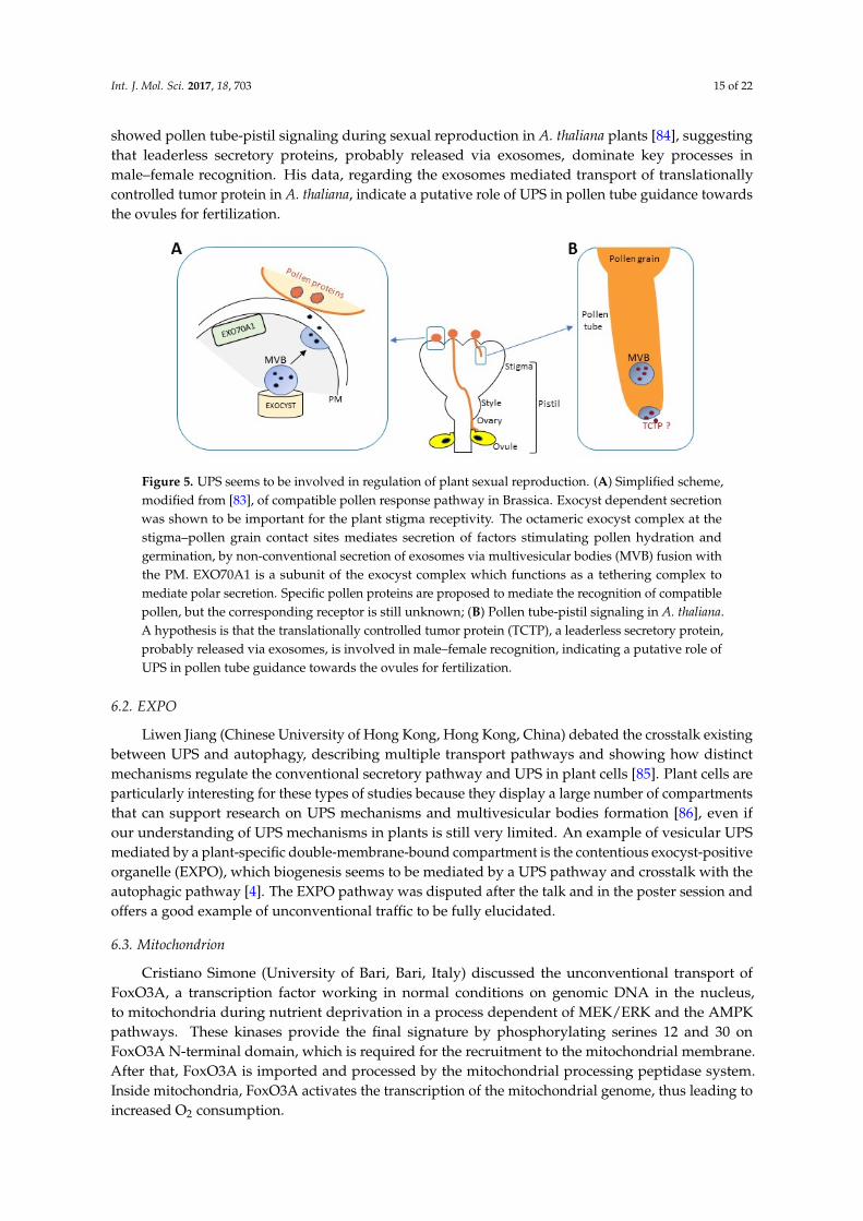
Daphne Goring (University of Toronto, Toronto, Canada) focused on the exocyst, exosomes andautophagy in the regulation of plant sexual reproduction (Figure 5). Normal exocyst dependentsecretion was shown to be important for the plant stigma receptivity. Reported observations indicatethat the exocyst at the stigma–pollen grain contact sites mediates secretion of factors stimulating pollenhydration and germination [82,83]. In both compatible as well as incompatible pollinations within thefamily Brassicaceae, also a non-conventional secretion of exosomes via multivesicular bodies fusionwith the PM is implied. In the incompatible pollinations, one of the exocyst subunits EXO70A1 isdegraded and the autophagy pathway is activated as a part of active self-pollen recognition andrejection. Furthermore, Said Hafidh (Institute of Experimental Botany ASCR, Prague, Czech Republic)
Int. J. Mol. Sci. 2017, 18, 703 15 of 22
showed pollen tube-pistil signaling during sexual reproduction in A. thaliana plants [84], suggestingthat leaderless secretory proteins, probably released via exosomes, dominate key processes inmale–female recognition. His data, regarding the exosomes mediated transport of translationallycontrolled tumor protein in A. thaliana, indicate a putative role of UPS in pollen tube guidance towardsthe ovules for fertilization.
Int. J. Mol. Sci. 2017, 18, 703 14 of 21
This section is a miniature showcase of the diversity of topics that UPMT meeting attendees work on. With such diversity in proteins of the endomembrane system and organisms presented in this section, it can be hard to find a point of commonality. However, the molecular mechanisms of secretion and membrane trafficking, conventional or not, provided a thread of continuity between the subjects. The same concept can be applied to the next Section 6.
6. Unusual or Unexplored Intra- and Intercellular Pathways and Organelle Biogenesis
Intra- and intercellular communication is a fundamental mechanism for the coordination of organs during development and growth, stress adaptation, immunity and host-pathogen interactions in different biological kingdoms. Traffic and exchange of proteins, hormonal peptides and nucleic acids among cells play an essential role. Section 6 is characterized by talks in several research fields, showing how RNA/peptides as molecular signals regulate different processes in relation to trafficking mechanisms, including plant sexual reproduction or organelle function and dynamics.
6.1. Plant Sexuality
Daphne Goring (University of Toronto, Toronto, Canada) focused on the exocyst, exosomes and autophagy in the regulation of plant sexual reproduction (Figure 5). Normal exocyst dependent secretion was shown to be important for the plant stigma receptivity. Reported observations indicate that the exocyst at the stigma–pollen grain contact sites mediates secretion of factors stimulating pollen hydration and germination [82,83]. In both compatible as well as incompatible pollinations within the family Brassicaceae, also a non-conventional secretion of exosomes via multivesicular bodies fusion with the PM is implied. In the incompatible pollinations, one of the exocyst subunits EXO70A1 is degraded and the autophagy pathway is activated as a part of active self-pollen recognition and rejection. Furthermore, Said Hafidh (Institute of Experimental Botany ASCR, Prague, Czech Republic) showed pollen tube-pistil signaling during sexual reproduction in A. thaliana plants [84], suggesting that leaderless secretory proteins, probably released via exosomes, dominate key processes in male–female recognition. His data, regarding the exosomes mediated transport of translationally controlled tumor protein in A. thaliana, indicate a putative role of UPS in pollen tube guidance towards the ovules for fertilization.
Figure 5. UPS seems to be involved in regulation of plant sexual reproduction. (A) Simplified scheme, modified from [83], of compatible pollen response pathway in Brassica. Exocyst dependent secretion was shown to be important for the plant stigma receptivity. The octameric exocyst complex at the stigma–pollen grain contact sites mediates secretion of factors stimulating pollen hydration and germination, by non-conventional secretion of exosomes via multivesicular bodies (MVB) fusion with the PM. EXO70A1 is a subunit of the exocyst complex which functions as a tethering complex to mediate polar secretion. Specific pollen proteins are proposed to mediate the recognition of compatible pollen, but the corresponding receptor is still unknown; (B) Pollen tube-pistil signaling in A. thaliana. A hypothesis is that the translationally controlled tumor protein (TCTP), a leaderless
Figure 5. UPS seems to be involved in regulation of plant sexual reproduction. (A) Simplified scheme,modified from [83], of compatible pollen response pathway in Brassica. Exocyst dependent secretionwas shown to be important for the plant stigma receptivity. The octameric exocyst complex at thestigma–pollen grain contact sites mediates secretion of factors stimulating pollen hydration andgermination, by non-conventional secretion of exosomes via multivesicular bodies (MVB) fusion withthe PM. EXO70A1 is a subunit of the exocyst complex which functions as a tethering complex tomediate polar secretion. Specific pollen proteins are proposed to mediate the recognition of compatiblepollen, but the corresponding receptor is still unknown; (B) Pollen tube-pistil signaling in A. thaliana.A hypothesis is that the translationally controlled tumor protein (TCTP), a leaderless secretory protein,probably released via exosomes, is involved in male–female recognition, indicating a putative role ofUPS in pollen tube guidance towards the ovules for fertilization.
6.2. EXPO
Liwen Jiang (Chinese University of Hong Kong, Hong Kong, China) debated the crosstalk existingbetween UPS and autophagy, describing multiple transport pathways and showing how distinctmechanisms regulate the conventional secretory pathway and UPS in plant cells [85]. Plant cells areparticularly interesting for these types of studies because they display a large number of compartmentsthat can support research on UPS mechanisms and multivesicular bodies formation [86], even ifour understanding of UPS mechanisms in plants is still very limited. An example of vesicular UPSmediated by a plant-specific double-membrane-bound compartment is the contentious exocyst-positiveorganelle (EXPO), which biogenesis seems to be mediated by a UPS pathway and crosstalk with theautophagic pathway [4]. The EXPO pathway was disputed after the talk and in the poster session andoffers a good example of unconventional traffic to be fully elucidated.
6.3. Mitochondrion
Cristiano Simone (University of Bari, Bari, Italy) discussed the unconventional transport ofFoxO3A, a transcription factor working in normal conditions on genomic DNA in the nucleus,to mitochondria during nutrient deprivation in a process dependent of MEK/ERK and the AMPKpathways. These kinases provide the final signature by phosphorylating serines 12 and 30 onFoxO3A N-terminal domain, which is required for the recruitment to the mitochondrial membrane.After that, FoxO3A is imported and processed by the mitochondrial processing peptidase system.Inside mitochondria, FoxO3A activates the transcription of the mitochondrial genome, thus leading toincreased O2 consumption.
Int. J. Mol. Sci. 2017, 18, 703 16 of 22
6.4. Chloroplast
Jürgen Soll and his coworkers (Ludwig-Maximilians-Universität München, Munich, Germany)use biochemical and electron microscopic techniques to uncover transport and signal transductionprocesses in green plants [87]. One focus is on chloroplasts and its integration into the cell.The chloroplast contains an extensive thylakoid membrane system that is critical for photosynthesis.During chloroplast differentiation from non-green proplastids the complex internal membrane systemis formed. This process involves vesicle formation and membrane invagination [88]. In his presentation,Soll summarized the current knowledge on chloroplast vesicle transport and thylakoid biogenesis.He stressed that despite of extensive research in this area not much is known about this unusualmembrane system and while bioinformatics predict their presence no single protein factor involved inthe formation has been identified yet.
6.5. RNA as Molecular Signals in Plant Biology
Daniel Garcia Cabanillas (INRS Institut Armand-Frappier, Laval, QC, Canada) covered thereplication of the Turnip mosaic virus, a positive-sense (+) RNA plant virus, which induces substantialendomembrane system remodeling during infection. The virus, indeed, is known to induce ER-derivedvesicles, commonly known as “viral replication factories”, which house viral RNA as well as viraland host proteins required for its replication. These vesicles are also involved in the intercellulartrafficking of the Turnip mosaic virus and are released at the ER in a COPII-dependent transport.The viral membrane associated protein 6K2 has an essential role in vesicle formation [89]. The work ofCabanillas was focused on this protein, identifying a transmembrane stretch of five amino acids, GxxxG,responsible for the 6K2 protein bypassing the Golgi apparatus. The substitution of the glycine residueswith valine resulted in a delocalization of the protein in the Golgi apparatus and PM and preventednormal replication vesicle production. Cabanillas suggested a “tug of war” between conventional andunconventional trafficking pathways during virus replication, which was confirmed by an increase ofvirus cell-to-cell movement in dominant negative mutants of Golgi SNAREs where the ER-Golgi trafficis disrupted. The presentation by Julia Kehr (Hamburg University, Hamburg, Germany) can be seenas a corollary to this previous report. She presented an overview of the role of RNA as a molecularsignal in plant biology [55,90], discussing results on the intercellular transport of the RNA and its rolein plant development and defense reactions against pathogens. She also showed different experimentsin which several RNAs have been identified in the phloem, suggesting that mobile RNAs can act aslong-distance signals in higher plants and that siRNAs and miRNAs can be involved in plant-virusinteractions during the infection process.
6.6. Other Examples of Unusual or Unexplored Intra- and Intercellular Pathways
Valeria Crippa from the Angelo Poletti’s lab (CEND University of Milano, Milano, Italy) presentedresults devoted to the function of small heat shock protein chaperon B8 in autophagic disposal ofmisfolded proteins implied in motor neuron diseases. The B8 protein, upregulated in motor neurondiseases patients, was shown to increase an autophagic/lysosomal degradation of misfolded proteinsin a complex with co-chaperons (including E3 ligase CHIP) in cellular models and a clear protectivefunction in Drosophila model was also observed [91]. Moving to autophagy induced by nutrientstarvation, Xiaoqiang Yao (Chinese University of Hong Kong, Hong Kong, China) reported on a newpotassium channel crucial for this kind of autophagy in animal cells. The K+ channel TM9SF4 isespecially abundant in kidneys and its knockdown inhibits autophagy normally activated via mTORsignaling pathway.
Andrea Pompa (Institute of Biosciences and Bioresources, CNR, Perugia, Italy) presentedunpublished data on the maturation and unconventional trafficking of the CLV3 protein in transgenictobacco plants. The CLV3 protein is the precursor of a small 12 amino acid active peptide recognized atthe cell surface by CLV1/2 receptor complexes [92] and cooperates in a signaling mechanism that limits
Int. J. Mol. Sci. 2017, 18, 703 17 of 22
excessive proliferation of pluripotent cells in plant apical meristems [93]. Despite the presence of asignal peptide, CLV3 does not follow the conventional secretory pathway, but is retro-translocated fromER to the cytosol. In this process, the proteasome machinery seems to have a role in the productionof the active CLV3 peptide. Furthermore, he postulated that the active ligand, once released into thecytosol, reaches the apoplast by an unconventional secretion process yet to be discovered.
Muhammad Akbar Abdul Ghaffar (Ohio State University, Columbus, OH, USA) describedanother process of intracellular trafficking. He applied transmission electron microscopy to studyrubber ontogeny in the alternative rubber crop T. kok-saghyz. Natural rubber is synthetized andcompartmentalized in particles of some plants and fungi, but the ontogeny and development of theseparticles is not well understood [94]. In contrast to the current main rubber producer Hevea brasiliensisor its alternative Parthenium argentatum growing in arid regions, the plant Taraxacum would be ofinterest for rubber production in temperate areas like parts of the US or Europe. Muhammad Ghaffarpresented first insights into rubber ontogeny in secretory laticifer cells of Taraxacum. He observedrubber being initially formed in the ER-Golgi membrane system. Rubber particles were also foundin vesicles located in the cytoplasm and in plastids of laticifer cells. The findings contribute tounderstanding rubber ontogeny and hence to establishing T. kok-saghyz as a novel rubber producer.
7. Conclusions
The first “Unconventional Protein and Membrane Traffic” meeting was something of a gamblewith a happy conclusion. The UPS topic is intrinsically very diverse and heterogeneous because itinvolves many pathways, cargoes, cell types, and triggering conditions. Moreover, the organizershad not been sure that bringing together scientists working on different model systems, from primaryhuman monocytes to yeast cells, or from the plant A. thaliana to the fungus U. maydis, was a good ideabecause the audience might struggle to follow the talks if the speaker’s research background is toodistant from their own research field. Occasionally this might have been the case, but in general themeeting was very successful and on the last day, during an open final discussion, there was unanimityamong the participants that the second UPMT meeting should be held in 2018.
Acknowledgments: The UPMT committee is grateful to the sponsors supporting the meeting: The Company ofBiologists, Fondazione Puglia, the American Society of Plant Biologists, the Biochemical Society, University ofSalento, CNR-Nanotec, MDPI-International Journal of Molecular Science and Merck.
Author Contributions: All the authors wrote the manuscript. Gian Pietro Di Sansebastiano, Francesca De Marchisand Michele Bellucci prepared the Figures. Moreover, Andrea Pompa, Maria Teresa Pallotta, Yoselin Benitez Alfonso,Alexandra Jones, Kerstin Schipper, Kevin Moreau, Viktor Žárský, and Gian Pietro Di Sansebastiano gavepresentations at the meeting. All authors read and approved the final manuscript prior to submission.
Conflicts of Interest: The authors declare no conflict of interest.
References
1. Vitale, A.; Denecke, J. The Endoplasmic Reticulum—Gateway of the Secretory Pathway. Plant Cell 1999, 11,615–628. [CrossRef] [PubMed]
2. Bonifacino, J.S.; Glick, B.S. The mechanisms of vesicle budding and fusion. Cell 2004, 116, 153–166. [CrossRef]3. Viotti, C. ER to Golgi-Dependent Protein Secretion: The Conventional Pathway. In Unconventional Protein
Secretion, Methods in Molecular Biology; Pompa, A., De Marchis, F., Eds.; Humana Press Inc.: New York, NY,USA, 2016; Volume 1459, pp. 3–29.
4. Robinson, D.G.; Ding, Y.; Jiang, L. Unconventional protein secretion in plants: A critical assessment.Protoplasma 2016, 253, 31–43. [CrossRef] [PubMed]
5. Davis, D.J.; Kang, B.-H.; Heringer, A.S.; Wilkop, T.E.; Drakakaki, G. Unconventional Protein Secretion inPlants. In Unconventional Protein Secretion, Methods in Molecular Biology; Pompa, A., De Marchis, F., Eds.;Humana Press Inc.: New York, NY, USA, 2016; Volume 1459, pp. 47–63.
6. Ng, F.; Tang, B.L. Unconventional Protein Secretion in Animal Cells. In Unconventional Protein Secretion,Methods in Molecular Biology; Pompa, A., De Marchis, F., Eds.; Humana Press Inc.: New York, NY, USA, 2016;Volume 1459, pp. 31–46.
Int. J. Mol. Sci. 2017, 18, 703 18 of 22
7. Bhattacharya, A.; Prakash, Y.S.; Eissa, N.T. Secretory function of autophagy in innate immune cells.Cell Microbiol. 2014, 16, 1637–1645. [CrossRef] [PubMed]
8. Nickel, W.; Rabouille, C. Mechanisms of regulated unconventional protein secretion. Nat. Rev. Mol. Cell Biol.2009, 10, 148–155. [CrossRef] [PubMed]
9. Gerke, V.; Moss, S.E. Annexins: From structure to function. Physiol. Rev. 2002, 82, 331–371. [CrossRef][PubMed]
10. Valapala, M.; Maji, S.; Borejdo, J.; Vishwanatha, J.K. Cell surface translocation of annexin A2 facilitatesglutamate-induced extracellular proteolysis. J. Biol. Chem. 2014, 289, 15915–15926. [CrossRef] [PubMed]
11. Zheng, L.; Maji, S.; Borejdo, J.; Vishwanatha, J.K. Tyrosine 23 phosphorylation-dependent cell-surfacelocalization of annexin A2 is required for invasion and metastases of pancreatic cancer. PLoS ONE 2011,6, e19390. [CrossRef] [PubMed]
12. Hayes, M.J.; Longbottom, R.E.; Evans, M.A.; Moss, S.E. Annexinopathies. Subcell. Biochem. 2007, 45, 1–28.[PubMed]
13. Sidera, K.; Patsavoudi, E. Extracellular HSP90: Conquering the cell surface. Cell Cycle 2008, 7, 1564–1568.[CrossRef] [PubMed]
14. Sheokand, N.; Malhotra, H.; Kumar, S.; Tillu, V.A.; Chauhan, A.S.; Raje, C.I.; Raje, M. Moonlightingcell-surface GAPDH recruits apotransferrin to effect iron egress from mammalian cells. J. Cell Sci. 2014, 127,4279–4291. [CrossRef]
15. Rabinovich, G.A.; Rubinstein, N.; Fainboim, L. Unlocking the secrets of galectins: A challenge at the frontierof glyco-immunology. J. Leukoc. Biol. 2002, 71, 741–752. [PubMed]
16. Pinedo, M.; Regente, M.; Elizalde, M.; Quiroga, I.Y.; Pagnussat, L.A.; Jorrin-Novo, J.; Maldonado, A.;de la Canal, L. Extracellular sunflower proteins: Evidence on non-classical secretion of a jacalin-related lectin.Protein Pept. Lett. 2012, 19, 270–276. [CrossRef] [PubMed]
17. Tkach, M.; Théry, C. Communication by extracellular vesicles: Where we are and where we need to go. Cell2016, 164, 1226–1232. [CrossRef] [PubMed]
18. Cocucci, E.; Meldolesi, J. Ectosomes and exosomes: Shedding the confusion between extracellular vesicles.Trends Cell Biol. 2015, 25, 364–372. [CrossRef] [PubMed]
19. Kowala, J.; Arras, G.; Colombo, M.; Jouvea, M.; Morath, J.P.; Primdal-Bengtson, B.; Dingli, F.; Loew, D.;Tkach, M.; Théry, C. Proteomic comparison defines novel markers to characterize heterogeneous populationsof extracellular vesicle subtypes. Proc. Natl. Acad. Sci. USA 2016, 113, E968–E977. [CrossRef] [PubMed]
20. Prada, I.; Meldolesi, J. Binding and Fusion of Extracellular Vesicles to the Plasma Membrane of Their CellTargets. Int. J. Mol. Sci. 2016, 17, E1296. [CrossRef] [PubMed]
21. Lavieri, R.; Rubartelli, A.; Carta, S. Redox stress unbalances the inflammatory cytokine network: Role inautoinflammatory patients and healthy subjects. J. Leukoc. Biol. 2016, 99, 79–86. [CrossRef] [PubMed]
22. Rubartelli, A. Autoinflammatory diseases. Immunol. Lett. 2014, 161, 226–230. [CrossRef] [PubMed]23. Piccioli, P.; Rubartelli, A. The secretion of IL-1β and options for release. Semin. Immunol. 2013, 25, 425–429.
[CrossRef] [PubMed]24. Zacherl, S.; La Venuta, G.; Muller, H.M.; Wegehingel, S.; Dimou, E.; Sehr, P.; Lewis, J.D.; Erfle, H.;
Pepperkok, R.; Nickel, W. A direct role for ATP1A1 in unconventional secretion of fibroblast growthfactor 2. J. Biol. Chem. 2015, 290, 3654–3665. [CrossRef] [PubMed]
25. Steringer, J.P.; Bleicken, S.; Andreas, H.; Zacherl, S.; Laussmann, M.; Temmerman, K.; Contreras, F.X.;Bharat, T.A.M.; Lecgner, J.; Muller, H.M.; et al. Phosphatidylinositol 4,5-bisphosphate (PI(4,5)P2)-dependentoligomerization of fibroblast growth factor 2 (FGF2) triggers the formation of a lipidic membrane poreimplicated in unconventional secretion. J. Biol. Chem. 2012, 287, 27659–27669. [CrossRef]
26. Muller, H.M.; Steringer, J.P.; Wegehingel, S.; Bleicken, S.; Münster, M.; Dimou, E.; Unger, S.; Weidmann, G.;Andreas, H.; García-Sáez, A.J.; et al. Formation of disulfide bridges drives oligomerization, membrane poreformation, and translocation of fibroblast growth factor 2 to cell surfaces. J. Biol. Chem. 2015, 290, 8925–8937.[CrossRef] [PubMed]
27. Mandelkow, E.M.; Mandelkow, E. Biochemistry and cell biology of tau protein in neurofibrillary degeneration.Cold Spring Harb. Perspect. Med. 2012, 2, a006247. [CrossRef] [PubMed]
28. De Torre-Minguela, C.; Barberà-Cremades, M.; Gómez, A.I.; Martín-Sánchez, F.; Pelegrín, P. Macrophageactivation and polarization modify P2X7 receptor secretome influencing the inflammatory process. Sci. Rep.2016, 6, 22586. [CrossRef] [PubMed]
Int. J. Mol. Sci. 2017, 18, 703 19 of 22
29. Martin-Sanchez, F.; Diamond, C.; Zeitler, M.; Gomez, A.I.; Baroja-Mazo, A.; Bagnall, J.; Spiller, D.; White, M.;Daniels, M.J.; Mortellaro, A.; et al. Inflammasome-dependent IL-1β release depends upon membranepermeabilisation. Cell Death Differ. 2016, 23, 1219–1231. [CrossRef] [PubMed]
30. Bianco, F.; Perrotta, C.; Novellino, L.; Francolini, M.; Riganti, L.; Menna, E.; Saglietti, L.; Schuchman, E.H.;Furlan, R.; Clementi, E.; et al. Acid sphingomyelinase activity triggers microparticle release from glial cells.EMBO J. 2009, 28, 1043–1054. [CrossRef] [PubMed]
31. Antonucci, F.; Turola, E.; Riganti, L.; Caleo, M.; Gabrielli, M.; Perrotta, C.; Novellino, L.; Clementi, E.;Giussani, P.; Viani, P.; et al. Microvesicles released from microglia stimulate synaptic activity via enhancedsphingolipid metabolism. EMBO J. 2012, 31, 1231–1240. [CrossRef] [PubMed]
32. Verderio, C.; Muzio, L.; Turola, E.; Bergami, A.; Novellino, L.; Ruffini, F.; Riganti, L.; Corradini, I.;Francolini, M.; Garzetti, L.; et al. Myeloid microvesicles are a marker and therapeutic target forneuroinflammation. Ann. Neurol. 2012, 72, 610–614. [CrossRef] [PubMed]
33. Joshi, P.; Turola, E.; Ruiz, A.; Bergami, A.; Libera, D.D.; Benussi, L.; Giussani, P.; Magnani, G.; Comi, G.;Legname, G.; et al. Microglia convert aggregated amyloid-β into neurotoxic forms through the shedding ofmicrovesicles. Cell Death Differ. 2014, 21, 582–593. [CrossRef] [PubMed]
34. Agosta, F.M.; Dalla Libera, D.; Spinelli, E.G.; Finardi, A.; Canu, E.; Bergami, A.; Bocchio Chiavetto, L.;Baronio, M.; Comi, G.; Martino, G.; et al. Myeloid microvescicles in cerebrospinal fluid are associated withmyelin damage and neuronal loss in mild cognitive impairment and Alzheimer disease. Ann. Neurol. 2014,76, 813–825. [CrossRef] [PubMed]
35. Richards, A.L.; Jackson, W.T. Intracellular Vesicle Acidification Promotes Maturation of Infectious PoliovirusParticles. PLoS Pathog. 2012, 8, e1003046. [CrossRef] [PubMed]
36. Curwin, A.J.; Brouwers, N.; Alonso, Y.; Adell, M.; Teis, D.; Turacchio, G.; Parashuraman, S.; Ronchi, P.;Malhotra, V. ESCRT-III drives the final stages of CUPS maturation for unconventional protein secretion. Elife2016, 26, e16299. [CrossRef] [PubMed]
37. Cruz-Garcia, D.; Curwin, A.J.; Popoff, J.F.; Bruns, C.; Duran, J.M.; Malhotra, V. Remodeling of secretorycompartments creates CUPS during nutrient starvation. J. Cell Biol. 2014, 207, 695–703. [CrossRef] [PubMed]
38. Langner, T.; Özturk, M.; Hartmann, S.; Cord-Landwehr, S.; Moerschbacher, B.; Walton, J.D.; Göhre, V.Chitinases are essential for cell separation in Ustilago maydis. Eukaryot. Cell 2015, 14, 846–857. [CrossRef][PubMed]
39. Stock, J.; Terfrüchte, M.; Schipper, K. A Reporter System to Study Unconventional Secretion of ProteinsAvoiding N-glycosylation in Ustilago maydis. In Unconventional Protein Secretion, Methods in MolecularBiology; Pompa, A., de Marchis, F., Eds.; Humana Press Inc.: New York, NY, USA, 2016; Volume 1459,pp. 149–160.
40. Stock, J.; Sarkari, P.; Kreibich, S.; Brefort, T.; Feldbrügge, M.; Schipper, K. Applying unconventional secretionof the endochitinase Cts1 to export heterologous proteins in Ustilago maydis. J. Biotechnol. 2012, 161, 80–91.[CrossRef] [PubMed]
41. Pallotta, M.T.; Orabona, C.; Volpi, C.; Vacca, C.; Belladonna, M.L.; Bianchi, R.; Servillo, G.; Brunacci, C.;Calvitti, M.; Bicciato, S.; et al. Indoleamine 2,3-dioxygenase is a signaling protein in long-term tolerance bydendritic cells. Nat. Immunol. 2011, 12, 870–878. [CrossRef] [PubMed]
42. De Marchis, F.; Bellucci, M.; Pompa, A. Unconventional pathways of secretory plant proteins from theendoplasmic reticulum to the vacuole bypassing the Golgi complex. Plant Signal. Behav. 2013, 8, e25129.[CrossRef] [PubMed]
43. Viotti, C. ER and vacuoles: Never been closer. Front. Plant. Sci. 2014, 5, 20. [CrossRef] [PubMed]44. Jennelle, L.; Hunegnaw, R.; Dubrovsky, L.; Pushkarsky, T.; Fitzgerald, M.L.; Sviridov, D.; Popratiloff, A.;
Brichacek, B.; Bukrinsky, M. HIV-1 protein Nef inhibits activity of ATP-binding cassette transporter A1 bytargeting Endoplasmic Reticulum chaperone Calnexin. J. Biol. Chem. 2014, 289, 28870–28884. [CrossRef]
45. Kulich, I.; Pecenková, T.; Sekereš, J.; Smetana, O.; Fendrych, M.; Foissner, I.; Höftberger, M.; Zárský, V.Arabidopsis exocyst subcomplex containing subunit EXO70B1 is involved in autophagy-related transport tothe vacuole. Traffic 2013, 14, 1155–1165. [CrossRef] [PubMed]
46. Pedrazzini, E.; Caprera, A.; Fojadelli, I.; Stella, A.; Rocchetti, A.; Bassin, B.; Martinoia, E.; Vitale, A.The Arabidopsis tonoplast is almost devoid of glycoproteins with complex N-glycans, unlike the rat lysosomalmembrane. J. Exp. Bot. 2016, 67, 1769–1781. [CrossRef] [PubMed]
Int. J. Mol. Sci. 2017, 18, 703 20 of 22
47. Pedrazzini, E.; Komarova, N.Y.; Rentsch, D.; Vitale, A. Traffic routes and signals for the tonoplast. Traffic2013, 14, 622–628. [CrossRef] [PubMed]
48. Stigliano, E.; Faraco, M.; Neuhaus, J.M.; Montefusco, A.; Dalessandro, G.; Piro, G.; Di Sansebastiano, G.P.Two glycosylated vacuolar GFPs are new markers for ER-to-vacuole sorting. Plant Physiol. Biochem. 2013, 73,337–343. [CrossRef] [PubMed]
49. Occhialini, A.; Gouzerh, G.; Di Sansebastiano, G.P.; Neuhaus, J.M. Dimerization of the Vacuolar ReceptorsAtRMR1 and -2 from Arabidopsis thaliana Contributes to Their Localization in the trans-Golgi Network.Int. J. Mol. Sci. 2016, 17, E1661. [CrossRef] [PubMed]
50. Staudt, C.; Puissant, E.; Boonen, M. Subcellular Trafficking of Mammalian Lysosomal Proteins: An ExtendedView. Int. J. Mol. Sci. 2017, 18, 47. [CrossRef] [PubMed]
51. De Marcos Lousa, C.; Denecke, J. Lysosomal and vacuolar sorting: Not so different after all!Biochem. Soc. Trans. 2016, 44, 891–897. [CrossRef] [PubMed]
52. Hilscher, J.; Kapusi, E.; Stoger, E.; Ibl, V. Cell layer-specific distribution of transiently expressed barleyESCRT-III component HvVPS60 in developing barley endosperm. Protoplasma 2016, 253, 137–153. [CrossRef][PubMed]
53. Meyer, D.; Pajonk, S.; Micali, C.; O’Connell, R.; Schulze-Lefert, P. Extracellular transport and integration ofplant secretory proteins into pathogen-induced cell wall compartments. Plant J. 2009, 57, 986–999. [CrossRef][PubMed]
54. Richter, S.; Kientz, M.; Brumm, S.; Nielsen, M.E.; Park, M.; Gavidia, R.; Krause, C.; Voss, U.; Beckmann, H.;Mayer, U.; et al. Delivery of endocytosed proteins to the cell-division plane requires change of pathway fromrecycling to secretion. Elife 2014, 8, e02131. [CrossRef] [PubMed]
55. Otero, S.; Helariutta, Y.; Benitez-Alfonso, Y. Symplastic communication in organ formation and tissuepatterning. Curr. Opin. Plant Biol. 2016, 29, 21–28. [CrossRef] [PubMed]
56. Knox, J.P.; Benitez-Alfonso, Y. Roles and regulation of plant cell walls surrounding plasmodesmata.Curr. Opin. Plant Biol. 2014, 22, 93–100. [CrossRef] [PubMed]
57. Gerdes, H.H.; Rustom, A.; Wang, X. Tunneling nanotubes, an emerging intercellular communication route indevelopment. Mech. Dev. 2013, 130, 381–387. [CrossRef] [PubMed]
58. Knox, K.; Wang, P.; Kriechbaumer, V.; Tilsner, J.; Frigerio, L.; Sparkes, I.; Hawes, C.; Oparka, K. Putting theSqueeze on Plasmodesmata: A Role for Reticulons in Primary Plasmodesmata Formation. Plant Physiol.2015, 168, 1563–1572. [CrossRef] [PubMed]
59. Kriechbaumer, V.; Botchway, S.W.; Slade, S.E.; Knox, K.; Frigerio, L.; Oparka, K.; Hawes, C. Reticulomics:Protein-Protein Interaction Studies with Two Plasmodesmata-Localized Reticulon Family Proteins IdentifyBinding Partners Enriched at Plasmodesmata, Endoplasmic Reticulum, and the Plasma Membrane.Plant Physiol. 2015, 169, 1933–1945. [CrossRef] [PubMed]
60. Heinlein, M. Plasmodesmata: Channels for viruses on the move. Methods Mol. Biol. 2015, 1217, 25–52.[PubMed]
61. Amari, K.; Di Donato, M.; Dolja, V.V.; Heinlein, M. Myosins VIII and XI play distinct roles in reproductionand transport of tobacco mosaic virus. PLoS Pathog. 2014, 10, e1004448. [CrossRef] [PubMed]
62. Niehl, A.; Wyrsch, I.; Boller, T.; Heinlein, M. Double-stranded RNAs induce a pattern-triggered immunesignaling pathway in plants. New Phytol. 2016, 211, 1008–1019. [CrossRef] [PubMed]
63. Dettmer, J.; Ursache, R.; Campilho, A.; Miyashima, S.; Belevich, I.; O’Regan, S.; Mullendore, D.L.; Yadav, S.R.;Lanz, C.; Beverina, L.; et al. CHOLINE TRANSPORTER-LIKE1 is required for sieve plate development tomediate long-distance cell-to-cell communication. Nat. Commun. 2014, 5, 4276. [CrossRef] [PubMed]
64. Vatén, A.; Dettmer, J.; Wu, S.; Stierhof, Y.D.; Miyashima, S.; Yadav, S.R.; Roberts, C.J.; Campilho, A.; Bulone, V.;Lichtenberger, R.; et al. Callose biosynthesis regulates symplastic trafficking during root development.Dev. Cell. 2011, 21, 1144–1155. [CrossRef] [PubMed]
65. Mattila, P.K.; Lappalainen, P. Filopodia: Molecular architecture and cellular functions. Nat. Rev. Mol. Cell Biol.2008, 9, 446–454. [CrossRef] [PubMed]
66. Delage, E.; Cervantes, D.C.; Pénard, E.; Schmitt, C.; Syan, S.; Disanza, A.; Scita, G.; Zurzolo, C. Differentialidentity of Filopodia and Tunneling Nanotubes revealed by the opposite functions of actin regulatorycomplexes. Sci. Rep. 2016, 6, 39632. [CrossRef] [PubMed]
Int. J. Mol. Sci. 2017, 18, 703 21 of 22
67. Abounit, S.; Bousset, L.; Loria, F.; Zhu, S.; de Chaumont, F.; Pieri, L.; Olivo-Marin, J.C.; Melki, R.; Zurzolo, C.Tunneling nanotubes spread fibrillar α-synuclein by intercellular trafficking of lysosomes. EMBO J. 2016, 35,2120–2138. [CrossRef] [PubMed]
68. Bodemann, B.O.; Orvedahl, A.; Chenget, T.; Ram, R.R.; Ou, Y.H.; Formstecher, E.; Maiti, M.; Hazelett, C.C.;Wauson, E.M.; Balakireva, M.; et al. RalB and the exocyst mediate the cellular starvation response by directactivation of autophagosome assembly. Cell 2011, 144, 253–267. [CrossRef] [PubMed]
69. Pecenková, T.; Hála, M.; Kulich, I.; Kocourková, D.; Drdová, E.; Fendrych, M.; Toupalová, H.; Žársky, V.The role for the exocyst complex subunits Exo70B2 and Exo70H1 in the plant-pathogen interaction. J. Exp. Bot.2011, 62, 2107–2116. [CrossRef] [PubMed]
70. Robert, S.; Raikhel, N.V.; Hicks, G.R. Powerful partners: Arabidopsis and chemical genomics. Arabidopsis Book2009, 7, e0109. [CrossRef]
71. Zhang, C.; Brown, M.Q.; van de Ven, W.; Zhang, Z.M.; Wu, B.; Young, M.C.; Synek, L.; Borchardt, D.;Harrison, R.; Pan, S.; et al. Endosidin2 targets conserved exocyst complex subunit EXO70 to inhibitexocytosis. Proc. Natl. Acad. Sci. USA 2016, 113, E41–E50. [CrossRef] [PubMed]
72. Di Sansebastiano, G.P. Defining new SNARE functions: The i-SNARE. Front. Plant Sci. 2013, 16, 99. [CrossRef][PubMed]
73. Morelli, E.; Ginefra, P.; Mastrodonato, V.; Beznoussenko, G.V.; Rusten, T.E.; Bilder, D.; Stenmark, H.;Mironov, A.A.; Vaccari, T. Multiple functions of the SNARE protein Snap29 in autophagy, endocytic, andexocytic trafficking during epithelial formation in Drosophila. Autophagy 2014, 10, 2251–2268. [CrossRef][PubMed]
74. Morelli, E.; Mastrodonato, V.; Beznoussenko, G.V.; Mironov, A.A.; Tognon, E.; Vaccari, T. An essential step ofkinetochore formation controlled by the SNARE protein Snap29. EMBO J. 2016, 35, 2223–2237. [CrossRef][PubMed]
75. Faraco, M.; Latino, A.; de Benedictis, M.; Piro, G.; Di Sansebastiano, G.P. Atsyp51 and Atsyp52 differentlyaffect sorting of Gfpchi and Aleugfp vacuolar markers. J. Plant Biochem. Physiol. 2013, 1, e105.
76. Borg, M.; Bakke, O.; Progida, C. A novel interaction between Rab7b and actomyosin reveals a dual role inintracellular transport and cell migration. J. Cell Sci. 2014, 127, 4927–4939. [CrossRef] [PubMed]
77. Margiotta, A.; Progida, C.; Bakke, O.; Bucci, C. Rab7a regulates cell migration through Rac1 and vimentin.Biochim. Biophys. Acta 2016, 1864, 367–381. [CrossRef]
78. Heard, W.; Sklenar, J.; Tome, D.F.; Robatzek, S.; Jones, A.M. Identification of regulatory and cargo proteins ofendosomal and secretory pathways in Arabidopsis thaliana by proteomic dissection. Mol. Cell. Prot. 2015, 14,1796–1813. [CrossRef] [PubMed]
79. Sechet, J.; Frey, A.; Effroy-Cuzzi, D.; Berger, A.; Perreau, F.; Cueff, G.; Charif, D.; Rajjou, L.; Mouille, G.;North, H.M.; et al. Xyloglucan metabolism differentially impacts the cell wall characteristics of theendosperm and embryo during Arabidopsis seed germination. Plant Phys. 2016, 170, 1367–1380. [CrossRef][PubMed]
80. Poulsen, C.P.; Dilokpimol, A.; Mouille, G.; Burow, M.; Geshi, N. Arabinogalactan glycosyltransferases targetto a unique subcellular compartment that may function in unconventional secretion in plants. Traffic 2014,15, 1219–1234. [CrossRef] [PubMed]
81. Landsverk, O.J.; Barois, N.; Gregers, T.F.; Bakke, O. Invariant chain increases the half-life of MHC II bydelaying endosomal maturation. Immunol. Cell Biol. 2011, 89, 619–629. [CrossRef] [PubMed]
82. Safavian, D.; Zayed, Y.; Indriolo, E.; Chapman, L.; Ahmed, A.; Goring, D.R. RNA Silencing of Exocyst Genesin the Stigma Impairs the Acceptance of Compatible Pollen in Arabidopsis. Plant. Phys. 2015, 169, 2526–2538.[CrossRef] [PubMed]
83. Doucet, J.; Lee, H.K.; Goring, D.R. Pollen acceptance or rejection: A tale of two pathways. Trends Plant Sci.2016, 21, 1058–1067. [CrossRef] [PubMed]
84. Hafidh, S.; Potešil, D.; Fíla, J.; Capková, V.; Zdráhal, Z.; Honys, D. Quantitative proteomics of the tobaccopollen tube secretome identifies novel pollen tube guidance proteins important for fertilization. Genome Biol.2016, 17, 81. [CrossRef] [PubMed]
85. Chung, K.P.; Zeng, Y.; Jiang, L. COPII paralogs in plants—Functional redundancy or diversity?Trends Plant Sci. 2016, 21, 758–769. [CrossRef] [PubMed]
86. Cui, Y.; Shen, J.; Gao, C.; Zhuang, X.; Wang, J.; Jiang, L. Biogenesis of Plant Prevacuolar MultivesicularBodies. Mol. Plant 2016, 9, 774–786. [CrossRef] [PubMed]
Int. J. Mol. Sci. 2017, 18, 703 22 of 22
87. Bolter, B.; Soll, J. Once upon a time—Chloroplast protein import research from infancy to future challenges.Mol. Plant 2016, 9, 798–812. [CrossRef] [PubMed]
88. Vothknecht, U.C.; Soll, J. Chloroplast membrane transport: Interplay of prokaryotic and eukaryotic traits.Gene 2005, 354, 99–109. [CrossRef] [PubMed]
89. Wan, J.; Cabanillas, D.G.; Zheng, H.; Laliberté, J.F. Turnip mosaic virus moves systemically through bothphloem and xylem as membrane-associated complexes. Plant Physiol. 2015, 167, 1374–1388. [CrossRef][PubMed]
90. Saplaoura, E.; Kragler, F. Mobile Transcripts and Intercellular Communication in Plants. Enzymes 2016, 40,1–29. [PubMed]
91. Crippa, V.; D’Agostino, V.G.; Cristofani, R.; Rusmini, P.; Cicardi, M.E.; Messi, E.; Loffredo, R.; Pancher, M.;Piccolella, M.; Galbiati, M.; et al. Transcriptional induction of the heat shock protein B8 mediates theclearance of misfolded proteins responsible for motor neuron diseases. Sci. Rep. 2016, 10, 22827. [CrossRef][PubMed]
92. Ogawa, M.; Shinohara, H.; Sakagami, Y.; Matsubayashi, Y. Arabidopsis CLV3 peptide directly binds CLV1ectodomain. Science 2008, 319, 294. [CrossRef] [PubMed]
93. Fletcher, J.C.; Brand, U.; Running, M.P.; Simon, R.; Meyerowitz, E.M. Signaling of cell fate decisions byCLAVATA3 in Arabidopsis shoot meristems. Science 1999, 283, 1911–1914. [CrossRef] [PubMed]
94. Cornish, K.; Xie, W. Natural rubber biosynthesis in plants: Rubber transferase. Methods Enzymol. 2012, 515,63–82. [PubMed]
© 2017 by the authors. Licensee MDPI, Basel, Switzerland. This article is an open accessarticle distributed under the terms and conditions of the Creative Commons Attribution(CC BY) license (http://creativecommons.org/licenses/by/4.0/).