ultrasound cartilage defect analysis.pdf
8
Open Access Available online http://arthritis-research.com/content/7/3/R552 R552 Vol 7No 3 Research article Quantitative ultrasound can assess the regeneration process of tissue-engineered cartilage using a complex between adherent bone marrow cells and a three-dimensional scaffold Koji Hattori 1 , Yoshinori Takak ura 1 , Hajime Ohgushi 2 , Takashi Habata 1 , Kot a Uemats u 1 , Jun Yam auchi 1 , Kenji Yamashita 3 , Takashi Fukuchi 3 , Masao Sato 3 and Ken Ikeuchi 4 1 Department of Orthopaedic Surgery, Nara Medical University, Nara, Japan 2 National Institute of Advanced Industrial Science and Technology, Amagasaki Site, Hyogo, Japan 3 Life Science Laboratories, Life Science RD Center, Kaneka Corporation, Takasago, Hyogo, Japan 4 Institute for Frontier Medical Sciences, Kyoto University, Kyoto, Japan Corresp onding author: Koji Hattori , hattor i@nara med-u. ac.jp Received: 1 0 Jan 2005 Revisio ns re quested : 25 Jan 2005 Revisions re ceived : 1 Feb 2005 Accept ed: 8 Feb 2 005 Published: 1 Mar 2005 Arthritis Research & Therapy 2005, 7:R552-R559 (DOI 10.1186/ar1710) This article is online at: http://arthritis-research.com/content/7/3/R552 © 2005 Hattori et al .; licensee BioMed Central Ltd. This is an Open Access article distributed under the terms of the Creative Commons Attribution License (http://creativecommons.org/licenses/by/ 2.0 ), which permits unrestricted use, distribution, and reproduction in any medium, provided the original work is properly cited. Abstract Articular cartilage (hyaline cartilage) defects resulting from traumatic injury or degenerative joint disease do not repair themselves spontaneously. Therefore, such defects may require novel regenerative strategies to restore biologically and biomechanically functional tissue. Recently, tissue engineering using a complex of cells and scaffold has emerged as a new approach for repairing cartilage defects and restoring cartilage function. With the advent of this new technology, accurate methods for evaluating articular cartilage have become important. In particular, in vivo evaluation is essential for determining the best treatment. However, without a biopsy, which causes damage, articular cartilage cannot be accurately evaluated in a clinical context. We have developed a novel system for evaluating articular cartilage, in which the acoustic properties of the cartilage are measured by introducing an ultrasonic probe during arthroscopy of the knee joint. The purpose of the current study was to determine the efficacy of this ultrasound system for evaluating tissue-engineered cartilage in an experimental model involving implantation of a cell/scaffold complex into rabbit knee joint defects. Ultrasonic echoes from the articular cartilage were converted into a wavelet map by wavelet transformation. On the wavelet map, the percentage maximum magnitude (the maximum magnitude of the measurement area of the operated knee divided by that of the intact cartilage of the opposite, nonoperated knee; %MM) was used as a quantitative index of cartilage regeneration. Using this index, the tissue-engineered cartilage was examined to elucidate the relations between ultrasonic analysis and biochemical and histological analyses. The %MM increased over the time course of the implant and all the hyaline-like cartilage samples from the histological findings had a high %MM. Correlations were observed between the %MM and the semiquantitative histologic grading scale scores from the histological findings. In the biochemical findings, the chondroitin sulfate content increased over the time course of the implant, whereas the hydroxyproline content remained constant. The chondroitin sulfate content showed a similarity to the results of the %MM values. Ultrasonic measurements were found to predict the regeneration process of the tissue-engineered cartilage as a minimally invasive method. Therefore, ultrasonic evaluation using a wavelet map can support the evaluation of tissue-engine ered cartilage using cell/scaffold complexes. Introduction Defects in articular cartilage (hyaline cartilage) resulting from traumatic injury or degenerative joint disease do not repair themselves spontaneously, becaus e of the low mitotic activity of chondrocytes and the avascular nature of this type of carti- lage [1,2]. Therefore, defects may require novel regenerative strategies to restore the biological and biomechanical function of the tissue. Recently, tissue engineering using cell/scaffold complexes has emerged as an approach for repairing cartilage defects and restoring cartilage function [ 3-5]. However, little is known about which scaffolds and which cells (chondrocytes or cells derived from bone marrow) are effective for the %MM = maximum magnitude of the measurement area divided by that of the intact cartilage of the nonoperated knee; PLGA = poly(lactic-glycolic acid).
Transcript of ultrasound cartilage defect analysis.pdf
7/18/2019 ultrasound cartilage defect analysis.pdf
http://slidepdf.com/reader/full/ultrasound-cartilage-defect-analysispdf 1/8
Open Access
Available online http://arthritis-research.com/content/7/3/R552
R
Vol 7No 3
Research articleQuantitative ultrasound can assess the regeneration process oftissue-engineered cartilage using a complex between adherentbone marrow cells and a three-dimensional scaffoldKoji Hattori1, Yoshinori Takakura1, Hajime Ohgushi2, Takashi Habata1, Kota Uematsu1,
Jun Yamauchi1, Kenji Yamashita3, Takashi Fukuchi3, Masao Sato3 and Ken Ikeuchi4
1Department of Orthopaedic Surgery, Nara Medical University, Nara, Japan2National Institute of Advanced Industrial Science and Technology, Amagasaki Site, Hyogo, Japan3Life Science Laboratories, Life Science RD Center, Kaneka Corporation, Takasago, Hyogo, Japan4Institute for Frontier Medical Sciences, Kyoto University, Kyoto, Japan
Corresponding author: Koji Hattori, [email protected]
Received: 10 Jan 2005 Revisions requested: 25 Jan 2005 Revisions received: 1 Feb 2005 Accepted: 8 Feb 2005 Published: 1 Mar 2005
Arthritis Research & Therapy 2005, 7:R552-R559 (DOI 10.1186/ar1710)
This article is online at: http://arthritis-research.com/content/7/3/R552© 2005 Hattori et al .; licensee BioMed Central Ltd.
This is an Open Access article distributed under the terms of the Creative Commons Attribution License (http://creativecommons.org/licenses/by/
2.0), which permits unrestricted use, distribution, and reproduction in any medium, provided the original work is properly cited.
Abstract
Articular cartilage (hyaline cartilage) defects resulting fromtraumatic injury or degenerative joint disease do not repairthemselves spontaneously. Therefore, such defects may requirenovel regenerative strategies to restore biologically andbiomechanically functional tissue. Recently, tissue engineeringusing a complex of cells and scaffold has emerged as a newapproach for repairing cartilage defects and restoring cartilagefunction. With the advent of this new technology, accuratemethods for evaluating articular cartilage have becomeimportant. In particular, in vivo evaluation is essential fordetermining the best treatment. However, without a biopsy,which causes damage, articular cartilage cannot be accuratelyevaluated in a clinical context. We have developed a novelsystem for evaluating articular cartilage, in which the acousticproperties of the cartilage are measured by introducing anultrasonic probe during arthroscopy of the knee joint. Thepurpose of the current study was to determine the efficacy ofthis ultrasound system for evaluating tissue-engineered cartilagein an experimental model involving implantation of a cell/scaffoldcomplex into rabbit knee joint defects. Ultrasonic echoes fromthe articular cartilage were converted into a wavelet map by
wavelet transformation. On the wavelet map, the percentagemaximum magnitude (the maximum magnitude of themeasurement area of the operated knee divided by that of theintact cartilage of the opposite, nonoperated knee; %MM) wasused as a quantitative index of cartilage regeneration. Using thisindex, the tissue-engineered cartilage was examined toelucidate the relations between ultrasonic analysis andbiochemical and histological analyses. The %MM increasedover the time course of the implant and all the hyaline-likecartilage samples from the histological findings had a high%MM. Correlations were observed between the %MM and thesemiquantitative histologic grading scale scores from thehistological findings. In the biochemical findings, the chondroitinsulfate content increased over the time course of the implant,whereas the hydroxyproline content remained constant. Thechondroitin sulfate content showed a similarity to the results ofthe %MM values. Ultrasonic measurements were found topredict the regeneration process of the tissue-engineeredcartilage as a minimally invasive method. Therefore, ultrasonicevaluation using a wavelet map can support the evaluation oftissue-engineered cartilage using cell/scaffold complexes.
IntroductionDefects in articular cartilage (hyaline cartilage) resulting fromtraumatic injury or degenerative joint disease do not repair
themselves spontaneously, because of the low mitotic activity
of chondrocytes and the avascular nature of this type of carti-
lage [1,2]. Therefore, defects may require novel regenerative
strategies to restore the biological and biomechanical function
of the tissue. Recently, tissue engineering using cell/scaffoldcomplexes has emerged as an approach for repairing cartilage
defects and restoring cartilage function [3-5]. However, little is
known about which scaffolds and which cells (chondrocytes
or cells derived from bone marrow) are effective for the
%MM = maximum magnitude of the measurement area divided by that of the intact cartilage of the nonoperated knee; PLGA = poly(lactic-glycolicacid).
7/18/2019 ultrasound cartilage defect analysis.pdf
http://slidepdf.com/reader/full/ultrasound-cartilage-defect-analysispdf 2/8
Arthritis Research & Therapy Vol 7 No 3 Hattori et al.
553
treatment of cartilage defects. Furthermore, the length of time
required for chondrocyte maturation or stem cell differentiation
into hyaline cartilage is unknown.
With the advent of new technologies in scaffold processing
and cell biology, accurate methods for evaluating articular car-tilage have become important. In particular, in vivo evaluation
is essential for determining the best treatment. However, with-
out a biopsy, which causes damage, articular cartilage cannot
be accurately evaluated in a clinical context.
We therefore developed a new ultrasonic evaluation system
for articular cartilage and showed that this system can quanti-
tatively evaluate cartilage degeneration clinically [6,7]. The
analysis system is based on wavelet transformation of thereflex echogram from articular cartilage. Our previous study
revealed that the system could predict the histological findings
for tissue-engineered cartilage [8,9]. However, it remained to
be seen whether this system could accurately evaluate tissue-engineered cartilage from cell/scaffold complexes, especially
the regeneration process. The purpose of the present study
was to find out. Therefore, we fabricated three-dimensional
scaffolds using a biodegradable polymer to engineer hyaline-cartilage-like tissue derived from adherent bone marrow cells
and evaluated the tissue-engineered cartilage after implanta-
tion in rabbit cartilage defects. We investigated whether ultra-
sound could evaluate the regeneration process at 4 and 12
weeks after the implantation of a cell/scaffold complex. The
relations between the ultrasonic examination and histological
or biochemical examinations were analyzed.
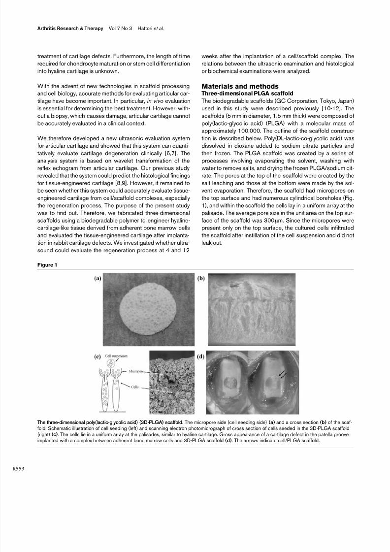
Materials and methodsThree-dimensional PLGA scaffold
The biodegradable scaffolds (GC Corporation, Tokyo, Japan)
used in this study were described previously [10-12]. The
scaffolds (5 mm in diameter, 1.5 mm thick) were composed of
poly(lactic-glycolic acid) (PLGA) with a molecular mass of
approximately 100,000. The outline of the scaffold construc-
tion is described below. Poly(DL-lactic-co-glycolic acid) was
dissolved in dioxane added to sodium citrate particles and
then frozen. The PLGA scaffold was created by a series of
processes involving evaporating the solvent, washing withwater to remove salts, and drying the frozen PLGA/sodium cit-
rate. The pores at the top of the scaffold were created by the
salt leaching and those at the bottom were made by the sol-
vent evaporation. Therefore, the scaffold had micropores onthe top surface and had numerous cylindrical boreholes (Fig.
1), and within the scaffold the cells lay in a uniform array at the
palisade. The average pore size in the unit area on the top sur-
face of the scaffold was 300µm. Since the micropores werepresent only on the top surface, the cultured cells infiltrated
the scaffold after instillation of the cell suspension and did not
leak out.
Figure 1
The three-dimensional poly(lactic-glycolic acid) (3D-PLGA) scaffoldThe three-dimensional poly(lactic-glycolic acid) (3D-PLGA) scaffold. The micropore side (cell seeding side) (a) and a cross section (b) of the scaf-fold. Schematic illustration of cell seeding (left) and scanning electron photomicrograph of cross section of cells seeded in the 3D-PLGA scaffold(right) (c). The cells lie in a uniform array at the palisades, similar to hyaline cartilage. Gross appearance of a cartilage defect in the patella grooveimplanted with a complex between adherent bone marrow cells and 3D-PLGA scaffold (d). The arrows indicate cell/PLGA scaffold.
7/18/2019 ultrasound cartilage defect analysis.pdf
http://slidepdf.com/reader/full/ultrasound-cartilage-defect-analysispdf 3/8
Available online http://arthritis-research.com/content/7/3/R552
R
Culture of adherent bone marrow cells
Twenty adult male Japanese white rabbits (3 to 4 kg) were
used in this study; they were individually maintained in
stainless-steel cages. The rabbits were anesthetized with a
mixture of ketamine (50 mg/ml) and xylazine (20 mg/ml) at a
ratio of 2:1, via a dose of 1 ml/kg injected intramuscularly intothe gluteal muscle. Bone marrow was then isolated from the
humeral head using an 18-gauge bone marrow needle, and 5
ml of the marrow was drawn into a 10-ml syringe containing
0.1 ml heparin (3,000 U/ml). The released cells were trans-
ferred to a T-75 flask (Costar, Cambridge, MA, USA) contain-
ing 15 ml of medium. The medium used was Eagle's minimal
essential medium (MEM) containing 10% fetal bovine serum
and antibiotics (penicillin, 100 U/ml; streptomycin, 0.1 mg/ml;
and amphotericin B (Fungizone), 0.25 g/ml; all from SigmaChemicals, St Louis, MO, USA). The cells were grown in a
humidified atmosphere of 5% carbon dioxide at 37°C and the
medium was replaced with fresh medium every 2 days. No
growth factors were added. The cell culture was maintainedfor 2 weeks until the cells reached confluence, and then the
cultured adherent bone marrow cells were released from the
substratum using 0.25% trypsin and counted in a hemocytom-
eter. The cultured cells obtained from each rabbit werereseeded onto three-dimensional PLGA scaffolds by simply
dropping the cell suspension onto the scaffolds. The density
of the cultured cells in a scaffold was 1 × 10⁷ cells/cm3. To
these composites in 35-mm tissue-culture plates we added 2
ml of fresh medium for subculture and the cultures were left to
stand overnight at 37°C in 5% carbon dioxide atmosphere.During this static overnight culture, the cultured cells in the
scaffold lay in uniform arrays in the palisades. The next day, the
composites of adherent bone marrow cells with the three-dimensional PLGA scaffold were implanted into osteochon-
dral defects in rabbit knee joints.
Implantation
Under general anesthesia as described above, an anterome-
dial arthrotomy was performed in one knee with the joint flexedmaximally. The patella was dislocated laterally and the surface
of the femoropatellar groove was exposed. A full-thickness
cylindrical cartilage defect (5 mm in diameter, 1.5 mm deep)
was created in the patellar groove of the knee using a chisel
and a disposable stainless-steel punch. After washing the
knee with saline solution and drying with a swab to remove any
debris, in some rabbits the defect in one knee was coveredwith a cell/PLGA scaffold, with the surface bearing the micro-
pores facing the subchondral bone; this was the tissue-engi-
neered-cartilage group (group T; n = 14). In a control group(group C; n = 6), defects were washed with saline solution
and dried in the same way but were left without any further
treatment. Finally, fibrin sealant (Tisseel®; Baxter AG, Vienna,
Austria) was applied between the scaffold and the edge of the
defect in group T and to the edge of the defect in group C. The
wound was then closed in layers with 2-0 vicryl sutures.
The rabbits were returned to their cages and allowed to move
freely without joint immobilization. The rabbits were humanely
killed with an overdose of phenobarbital sodium salt at 4 and
12 weeks in group T (groups T-4 (n = 8) and T-12 (n = 6),
respectively) and at 12 weeks in group C (n = 6). All the knee
joints were opened and the cartilage surfaces were observedwith the naked eye and photographed. The knee joint was dis-
sected free from all the soft tissues and the tibia was removed.
The distal femur was cut proximal to the patellofemoral joint
and cartilage samples were taken. All the animals were oper-
ated on in accordance with the guidelines for animal experi-
ments of the Nara Medical University Ethics Committee.
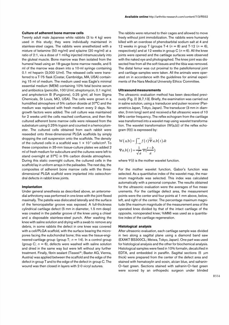
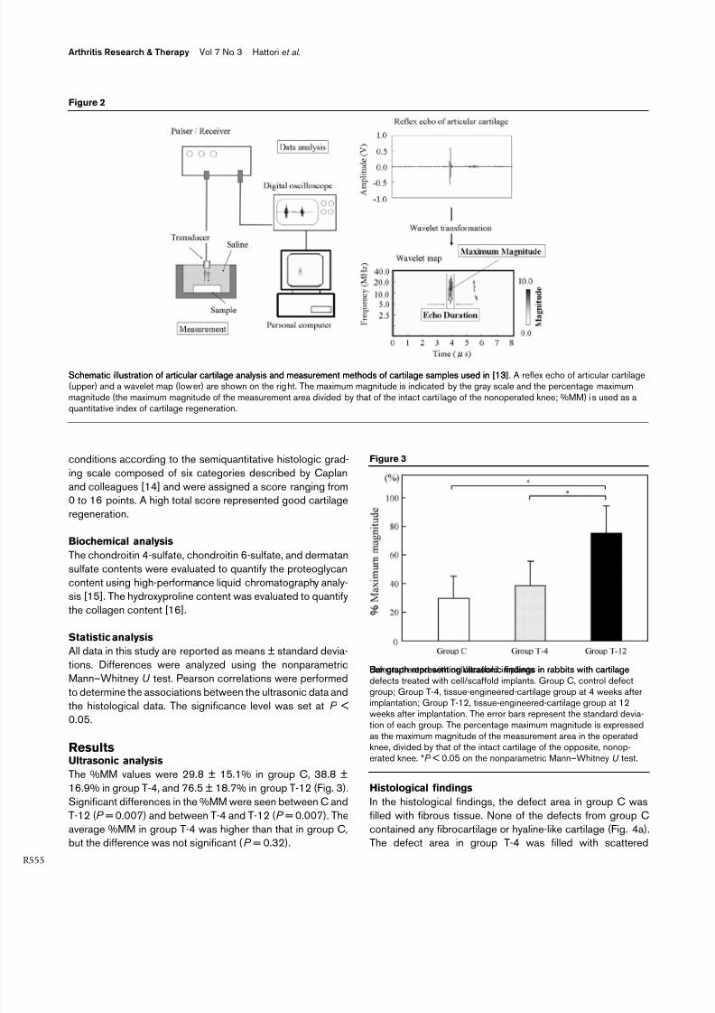
Ultrasound measurements
The ultrasonic evaluation method has been described previ-ously (Fig. 2) [6,7,13]. Briefly, the examination was carried out
in saline solution, using a transducer and pulser receiver (Pan-
ametrics Japan, Tokyo, Japan). The transducer (3 mm in diam-
eter, 3 mm long) sent and received a flat ultrasonic wave of 10MHz center frequency. The reflex echogram from the cartilage
was transformed into a wavelet map using wavelet transforma-
tion. The wavelet transformation (W(a,b)) of the reflex echo-
gram (f(t)) is expressed by:
whereΨ(t ) is the mother wavelet function.
For the mother wavelet function, Gabor's function wasselected. As a quantitative index of the wavelet map, the max-
imum magnitude was selected. This index was calculated
automatically with a personal computer. The results obtained
for the ultrasonic evaluation were the averages of five meas-
urements. For the cartilage defect area, the measurement
points were the center and four points at 1 mm above, below,left, and right of the center. The percentage maximum magni-
tude (the maximum magnitude of the measurement area of the
operated knee divided by that of the intact cartilage of the
opposite, nonoperated knee; %MM) was used as a quantita-
tive index of the cartilage regeneration.
Histological analysisAfter ultrasonic evaluation, each cartilage sample was divided
in two along a sagittal plane using a diamond band saw
(EXAKT BS300CL; Meiwa, Tokyo, Japan). One part was usedfor histological analysis and the other for biochemical analysis.
Histological samples were fixed in 10% formalin, decalcified in
EDTA, and embedded in paraffin. Sagittal sections (5 µm
thick) were prepared from the center of the defect area and
stained with hematoxylin and eosin, alcian blue, and safranin-
O–fast green. Sections stained with safranin-O–fast greenwere scored by an orthopedic surgeon under blinded
W a,b( ) = ( ) ( )
( ) = −
−∞
∞
∫ f t a b t dt
a b t a
t b
a
Ψ
Ψ Ψ
,
, ( )1
7/18/2019 ultrasound cartilage defect analysis.pdf
http://slidepdf.com/reader/full/ultrasound-cartilage-defect-analysispdf 4/8
Arthritis Research & Therapy Vol 7 No 3 Hattori et al.
555
conditions according to the semiquantitative histologic grad-
ing scale composed of six categories described by Caplan
and colleagues [14] and were assigned a score ranging from0 to 16 points. A high total score represented good cartilage
regeneration.
Biochemical analysis
The chondroitin 4-sulfate, chondroitin 6-sulfate, and dermatan
sulfate contents were evaluated to quantify the proteoglycan
content using high-performance liquid chromatography analy-
sis [15]. The hydroxyproline content was evaluated to quantify
the collagen content [16].
Statistic analysis
All data in this study are reported as means ± standard devia-
tions. Differences were analyzed using the nonparametric
Mann–Whitney U test. Pearson correlations were performed
to determine the associations between the ultrasonic data and
the histological data. The significance level was set at P <0.05.
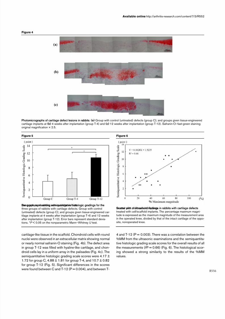
ResultsUltrasonic analysis
The %MM values were 29.8 ± 15.1% in group C, 38.8 ±
16.9% in group T-4, and 76.5 ± 18.7% in group T-12 (Fig. 3).
Significant differences in the %MM were seen between C and
T-12 (P = 0.007) and between T-4 and T-12 (P = 0.007). The
average %MM in group T-4 was higher than that in group C,but the difference was not significant (P = 0.32).
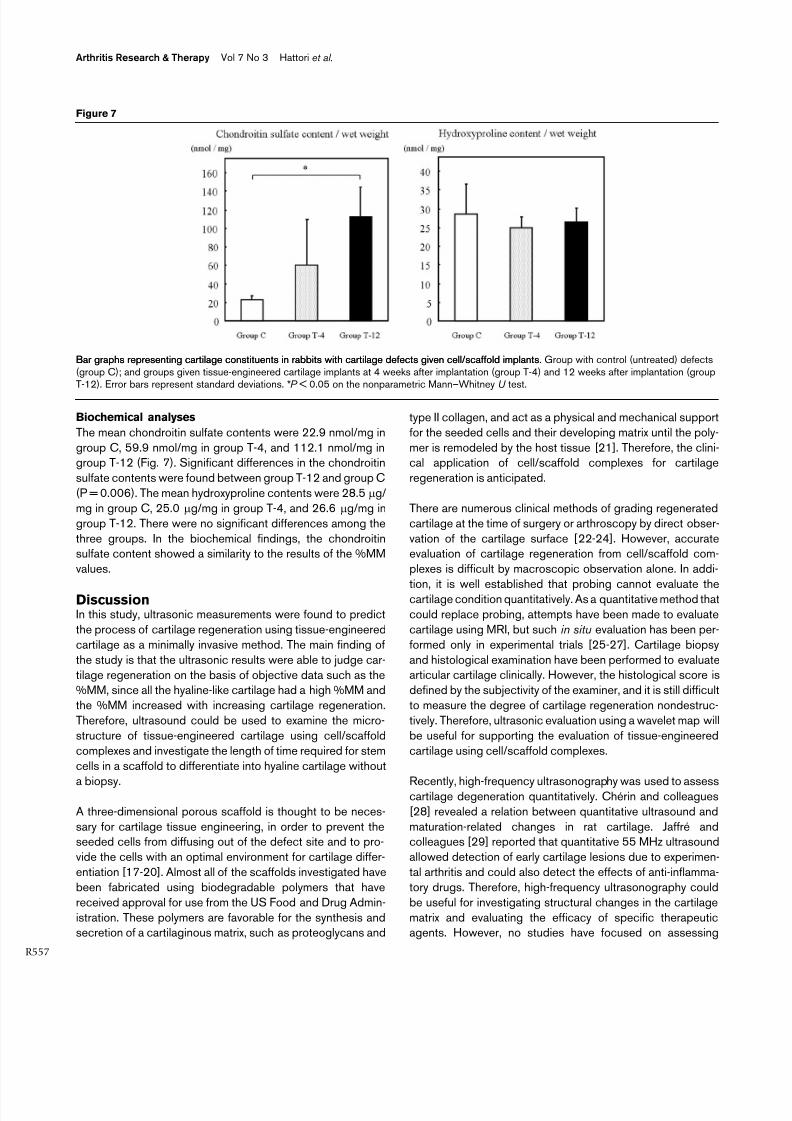
Histological findings
In the histological findings, the defect area in group C was
filled with fibrous tissue. None of the defects from group C
contained any fibrocartilage or hyaline-like cartilage (Fig. 4a).The defect area in group T-4 was filled with scattered
Figure 2
Schematic illustration of articular cartilage analysis and measurement methods of cartilage samples used in [13]Schematic illustration of articular cartilage analysis and measurement methods of cartilage samples used in [13]. A reflex echo of articular cartilage(upper) and a wavelet map (lower) are shown on the right. The maximum magnitude is indicated by the gray scale and the percentage maximummagnitude (the maximum magnitude of the measurement area divided by that of the intact carti lage of the nonoperated knee; %MM) is used as aquantitative index of cartilage regeneration.
Figure 3
Bar graph representing ultrasonic findings in rabbits with cartilagedefects treated with cell/scaffold implantsBar graph representing ultrasonic findings in rabbits with cartilagedefects treated with cell/scaffold implants. Group C, control defectgroup; Group T-4, tissue-engineered-cartilage group at 4 weeks afterimplantation; Group T-12, tissue-engineered-cartilage group at 12weeks after implantation. The error bars represent the standard devia-tion of each group. The percentage maximum magnitude is expressedas the maximum magnitude of the measurement area in the operatedknee, divided by that of the intact cartilage of the opposite, nonop-erated knee. *P < 0.05 on the nonparametric Mann–Whitney U test.
7/18/2019 ultrasound cartilage defect analysis.pdf
http://slidepdf.com/reader/full/ultrasound-cartilage-defect-analysispdf 5/8
Available online http://arthritis-research.com/content/7/3/R552
R
cartilage-like tissue in the scaffold. Chondroid cells with round
nuclei were observed in an extracellular matrix showing normal
or nearly normal safranin-O staining (Fig. 4b). The defect area
in group T-12 was filled with hyaline-like cartilage, and chon-
droid cells lay in a uniform array in the palisades (Fig. 4c). The
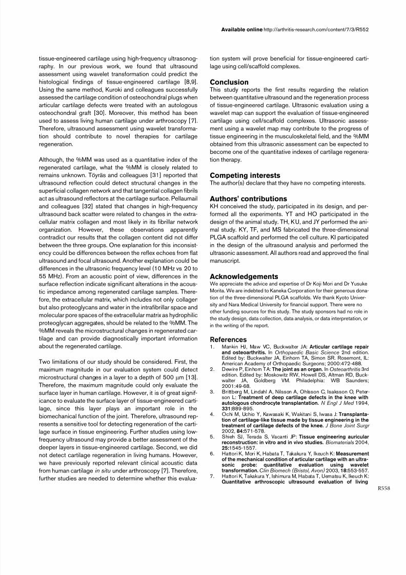
semiquantitative histologic grading scale scores were 4.17 ±1.72 for group C, 4.88 ± 1.81 for group T-4, and 10.7 ± 0.82
for group T-12 (Fig. 5). Significant differences in the scores
were found between C and T-12 (P = 0.004), and between T-
4 and T-12 (P = 0.003). There was a correlation between the
%MM from the ultrasonic examinations and the semiquantita-
tive histologic grading scale scores for the overall results of all
the measurements (R 2 = 0.66) (Fig. 6). The histological scor-
ing showed a strong similarity to the results of the %MM
values.
Figure 4
Photomicrographs of cartilage defect lesions in rabbitsPhotomicrographs of cartilage defect lesions in rabbits. (a) Group with control (untreated) defects (group C); and groups given tissue-engineeredcartilage implants at (b) 4 weeks after implantation (group T-4) and (c) 12 weeks after implantation (group T-12). Safranin-O–fast-green staining;original magnification × 2.5.
Figure 5
Bar graph representing semiquantitative histologic gradings for thethree groups of rabbits with cartilage defectsBar graph representing semiquantitative histologic gradings for thethree groups of rabbits with cartilage defects. Group with control(untreated) defects (group C); and groups given tissue-engineered car-tilage implants at 4 weeks after implantation (group T-4) and 12 weeksafter implantation (group T-12). Error bars represent standard devia-
tions. *P < 0.05 on the nonparametric Mann–Whitney U test.
Figure 6
Scatter plot of ultrasound findings in rabbits with cartilage defectstreated with cell/scaffold implantsScatter plot of ultrasound findings in rabbits with cartilage defectstreated with cell/scaffold implants. The percentage maximum magni-tude is expressed as the maximum magnitude of the measurement areain the operated knee, divided by that of the intact cartilage of the oppo-
site, nonoperated knee.
7/18/2019 ultrasound cartilage defect analysis.pdf
http://slidepdf.com/reader/full/ultrasound-cartilage-defect-analysispdf 6/8
Arthritis Research & Therapy Vol 7 No 3 Hattori et al.
557
Biochemical analyses
The mean chondroitin sulfate contents were 22.9 nmol/mg in
group C, 59.9 nmol/mg in group T-4, and 112.1 nmol/mg ingroup T-12 (Fig. 7). Significant differences in the chondroitin
sulfate contents were found between group T-12 and group C
(P = 0.006). The mean hydroxyproline contents were 28.5 µg/
mg in group C, 25.0 µg/mg in group T-4, and 26.6 µg/mg in
group T-12. There were no significant differences among the
three groups. In the biochemical findings, the chondroitinsulfate content showed a similarity to the results of the %MM
values.
DiscussionIn this study, ultrasonic measurements were found to predict
the process of cartilage regeneration using tissue-engineered
cartilage as a minimally invasive method. The main finding of
the study is that the ultrasonic results were able to judge car-
tilage regeneration on the basis of objective data such as the%MM, since all the hyaline-like cartilage had a high %MM and
the %MM increased with increasing cartilage regeneration.
Therefore, ultrasound could be used to examine the micro-
structure of tissue-engineered cartilage using cell/scaffold
complexes and investigate the length of time required for stem
cells in a scaffold to differentiate into hyaline cartilage without
a biopsy.
A three-dimensional porous scaffold is thought to be neces-
sary for cartilage tissue engineering, in order to prevent theseeded cells from diffusing out of the defect site and to pro-
vide the cells with an optimal environment for cartilage differ-
entiation [17-20]. Almost all of the scaffolds investigated have
been fabricated using biodegradable polymers that have
received approval for use from the US Food and Drug Admin-
istration. These polymers are favorable for the synthesis andsecretion of a cartilaginous matrix, such as proteoglycans and
type II collagen, and act as a physical and mechanical support
for the seeded cells and their developing matrix until the poly-
mer is remodeled by the host tissue [21]. Therefore, the clini-cal application of cell/scaffold complexes for cartilage
regeneration is anticipated.
There are numerous clinical methods of grading regenerated
cartilage at the time of surgery or arthroscopy by direct obser-
vation of the cartilage surface [22-24]. However, accurateevaluation of cartilage regeneration from cell/scaffold com-
plexes is difficult by macroscopic observation alone. In addi-
tion, it is well established that probing cannot evaluate thecartilage condition quantitatively. As a quantitative method that
could replace probing, attempts have been made to evaluate
cartilage using MRI, but such in situ evaluation has been per-
formed only in experimental trials [25-27]. Cartilage biopsy
and histological examination have been performed to evaluatearticular cartilage clinically. However, the histological score is
defined by the subjectivity of the examiner, and it is still difficult
to measure the degree of cartilage regeneration nondestruc-
tively. Therefore, ultrasonic evaluation using a wavelet map will
be useful for supporting the evaluation of tissue-engineered
cartilage using cell/scaffold complexes.
Recently, high-frequency ultrasonography was used to assesscartilage degeneration quantitatively. Chérin and colleagues
[28] revealed a relation between quantitative ultrasound and
maturation-related changes in rat cartilage. Jaffré andcolleagues [29] reported that quantitative 55 MHz ultrasound
allowed detection of early cartilage lesions due to experimen-
tal arthritis and could also detect the effects of anti-inflamma-
tory drugs. Therefore, high-frequency ultrasonography could
be useful for investigating structural changes in the cartilage
matrix and evaluating the efficacy of specific therapeuticagents. However, no studies have focused on assessing
Figure 7
Bar graphs representing cartilage constituents in rabbits with cartilage defects given cell/scaffold implantsBar graphs representing cartilage constituents in rabbits with cartilage defects given cell/scaffold implants. Group with control (untreated) defects(group C); and groups given tissue-engineered cartilage implants at 4 weeks after implantation (group T-4) and 12 weeks after implantation (groupT-12). Error bars represent standard deviations. *P < 0.05 on the nonparametric Mann–Whitney U test.
7/18/2019 ultrasound cartilage defect analysis.pdf
http://slidepdf.com/reader/full/ultrasound-cartilage-defect-analysispdf 7/8
Available online http://arthritis-research.com/content/7/3/R552
R
tissue-engineered cartilage using high-frequency ultrasonog-
raphy. In our previous work, we found that ultrasound
assessment using wavelet transformation could predict the
histological findings of tissue-engineered cartilage [8,9].
Using the same method, Kuroki and colleagues successfully
assessed the cartilage condition of osteochondral plugs whenarticular cartilage defects were treated with an autologous
osteochondral graft [30]. Moreover, this method has been
used to assess living human cartilage under arthroscopy [7].
Therefore, ultrasound assessment using wavelet transforma-
tion should contribute to novel therapies for cartilage
regeneration.
Although, the %MM was used as a quantitative index of the
regenerated cartilage, what the %MM is closely related toremains unknown. Töyräs and colleagues [31] reported that
ultrasound reflection could detect structural changes in the
superficial collagen network and that tangential collagen fibrils
act as ultrasound reflectors at the cartilage surface. Pellaumailand colleagues [32] stated that changes in high-frequency
ultrasound back scatter were related to changes in the extra-
cellular matrix collagen and most likely in its fibrillar network
organization. However, these observations apparentlycontradict our results that the collagen content did not differ
between the three groups. One explanation for this inconsist-
ency could be differences between the reflex echoes from flat
ultrasound and focal ultrasound. Another explanation could be
differences in the ultrasonic frequency level (10 MHz vs 20 to
55 MHz). From an acoustic point of view, differences in thesurface reflection indicate significant alterations in the acous-
tic impedance among regenerated cartilage samples. There-
fore, the extracellular matrix, which includes not only collagenbut also proteoglycans and water in the intrafibrillar space and
molecular pore spaces of the extracellular matrix as hydrophilic
proteoglycan aggregates, should be related to the %MM. The
%MM reveals the microstructural changes in regenerated car-
tilage and can provide diagnostically important informationabout the regenerated cartilage.
Two limitations of our study should be considered. First, the
maximum magnitude in our evaluation system could detect
microstructural changes in a layer to a depth of 500 µm [13].
Therefore, the maximum magnitude could only evaluate the
surface layer in human cartilage. However, it is of great signif-
icance to evaluate the surface layer of tissue-engineered carti-lage, since this layer plays an important role in the
biomechanical function of the joint. Therefore, ultrasound rep-
resents a sensitive tool for detecting regeneration of the carti-lage surface in tissue engineering. Further studies using low-
frequency ultrasound may provide a better assessment of the
deeper layers in tissue-engineered cartilage. Second, we did
not detect cartilage regeneration in living humans. However,
we have previously reported relevant clinical acoustic data
from human cartilage in situ under arthroscopy [7]. Therefore,further studies are needed to determine whether this evalua-
tion system will prove beneficial for tissue-engineered carti-
lage using cell/scaffold complexes.
ConclusionThis study reports the first results regarding the relation
between quantitative ultrasound and the regeneration processof tissue-engineered cartilage. Ultrasonic evaluation using a
wavelet map can support the evaluation of tissue-engineered
cartilage using cell/scaffold complexes. Ultrasonic assess-
ment using a wavelet map may contribute to the progress of
tissue engineering in the musculoskeletal field, and the %MM
obtained from this ultrasonic assessment can be expected to
become one of the quantitative indexes of cartilage regenera-
tion therapy.
Competing interestsThe author(s) declare that they have no competing interests.
Authors' contributionsKH conceived the study, participated in its design, and per-
formed all the experiments. YT and HO participated in the
design of the animal study. TH, KU, and JY performed the ani-
mal study. KY, TF, and MS fabricated the three-dimensionalPLGA scaffold and performed the cell culture. KI participated
in the design of the ultrasound analysis and performed the
ultrasonic assessment. All authors read and approved the final
manuscript.
AcknowledgementsWe appreciate the advice and expertise of Dr Koji Mori and Dr Yusuke
Morita. We are indebted to Kaneka Corporation for their generous dona-
tion of the three-dimensional PLGA scaffolds. We thank Kyoto Univer-
sity and Nara Medical University for financial support. There were no
other funding sources for this study. The study sponsors had no role in
the study design, data collection, data analysis, or data interpretation, or
in the writing of the report.
References1. Mankin HJ, Maw VC, Buckwalter JA: Articular cartilage repair
and osteoarthritis. In Orthopaedic Basic Science 2nd edition.Edited by: Buckwalter JA, Einhorn TA, Simon SR. Rosemont, IL:American Academy of Orthopaedic Surgeons; 2000:472-488.
2. Dewire P, Einhorn TA: The joint as an organ. In Osteoarthritis 3rdedition. Edited by: Moskowitz RW, Howell DS, Altman RD, Buck-walter JA, Goldberg VM. Philadelphia: WB Saunders;2001:49-68.
3. Brittberg M, Lindahl A, Nilsson A, Ohlsson C, Isaksson O, Peter-son L: Treatment of deep cartilage defects in the knee with
autologous chondrocyte transplantation. N Engl J Med 1994,331:889-895.
4. Ochi M, Uchio Y, Kawasaki K, Wakitani S, Iwasa J: Transplanta-tion of cartilage-like tissue made by tissue engineering in thetreatment of cartilage defects of the knee. J Bone Joint Surgr 2002, 84:571-578.
5. Shieh SJ, Terada S, Vacanti JP: Tissue engineering auricularreconstruction: in vitro and in vivo studies. Biomaterials 2004,25:1545-1557.
6. Hattori K, Mori K, Habata T, Takakura Y, Ikeuch K: Measurementof the mechanical condition of articular cartilage with an ultra-sonic probe: quantitative evaluation using wavelettransformation. Clin Biomech (Bristol, Avon) 2003, 18:553-557.
7. Hattori K, Takakura Y, Ishimura M, Habata T, Uematsu K, Ikeuch K:Quantitative arthroscopic ultrasound evaluation of living
http://www.ncbi.nlm.nih.gov/entrez/query.fcgi?cmd=Retrieve&db=PubMed&dopt=Abstract&list_uids=8078550
http://www.ncbi.nlm.nih.gov/entrez/query.fcgi?cmd=Retrieve&db=PubMed&dopt=Abstract&list_uids=8078550
http://www.ncbi.nlm.nih.gov/entrez/query.fcgi?cmd=Retrieve&db=PubMed&dopt=Abstract&list_uids=8078550
7/18/2019 ultrasound cartilage defect analysis.pdf
http://slidepdf.com/reader/full/ultrasound-cartilage-defect-analysispdf 8/8
Arthritis Research & Therapy Vol 7 No 3 Hattori et al.
559
human cartilage. Clin Biomech (Bristol, Avon) 2004,19:213-216.
8. Hattori K, Takakura Y, Morita Y, Takenaka M, Uematsu K, IkeuchiK: Can ultrasound predict histological findings in regeneratedcartilage? Rheumatology 2004, 43:302-305.
9. Hattori K, Takakura Y, Ohgushi H, Habata T, Uematsu K, TakenakaM, Ikeuchi K: Which cartilage is regenerated, hyaline cartilageor fibrocartilage? Non-invasive ultrasonic evaluation of tissue-engineered cartilage. Rheumatology 2004, 43:1106-1108.
10. Uematsu K, Hattori K, Ishimoto Y, Yamauchi J, Habata T, TakakuraY, Ohgushi H, Fukuchi T, Satoh M: Cartilage repair with a spe-cially three-dimensional poly-lactic-glycolic acid scaffold[abstract]. Trans Orthop Research Soc 2004, 29:676.
11. Kawanishi M, Ushida T, Kaneko T, Niwa H, Fukubayashi T, Naka-mura K, Oda H, Tanaka S, Tateishi T: New type of biodegradableporous scaffolds for tissue-engineered articular cartilage.Mater Sci Eng C 2004, 24:431-435.
12. Uematsu K, Hattori K, Ishimoto Y, Yamauchi J, Habata T, TakakuraY, Ohgushi H, Fukuchi T, Sato M: Cartilage regeneration usingmesenchymal stem cells and a three-dimensional poly-lactic-glycolic acid (PLGA) scaffold. Biomaterials 2005,26:4273-4279.
13. Hattori K, Ikeuchi K, Morita Y, Takakura Y: Quantitative ultrasonicassessment for detecting microscopic cartilage damage inosteoarthritis. Arthritis Res Ther 2005, 7:R38-46.
14. Caplan AI, Elyaderani M, Mochizuki Y, Wakitani S, Goldberg VM:Principles of cartilage repair and regeneration. Clin Orthop1997, 342:254-269.
15. Shinmei M, Miyauchi S, Machida A, Miyazaki K: Quantification ofchondroitin 4-sulfate and chondroitin 6-sulfate in pathologicjoint fluid. Arthritis Rheum 1992, 35:1304-1308.
16. Woessner J: The determination of hydroxyproline in tissue andprotein samples containing small proportions of this iminoacid. Arch Biochem Biophys 1961, 93:440-447.
17. Chen G, Sato T, Ushida T, Ochiai N, Tateishi T: Tissue engineer-ing of cartilage using a hybrid scaffold of synthetic polymerand collagen. Tissue Eng 2004, 10:323-330.
18. Woodfield TB, Malda J, de Wijn J, Peters F, Riesle J, van Blittersw-ijk CA: Design of porous scaffolds for cartilage tissue engi-neering using a three-dimensional fiber-deposition technique.Biomaterials 2004, 25:4149-4161.
19. Peter SJ, Miller MJ, Yasko AW, Yaszemski MJ, Mikos AG: Polymerconcepts in tissue engineering. J Biomed Mater Res 1998,
43:422-427.20. Isogai N, Landis W, Kim TH, Gerstenfeld LC, Upton J, Vacanti JP:Formation of phalanges and small joints by tissue-engineer-ing. J Bone Joint Surg Am 1999, 81:306-316.
21. Isogai N, Asamura S, Higashi T, Ikada Y, Morita S, Hillyer J, JacquetR, Landis WJ: Tissue engineering of an auricular cartilagemodel utilizing cultured chondrocyte-poly(L-lactide-epsilon-caprolactone) scaffolds. Tissue Eng 2004, 10:673-687.
22. Outerbridge RE: The etiology of chondromalacia patellae. J Bone Joint Surg 1961, 43B:752-757.
23. Dougados M, Ayral X, Listrat V, Gueguen A, Bahuaud J, Beaufils P,Beguin JA, Bonvarlet JP, Boyer T, Coudane H: The SFA systemfor assessing articular cartilage lesions at arthroscopy of theknee. Arthroscopy 1994, 10:69-77.
24. Brismar BH, Wredmark T, Movin T, Leandersson J, Svensson O:Observation reliability in the arthroscopic classification ofosteoarthritis of the knee. J Bone Joint Surg 2002, 84B:42-47.
25. Nissi MJ, Toyras J, Laasanen MS, Rieppo J, Saarakkala S, Lappal-
ainen R, Jurvelin JS, Nieminen MT: Proteoglycan and collagensensitive MRI evaluation of normal and degenerated articularcartilage. J Orthop Res 2004, 22:557-564.
26. Graichen H, von Eisenhart-Rothe R, Vogl T, Englmeier KH, Eck-stein F: Quantitative assessment of cartilage status in osteoar-thritis by quantitative magnetic resonance imaging: technicalvalidation for use in analysis of cartilage volume and furthermorphologic parameters. Arthritis Rheum 2004, 50:811-816.
27. Watrin-Pinzano A, Ruaud JP, Cheli Y, Gonord P, Grossin L, GilletP, Blum A, Payan E, Olivier P, Guillot G, et al.: T2 mapping: anefficient MR quantitative technique to evaluate spontaneouscartilage repair in rat patella. Osteoarthritis Cartilage 2004,12:191-200.
28. Chérin E, Saïed A, Pellaumail B, Loeuille D, Laugier P, Gillet P, Net-ter P, Berger G: Assessment of rat articular cartilage matura-
tion using 50-MHz quantitative ultrasonography. OsteoarthritisCartilage 2001, 9:178-186.
29. Jaffré B, Watrin A, Loeuille D, Gillet P, Netter P, Laugier P, SaïedA: Effects of antiinflammatory drugs on arthritic cartilage: ahigh-frequency quantitative ultrasound study in rats. ArthritisRheum 2003, 48:1594-1601.
30. Kuroki H, Nakagawa Y, Mori K, Ohba M, Suzuki T, Mizuno Y, AndoK, Takenaka M, Ikeuchi K, Nakamura T: Acoustic stiffness andchange in plug cartilage over time after autologous osteo-chondral grafting: correlation between ultrasound signalintensity and histological score in a rabbit model. Arthritis ResTher 2004, 6:R492-504.
31. Töyräs J, Rieppo J, Nieminen MT, Helminen HJ, Jurvelin JS: Char-acterization of enzymatically induced degradation of articularcartilage using high frequency ultrasound. Phys Med Biol 1999, 44:2723-2733.
32. Pellaumail B, Watrin A, Loeuille D, Netter P, Berger G, Laugier P,Saïed A: Effect of articular cartilage proteoglycan depletion onhigh frequency ultrasound backscatter. Osteoarthritis Cartilage2002, 10:535-541.
http://www.ncbi.nlm.nih.gov/entrez/query.fcgi?cmd=Retrieve&db=PubMed&dopt=Abstract&list_uids=9308548
http://www.ncbi.nlm.nih.gov/entrez/query.fcgi?cmd=Retrieve&db=PubMed&dopt=Abstract&list_uids=1445446
http://www.ncbi.nlm.nih.gov/entrez/query.fcgi?cmd=Retrieve&db=PubMed&dopt=Abstract&list_uids=1445446
http://www.ncbi.nlm.nih.gov/entrez/query.fcgi?cmd=Retrieve&db=PubMed&dopt=Abstract&list_uids=1445446
http://www.ncbi.nlm.nih.gov/entrez/query.fcgi?cmd=Retrieve&db=PubMed&dopt=Abstract&list_uids=9855200
http://www.ncbi.nlm.nih.gov/entrez/query.fcgi?cmd=Retrieve&db=PubMed&dopt=Abstract&list_uids=9855200
http://www.ncbi.nlm.nih.gov/entrez/query.fcgi?cmd=Retrieve&db=PubMed&dopt=Abstract&list_uids=8166905
http://www.ncbi.nlm.nih.gov/entrez/query.fcgi?cmd=Retrieve&db=PubMed&dopt=Abstract&list_uids=8166905
http://www.ncbi.nlm.nih.gov/entrez/query.fcgi?cmd=Retrieve&db=PubMed&dopt=Abstract&list_uids=8166905
http://www.ncbi.nlm.nih.gov/entrez/query.fcgi?cmd=Retrieve&db=PubMed&dopt=Abstract&list_uids=8166905
http://www.ncbi.nlm.nih.gov/entrez/query.fcgi?cmd=Retrieve&db=PubMed&dopt=Abstract&list_uids=8166905
http://www.ncbi.nlm.nih.gov/entrez/query.fcgi?cmd=Retrieve&db=PubMed&dopt=Abstract&list_uids=8166905
http://www.ncbi.nlm.nih.gov/entrez/query.fcgi?cmd=Retrieve&db=PubMed&dopt=Abstract&list_uids=9855200
http://www.ncbi.nlm.nih.gov/entrez/query.fcgi?cmd=Retrieve&db=PubMed&dopt=Abstract&list_uids=9855200
http://www.ncbi.nlm.nih.gov/entrez/query.fcgi?cmd=Retrieve&db=PubMed&dopt=Abstract&list_uids=1445446
http://www.ncbi.nlm.nih.gov/entrez/query.fcgi?cmd=Retrieve&db=PubMed&dopt=Abstract&list_uids=1445446
http://www.ncbi.nlm.nih.gov/entrez/query.fcgi?cmd=Retrieve&db=PubMed&dopt=Abstract&list_uids=1445446