ukmss-37127
20
Diagnosis and management of ectopic pregnancy Vanitha N Sivalingam 1 , W Colin Duncan 2 , Emma Kirk 3 , Lucy A Shephard 2 , and Andrew W Horne 2 1 Simpson Centre for Reproductive Health, Royal Infirmary of Edinburgh, 51 Little France Crescent, Edinburgh EH16 4SA 2 MRC Centre for Reproductive Health, University of Edinburgh, Queen’s Medical Research Institute, 47 Little France Crescent, Edinburgh EH16 4TJ, UK 3 North Middlesex University Hospital, Sterling Way, London, N18 1QX Keywords ectopic pregnancy; beta-human chorionic gonadotrophin; transvaginal ultrasound; methotrexate; salpingectomy; salpingostomy Overview An ectopic pregnancy occurs when a fertilised ovum implants outside the normal uterine cavity. 1-3 It is a common cause of morbidity and occasionally of mortality in women of reproductive age. The aetiology of ectopic pregnancy remains uncertain although a number of risk factors have been identified. 4 Its diagnosis can be difficult. In current practice, in developed countries, diagnosis relies on a combination of ultrasound scanning and serial serum beta-human chorionic gonadotrophin (β-hCG) measurements. 5 Ectopic pregnancy is one of the few medical conditions that can be managed expectantly, medically or surgically. 136 Incidence In the developed world, between 1% and 2% of all reported pregnancies are ectopic pregnancies (comparable to the incidence of spontaneous twin pregnancy). 7 The incidence is thought to be higher in developing countries, but specific numbers are unknown. Although the incidence in the developed world has remained relatively static in recent years, between 1972 and 1992 there was an estimated six-fold rise in the incidence of ectopic pregnancy. 8 This increase was attributed to three factors: an increase in risk factors such as pelvic inflammatory disease and smoking in women of reproductive age, the increased use of assisted reproductive technology (ART) and increased awareness of the condition, facilitated by the development of specialised early pregnancy units (EPUs). CORRESPONDING AUTHOR: Dr Andrew Horne, MRC Centre for Reproductive Health, University of Edinburgh, Queen’s Medical Research Institute, 47 Little France Crescent, Edinburgh EH16 4TJ, UK, [email protected] Tel +44 131 242 6609, Fax +44 131 242 6441. COMPETING INTEREST: Andrew Horne holds a UK patent for a diagnostic biomarker for ectopic pregnancy (# 0712801.0). Europe PMC Funders Group Author Manuscript J Fam Plann Reprod Health Care. Author manuscript; available in PMC 2012 January 01. Published in final edited form as: J Fam Plann Reprod Health Care. 2011 October ; 37(4): 231–240. doi:10.1136/jfprhc-2011-0073. Europe PMC Funders Author Manuscripts Europe PMC Funders Author Manuscripts
-
Upload
deby-nelsya-eka-zein -
Category
Documents
-
view
3 -
download
0
description
ksxn
Transcript of ukmss-37127
Diagnosis and management of ectopic pregnancy
Vanitha N Sivalingam1, W Colin Duncan2, Emma Kirk3, Lucy A Shephard2, and Andrew WHorne2
1Simpson Centre for Reproductive Health, Royal Infirmary of Edinburgh, 51 Little FranceCrescent, Edinburgh EH16 4SA2MRC Centre for Reproductive Health, University of Edinburgh, Queen’s Medical ResearchInstitute, 47 Little France Crescent, Edinburgh EH16 4TJ, UK3North Middlesex University Hospital, Sterling Way, London, N18 1QX
Keywordsectopic pregnancy; beta-human chorionic gonadotrophin; transvaginal ultrasound; methotrexate;salpingectomy; salpingostomy
OverviewAn ectopic pregnancy occurs when a fertilised ovum implants outside the normal uterinecavity.1-3 It is a common cause of morbidity and occasionally of mortality in women ofreproductive age. The aetiology of ectopic pregnancy remains uncertain although a numberof risk factors have been identified.4 Its diagnosis can be difficult. In current practice, indeveloped countries, diagnosis relies on a combination of ultrasound scanning and serialserum beta-human chorionic gonadotrophin (β-hCG) measurements.5 Ectopic pregnancy isone of the few medical conditions that can be managed expectantly, medically orsurgically.136
IncidenceIn the developed world, between 1% and 2% of all reported pregnancies are ectopicpregnancies (comparable to the incidence of spontaneous twin pregnancy).7 The incidence isthought to be higher in developing countries, but specific numbers are unknown. Althoughthe incidence in the developed world has remained relatively static in recent years, between1972 and 1992 there was an estimated six-fold rise in the incidence of ectopic pregnancy.8
This increase was attributed to three factors: an increase in risk factors such as pelvicinflammatory disease and smoking in women of reproductive age, the increased use ofassisted reproductive technology (ART) and increased awareness of the condition, facilitatedby the development of specialised early pregnancy units (EPUs).
CORRESPONDING AUTHOR: Dr Andrew Horne, MRC Centre for Reproductive Health, University of Edinburgh, Queen’sMedical Research Institute, 47 Little France Crescent, Edinburgh EH16 4TJ, UK, [email protected] Tel +44 131 242 6609, Fax+44 131 242 6441.
COMPETING INTEREST:Andrew Horne holds a UK patent for a diagnostic biomarker for ectopic pregnancy (# 0712801.0).
Europe PMC Funders GroupAuthor ManuscriptJ Fam Plann Reprod Health Care. Author manuscript; available in PMC 2012 January 01.
Published in final edited form as:J Fam Plann Reprod Health Care. 2011 October ; 37(4): 231–240. doi:10.1136/jfprhc-2011-0073.
Europe PM
C Funders A
uthor Manuscripts
Europe PM
C Funders A
uthor Manuscripts
Morbidity and mortalityIn the UK, ectopic pregnancy remains the leading cause of pregnancy-related first trimesterdeath (0.35/1000 ectopic pregnancies).369 However, in the developing world it has beenestimated that 10% of women admitted to hospital with a diagnosis of ectopic pregnancyultimately die from the condition.10 Ectopic pregnancy is a considerable cause of maternalmorbidity, causing acute symptoms such as pelvic pain and vaginal bleeding and long-termproblems such as infertility.3 Short- and long-term consequences of ectopic pregnancy onhealth-related quality of life and on bereavement issues are likely to be significant but havenot been formally quantified.
Risk factorsAlthough women with ectopic pregnancy frequently have no identifiable risk factors, aprospective case-controlled study has shown that increased awareness of ectopic pregnancyand a knowledge of the associated risk factors helps identify women at higher risk in orderto facilitate early and more accurate diagnosis.11 Most risk factors are associated with risksof prior damage to the Fallopian tube (Box 1). These factors include any previous pelvic orabdominal surgery, and pelvic infection.11 Chlamydia trachomatis has been linked to30-50% of all ectopic pregnancies.12 The exact mechanism of this association is not knownbut it has been proposed that in addition to distortion of tubal architecture, it may to be dueto an effect on the tubal microenvironment.13
Ectopic pregnancy is more common in women attending infertility clinics14 even in theabsence of tubal disease. In addition, the use of ART increases the rate of ectopicpregnancies. In vitro fertilisation (IVF) is associated with an ectopic pregnancy risk of 2-5%and it may be higher than this where there is tubal disease. Indeed the first IVF pregnancy,before the first IVF live birth, was a tubal ectopic pregnancy.15
Some types of contraception, such as progestogen-only contraception and the intrauterinecontraceptive device, are associated with an increased incidence of ectopic pregnancy whenthere is contraceptive failure, without necessarily increasing the absolute risk of ectopicpregnancy.16
One third of all cases of ectopic pregnancy are thought to be associated with smoking.17
There is a dose–effect relationship, with the highest adjusted odds ratio (OR) (3.9) whenmore than 20 cigarettes are smoked a day.18 Several mechanisms for this association havebeen suggested, including one or more of the following: delayed ovulation, altered tubal anduterine motility and microenvironment, or altered immunity.1920
The risk of ectopic pregnancy increases with advancing maternal age, with age over 35 yearsbeing a significant risk factor.6 Hypotheses for this association include the higherprobability of exposure to most other risk factors with advancing age, increase inchromosomal abnormalities in trophoblastic tissue and age-related changes in tubal functiondelaying ovum transport, resulting in tubal implantation.18
Women with a previous history of ectopic pregnancy also have an increased risk, whichincreases further in proportion to the number of previous ectopic pregnancies. In one studythe OR for having an ectopic pregnancy was 12.5 after one previous ectopic pregnancy and76.6 after two.18
Sivalingam et al. Page 2
J Fam Plann Reprod Health Care. Author manuscript; available in PMC 2012 January 01.
Europe PM
C Funders A
uthor Manuscripts
Europe PM
C Funders A
uthor Manuscripts
AetiologyThe exact aetiology of ectopic pregnancy is unknown. It is notable that it is unique tohumans, and perhaps the higher apes, so that there are no good animal models that could beused to further our understanding.21 However, it is thought that tubal implantation occurs asa result of a combination of arrest of the embryo in the Fallopian tube and changes in thetubal microenvironment that allow early implantation to occur.4 Inflammation within thetube, resulting from infection or smoking, may affect embryo-tubal transport by disruptingsmooth muscle contractility and ciliary beat activity and may also provide pro-implantationsignals. Molecular research generally involves studying Fallopian tube biopsies taken fromwomen with ectopic pregnancies. Interpretation is limited as comparable Fallopian tubesamples are not available from women with an intrauterine pregnancy (IUP) or in advanceof an ectopic pregnancy occurring. Thus, it is difficult to ascertain whether any molecularchanges observed are a cause or a consequence of ectopic implantation. Novel studiesfocusing on the functional consequences of smoking and infection on Fallopian tubephysiology and pathobiology are required.
Clinical presentationPatients with an ectopic pregnancy commonly present with pain and vaginal bleedingbetween 6 and 10 weeks’ gestation.1 However, these are common symptoms in earlypregnancy, with one third of women experiencing some pain and/or bleeding.22-24 The paincan be persistent and severe and is often unilateral. However unilateral pain is not alwaysindicative of ectopic pregnancy as, in early pregnancy, a prominent painful ovarian corpusluteum cyst is common. Shoulder tip pain, syncope and shock occur in up to 20% of womenand abdominal tenderness in more than 75%. Bimanual examination, if performed at all,should be done cautiously and gently. Cervical motion tenderness has been reported in up to67% of cases, and a palpable adnexal mass in about 50%.23-25 More recently, it has beenreported that one third of women with ectopic pregnancy have no clinical signs and 9% haveno symptoms.2627
A ruptured ectopic pregnancy should be strongly suspected if a woman has a positivepregnancy test and presents with syncope and signs of shock including tachycardia, pallorand collapse. There may be abdominal distension and marked tenderness. While a bimanualexamination may reveal tenderness, cervical excitation and an adnexal mass, great caution isrequired as this may exacerbate bleeding. As ectopic pregnancy affects young, fit womenthey are often able to mount remarkable haemodynamic compensation. Tachycardia is aparticularly important sign, but decompensation with shock is a sign of significantintraperitoneal bleeding. In an emergency, where the patient has collapsed and there is highclinical suspicion of tubal rupture, extensive clinical examination is inappropriate andimmediate surgical intervention is indicated.
Unfortunately, atypical presentation is also relatively common. Ectopic pregnancy maymimic other gynaecological disorders and gastrointestinal or urinary tract disease, includingappendicitis, salpingitis, ruptured corpus luteum or follicular cysts, threatened or inevitablespontaneous abortion, ovarian torsion and urinary tract infection. The 1997-1999 and2003-2005 Confidential Enquiries into Maternal Deaths reports highlighted that most of thewomen who died from ectopic pregnancy were misdiagnosed in the primary care or accidentand emergency settings.2829 It was therefore recommended that all clinicians should bemade aware of the atypical clinical presentations of ectopic pregnancy. While there has beena welcome decline in the case death rate in women with ectopic pregnancies, a key lessonemphasised in these reports does not appear to have been learnt. In the 2006-2008 Centre forMaternal and Child Enquiries (CMACE) report, four of the six women who died from early
Sivalingam et al. Page 3
J Fam Plann Reprod Health Care. Author manuscript; available in PMC 2012 January 01.
Europe PM
C Funders A
uthor Manuscripts
Europe PM
C Funders A
uthor Manuscripts

ectopic pregnancy complained of diarrhoea, dizziness or vomiting as early symptoms,without triggering any consideration of extrauterine pregnancy by their medicalattendants.30
However, it remains difficult to diagnose an ectopic pregnancy from risk factors, history andexamination alone. Clinicians should be suspicious of pregnancy in any such woman whopresents with abdominal or pelvic symptoms and should always bear in mind the possibilityof ectopic pregnancy in any woman of reproductive age who presents with any of thesymptoms mentioned above.
DiagnosisDiagnosis of ectopic pregnancy has improved significantly due to advances in ultrasoundtechnology, rapid and sensitive serum hormone assays, the development of EPUs and anincreased awareness and understanding of the associated risk factors. Despite this, aroundhalf of the women with an eventual diagnosis of ectopic pregnancy are not diagnosed at theirfirst presentation.3132 Early diagnosis reduces the risk of tubal rupture and allows moreconservative medical treatments to be employed.133
Currently, diagnosis in unruptured ectopic pregnancy is achieved using a combination oftransvaginal ultrasonography and measurement of serum β-hCG concentrations. One of thekey elements in the diagnosis is the exclusion of a viable or non-viable IUP. Diagnosis canbe straightforward when a transvaginal ultrasound scan (TVS) positively identifies an IUPor ectopic pregnancy34 (Figure 1). However, TVS fails to identify the location of apregnancy in a significant number of women and such women are currently diagnosed ashaving a ‘pregnancy of unknown location’ (PUL).3536
The 2006–2008 CMACE report drew attention to a maternal death secondary to rupturedectopic pregnancy where a diagnosis of PUL had been made.30 Although most patients witha PUL will subsequently be diagnosed with either a failed IUP (a spontaneous abortion) orviable IUP, the report highlights that 7-20% will be diagnosed with an ectopic pregnancy. Itis therefore very important that a diagnosis of PUL should trigger further diagnosticpathways and follow-up until the final outcome of the pregnancy is known.
The concept of a ‘discriminatory β-hCG level’ was introduced in 1985 to highlight theserum concentration of β-hCG when a pregnancy should be visible on an ultrasound scan.Using transabdominal ultrasound examination, it was reported then that the absence of anintrauterine gestational sac at a β-hCG concentration over 6500 IU/l had a sensitivity of100%, specificity of 96%, positive predictive value of 87% and negative predictive value of100% for the prediction of ectopic pregnancy. In the context of a 19.4% prevalence ofectopic pregnancies in the study group, this diagnostic paradigm was 98% efficient.37 Withthe introduction of high-resolution TVS, the discriminatory β-hCG level of 6500 IU/l is nowless helpful.3538 An ectopic pregnancy can be detected at β-hCG concentrations well belowthis level and an ultrasound scan should not be delayed because of low β-hCGconcentrations.
Transvaginal ultrasonographyHigh-definition ultrasonography, particularly using the transvaginal route, hasrevolutionised the assessment of patients with early pregnancy problems, allowing forclearer visualisation of both normal and abnormal gestations.39 In a healthy IUP, a TVSshould identify the intrauterine gestation sac with almost 100% accuracy at a gestational ageof 5.5 weeks.4041 Even so, it is recognised radiographic practice that an IUP is onlydefinitively diagnosed by ultrasound visualisation of a yolk sac or embryo in addition to a
Sivalingam et al. Page 4
J Fam Plann Reprod Health Care. Author manuscript; available in PMC 2012 January 01.
Europe PM
C Funders A
uthor Manuscripts
Europe PM
C Funders A
uthor Manuscripts

gestation sac.42-44 This is because an ectopic pregnancy can be accompanied by a‘pseudosac’, a collection of fluid within the endometrial cavity that may be the result oflocalised breakdown of the decidualised endometrium. However, its central location withinthe endometrial cavity distinguishes it from the very early gestation sac that is typicallyeccentrically placed.45 In addition, pseudosacs are transient rather than consistent and theydo not have a hyperechoic decidual reaction around them. Additional embryonic featuresincluding the yolk sac and cardiac activity should be clearly visible after 6 weeks’ gestation.A sonographer with experience in early pregnancy scanning should generally be able to tellthe difference between a pseudosac and an empty early intrauterine sac.
The identification of an IUP can rule out ectopic pregnancy in most settings unless aheterotopic pregnancy is suspected, where an ectopic pregnancy coexists with an IUP.46
They are rare (1 in 40 000), although more common after assisted conception, and difficultto diagnose.
In the absence of an intrauterine gestation sac, an ectopic pregnancy can be diagnosed by thepresence of an adnexal mass, often visible within the Fallopian tube. The positiveidentification of a non-cystic adnexal mass with an empty uterus has a sensitivity of 84-90%and a specificity of 94-99% for the diagnosis of an ectopic gestation.47 In one largeprospective study of 6621 patients, ectopic pregnancy was correctly diagnosed by TVS witha sensitivity of 90.9% and specificity of 99.9%.24 False positives can, however, occur ifother structures such as the corpus luteum, bowel, a paratubal cyst, a hydrosalpinx or anendometrioma are mistaken for an ectopic pregnancy. False negatives can occur if theectopic is small or if it is concealed by bowel or uterine anomalies such as fibroids. It istherefore possible for an ectopic pregnancy to go unnoticed on an ultrasound scan,especially if the patient is asymptomatic.
Around 80% of ectopic pregnancies will be on the same side as the ovarian corpus luteum,the identification of which can help in the search for an adnexal mass. The mass may appearas an inhomogenous echogenic area adjacent to the ovary that moves separately from it ongentle pressure; a gestation sac enclosed by a hyperechoic ring (the so-called ‘bagel’appearance); or a gestation sac with a fetal pole, with or without cardiac activity.
Suspicion of an ectopic pregnancy increases if free fluid (representing blood) is visualised,either surrounding the uterus or in the Pouch of Douglas,48 although a small amount of freefluid in the Pouch of Douglas, a transudate due to increased vascular permeability, iscommon in early pregnancy.
Box 2 summarises ultrasonographic findings that are useful in diagnosing an ectopicpregnancy.
Serum β-hCG concentrationsThe changes in serum β-hCG concentrations over time have been used to predict theoutcome of PULs.49 Kadar and Romero50 were the first to describe these serial changes onthe basis of a small sample of 20 women using an 85% confidence interval (CI). Theyshowed that in a normal ongoing pregnancy, the minimal rate of increase in β-hCG is 66%in 2 days. In a recent study of 287 patients with pain or bleeding, the minimum rise in β-hCG for a viable IUP was 24% at 24 hours and 53% at 48 hours.51 In addition, Seeber etal.52 produced data with a 99% CI that suggested a more conservative minimum rise of 35%over 2 days. In current practice most units use a minimum value of between 50% and 66%for the acceptable 48-hour increase in β-hCG in a normal pregnancy.53 Some non-viableIUPs will also demonstrate an exponential increase in serum β-hCG, so normal β-hCG
Sivalingam et al. Page 5
J Fam Plann Reprod Health Care. Author manuscript; available in PMC 2012 January 01.
Europe PM
C Funders A
uthor Manuscripts
Europe PM
C Funders A
uthor Manuscripts
changes do not necessarily confirm viability. However, absence of this expected risesuggests early pregnancy failure.
A rapid decline in β-hCG concentrations over 2 days, commonly by 21-35% or more, isindicative of a spontaneous abortion52 or a resolving ectopic pregnancy. In an ectopicpregnancy, β-hCG concentrations are just as likely to fall as to rise, with no single patternable to characterise the condition.54 However, 71% have serial serum β-hCG values thatincrease more slowly than would be expected with a viable IUP and decrease more slowlythan would be expected with a spontaneous abortion.9
If the history is not compatible with a spontaneous abortion, or the β-hCG concentrationscontinue to rise and the scan location of the pregnancy is still unknown, an ectopicpregnancy is likely and a clear management strategy should be put in place.
Serum progesteroneAlthough there are no definitive values that demarcate an ectopic pregnancy from an IUP,the measurement of serum progesterone levels is a potentially useful adjunct in theassessment of PULs.55 Serum progesterone concentrations in a viable IUP are >50 ng/ml.Although progesterone assessment cannot easily discriminate between an ectopic pregnancyand a failing IUP56 some EPUs use a low progesterone (<5 ng/ml) to differentiate between‘low-risk’ patients, when a PUL may be suitable for conservative management, and ‘at-risk’patients who require definitive treatment.57
Other serum biomarkersAlthough other potential serum biomarkers have been proposed,58 none of these are used incommon clinical practice. New biomarkers with clinical utility would be helpful inimproving the diagnosis of ectopic pregnancy, with the potential benefits of greater safetyand reduced diagnostic costs.532
Diagnostic laparoscopyIn cases where an ectopic pregnancy is suspected and ultrasound is inconclusive, adiagnostic laparoscopy may be required. This is believed by many to be the ‘gold standard’investigation in ectopic pregnancy. Indeed reluctance or delay in performing a diagnosticlaparoscopy has been highlighted as a factor in fatal cases.30 However, some small ectopicpregnancies may be missed at the time of laparoscopy or laparotomy. In one study, 2 of 44(4.5%) women reported to have no evidence of an ectopic pregnancy at the time oflaparoscopy were subsequently diagnosed with one.55 An alternative to diagnosticlaparoscopy may involve a repeat ultrasound examination, particularly when β-hCGconcentrations are close to 1500 IU/l. Other strategies include alternative diagnostic tests,such as serum progesterone or an endometrial biopsy, or empirical medical treatment as thepatient may well have an ectopic pregnancy. If β-hCG concentrations are falling but anectopic has not been excluded, consideration should be given to performing serial β-hCGmeasurements until levels become undetectable, as rupture can still occur.40
Endometrial biopsyIn selected cases of PUL, an endometrial biopsy may be taken and analysed for the presenceor absence of chorionic villi. Their absence in the presence of a static β-hCG is suggestive ofan ectopic pregnancy. A dilatation and curettage may be useful when performed inassociation with a ‘negative’ diagnostic laparoscopy for a suspected ectopic pregnancy. The
Sivalingam et al. Page 6
J Fam Plann Reprod Health Care. Author manuscript; available in PMC 2012 January 01.
Europe PM
C Funders A
uthor Manuscripts
Europe PM
C Funders A
uthor Manuscripts

clinician should be certain that the pregnancy, if intrauterine, is non-viable and appropriateconsent obtained, as this procedure could potentially interrupt a continuing pregnancy.
ManagementEctopic pregnancy may be managed surgically, medically or expectantly. In these days ofincreasing outpatient diagnosis and management it is important to remember the risks ofruptured ectopic pregnancy. Clear documentation of diagnostic and management strategies –with clinical, sonographic and biochemical assessment of the patient – is thereforeimportant. Which management is most appropriate depends on ongoing assessment and onnumerous clinical factors. Management is tailored to individual patients, based on theirpresentation and on the severity of their condition, suitability of treatment options andpatient preference. Figure 2 demonstrates a suggested diagnosis and management pathway.
SurgerySurgical management is imperative in the clinical scenario of a ruptured ectopic pregnancy.A laparoscopic approach is preferable to an open approach in a patient who ishaemodynamically stable. Laparoscopic procedures are associated with shorter operativetimes, less intraoperative blood loss, shorter hospital stays and lower analgesiarequirements.59-61 Laparotomy should be reserved for patients who present with rupture andare in a state of hypovolaemic shock and compromise. If the contralateral tube is healthy, thepreferred option is salpingectomy, where the entire Fallopian tube, or the affected segmentcontaining the ectopic gestation, is removed (Figure 3). A salpingostomy is the removal ofthe ectopic pregnancy, by dissecting it out of the tube, leaving the Fallopian tube in situ inan attempt to preserve fertility on that side.
A number of systematic reviews have examined reproductive outcomes following the twoprocedures in patients with a healthy contralateral tube. Studies in this area can be criticisedwith regard to patient selection, surgical techniques and follow-up times62-64 and somestudies report conflicting results.6566 However, it is generally accepted that the chance ofsubsequent IUP is not increased after salpingostomy compared with salpingectomy. Inaddition, the use of conservative surgical techniques exposes women to a small risk of tubalbleeding in the immediate postoperative period and the potential need for further treatmentof persistent trophoblast.9 This supports current guidelines stating that the operation ofchoice, where there is a healthy contralateral tube, is laparoscopic salpingectomy.67
In the presence of contralateral tubal disease, a laparoscopic salpingostomy should beconsidered if future fertility is desired. Persistent trophoblast is the main concern after asalpingostomy. This is usually detected by a failure of serum β-hCG levels to fall and ismore common following active tubal bleeding, where the ectopic pregnancy size was >2 cmor if serum β-hCG concentrations are >3000 IU/l or rising prior to surgery.68 Women shouldbe followed up with serial β-hCG measurements and systemic methotrexate treatment maybe required if the levels fail to fall as expected. While the short-term costs of postoperativefollow-up and treatment of persistent trophoblast are greater following a salpingostomy,69
the potential avoidance of the subsequent need for assisted conception will make it morecost effective compared with salpingectomy.66
Medical treatment with methotrexateMedical treatment is useful for patients with an unruptured tubal ectopic pregnancy who arehaemodynamically stable and have minimal symptoms and a low volume of freeintraperitoneal fluid on ultrasound scan.70 Intramuscular methotrexate is the most widelyused and successful medical therapy for ectopic pregnancy and is generally administered in
Sivalingam et al. Page 7
J Fam Plann Reprod Health Care. Author manuscript; available in PMC 2012 January 01.
Europe PM
C Funders A
uthor Manuscripts
Europe PM
C Funders A
uthor Manuscripts
a single-dose protocol.3469 Methotrexate is a folic acid antagonist that targets rapidlydividing cells and arrests mitosis.971 In ectopic pregnancy, the drug prevents theproliferation of cytotrophoblast cells, reducing cell viability and β-hCG secretion and thusprogesterone support for the pregnancy. This facilitates the resolution of the ectopicpregnancy and tissue remodelling.
After assessing patient suitability for medical management (Box 3), body surface area iscalculated using height and weight measurements. In addition, a baseline full blood countand renal and liver function tests are obtained. In general, apart from some abdominaldiscomfort 1-3 days after treatment and abdominal bloating, side effects are not commonand return to normal activities is quicker than after surgery. Potential serious side effectssuch as significant hepatotoxicity, bone marrow toxicity or alopecia are extremely rare withectopic pregnancy treatment regimens. Patients require careful monitoring to ensurecomplete resolution of the ectopic gestation using serial assessment of β-hCG levels every4-7 days (protocols vary between units) until the β-hCG level is <5 IU/l.72
The commonly used single-dose methotrexate treatment regimen involves a deepintramuscular injection at a dose of 50 mg/m2 of the calculated body surface area.Approximately 14-20% of patients receiving single-dose treatment will require a repeatdose,7374 usually decided on following a fall of the β-hCG concentration of less than 15%from Day 4 to 7 after treatment. This timescale is used as methotrexate can cause a transientrise in serum β-hCG after initial treatment. Approximately 10% of women will requiresurgical intervention,75 although most of these are for slowly falling β-hCG levels ratherthan for acute tubal rupture. However, rupture still remains a possibility during treatment.Close treatment surveillance, and staff and patient awareness of potential treatment failure,are vital.
Two much less common uses of methotrexate for the treatment of ectopic gestation are themulti-dose protocol and direct injection of methotrexate into the ectopic pregnancy. Themulti-dose regimen consists of methotrexate treatment on Days 1, 3, 5 and 7 to a maximumof four doses and leucovorine ‘rescue-therapy’ at a dose of 0.1 mg/kg on alternate Days 2, 4,6 and 8. This treatment may be more appropriate for patients who present with a largeradnexal masses and greater initial β-hCG levels (>5000 IU/l). Direct injection ofmethotrexate into the ectopic sac, either laparoscopically or with ultrasound guidance, limitssystemic toxicity and maintains a higher therapeutic level. However, local injection has nosignificant advantage in most patients and is accompanied by a risk of provoking tubalrupture.
Methotrexate treatment is very successful for small stable ectopic pregnancies. A meta-analysis of non-randomised studies showed success rates of 93% (95% CI 89-96%) formulti-dose protocols and 88% (95% CI 86-90%) for single dose therapy.76 Failure of single-dose medical management is associated with initial serum β-hCG concentrations >5000 IU/l, a moderate or large amount of free fluid on ultrasound, the presence of fetal cardiacactivity and a pretreatment increase in serum β-hCG of >50% over a 48-hour period. It is notknown whether methotrexate treatment has better fertility outcomes than surgery but this islikely to be the case when the ectopic gestation occurs in the only functioning tube.
Expectant managementSome ectopic pregnancies resolve spontaneously through either regression or tubal abortion,without causing harm to the patient. Expectant management is a conservative strategyconsisting of observation and assessment of whether the ectopic pregnancy is continuing toresolve spontaneously and successfully without intervention.34 A suitable candidate forexpectant management must have an ectopic pregnancy with no evidence of rupture, be
Sivalingam et al. Page 8
J Fam Plann Reprod Health Care. Author manuscript; available in PMC 2012 January 01.
Europe PM
C Funders A
uthor Manuscripts
Europe PM
C Funders A
uthor Manuscripts

clinically stable and asymptomatic, and have consistently declining β-hCG concentrations.A low serum progesterone is also a possible marker of suitability for the expectant approach.Follow-up should be between one and three times weekly with β-hCG measurement andultrasonography as required. Expectant management is reported to be most useful when theinitial β-hCG is <1000 IU/l.58 A rapidly declining β-hCG level also appears to predict afavourable outcome.77 Success rates between 47% and 82% are reported, depending on thepatient’s initial status.78
The importance of compliance with follow-up and ease of access to the hospital should beemphasised. If β-hCG levels remain static or decline suboptimally, consideration should begiven to reverting to surgical or medical management.
Unusual sites of implantationOver 98% of ectopic pregnancies implant in the Fallopian tube, in its ampullary region(70%), isthmus (12%) or fimbria (11.1%). Interstitial or cornual ectopics, where thepregnancy implants in the intramyometrial portion of the Fallopian tube, are less common(2.4%) but have a mortality twice that of any other type of Fallopian tube ectopicpregnancy.77 Rarely, an ectopic pregnancy implants at an extratubal location, such as thecervix, ovary, abdomen, liver, spleen or Caesarean section scar.1 This produces a diagnosticchallenge and colour Doppler visualisation aids in the identification of the ectopicpregnancy by creating awareness of vasculature supplying the implanted gestation.77
Surgical treatment is difficult and systemic methotrexate is considered first-line treatment,with an early recourse to more than one dose, for the majority of extratubal ectopicpregnancies.78 A more detailed description of the management of these unusual cases isbeyond the scope of this review.
Subsequent pregnanciesStudies suggest that around 60% of women affected by an ectopic pregnancy go on to have aviable IUP.79 This figure includes those who do not plan to have another pregnancy and sothe proportion will be higher if further pregnancy is planned. There is thought to be a 5-20%risk of a recurrence of ectopic pregnancy with one previous ectopic pregnancy and a risk of32% or more following more than one previous ectopic.79 However, the risk is reduced aftereach subsequent IUP.80 Even when there has been a bilateral salpingectomy there is still arisk of ectopic pregnancy in the interstitial tube or in tubal remnants following IVF. Womenshould receive an early scan in their next pregnancy to exclude a recurrent ectopicpregnancy.
The futureThere have been major advances in the diagnosis and management of ectopic pregnanciesduring the last 20 years. However, even now a significant proportion of ectopic pregnanciesare not diagnosed at presentation and there are wide variations in management strategiesbetween different units. Current screening methods have a high false-positive rate, and arenot cost effective. Consequently, there are a number of ongoing studies developingbiomarkers that allow definitive diagnosis.5358 81 In addition, there is a lack of randomisedtrials investigating the optimal management of ectopic pregnancy, particularly focusing onrecurrence rates and impact on future fertility. Results are awaited from a large randomisedtrial comparing laparoscopic salpingectomy with salpingostomy.82
AcknowledgmentsThe authors thank Ronnie Grant for graphics support and Dr Graeme Walker for images.
Sivalingam et al. Page 9
J Fam Plann Reprod Health Care. Author manuscript; available in PMC 2012 January 01.
Europe PM
C Funders A
uthor Manuscripts
Europe PM
C Funders A
uthor Manuscripts

FINANCIAL SUPPORT:
AWH receives grant support from UK Medical Research Council (2009-13) (G0802808), IKTF (2009-2011) and anAlbert McKern Bequest (2010-11). WCD holds a Scottish Senior Clinical Fellowship and has grant support fromThe Cunningham Trust.
Funding
Andrew Horne receives grant support from the UK Medical Research Council (2009-2013), IKTF (2009-2011) andan Albert McKern Bequest (2010-2011). Colin Duncan holds a Scottish Senior Clinical Fellowship and has grantsupport from The Cunningham Trust.
References1. Walker JJ. Ectopic pregnancy. Clin Obstet Gynecol. 2007; 50:89–99. [PubMed: 17304026]
2. Della-Giustina D, Denny M. Ectopic pregnancy. Emerg Med Clin North Am. 2003; 21:565–584.[PubMed: 12962347]
3. Varma R, Gupta J. Tubal ectopic pregnancy. Clin Evid (Online). 2009; 2009:1406. pii. [PubMed:19445747]
4. Shaw JL, Dey SK, Critchley HO, et al. Current knowledge of the aetiology of human tubal ectopicpregnancy. Hum Reprod Update. 2010; 16:432–444. [PubMed: 20071358]
5. Horne AW, Duncan WC, Critchley HO. The need for serum biomarker development for diagnosingand excluding tubal ectopic pregnancy. Acta Obstet Gynecol Scand. 2010; 89:299–301. [PubMed:20199347]
6. Farquhar CM. Ectopic pregnancy. Lancet. 2005; 366:583–591. [PubMed: 16099295]
7. Goldner TE, Lawson HW, Xia Z, et al. Surveillance for ectopic pregnancy - United States,1970-1989. MMWR CDC Surveill Summ. 1993; 42:73–85. [PubMed: 8139528]
8. Chang J, Elam-Evans LD, Berg CJ, et al. Pregnancy-related mortality surveillance - United States,1991-1999. MMWR Surveill Summ. 2003; 52:1–8. [PubMed: 12825542]
9. Nama V, Manyonda I. Tubal ectopic pregnancy: diagnosis and management. Arch Gynecol Obstet.2009; 279:443–453. [PubMed: 18665380]
10. Leke RJ, Goyaux N, Matsuda T, et al. Ectopic pregnancy in Africa: a population-based study.Obstet Gynecol. 2004; 103:692–697. [PubMed: 15051561]
11. Karaer A, Avsar FA, Batioglu S. Risk factors for ectopic pregnancy: a case-control study. Aust NZ J Obstet Gynaecol. 2006; 46:521–527. [PubMed: 17116058]
12. Akande V, Turner C, Horner P, et al. British Fertility Society. Impact of Chlamydia trachomatis inthe reproductive setting: British Fertility Society Guidelines for practice. Hum Fertil (Camb).2010; 13:115–125. [PubMed: 20849196]
13. Shaw JL, Wills GS, Lee KF, et al. Chlamydia trachomatis infection increases fallopian tubePROKR2 via TLR2 and NF?B activation resulting in a microenvironment predisposed to ectopicpregnancy. Am J Pathol. 2011; 178:253–260. [PubMed: 21224062]
14. Clayton HB, Schieve LA, Peterson HB, et al. Ectopic pregnancy risk with assisted reproductivetechnology procedures. Obstet Gynecol. 2006; 107:595–604. [PubMed: 16507930]
15. Steptoe PC, Edwards RG. Reimplantation of a human embryo with subsequent tubal pregnancy.Lancet. 1976; 1:880–882. [PubMed: 58146]
16. Furlong LA. Ectopic pregnancy risk when contraception fails. A review. J Reprod Med. 2002;47:881–885. [PubMed: 12497674] Ankum WM, Mol BW, Van der Veen F, et al. Risk factors forectopic pregnancy: a meta-analysis. Fertil Steril. 1996; 65:1093–1099. [PubMed: 8641479]
17. Bouyer J, Coste J, Shojaei T, et al. Risk factors for ectopic pregnancy: a comprehensive analysisbased on a large case-control, population-based study in France. Am J Epidemiol. 2003; 157:185–194. [PubMed: 12543617]
18. Shaw JL, Oliver E, Lee KF, et al. Cotinine exposure increases Fallopian tube PROKR1 expressionvia nicotinic AChRalpha-7: a potential mechanism explaining the link between smoking and tubalectopic pregnancy. Am J Pathol. 2010; 177:2509–2515. [PubMed: 20864676]
Sivalingam et al. Page 10
J Fam Plann Reprod Health Care. Author manuscript; available in PMC 2012 January 01.
Europe PM
C Funders A
uthor Manuscripts
Europe PM
C Funders A
uthor Manuscripts

19. Talbot P, Riveles K. Smoking and reproduction: the oviduct as a target of cigarette smoke. ReprodBiol Endocrinol. 2005; 3:52. [PubMed: 16191196]
20. Corpa JM. Ectopic pregnancy in animals and humans. Reproduction. 2006; 131:631–640.[PubMed: 16595714]
21. Hasan R, Baird DD, Herring AH, et al. Patterns and predictors of vaginal bleeding in the firsttrimester of pregnancy. Ann Epidemiol. 2010; 20:524–531. [PubMed: 20538195]
22. Weckstein LN, Boucher AR, Tucker H, et al. Accurate diagnosis of early ectopic pregnancy.Obstet Gynecol. 1985; 65:393–397. [PubMed: 3883266]
23. Jehle D, Krause R, Braen GR. Ectopic pregnancy. Emerg Med Clin North Am. 1994; 12:55–71.[PubMed: 8306937]
24. Chez RA, Moore JG. Diagnostic errors in the management of ectopic pregnancy. Surg GynecolObstet. 1963; 117:589–596. [PubMed: 14075753]
25. Tay JI, Moore J, Walker JJ. Ectopic pregnancy. BMJ. 2000; 320:916–919. [PubMed: 10742003]
26. Kaplan BC, Dart RG, Moskos M, et al. Ectopic pregnancy: prospective study with improveddiagnostic accuracy. Ann Emerg Med. 1996; 28:10–17. [PubMed: 8669724]
27. Lewis, G.; Drife, J. Why Mothers Die 1997-1999: The Confidential Enquiries into MaternalDeaths in the United Kingdom. RCOG Press; London, UK: 2001.
28. Lewis, G. The Confidential Enquiry into Maternal and Child Health (CEMACH). Saving Mothers’Lives: Reviewing Maternal Deaths to Make Motherhood Safe 2003-2005. RCOG Press; London,UK: 2007.
29. Cantwell, R.; Clutton-Brock, T.; Cooper, G.; O'Herlihy, C., et al. Deaths in early pregnancy. In:Cantwell, R.; Clutton-Brock, T.; Cooper, G., et al., editors. Centre for Maternal and ChildEnquiries (CMACE). Saving Mothers’ Lives: Reviewing Maternal Deaths to Make MotherhoodSafe 2006-2008. RCOG Press; London, UK: 2011. p. 81-84.
30. Robson SJ, O’Shea RT. Undiagnosed ectopic pregnancy: a retrospective analysis of 31 ‘missed’ectopic pregnancies at a teaching hospital. Aust N Z J Obstet Gynaecol. 1996; 36:182–185.[PubMed: 8798311]
31. Wedderburn CJ, Warner P, Graham B, et al. Economic evaluation of diagnosing and excludingectopic pregnancy. Hum Reprod. 2010; 25:328–333. [PubMed: 19933287]
32. Murray H, Baakdah H, Bardell T, et al. Diagnosis and treatment of ectopic pregnancy. CMAJ.2005; 173:905–912. [PubMed: 16217116]
33. Barnhart KT. Clinical practice. Ectopic pregnancy. N Engl J Med. 2009; 361:379–387. [PubMed:19625718]
34. Condous G, Timmerman D, Goldstein S, et al. Pregnancies of unknown location: consensusstatement. Ultrasound Obstet Gynecol. 2006; 28:121–122. [PubMed: 16933302]
35. Barnhart K, van Mello NM, Bourne T, et al. Pregnancy of unknown location: a consensusstatement of nomenclature, definitions, and outcome. Fertil Steril. 2011; 95:857–866. [PubMed:20947073] Romero R, Kadar N, Jeanty P, et al. Diagnosis of ectopic pregnancy: value of thediscriminatory human chorionic gonadotropin zone. Obstet Gynecol. 1985; 66:357–360. [PubMed:3895079] Barnhart KT, Simhan H, Kamelle SA. Diagnostic accuracy of ultrasound above andbelow the beta-hCG discriminatory zone. Obstet Gynecol. 1999; 94:583–587. [PubMed:10511363]
36. Kirk E, Bourne T. Diagnosis of ectopic pregnancy with ultrasound. Best Pract Res Clin ObstetGynaecol. 2009; 23:501–508. [PubMed: 19356985]
37. Barnhart K, Mennuti MT, Benjamin I, et al. Prompt diagnosis of ectopic pregnancy in anemergency department setting. Obstet Gynecol. 1994; 84:1010–1015. [PubMed: 7970455]
38. Shalev E, Yarom I, Bustan M, et al. Transvaginal sonography as the ultimate diagnostic tool for themanagement of ectopic pregnancy: experience with 840 cases. Fertil Steril. 1998; 69:62–65.[PubMed: 9457934]
39. American Institute of Ultrasound in Medicine. AIUM practice guideline for the performance ofobstetric ultrasound examinations. J Ultrasound Med. 2010; 29:157–166. [PubMed: 20040792]
40. Morin L, Van den Hof MC. Diagnostic Imaging Committee, Society of Obstetricians andGynaecologists of Canada. Ultrasound evaluation of first trimester pregnancy complications. JObstet Gynaecol Can. 2005; 27:581–591. [PubMed: 16100636]
Sivalingam et al. Page 11
J Fam Plann Reprod Health Care. Author manuscript; available in PMC 2012 January 01.
Europe PM
C Funders A
uthor Manuscripts
Europe PM
C Funders A
uthor Manuscripts

41. Royal College of Obstetricians and Gynaecologists and Royal College of Radiologists Faculty ofClinical Radiology. Guidance on Ultrasound Procedures in Early Pregnancy. RCOG Press;London, UK: 2005.
42. Ahmed AA, Tom BD, Calabrese P. Ectopic pregnancy diagnosis and the pseudo-sac. Fertil Steril.2004; 81:1225–1228. [PubMed: 15136081]
43. Reyftmann L, Dechaud H, Hedon B. Alert for heterotopic pregnancy. Fertil Steril. 2007; 88:759–60. [PubMed: 17681335]
44. Condous G, Okaro E, Khalid A, et al. The accuracy of transvaginal ultrasonography for thediagnosis of ectopic pregnancy prior to surgery. Hum Reprod. 2005; 20:1404–1409. [PubMed:15695311]
45. Perriera L, Reeves MF. Ultrasound criteria for diagnosis of early pregnancy failure and ectopicpregnancy. Semin Reprod Med. 2008; 26:373–382. [PubMed: 18825605]
46. Condous G, Lu C, Van Huffel SV, et al. Human chorionic gonadotrophin and progesterone levelsin pregnancies of unknown location. Int J Gynaecol Obstet. 2004; 86:351–357. [PubMed:15325852]
47. Kadar N, Romero R. HCG assays and ectopic pregnancy. Lancet. 1981; 1:1205–1206. [PubMed:6112542]
48. Barnhart KT, Sammel MD, Rinaudo PF, et al. Symptomatic patients with an early viableintrauterine pregnancy: HCG curves redefined. Obstet Gynecol. 2004; 104:50–55. [PubMed:15229000]
49. Seeber BE, Sammel MD, Guo W, et al. Application of redefined human chorionic gonadotropincurves for the diagnosis of women at risk for ectopic pregnancy. Fertil Steril. 2006; 86:454–459.[PubMed: 16753158]
50. Horne AW, McBride R, Denison FC. Normally rising hCG does not predict live birth in womenpresenting with pain and bleeding in early pregnancy. Eur J Obstet Gynecol Reprod Biol. 2011;156:120–121. [PubMed: 21334129]
51. Silva C, Sammel MD, Zhou L, et al. Human chorionic gonadotropin profile for women withectopic pregnancy. Obstet Gynecol. 2006; 107:605–610. [PubMed: 16507931]
52. Li TC, Tristram A, Hill AS, et al. A review of 254 ectopic pregnancies in a teaching hospital in theTrent Region, 1977-1990. Hum Reprod. 1991; 6:1002–1007. [PubMed: 1722218]
53. Stovall TG, Kellerman AL, Ling FW, et al. Emergency department diagnosis of ectopic pregnancy.Ann Emerg Med. 1990; 19:1098–1103. [PubMed: 2221515]
54. McCord ML, Muram D, Buster JE, et al. Single serum progesterone as a screen for ectopicpregnancy: exchanging specificity and sensitivity to obtain optimal test performance. Fertil Steril.1996; 66:513–516. [PubMed: 8816609]
55. Horne AW, Shaw JL, Murdoch A, et al. Placental growth factor: a promising diagnostic biomarkerfor tubal ectopic pregnancy. J Clin Endocrinol Metab. 2011; 96:E104–E108. [PubMed: 21047920]
56. Lundorff P, Thorburn J, Hahlin M, et al. Laparoscopic surgery in ectopic pregnancy. A randomizedtrial versus laparotomy. Acta Obstet Gynecol Scand. 1991; 70:343–348. [PubMed: 1836087]
57. Vermesh M, Silva PD, Rosen GF, et al. Management of unruptured ectopic gestation by linearsalpingostomy: a prospective, randomized clinical trial of laparoscopy versus laparotomy. ObstetGynecol. 1989; 73:400–404. [PubMed: 2464777]
58. Murphy AA, Nager CW, Wujek JJ, et al. Operative laparoscopy versus laparotomy for themanagement of ectopic pregnancy: a prospective trial. Fertil Steril. 1992; 57:1180–1185.[PubMed: 1534771]
59. Parker J, Bisits A. Laparoscopic surgical treatment of ectopic pregnancy: salpingectomy orsalpingostomy? Aust N Z J Obstet Gynaecol. 1997; 37:115–117. [PubMed: 9075562]
60. Clausen I. Conservative versus radical surgery for tubal pregnancy. A review. Acta Obstet GynecolScand. 1996; 75:8–12. [PubMed: 8561006]
61. Thornton KL, Diamond MP, DeCherney AH. Linear salpingostomy for ectopic pregnancy. ObstetGynecol Clin North Am. 1991; 18:95–109. [PubMed: 1923258]
62. Bangsgaard N, Lund CO, Ottesen B, et al. Improved fertility following conservative surgicaltreatment of ectopic pregnancy. BJOG. 2003; 110:765–770. [PubMed: 12892689]
Sivalingam et al. Page 12
J Fam Plann Reprod Health Care. Author manuscript; available in PMC 2012 January 01.
Europe PM
C Funders A
uthor Manuscripts
Europe PM
C Funders A
uthor Manuscripts

63. Mol BW, Matthijsse HC, Tinga DJ, et al. Fertility after conservative and radical surgery for tubalpregnancy. Hum Reprod. 1998; 13:1804–1809. [PubMed: 9740428]
64. Royal College of Obstetricians and Gynaecologists. The Management of Tubal Pregnancy(Guideline No. 21). RCOG Press; London, UK: 2004.
65. Gracia CR, Barnhart KT. Diagnosing ectopic pregnancy: decision analysis comparing sixstrategies. Obstet Gynecol. 2001; 97:464–470. [PubMed: 11239658]
66. Rulin MC. Is salpingostomy the surgical treatment of choice for unruptured tubal pregnancy?Obstet Gynecol. 1995; 86:1010–1013. [PubMed: 7501323]
67. Mukul LV, Teal SB. Current management of ectopic pregnancy. Obstet Gynecol Clin North Am.2007; 34:403–19. [PubMed: 17921007]
68. Gilman, A.; Goodman, LS.; Goodman, A.; Calabresi, P.; Chabner, BA. Antineoplastic agents. In:Gilman, A.; Goodman, LS.; Goodman, A., editors. The Pharmacologic Basis of Therapeutics. 8thedn. Macmillan Publishing; New York, NY: 1990. p. 1275-1276.
69. Stovall TG, Ling FW, Gray LA. Single-dose methotrexate for treatment of ectopic pregnancy.Obstet Gynecol. 1991; 77:754–757. [PubMed: 2014091]
70. Lipscomb GH, McCord ML, Stovall TG, et al. Predictors of success of methotrexate treatment inwomen with tubal ectopic pregnancies. N Engl J Med. 1999; 341:1974–1978. [PubMed:10607814] Lipscomb GH, Bran D, McCord ML, et al. Analysis of three hundred fifteen ectopicpregnancies treated with single-dose methotrexate. Am J Obstet Gynecol. 1998; 178:1354–1358.[PubMed: 9662322] Sowter MC, Farquhar CM, Petrie KJ, et al. A randomised trial comparingsingle dose systemic methotrexate and laparoscopic surgery for the treatment of unruptured tubalpregnancy. BJOG. 2001; 108:192–203. [PubMed: 11236120]
71. Barnhart KT, Gosman G, Ashby R, et al. The medical management of ectopic pregnancy: a meta-analysis comparing “single dose” and “multidose” regimens. Obstet Gynecol. 2003; 101:778–784.[PubMed: 12681886]
72. Korhonen J, Stenman UH, Ylöstalo P. Serum human chorionic gonadotropin dynamics duringspontaneous resolution of ectopic pregnancy. Fertil Steril. 1994; 61:632–636. [PubMed: 8150103]
73. Shalev E, Peleg D, Tsabari A, et al. Spontaneous resolution of ectopic tubal pregnancy: naturalhistory. Fertil Steril. 1995; 63:15–19. [PubMed: 7805905]
74. Lozeau AM, Potter B. Diagnosis and management of ectopic pregnancy. Am Fam Physician. 2005;72:1707–1714. [PubMed: 16300032]
75. Butts S, Sammel M, Hummel A, et al. Risk factors and clinical features of recurrent ectopicpregnancy: a case control study. Fertil Steril. 2003; 80:1340–1344. [PubMed: 14667866]
76. Horne AW, van den Driesche S, King AE, et al. Endometrial inhibin/activin beta-B subunitexpression is related to decidualization and is reduced in tubal ectopic pregnancy. J ClinEndocrinol Metab. 2008; 93:2375–2382. [PubMed: 18381568] Mol F, Strandell A, Jurkovic D, etal. European Surgery in Ectopic Pregnancy study group. The ESEP study: salpingostomy versussalpingectomy for tubal ectopic pregnancy; the impact on future fertility: a randomised controlledtrial. BMC Womens Health. 2008; 8:11. [PubMed: 18582372]
Sivalingam et al. Page 13
J Fam Plann Reprod Health Care. Author manuscript; available in PMC 2012 January 01.
Europe PM
C Funders A
uthor Manuscripts
Europe PM
C Funders A
uthor Manuscripts

Key message points
□ Clinicians should be suspicious of ectopic pregnancy in any woman ofreproductive age presenting with abdominal or pelvic symptoms.
□ The diagnosis of ectopic pregnancy can be difficult and protracted.
□ A diagnosis of ‘pregnancy of unknown location’ should trigger furtherdiagnostic pathways and follow-up until the final outcome of the pregnancyis known.
□ Medical management with methotrexate is successful for small, stableectopic pregnancies.
Sivalingam et al. Page 14
J Fam Plann Reprod Health Care. Author manuscript; available in PMC 2012 January 01.
Europe PM
C Funders A
uthor Manuscripts
Europe PM
C Funders A
uthor Manuscripts
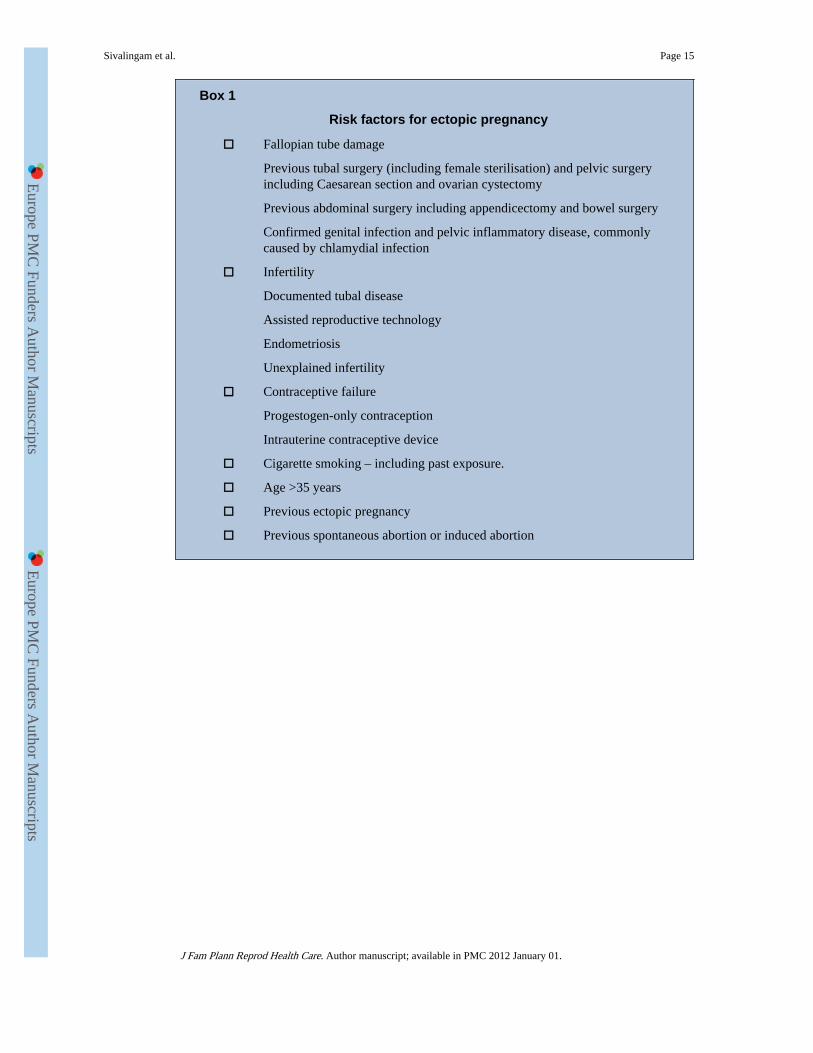
Box 1
Risk factors for ectopic pregnancy
□ Fallopian tube damage
Previous tubal surgery (including female sterilisation) and pelvic surgeryincluding Caesarean section and ovarian cystectomy
Previous abdominal surgery including appendicectomy and bowel surgery
Confirmed genital infection and pelvic inflammatory disease, commonlycaused by chlamydial infection
□ Infertility
Documented tubal disease
Assisted reproductive technology
Endometriosis
Unexplained infertility
□ Contraceptive failure
Progestogen-only contraception
Intrauterine contraceptive device
□ Cigarette smoking – including past exposure.
□ Age >35 years
□ Previous ectopic pregnancy
□ Previous spontaneous abortion or induced abortion
Sivalingam et al. Page 15
J Fam Plann Reprod Health Care. Author manuscript; available in PMC 2012 January 01.
Europe PM
C Funders A
uthor Manuscripts
Europe PM
C Funders A
uthor Manuscripts

Box 2
Useful ultrasonographic findings in the diagnosis of ectopic pregnancy
□ Absence of intrauterine pregnancy (IUP)
□ Positive identification of an ectopic pregnancy mass: inhomogenous mass,empty adnexal gestation sac or adnexal sac containing yolk sac or fetal pole
□ Free fluid (i.e. blood): suggestive of ectopic pregnancy in the absence of IUP,but not diagnostic (small amount may be physiological)
Sivalingam et al. Page 16
J Fam Plann Reprod Health Care. Author manuscript; available in PMC 2012 January 01.
Europe PM
C Funders A
uthor Manuscripts
Europe PM
C Funders A
uthor Manuscripts

Box 3
Inclusion criteria for medical management of ectopic pregnancy withmethotrexate
□ Patient characteristics
Would prefer medical option
Willing to attend follow-up for up to 6 weeks
Willing to abstain from alcohol for 7 days following the treatment
Not breastfeeding or willing to stop
□ Clinical features
Haemodynamically stable
Minimal abdominal pain
□ Ultrasound scan findings
No fetal heart activity or clear yolk sac in adnexal mass
Small amount of free fluid
Unlikely to be early intrauterine pregnancy failure
□ Serum beta-human chorionic gonadotrophin (β-hCG) concentrations
Usually <3000 IU/l (Although limits of <5000 IU/l are used in some unitsand earlier studies, treatment success rates are higher when this morecommonly used lower limit applies.)
□ Medical history
No active peptic ulcer disease
No severe medical conditions including renal disease, hepatic disease, severeanaemia, leucopenia or thrombocytopenia
□ Should not be on concurrent medication
Non-steroidal anti-inflammatory agents (NSAIDs), aspirin, penicillins,sulphonamides, trimethoprim, tetracyclines, diuretics, phenytoin,antimalarials, ciclosporin, retinoids, probenecid, folic acid, hypoglycaemics,live vaccines, nephrotoxic or hepatotoxic drugs
Sivalingam et al. Page 17
J Fam Plann Reprod Health Care. Author manuscript; available in PMC 2012 January 01.
Europe PM
C Funders A
uthor Manuscripts
Europe PM
C Funders A
uthor Manuscripts

Figure 1.Transvaginal ultrasound images of an intrauterine pregnancy (IUP) and ectopic pregnancy.(A) An IUP at 6 weeks. The central dark area is the intrauterine gestational sac and withinthe sac is a circular ringed structure that is the yolk sac. The small oval structure below theyolk sac is the fetus. (B) An ectopic pregnancy. To the right of the image is the normaluterus and to the left of the uterus is the doughnut-shaped ectopic pregnancy.
Sivalingam et al. Page 18
J Fam Plann Reprod Health Care. Author manuscript; available in PMC 2012 January 01.
Europe PM
C Funders A
uthor Manuscripts
Europe PM
C Funders A
uthor Manuscripts
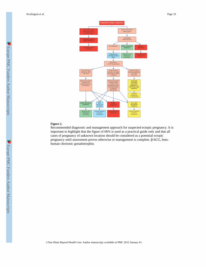
Figure 2.Recommended diagnostic and management approach for suspected ectopic pregnancy. It isimportant to highlight that the figure of 66% is used as a practical guide only and that allcases of pregnancy of unknown location should be considered as a potential ectopicpregnancy until assessment proves otherwise or management is complete. β-hCG, beta-human chorionic gonadotrophin.
Sivalingam et al. Page 19
J Fam Plann Reprod Health Care. Author manuscript; available in PMC 2012 January 01.
Europe PM
C Funders A
uthor Manuscripts
Europe PM
C Funders A
uthor Manuscripts

Figure 3.(A) Left tubal ectopic pregnancy at laparoscopy. (B) Tubal ectopic pregnancy has beenremoved by salpingectomy.
Sivalingam et al. Page 20
J Fam Plann Reprod Health Care. Author manuscript; available in PMC 2012 January 01.
Europe PM
C Funders A
uthor Manuscripts
Europe PM
C Funders A
uthor Manuscripts