ugst195 ltrnbaicVertigo-Lundcgren Vetg-udrnMEDICAL 513 file28 August 1965 28Alternobaricugst195...
4
28 ugst195 ltrnbaicVertigo-Lundcgren MEDIBRIMHS5 28 August 1965 Alternobaric Vetg-udrnMEDICAL JOURNAL 513 From the point of view of prognosis alternobaric vertigo itself seems to be essentially benign. However, because of its potentially deleterious effects all possible steps should be taken to avoid it-for example, diving should not be undertaken when the person has a cold, nor preferably immediately after a cold when it is more difficult than usual to clear the ears. The correct use of nasal decongestants is also recommended in cases where pressure equilibration is difficult despite the absence of recent infection. Great care should be taken always to keep pressure equilibration timed with external pressure change and to avoid unnecessarily rapid ascents and descents. Intermittent swallowing during ascent, though not necessary for pressure equilibration, may possibly facilitate it, thus further reducing the risk. Divers who are inclined to vertiginous reactions of the type described should avoid pressure exposures which make the use of stage decompression necessary. The author would like to acknowledge the valuable criticism and discussion of the manuscript offered by Surgeon Captain S. Miles and Surgeon Lieutenant-Commander R. R. A. Coles, R.N. Summary The occurrence of vertigo in connexion with external pressure variations has been studied among sport divers by means of a questionary. The phenomenon, which is here called "alternobaric vertigo," appears from this investigation to be a serious poten- tial menace to divers. Some aetiological mechanisms and preventive or therapeutic measures which might prove important are discussed. Appendix. Questionary 1. I have/have not undergone medical examination in connexion with diving. Date........................ 2. The result was/was not satisfactory, owing to .................. 3. In connexion with diving I have always/often/seldom/never had difficulties with pressure equilibration. 4. In case of difficulties with pressure equilibration I have more! equal difficulties with the right (and) left ear(s). I usually/never! also/only have difficulties with the nasal sinuses/cheeks/forehead. 5. To obtain pressure equilibration I usually open the mouth (move the jaw)/swallow/blow against the clamped nose/do nothing special. 6. When I dive I always/often/occasionally/seldom/never experience vertigo (a whirling sensation). 7. The vertigo usually appears at the surface (upon ascent)/ during ascent/during descent/at the bottom. The greatest depth reached in dives in which vertigo was experienced was .........metres. (Not necessarily the same as the depth at which the vertigo appeared.) 8. The visibility in the water when the vertigo appeared was .metres. 9. When I had vertigo I had not/possibly/for certain a ruptured right/left eardrum. I wore/did not wear a hood. I can/cannot induce vertigo by forced blowing against the clamped nose. 10. I believe/do not believe that the vertigo was due to sea- sickness. It may/cannot have been caused by breathing impure air! food-poisoning/or ............................................................ 11. When I had vertigo I never/sometimes/always wore breath- ing apparatus. 12. The vertigo I had was very/rather/scarcely/not at all troublesome. Mention if possible how many times you have had vertigo and how troublesome it was (e.g., difficulty in finding the way to the surface, swimming ashore, etc.) ..................... 13. When I dive I always/often/occasionally/seldom/never have nausea. The nausea is then always/sometimes/never connected with the vertigo mentioned above. [A number of questions treating nausea as an entity separate from the vertigo were then put. The results will be presented in a separate publication.] 14. If you have had vertigo, mention how long it persisted and what you did to eliminate it ................................................ 15. Additional information ............................................. [In an additional inquiry 15 of the subjects who had had vertigo (including some of the most serious cases) were asked for their experiences of vertigo on land.] REFERENCES Alt, F. (1896). Mschr. Ohrenheilk., 30, 341. Coles, R. R. A., and Knight, J. J. (1961). Aural and Audiometric Survey of Qualified Divers and Submarine Escape Training Tank Instruc- tors, R.N.P. 61/1011 (Royal Naval Personnel Research Committee, R.N., G.B. August 1961). Fields, J. A. (1958). Arch. Otolaryng., 68, 531. Filippin, S. (1963). Ann. Laring. (Torino), 62, 244. Heller, R., Mager, W., and Schrotter, H. von (1900). Cited by L. Hill (see Keays, 1909). Keays, F. L. (1909). Cited by L. Hill, Caisson Sickness and the Physio- logy of Work in Compressed Air. Longmans, Green, New York. 1912. Melvill Jones, G. (1957). Review of Current Problems Associated with Disorientation in Man-Controlled Flight, FPRC 1021 (Flying Per- sonnel Research Committee, R.A.F., G.B. October 1957), (Restricted). Miles, S. (1962). Underwater Medicine. Staples Press, London. Rawlins, J. S. P. (1960). "Psychological Stress in Divers," paper read at the Second International Symposium on Submarine and Space Medi- cine in Stockholm, 18-19 August 1960. Rowe, B. (1961). Med. 7. Aust., 2, 1038. Vail, H. H. (1929). Arch. Otolaryng., 10, 113. Estimations of Blood Volume During Course of Renal Failure VERNON C. MARSHALL,* F.R.A.C.S.; DAVID YOFFA,* F.R.A.C.S Brit. med. J., 1965, 2, 513-516 Oliguric renal failure may be associated with a considerable dislocation of the normal partitioning of the body fluids, but relatively few studies of such changes have been made in man. We have performed serial estimations of blood volume in 50 patients with oliguric renal failure. Observations were made throughout the course of the disease, and the effects of haemo- dialysis and of peritoneal dialysis have been observed. This paper outlines some of the results, which have provided information on the fluid shifts occurring as a result of renal failure, and have also on occasion materially assisted in the management of these patients. * Department of Surgery, University of Melbourne, and Department of Surgical Metabolism, Royal Melbourne Hospital. Method and Materials An automatic isotope dilution technique was employed, using a machine, the Volemetron, which can be set for iodinated serum albumin, or chromated red cells, or both (Williams and Fine, 1961). Because of its greater simplicity, we have used iodinated on 10 May 2019 by guest. Protected by copyright. http://www.bmj.com/ Br Med J: first published as 10.1136/bmj.2.5460.513 on 28 August 1965. Downloaded from
Transcript of ugst195 ltrnbaicVertigo-Lundcgren Vetg-udrnMEDICAL 513 file28 August 1965 28Alternobaricugst195...

28 ugst195 ltrnbaicVertigo-Lundcgren MEDIBRIMHS528 August 1965 Alternobaric Vetg-udrnMEDICAL JOURNAL 513
From the point of view of prognosis alternobaric vertigoitself seems to be essentially benign. However, because of itspotentially deleterious effects all possible steps should be takento avoid it-for example, diving should not be undertakenwhen the person has a cold, nor preferably immediately aftera cold when it is more difficult than usual to clear the ears. Thecorrect use of nasal decongestants is also recommended in caseswhere pressure equilibration is difficult despite the absence ofrecent infection. Great care should be taken always to keeppressure equilibration timed with external pressure change andto avoid unnecessarily rapid ascents and descents. Intermittentswallowing during ascent, though not necessary for pressureequilibration, may possibly facilitate it, thus further reducingthe risk. Divers who are inclined to vertiginous reactions ofthe type described should avoid pressure exposures which makethe use of stage decompression necessary.
The author would like to acknowledge the valuable criticismand discussion of the manuscript offered by Surgeon Captain S.Miles and Surgeon Lieutenant-Commander R. R. A. Coles, R.N.
SummaryThe occurrence of vertigo in connexion with external pressure
variations has been studied among sport divers by means of aquestionary.The phenomenon, which is here called "alternobaric
vertigo," appears from this investigation to be a serious poten-tial menace to divers.Some aetiological mechanisms and preventive or therapeutic
measures which might prove important are discussed.
Appendix. Questionary1. I have/have not undergone medical examination in connexion
with diving. Date........................2. The result was/was not satisfactory, owing to ..................3. In connexion with diving I have always/often/seldom/never
had difficulties with pressure equilibration.4. In case of difficulties with pressure equilibration I have more!
equal difficulties with the right (and) left ear(s). I usually/never!also/only have difficulties with the nasal sinuses/cheeks/forehead.
5. To obtain pressure equilibration I usually open the mouth(move the jaw)/swallow/blow against the clamped nose/do nothingspecial.
6. When I dive I always/often/occasionally/seldom/neverexperience vertigo (a whirling sensation).
7. The vertigo usually appears at the surface (upon ascent)/during ascent/during descent/at the bottom. The greatest depthreached in dives in which vertigo was experienced was .........metres.(Not necessarily the same as the depth at which the vertigoappeared.)
8. The visibility in the water when the vertigo appeared was.metres.
9. When I had vertigo I had not/possibly/for certain a rupturedright/left eardrum. I wore/did not wear a hood. I can/cannotinduce vertigo by forced blowing against the clamped nose.
10. I believe/do not believe that the vertigo was due to sea-sickness. It may/cannot have been caused by breathing impure air!food-poisoning/or ............................................................
11. When I had vertigo I never/sometimes/always wore breath-ing apparatus.
12. The vertigo I had was very/rather/scarcely/not at alltroublesome. Mention if possible how many times you have hadvertigo and how troublesome it was (e.g., difficulty in finding theway to the surface, swimming ashore, etc.) .....................
13. When I dive I always/often/occasionally/seldom/never havenausea. The nausea is then always/sometimes/never connectedwith the vertigo mentioned above.
[A number of questions treating nausea as an entity separate fromthe vertigo were then put. The results will be presented in aseparate publication.]
14. If you have had vertigo, mention how long it persisted andwhat you did to eliminate it ................................................
15. Additional information .............................................[In an additional inquiry 15 of the subjects who had had vertigo
(including some of the most serious cases) were asked for theirexperiences of vertigo on land.]
REFERENCES
Alt, F. (1896). Mschr. Ohrenheilk., 30, 341.Coles, R. R. A., and Knight, J. J. (1961). Aural and Audiometric Survey
of Qualified Divers and Submarine Escape Training Tank Instruc-tors, R.N.P. 61/1011 (Royal Naval Personnel Research Committee,R.N., G.B. August 1961).
Fields, J. A. (1958). Arch. Otolaryng., 68, 531.Filippin, S. (1963). Ann. Laring. (Torino), 62, 244.Heller, R., Mager, W., and Schrotter, H. von (1900). Cited by L. Hill
(see Keays, 1909).Keays, F. L. (1909). Cited by L. Hill, Caisson Sickness and the Physio-
logy of Work in Compressed Air. Longmans, Green, New York.1912.
Melvill Jones, G. (1957). Review of Current Problems Associated withDisorientation in Man-Controlled Flight, FPRC 1021 (Flying Per-sonnel Research Committee, R.A.F., G.B. October 1957),(Restricted).
Miles, S. (1962). Underwater Medicine. Staples Press, London.Rawlins, J. S. P. (1960). "Psychological Stress in Divers," paper read at
the Second International Symposium on Submarine and Space Medi-cine in Stockholm, 18-19 August 1960.
Rowe, B. (1961). Med. 7. Aust., 2, 1038.Vail, H. H. (1929). Arch. Otolaryng., 10, 113.
Estimations of Blood Volume During Course of Renal Failure
VERNON C. MARSHALL,* F.R.A.C.S.; DAVID YOFFA,* F.R.A.C.S
Brit. med. J., 1965, 2, 513-516
Oliguric renal failure may be associated with a considerabledislocation of the normal partitioning of the body fluids, butrelatively few studies of such changes have been made in man.We have performed serial estimations of blood volume in 50patients with oliguric renal failure. Observations were madethroughout the course of the disease, and the effects of haemo-dialysis and of peritoneal dialysis have been observed. Thispaper outlines some of the results, which have provided
information on the fluid shifts occurring as a result of renalfailure, and have also on occasion materially assisted in themanagement of these patients.
* Department of Surgery, University of Melbourne, and Department ofSurgical Metabolism, Royal Melbourne Hospital.
Method and MaterialsAn automatic isotope dilution technique was employed, usinga machine, the Volemetron, which can be set for iodinated serumalbumin, or chromated red cells, or both (Williams and Fine,1961). Because of its greater simplicity, we have used iodinated
on 10 May 2019 by guest. P
rotected by copyright.http://w
ww
.bmj.com
/B
r Med J: first published as 10.1136/bm
j.2.5460.513 on 28 August 1965. D
ownloaded from

serum albumin on most occasions. A result can be obtainedat the bedside within 15 minutes as a direct dial reading. Theaccuracy of the machine is high ; the error for a singledetermination is below 2%, and after five consecutive estima-tions is still less than 5 % (Williams and Frank, 1962).Quantitative errors of volumetric measurement are eliminatedby providing a direct assay of radioactivity, and the accuracy
7-0-
6-0-
5 5.0-z
sm 40-
e 1-0-
&VI
0
em
BVd
0_l.-:01
FIG. 1.-Blood volume (B.V.)estimations before and immedi-ately on starting dialysis areshown. In this and subse-quent figures the red-cellvolume (R.C.V.) is stippledand plasma volume (P.V.)clear. Blood added: 1,200B.V., 516 R.C.V., 684 P.V.Calculation of blood added:1,220 B.V., 520 R.C.V., 700
P.V.
is thus greater than that obtained from existing dye dilution andindirect radioisotopic methods, especially in repeated studies(Greep et al., 1963). Our observations, both in vitro and inpatients, confirm these levels of accuracy. A representativestudy is shown in Fig. 1. Blood volumes are compared beforeand immediately at the beginning of haemodialysis, when a
known amount of blood has been added to the circulation. Theclose correlation between measured and calculated volumes isevident.
Results
The 50 patients studied were drawn from a larger group of127 patients referred to our unit over a period of 18 monthswith acute or chronic renal failure. The majority (40) of the50 patients were suffering from acute oliguric renal failure,seven had acute exacerbations of chronic renal disease, and threewere treated by renal transplantation. The largest group (32)was of acute renal failure after operation or trauma. In all,148 estimations have been performed on the 50 patients.
1. Shifts in Blood Volume During the Course of Acute RenalFailure
Patients with acute renal failure who are studied from theonset of their illness, if managed by suitable fluid and electrolyterestriction, will usually show a relative constancy of bloodvolume. Fig. 2 gives the results of serial studies in a woman
aged 29 with cardiac arrest and renal failure due to haemorrhagefrom a central placenta praevia who was referred on the thirdday of oliguria. There has been minimal change in bloodvolume over the course of three haemodialyses, but the increase
Dil D,2 D3
CU 60.60I-~~~~~V
.0 BV constant 0
4-0 4030
3-03> ~~~~~~~~~~~~~~~~~~~~-4
a~~~~~~~~~~~~~~~00
IDAYSFIG. 2.-Blood volumes are shown by the histograms andhaematocrit levels by the circles. D1, D2, and D3 refer to times
of haemodialyses.
BRITISHMEDICAL JOURNAL
in red-cell volume due to dialysis has caused a 13 % rise in
haematocrit to give acceptable levels of red-cell, plasma, andtotal blood volume at the end of the study, when diuresis was
beginning.
2. Fallibility of Haematocrit or Haemoglobin Levels
The peripheral large vessel haematocrit or haemoglobin levelcannot predict either the direction or extent of shifts in bloodvolume, and we have repeatedly observed, in both acute andchronic renal failure, relatively small shifts in blood volumeassociated with progressive and severe diminution in red-cellvolume, with a falling haematocrit, unless this is corrected byhaemodialysis or by transfusion. We have in such instancescombined ultrafiltration with packed cell infusion duringhaemodialysis to restore red-cell mass without danger ofcirculatory overloading.
Case Report (Fig. 3).-A woman aged 63 with Escherichia colisepticaemia from pyelonephritis was clinically overhydrated whenfirst seen, and a peritoneal dialysis caused an initial fall in bloodvolume of approximately 1 litre. Subsequent blood volumesremained constant, but red-cell volume progressively fell to below
7- PRITONEAL DIALYSIS
I-Im 5-0. V B4
z
z ~~~~~~~~~~ASVu*+2Zomi.-j3-0- A RV +SCOmI.
:__ A Pv---- - r-
i 3 5 7DAYS
9 I1
70
60
50
40
30
20
10
0
;I-,
FIG. 3
1 litre, lowering the haematocrit to 17 %. Haemodialysis withultrafiltration and packed cell infusion caused a slight rise in bloodvolume (220 ml.). The red-cell volume rose by 800 ml. to givea near normal haematocrit of 3300 and plasma volume over thesame period fell by 580 ml.The unreliability of the haematocrit has been seen on many
other occasions when significant acute shifts in blood volumehave been accompanied by haematocrit shifts in similar or in
opposite directions.
3. Value of Estimations in Renal Failure in Surgical Patients
Surgical causes have accounted for 59 of a total number of127 cases referred to our unit. In these patients the complica-tions of severe blood loss, or of extrarenal electrolyte losses fromvomiting, ileus, or fistulae, can often greatly increase thedifficulties of achieving metabolic balance. Monitoring theblood volume in these instances can be of material assistance.When acute loss of blood prejudices survival it is again
stressed that the peripheral haematocrit is of little or no valuein estimating the volume of replacement fluid required, and itwill in time merely reflect the haematocrit of bank blood (Moore,1959).
Case Report (Fig. 4).-A man aged 25 with Peutz-Jeghers'ssyndrome had a jejunojejunal intussusception which was treated byoperative reduction. He developed signs of peritonitis and ofoliguric renal failure after operation, and was transferred to our
unit on the fourth day. Immediate reoperation was required andthe gangrenous segment of jejunum was resected. A fall in, bloodvolume of over 1 litre immediately after operation (BV1 and BV2)was unaccompanied by any change of haematocrit, and over the
514 28 August 1965 Renal Failure-Marshall and Yoffa
'I
r-q
|
so
on 10 May 2019 by guest. P
rotected by copyright.http://w
ww
.bmj.com
/B
r Med J: first published as 10.1136/bm
j.2.5460.513 on 28 August 1965. D
ownloaded from

28 August 1965 Renal Failure-Marshall and Yoffasucceeding five days transcapillary refilling of the circulation hadresulted in an increase in blood volume to the preoperative level(BV4) with a stable haemodilutional anaemia (Lister et al., 1963).Haemodialysis was required on the ninth day, but he developedmassive gastro-intestinal bleeding during the diuretic phase. Re-placement therapy was guided by blood-volume estimations, andafter 48 hours replacement appeared to be adequate with normalvolume parameters (BV7). However, recurrent bleeding loweredthe blood volume to near shock levels despite massive transfusion,and at operation on the 13th day multiple acute gastric ulcers werefound. Despite vagotomy and gastrectomy he died from sutureleakage and peritonitis on the 15th day.
Iomatomesisnd Me aenablod 12 5 litres
I-
x
4"I.-140
A
DAYS AFTER LAPAROTOMYFIG. 4
Gastro-intestinal haemorrhage of a degree to threaten survivalhas occurred in 7 of our 59 surgical patients, and is a consider-able hazard in this group, as has been described by Shackmanet al. (1960). It has occurred in dialysed and non-dialysedpatients, is often due to multiple gastric or duodenal ulcers, andhas been treated conservatively except in the case described.Serial measurements of blood volume are of great value inassessment of volume replacement in such patients.
Case Report (Fig. 5).-A man aged 45, weighing 105 kg., hadan appendicectomy for a perforated gangrenous appendicitis. Hesubsequently developed an E. coli septicaemia with severe renalfailure, complicated by jaundice and recurrent severe haematemesis.He required haemodialysis on three occasions. A progressive slight
IJS5-
z
au
0
m
-4
0
-R0
DAYSFIG. 5.-D1, D2, and D3 refer to times of haemodialyses.
fall in total blood volume was noted with a declining haematocritdespite repeated transfusions over the course of his illness. Duringthis time loss of weight was considerable. He made a completerecovery, and four months later when his original weight had beenregained his blood volume had returned to a stable level of justbelow 8 litres.
4. Monitoring Fluid Removal During Peritoneal Dialysis andHaemodialysis
When late presentation to the unit is associated with severeoverhydration, removal of fluid may be of pressing urgency.
We have found peritoneal dialysis to be considerably superior tohaemodialysis for fluid removal, and by using hypertonic solu-tions very rapid shifts can be achieved. However, injudicioususe. of the hypertonic solutions can readily precipitate hypo-volaemia with signs of peripheral circulatory failure (Burnset al., 1962), and monitoring of the blood volume during theprocedure adds further precision and safety to the method.
Case Report (Fig. 6).-A man aged 46 presented with a five-dayhistory of bilateral renal pain followed by anuria. He was stuporouswhen admitted, with gross oedema and congestive heart failure.Immediate peritoneal dialysis removed 2 litres of fluid over thecourse of 24 hours and his blood volume fell by a similar figure(BV2). A history of phenacetin ingestion and confirmatory retro-grade pyelograms led to the diagnosis of papillary necrosis withobstruction. Bilateral nephrostomies with removal of necroticpapillae from the pelvi-ureteric junctions were done on the eighthday and he subsequently improved with a maintained diureticresponse. Blood volume remained constant but a progressiveanaemia was revealed with a decline in red-cell volume and
PeriOnea 2,Fl
2 litres
7.0
W% 6* 601 2 4 1DAYS~ ~ ~~~S
0o 12 1 1IDAYS
FIG. 6.-Weight is indicated by the closed circles.
haematocrit. The nephrostomy tubes were ultimately removed andhis subsequent health has been good, with a stable but elevatedblood urea.
When fluid removal during haemodialysis is requiredmonitoring of 'shifts in blood volume gives precise control.Considerable fluid removal by ultrafiltration, as measured byan increase in the volume of the bath and by decrease in weightof the patient, can be combined with infusion of packed redcells during dialysis, so that anaemia may be corrected withoutfear of circulatory overloading, while maximal fluid removal ispossible without danger of precipitating oligaemia. It is ourpractice routinely to use regional heparinization during haemo-dialysis, so that normally about 500 ml. of electrolyte solutionis infused 1uring dialysis. However, when fluid removal is ofvital importance, the heparin infusion on the arterial side hasbeen delivered in a bottle of packed erythrocytes, so that aminimal amount of fluid administration obtains from' theprotamine infusion on the venous side (250 ml. in five hours).
5. Differentiation Between Depletional Oliguria and IntrinsicRenal Failure
The difficulties of differentiation between depletional oliguriafrom renal circulatory insufficiency, which is reversible, and theoliguria of established intrinsic renal failure are well known.Physical signs, blood indices, and urine composition are all ofvalue in distinguishing the two (Shackman et al., 1960). Theestimation of blood volume will on occasion be of materialassistance, and serial estimations aid in assessment of theefficiency of treatment.We have also on several occasions used mannitol infusions in
an attempt to promote a diuresis when volume studies haveindicated adequate circulatory filling. Although clinical proof
BRITISHMEDICAL JOURNAL 515
on 10 May 2019 by guest. P
rotected by copyright.http://w
ww
.bmj.com
/B
r Med J: first published as 10.1136/bm
j.2.5460.513 on 28 August 1965. D
ownloaded from
516 28 August 1965 Renal Failure-Marshall and Yoffa BRITNA
of its efficacy is difficult to provide, we, like others (Moore,1963), have little doubt that on occasions its use will avert thedevelopment of renal failure. We have studied its volume-expanding effects which are transient, and with care in its useno complications have been seen.
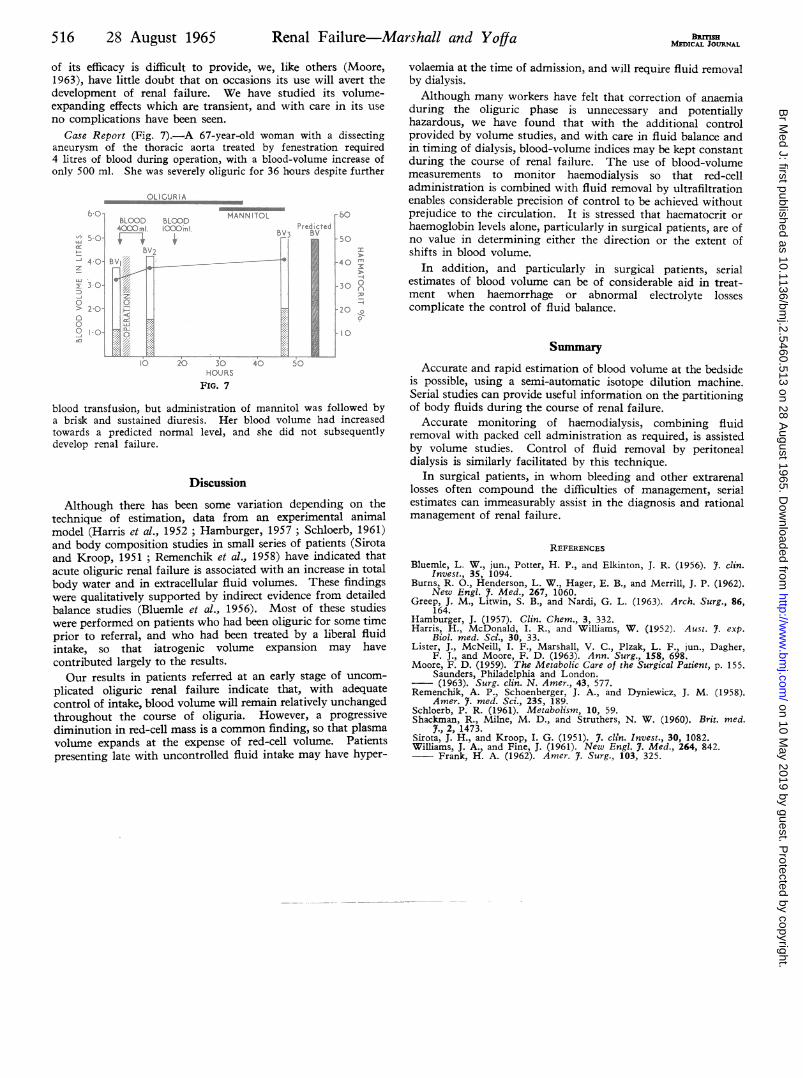
Case Report (Fig. 7).-A 67-year-old woman with a dissectinganeurysm of the thoracic aorta treated by fenestration required4 litres of blood during operation, with a blood-volume increase ofonly 500 ml. She was severely oliguric for 36 hours despite further
blood transfusion, but administration of mannitol was followed bya brisk and sustained diuresis. Her blood volume had increasedtowards a predicted normal level, and she did not subsequentlydevelop renal failure.
OLIGURIA
b o7bOD LOHANNITOL bo{>mi. 510 0^. OO~mJ. AdPredicted
C S'4OBV 40;f
2100 204
~1.0 ~ 1 300s
HOURSFIG. 7
Discussion
Although there has been some variation depending on thetechnique of estimation, data from an experimental animalmodel (Harris et al., 1952 ; Hamburger, 1957 ; Schloerb, 1961)and body composition studies in small series of patients (Sirotaand Kroop, 1951; Remenchik et al., 1958) have indicated thatacute oliguric renal failure is associated with an increase in totalbody water and in extracellular fluid volumes. These findingswere qualitatively supported by indirect evidence from detailedbalance studies (Bluemle et al., 1956). Most of these studieswere performed on patients who had been oliguric for some timeprior to referral, and who had been treated by a liberal fluidintake, so that iatrogenic volume expansion may havecontributed largely to the results.Our results in patients referred at an early stage of uncom-
plicated oliguric renal failure indicate that, with adequatecontrol of intake, blood volume will remain relatively unchangedthroughout the course of oliguria. However, a progressivediminution in red-cell mass is a common finding, so that plasmavolume expands at the expense of red-cell volume. Patientspresenting late with uncontrolled fluid intake may have hyper-
volaemia at the time of admission, and will require fluid removalby dialysis.Although many workers have felt that correction of anaemia
during the oliguric phase is unnecessary and potentiallyhazardous, we have found that with the additional controlprovided by volume studies, and with care in fluid balance andin timing of dialysis, blood-volume indices may be kept constantduring the course of renal failure. The use of blood-volumemeasurements to monitor haemodialysis so that red-celladministration is combined with fluid removal by ultrafiltrationenables considerable precision of control to be achieved withoutprejudice to the circulation. It is stressed that haematocrit orhaemoglobin levels alone, particularly in surgical patients, are ofno value in determining either the direction or the extent ofshifts in blood volume.
In addition, and particularly in surgical patients, serialestimates of blood volume can be of considerable aid in treat-ment when haemorrhage or abnormal electrolyte lossescomplicate the control of fluid balance.
SummaryAccurate and rapid estimation of blood volume at the bedside
is possible, using a semi-automatic isotope dilution machine.Serial studies can provide useful information on the partitioningof body fluids during the course of renal failure.
Accurate monitoring of haemodialysis, combining fluidremoval with packed cell administration as required, is assistedby volume studies. Control of fluid removal by peritonealdialysis is similarly facilitated by this technique.
In surgical patients, in whom bleeding and other extrarenallosses often compound the difficulties of management, serialestimates can immeasurably assist in the diagnosis and rationalmanagement of renal failure.
REFERENCES
Bluemle, L. W., jun., Potter, H. P., and Elkinton, J. R. (1956). 7. clin.Invest., 35, 1094.
Burns, R. O., Henderson, L. W., Hager, E. B., and Merrill, J. P. (1962).New Engl. 7. Med., 267, 1060.
Greep, J. M., Litwin, S. B., and Nardi, G. L. (1963). Arch. Surg., 86,164.
Hamburger, J. (1957). Clin. Chem., 3, 332.Harris, H., McDonald, I. R., and Williams, W. (1952). Aust. 7. exp.
Biol. med. Scan., 30, 33.Lister, J., McNeill, I. F., Marshall, V. C., Plzak, L. F., jun., Dagher,
F. J., and Moore, F. D. (1963). Ann. Surg., 158, 698.Moore, F. D. (1959). The Metabolic Care of the Surgical Patient, p. 155.
Saunders, Philadelphia and London.(1963). Surg. clin. N. Amer., 43, 577.
Remenchik, A. P., Schoenberger, J. A., and Dyniewicz, J. M. (1958).Amer. 7. med. Sci., 235, 189.
Schloerb, P. R. (1961). Metabolism, 10, 59.Shackman, R., Milne, M. D., and Struthers, N. W. (1960). Brit. med.
7., 2, 1473.Sirota, J. H., and Kroop, I. G. (1951). 7. cldn. Invest., 30, 1082.Williams, J. A., and Fine, J. (1961). New Engl. 7. Med., 264, 842.
Frank, H. A. (1962). Amer. 7. Surg., 103, 325.
on 10 May 2019 by guest. P
rotected by copyright.http://w
ww
.bmj.com
/B
r Med J: first published as 10.1136/bm
j.2.5460.513 on 28 August 1965. D
ownloaded from