Tyrosine-Derived Nanospheres for Topical Skin Delivery · Maghraby et al. J Pharm Pharmacol 58,...
42
Tyrosine-Derived Nanospheres for Topical Skin Delivery Larisa Sheihet, PhD SPOTLIGHT ON SKIN RESEARCH New Jersey Center for Biomaterials June 25, 2007
Transcript of Tyrosine-Derived Nanospheres for Topical Skin Delivery · Maghraby et al. J Pharm Pharmacol 58,...

Tyrosine-Derived Nanospheresfor Topical Skin Delivery
Larisa Sheihet, PhD
SPOTLIGHT ON SKIN RESEARCHNew Jersey Center for Biomaterials
June 25, 2007
Team:Larisa Sheihet, Prafulla Chandra, Priya Batheja
David Devore, Joachim Kohn, and Bozena Michniak
3
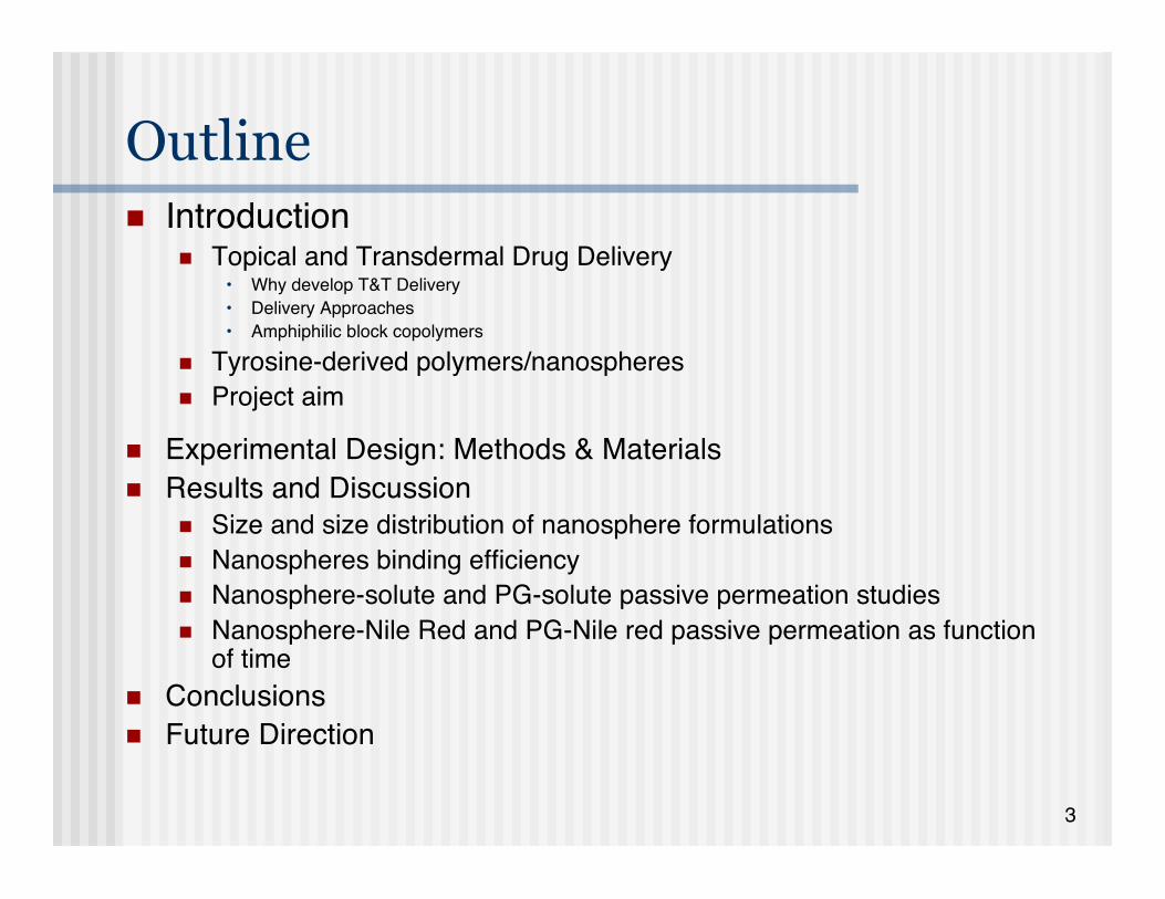
Outline Introduction
Topical and Transdermal Drug Delivery• Why develop T&T Delivery• Delivery Approaches• Amphiphilic block copolymers
Tyrosine-derived polymers/nanospheres Project aim
Experimental Design: Methods & Materials Results and Discussion
Size and size distribution of nanosphere formulations Nanospheres binding efficiency Nanosphere-solute and PG-solute passive permeation studies Nanosphere-Nile Red and PG-Nile red passive permeation as function
of time Conclusions Future Direction

4
Introduction

5
Drug Delivery “…In the context of drug delivery, the needs for materials can
generally be broken into two categories: the creation of newmaterials and/or better understanding of how to manipulateexisting materials
Current needs include reducing the toxicity of drugs,increasing their absorption, improving their administration andrelease profile…”
Robert S. Langer, Institute Professor, MIT C&EN, Cover Story, August 26, 2002

6
Drug Delivery
http://www.azonano.com/details.asp?ArticleID=1538
AdministrationRoutes Peroral
Pulmonary
Parenteral• Intravenous• Intramuscular• Subcutaneous
Percutaneous• Topical• Transdermal
7
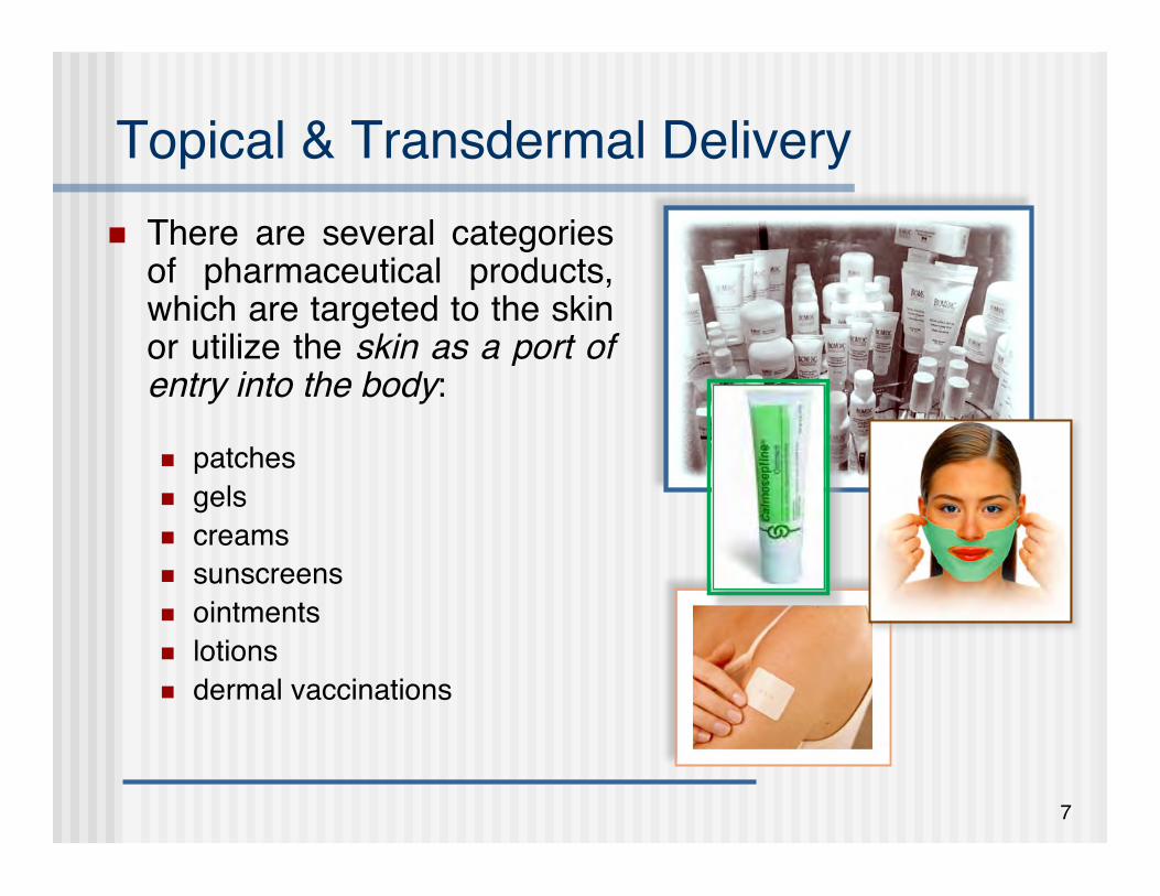
Topical & Transdermal Delivery There are several categories
of pharmaceutical products,which are targeted to the skinor utilize the skin as a port ofentry into the body:
patches gels creams sunscreens ointments lotions dermal vaccinations
Skin Care ProductsSkin Care Products

8
Common Skin Diseases Skin cancer Warts Fungal infections Dermatitis Psoriasis Acne Hand dermatitis Atopic eczema (one in six of all children) Cold sores (herpes simplex, one in five persons)
http://www.dermnetnz.org/dermatologist.htmlhttp://www.skinsite.com/index_dermatology_diseases.htm
7.5 million Americans have psoriasis2% to 3% of the world is affectedNational Psoriasis Foundation, 2007

9
Main Challenges in Transporting Drugs Intothe Skin
Low diffusion rate of drugs through the the stratumcorneum (SC)
Overcoming the resistance of the skin in a reversibleand non-damaging manner
Intra-inter variability associated with permeability ofintact and diseased skin
Pre-systemic metabolism Immunological barrier The limited number of suitable drugs is reduced to:
Low molecular mass and melting point Optimal partitioning coefficient
• Sufficient lipid (SC) and aqueous solubility Moderately lipophilic
B.W. Barry Nat Biotechnol 22 (2004) 192-197H. Patel et al. Cronin Chemosphere 48 (2002) 603-613A. Nanda et al. Curr Drug Deliv 3 (2006) 233-242
10
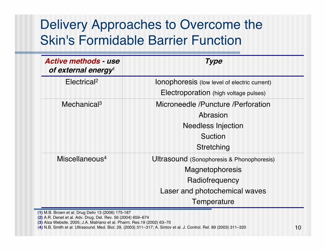
Delivery Approaches to Overcome theSkin's Formidable Barrier Function
(1) M.B. Brown et al. Drug Deliv 13 (2006) 175-187(2) A.R. Denet et al. Adv. Drug. Del. Rev. 56 (2004) 659–674(3) Alza Website. 2005; J.A. Matriano et al. Pharm. Res.19 (2002) 63–70(4) N.B. Smith et al. Ultrasound. Med. Biol. 29, (2003) 311–317; A. Sintov et al. J. Control. Rel. 89 (2003) 311–320
Ultrasound (Sonophoresis & Phonophoresis)
MagnetophoresisRadiofrequency
Laser and photochemical wavesTemperature
Miscellaneous4
Microneedle /Puncture /PerforationAbrasion
Needless InjectionSuction
Stretching
Mechanical3
Ionophoresis (low level of electric current)
Electroporation (high voltage pulses)
Electrical2
TypeTypeActive methods - - useof external energy1

11
Delivery Approaches to Overcome theSkin's Formidable Barrier Function
Passive methods penetration enhancers supersaturated systems prodrug or metabolic approach liposomes, microemulsions, colloidal polymeric suspensions*
A.C. Williams et al. Adv Drug Deliv Rev. 56, 603-618, 2004; B. Godin et al. Crit Rev Ther Drug Carrier Syst 20, 63-102, 2003; G.M. ElMaghraby et al. J Pharm Pharmacol 58, 415-429, 2006; S.L. Borgia et al. J Control Release 110, 151-163, 2005;
(*) A. Berthold et al. Eur J Pharm Biopharm 45, 23-29, 1998; E.G. de Jalon et al. Int J Pharm 226, 181-184, 2001; S. Santoyoet al. Int J Pharm 242, 107-113, 2002.
However:• the amount of drug that can be delivered is still limited
• most of the enhancers cause irreversible membrane damage• just a limited number of suitable biodegradable, polymer and solid-lipid particles areavailable

12
Choice of Formulation
Solubilise/disperse both lipophilic and hydrophilic substances
Improve the transport of highly lipophilic molecules from themainly lipidic SC into the more aqueous viable epidermis
Obtain a suitable release profile and stability of the drug
Increase uptake efficiency without causing notable damage tothe skin

13
Choice of Formulation Nanotechnology may allow the development of drug
carriers that improve the generally low skin permeation andmay also induce targeting to the specific skin strata
In particular, protect labile compounds from degradation produce occlusive properties favor drug penetration into the skin provide sustained release reduce systemic adsorption reduce irritation improve tolerance
14
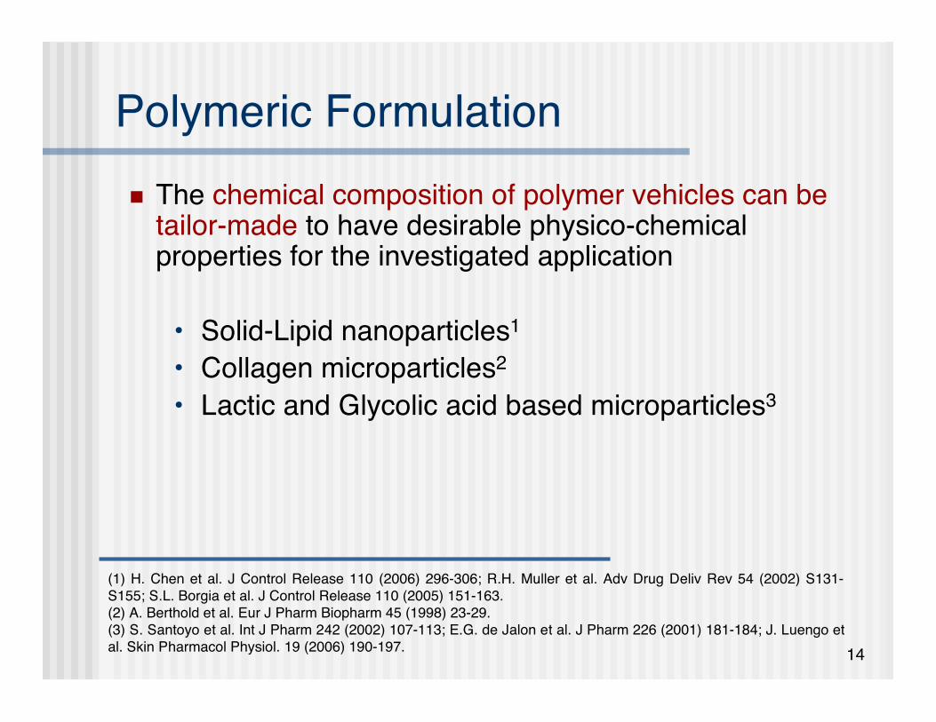
Polymeric Formulation The chemical composition of polymer vehicles can be
tailor-made to have desirable physico-chemicalproperties for the investigated application
• Solid-Lipid nanoparticles1
• Collagen microparticles2
• Lactic and Glycolic acid based microparticles3
(1) H. Chen et al. J Control Release 110 (2006) 296-306; R.H. Muller et al. Adv Drug Deliv Rev 54 (2002) S131-S155; S.L. Borgia et al. J Control Release 110 (2005) 151-163.(2) A. Berthold et al. Eur J Pharm Biopharm 45 (1998) 23-29.(3) S. Santoyo et al. Int J Pharm 242 (2002) 107-113; E.G. de Jalon et al. J Pharm 226 (2001) 181-184; J. Luengo etal. Skin Pharmacol Physiol. 19 (2006) 190-197.
15
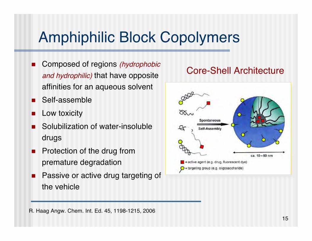
Amphiphilic Block Copolymers Composed of regions (hydrophobic
and hydrophilic) that have oppositeaffinities for an aqueous solvent
Self-assemble Low toxicity Solubilization of water-insoluble
drugs Protection of the drug from
premature degradation Passive or active drug targeting of
the vehicle
Core-Shell Architecture
R. Haag Angw. Chem. Int. Ed. 45, 1198-1215, 2006

16
Requirements for the TherapeuticPolymeric Nanoparticles
Core-Shell architecture: Mw of macromolecules in the range of 20-50KDa1
Inner hydrophobic block serves as the reservoir for lipophiliccompounds5
• Biocompatible polymer: poly(ester)s, poly(ether)s, poly(amino acid)s2,3,4
• Low CMC in water - stable aqueous dispersions of lipophilic drugs Outer hydrophilic block: PEG or PEO6
• Modulation of cell behavior and non-fouling characteristics• Could provide superior hydration of the skin thereby increasing the skin permeation ability of
nanospheres
Particle diameter < 200 nm penetrate SC via surface furrows on the skin6
avoid entrapment by RES7
(1) H. Maeda et al. J Cont. Rel. 74 (2001) 47-61; (2) C. Allen et al. J Cont. Rel. 63 (2000) 275-286; (3) A.V. Kabanov et al.Adv. Drug Deliv. Rev. 54 (2002) 759-779; (4) D. Shenoy et al. Pharm. Res. 22 (2005) 2107-2114; (5) R. Haag Angw. Chem.Int. Ed. 45 (2006) 1198-1215; (6) G. Cevc et al. Adv Drug Deliv Rev 56 (2004) 675-711. (7) R.B. Greenwald Adv. DrugDelivery Rev. 55 (2003) 217-250
17
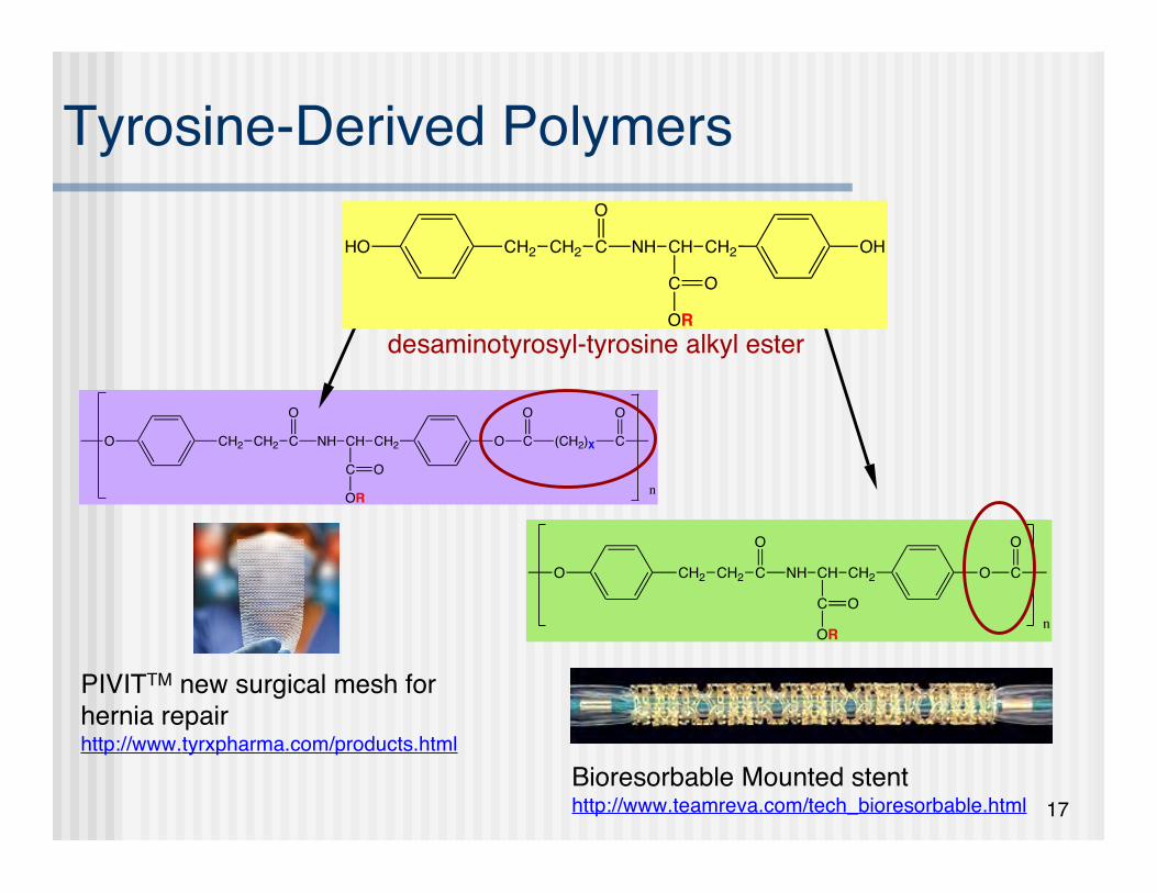
Tyrosine-Derived Polymers
PIVITTM new surgical mesh forhernia repairhttp://www.tyrxpharma.com/products.html
O CH2 CH2 C NH
O
CH CH2
C
O
O
OR
C (CH2)X
O
n
C
O
Bioresorbable Mounted stenthttp://www.teamreva.com/tech_bioresorbable.html
OR
O CH2 CH2 C NH
O
CH CH2
C
O
O
OR
C
O
n
desaminotyrosyl-tyrosine alkyl ester
HO CH2 CH2 C NH
O
CH CH2
C
OH
O
OR

18
Tyrosine-Based Nanospheres
CH3O(CH2CH2O)n C (CH2)X C O CH2 CH2 C NH
CH
C
CH2
O
R
O C (CH2)X C (OCH2CH2)nOCH3
O O O O O
O
m
C. Nardin, D. Bolikal and J. Kohn Langmuir 20 (2004) 11721-11725L. Sheihet et al. Biomacromolecules 2005 & 2007
By changing the length of the diacid (X) or the pendent ester chain of the dipeptide (R) or the molecular weight of PEG (n) or the molecular weight of hydrophobic core (m),
a large number of inter-related polymers can be obtained
19
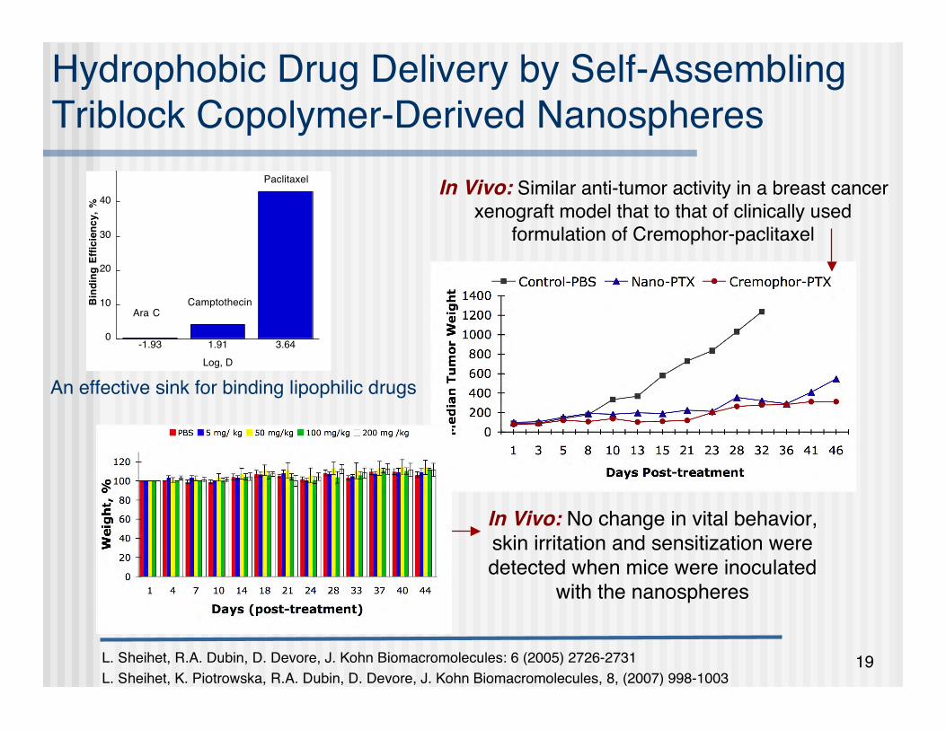
Hydrophobic Drug Delivery by Self-AssemblingTriblock Copolymer-Derived Nanospheres
L. Sheihet, R.A. Dubin, D. Devore, J. Kohn Biomacromolecules: 6 (2005) 2726-2731L. Sheihet, K. Piotrowska, R.A. Dubin, D. Devore, J. Kohn Biomacromolecules, 8, (2007) 998-1003
In Vivo: Similar anti-tumor activity in a breast cancerxenograft model that to that of clinically used
formulation of Cremophor-paclitaxel
In Vivo: No change in vital behavior,skin irritation and sensitization weredetected when mice were inoculated
with the nanospheres
0
10
20
30
40
50
-1.93 1.91 3.64
Bin
din
g E
ffic
ien
cy
, %
Log, D
Ara CCamptothecin
Paclitaxel
An effective sink for binding lipophilic drugs
20
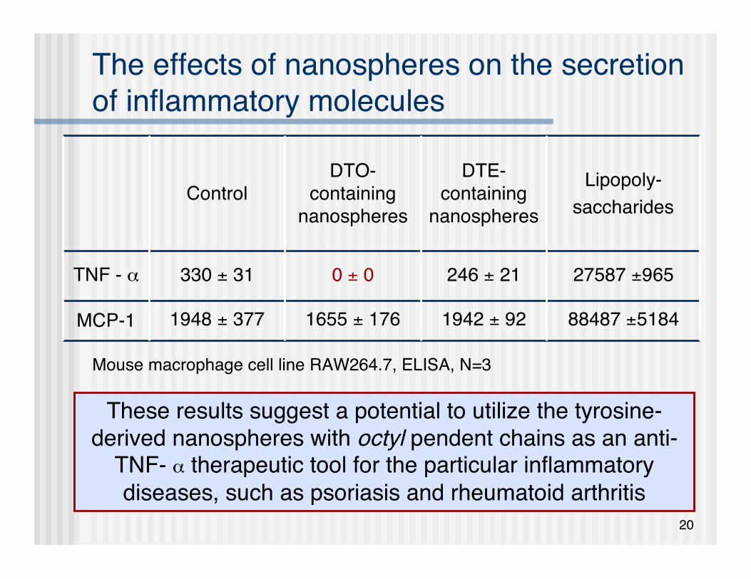
The effects of nanospheres on the secretionof inflammatory molecules
88487 ±51841942 ± 921655 ± 1761948 ± 377MCP-1
27587 ±965246 ± 210 ± 0330 ± 31TNF - α
Lipopoly-saccharides
DTE-containing
nanospheres
DTO-containing
nanospheresControl
These results suggest a potential to utilize the tyrosine-derived nanospheres with octyl pendent chains as an anti-
TNF- α therapeutic tool for the particular inflammatorydiseases, such as psoriasis and rheumatoid arthritis
Mouse macrophage cell line RAW264.7, ELISA, N=3

21
Project Aim
Evaluation of tyrosine-derived nanospheres asdelivery vehicles for highly lipophilic moleculesfor passive skin permeation
Fluorescent dyes as model agents were used todetermine the efficiency of the nanosphere approachas compared to non-particulate enhancerrepresented by propylene glycol

22
Materials & Methods

23
Experimental Design: Materials
Log D values (pH 7) were obtained from ACD/Labs (© 1994-2006 ACD/Labs).
CH3O(CH2CH2O)n C (CH2)6 C O CH2 CH2 CHN CH
C
CH2
O
R
O C (CH2)6 C
O O O O O
O
m
R = DTO (Octyl)
(OCH2CH2)nOCH3
OHO O
C
OH
O
HN
C
(CH2)10
O
CH3
DAF (Log D 7.54)
O
N
Et2N O
Nile Red (Log D 3.10)
24
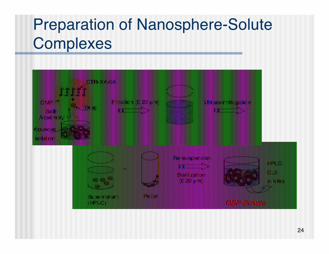
Preparation of Nanosphere-SoluteComplexes
NSP-Solute

25
Solute Binding Efficiency
Solute extraction procedure: Freeze dry predetermined aliquot of the purified
nanosphere-solute complexes suspension Accurately weight the dry residue Thoroughly dissolve in extraction solvent
Solute concentration assay (for NSP and/or PG) RP-HPLC method developed for each solute
!
BindingEfficiency(%)=massof solute inthenanospheresmassof solute inthe feed
" 100%
26
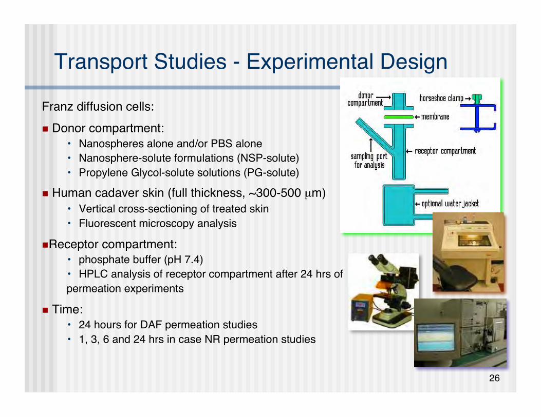
Transport Studies - Experimental Design
Franz diffusion cells: Donor compartment:
• Nanospheres alone and/or PBS alone• Nanosphere-solute formulations (NSP-solute)• Propylene Glycol-solute solutions (PG-solute)
Human cadaver skin (full thickness, ~300-500 µm)• Vertical cross-sectioning of treated skin• Fluorescent microscopy analysis
Receptor compartment:• phosphate buffer (pH 7.4)• HPLC analysis of receptor compartment after 24 hrs ofpermeation experiments
Time:• 24 hours for DAF permeation studies• 1, 3, 6 and 24 hrs in case NR permeation studies

27
Results & Discussion

28
NSP Morphology - TEM
(A) Freeze-fracture (the largest spheres are 60 nm in size, magnification54000 x )
(B) Negative staining(C) Cryo-transmission - membrane thickness 6 mm
(A) (B) (C)

29
Size Distribution of NSP-Solute Formulations
Neither the presence of thesolute in the nanospherespreparation nor the solute
hydrophobicity have asignificant effect on
nanosphere size and SD
() NSP alone() NSP-DAF() NSP-Nile Red
Assuming that the average width of transepidermal hydrophilic pathwaysis in order of 0.4 (water evaporation pathways) to ~ 100 nm
(intercorneocyte space), the relatively small size of tyrosine-derivednanospheres will easily allow their penetration into the stratum corneum
along the surface furrows on the skin
0
0.2
0.4
0.6
0.8
1
1.2
0 12 18 28 43 66 101 156 200
Hydrodynamic Diameter (nm)
Inte
nsit
y
30
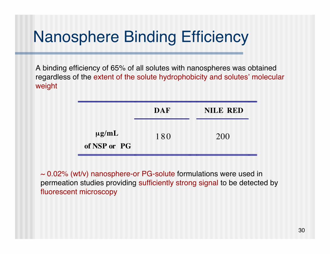
Nanosphere Binding Efficiency
~ 0.02% (wt/v) nanosphere-or PG-solute formulations were used inpermeation studies providing sufficiently strong signal to be detected byfluorescent microscopy
DAF NILE RED
g/mL
of NSP or PG
1 8 0
200
A binding efficiency of 65% of all solutes with nanospheres was obtainedregardless of the extent of the solute hydrophobicity and solutes’ molecularweight
31
HPLC Analysis Tyrosine-derived nanospheres do not facilitate
transport across the human cadaver skin• the amount of solutes in the receptor compartments was
below the limit of detection
1. E.G. de Jalon, M.J. Blanco-Prieto, P. Ygartua, S. Santoyo Int J Pharm 226, 181-184, 20012. R. Alvarez-Roman, A. Naik, Y.N. Kalia, R.H. Guy, H. Fessi Pharm Res 21, 1818-1825, 2004
Similar observations were previously reported:• PLGA-fluorescent microparticles were clearly visualized withinthe skin layers but were not able to reach the receptorcompartment of the diffusion cells1
• use of particulate drug carriers appeared to improve the drugresidence in skin without increasing transdermal transport2

32
Histology of Cryo-Sectioned Skin UsingH&E Staining
Schematic representation of cryo-sectioned skin (Left) and Hematoxylin & Eosinstaining (Right)
33
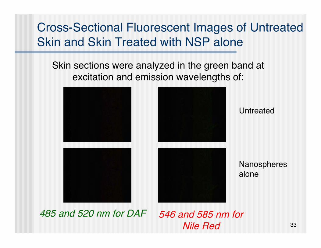
Cross-Sectional Fluorescent Images of UntreatedSkin and Skin Treated with NSP alone
485 and 520 nm for DAF
Skin sections were analyzed in the green band atexcitation and emission wavelengths of:
546 and 585 nm forNile Red
Untreated
Nanospheresalone
34
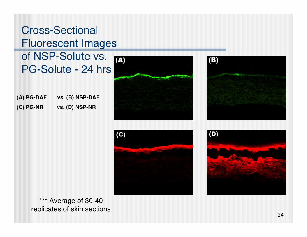
Cross-SectionalFluorescent Imagesof NSP-Solute vs.PG-Solute - 24 hrs
(A) PG-DAF vs. (B) NSP-DAF(C) PG-NR vs. (D) NSP-NR
*** Average of 30-40replicates of skin sections
35
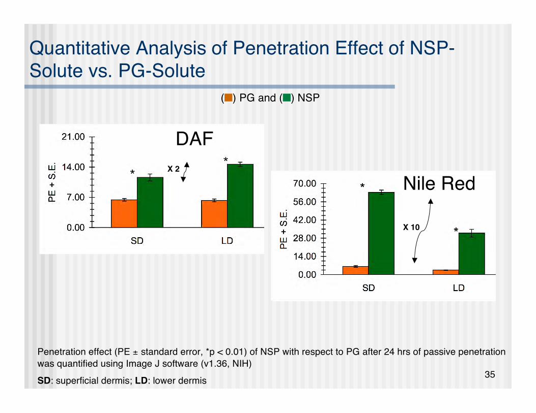
Quantitative Analysis of Penetration Effect of NSP-Solute vs. PG-Solute
Penetration effect (PE ± standard error, *p < 0.01) of NSP with respect to PG after 24 hrs of passive penetrationwas quantified using Image J software (v1.36, NIH)SD: superficial dermis; LD: lower dermis
() PG and () NSP
DAF
Nile RedX 2
X 10

36
Cross-SectionalFluorescent Imagesof NSP-NR vs. PG-NR PassivePermeation as aFunction of Time
1: 1 hour3: 3 hrs6: 6 hrs
(A) PG-NR vs. (B) NSP-NR

37
Cross-Sectional Fluorescent Images of NSP-NR vs.PG-NR Passive Permeation as Function of Time
Penetration effect (PE ± standard error, *p < 0.01)
of NSP with respect to PGSD: superficial dermis; LD: lower dermis
() PG and () NSP
x 1.8
x 3

38
Proposed Explanations forNanospheres Enhanced Permeability
Vehicle nature Neat PG ⇒ dehydrating effect ⇒ less flux PEG in the nanosphere corona ⇒ superior hydration of the skin -
permeation enhancing effect An individual nanosphere could be considered as a supersaturated
system ⇒ higher thermodynamic activity ⇒ increase the partitioning Size
Given the small size, size distribution and dynamic structure ofnanosphere-dye complexes, it is conceivable that they may traversethrough the intercorneocyte spaces
Mechanism of penetration Alteration in the barrier properties and/or a greater degree of partitioning
of the nanospheres into the stratum corneum It is possible that the furrows between the corneocyte islands provide a
place into which nanospheres may accumulate within the skin

39
Conclusions Tyrosine-derived nanospheres significantly enhanced skin penetration of
highly lipophilic model agents (DAF and Nile Red) in human cadaver skinas compared to a non-particulate formulation
No detectable transdermal permeation was observed even after 24 hoursapplication, suggesting that these nanospheres can be used in topicaldrug delivery
An increase in rate and extent of dyes penetration to deeper skin layerscould be due to the higher thermodynamic activity of the dye (relative tothat in propylene glycol), small size and hydration properties of thenanospheres
No acute toxicity, skin irritation or sensitization was detected when micewere injected with nanospheres
Hence, these nanospheres can offer a promising tool for the topical skindelivery of lipophilic drugs and personal care agents such as Vitamins Aand D, sunscreens, glucocorticoids, or retinoids
40
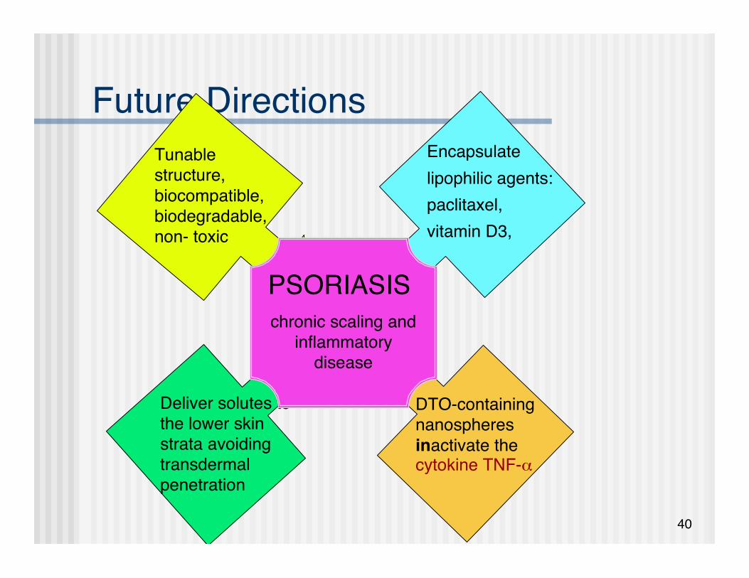
Future Directions
Deliver solutes tothe lower skinstrata avoidingtransdermalpenetration
Tunablestructure,biocompatible,biodegradable,non- toxic
Encapsulatelipophilic agents:paclitaxel,vitamin D3,
DTO-containingnanospheresinactivate thecytokine TNF-α
PSORIASISchronic scaling and
inflammatorydisease

41
Future Directions
PSORIASIS
Targets of therapeuticintervention:
1. Hyperproliferation andabnormal differentiationkeratinocytes
• Increased release of TNF-α
2. Vascular changes withelongated and dilated bloodvessels
• An angiogenic phenotype3. Dermal inflammation with
prominent presence of Tlymphocytes
• Type 1 T-cell cytokines ⇒elevated levels of TNF-αand IL-2
Therapies1. Calcipotriol (Vitamin D3
analogue, Log D ~ 3)• “Normalization” of
keratinocytes• Antiangiogenic activity
2. Anti-TNF- α biologicals
3. Anti-cancer drugs (paclitaxel!)
• Antiangiogenic,antiproliferative andantiflammatory
√
√
√

42
Acknowledgements The TEAM Dr. Gleb Shumyatsky Mr. Matt Treiser NJCBM and CEMBR (W81XWH-04-2-003) for
funding
Thank You!Thank You!