Type-2 innate lymphoid cells control the development of ...
11
ARTICLE Received 27 May 2016 | Accepted 27 Apr 2017 | Published 7 Jun 2017 Type-2 innate lymphoid cells control the development of atherosclerosis in mice Stephen A. Newland 1 , Sarajo Mohanta 2 , Marc Cle ´ment 1 , Soraya Taleb 3 , Jennifer A. Walker 4 , Meritxell Nus 1 , Andrew P. Sage 1 , Changjun Yin 2 , Desheng Hu 5 , Lauren L. Kitt 1 , Alison J. Finigan 1 , Hans-Reimer Rodewald 6 , Christoph J. Binder 7 , Andrew N.J. McKenzie 4 , Andreas J. Habenicht 2 & Ziad Mallat 1,3 Type-2 innate lymphoid cells (ILC2) are a prominent source of type II cytokines and are found constitutively at mucosal surfaces and in visceral adipose tissue. Despite their role in limiting obesity, how ILC2s respond to high fat feeding is poorly understood, and their direct influence on the development of atherosclerosis has not been explored. Here, we show that ILC2 are present in para-aortic adipose tissue and lymph nodes and display an inflammatory-like phenotype atypical of adipose resident ILC2. High fat feeding alters both the number of ILC2 and their type II cytokine production. Selective genetic ablation of ILC2 in Ldlr / mice accelerates the development of atherosclerosis, which is prevented by reconstitution with wild type but not Il5 / or Il13 / ILC2. We conclude that ILC2 represent a major innate cell source of IL-5 and IL-13 required for mounting atheroprotective immunity, which can be altered by high fat diet. DOI: 10.1038/ncomms15781 OPEN 1 Department of Medicine, Division of Cardiovascular Medicine, University of Cambridge, Cambridge CB2 0SZ, UK. 2 Institute for Cardiovascular Prevention, Ludwig-Maximilians-University (LMU), 80336 Munich, Germany. 3 Institut National de la Sante ´ et de la Recherche Me ´dicale, U970 Paris, France. 4 Division of Protein and Nucleic Acid Chemistry, MRC Laboratory of Molecular Biology, Cambridge CB2 0QH, UK. 5 State Key Laboratory of Molecular Vaccinology and Molecular Diagnostics, Xiamen University, Xiamen, Fujian 361102, China. 6 Division of Cellular Immunology, German Cancer Research Center, 69120 Heidelberg, Germany. 7 Department of Laboratory Medicine, Medical University of Vienna and Center for Molecular Medicine (CeMM) of the Austrian Academy of Sciences, 1090 Vienna, Austria. Correspondence and requests for materials should be addressed to Z.M. (email: [email protected]). NATURE COMMUNICATIONS | 8:15781 | DOI: 10.1038/ncomms15781 | www.nature.com/naturecommunications 1
Transcript of Type-2 innate lymphoid cells control the development of ...

ARTICLE
Received 27 May 2016 | Accepted 27 Apr 2017 | Published 7 Jun 2017
Type-2 innate lymphoid cells control thedevelopment of atherosclerosis in miceStephen A. Newland1, Sarajo Mohanta2, Marc Clement1, Soraya Taleb3, Jennifer A. Walker4, Meritxell Nus1,
Andrew P. Sage1, Changjun Yin2, Desheng Hu5, Lauren L. Kitt1, Alison J. Finigan1, Hans-Reimer Rodewald6,
Christoph J. Binder7, Andrew N.J. McKenzie4, Andreas J. Habenicht2 & Ziad Mallat1,3
Type-2 innate lymphoid cells (ILC2) are a prominent source of type II cytokines and are found
constitutively at mucosal surfaces and in visceral adipose tissue. Despite their role in limiting
obesity, how ILC2s respond to high fat feeding is poorly understood, and their direct influence
on the development of atherosclerosis has not been explored. Here, we show that ILC2 are
present in para-aortic adipose tissue and lymph nodes and display an inflammatory-like
phenotype atypical of adipose resident ILC2. High fat feeding alters both the number of ILC2
and their type II cytokine production. Selective genetic ablation of ILC2 in Ldlr�/� mice
accelerates the development of atherosclerosis, which is prevented by reconstitution with
wild type but not Il5� /� or Il13� /� ILC2. We conclude that ILC2 represent a major innate
cell source of IL-5 and IL-13 required for mounting atheroprotective immunity, which can be
altered by high fat diet.
DOI: 10.1038/ncomms15781 OPEN
1 Department of Medicine, Division of Cardiovascular Medicine, University of Cambridge, Cambridge CB2 0SZ, UK. 2 Institute for Cardiovascular Prevention,Ludwig-Maximilians-University (LMU), 80336 Munich, Germany. 3 Institut National de la Sante et de la Recherche Medicale, U970 Paris, France.4 Division of Protein and Nucleic Acid Chemistry, MRC Laboratory of Molecular Biology, Cambridge CB2 0QH, UK. 5 State Key Laboratory of MolecularVaccinology and Molecular Diagnostics, Xiamen University, Xiamen, Fujian 361102, China. 6 Division of Cellular Immunology, German Cancer ResearchCenter, 69120 Heidelberg, Germany. 7 Department of Laboratory Medicine, Medical University of Vienna and Center for Molecular Medicine (CeMM)of the Austrian Academy of Sciences, 1090 Vienna, Austria. Correspondence and requests for materials should be addressed to Z.M. (email:[email protected]).
NATURE COMMUNICATIONS | 8:15781 | DOI: 10.1038/ncomms15781 | www.nature.com/naturecommunications 1

Cardiovascular disease is the leading cause of deathworldwide, increasing in incidence year on year and wasaccountable for one in four deaths globally in 2010 (ref. 1).
Atherosclerosis is the major cause of cardiovascular disease wheredeposits of low-density lipoproteins in the arterial wall lead tothe infiltration of immune cells, inflammation and growth offibro-fatty plaques. This process can culminate in occlusion of theartery following plaque disruption and thrombosis2.
Plaque maturation is influenced by the populations of innateand adaptive immune cells infiltrating the lesion, their activationstate and how they communicate with non-immune cells in thesurrounding arterial tissue3–5. Hypercholesterolaemia and highfat diet (HFD) also trigger systemic immune responses thatmodulate the atherosclerotic process, which may explain theprofound impact of spleen-dependent responses on severalaspects of the atherosclerotic immune response6–8.
Innate lymphoid cells (ILC) are a rare cell population that areclosely related to T and B lymphocytes, but which do not expressrecombined antigen receptors such as the T-cell receptor andB-cell receptor. Early research identified many different subtypesincluding conventional natural killer (NK) cells9, lymphoid tissueinducer cells10,11, nuocytes12 and natural helper cells13. ILC canbe assigned to one of three groups, ILC1, ILC2 or ILC3 (ref. 14).These mirror the T helper (Th)1, Th2 and Th17 paradigm ofT-cell biology and share effector cytokines and transcriptionfactors. Th1 cells promote atherogenesis4, which is also the casefor ILC1-related NK cells15. However, the impact of Th2 andTh17 bias on the atherosclerotic process is more complex; theymay either enhance or limit the disease4,16.
ILC2 were initially identified as an innate source of IL-13during helminth infection12. Subsequently they have beenobserved secreting large quantities of type II cytokines (IL-5,IL-13, IL-9), regulating innate and adaptive immune responses inseveral inflammatory settings (reviewed in ref. 17), modulatingwound healing/tissue repair18, and influencing adipose tissuefunction and metabolic homeostasis19. Furthermore, there isgrowing evidence that some type II cytokines are protective inmouse models of atherosclerosis. For example, IL-13 has beenshown to protect from lesion development and promote plaquestability by increasing collagen deposition, and skewing themacrophage infiltrate towards an alternative activatedphenotype20. IL-5 on the other hand may be protective viaincreasing titres of natural IgM antibodies specific for modifiedLDL epitopes21. Finally, the atheroprotective cytokines IL-33(ref. 22) and IL-25 (ref. 23) can drive expansion of ILC2(refs 24,25) and these cells may provide a crucial component ofthe protective mechanism. However, IL33 and IL-25 activate mayother cellular responses independently of ILC2, and type IIcytokines are also secreted by other cell types and may act onatherosclerosis independently of ILC2.
Two recent studies suggested that ILC2 expansion in mice mayhave an athero-protective role23,26. However, the results werebased on pharmacologic expansion of an ILC2 population,sometimes in immunodeficient mice, and were confounded bydramatic alterations in plasma cholesterol levels after treatment,or by alterations in other immune cell populations. Another studyshowed that total deficiency of Id3, which leads to increasedatherosclerosis in mice, may reduce IL-5 production by ILC2(after exogenous IL-33 stimulation)27. However, no directrelationship was provided to link the ILC2 and atherosclerosisphenotypes27. Thus, the role of naturally occurring ILC2 and themechanisms through which they may regulate atherosclerosis arestill unknown.
Thus, the focus of this work is in defining how ILC2 respond tohypercholesterolaemia, and how atherosclerosis develops in anenvironment where this cell type is absent. Our results show that
ILC2 control the development of atherosclerosis, in part throughproduction of type 2 cytokines.
ResultsCharacterization of ILC2 in atherosclerosis-prone mice.We first addressed the frequency of ILC2 in atherosclerosis-sus-ceptible apolipoprotein e-deficient (Apoe� /� ) mice fednormal chow diet. A typical overview of a transverse sectionof the aorta reveals several aortic and para-aortic structures(Supplementary Fig. 1A) where ILC2 may accumulate. Thosestructures include the atherosclerotic plaque, the aortic adventitiaand associated tertiary lymphoid structures (ATLO), the para-aortic lymph nodes (PaLN), the para-aortic adipose tissue (PaAT)and fat-associated lymphoid clusters (FALCs). We thereforeexamined and quantified the presence of ILC2 in each of thosestructures using flow cytometry and immunofluorescence. Ouranalyses first revealed the presence of ILC2 (Lin� ICOSþ
CD25þ CD127þ ) in PaLN and PaAT of chow-fed Apoe� /�
mice (Fig. 1a), which is consistent with the previously reportedpresence of ILC2 in secondary lymphoid organs (GATA3þ
ICOSþCD3� cells in Supplementary Fig. 1B,C) and theirtropism for adipose tissue (for example, peri-gonadal WAT)(Fig. 1a)28. The percentage of ILC2 among CD45þ cells in PaATwas smaller than in peri-gonadal WAT (Fig. 1b), but wassubstantially higher than the percentage of ILC2 in PaLN(Fig. 1b), and mesenteric lymph nodes (MLN) (Fig. 1b) of thesame animals. Supplementary Fig. 1D shows the absolute numberof ILC2 recovered from different locations in 420-week-oldApoe� /� mice.
The phenotype, activation state and function of ILC2 maychange dependent on the tissue where they reside and thecytokine microenvironment29–31. We found that Lin� ICOSþ
CD25þ CD127þ ILC2 of peri-gonadal WAT (GWAT) weremostly KLRG1þST2þ (Fig. 1a,b) and were comparable tonatural ILC2 (ref. 29), whereas ILC2 of MLN and PaLN were inlarge majority KLRG1þST2� or ST2low (Fig. 1a,b), similar to arecently described population of inflammatory ILC2 with reducedability to produce IL-5 and IL-13 (ref. 29). Interestingly, the ILC2population in para-aortic fat differed significantly from that ofperi-gonadal fat, and comprised a population expressing lowamounts of ST2 on their surface (Fig. 1c). The number of PaATILC2 remained relatively constant during aging (SupplementaryFig. 1E). ILC2 were also found in ATLO of 80-week-oldApoe� /� mice with advanced atherosclerosis (SupplementaryFig. 2A,B).
Recent studies showed that inflammatory stimuli promote theformation of FALCs within WAT32. FALCs have been detectedmostly in peri-gonadal, mesenteric and mediastinal WAT, withthe pericardium accumulating a substantial number of clusters32.However, whether FALCs may also accumulate in the para-aorticregion is still unknown. Given the role of inflammation in FALCformation, we reasoned that those clusters may be more prevalentin old animals (for example, 80 weeks). Indeed, we detectedFALCs in the para-aortic WAT of both old WT and Apoe� /�
mice (Fig. 1d,e, Supplementary Fig. 1A). As reported for otherlocations, para-aortic FALCs were rich in CD3þ T cells, B220þ
B cells, CD138þ plasma cells (Fig. 1f), PNAþKi67þ germinalcentre-like B cells and Foxp3þ Tregs (Supplementary Fig. 2C),accumulated a few ILC2 (Fig. 1g), no follicular dendritic cells(CD35 staining in Fig. 1f) and were supplied with blood vessels,lymph vessels, high endothelial venules and ERTR7þ conduits(Supplementary Fig. 2C). The number and size of peri-aorticFALCs were significantly greater in Apoe� /� mice compared toWT mice (Fig. 1e), supporting a role for vascular inflammation inpromoting para-aortic FALC formation.
ARTICLE NATURE COMMUNICATIONS | DOI: 10.1038/ncomms15781
2 NATURE COMMUNICATIONS | 8:15781 | DOI: 10.1038/ncomms15781 | www.nature.com/naturecommunications
Thus, besides their presence in WAT and secondarylymphoid organs, ILC2 are also present in para-aortic fatof athero-prone mice, where they display an inflammatoryphenotype, distinct from the natural ILC2 phenotype of distantWAT and more similar to the inflammatory phenotype of lymphnode ILC2.
High fat feeding alters ILC2 numbers and cytokine production.Mice fed a defined HFD for a period of weeks to months developaccelerated atherosclerosis. We therefore hypothesized that highfat feeding may alter the accumulation and function of ILC2systemically, and observed the ILC2 populations in the bonemarrow (BM), spleen and peripheral lymph nodes of low-density
Par
a-ao
rtic
LNP
ara-
aort
icAT
Per
i-gon
adal
WAT
SS
C-A
SS
C-A
SS
C-A
Lin
FIT
C
KLR
G1
Pe
Cy7
Lin- ICOS+0.043
Lin- ICOS+0.21
Lin- ICOS+10.2
250Ka
b
d
f g
e
c
105
104
103
102
105
104
103
–103
KLR
G1
Pe
Cy7
105
104
103
–103
Lymphocytes46.2
Lymphocytes11.7
Lymphocytes0.95
14.3 0
85.7 0Q1
Q4
42.9
23.8Q1
Q4
1.75
11.8Q1
Q4
Q3
Q2
0
33.3
Q3
Q2
1.75
84.7
Q3
Q2
200K
150K
100K
50K
0
250K
200K
150K
100K
50K
0
250K
200K
150K
100K
10 ST2 expression
* **
2,000
1,500
1,000
500
0
0.1%IL
C2
MF
I
0.01
Apoe–/–
B220CD3eDAPI
GATA3DAPI
ICOSCD3e
GATA3DAPI
ICOSGATA3DAPI
ICOSCD3e
CD138CD3eDAPI
CD35CD3eDAPI
WT Apoe–/–WT
8 0.03
0.02
0.01
0
6
4
2
Clu
ster
num
ber
Clu
ster
siz
e (m
m2 )
0
MLN
PaLN
PaLN
PaAT
GWAT
Peri-a
ortic
AT
Peri-g
onad
al AT
1
50K
0
0 0 050K 103 –103 103 104 105
0–103 103 104 105
104100K 150K
FSC-A ICOS PE
Lin
FIT
C
105
104
103
102
0 103 104
ICOS PE
Lin
FIT
C
105
104
103
102
0 103 104
ICOS PE
ST2 e710
ST2 e710
KLR
G1
Pe
Cy7
105
104
103
–103
0–103 103 104 105
ST2 e710
200K 250K
0 50K 100K 150KFSC-A
200K 250K
0 50K 100K 150KFSC-A
200K 250K
Figure 1 | ILC2 in para-aortic adipose tissue and FALCs of atherosclerotic mice. Flow cytometric analysis (a) and quantification (b,c) of ILC2 present in
the para-aortic adipose tissue (AT) show a phenotype similar to KLRG1hi ST2� ILC2 (iILC2-like) in the lymph node in contrast to KLRG1þ ST2þ ILC2
(nILC2-like) prevalent in the peri-gonadal WAT. Mean fluorescence intensity (MFI) for ST2 expression in ILC2 is shown in c. Bars represent mean values.
(d) Oil red O and Haematoxylin show that FALCs are present in the para-aortic AT of aged Apoe� /� mice (78–80 weeks of age n¼ 10, scale bar 100mm)
(e) and that Apoe� /� FALCs are greater in both number and size compared to WT (f). Immunoflorescence staining demonstrates that para-aortic FALCs
are rich in CD3þ T cells, B220þ B cells and CD138þ plasma cells. CD35þ follicular dendritic cells were absent from these structures (scale bar 50mm).
Additionally, para-aortic FALC-resident CD3� GATA3þ ICOSþ ILC2 cells were also detected (g, scale bar 10mm). Representative images shown. Graph
data points represent individual mice. Statistical significance was determined by Mann–Whitney U-test.
NATURE COMMUNICATIONS | DOI: 10.1038/ncomms15781 ARTICLE
NATURE COMMUNICATIONS | 8:15781 | DOI: 10.1038/ncomms15781 | www.nature.com/naturecommunications 3

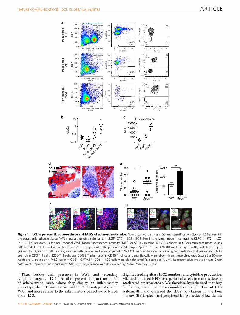
lipoprotein receptor-deficient (Ldlr� /� ) mice, a second athero-sclerosis-susceptible strain, that had been maintained on HFD for8 weeks. Flow cytometric analysis of ILC2 populations (Fig. 2a)demonstrated that although there was no difference in theproportion of precursor cells in the BM (Fig. 2b), the matureILC2 were significantly under-represented (2–3-fold loss) in MLNand PaLN of mice maintained on HFD (Fig. 2b).
To examine any change in functional capability associated withthe suppression of this population, Lin� ICOSþ ILC2 cells weresorted from the spleens and MLN of conventional chow- andHFD-fed mice (purity495%, Supplementary Fig. 2D). Not onlywere fewer cells recovered from the organs of HFD mice(Supplementary Fig. 2E and consistent with Fig. 2b) but, duringex-vivo expansion with IL-7 and IL-33, they also secretedsubstantially less IL-5 and IL-13 (Fig. 2c). To confirm that thealteration of type II cytokine production occurred in vivo,we repeated the experiments and performed QPCR analysis oncell-sorted ILC2 isolated from the spleens of Ldlr� /� mice thathad been maintained on chow or HFD for 8 weeks. ILC2 werealso cell-sorted from the aortas (two pools of three mice each)and GWAT for comparison (Fig. 2d). QPCR analysis indicated a
significant decrease of GATA3 and IL-13 transcripts (Fig. 2d) anda similar trend observed with IL-5 (Fig. 2d), in spleen-derivedILC2 of mice on HFD compared to chow diet. Interestingly,GWAT-derived ILC2 showed no significant change, whereasaorta-derived ILC2 tended to upregulate their expression ofGATA3, IL-5 and IL-13 after HFD (Fig. 2d). This is a strongindication that HFD differentially alters ILC2 phenotype in theperiphery, and that continuous production of type II cytokines byaortic-ILC2 may be critical to maintain a counter-regulatorypathway, and limit the progression of aortic inflammation inface of a sharp decline of type II cytokine production byperipheral ILC2.
Expansion of ILC2 reduces atherogenesis. Similarly to others26,we hypothesized that reconstitution of ILC2 cells by treatingwith IL-2 during HFD would replenish an atheroprotectiveenvironment. To minimize off-target effects of IL-2 on otherCD25-expressing cells such as Tregs, we used T- and B-cell-deficient Apoe� /� /Rag2� /� mice maintained on HFD for 8weeks. The mice received three weekly injections of IL-2/Jes6-1
104
103
1030 104 105
Lin
FT
C
104
103
Lin
FT
C
dCT
/18s
RN
A
dCT
/18s
RN
A
dCT
/18s
RN
A
Lin-
%S
ca1+
ST
2+
% IL
C2
% IL
C2
% IL
C2
pg m
l–1pg
ml–1
ICOS PE
1030 104 105
ICOS PE
Spleen Aorta GWAT Spleen Aorta GWAT Spleen Aorta GWAT
Chow
MLN
IL-5 IL-13 GATA3
PaLN
0.0317
0.055 0.0079 0.0079
0.0079
HFD
Chow HFD
Chow
HFDCho
wHFD
Chow
HFDCho
wHFD
Chow
HFDCho
wHFD
Chow
HFDCho
wHFD
Chow
HFD
Chow HFD
Chow HFD
Chow M
LN
HFD MLN
Chow M
LN
HFD MLN
Lin- ICOS+0.52
Lin- ICOS+0.054
2.0
Bone marrow
0.11
Spleen ILC2-derived IL-13
ILC2-derived IL-5
0.05
0.201,000
750
500
250
0
0.15
0.10
0.05
0.00
0.03
15 10 2015105
–5–10–15
0
0
10
5
–5
–10
–10
–20–15
0
0.02
0.01
0.00
250
200
150
100
50
0
0.04
0.03
0.02
0.01
0.00
1.5
1.0
0.5
0.0
a
d
b c
Figure 2 | ILC2 populations are decreased during high fat diet. Spleen, bone marrow (BM), mesenteric lymph node (MLN, shown) and para-aortic lymph
node (PaLN, shown) from Ldlr�/�mice maintained on high fat diet (HFD) for 8 weeks were analysed for ILC2 populations by flow cytometry (a). While
BM resident ILC2 were unchanged (top left) a downward trend in splenic ILC2 and a statistically significant decrease in ILC2 was observed in MLN and
PaLN (b). ELISA analysis of IL-5 and IL-13 in the supernatants of sorted and cultured ILC2 (1� 105 cells per well) also demonstrated a decrease in cytokine
secretion (c). QPCR on sorted splenic, aortic and GWAT ILC2 from Ldlr�/�mice maintained on chow or high fat diet (HFD) for 8 weeks. Data from aortic
ILC2 represent two pools of three mice each. Each other square or triangle represents data from one separate mouse (d). Statistical significance was
determined by Mann–Whitney U-test.
ARTICLE NATURE COMMUNICATIONS | DOI: 10.1038/ncomms15781
4 NATURE COMMUNICATIONS | 8:15781 | DOI: 10.1038/ncomms15781 | www.nature.com/naturecommunications

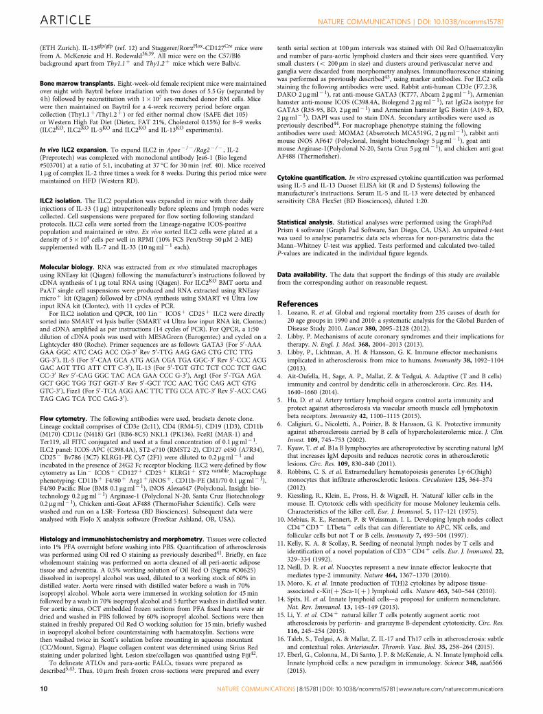
complex, which increases IL-2 biological activity33, for theduration of the experiment. Following this treatment, flowcytometry demonstrated ILC2 populations were significantlyexpanded in spleen and BM compared to PBS-treated controls(Fig. 3a). In addition to ILC2 expansion in peripheral lymphoidtissue, clusters of ICOSþ KLRG1þ (Fig. 3b) ILC2 cells wereobserved in the adventitia of the aorta adjacent to the aorticsinus by immunofluorescence. The adventitia has been suggestedas a source of precursor cells, which may influence plaque
architecture34 and the presence of expanded ILC2 in this tissuemay suggest a direct localized effect. Whether these ILC2 haveexpanded in situ (as recent publications may suggest35) or havemigrated into the tissue from the periphery remains to beinvestigated.
Further phenotypic changes occurred during this IL-2/Jes6-1treatment, namely an expanded population of IL-5þ ILC2(Lin� ICOSþ ), associated eosinophilia and decreased CD11bþ
Ly6G� Ly6Chi inflammatory monocytes (Supplementary Fig. 3A).
0.30
a
b
c
0.4 0.80.70.60.50.40.30.20.10.0
0.3
0.2
0.1
0.0
Bone marrow
PBS
Aortic sinus
PBS IL-2
0.0041200,000 50
40
30
20
10
0
150,000
100,000
50,000
Pla
que
area
per
μm
2
% P
laqu
e ar
ea
0
0.0728En face
IL-2
Spleen MLN
0.0012 0.0022 0.051
PBS IL-2 Jes6-1
PBS IL-2+Jes6-1 PBS IL-2+Jes6-1
PBS IL-2 Jes6-1 PBS IL-2 Jes6-1
0.25
0.20
0.15
% IL
C2
% IL
C2
% IL
C2
0.10
0.05
0.00
Figure 3 | ILC2 expansion reduces atherosclerosis in Apoe� /�/Rag2� /� mice. IL-2/Jes6-1 complexes can expand ILC2 in the bone marrow and spleen
of Apoe� /�/Rag2� /� mice (a) as well as inducing clusters of ICOSþ KLRG1þ cells (b, ICOS Green, KLRG1 Red, scale bar 25mm) adjacent to the aortic
sinus. IL-2/Jes6-1 treatment significantly decreases plaque area in the aortic sinus compared to vehicle alone (c, scale bar 270mm). Graph data points
represent individual mice and statistical significance was determined by Mann–Whitney U-test.
NATURE COMMUNICATIONS | DOI: 10.1038/ncomms15781 ARTICLE
NATURE COMMUNICATIONS | 8:15781 | DOI: 10.1038/ncomms15781 | www.nature.com/naturecommunications 5

There was also an increase in splenic NK cells (SupplementaryFig. 3A). This treatment resulted in a significant decrease in thearea of atherosclerotic plaques at the aortic sinus (Fig. 3c) and asimilar trend was also observed at the aortic arch although thiswas not statistically significant. In contrast to a recent studywhere IL-2/Jes6-1 treatment of Ldlr� /� /Rag2� /� mice led to asubstantial reduction of plasma VLDL-cholesterol levels26,our treatment of Apoe� /� /Rag2� /� mice with IL-2 wassuccessful in reducing the effect of HFD on the progression ofatherosclerosis, without any change in plasma lipid levels(Supplementary Fig. 3B). However, as reported in previouswork26, IL-2 treatment was not without a number of off-targeteffects, the most common of which was an increase in splenicfibrosis in the IL-2 treated group, which was severe in some mice(Supplementary Fig. 3C). Additionally, weight gain upon HFDfeeding was slower in the group receiving IL-2 compared tocontrols (Supplementary Fig. 3D).
Selective genetic ILC2 ablation exacerbates atherosclerosis.Artificial expansion of ILC2 does not inform about the true roleof the endogenous ILC2 population that develops during thecourse of atherogenesis. To allow the specific depletion of ILC2 inan otherwise replete immune system, Staggerer/RoraFlox-Cd127Cre
mice (which are selectively deficient in ILC2, hereafter known asILC2KO (ref. 36)) were used as donors in a BM transplant modelinto atherosclerosis prone Ldlr� /� recipients. Given recentobservations that in steady-state conditions tissue resident ILC2are not replenished from circulating ILC2 (ref. 35), we validatedthe ability for BM ILC2 from Thy1.1 congenic mice toreconstitute lymphatic and tissue compartments. Thy1.2þ
recipient mice were irradiated and reconstituted with Thy1.1þ
BM. Following a 4-week recovery period, spleen, MLN andGWAT were collected and the proportion of Thy1.1þ donorILC2 was determined by flow cytometry. We found that donorILC2Thy1.1 fully reconstituted the lymphatic compartments(0% Thy1.2þ ILC2) as well as the majority of the GWATtissue resident ILC2 (5% Thy1.2þ ) (Fig. 4a). Therefore, BMtransplants are an effective method for replacing host with donorBM-derived ILC2. This was then repeated using either WT orILC2KO BM followed by recovery and HFD for 9 weeks. Toensure the BM graft was effective and very few endogenousILC2 remained, IL-33 was given to recipients 24 h before organcollection. Subsequent flow cytometry analysis demonstrated thatrecipients of ILC2KO BM had significantly decreased ILC2 in BMand peripheral MLN compared to ILC2WT recipients (Fig. 4b).Quantification of serum cytokines also demonstrated decreasedIL-5 and IL-13 (Fig. 4c). Moreover, gene expression analysis onaorta and PaAT confirmed a substantial reduction of IL-5 andIL-13 expression in tissues recovered from ILC2KO mice (Fig. 4d).The extent of lipid accumulation in aortas of ILC2KO mice wassignificantly increased in the aortic arch (Fig. 4e) and aortic sinus(Fig. 4f) compared to ILC2WT recipients, despite no change inplasma lipid levels (Supplementary Fig. 4A).
As discussed above, ILC2 are a potent source of the type IIcytokines IL-5 and IL-13, which are known to be atheroprotectivethrough differing mechanisms. It was therefore logical toinvestigate whether the increase of disease severity in the ILC2KO
model could be accounted for by deficiencies in these pathways.The potential expansion and maintenance of B1a B cells andassociated increase in natural IgM antibodies by ILC2-derivedIL-5 were investigated by flow cytometry in ILC2KO BMrecipients. Although pooled results from a number of experi-ments suggested that B1a B cells were less abundant in the spleenand MLN of ILC2KO recipient mice (Supplementary Fig. 4B),there was considerable variation between biological replicates and
not all repeats followed the same trend. Furthermore, there wasno associated decrease in natural IgM isotypes in the serum ofILC2KO recipient mice after 8 weeks of HFD (SupplementaryFig. 4B) or difference in plaque IgM deposits (SupplementaryFig. 4C). Thus, changes in B1a B-cell subset are unlikely toaccount for the effect of ILC2 deletion on atherosclerosis althoughthis remains to be clarified.
ILC2 alter plaque composition. To examine if ILC2 deletionimpacts immune cell accumulation and activation in vivo,we analysed plaque composition. The number of CD3þ T cells inlesions of ILC2KO recipients was reduced (SupplementaryFig. 5A) indicating that the increase in lesion size was unlikelyto be driven by T-cell activation. However, immune-fluorescentlabelling of MOMA2þ myeloid cells in the aortic sinus revealed alarger lipid-containing core of foam cells in the absence of ILC2(Fig. 5a). Interestingly, the expression of Arg1 was significantlydecreased (Fig. 5b). There was no difference in the proportionof a-smooth muscle actin-expressing cells in plaques or thedeposition of collagen throughout the plaque detected by Siriusred staining (Supplementary Fig. 5B). It is usual for larger, moreadvanced plaques in this model to contain more collagendeposits, and this absence of increased collagen deposition inthe larger plaques of ILC2KO mice coupled with less Arg1expression might indicate disrupted tissue repair mechanisms.The macrophage phenotype was therefore further investigated byflow cytometry. Here, we observed a significant decrease inCD11bþ F4/80þArg1þ and CD11bþ F4/80þ Arg1þ iNOSþ
macrophage population in the aorta and peri-aortic adipose tissueof ILC2KO mice and an expansion of CD11bþ F4/80þ iNOSþ
macrophages (Fig. 5c). This shows that, although ILC2 are a rarepopulation of cells, in mouse models of atherosclerosis theyperform a critical role in preventing plaque development andtheir ablation alters macrophage phenotype and increases diseaseseverity.
ILC2-derived IL-5 and IL-13 are required for atheroprotection.We designed reconstitution experiments to address the specificroles of ILC2-derived IL-5 or IL-13 in the control of athero-sclerosis. BM transplantation experiments were performed inLdlr� /� mice which received mixed BM transplants of ILC2WT,ILC2KO, 80% ILC2KO with 20% IL-5þ (ILC2-deficient micereconstituted with IL-5 sufficient ILC2) or 80% ILC2KO with 20%IL-5KO (ILC2-deficient mice reconstituted with IL-5-deficientILC2; 80% of all other cell types are still capable of IL-5production). After recovery, mice were put on HFD for 8 weeks.Reproducing the original observation, ILC2KO mice showedincreased atherosclerosis of the aortic arch (Fig. 6a). Additionally,ILC2KO mice reconstituted with IL-5þ ILC2 did not developincreased atherosclerosis compared to ILC2WT mice (Fig. 6a),further supporting the requirement for a competent ILC2population to limit atherogenesis. Crucially however, recipients ofILC2KO/IL-5� BM, which are replete with IL-5-deficient ILC2,developed severe atherosclerosis comparable to the full ILC2knockout condition (Fig. 6a). However, absence of ILC2-derivedIL-5 did not alter lesion size in the aortic sinus (SupplementaryFig. 5C), suggesting the involvement of other pathways.Therefore, complementing the observations with ILC2 sourcedIL-5, we examined the function of ILC2-derived IL-13 in aseparate set of experiments by reconstituting BMT recipients with80% ILC2KO and either 20% IL-13þ or 20% IL-13KO. As wasobserved with the ILC2-specific IL-5 deficiency, the inability ofILC2 to produce IL-13 significantly increased atherosclerosis inthe aortic arch (Fig. 6a). Furthermore, there was a significantincrease in lesion size in the aortic sinus of ILC2KO IL-13�
ARTICLE NATURE COMMUNICATIONS | DOI: 10.1038/ncomms15781
6 NATURE COMMUNICATIONS | 8:15781 | DOI: 10.1038/ncomms15781 | www.nature.com/naturecommunications

Lymphocytes
Lin- ICOS+
CD127+ CD25+33.9
0.39
104
105 105
104
103
0
104
103
–103
103 104
–103
105
104
103
0
1
2 4
2
0
–2
–4
–6
–1
–4
–7
–10
3
0
–3
–6
–9
0
–1
–2
–3
–4
0
0 103 1040 103 104 1050
104
103
103
ICOS APC CD25 Bv786
0
0 1041030
104
2.0 0.25
0.20
0.15
0.10
0.05
0.00
Bone marrow
Wild type ILC2 KO
Wild type ILC2 KOAortic sinus
20
15
10
5
0
450,000
300,000
150,000
00 1 2 3 4 5
Depth6 7 8 9 10
En face
% A
rea
plaq
ue
Are
a pe
r μm
2
MLNa
c
e
f
b
d
IL-5
200 15,000
12,500
10,0002,000
1,000
0
150
100
50
0
IL-13
IL-13 IL-5
IL-13 IL-5
0.0159
0.0193
0.01
0.0115
<0.0001
0.0047
0.17
0.0080.1
0.0023
ILC2
WT
ILC2
KO
ILC2
WT
ILC2
KO
ILC2
WT
ILC2
KO
ILC2WT
ILC2KO
ILC2
WT
ILC2
KO
ILC2
WT
ILC2
KO
ILC2
WT
ILC2
KO
ILC2
WT
ILC2
KO
ILC2
WT
ILC2
KO
ILC2
WT
ILC2
KO
0.5
0.1%IL
C2
%IL
C2
pg m
l–1
Aor
taP
aAT
ΔCT
/18s
RN
AΔC
T/1
8sR
NA
ΔCT
/18s
RN
AΔC
T/1
8sR
NA
pg m
l–1
0.0
103
0
Lin
FIT
C
Thy
1.2
PE
Thy1.1 Bv650 Thy1.1 Bv650 Thy1.1 Bv650
Thy
1.2
PE
Thy
1.2
PE
Spleen
Q50.55
Q10
Q15.65
Q86.39
Q45.34
Q411.3
Q394.1
Q380.9
Q792.4
Q60.63
Q20.54
Q22.12
MLN GWAT
CD
127
Bv4
50
17.9
Figure 4 | Genetic deletion of ILC2 exacerbates atherosclerosis in Ldlr�/� mice. During bone marrow transplant, ILC2Thy1.1 fully restore Spleen
MLN and GWAT resident compartments in Thy1.2þ mice (a). Irradiated Ldlr�/� mice received bone marrow from Staggerer/RoraFlox -CD127Cre (ILC2KO)
or Staggerer/Roraþ -CD127Cre (ILC2WT) donor mice before being maintained on HFD for 8 weeks (b). Intra-peritoneal injections of IL-33 24 h before
organ collection demonstrated recipients of ILC2KO BM had decreased Lin� ICOSþ Sca1þ ST2þ ILC2 in bone marrow and Lin� ICOSþ CD25þ in
peripheral MLN compared to ILC2WT recipients. Serum levels of IL-5 and IL-13 were also decreased (c), as was expression of IL-5 and IL-13 transcripts in
aorta and PaAT (d). Oil Red O quantified atherosclerotic lesions at the aortic arch and aortic sinus (e and f respectively, representative images shown)
indicated increased plaque size (all surface of intimal lesion is taken into account) in ILC2KO recipients compared to ILC2WT controls for both sites.
Graph data points represent individual mice and statistical significance was determined by Mann–Whitney U-test. Scale bars: 270 mm.
NATURE COMMUNICATIONS | DOI: 10.1038/ncomms15781 ARTICLE
NATURE COMMUNICATIONS | 8:15781 | DOI: 10.1038/ncomms15781 | www.nature.com/naturecommunications 7

recipients (Fig. 6b). Although no significant change in MOMA2þ
expression was detected (Fig. 6c), Arg1 expression was decreased(Fig. 6c) as previously observed in ILC2KO recipients. We alsofound a significant reduction of collagen deposition in lesions ofILC2KO IL-13� recipients (Fig. 6d), indicating impaired vascularhealing. ILC2-derived cytokines (IL-5 and IL-13) are thereforevital components controlling the progression of atherosclerosis,particularly IL-13 which may alter macrophage phenotype,and its absence leads to larger and potentially more vulnerableplaques.
DiscussionHere, we show that ILC2 constitute a major atheroprotective celltype. High fat feeding reduces the frequency of ILC2 in theperiphery and profoundly alters their protective phenotype,concomitant with an acceleration of atherosclerosis. Usingmice specifically deficient in ILC2, we show that endogenousILC2 perform a central role in controlling the progressionof atherosclerosis and this effect is in part dependent onILC2-derived IL-5 and IL-13. Remarkably, production of IL-5and IL-13 by other cell types is unable to compensate for thelack of those ILC2-derived cytokines, particularly IL-13, andtheir atheroprotective effects. IL-5-dependent atheroprotectionwas limited to the thoracic aorta and could not be attributed to
changes in macrophage phenotype or B1-dependent natural IgMproduction. IL-13-dependent atheroprotection was associatedwith important changes in collagen deposition and macrophagephenotype, suggestive of alternative activation. However, thedirect links between changes of macrophage phenotype andatheroprotection were not addressed. Future studies should try tounderstand the differential impact of HFD on peripheral versusaortic ILC2, and define their distinct contributions to limitingvascular inflammation and atherosclerotic lesion development.
Previous studies suggested a potential role for ILC2 in themodulation of atherosclerosis23,26. However, those studies usedimmune-compromised animals and relied on non-physiologicalexogenous and chronic administration of cytokines (that is, IL-2and IL-25) that are not specific for the ILC2 population, and thatcan promote ILC2-independent immune responses. Moreover,those studies failed to provide any direct evidence of theinvolvement of ILC2 in atherosclerosis and were confounded byprofound alterations of hepatic and lipid metabolism26 followingchronic exogenous cytokine administration. We also found thatchronic administration of IL-2/anti-IL2 complexes in immune-compromised animals reduced atherogenesis but in agreementwith previous findings, the effect was associated with severaladverse side effects. Most probably, the amount of IL-2/IL-2 mAbused in murine models is not physiologically relevant and further
MOMA-2
a
b c
Wild type ILC2 KO
0.0244
Arg1+ Cell count
Arg1% Cells
60
2030 40
30
20
10
0
20
10
0
15
10
5
0
35 25
20
15
10
5
0
302520151050
50
40
30
20
10
0No.
Arg
1+ p
er p
laqu
e
Aor
taP
eri-a
ortic
AT
% A
rg1+
% M
φ A
rg1+
% M
φ iN
OS
+
% M
φ A
rg1+
% M
φ iN
OS
+A
rg1+
% M
φ iN
OS
+A
rg1+
% M
φ iN
OS
+
0.024
0.024 0.0640.0043
0.81
0.0022 0.0043 0.0411
70
6050
4030
% M
OM
A-2
+ p
laqu
e
20100
500
400
300
200
100
0
35302520151050
ILC2
WT
ILC2
KO
ILC2
WT
ILC2
KO
ILC2
WT
ILC2
KO
ILC2
WT
ILC2
KO
ILC2
WT
ILC2
KO
ILC2
WT
ILC2
KO
ILC2
WT
ILC2
KO
ILC2
WT
ILC2
KO
ILC2
WT
ILC2
KO
Figure 5 | ILC2 alter the plaque composition. Phenotypic changes in the enlarged plaque (a) favour increased MOMA2þ . Proportional and numerical
decrease in Arginase1 (Arg1) expressing cells (b). Flow cytometry of CD11bþ F4/80þ aorta resident macrophages indicated bias towards reduced Arg1
and increased iNOS expression (c). Representative micrographs of MOMA2þ staining are shown. Graph data points represent individual mice and
statistical significance was determined by Mann–Whitney U-test. Scale bars: 270 mm.
ARTICLE NATURE COMMUNICATIONS | DOI: 10.1038/ncomms15781
8 NATURE COMMUNICATIONS | 8:15781 | DOI: 10.1038/ncomms15781 | www.nature.com/naturecommunications

work is required to titrate the dose of cytokine required to protectagainst atherosclerosis without inducing undesirable side effects.
It is interesting to note, however, that low dose IL-2 therapy isreported to be safe in humans and has been successful in severalclinical trials of immune-mediated diseases (reviewed in ref. 37)where exogenous IL-2 was used to expand Tregs. Remarkably,low-dose IL-2 in humans also increases the production ofIL-5 (ref. 38) and this is attributed to the dose-dependentexpansion of ILC2.
Thus, augmentation of ILC2 and ILC2-derived IL-5 or IL-13on top of Treg expansion might constitute a potentially attractivedouble-hit therapy to limit accelerated atherosclerosis.
MethodsMice. All work was conducted under UK Home Office project license regulationsafter approval by the Ethical Review Committee of the University of Cambridge.
Mice used in this investigation were Ldlr� /� (Jackson Labs 002207),Apoe� /� /Rag2� /� (Jackson Labs), and IL5� /� were from Manfred Kopj
Aortic arch en face
20
a
b
c
d
0.01520.0152 0.0037NS
15
10
5
0
50 15 1751501251007550250
10
5
0
MOMA-2
Collagen0.0140
15
10
5
0
Arg1+ Arg1+
40
30
20
10
0
20
15
10
5
0
Aortic arch en face
% A
rea
plaq
ue
% A
rea
plaq
ue
ILC2
WT
ILC2
KO
ILC2KO IL-13+
ILC2KO IL-13+ ILC2KO IL-13–
ILC2KO IL-13WT
ILC2KO IL-13KO
ILC2KO IL-13–
ILC2
KO IL-1
3+
ILC2
KO IL-1
3–
ILC2
KO IL-1
3+
ILC2
KO IL-1
3–
ILC2
KO IL-1
3+
ILC2
KO IL-1
3–
ILC2
KO IL-1
3+
ILC2
KO IL-1
3–
Aortic sinus
0.0001
0.02890.014
600,000
500,000
400,000
300,000
200,000
100,000
00 1 2 3 4 5
Depth
6 7 8 9 10
ILC2
KO IL-5+
ILC2
KO IL-5–
ILC2
KO IL-1
3+
ILC2
KO IL-1
3–
Are
a pe
r μm
2
% M
OM
A2+
pla
que
% A
rea
No.
cel
ls A
rg1+
% N
ucle
ated
cel
ls
Figure 6 | ILC2 derived IL-5 and IL-13 are required to reduce atherosclerosis. Ldlr�/� recipients of mixed bone marrow transplants of ILC2WT, ILC2KO,
80% ILC2KO with 20% IL-5þ or 80% ILC2KO with 20% IL-5KO, 80% ILC2KO with 20% IL-13þ 80% ILC2KO with 20% IL-13� were maintained on HFD for
8 weeks. Atherosclerosis in the aortic arch was quantified after Oil Red O staining, multiple comparison using Kruskal–Wallis yielded a P value of 0.03 (a).
Representative images and quantification of atherosclerotic lesion size in aortic sinus of ILC2KO IL-13� and ILC2KO IL-13þ recipients (b). Proportional and
numerical decrease of Arg1þ cells in aortic sinus of ILC2KO IL-13� recipients (c). Reduced collagen deposition in lesions of ILC2KO IL-13� recipients (d).
Graph data points represent individual mice and statistical significance was determined by Mann–Whitney U-test or two-way ANOVA where appropriate.
Scale bars: 270mm.
NATURE COMMUNICATIONS | DOI: 10.1038/ncomms15781 ARTICLE
NATURE COMMUNICATIONS | 8:15781 | DOI: 10.1038/ncomms15781 | www.nature.com/naturecommunications 9
(ETH Zurich). IL-13gfp/gfp (ref. 12) and Staggerer/RoraFlox-CD127Cre mice werefrom A. McKenzie and H. Rodewald36,39. All mice were on the C57/Bl6background apart from Thy1.1þ and Thy1.2þ mice which were Balb/c.
Bone marrow transplants. Eight-week-old female recipient mice were maintainedover night with Baytril before irradiation with two doses of 5.5 Gy (separated by4 h) followed by reconstitution with 1� 107 sex-matched donor BM cells. Micewere then maintained on Baytril for a 4-week recovery period before organcollection (Thy1.1þ /Thy1.2þ ) or fed either normal chow (SAFE diet 105)or Western High Fat Diet (Dietex, FAT 21%, Cholesterol 0.15%) for 8–9 weeks(ILC2KO, ILC2KO IL-5KO and ILC2KO and IL-13KO experiments).
In vivo ILC2 expansion. To expand ILC2 in Apoe� /� /Rag2� /� , IL-2(Preprotech) was complexed with monoclonal antibody Jes6-1 (Bio legend#503701) at a ratio of 5:1, incubating at 37 �C for 30 min (ref. 40). Mice received1 mg of complex IL-2 three times a week for 8 weeks. During this period mice weremaintained on HFD (Western RD).
ILC2 isolation. The ILC2 population was expanded in mice with three dailyinjections of IL-33 (1mg) intraperitoneally before spleens and lymph nodes werecollected. Cell suspensions were prepared for flow sorting following standardprotocols. ILC2 cells were sorted from the Lineage-negative ICOS-positivepopulation and maintained in vitro. Ex vivo sorted ILC2 cells were plated at adensity of 5� 104 cells per well in RPMI (10% FCS Pen/Strep 50 mM 2-ME)supplemented with IL-7 and IL-33 (10 ng ml� 1 each).
Molecular biology. RNA was extracted from ex vivo stimulated macrophagesusing RNEasy kit (Qiagen) following the manufacturer’s instructions followed bycDNA synthesis of 1 mg total RNA using (Qiagen). For ILC2KO BMT aorta andPaAT single cell suspensions were produced and RNA extracted using RNEasymicroþ kit (Qiagen) followed by cDNA synthesis using SMART v4 Ultra lowinput RNA kit (Clontec), with 11 cycles of PCR.
For ILC2 isolation and QPCR, 100 Lin� ICOSþ CD25þ ILC2 were directlysorted into SMART v4 lysis buffer (SMART v4 Ultra low input RNA kit, Clontec)and cDNA amplified as per instructions (14 cycles of PCR). For QPCR, a 1:50dilution of cDNA pools was used with MESAGreen (Eurogentec) and cycled on aLightcycler 480 (Roche). Primer sequences are as follows: GATA3 (For 50-AAAGAA GGC ATC CAG ACC CG-30 Rev 50-TTG AAG GAG CTG CTC TTGGG-30), IL-5 (For 50-CAA GCA ATG AGA CGA TGA GGC-30 Rev 50-CCC ACGGAC AGT TTG ATT CTT C-30), IL-13 (For 50-TGT GTC TCT CCC TCT GACCC-30 Rev 50-CAG GGC TAC ACA GAA CCC G-30), Arg1 (For 50-TGA AGAGCT GGC TGG TGT GGT-30 Rev 50-GCT TCC AAC TGC CAG ACT GTGGTC-30), Fizz1 (For 50-TCA AGG AAC TTC TTG CCA ATC-30 Rev 50-ACC CAGTAG CAG TCA TCC CAG-30).
Flow cytometry. The following antibodies were used, brackets denote clone.Lineage cocktail comprises of CD3e (2c11), CD4 (RM4-5), CD19 (1D3), CD11b(M170) CD11c (N418) Gr1 (RB6-8C5) NK1.1 (PK136), FceRI (MAR-1) andTer119, all FITC conjugated and used at a final concentration of 0.1 mg ml� 1.ILC2 panel: ICOS-APC (C398.4A), ST2-e710 (RMST2-2), CD127 e450 (A7R34),CD25� Bv786 (3C7) KLRG1-PE Cy7 (2F1) were diluted to 0.2 mg ml� 1 andincubated in the presence of 24G2 Fc receptor blocking. ILC2 were defined by flowcytometry as Lin� ICOSþ CD127þ CD25þ KLRG1þ ST2 variable. Macrophagephenotyping: CD11bþ F4/80þ Arg1þ /iNOSþ . CD11b-PE (M1/70 0.1 mg ml� 1),F4/80 Pacific Blue (BM8 0.1 mg ml� 1), iNOS Alexa647 (Polyclonal, Insight bio-technology 0.2 mg ml� 1) Arginase-1 (Polyclonal N-20, Santa Cruz Biotechnology0.2 mg ml� 1), Chicken anti-Goat AF488 (ThermoFisher Scientific). Cells werewashed and run on a LSR- Fortessa (BD Biosciences). Subsequent data wereanalysed with FloJo X analysis software (FreeStar Ashland, OR, USA).
Histology and immunohistochemistry and morphometry. Tissues were collectedinto 1% PFA overnight before washing into PBS. Quantification of atherosclerosiswas performed using Oil red O staining as previously described41. Briefly, en facewholemount staining was performed on aorta cleaned of all peri-aortic adiposetissue and adventitia. A 0.5% working solution of Oil Red O (Sigma #O0625)dissolved in isopropyl alcohol was used, diluted to a working stock of 60% indistilled water. Aorta were rinsed with distilled water before a wash in 70%isopropyl alcohol. Whole aorta were immersed in working solution for 45 minfollowed by a wash in 70% isopropyl alcohol and 5 further washes in distilled water.For aortic sinus, OCT embedded frozen sections from PFA fixed hearts were airdried and washed in PBS followed by 60% isopropyl alcohol. Sections were thenstained in freshly prepared Oil Red O working solution for 15 min, briefly washedin isopropyl alcohol before counterstaining with haematoxylin. Sections werethen washed twice in Scott’s solution before mounting in aqueous mountant(CC/Mount, Sigma). Plaque collagen content was determined using Sirius Redstaining under polarized light. Lesion size/collagen was quantified using Fiji42.
To delineate ATLOs and para-aortic FALCs, tissues were prepared asdescribed5,43. Thus, 10mm fresh frozen cross-sections were prepared and every
tenth serial section at 100mm intervals was stained with Oil Red O/haematoxylinand number of para-aortic lymphoid clusters and their sizes were quantified. Verysmall clusters (o 200 mm in size) and clusters around perivascular nerve andganglia were discarded from morphometry analyses. Immunofluorescence stainingwas performed as previously described43, using marker antibodies. For ILC2 cellsstaining the following antibodies were used. Rabbit anti-human CD3e (F7.2.38,DAKO 2 mg ml� 1), rat anti-mouse GATA3 (KT77, Abcam 2 mg ml� 1), Armenianhamster anti-mouse ICOS (C398.4A, Biolegend 2 mg ml� 1), rat IgG2a isotype forGATA3 (R35-95, BD, 2 mg ml� 1) and Armenian hamster IgG Biotin (A19-3, BD,2 mg ml� 1). DAPI was used to stain DNA. Secondary antibodies were used aspreviously described44. For macrophage phenotype staining the followingantibodies were used: MOMA2 (Abserotech MCA519G, 2 mg ml� 1), rabbit antimouse iNOS AF647 (Polyclonal, Insight biotechnology 5 mg ml� 1), goat antimouse Arginase-1(Polyclonal N-20, Santa Cruz 5 mg ml� 1), and chicken anti goatAF488 (Thermofisher).
Cytokine quantification. In vitro expressed cytokine quantification was performedusing IL-5 and IL-13 Duoset ELISA kit (R and D Systems) following themanufacturer’s instructions. Serum IL-5 and IL-13 were detected by enhancedsensitivity CBA FlexSet (BD Biosciences), diluted 1:20.
Statistical analysis. Statistical analyses were performed using the GraphPadPrism 4 software (Graph Pad Software, San Diego, CA, USA). An unpaired t-testwas used to analyse parametric data sets whereas for non-parametric data theMann–Whitney U-test was applied. Tests performed and calculated two-tailedP-values are indicated in the individual figure legends.
Data availability. The data that support the findings of this study are availablefrom the corresponding author on reasonable request.
References1. Lozano, R. et al. Global and regional mortality from 235 causes of death for
20 age groups in 1990 and 2010: a systematic analysis for the Global Burden ofDisease Study 2010. Lancet 380, 2095–2128 (2012).
2. Libby, P. Mechanisms of acute coronary syndromes and their implications fortherapy. N. Engl. J. Med. 368, 2004–2013 (2013).
3. Libby, P., Lichtman, A. H. & Hansson, G. K. Immune effector mechanismsimplicated in atherosclerosis: from mice to humans. Immunity 38, 1092–1104(2013).
4. Ait-Oufella, H., Sage, A. P., Mallat, Z. & Tedgui, A. Adaptive (T and B cells)immunity and control by dendritic cells in atherosclerosis. Circ. Res. 114,1640–1660 (2014).
5. Hu, D. et al. Artery tertiary lymphoid organs control aorta immunity andprotect against atherosclerosis via vascular smooth muscle cell lymphotoxinbeta receptors. Immunity 42, 1100–1115 (2015).
6. Caligiuri, G., Nicoletti, A., Poirier, B. & Hansson, G. K. Protective immunityagainst atherosclerosis carried by B cells of hypercholesterolemic mice. J. Clin.Invest. 109, 745–753 (2002).
7. Kyaw, T. et al. B1a B lymphocytes are atheroprotective by secreting natural IgMthat increases IgM deposits and reduces necrotic cores in atheroscleroticlesions. Circ. Res. 109, 830–840 (2011).
8. Robbins, C. S. et al. Extramedullary hematopoiesis generates Ly-6C(high)monocytes that infiltrate atherosclerotic lesions. Circulation 125, 364–374(2012).
9. Kiessling, R., Klein, E., Pross, H. & Wigzell, H. ‘Natural’ killer cells in themouse. II. Cytotoxic cells with specificity for mouse Moloney leukemia cells.Characteristics of the killer cell. Eur. J. Immunol. 5, 117–121 (1975).
10. Mebius, R. E., Rennert, P. & Weissman, I. L. Developing lymph nodes collectCD4þCD3� LTbetaþ cells that can differentiate to APC, NK cells, andfollicular cells but not T or B cells. Immunity 7, 493–504 (1997).
11. Kelly, K. A. & Scollay, R. Seeding of neonatal lymph nodes by T cells andidentification of a novel population of CD3�CD4þ cells. Eur. J. Immunol. 22,329–334 (1992).
12. Neill, D. R. et al. Nuocytes represent a new innate effector leukocyte thatmediates type-2 immunity. Nature 464, 1367–1370 (2010).
13. Moro, K. et al. Innate production of T(H)2 cytokines by adipose tissue-associated c-Kit(þ )Sca-1(þ ) lymphoid cells. Nature 463, 540–544 (2010).
14. Spits, H. et al. Innate lymphoid cells—a proposal for uniform nomenclature.Nat. Rev. Immunol. 13, 145–149 (2013).
15. Li, Y. et al. CD4þ natural killer T cells potently augment aortic rootatherosclerosis by perforin- and granzyme B-dependent cytotoxicity. Circ. Res.116, 245–254 (2015).
16. Taleb, S., Tedgui, A. & Mallat, Z. IL-17 and Th17 cells in atherosclerosis: subtleand contextual roles. Arterioscler. Thromb. Vasc. Biol. 35, 258–264 (2015).
17. Eberl, G., Colonna, M., Di Santo, J. P. & McKenzie, A. N. Innate lymphoid cells.Innate lymphoid cells: a new paradigm in immunology. Science 348, aaa6566(2015).
ARTICLE NATURE COMMUNICATIONS | DOI: 10.1038/ncomms15781
10 NATURE COMMUNICATIONS | 8:15781 | DOI: 10.1038/ncomms15781 | www.nature.com/naturecommunications

18. Rak, G. D. et al. IL-33-dependent group 2 innate lymphoid cells promotecutaneous wound healing. J. Invest. Dermatol. 136, 487–496 (2016).
19. Brestoff, J. R. et al. Group 2 innate lymphoid cells promote beiging of whiteadipose tissue and limit obesity. Nature 519, 242–246 (2015).
20. Cardilo-Reis, L. et al. Interleukin-13 protects from atherosclerosis andmodulates plaque composition by skewing the macrophage phenotype. EMBOMol. Med. 4, 1072–1086 (2012).
21. Binder, C. J. et al. IL-5 links adaptive and natural immunity specific for epitopesof oxidized LDL and protects from atherosclerosis. J. Clin. Invest. 114, 427–437(2004).
22. Miller, A. M. et al. IL-33 reduces the development of atherosclerosis. J. Exp.Med. 205, 339–346 (2008).
23. Mantani, P. T. et al. IL-25 inhibits atherosclerosis development inapolipoprotein E deficient mice. PLoS ONE 10, e0117255 (2015).
24. Fallon, P. G. et al. Identification of an interleukin (IL)-25-dependent cellpopulation that provides IL-4, IL-5, and IL-13 at the onset of helminthexpulsion. J. Exp. Med. 203, 1105–1116 (2006).
25. Hung, L. Y. et al. IL-33 drives biphasic IL-13 production for noncanonicalType 2 immunity against hookworms. Proc. Natl Acad. Sci. USA 110, 282–287(2013).
26. Engelbertsen, D. et al. Expansion of CD25þ innate lymphoid cells reducesatherosclerosis. Arterioscler. Thromb. Vasc. Biol. 35, 2526–2535 (2015).
27. Perry, H. M. et al. Helix-loop-helix factor inhibitor of differentiation 3 regulatesinterleukin-5 expression and B-1a B cell proliferation. Arterioscler. Thromb.Vasc. Biol. 33, 2771–2779 (2013).
28. Molofsky, A. B. et al. Innate lymphoid type 2 cells sustain visceral adipose tissueeosinophils and alternatively activated macrophages. J. Exp. Med. 210, 535–549(2013).
29. Huang, Y. et al. IL-25-responsive, lineage-negative KLRG1(hi) cells aremultipotential ‘inflammatory’ type 2 innate lymphoid cells. Nat. Immunol. 16,161–169 (2015).
30. Molofsky, A. B. et al. Interleukin-33 and interferon-gamma counter-regulategroup 2 innate lymphoid cell activation during immune perturbation.Immunity 43, 161–174 (2015).
31. Moro, K. et al. Interferon and IL-27 antagonize the function of group 2 innatelymphoid cells and type 2 innate immune responses. Nat. Immunol. 17, 76–86(2016).
32. Benezech, C. et al. Inflammation-induced formation of fat-associated lymphoidclusters. Nat. Immunol. 16, 819–828 (2015).
33. Boyman, O., Kovar, M., Rubinstein, M. P., Surh, C. D. & Sprent, J. Selectivestimulation of T cell subsets with antibody-cytokine immune complexes.Science 311, 1924–1927 (2006).
34. Campbell, K. A. et al. Lymphocytes and the adventitial immune response inatherosclerosis. Circ. Res. 110, 889–900 (2012).
35. Gasteiger, G., Fan, X., Dikiy, S., Lee, S. Y. & Rudensky, A. Y. Tissue residencyof innate lymphoid cells in lymphoid and nonlymphoid organs. Science 350,981–985 (2015).
36. Oliphant, C. J. et al. MHCII-mediated dialog between group 2 innate lymphoidcells and CD4(þ ) T cells potentiates type 2 immunity and promotes parasitichelminth expulsion. Immunity 41, 283–295 (2014).
37. Klatzmann, D. & Abbas, A. K. The promise of low-dose interleukin-2 therapyfor autoimmune and inflammatory diseases. Nat. Rev. Immunol. 15, 283–294(2015).
38. Van Gool, F. et al. Interleukin-5-producing group 2 innate lymphoid cellscontrol eosinophilia induced by interleukin-2 therapy. Blood 124, 3572–3576(2014).
39. Schlenner, S. M. et al. Fate mapping reveals separate origins of T cells andmyeloid lineages in the thymus. Immunity 32, 426–436 (2010).
40. Polhill, T. et al. IL-2/IL-2Ab complexes induce regulatory T cell expansion andprotect against proteinuric CKD. J. Am. Soc. Nephrol. 23, 1303–1308 (2012).
41. Sage, A. P. et al. BAFF receptor deficiency reduces the development ofatherosclerosis in mice—brief report. Arterioscler. Thromb. Vasc. Biol. 32,1573–1576 (2012).
42. Schindelin, J. et al. Fiji: an open-source platform for biological-image analysis.Nat. Methods 9, 676–682 (2012).
43. Grabner, R. et al. Lymphotoxin beta receptor signaling promotes tertiarylymphoid organogenesis in the aorta adventitia of aged ApoE� /� mice. J. Exp.Med. 206, 233–248 (2009).
44. Zhao, L. et al. The 5-lipoxygenase pathway promotes pathogenesis ofhyperlipidemia-dependent aortic aneurysm. Nat. Med. 10, 966–973 (2004).
AcknowledgementsThis research was supported by the Cambridge NIHR BRC Cell Phenotyping Hub.We thank Yuanfang Li (IPEK, LMU Munich) for para-aortic FALC morphometry.Funding bodies: British Heart Foundation RG/15/11/31593 and PG/15/99/31865 to Z.M.;ERC Starting Grant GA281164 to Z.M.; German Research Council YI 133/2-1 to C.Y.;HA 1083/15-4 to A.J.R.H.; and MO 3054/1-1 to S.M.
Author contributionsS.A.N. contributed to the design of the experiments, conducted the experiments and wasinvolved in writing the manuscript. S.M. and A.P.S. contributed to the design of theexperiments and conducted the experiments. M.C., S.T., M.N., C.Y., D.H., L.L.K. andA.J.F. conducted the experiments. H.-R.R. contributed IL-7cre transgenic mice. J.A.W.and A.N.J.M. contributed to the design of the experiments, provided reagents andRorasg/fl IL7rCre mice. C.J.B. and A.J.H. contributed to the design of the experiments andprovided reagents. Z.M. contributed to the design of the experiments and supervised andwas involved in the writing of the manuscript.
Additional informationSupplementary Information accompanies this paper at http://www.nature.com/naturecommunications
Competing interests: The authors declare no competing financial interests.
Reprints and permission information is available online at http://npg.nature.com/reprintsandpermissions/
How to cite this article: Newland, S. A. et al. Type-2 innate lymphoid cellscontrol the development of atherosclerosis in mice. Nat. Commun. 8, 15781doi: 10.1038/ncomms15781 (2017).
Publisher’s note: Springer Nature remains neutral with regard to jurisdictional claims inpublished maps and institutional affiliations.
Open Access This article is licensed under a Creative CommonsAttribution 4.0 International License, which permits use, sharing,
adaptation, distribution and reproduction in any medium or format, as long as you giveappropriate credit to the original author(s) and the source, provide a link to the CreativeCommons license, and indicate if changes were made. The images or other third partymaterial in this article are included in the article’s Creative Commons license, unlessindicated otherwise in a credit line to the material. If material is not included in thearticle’s Creative Commons license and your intended use is not permitted by statutoryregulation or exceeds the permitted use, you will need to obtain permission directly fromthe copyright holder. To view a copy of this license, visit http://creativecommons.org/licenses/by/4.0/
r The Author(s) 2017
NATURE COMMUNICATIONS | DOI: 10.1038/ncomms15781 ARTICLE
NATURE COMMUNICATIONS | 8:15781 | DOI: 10.1038/ncomms15781 | www.nature.com/naturecommunications 11


![Pulmonary type2 innate lymphoid cells in paediatric severe ... · the induction of type 2 innate lymphoid cells (ILCs) [12-14]. ILCs are a rare population of cells of lymphoid lineage,](https://static.fdocuments.us/doc/165x107/5e17e22a8563b16def417337/pulmonary-type2-innate-lymphoid-cells-in-paediatric-severe-the-induction-of.jpg)