TwoImmigrantswithTuberculosisoftheEar,Nose,and...
6
Hindawi Publishing Corporation Case Reports in Medicine Volume 2011, Article ID 675807, 5 pages doi:10.1155/2011/675807 Case Report Two Immigrants with Tuberculosis of the Ear, Nose, and Throat Region with Skull Base and Cranial Nerve Involvement Renate A. Richardus, 1 Jeroen C. Jansen, 2 Stefan C. A. Steens, 3 and Sandra M. Arend 1 1 Department of Infectious Diseases, Leiden University Medical Center, P.O. Box 9600, 2300 RC Leiden, The Netherlands 2 Department of Ear, Nose and Throat Diseases, Leiden University Medical Center, 2300 RC Leiden, The Netherlands 3 Department of Radiology, Leiden University Medical Center, Leiden, 2300 RC, The Netherlands Correspondence should be addressed to Sandra M. Arend, [email protected] Received 20 December 2010; Accepted 16 February 2011 Academic Editor: Peter M. Van Ooijen Copyright © 2011 Renate A. Richardus et al. This is an open access article distributed under the Creative Commons Attribution License, which permits unrestricted use, distribution, and reproduction in any medium, provided the original work is properly cited. We report two immigrants with tuberculosis of the skull base and a review of the literature. A Somalian man presented with bilateral otitis media, hearing loss, and facial and abducens palsy. Imaging showed involvement of both mastoid and petrous bones, extending via the skull base to the nasopharynx, suggesting tuberculosis which was confirmed by characteristic histology and positive auramine staining, while Ziehl-Neelsen staining and PCR were negative. A Sudanese man presented with torticollis and deviation of the uvula due to paresis of N. IX and XI. Imaging showed a retropharyngeal abscess and lysis of the clivus. Histology, acid-fast staining, and PCR were negative. Both patients had a positive Quantiferon TB Gold in-tube result and improved rapidly after empiric treatment for tuberculosis. Cultures eventually yielded M. tuberculosis. These unusual cases exemplify the many faces of tuberculosis and the importance to include tuberculosis in the differential diagnosis of unexplained problems. 1. Introduction Tuberculosis (TB) remains a major health problem in the developing world, where almost yearly over 8 million new cases of TB occur [1]. In comparison, in most industrialized countries the incidence of TB is considerably lower, and the majority of TB cases are found among immigrants originating from high-endemic areas. With improvement in economic and social conditions and the use of effective antituberculosis therapy, most high-income countries have initially enjoyed a decline of TB rates during the last several decades. However, with the upsurge of HIV, drug resistance, and the influx of immigrants in, for example, Western- Europe, the USA and Australia, the incidence of TB has not decreased further. It therefore remains important for clinicians to be able to recognize TB, but TB can manifest in virtually every organ or tissue, and complaints are often nonspecific, resulting in delay of diagnosis or worse. Pulmonary TB is the most common clinical presentation, while 15–20% of cases manifest as extrapulmonary or disseminated TB. The most frequent sites of extrapulmonary TB are lymph nodes (48.9%), pleura (25.5%), skeleton (22.7%), genitourinary tract (5.7%), and meninges (5%) [2], but TB can also manifest in the eye, brain, pericardium, peritoneum, abdominal organs, skin, and other sites. In low-endemic areas, individual physicians may see very few, if any TB patients during their career depending on their specialization area. This report describes two patients with rare localizations of extrapulmonary TB in the ear, nose, and throat (ENT) area with skull base and cranial nerve involvement, followed by a review of the literature. These cases illustrate the lack of specific symptoms and signs, the importance of including TB in the differential diagnosis, and the need of an aggressive diagnostic approach of suspected TB in unusual locations. 2. Case Report A A 27-year-old Somalian man, who had immigrated to the Netherlands in 1998, was referred in March 2005 with persistent bilateral otitis media, progressive hearing loss, and tinnitus since eight months, which had been unresponsive
Transcript of TwoImmigrantswithTuberculosisoftheEar,Nose,and...
Hindawi Publishing CorporationCase Reports in MedicineVolume 2011, Article ID 675807, 5 pagesdoi:10.1155/2011/675807
Case Report
Two Immigrants with Tuberculosis of the Ear, Nose, andThroat Region with Skull Base and Cranial Nerve Involvement
Renate A. Richardus,1 Jeroen C. Jansen,2 Stefan C. A. Steens,3 and Sandra M. Arend1
1 Department of Infectious Diseases, Leiden University Medical Center, P.O. Box 9600, 2300 RC Leiden, The Netherlands2 Department of Ear, Nose and Throat Diseases, Leiden University Medical Center, 2300 RC Leiden, The Netherlands3 Department of Radiology, Leiden University Medical Center, Leiden, 2300 RC, The Netherlands
Correspondence should be addressed to Sandra M. Arend, [email protected]
Received 20 December 2010; Accepted 16 February 2011
Academic Editor: Peter M. Van Ooijen
Copyright © 2011 Renate A. Richardus et al. This is an open access article distributed under the Creative Commons AttributionLicense, which permits unrestricted use, distribution, and reproduction in any medium, provided the original work is properlycited.
We report two immigrants with tuberculosis of the skull base and a review of the literature. A Somalian man presented withbilateral otitis media, hearing loss, and facial and abducens palsy. Imaging showed involvement of both mastoid and petrous bones,extending via the skull base to the nasopharynx, suggesting tuberculosis which was confirmed by characteristic histology andpositive auramine staining, while Ziehl-Neelsen staining and PCR were negative. A Sudanese man presented with torticollis anddeviation of the uvula due to paresis of N. IX and XI. Imaging showed a retropharyngeal abscess and lysis of the clivus. Histology,acid-fast staining, and PCR were negative. Both patients had a positive Quantiferon TB Gold in-tube result and improved rapidlyafter empiric treatment for tuberculosis. Cultures eventually yielded M. tuberculosis. These unusual cases exemplify the many facesof tuberculosis and the importance to include tuberculosis in the differential diagnosis of unexplained problems.
1. Introduction
Tuberculosis (TB) remains a major health problem in thedeveloping world, where almost yearly over 8 million newcases of TB occur [1]. In comparison, in most industrializedcountries the incidence of TB is considerably lower, andthe majority of TB cases are found among immigrantsoriginating from high-endemic areas. With improvementin economic and social conditions and the use of effectiveantituberculosis therapy, most high-income countries haveinitially enjoyed a decline of TB rates during the last severaldecades. However, with the upsurge of HIV, drug resistance,and the influx of immigrants in, for example, Western-Europe, the USA and Australia, the incidence of TB hasnot decreased further. It therefore remains important forclinicians to be able to recognize TB, but TB can manifestin virtually every organ or tissue, and complaints areoften nonspecific, resulting in delay of diagnosis or worse.Pulmonary TB is the most common clinical presentation,while 15–20% of cases manifest as extrapulmonary ordisseminated TB. The most frequent sites of extrapulmonary
TB are lymph nodes (48.9%), pleura (25.5%), skeleton(22.7%), genitourinary tract (5.7%), and meninges (5%) [2],but TB can also manifest in the eye, brain, pericardium,peritoneum, abdominal organs, skin, and other sites. Inlow-endemic areas, individual physicians may see very few,if any TB patients during their career depending on theirspecialization area. This report describes two patients withrare localizations of extrapulmonary TB in the ear, nose,and throat (ENT) area with skull base and cranial nerveinvolvement, followed by a review of the literature. Thesecases illustrate the lack of specific symptoms and signs, theimportance of including TB in the differential diagnosis, andthe need of an aggressive diagnostic approach of suspectedTB in unusual locations.
2. Case Report A
A 27-year-old Somalian man, who had immigrated to theNetherlands in 1998, was referred in March 2005 withpersistent bilateral otitis media, progressive hearing loss, andtinnitus since eight months, which had been unresponsive

2 Case Reports in Medicine
P
(a)
P
(b)
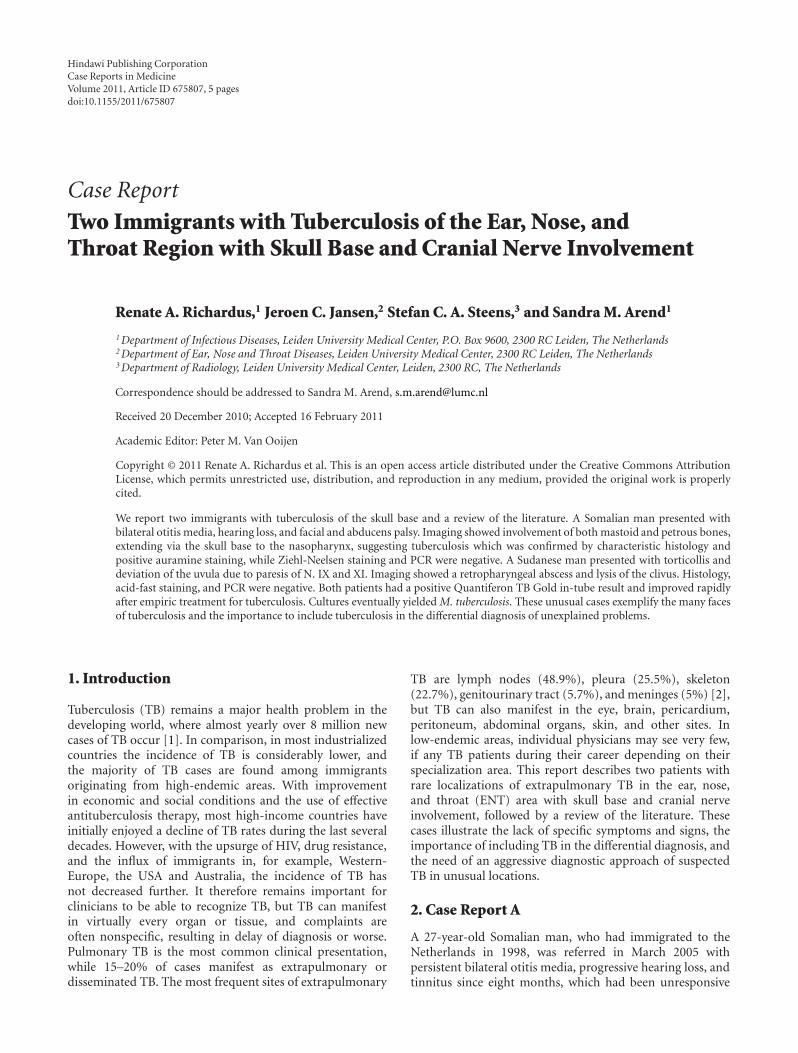
Figure 1: (Case A) Axial T1-weighted MR image with fat suppression after administration of Gadolinium at the level of the petrous bones(a) and just below the skull base (b). Indicated in a are the bilateral inflammatory changes in the petrous apices (short open arrows),bilateral involvement of the basal turn of the cochlea (long open arrows), extensive bilateral pachymeningeal involvement at the temporallobes (short closed arrows), opacification of both mastoids and middle ears (medium closed arrows), and the mass in the left middle nasalpassage extending up to the left Eustachian tube as visualized during fiber endoscopy (long closed arrow). Extension of the abscess into theparapharyngeal space, resulting in a bulge of the contour of the nasopharynx, is also shown (b, medium closed arrow).
to several courses of antibiotics. Other complaints weremalaise, fever, anorexia, and weight loss for two weeksaccompanied by dizziness and unsteady gait, one week laterfollowed by diplopia and drooping mouth. His sister hadbeen diagnosed with TB during screening on immigrationin 1998. At that time, the patient’s chest radiography showedabnormalities consistent with past healed TB infection. Onphysical examination, a malnourished man was seen withcomplete deafness of both ears. Neurological examinationconfirmed a right facial palsy and left abducens palsy.Otoscopy showed an extensive polypous inflammatory massin both external ear canals. During fiber endoscopy a similarmass with purulence was seen in the left middle nasal passageextending up to the left Eustachian tube. The remainder ofthe examination was unremarkable. Laboratory investigationshowed a hemoglobin level of 7.2 mmol/L after hydration,an ESR of 56 mm/first h, and mild elevation of serumtransaminases. HIV serology was negative. Tone audiometryshowed symmetric mixed hearing loss of 80 to 120 dB. Onchest radiography, fibrosis in the apex of the left inferior lobewas seen, unchanged compared to 1998. An MRI showed aninflammatory process of both mastoid and petrous boneswith abscess formation on the left extending downwardsinto the parapharyngeal space resulting in a bulge of thecontour of the nasopharynx (Figure 1). An extensive bilateraltemporal pachymeningitis was observed as well as bilateralinvolvement of the inner ear. A low-grade infectious diseaseprocess such as TB was suggested. Awaiting further testresults, empirical treatment with corticosteroids was started.Histology of biopsies from the external ear canals as well asfrom the mass extending from the nasopharynx and skull
base to the nose showed necrotizing granulomatous inflam-mation with giant cells. Auramine staining was positive withone acid-fast rod in the nasopharyngeal biopsy, and Ziehl-Neelsen staining and PCR of all biopsies were negative. TheQuantiferon TB Gold in-tube assay (Cellestis, Carnegie, Aus-tralia), performed three days after starting prednisone, waspositive (>10 IU/mL interferon-γ, cut-off value 0.35 IU/mL).Hence, the patient was diagnosed with tuberculous otitismedia with petrositis extending via the skull base to thenasopharynx. Treatment with four antituberculous drugs(rifampicin, isoniazide, pyrazinamide, and ethambutol) andpyridoxine was initiated. Within several weeks there wasclinical improvement with functional recovery of the nervusfacialis and abducens. However, severe hearing loss persisted(>90 dB in both ears at all frequencies), requiring a hearingaid. Culture of the nasopharyngeal biopsy yielded susceptibleM. tuberculosis. Isoniazide and rifampin were continued fora total duration of 12 months.
3. Case Report B
In 2005, a 35-y-old man from Sudan, who resided as arefugee in the Netherlands since 3 years, presented with painin the neck since several months, a sore throat, problemswith swallowing, and torticollis to the right. He reportedweight loss of five kg in two months. Three months earlierhe had been analyzed for unproductive cough, but chestradiography was without abnormalities, and no specific diag-nosis was made. On physical examination an ill, transpiringman without a fever was seen. There was a repositionabletorticollis to the right shoulder and deviation of the uvula

Case Reports in Medicine 3
(a)
A
P
(b)
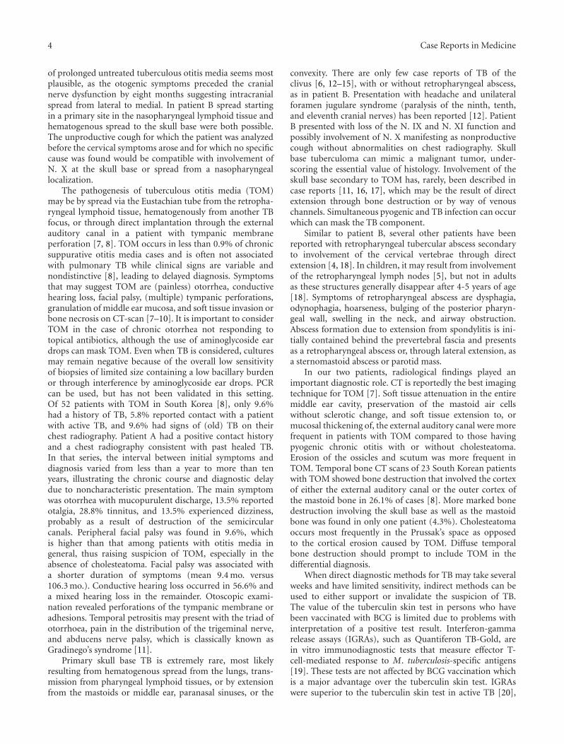
Figure 2: (Case B) Axial contrast-enhanced CT image in bone window (a) and axial T1-weighted MR image with fat suppression afteradministration of Gadolinium (b) at the level of the foramen magnum. Indicated in (a) are the mass on the right side of the nasopharynx(medium open arrow) and lysis of the clivus (medium closed arrow). The abscess in the retropharyngeal space and prevertebral muscleis better appreciated on MR (b, medium open arrow); edema in the clivus and occipital bone as well as paravertebral soft tissues are alsoindicated (short open arrow).
to the right due to paresis of the right N. IX and N.XI. On nasendoscopy, an asymmetric mass with a glazedaspect was seen on the right side of the nasopharynx.The remainder of the examination and the routine lab-oratorium examination were unremarkable. HIV serologywas negative. The Quantiferon TB Gold in-tube assay waspositive (>10 IU/mL interferon-γ; cut-off value 0.35 IU/mL).Chest radiography showed no abnormalities. CT and MRIshowed abscess formation and surrounding edema in theretropharyngeal space and prevertebral muscles on theright, accompanied by usurpation of the clivus and C0-C1 joint (Figure 2). The differential diagnosis includedTB. Biopsies of the nasopharyngeal mass obtained duringnasendoscopy were not diagnostic. Next, a CT-guided biopsyof the prevertebral mass was obtained under completeanesthesia, showing necrotic material with chronic activeinflammation. Auramine and Ziehl-Neelsen staining andPCR for M. tuberculosis were negative. Based on the positiveQuantiferon test result in the absence of an alternativediagnosis, treatment with four anti-TB drugs was startedfollowed by rapid clinical and radiological improvement withrecovery of cranial nerve function. The culture yielded fullysusceptible M. tuberculosis. Isoniazide and rifampicin werecontinued for a total of 12 months. At the end of treatment,only partial destruction of the right C0-C1 joint persisted.
4. Discussion
Both patients presented with neurological symptoms causedby TB in the ENT region with skull base and cranial nerveinvolvement, representing a rare manifestation of TB. In aseries of 323 cases of extrapulmonary TB, 23.2% presentedas ENT localization [3] of which 94.1% in cervical lymph
nodes, 4.33% in the larynx, 0.62% in the tonsil, 0.31% in theoral cavity, 0.31% in the middle ear, and 0.31% in the nose.Two smaller studies from India reported roughly similarfindings, but included rare cases of TB of the cervical spine,parotid, temporomandibular joint and a retropharyngealabscess [4, 5]. Mancusi et al. reported that 0.2–1.3% ofskeletal TB is localized in the skull, with involvement of theskull base occurring in only a few cases [6]. Thus, our twopatients with otitis media, retropharyngeal abscess, and skullbase TB represent extremely rare forms of extrapulmonaryTB.
The pathogenesis of ENT TB is thought to resulteither from primary infection of Waldeyer’s ring followingtransmission of infectious droplets expectorated by a patientwith smear-positive TB or by hematogenous spread froma TB focus in the lung or elsewhere. Another possibilityis direct inoculation from an endogenous pulmonary TBfocus to the larynx, oral cavity, or nasopharynx, yet manypatients with ENT TB, including those described in thispaper, have no signs of active pulmonary TB at the time ofdiagnosis. The clinical manifestations of ENT TB may becaused either by a mass effect of the inflammatory process orby destruction of anatomical structures, both of which oftenoccur simultaneously in TB. Due to the complex anatomy ofthe ENT area, with the skull base, cranial nerves, and C0-C1and atlantoaxial joints all in close proximity, TB in this areacan present with manifestations of different systems as wasalso the case in our two patients.
That isolated skull base TB is extremely rare may beexplained by the fact that bone TB has a predilection forlarge and weight-baring bones such as the vertebrae and largejoints, while the skull base is a nonweightbaring bone withlimited articulation. In patient A, direct spread from a focus
4 Case Reports in Medicine
of prolonged untreated tuberculous otitis media seems mostplausible, as the otogenic symptoms preceded the cranialnerve dysfunction by eight months suggesting intracranialspread from lateral to medial. In patient B spread startingin a primary site in the nasopharyngeal lymphoid tissue andhematogenous spread to the skull base were both possible.The unproductive cough for which the patient was analyzedbefore the cervical symptoms arose and for which no specificcause was found would be compatible with involvement ofN. X at the skull base or spread from a nasopharyngeallocalization.
The pathogenesis of tuberculous otitis media (TOM)may be by spread via the Eustachian tube from the retropha-ryngeal lymphoid tissue, hematogenously from another TBfocus, or through direct implantation through the externalauditory canal in a patient with tympanic membraneperforation [7, 8]. TOM occurs in less than 0.9% of chronicsuppurative otitis media cases and is often not associatedwith pulmonary TB while clinical signs are variable andnondistinctive [8], leading to delayed diagnosis. Symptomsthat may suggest TOM are (painless) otorrhea, conductivehearing loss, facial palsy, (multiple) tympanic perforations,granulation of middle ear mucosa, and soft tissue invasion orbone necrosis on CT-scan [7–10]. It is important to considerTOM in the case of chronic otorrhea not responding totopical antibiotics, although the use of aminoglycoside eardrops can mask TOM. Even when TB is considered, culturesmay remain negative because of the overall low sensitivityof biopsies of limited size containing a low bacillary burdenor through interference by aminoglycoside ear drops. PCRcan be used, but has not been validated in this setting.Of 52 patients with TOM in South Korea [8], only 9.6%had a history of TB, 5.8% reported contact with a patientwith active TB, and 9.6% had signs of (old) TB on theirchest radiography. Patient A had a positive contact historyand a chest radiography consistent with past healed TB.In that series, the interval between initial symptoms anddiagnosis varied from less than a year to more than tenyears, illustrating the chronic course and diagnostic delaydue to noncharacteristic presentation. The main symptomwas otorrhea with mucopurulent discharge, 13.5% reportedotalgia, 28.8% tinnitus, and 13.5% experienced dizziness,probably as a result of destruction of the semicircularcanals. Peripheral facial palsy was found in 9.6%, whichis higher than that among patients with otitis media ingeneral, thus raising suspicion of TOM, especially in theabsence of cholesteatoma. Facial palsy was associated witha shorter duration of symptoms (mean 9.4 mo. versus106.3 mo.). Conductive hearing loss occurred in 56.6% anda mixed hearing loss in the remainder. Otoscopic exami-nation revealed perforations of the tympanic membrane oradhesions. Temporal petrositis may present with the triad ofotorrhoea, pain in the distribution of the trigeminal nerve,and abducens nerve palsy, which is classically known asGradinego’s syndrome [11].
Primary skull base TB is extremely rare, most likelyresulting from hematogenous spread from the lungs, trans-mission from pharyngeal lymphoid tissues, or by extensionfrom the mastoids or middle ear, paranasal sinuses, or the
convexity. There are only few case reports of TB of theclivus [6, 12–15], with or without retropharyngeal abscess,as in patient B. Presentation with headache and unilateralforamen jugulare syndrome (paralysis of the ninth, tenth,and eleventh cranial nerves) has been reported [12]. PatientB presented with loss of the N. IX and N. XI function andpossibly involvement of N. X manifesting as nonproductivecough without abnormalities on chest radiography. Skullbase tuberculoma can mimic a malignant tumor, under-scoring the essential value of histology. Involvement of theskull base secondary to TOM has, rarely, been described incase reports [11, 16, 17], which may be the result of directextension through bone destruction or by way of venouschannels. Simultaneous pyogenic and TB infection can occurwhich can mask the TB component.
Similar to patient B, several other patients have beenreported with retropharyngeal tubercular abscess secondaryto involvement of the cervical vertebrae through directextension [4, 18]. In children, it may result from involvementof the retropharyngeal lymph nodes [5], but not in adultsas these structures generally disappear after 4-5 years of age[18]. Symptoms of retropharyngeal abscess are dysphagia,odynophagia, hoarseness, bulging of the posterior pharyn-geal wall, swelling in the neck, and airway obstruction.Abscess formation due to extension from spondylitis is ini-tially contained behind the prevertebral fascia and presentsas a retropharyngeal abscess or, through lateral extension, asa sternomastoid abscess or parotid mass.
In our two patients, radiological findings played animportant diagnostic role. CT is reportedly the best imagingtechnique for TOM [7]. Soft tissue attenuation in the entiremiddle ear cavity, preservation of the mastoid air cellswithout sclerotic change, and soft tissue extension to, ormucosal thickening of, the external auditory canal were morefrequent in patients with TOM compared to those havingpyogenic chronic otitis with or without cholesteatoma.Erosion of the ossicles and scutum was more frequent inTOM. Temporal bone CT scans of 23 South Korean patientswith TOM showed bone destruction that involved the cortexof either the external auditory canal or the outer cortex ofthe mastoid bone in 26.1% of cases [8]. More marked bonedestruction involving the skull base as well as the mastoidbone was found in only one patient (4.3%). Cholesteatomaoccurs most frequently in the Prussak’s space as opposedto the cortical erosion caused by TOM. Diffuse temporalbone destruction should prompt to include TOM in thedifferential diagnosis.
When direct diagnostic methods for TB may take severalweeks and have limited sensitivity, indirect methods can beused to either support or invalidate the suspicion of TB.The value of the tuberculin skin test in persons who havebeen vaccinated with BCG is limited due to problems withinterpretation of a positive test result. Interferon-gammarelease assays (IGRAs), such as Quantiferon TB-Gold, arein vitro immunodiagnostic tests that measure effector T-cell-mediated response to M. tuberculosis-specific antigens[19]. These tests are not affected by BCG vaccination whichis a major advantage over the tuberculin skin test. IGRAswere superior to the tuberculin skin test in active TB [20],
Case Reports in Medicine 5
although the sensitivity of IGRA for active TB is incomplete[21]. A recent study found that higher quantitative interferongamma values were associated with active TB [22]. In bothour patients, the result of the Quantiferon TB-Gold assaywas strongly positive, which confirmed the suspicion ofTB, justifying the initiation of treatment after biopsies thatwere obtained. However, IGRA cannot differentiate betweenpast latent TB infection and active TB. Thus, it remains ofutmost importance to obtain direct proof through histology,staining, PCR, and/or culture. If adequate facilities for imag-ing, targeted biopsy, and microbiological diagnostics are notavailable and TB is strongly considered, it may be justifiedto treat a patient empirically for TB and evaluate carefullyfor a clinical response, on condition that it is recognized thatclinical deterioration not necessarily invalidates the diagnosisTB, but may indicate a paradoxical response to treatment asoccurs frequently during TB treatment [23].
5. Conclusion
These two cases of rare manifestations of TB, with skullbase and cranial nerve involvement, illustrate the importanceto include TB in the differential diagnosis of unexplainedclinical problems. Clues that should prompt the clinicianto consider skull base TB are cranial nerve dysfunction,radiographic finding of a mass, destructive process, ordiffuse temporal bone destruction in the absence of anotherexplanation. TOM should be considered in case of chronicotorrhea especially if occurring in an individual originatingfrom a TB-endemic region.
References
[1] E. L. Corbett, C. J. Watt, N. Walker et al., “The growing burdenof tuberculosis: global trends and interactions with the HIVepidemic,” Archives of Internal Medicine, vol. 163, no. 9, pp.1009–1021, 2003.
[2] O. Fain, O. Lortholary, V. Lascaux et al., “Extrapulmonarytuberculosis in the northeastern suburbs of Paris: 141 cases,”European Journal of Internal Medicine, vol. 11, no. 3, pp. 145–150, 2000.
[3] F. Ricciardiello, S. Martufi, M. Cardone, M. Cavaliere, P.D’Errico, and M. Iengo, “Otorhinolaryngology-related tuber-culosis,” Acta Otorhinolaryngologica Italica, vol. 26, no. 1, pp.38–42, 2006.
[4] K. C. Prasad, S. Sreedharan, Y. Chakravarthy, and S. C. Prasad,“Tuberculosis in the head and neck: experience in India,”Journal of Laryngology and Otology, vol. 121, no. 10, pp. 979–985, 2007.
[5] B. Nalini and S. Vinayak, “Tuberculosis in ear, nose, and throatpractice: its presentation and diagnosis,” American Journal ofOtolaryngology, vol. 27, no. 1, pp. 39–45, 2006.
[6] G. Mancusi, B. Marks, C. Czerny et al., “Tuberculosis of theclivus and the nasopharynx,” HNO, vol. 53, no. 12, pp. 1081–1084, 2005.
[7] M. H. Rho, D. W. Kim, S. S. Kim, Y. S. Sung, J. S. Kwon,and S. W. Lee, “Tuberculous otomastoiditis on high-resolutiontemporal bone CT: comparison with nontuberculous otomas-toiditis with and without cholesteatoma,” American Journal ofNeuroradiology, vol. 28, no. 3, pp. 493–496, 2007.
[8] Y. S. Cho, H. S. Lee, S. W. Kim et al., “Tuberculous otitis media:a clinical and radiologic analysis of 52 patients,” Laryngoscope,vol. 116, no. 6, pp. 921–927, 2006.
[9] T. Halvorsen, H. Townsend, W. Stauffer, K. Belani, and D.Kamat, “A case of tuberculous otitis media,” Clinical Pediatrics,vol. 45, no. 1, pp. 83–87, 2006.
[10] R. Sharma, I. Tyagi, R. Kumar, and R. V. Phadke, “Tuberculosisof the skull. A case report and review of the literature,”Neurosurgical Review, vol. 23, no. 2, pp. 104–106, 2000.
[11] A. Sethi, A. Sabherwal, A. Gulati, and D. Sareen, “Primarytuberculous petrositis,” Acta Oto-Laryngologica, vol. 125, no.11, pp. 1236–1239, 2005.
[12] B. Indira Devi, A. K. Tyagi, D. I. Bhat, and V. Santosh,“Tuberculous osteitis of clivus,” Neurology India, vol. 51, no.1, pp. 69–70, 2003.
[13] S. Selvapandian and M. J. Chandy, “Tuberculous granulomaof the clivus,” British Journal of Neurosurgery, vol. 7, no. 5, pp.581–582, 1993.
[14] S. Sencer, A. Sencer, K. Aydin, K. Hepgul, A. Poyanli, and O.Minareci, “Imaging in tuberculosis of the skull and skull-base:case report,” Neuroradiology, vol. 45, no. 3, pp. 160–163, 2003.
[15] S. N. Shenoy and A. Raja, “Tuberculous granuloma of thespheno-clival region,” Neurology India, vol. 52, no. 1, pp. 129–130, 2004.
[16] A. Sethi, D. Sethi, S. Mrig, J. C. Passey, and N. Srivastav,“Coexistent acute pyogenic and tubercular petrous apicitis: adiagnostic dilemma,” Journal of Laryngology and Otology, vol.120, no. 10, pp. 875–878, 2006.
[17] K. Mongkolrattanothai, R. Oram, M. Redleaf, J. Bova, andJ. A. Englund, “Tuberculous otitis media with mastoiditisand central nervous system involvement,” Pediatric InfectiousDisease Journal, vol. 22, no. 5, pp. 453–456, 2003.
[18] O. Yoruk, V. Fidan, and Y. Sutbeyaz, “Hearing loss unusuallycaused by tubercular retropharyngeal abscess,” Journal ofCraniofacial Surgery, vol. 20, no. 3, pp. 955–957, 2009.
[19] M. Pai, A. Zwerling, and D. Menzies, “Systematic review:T-cell-based assays for the diagnosis of latent tuberculosisinfection: an update,” Annals of Internal Medicine, vol. 149, no.3, pp. 177–184, 2008.
[20] R. Diel, R. Loaddenkemper, and A. Nienhaus, “Evidence-based comparison of commercial interferon-γ release assaysfor detecting active TB—a meta analysis,” Chest, vol. 137, no.4, pp. 952–968, 2010.
[21] P. K. Dewan, J. Grinsdale, and L. M. Kawamura, “Lowsensitivity of a whole-blood interferon-γ release assay fordetection of active tuberculosis,” Clinical Infectious Diseases,vol. 44, no. 1, pp. 69–73, 2007.
[22] J. Z. Metcalfe, A. Cattamanchi, E. Vittinghoff et al., “Evalu-ation of quantitative IFN-γ response for risk stratification ofactive tuberculosis suspects,” American Journal of Respiratoryand Critical Care Medicine, vol. 181, no. 1, pp. 87–93, 2010.
[23] V. C. C. Cheng, P. L. Ho, R. A. Lee et al., “Clinical spectrumof paradoxical deterioration during antituberculosis therapyin non-HIV-infected patients,” European Journal of ClinicalMicrobiology and Infectious Diseases, vol. 21, no. 11, pp. 803–809, 2002.

Submit your manuscripts athttp://www.hindawi.com
Stem CellsInternational
Hindawi Publishing Corporationhttp://www.hindawi.com Volume 2014
Hindawi Publishing Corporationhttp://www.hindawi.com Volume 2014
MEDIATORSINFLAMMATION
of
Hindawi Publishing Corporationhttp://www.hindawi.com Volume 2014
Behavioural Neurology
EndocrinologyInternational Journal of
Hindawi Publishing Corporationhttp://www.hindawi.com Volume 2014
Hindawi Publishing Corporationhttp://www.hindawi.com Volume 2014
Disease Markers
Hindawi Publishing Corporationhttp://www.hindawi.com Volume 2014
BioMed Research International
OncologyJournal of
Hindawi Publishing Corporationhttp://www.hindawi.com Volume 2014
Hindawi Publishing Corporationhttp://www.hindawi.com Volume 2014
Oxidative Medicine and Cellular Longevity
Hindawi Publishing Corporationhttp://www.hindawi.com Volume 2014
PPAR Research
The Scientific World JournalHindawi Publishing Corporation http://www.hindawi.com Volume 2014
Immunology ResearchHindawi Publishing Corporationhttp://www.hindawi.com Volume 2014
Journal of
ObesityJournal of
Hindawi Publishing Corporationhttp://www.hindawi.com Volume 2014
Hindawi Publishing Corporationhttp://www.hindawi.com Volume 2014
Computational and Mathematical Methods in Medicine
OphthalmologyJournal of
Hindawi Publishing Corporationhttp://www.hindawi.com Volume 2014
Diabetes ResearchJournal of
Hindawi Publishing Corporationhttp://www.hindawi.com Volume 2014
Hindawi Publishing Corporationhttp://www.hindawi.com Volume 2014
Research and TreatmentAIDS
Hindawi Publishing Corporationhttp://www.hindawi.com Volume 2014
Gastroenterology Research and Practice
Hindawi Publishing Corporationhttp://www.hindawi.com Volume 2014
Parkinson’s Disease
Evidence-Based Complementary and Alternative Medicine
Volume 2014Hindawi Publishing Corporationhttp://www.hindawi.com