Two identified byapplication of - pnas.org fileProc. Nadl. Acad. Sci. USA Vol. 86, pp. 1603-1607,...
5
Proc. Nadl. Acad. Sci. USA Vol. 86, pp. 1603-1607, March 1989 Genetics Two putative protein-tyrosine kinases identified by application of the polymerase chain reaction (gene families/DNA amplification in vitro/degenerate oligonucleotides) ANDREW F. WILKS Ludwig Institute for Cancer Research, Royal Melbourne Hospital, Parkville, Victoria 3050, Australia Communicated by G. J. V. Nossal, November 28, 1988 ABSTRACT The pivotal role that protein-tyrosine kinases (PTKs) play in the growth regulation of eukaryotic cells is manifest in the frequent appearance of members of the PTK family as growth factor receptors or as the transforming agents of acutely transforming retroviruses. A feature common to all members of the PTK family is a highly conserved catalytic domain which is characteristic of the group as a whole and whose activity appears to be tightly regulated within the cell by other domains of the PTK. Degenerate oligonucleotide probes corresponding to two invariant amino acid sequence motifs within the catalytic domains of all PTK family members were synthesized and employed in the polymerase chain reaction (PCR) to amplify cDNA sequences between them. An M13 PCR library was produced in this way from cDNA prepared against mRNA from the murine hemopoietic cell line FDC-P1. The PCR library was then screened by DNA sequencing for PTK- related sequences. Two sequences were identified that, on the basis of sequence comparison with known PTKs, may encode representatives of a new class of PTK. Protein-tyrosine kinases (PTKs; EC 2.7.1.112) are believed to play an important role(s) in the metabolism of the cell, most probably as components of signal-transduction pathways. Indirect evidence in support of this presumption is found in the frequent identification of members of the PI7K family as cellular receptors for certain growth factors (1-8) and as products of the oncogenes of many of the acutely transform- ing retroviruses (7-12). Hanks et al. (13) recently suggested that the protein kinases could be mapped phylogenetically, based on the conservation of the amino acid sequences of their catalytic domains. Similar phylogenetic mapping of the PTK family suggested that there may be clusters of related subfamilies, such as the Src family [including c-Yes, c-Lyn, and hemopoietic cell kinase (HCK), among others], the Fes family (currently containing only c-Fes), and the colony- stimulating factor 1 (CSF-1) receptor family [including c-Kit and the platelet-derived growth factor (PDGF) receptor]. Such speciation is also evident in the noncatalytic domains of the PTKs; it is highly probable that the PTK catalytic domain has been recombined with a wide variety of noncatalytic domains, so that the use of the tyrosine kinase activity can be deployed in a regulated manner in as wide a variety of situations as possible. Several approaches have been tried in order to identify novel members of the protein kinase family, such as low- stringency screening of cDNA libraries with previously characterized cDNA probes (14) or the use of degenerate oligonucleotide probes based on the highly conserved regions of the protein kinase catalytic domain (15). Each of these approaches has been successfully applied. However, more recently, a technique has been developed that is capable of greatly facilitating the isolation of any gene for which some sequence data are known. The polymerase chain reaction (PCR) (16, 17) technique has already demonstrated consid- erable flexibility in its application to a wide variety of diagnostic and molecular biological situations. The technique is based on the enzymatic amplification of sequences residing between two oligonucleotide primers that define the 5' and 3' borders of the amplified segment. The technique has recently been applied to the cloning of cDNA sequences defined by mixed oligonucleotides derived from selected amino acid sequences (18). A natural extension of this application is in cloning members of gene families, such as those comprising the PTK family. To detect members of the PTK family expressed in hemopoietic cells, the technique has been applied to cDNA from a murine factor-dependent hemopoi- etic cell line, FDC-P1 (19), and PTK-related sequences have been isolated.* The isolation of new PTK-related sequences will aid in the understanding of the response of these cells to growth factors such as the colony-stimulating factors, a response that is known to be accompanied by changes in the patterns of intracellular protein phosphotyrosine metabolism (20). MATERIALS AND METHODS Nucleic Acids. Poly(A)+ RNA was prepared by the method of Gonda et al. (21). In brief, cells were washed in isotonic phosphate-buffered saline and resuspended at 107 per ml in 0.1 M NaCl/10 mM Tris, pH 7.5/1 mM EDTA. Before homogenization at high speed in a Polytron (Selby-Anax), the cell lysate was made 1.0%o with respect to SDS, and proteinase K was added to a final concentration of 200 .ug/ml. After digestion for 4 hr at 370C, poly(A)+ RNA was isolated by the addition of 500 mg of oligo(dT)-cellulose (Sigma), followed by low-salt elution. RNA was recovered by ethanol precipitation. Oligonucleotides were synthesized on an Ap- plied Biosystems 380A oligonucleotide synthesizer using standard chemistry; the crude oligonucleotide preparation was not purified further. DNA Amplification. Poly(A)+ mRNA from the growth factor-dependent hemopoietic cell line FDC-P1 and an Am- ersham cDNA-synthesis kit were used to generate oligo(dT)- primed double-stranded cDNA (1 tug). Five microcuries of [a-32P]dATP (3000 Ci/mmol; 1 Ci = 37 GBq) was included in the reaction mixture to label the cDNA product. PCR was performed with a Geneamp kit (Cetus), using the manufac- turer's buffers and modified conditions, and the PTK I and PTK II oligonucleotides (1 ,ug each). The PCR cycle was 1.5 min at 950C (denaturation), 2 min at 37C (annealing), and 3 min at 630C (elongation). An additional 5 gCi of [a-32P]dATP was included to label the PCR product. For cloning, the Abbreviations: PCR, polymerase chain reaction; PTK, protein- tyrosine kinase. *The sequences reported in this paper have been deposited in the EMBL/GenBank data base (accession no. J04523). 1603 The publication costs of this article were defrayed in part by page charge payment. This article must therefore be hereby marked "advertisement" in accordance with 18 U.S.C. §1734 solely to indicate this fact.
Transcript of Two identified byapplication of - pnas.org fileProc. Nadl. Acad. Sci. USA Vol. 86, pp. 1603-1607,...
Proc. Nadl. Acad. Sci. USAVol. 86, pp. 1603-1607, March 1989Genetics
Two putative protein-tyrosine kinases identified by application ofthe polymerase chain reaction
(gene families/DNA amplification in vitro/degenerate oligonucleotides)
ANDREW F. WILKSLudwig Institute for Cancer Research, Royal Melbourne Hospital, Parkville, Victoria 3050, Australia
Communicated by G. J. V. Nossal, November 28, 1988
ABSTRACT The pivotal role that protein-tyrosine kinases(PTKs) play in the growth regulation of eukaryotic cells ismanifest in the frequent appearance of members of the PTKfamily as growth factor receptors or as the transforming agentsof acutely transforming retroviruses. A feature common to allmembers of the PTK family is a highly conserved catalyticdomain which is characteristic of the group as a whole andwhose activity appears to be tightly regulated within the cell byother domains of the PTK. Degenerate oligonucleotide probescorresponding to two invariant amino acid sequence motifswithin the catalytic domains of all PTK family members weresynthesized and employed in the polymerase chain reaction(PCR) to amplify cDNA sequences between them. An M13 PCRlibrary was produced in this way from cDNA prepared againstmRNA from the murine hemopoietic cell line FDC-P1. ThePCR library was then screened by DNA sequencing for PTK-related sequences. Two sequences were identified that, on thebasis of sequence comparison with known PTKs, may encoderepresentatives of a new class of PTK.
Protein-tyrosine kinases (PTKs; EC 2.7.1.112) are believedto play an important role(s) in the metabolism ofthe cell, mostprobably as components of signal-transduction pathways.Indirect evidence in support of this presumption is found inthe frequent identification of members of the PI7K family ascellular receptors for certain growth factors (1-8) and asproducts of the oncogenes ofmany of the acutely transform-ing retroviruses (7-12). Hanks et al. (13) recently suggestedthat the protein kinases could be mapped phylogenetically,based on the conservation of the amino acid sequences oftheir catalytic domains. Similar phylogenetic mapping of thePTK family suggested that there may be clusters of relatedsubfamilies, such as the Src family [including c-Yes, c-Lyn,and hemopoietic cell kinase (HCK), among others], the Fesfamily (currently containing only c-Fes), and the colony-stimulating factor 1 (CSF-1) receptor family [including c-Kitand the platelet-derived growth factor (PDGF) receptor].Such speciation is also evident in the noncatalytic domains ofthe PTKs; it is highly probable that the PTK catalytic domainhas been recombined with a wide variety of noncatalyticdomains, so that the use ofthe tyrosine kinase activity can bedeployed in a regulated manner in as wide a variety ofsituations as possible.
Several approaches have been tried in order to identifynovel members of the protein kinase family, such as low-stringency screening of cDNA libraries with previouslycharacterized cDNA probes (14) or the use of degenerateoligonucleotide probes based on the highly conserved regionsof the protein kinase catalytic domain (15). Each of theseapproaches has been successfully applied. However, morerecently, a technique has been developed that is capable of
greatly facilitating the isolation of any gene for which somesequence data are known. The polymerase chain reaction(PCR) (16, 17) technique has already demonstrated consid-erable flexibility in its application to a wide variety ofdiagnostic and molecular biological situations. The techniqueis based on the enzymatic amplification of sequences residingbetween two oligonucleotide primers that define the 5' and 3'borders ofthe amplified segment. The technique has recentlybeen applied to the cloning of cDNA sequences defined bymixed oligonucleotides derived from selected amino acidsequences (18). A natural extension of this application is incloning members of gene families, such as those comprisingthe PTK family. To detect members of the PTK familyexpressed in hemopoietic cells, the technique has beenapplied to cDNA from a murine factor-dependent hemopoi-etic cell line, FDC-P1 (19), and PTK-related sequences havebeen isolated.* The isolation of new PTK-related sequenceswill aid in the understanding of the response of these cells togrowth factors such as the colony-stimulating factors, aresponse that is known to be accompanied by changes in thepatterns of intracellular protein phosphotyrosine metabolism(20).
MATERIALS AND METHODSNucleic Acids. Poly(A)+ RNA was prepared by the method
of Gonda et al. (21). In brief, cells were washed in isotonicphosphate-buffered saline and resuspended at 107 per ml in0.1 M NaCl/10 mM Tris, pH 7.5/1 mM EDTA. Beforehomogenization at high speed in a Polytron (Selby-Anax),the cell lysate was made 1.0%o with respect to SDS, andproteinase K was added to a final concentration of200 .ug/ml.After digestion for 4 hr at 370C, poly(A)+ RNA was isolatedby the addition of 500 mg of oligo(dT)-cellulose (Sigma),followed by low-salt elution. RNA was recovered by ethanolprecipitation. Oligonucleotides were synthesized on an Ap-plied Biosystems 380A oligonucleotide synthesizer usingstandard chemistry; the crude oligonucleotide preparationwas not purified further.DNA Amplification. Poly(A)+ mRNA from the growth
factor-dependent hemopoietic cell line FDC-P1 and an Am-ersham cDNA-synthesis kit were used to generate oligo(dT)-primed double-stranded cDNA (1 tug). Five microcuries of[a-32P]dATP (3000 Ci/mmol; 1 Ci = 37 GBq) was included inthe reaction mixture to label the cDNA product. PCR wasperformed with a Geneamp kit (Cetus), using the manufac-turer's buffers and modified conditions, and the PTK I andPTK II oligonucleotides (1 ,ug each). The PCR cycle was 1.5min at 950C (denaturation), 2 min at 37C (annealing), and 3min at 630C (elongation). An additional 5 gCi of [a-32P]dATPwas included to label the PCR product. For cloning, the
Abbreviations: PCR, polymerase chain reaction; PTK, protein-tyrosine kinase.*The sequences reported in this paper have been deposited in theEMBL/GenBank data base (accession no. J04523).
1603
The publication costs of this article were defrayed in part by page chargepayment. This article must therefore be hereby marked "advertisement"in accordance with 18 U.S.C. §1734 solely to indicate this fact.

Proc. Natl. Acad. Sci. USA 86 (1989)
amplified DNA was ethanol-precipitated, air-dried, and re-suspended in 20 1.l of double-distilled water before beingdigested in EcoRI buffer with EcoRI and BamHI (20 unitseach) for 4 hr at 370C. The DNA was then purified on anElutip-d column (Schleicher & Schuell) and ligated intoEcoRI- and BamHI-cleaved M13mpl9. Sequencing was car-ried out by the dideoxy chain-termination method (22), usinga Sequenase kit (United States Biochemical). In all cases[a-[35S]thio]dATP (Amersham, catalogue no. SJ304) was thepreferred radionucleotide.RNA Analysis. Poly(A)+ mRNA samples were prepared as
described (21), Aliquots (1 gg) were electrophoresed in 1%agarose gels containing 2.2 M formaldehyde, 20 mM Mops(pH 6.8), 1 mM EDTA, and 5 mM sodium acetate, and theelectrophoretically separated RNAs were transferred to Hy-bond N (Amersham, catalogue no. RPN303N) or nitrocellu-lose (Schleicher & Schuell, BA85, catalogue no. 401196)membranes. Filters were prehybridized for 4 hr in 50%formamide containing 3x SSC (lx is 0.15 M NaCl/0.015 Msodium citrate, pH 7.0), 5 x Denhardt's solution (lx is 0.02%Ficoll/0.02% polyvinylpyrrolidone/0.02% bovine serum al-bumin), 10 mM Hepes (pH 7.0), poly(C) at 100 ,g/ml,denatured herring sperm DNA at 100 ,ug/ml, Escherichia coliDNA at 10 ,ug/ml, and 0.1% SDS and were hybridized for 18hr at 42°C in the same solution with nick-translated 32p-labeled DNA insert. Filters were washed to a final stringencyof 0.2x SSC/0.1% SDS at 68°C before exposure to KodakXAR-5 x-ray film with two intensifying screens.
aCATALYTIC DOMAIN
PTK
PTK
PTK II
YAPE
b FDC-P1 mRNA
| oligo(dT)-primedcDNA synthesis
-Tim
j PCR withPTK I and PTK IIoligonucleotides
Amplified product
RESULTS AND DISCUSSIONStrategy. The basic strategy of the approach is outlined in
Fig. 1. Three regions of the catalytic domain of the PTKswere used to derive oligonucleotide primers. To selectsuitable regions from which to generate PTK oligonucleotideprimers, careful consideration was given to identifying themost highly conserved regions of the PTK catalytic domain.Fig. 2 shows a comparison of 13 PTK amino acid sequences,from which consensus sequences have been derived fromparticularly highly conserved regions. Of these, two partic-ular sequences designated PTK I and PTK II were selectedfor the derivation of oligonucleotide primers. The sequence-IHRDL- (PTK I) at position 392 in c-Src defined the 5'border of the region of the cDNA to be amplified, and thesequence -DVWSFG- (PTK II) located at position 453 inc-Src defined the 3' border. Between these two sequences isa characteristic and highly conserved sequence, -P(V/I)-(K/R)W(M/T)APES-, which served as a reference point withwhich to identify potential PTK sequences. The amino acidsequences of known PTKs in this region, although broadlyconserved across the family ofPTKs, are sufficiently diverseto permit the designation of any candidate PTK clones aspreviously known or novel and tentatively provided a moreprecise assignment-for example, to the cytoplasmic (Src-like) or the growth factor receptor subfamily.The oligonucleotide probes derived (Fig. 3) were based
upon the DNA sequences encoding the two consensus aminoacid sequences. The DNA sequences encoding these motifsexhibited considerably less degeneracy than a simple decod-ing of the amino acid sequences would predict. This ispresumably due to a high degree of evolutionary codonselection. The PTK I and PTK II probes were thus mixturesof 64 and 8 different oligonucleotide sequences, respectively.The predicted tm range of the oligonucleotides was 39-45°Cfor PTK I and 45-49.2°C for PTK II. Although this was wellbelow the 63°C elongation temperature selected for the PCRreaction, it was anticipated that the 37°C annealing stepincorporated into the protocol would additionally serve as anextension phase, during which the oligonucleotides would beelongated sufficiently to remain hybridized as the tempera-
j Clone into M13and sequence
B R
M13
FIG. 1. Strategy for cloning PTK-related sequences. (a) Con-served regions of PTK catalytic domains. Blocks of identity held incommon between the catalytic domains ofPTKs are shown as boxes,with the conserved motif written in one-letter code within. Oligo-nucleotide probes were initially derived from the -IHRDL- motif(PTK I) and the -DVWSFG- motif (PTK II). A further round of PCRwas performed using PTK I and the FWYAPE-based (YAPE)oligonucleotide. (b) Summary of the PCR cloning strategy. Aftersynthesis of FDC-P1 cDNA, the cDNA was amplified by using PTKI and PTK II oligonucleotides as primers. After codigestion withBamHI and EcoRI the amplified fragment was ligated into M13mpl8,and sequence analysis was performed on 200 clones. B, BamHI site;R, EcoRI site.
ture increased to 63°C. Each oligonucleotide was additionallydesigned to include a restriction enzyme-specific linkerflanking the consensus PTK sequence, facilitating the sub-sequent directional cloning of amplified fragments into M13phage.DNA Amplification. Fig. 4 shows the PCR-amplified prod-
uct of cDNA generated by using FDC-P1 mRNA as atemplate. After 22 cycles of amplification an obvious ethid-ium bromide-stained DNA band of the expected size (-210base pairs) was detected. This DNA fragment was radiola-beled during the PCR process, confirming its origin as part ofthe amplification process. Additionally, the background ra-dioactivity present in the track was increased, probably dueto nonspecific priming of the original template cDNA. Ap-proximately 1 ,g of amplified DNA was generated. After
1604 Genetics: Wilks

Proc. Natl. Acad. Sci. USA 86 (1989) 1605
c-Src YVEPDLRAANILVGENLtXVADFGLARLIED. .NEYTAR.. QGAK.EPIXTrAPZAALYGI'TIKSDVWSFGILLZRL2Tc-Yes YIERDLRAANILVGENLVCKIADFGLAPIIED. .NEYTAR.. QGAK.FPIIWTAPZAALYGRFTI(SDVWSFGILQ7XLVTHCK YIBPRDLRAANILVSASLVKADFGLAR VIED. .NEYTAR.. EGAK.VFPIITAPZAINFGSFTIKSDVWSFGILLIIVTc-Fes C1URDLARBNILVTEK&VLKISDFSEEAD . . GIYAAC. . SGLR YV1APZALNYGRYSSESDWSFGILLBTFSc-Abl FIE=DLMPZLVGENHLVKVADFGLSRLMTG. . D.TTAH. . . AGAK.VPI"ITAPZSLAYN SIKSDVWAFGVLLMhIATEGFR LVHDLAARNVLVKTPQNRKa7DFGLAKLGAEEKEYHA...EGGK.VPIUKXSILHRIYTHQSDVSYGVTVWLMTNeu LVHDLAARNVLVKSPMKITDFGLARLLDIDETEYHA...DGGK.VPXISIJgLRRXLRRRVTHTHQSDVWSYGVTVWZLMTCSF1R CVIL DVAAPUVLLTSGHVAKIGYGLARDIMND. SNYVVK. . GNA. .LPWMPZSIFDCVIYVQSDVWSYG.LLWBIFSPDGFR CVNDLAAPNVLICEGKLVKICDrGLARDIMRD. SNYISK. . GSTY.LPLKWMAPZSIFNSLYTTLSDWSFGILLWZIFTIR EVBRDLAAPRNCVAHDFTVYKGDFGIT7RDIYET.DYYRKG. . GKGL.LVRMAlPZSLIKDGVFITTSSDIGVV I TSIGFIR FVIBRDLA)JZOAEDFTVKIGDFQ(2RDIYET. DYYRKG. .GKGL .LnUSSLKDGKDGVFTTSWSrGVVLIIATc-Met EVBRDLAARCNLDEKF7VKVADFGLARDMYDK. EYYSVHNKTGAK.L 2SLQTQKFT TK SMWYVLWZLhTc-Trk FVIRDLA&7RCLVGQGLtVKcGDFGASRDIYST. DYYRVG. . GRTM.LPIRWMPPZSILYRKIaTESDVWSYGVVLWEIf
ICONSENSUS VUDLAARNCLV VXI DFGL RDSEQUENCE I R A vW c v MXKL
PTK I
yFLP IKMPZSIVVRT L L
FT SDVWSFGVLL TYS Y IV S
PTI II
FIG. 2. Derivation of consensus sequences for PCR primers. Alignment of amino acid sequences from conserved regions of various PTKcatalytic domains. The appropriate regions ofmurine neuronal c-Src [amino acids (aa) 391-466; ref. 9], human c-Yes (aa 392-367; ref. 10), murineHCK (aa 354-429; ref. 14), murine c-Fes (aa 677-753; ref. 11), human c-Abl (aa 359-434; ref. 12), human epidermal growth factor receptor(EGFR, aa 809-885; ref. 7), human Neu (aa 841-917; ref. 1), murine colony-stimulating factor 1 receptor (CSF1R, aa 772-848; ref. 8), humanplatelet-derived growth factor receptor (PDGFR, aa 789-886; ref. 2), human insulin receptor (IR, aa 1116-1192; ref. 3), human insulin-like growthfactor I receptor (IGFIR, aa 1101-1177; ref. 4), human c-Met (aa 1118-1192; ref. 5), and human c-Trk-2h (aa 289-365; ref. 6) are shown. Dotscorrespond to gaps introduced to optimize sequence alignment, and each dot represents one amino acid. Conserved amino acids are shown inbold type and the derived consensus sequences are shown below. Amino acid sequences chosen for the derivation ofPCR primers are designatedPTK I and PTK II.
BamHI/EcoRI digestion of the PCR product the DNA wascloned into BamHI/EcoRI-cleaved, phosphatase-treatedM13mpl9 replicative-form DNA.
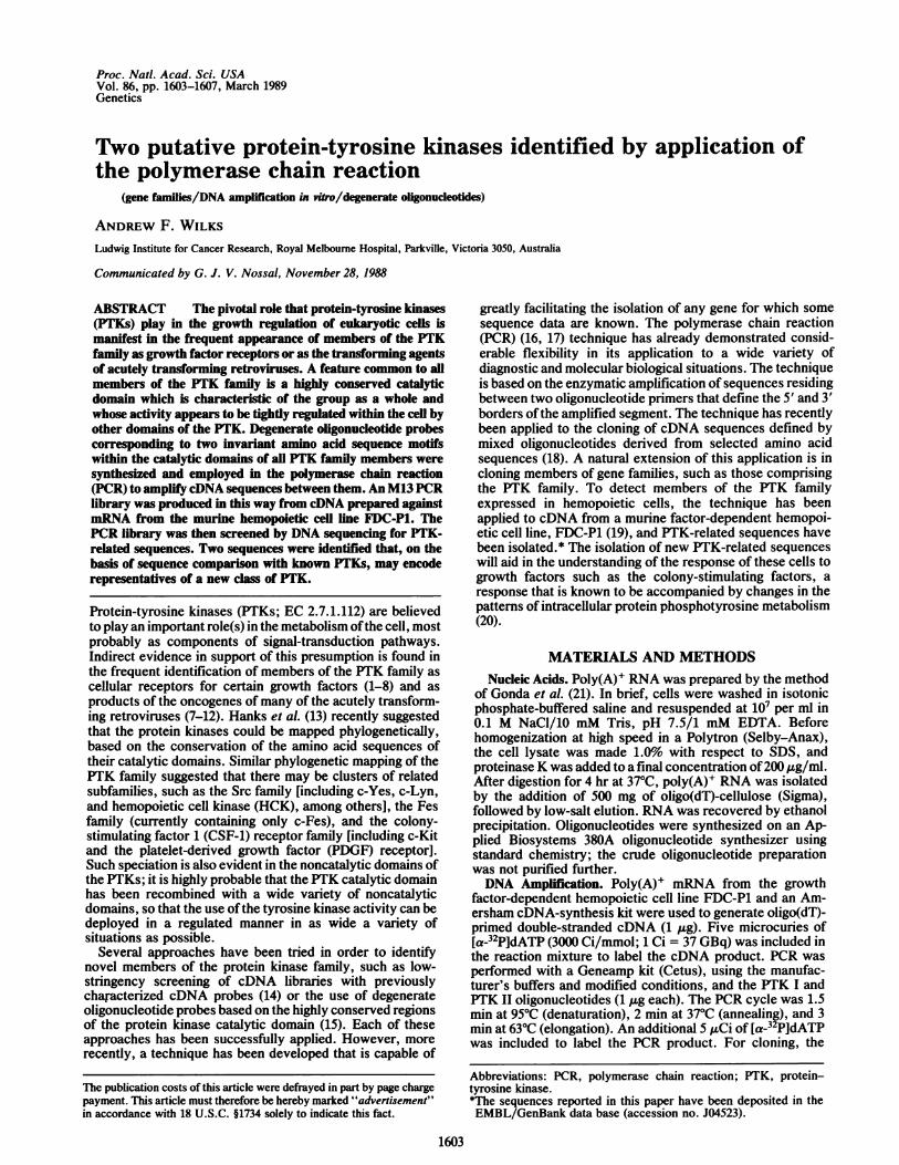
Initially, 24 M13 clones were fully sequenced by thedideoxy method (22), and the encoded protein sequenceswere compared with known PTK sequences. Subsequently,a total of 200 clones were examined by comparison of singlebase sequencing reactions and novel clones were fully se-quenced; the protein sequences of representative clones areshown in Fig. 5. Of these 200 clones, 133 contained aPTK-related sequence, 55 contained no insert, and theremainder contained head-to-tail oligonucleotide dimers orsequences unrelated to PTKs. Clone FD15 was identical tomurine c-fes (11). Clone FD16 showed 95% amino acidsequence identity to the human insulin-like growth factor I(IGF-I) receptor (4) and 92% amino acid sequence identity tothe human insulin receptor (3) and probably represented amurine equivalent of the IGF-I receptor. Clone FD19 was98% identical to human c-met (5) and is thus a strongcandidate for the murine homologue of this gene. Finally,clone FD175 showed 100o amino acid identity to a murinec-lyn clone described by others (S. McEwen, D. Holtzman,and A. R. Dunn, personal communication).
I E R D L5' CGG ATCCACAGNGACCT 3'
BamHI C TT
PTK I
Unassigned PTK-Related Sequences. Clones FD17 andFD22 currently remain without a known cellular homologue.The presence of an "acidic" tyrosine residue 10-12 aminoacids C-terminal to the conserved -DFG- motif is invariant inthe PTK family and is encoded in both FD17 and FD22. TheFD17 and FD22 sequences are thus highly similar to othermembers of the PTK family. Comparison of the amino acidsequences of FD17 and FD22 showed them to be moreclosely related to each other (73% identical) than to any othersequences in the data bases searched (EMBL/GenBank,National Biomedical Research Foundation, Swiss Oncogene,Melbourne; the highest score was with v-abl at 50% vs. FD17and 51.5% vs. FD22). Of particular interest is the sequencearound the highly conserved PTK-specific motif -(K/R)-W(M/T)APES- (Fig. 2). Two of the most highly conservedfeatures of this motif (the basic residue, lysine or arginine,preceding the tryptophan, and the methionine or threonineresidue following the tryptophan) are replaced in theFD17/FD22 motif with phenylalanine and tyrosine, respec-
1 2 2122
'J
S
D V W S F G3' CTGCGCGA C TTAAGG 5'
A C T EcoRI
PTK II
FIG. 3. Oligonucleotide sequences employed in the PCR ampli-fication ofPTK sequences. Nucleotide sequences encoding the PTKI and PTK II consensus sequences were derived from the amino acidsequences of the PTKs listed in Fig. 2. Amino acid sequences areshown in one-letter code above the oligonucleotide sequences;alternative nucleotides are shown below the sequence. The N atposition 12 in PTK I denotes the use of all four bases. The PTK IIcoding sequence was reversed and complemented before synthesis ofthe corresponding oligonucleotide. Restriction sites for BamHI andEcoRI have been built into the PTK I and PTK II oligonucleotides,respectively.
FIG. 4. Gel electrophoresis ofamplified PTK sequences. After 22cycles, the PCR product was electrophoresed in a 2.2% agarose geland the gel was dried and autoradiographed. (Left) Autoradiogramshows the radiolabeled cDNA before PCR (lane 1) and the labeledPCR product ofthe same material (lane 2). (Right) Ethidium bromidestaining of the same gel; PCR product (lane 2) and size standards[BRL 1-kilobase (kb) ladder] (lane S) are shown. Marker at rightpoints to an -210-base-pair DNA fragment amplified in the PCRreaction.
Genetics: Wilks
Proc. Natl. Acad. Sci. USA 86 (1989)
NO.IDENTITY CLONESSEQUENCE
15 ImDLWVTEKNVIISDNGKSREEA.DG1YAACS ... GLRQVVKWTAZPNALNYGRYSSESDW16 IRD&AM AFVKGDIFGATRDY.Z2DYYRKG. . GKGL. PVRVWSLZSLKDGVrT2HSDW19 EKFTVVADrGLARDMY.DKKYYSVHNKTGAK. LPVKIUALZSLQRQIFGTTKSD W
175 IBRDLRAANVLVSESLMCIIADVGLARVIE.DNYTAR ... EGAK. FPIKWAINFAINFGCVTIKSDV17 INRDLA&TNILVEZNRKZDFGLTKVLP DKZXYYK. . EPGE. SPIFNYAPZSLTESSVMSDW22 IHDL&NVZVZSKQgvXIGVGLTKAIETDKK!Y2VK. . DDRD. SPVFWYhPZCLIQCFYIMDW
C-fesIGFI-Rc-metc-
I
491543
4517
FIG. 5. Alignment of amino acid sequences encoded by clones isolated from the PCR library. Examples of each species of clone isolatedfrom the PCR library were aligned to maximize homology. The conventional one-letter amino acid code is used; conserved residues are displayedin bold type and nonconserved residues are in italic. The identities of those clones corresponding to known PTK sequences are shown in thethird column, and the number of individual isolates of each type in the 200 clones sequenced is shown in the right-hand column. Clones FD17and FD22 have not previously been described and are assumed to represent novel PTK-related sequences.
tively. Moreover, these changes are conserved in all of theFD17-type and FD22-type clones, including a human homo-logue of FD17 (C. M. Hovens and A.F.W., unpublisheddata). This modified motif has very different charge andhydrophilicity properties than the usual motif, and the con-sequences with respect to the substrate specificity andbiological properties of these molecules remain to be eluci-dated. In this motif, the presence of a methionine followingthe tryptophan is invariant in the growth factor receptorgroup, whereas in the Src-like PTKs, threonine is alwaysfound. Thus FD17 and FD22 may represent members of anovel PTK subfamily.To isolate additional members of this putative subfamily of
PTKs, a similar PCR-based strategy was employed that usedoligonucleotides tailored to the isolation of sequences withthe distinctive FD17/22-specific -FWYAPE- motif; this ap-proach is outlined in Fig. 1. The use of the PTK I and YAPEoligonucleotides to amplify sequences from murine genomicDNA was unsuccessful, and it is probable that the location ofintron sequences in this region renders the amplificationprocess inviable. However, by using murine brain cDNA asthe most complex source of cDNA sequences available, incombination with the same oligonucleotides, efficient ampli-fication of an t160-base-pair fragment was observed (datanot shown). This DNA was cloned into M13 as before, andanalysis of50 randomly selected clones revealed the presenceofonly FD17 and FD22 sequences (data not shown). Becauseof the complexity of brain RNA and the sensitivity of theamplification technique employed, it can be tentativelyconcluded that the existence of other related sequences isunlikely, although the existence of a very rare or highlytissue-specific related sequence cannot be ruled out.RNA Expression. To examine the range ofexpression ofthe
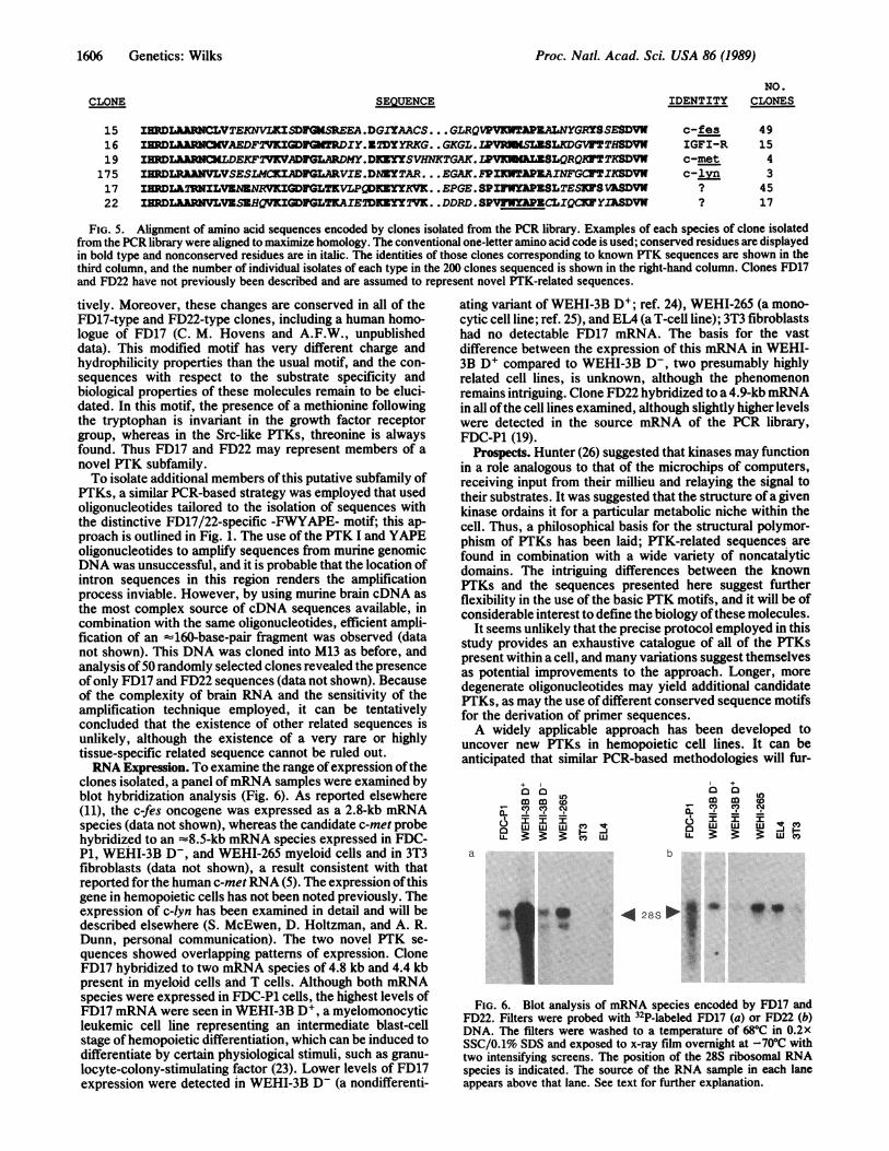
clones isolated, a panel ofmRNA samples were examined byblot hybridization analysis (Fig. 6). As reported elsewhere(11), the c-fes oncogene was expressed as a 2.8-kb mRNAspecies (data not shown), whereas the candidate c-met probehybridized to an -8.5-kb mRNA species expressed in FDC-P1, WEHI-3B D-, and WEHI-265 myeloid cells and in 3T3fibroblasts (data not shown), a result consistent with thatreported for the human c-metRNA (5). The expression ofthisgene in hemopoietic cells has not been noted previously. Theexpression of c-lyn has been examined in detail and will bedescribed elsewhere (S. McEwen, D. Holtzman, and A. R.Dunn, personal communication). The two novel PTK se-quences showed overlapping patterns of expression. CloneFD17 hybridized to two mkNA species of 4.8 kb and 4.4 kbpresent in myeloid cells and T cells. Although both mRNAspecies were expressed in FDC-P1 cells, the highest levels ofFD17 mRNA were seen in WEHI-3B D', a myelomonocyticleukemic cell line representing an intermediate blast-cellstage ofhemopoietic differentiation, which can be induced todifferentiate by certain physiological stimuli, such as granu-locyte-colony-stimulating factor (23). Lower levels of FD17expression were detected in WEHI-3B D- (a nondifferenti-
ating variant of WEHI-3B DI; ref. 24), WEHI-265 (a mono-cytic cell line; ref. 25), and EL4 (a T-cell line); 3T3 fibroblastshad no detectable FD17 mRNA. The basis for the vastdifference between the expression of this mRNA in WEHI-3B D+ compared to WEHI-3B D-, two presumably highlyrelated cell lines, is unknown, although the phenomenonremains intriguing. Clone FD22 hybridized to a 4.9-kb mRNAin all ofthe cell lines examined, although slightly higher levelswere detected in the source mRNA of the PCR library,FDC-P1 (19).
Prospects. Hunter (26) suggested that kinases may functionin a role analogous to that of the microchips of computers,receiving input from their millieu and relaying the signal totheir substrates. It was suggested that the structure ofa givenkinase ordains it for a particular metabolic niche within thecell. Thus, a philosophical basis for the structural polymor-phism of PTKs has been laid; PTK-related sequences arefound in combination with a wide variety of noncatalyticdomains. The intriguing differences between the knownPTKs and the sequences presented here suggest furtherflexibility in the use of the basic PTK motifs, and it will be ofconsiderable interest to define the biology ofthese molecules.
It seems unlikely that the precise protocol employed in thisstudy provides an exhaustive catalogue of all of the PTKspresent within a cell, and many variations suggest themselvesas potential improvements to the approach. Longer, moredegenerate oligonucleotides may yield additional candidatePTKs, as may the use of different conserved sequence motifsfor the derivation of primer sequences.A widely applicable approach has been developed to
uncover new PTKs in hemopoietic cell lines. It can beanticipated that similar PCR-based methodologies will fur-
+0 a
L
a. CV C CJ
3 'C") w
a
O.Z" .
+
m m 40C? C? CJ
6 I F IaU- W
b
*4 28S 0* |
FIG. 6. Blot analysis of mRNA species encoded by FD17 andFD22. Filters were probed with 32P-labeled FD17 (a) or FD22 (b)DNA. The filters were washed to a temperature of 68°C in 0.2xSSC/0.1% SDS and exposed to x-ray film overnight at -700C withtwo intensifying screens. The position of the 28S ribosomal RNAspecies is indicated. The source of the RNA sample in each laneappears above that lane. See text for further explanation.
CLONE
1606 Genetics: Wilks

Proc. Natl. Acad. Sci. USA 86 (1989) 1607
ther extend the list of PTK-related sequences. The approachmay also have a broader application in the discovery ofmembers of other gene families such as those encoding se-quences related to protein kinase C (27), Myc (28), Fos (29),Ras (30), and steroid hormone receptors (31).
I wish to thank Raja Kurban for excellent technical assistance,Gavin Reid and Richard Simpson for preparation of the oligonucle-otides, and Steve Ralph and Chris Hovens for critical reading of themanuscript. I am indebted to Tony Burgess, Ashley Dunn, andPatricia Wilks for advice, support, and encouragement.
1. Coussen5, L., Yang-Feng, T. L., Chen, E., Gray, A., Mc-Grath, J., Liberman, T. A., Schlessinger, J., Franke, U.,Levinson, A. & Ullrich, A. (1985) Science 230, 1132-1139.
2. Yarden, Y., Escobedo, J. A., Kuang, W.-J., Yang-Feng, T. L.,Daniel, T. O., Tremble, P. M., Chen, E. Y., Ando, M. E.,Harkins, R. N., Franke, U., Fried, V. A., JIIrich, A. &Williams, L. T. (1986) Nature (London) 323, 226-232.
3. Ebina, Y., Ellis, L., Jarnagin, K., Edery, M., Graf, L., Clauser,E., Ou, J.-H., Masiarz, F., Kan, Y. W., Godfine, I. D., Roth,R. A. & Rutter, W. J. (1985) Cell 40, 747-758.
4. Ullrich, A., Gray, A., Tam, A. W., Yang-Feng, T. L., Tsub-kawa, M., Collins, C., Henzel, W., LeBon, T., Katthuria, S.,Chen, E., Jacobs, S., Franke, U., Ramachandran, J. & Rojita-Yamaguchi, Y. (1986) EMBO J. 5, 2503-2512.
5. Park, M., Dean, M., Kaul, K., Braun, M. J., Gonda, M. A. &Vande Woude, G. (1987) Proc. Nati. Acad. Sci. USA 84, 6379-6383.
6. Kozma, S. C., Redford, S. M. S., Xioao-Chang, F., Saurer,S. M., Groner, B. & Hynes, N. E. (1988) EMBO J. 7, 147-154.
7. Ullrich, A., Coussens, L., Hayflick, J. S., Dull, T. J., Gray, A.,Tam, A. W., Lee, J., Yarden, Y., Liberman, T. A., Schles-singer, J., Downward, J., Mayes, E. L. V., Whittle, N., Wa-terfield, M. D. & Seeburg, P. H. (1984) Nature (London) 309,418-425.
8. Rothwell, V. M. & Rohrschneider, L. R. (1987) Oncogene Res.1 311-324.
9. Martinez, R., Mathey-Prevot, B., Bernards, A. & Baltimore,D. (1987) Science 237, 411-414.
10. Sukegawa, J., Semba, K., Yamanishi, Y., Nishizawa, M.,Miyajima, N., Yamamoto, T. & Toyoshima, K. (1987) Mol.Cell. Biol. 7, 41-47.
11. Wilks, A. F. & Kurban, R. R. (1988) Oncogene 3, 289-294.12. Shivelman, E., Lifshitz, B., Gale, R. P., Roe, B. A. & Canaani,
E. (1986) Cell 47, 277-284.13. Hanks, S. K., Quinn, A. M, & Hunter, T. (1988) Science 241,
42-52.14. Holtzman, D., Cook, W. D. & Dunn, A. R. (1987) Proc. Nati.
Acad. Sci. USA 84, 8325-8329.15. Hanks, S. K. (1987) Proc. Nati. Acad. Sci. USA 84, 388-393.16. Saiki, R. K., Scharf, S., Faloona, F., Mullis, K. B., Horn,
G. T., Erlich, H. A. & Arnheim, N. (1985) Science 230, 1350-1354.
17. Saiki, R. K., Gelfand, D. H., Stoffel, S., Scharf, S., Higuch,R., Horn, G. T., Mullis, K. B. & Erlich, H. A. (1988) Science239, 487-491.
18. Lee, C. C., Wu, X., Gibbs, R. A., Cook, R. G., Muzny, D. M.& Caskey, C. T. (1988) Science 239, 1288-1291.
19. Dexter, T. M., Garland, J., Scott, D., Scolnick, E. & Metcalf,D. (1980) J. Exp. Med. 152, 1036-1047.
20. Morla, A. O., Schreurs, J. & Wang, J. Y. J. (1988) Mol. Cell.Biol. 8, 2214-2218.
21. Gonda, T. J., Sheiness, D. K. & Bishop, J. M. (1982) Mol.Cell. Biol. 2, 617-624.
22. Sanger, F., Nicklen, S. & Coulson, A. R. (1977) Proc. Nati.Acad. Sci. USA 74, 5463-5467.
23. Metcalf, D. & Nicola, N. A. (1982) Int. J. Cancer 30, 773-780.24. Warner, N. L., Moore, M. A. S. & Metcalf, P. (1969) J. Nati.
Cancer Inst. 43, 963-982.25. Warner, N. L., Burchiel, S. W. & Walker, E. B. (1979) in
Immunobiology and Immunotherapy of Cancer, eds. Terry,W. D. & Yamura, Y. (Elsevier/North-Holland, Amsterdam),pp. 223-243.
26. Hunter, T. (1987) Cell 50, 823-829.27. Coussens, L., Parker, P. J., Rhee, L., Yang-Feng, T. L., Chen,
E., Waterfield, M. D. & Ullrich, A. (1986) Science 233, 859-866.
28. Legouy, E., DePinho, R., Zimmerman, K., Collum, R., Yan-copoulos, G., Mitsock, L., Kriz, R. & Alt, F. W. (1987) EMBOJ. 6, 3359-3366.
29. Cohen, D. R. & Curran, T. (1988) Mol. Cell. Biol. 8, 2063-2069.30. Touchot, N., Chardin, P. & Tavitan, A. (1987) Proc. Nati.
Acad. Sci. USA 84, 8210-8214.31. Evans, R. M. (1988) Science 240, 889-895.
Genetics: Wilks