Two Cases of Spontaneous Pneumoperitoneum Without...
4
CASE REPORT Two Cases of Spontaneous Pneumoperitoneum Without Peritonitis M.D. Shahrudin, MBBS Department of Surgery, Faculty of Medicine, University of Malaya, Lembah Pantai, 59700 Kuala Lumpur Summary Spontaneous pneumoperitoneum without peritonitis is a rare phenomenon which poses a dilemma to the surgeons faced with this problem. Two such cases and their outcome are presented. The first case was caused by tracheal rupture during emergency intubation and was treated by observation until complete resolution. The second case was caused by barotrauma during positive pressure ventilation and was treated by laparotomy. Both patients died for reasons unrelated to the pneumoperitoneum. The passage of air from the chest cavity into the abdominal cavity was along the great vessels in the first case and through the diaphragm in the second. A compilation of other aetiologies of pneumoperitoneum without peritonitis as extracted from the literature is presented. In the presence of pneumoperitoneum without peritonitis and when the clinical history does not suggest visceral perforation, an abdominal tap or lavage should be attempted. If negative, continued observation is advised. Key word: Pneumoperitoneum Introduction Spontaneous pneumoperitoneum signals perforation of a hollow viscus 1 in over 90% of the patients and is usually promptly treated by laparotomf. However, when no signs of peritonitis are evident i.e. fever or peritoneal irritation, that approach is questionable. In the case of patients already compromised by other illnesses it is to be avoided, as an unnecessary operation may further reduce their chances of survivaP. We report here two cases of spontaneous pneumoperitoneum without peritonitis treated at the University Hospital Kuala Lumpur in 1992, review the literature on the various aetiologies and present our recommendations for the management of this condition. Case Report Case 1 An 84 year old man was hospitalised after having collapsed suddenly in the street. On admission he was comatose with BP of 140/90 and pulse of 80/min which was regular and in sinus rhythm. He was ventilated through an endotrocheal tube. Neurological examination later showed severe anoxic brain damage. At this stage, there was subcutaneous emphysema of the face and thorax. Chest X- rays demonstrated pneumomediastinum and bilateral pneumothorax. Both pleural cavities were drained through two chest tubes, with full reexpansion of the lungs. A few hours later, an increase of the abdominal girth was noticed. Abdominal X-rays demonstrated pneumoperitoneum. There was no Med J Malaysia Vol 48 No 4 Dec 1993 449
Transcript of Two Cases of Spontaneous Pneumoperitoneum Without...
CASE REPORT
Two Cases of Spontaneous Pneumoperitoneum Without Peritonitis
M.D. Shahrudin, MBBS Department of Surgery, Faculty of Medicine, University of Malaya, Lembah Pantai, 59700 Kuala Lumpur
Summary
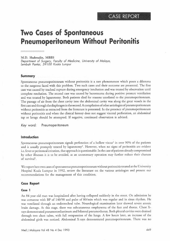
Spontaneous pneumoperitoneum without peritonitis is a rare phenomenon which poses a dilemma to the surgeons faced with this problem. Two such cases and their outcome are presented. The first case was caused by tracheal rupture during emergency intubation and was treated by observation until complete resolution. The second case was caused by barotrauma during positive pressure ventilation and was treated by laparotomy. Both patients died for reasons unrelated to the pneumoperitoneum. The passage of air from the chest cavity into the abdominal cavity was along the great vessels in the first case and through the diaphragm in the second. A compilation of other aetiologies of pneumoperitoneum without peritonitis as extracted from the literature is presented. In the presence of pneumoperitoneum without peritonitis and when the clinical history does not suggest visceral perforation, an abdominal tap or lavage should be attempted. If negative, continued observation is advised.
Key word: Pneumoperitoneum
Introduction
Spontaneous pneumoperitoneum signals perforation of a hollow viscus1 in over 90% of the patients and is usually promptly treated by laparotomf. However, when no signs of peritonitis are evident i.e. fever or peritoneal irritation, that approach is questionable. In the case of patients already compromised by other illnesses it is to be avoided, as an unnecessary operation may further reduce their chances of survivaP.
We report here two cases of spontaneous pneumoperitoneum without peritonitis treated at the University Hospital Kuala Lumpur in 1992, review the literature on the various aetiologies and present our recommendations for the management of this condition.
Case Report
Case 1
An 84 year old man was hospitalised after having collapsed suddenly in the street. On admission he was comatose with BP of 140/90 and pulse of 80/min which was regular and in sinus rhythm. He was ventilated through an endotrocheal tube. Neurological examination later showed severe anoxic brain damage. At this stage, there was subcutaneous emphysema of the face and thorax. Chest Xrays demonstrated pneumomediastinum and bilateral pneumothorax. Both pleural cavities were drained through two chest tubes, with full reexpansion of the lungs. A few hours later, an increase of the abdominal girth was noticed. Abdominal X-rays demonstrated pneumoperitoneum. There was no
Med J Malaysia Vol 48 No 4 Dec 1993 449
CASE REPORT
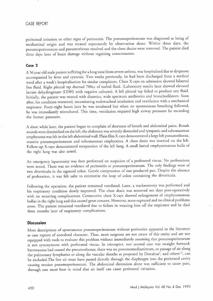
peritoneal irritation or other signs of peritonitis. The pneumoperitoneum was diagnosed as being of mediastinal origin and was treated expectantly by observation alone. Within three days, the pneumoperitoneum and pneumothorax resolved and the chest drains were removed. The patient died three days later of brain damage without regaining consciousness.
Case 2
A 56 year old male patient suffering for a long time from severe asthma, was hospitalised due to dyspnoea accompanied by fever and cyanosis. Two weeks previously, he had been discharged from a medical ward after a week's hospitalisation for similar complaints. Chest X-rays on admission showed bilateral free fluid. Right pleural tap drained 700cc of turbid fluid. (Laboratory results later showed elevated lactate dehydrogenase (LDH) with negative cultures). A left pleural tap failed to produce any fluid. Initially, the patient was treated with diuretics, wide spectrum antibiotics and bronchodilators. Soon after, his condition worsened, necessitating endotracheal intubation and ventilation with a mechanical respirator. Forty-eight hours later he was extubated but when no spontaneous breathing followed, he was immediately reintubated. This time, ventilation required high airway pressures far exceeding the former pressures.
A short while later, the patient began to complain of shortness of breath and abdominal pains. Breath sounds were diminished on the left, the abdomen was severely distended and tympanic and subcutaneous emphysema was felt in the left abdominal wall. Plain film X-rays demonstrated a large left pneumothorax, massive pneumoperitoneum and subcutaneous emphysema. A chest drain was inserted on the left. Follow-up X-rays demonstrated reexpansion of the left lung. A small lateral emphysematous bulla of the right lung was also noted.
An emergency laparotomy was then performed on suspicion of a perforated viscus. No perforations were noted. There was no evidence of peritonitis or pneumoperitoneum. The only findings were of two diverticula in the sigmoid colon. Gentle compression of one produced pus. Despite the absence of perforation, it was felt safer to exteriorize the loop of colon containing the diverticula.
Following the operation, the patient remained ventilated. Later, a tracheostomy was performed and his respiratory condition slowly improved. The chest drain was removed ten days post-operatively with no recurring complications. Consecutive chest X-rays showed enlargement of emphysematous bullae in the right lung and this caused great concern. However, none ruptured and no clinical problems arose. The patient remained ventilated due to failure in weaning him off the respirator and he died three months later of respiratory complications.
Discussion
Most descriptions of spontaneous pneumoperitoneum without peritonitis appeared in the literature as case reports of anecdotal character. Thus, most surgeons are not aware of this entity and are not equipped with tools to evaluate this problem without immediately assuming that pneumoperitoneum is not synonymous with perforated viscus. In retrospect, our second case was straight forward: barotrauma had caused the pneumothorax; there was no pneumomediastimum, so passage of air along the pulmonary lymphatics or along the vascular sheaths as proposed by Donahue\ and othersl,2, can be excluded. The free air must have passed directly through the diaphragm into the peritoneal cavity causing tension pneumoperitoneum. The abdominal distension alone was sufficient to cause pam, through one must bear in mind that air itself can cause peritoneal irritation.
450 Med J Malaysia Vol 48 No 4 Dec 1993
SPONTANEOUS PNEUMOPERITONEUM WITHOUT PERITONITIS
On the other hand, the first case demonstrated well the passage of air from the mediastinum into the peritoneum by the mechanism suggested above.
The mechanism for the rapid development of pneumoperitoneum in Case 2, deserves an explanation. We propose that there is direct passage of air from the pleural cavity into the peritoneal cavity through tiny diaphragmatic defects. These diaphragmatic defects normally remained asymptomatic throughout life but under certain conditions air or fluid may pass directly through these defects, or tear their way through the thin peritoneal lining of the muscular defects.
This route has been implicated in pneumothorax appearing in pneumoperitoneum therapy, insufflation for laparoscopy, post peritoneal dialysis, or post perforated gastric ulcer. It is also cited as the route of penetration of fluids into the chest in cirrhosis or peritoneal dialysis. We suggest likewise that under tension, these defects may serve as a passage from the thorax into the abdomen. This tension was relieved both by chest drainage and subsequent laparotomy eradicating the driving force and preventing any increase of the pneumoperitoneum. Unaware of such a possibility, the surgeon would not scrutinise the diaphragm during the abdominal exploration. In any case, apart from the respiratory problems, postoperative recovery was uneventful.
The various causes of spontaneous pneumoperitoneum have been extensively studied by Hillman2 .
The picture emerging from his review is that there are five basic sources from which air may enter the peritoneum namely (i) oropharyngeal surgery (ii) intrathoracic source (iii) intra-abdominal source (iv) extra-abdominal source (v) idiopathic.
Following oesophageal surgery, the air probably traverses via the mediastinal route.
Air from the thorax may enter the peritoneal cavity through two separate routes:
1) direct passage from the pleural cavity through a diaphragmatic defect (congenital or pathological);
2) via the mediastinum and along the great vessels leading from the chest to the abdomen, entering the intestinal mesentery and finally rupturing into the peritoneal cavity.
When neither traumatic, iatrogenic intervention or gastrointestinal pathology are implicated in females, the genital route is the most possible port of entry of air into the abdomen. Finally, one must bear in mind that even after elimination of all possible sources of air, its origin may remain obscure. Only then may it be classified as idiopathic.
When confronted with the problem of spontaneous pneumoperitoneum without peritonitis or suspicion of gastrointestinal perforation, the surgeon must rule out the possibility of lack of peritoneal irritation due to medical reasons such as immune depression or corticosteroid treatment. If indeed peritonitis is excluded, the medical history should be reviewed especially the medical treatments of the patient prior to the diagnosis of pneumoperitoneum. Based on this information, the surgeon should be able to determine whether the origin of air is supra or infradiaphragmatic. If supradiaphragmatic, lung pathology must be considered, the most common being pneumothorax due to barotrauma during positive pressure ventilation. This must be treated first by chest drainage, appropriate ventilation, etc. Then, an abdominal tap should be performed, the air drained and aspiration of peritoneal fluid attempted.
If the fluid recovered is normal, continued observation is indicated. If no fluid is recovered, lavage should be performed. Should the fluid recovered be contaminated, laparotomy is the next step. When
Med J Malaysia Vol 48 No 4 Dec 1993 451
CASE REPORT
infradiaphragmatic sources are suspected and there IS no clinical peritonitis the same approach IS
recommended.
To enhance the yield of an abdominal tap, one may instill methylene blue via a nasogastic tube. If staining of the peritoneal or lavage fluid is observed, there is an obvious perforation to be treated by operation.
Likewise, instillation of a water soluble radio-opague contrast medium via a nasogastic tube, followed by repeat abdominal X-rays, may indicate surgery if extravasation is observed.
If surgery is not indicated and the patient's condition remained stable after a period of observation, he may be discharged.
Pneumoperitoneum without clinical peritonitis may be treated without laparotomy provided that abdominal tap is negative for septic or inflammatory fluid. Continued observation for confirmation of the diagnosis is mandatory.
References
1. Hillman KM. Pneumoperitoneum. Crit Care Med 1982;10 : 476-81
2. McGlone PE. Spontaneous pneumoperitoneum. Gastroenterol 1966;51 : 393-98
3. Donahue PK. Pneumoperitoneum secondary to
pulmonary air leak. J Paediatr 1972;81 : 797-800
452 Med J Malaysia Vol 48 No 4 Dec 1993
I