Tumor Suppressive Role of miR-342-5p in Human ...
28
HAL Id: hal-03351650 https://hal-normandie-univ.archives-ouvertes.fr/hal-03351650 Submitted on 22 Sep 2021 HAL is a multi-disciplinary open access archive for the deposit and dissemination of sci- entific research documents, whether they are pub- lished or not. The documents may come from teaching and research institutions in France or abroad, or from public or private research centers. L’archive ouverte pluridisciplinaire HAL, est destinée au dépôt et à la diffusion de documents scientifiques de niveau recherche, publiés ou non, émanant des établissements d’enseignement et de recherche français ou étrangers, des laboratoires publics ou privés. Tumor Suppressive Role of miR-342-5p in Human Chondrosarcoma Cells and 3D Organoids Clément Veys, Abderrahim Benmoussa, Romain Contentin, Amandine Duchemin, Emilie Brotin, Jérôme Lafont, Yannick Saintigny, Laurent Poulain, Christophe Denoyelle, Magali Demoor, et al. To cite this version: Clément Veys, Abderrahim Benmoussa, Romain Contentin, Amandine Duchemin, Emilie Brotin, et al.. Tumor Suppressive Role of miR-342-5p in Human Chondrosarcoma Cells and 3D Organoids. International Journal of Molecular Sciences, MDPI, 2021, 22 (11), pp.5590. 10.3390/ijms22115590. hal-03351650
Transcript of Tumor Suppressive Role of miR-342-5p in Human ...

HAL Id: hal-03351650https://hal-normandie-univ.archives-ouvertes.fr/hal-03351650
Submitted on 22 Sep 2021
HAL is a multi-disciplinary open accessarchive for the deposit and dissemination of sci-entific research documents, whether they are pub-lished or not. The documents may come fromteaching and research institutions in France orabroad, or from public or private research centers.
L’archive ouverte pluridisciplinaire HAL, estdestinée au dépôt et à la diffusion de documentsscientifiques de niveau recherche, publiés ou non,émanant des établissements d’enseignement et derecherche français ou étrangers, des laboratoirespublics ou privés.
Tumor Suppressive Role of miR-342-5p in HumanChondrosarcoma Cells and 3D Organoids
Clément Veys, Abderrahim Benmoussa, Romain Contentin, AmandineDuchemin, Emilie Brotin, Jérôme Lafont, Yannick Saintigny, Laurent Poulain,
Christophe Denoyelle, Magali Demoor, et al.
To cite this version:Clément Veys, Abderrahim Benmoussa, Romain Contentin, Amandine Duchemin, Emilie Brotin, etal.. Tumor Suppressive Role of miR-342-5p in Human Chondrosarcoma Cells and 3D Organoids.International Journal of Molecular Sciences, MDPI, 2021, 22 (11), pp.5590. �10.3390/ijms22115590�.�hal-03351650�

International Journal of
Molecular Sciences
Article
Tumor Suppressive Role of miR-342-5p in HumanChondrosarcoma Cells and 3D Organoids
Clément Veys 1, Abderrahim Benmoussa 1,2 , Romain Contentin 1 , Amandine Duchemin 1, Emilie Brotin 3,4,5,Jérôme E. Lafont 6 , Yannick Saintigny 7,8 , Laurent Poulain 4,5 , Christophe Denoyelle 3,4,5, Magali Demoor 1 ,Florence Legendre 1 and Philippe Galéra 1,*
�����������������
Citation: Veys, C.; Benmoussa, A.;
Contentin, R.; Duchemin, A.; Brotin,
E.; Lafont, J.E.; Saintigny, Y.; Poulain,
L.; Denoyelle, C.; Demoor, M.; et al.
Tumor Suppressive Role of
miR-342-5p in Human
Chondrosarcoma Cells and 3D
Organoids. Int. J. Mol. Sci. 2021, 22,
5590. https://doi.org/10.3390/
ijms22115590
Academic Editors:
Magali Cucchiarini and Henning
Madry
Received: 8 April 2021
Accepted: 19 May 2021
Published: 25 May 2021
Publisher’s Note: MDPI stays neutral
with regard to jurisdictional claims in
published maps and institutional affil-
iations.
Copyright: © 2021 by the authors.
Licensee MDPI, Basel, Switzerland.
This article is an open access article
distributed under the terms and
conditions of the Creative Commons
Attribution (CC BY) license (https://
creativecommons.org/licenses/by/
4.0/).
1 Normandie Univ, UNICAEN, BIOTARGEN, 14000 Caen, France; [email protected] (C.V.);[email protected] (A.B.); [email protected] (R.C.); [email protected] (A.D.);[email protected] (M.D.); [email protected] (F.L.)
2 Research Center of the UHC Sainte-Justine and Department of Nutrition, Université de Montréal, Montréal,QC H3T 1C54, Canada
3 Normandie Univ, UNICAEN, ImpedanCELL Platform, Federative Structure 4206 ICORE, 14000 Caen, France;[email protected] (E.B.); [email protected] (C.D.)
4 Normandie Univ, UNICAEN, INSERM U1086 ANTICIPE, Biology and Innovative Therapeutics for OvarianCancer (BioTICLA), 14000 Caen, France; [email protected]
5 Unicancer, Comprehensive Cancer Center F. Baclesse, 14000 Caen, France6 CNRS UMR 5305, Laboratory of Tissue Biology and Therapeutic Engineering, Université Claude Bernard
Lyon 1, Univ Lyon, 69367 Lyon, France; [email protected] LARIA, iRCM, François Jacob Institute, DRF-CEA, 14000 Caen, France; [email protected] Normandie Univ, ENSICAEN, UNICAEN, CEA, CNRS, UMR6252 CIMAP, 14000 Caen, France* Correspondence: [email protected]
Abstract: Chondrosarcomas are malignant bone tumors. Their abundant cartilage-like extracellularmatrix and their hypoxic microenvironment contribute to their resistance to chemotherapy and radio-therapy, and no effective therapy is currently available. MicroRNAs (miRNAs) may be an interestingalternative in the development of therapeutic options. Here, for the first time in chondrosarcomacells, we carried out high-throughput functional screening using impedancemetry, and identified fivemiRNAs with potential antiproliferative or chemosensitive effects on SW1353 chondrosarcoma cells.The cytotoxic effects of miR-342-5p and miR-491-5p were confirmed on three chondrosarcoma celllines, using functional validation under normoxia and hypoxia. Both miRNAs induced apoptosis andmiR-342-5p also induced autophagy. Western blots and luciferase reporter assays identified for thefirst time Bcl-2 as a direct target of miR-342-5p, and also Bcl-xL as a direct target of both miR-342-5pand miR-491-5p in chondrosarcoma cells. MiR-491-5p also inhibited EGFR expression. Finally, onlymiR-342-5p induced cell death on a relevant 3D chondrosarcoma organoid model under hypoxia thatmimics the in vivo microenvironment. Altogether, our results revealed the tumor suppressive activityof miR-342-5p, and to a lesser extent of miR-491-5p, on chondrosarcoma lines. Through this study,we also confirmed the potential of Bcl-2 family members as therapeutic targets in chondrosarcomas.
Keywords: chondrosarcoma; miR-342-5p; miR-491-5p; Bcl-2/Bcl-xL; apoptosis; autophagy
1. Introduction
Chondrosarcomas are the second-most common primary malignant bone tumorsafter osteosarcomas [1,2]. They mainly affect adults between 30 and 70 years old. Chon-drosarcomas are an heterogeneous group of tumors characterized by the production of anextracellular matrix with cartilaginous characteristics [3]. They are classified into differenthistological grades, from low to high, related to their metastatic potential and associatedsurvival rates. Chondrosarcomas are resistant to conventional radiotherapy and chemother-apy. Several mechanisms are involved in their resistance [4]. For instance, they are poorlyvascularized, produce an abundant cartilaginous extracellular matrix and are composed of
Int. J. Mol. Sci. 2021, 22, 5590. https://doi.org/10.3390/ijms22115590 https://www.mdpi.com/journal/ijms

Int. J. Mol. Sci. 2021, 22, 5590 2 of 27
a limited number of proliferating cells which, as whole, hinder drug efficacy. The hypoxicmicroenvironment of chondrosarcomas is also a major contributor to their radio-resistance,because it prevents the formation of antineoplastic reactive oxygen species (ROS) afterirradiation [4]. Therefore, lethality varies between 10% and 50%. Consequently, the onlyeffective treatment for chondrosarcomas is extensive resection of the tumor [4]. However,surgery is not always feasible, especially for tumors located, for example, at the baseof the skull. Therefore, there is an urgent need for new therapeutic strategies to treatchondrosarcomas and/or overcome their resistance to conventional therapies.
MicroRNAs (miRNAs) are small non-coding RNAs of about 20–25 nucleotides, whichact as post-transcriptional regulators of mRNA, generally through base-pairing and asubsequent translation blockade or mRNA decay [5]. A single miRNA can regulate severalhundred different mRNAs. Additionally, several miRNAs can share the same mRNA as atarget, thereby allowing miRNAs to be key regulators of complex networks of targets [6,7].MiRNAs play an essential role in many physiological processes, but also in numerouspathological conditions, particularly in cancer progression, including chondrosarcomas.Several studies have reported an aberrant expression of miRNAs in chondrosarcomas [8,9].For example, miR-100, already identified as a tumor suppressor in many cancers, is down-regulated in chondrosarcomas compared with normal articular chondrocytes [8]. Anotherstudy found that miR-100 is able to re-sensitize resistant chondrosarcoma cells to cisplatinthrough the direct targeting of mammalian target of rapamycin kinase (mTOR) [10]. Conse-quently, miRNAs can be used as diagnostic and prognostic biomarkers, but may also allowthe discovery of novel therapeutic targets in chondrosarcomas [9,11,12]. Various pathways,regulated by miRNAs, that influence proliferation, progression, invasion, angiogenesis andchemosensitivity have been identified in chondrosarcomas. However, to the best of our knowl-edge, no study has explored the effect of miRNAs that directly target anti-apoptotic moleculessuch as B-cell lymphoma-2 (Bcl-2), Bcl-2 lymphoma-extra large (Bcl-xL) and Myeloid cellleukemia-1 (McL-1) in chondrosarcomas. Only two studies using siRNA designed to targetanti-apoptotic molecules Bcl-2, Bcl-xL, X-linked inhibitor of Apoptosis Protein (XIAP) orsurvivin have demonstrated an increase in sensitivity to doxorubicin or irradiation [13,14].The restoration of chemosensitivity to doxorubicin and cisplatin has also been obtained withthe BH3 mimetic ABT-737 in various chondrosarcoma cell lines [15,16].
In the present study, using functional high-throughput miRNA screening and miRNAtarget prediction, we selected five miRNAs that can induce apoptosis in the SW1353chondrosarcoma cell line by targeting BCL2, BCL2L1 or MCL1 mRNAs. Thereafter, weperformed functional studies with the individual miRNAs: miR-149-5p, miR-342-5p, miR-491-5p, miR-541-5p and miR-625-5p. The potential cytotoxic and chemosensitizing effectsof the miRNAs were studied under both normoxia and physioxia (hypoxia), the latterbeing more representative of the in situ physiopathological microenvironment of chon-drosarcomas [17]. We validated the apoptotic effects of miR-491-5p and miR-342-5p in threechondrosarcoma cell lines in both oxic conditions and identified key signaling pathwaysinvolved in their activity. Using a luciferase assay, we demonstrated that miR-342-5pdirectly inhibits both anti-apoptotic BCL2L1 and BCL2 mRNAs post-transcriptionally, andmiR-491-5p directly inhibits BCL2L1 mRNA post-transcriptionally. Considering the im-portance of autophagy in cancer biology and the growing number of autophagy-relatedmiRNAs [18], we also evaluated autophagy, and found that miR-342-5p can activate thisprocess. Finally, we demonstrated for the first time the tumor suppressive effect of miR-342-5p in a 3D organoid grown under hypoxia, a culture model more representative of thephysiopathology of chondrosarcomas than 2D cell cultures.
2. Results2.1. High-Throughput Screening Identifies miRNAs with Potential Antiproliferative andChemosensitive Effects on A Chondrosarcoma Cell Line
We carried out a functional high-throughput screening with a human library of 1200miRNA mimics on the SW1353 chondrosarcoma cell line. The study was based on thecontinuous measurement of impedance to analyze the dynamic behavior of the cells (adhesion,

Int. J. Mol. Sci. 2021, 22, 5590 3 of 27
proliferation and survival) after transfection with miRNA mimics combined with or withoutcisplatin treatment (CDDP). The effect of each miRNA was compared with that of the miR-Ctrl. We used three criteria to select the most potent cytotoxic miRNAs: the shape of thecurve, the area under the curve (AUC) and the cell index (CI) at the end of the experiment(Figure 1). Moreover, morphological observation of cell confluence provided complementaryinformation for the final selection of miRNAs (Figure S1). Finally, of all these miRNAs, fiveshowed a significant effect on cell proliferation/attachment (Figures 1 and S1).
Int. J. Mol. Sci. 2021, 22, x FOR PEER REVIEW 4 of 30
Figure 1. Identification of miRNAs with potential antiproliferative and chemosensitizing effects us-ing high-throughput screening. SW1353 cells were seeded in triplicate in 96-well E-plates VIEW. They were grown for 24 h before transfection (arrow labeled with a T) with 20 nM of miR-Ctrl, miR-491-5p, miR-342-5p, miR-541-5p, miR-625-5p or miR-149-5p. The cells were also treated with 1 µg/mL of CDDP 24 h post-transfection or left untreated (arrow labeled with a C). Each condition was performed in triplicate. Real-time growth curves were monitored using the xCELLigence Sys-tem. Impedance was recorded every 2 h for 120 h. Cell index profiles of one experiment per miRNA species, with mean ± SD, are shown. CDDP: cisplatin, Ctrl: control.
Figure 1. Identification of miRNAs with potential antiproliferative and chemosensitizing effects using high-throughputscreening. SW1353 cells were seeded in triplicate in 96-well E-plates VIEW. They were grown for 24 h before transfection(arrow labeled with a T) with 20 nM of miR-Ctrl, miR-491-5p, miR-342-5p, miR-541-5p, miR-625-5p or miR-149-5p. The cellswere also treated with 1 µg/mL of CDDP 24 h post-transfection or left untreated (arrow labeled with a C). Each conditionwas performed in triplicate. Real-time growth curves were monitored using the xCELLigence System. Impedance wasrecorded every 2 h for 120 h. Cell index profiles of one experiment per miRNA species, with mean ± SD, are shown. CDDP:cisplatin, Ctrl: control.

Int. J. Mol. Sci. 2021, 22, 5590 4 of 27
MiR-491-5p, an “apoptomiR” previously identified as cytotoxic in ovarian cancer cellswith the same xCELLigence cell analysis system [19], was initially used as a positive control.In SW1353 cells, compared with miR-Ctrl, miR-491-5p substantially altered the shape ofthe curve as early as 24 h after transfection. Consequently, at 96 h post-transfection, the cellindex decreased by 4.4-fold relative to miR-Ctrl (p < 0.01, unpaired Student’s t-test), clearlydemonstrating the potential cytotoxic effect of miR-491-5p on SW1353 cells.
MiR-342-5p modified the cell index from approximately 36 h post-transfection untilthe end of the experiment (5-fold decrease relative to miR-Ctrl, 120 h after plating thecells, p < 0.0001). In the presence of a sublethal dose of CDDP and miR-342-5p, the cellindex decreased further (7-fold relative to miR-Ctrl + CDDP, 120 h after seeding, p < 0.001),suggesting that miR-342-5p may sensitize SW1353 cells to CDDP.
We observed similar results when combining miR-541-5p and CDDP with a final cellindex lower than for miR-541-5p alone. However, the antiproliferative effect of miR-541-5pseemed less marked than that of miR-342-5p. It only decreased the cell index by 2.1-fold,120 h after seeding (p < 0.01 relative to miR-Ctrl), and its effect was delayed by 28 h (startedapproximately 60 h post-transfection versus 32 h for miR-342).
MiR-625-5p and miR-149-5p also decreased the cell index from 36 h and 48 h post-transfection, respectively, indicating their antiproliferative activity. Compared with miR-Ctrl, the transfection of these miRNAs led to a 3.8- and 3.2-fold decrease in cell index,respectively, 120 h after seeding (p < 0.001 and p < 0.01, respectively). Cell indices werehigher in the presence of CDDP for both miRNAs. The final cell index was the same formiR-149 with or without CDDP and higher for miR-625 with CDDP than without CDDP.
At the end of the experiment, morphological examination of the cells confirmed that allfive miRNAs under study reduced cell proliferation compared with miR-Ctrl (Figure S1).
2.2. MiR-491-5p and miR-342-5p Have Antimetabolic and Cytotoxic Effects on the SW1353Chondrosarcoma Cell Line
Next, we assessed the antiproliferative effects of these five selected miRNAs with afunctional analysis performed in both normoxia and hypoxia on SW1353 cells.
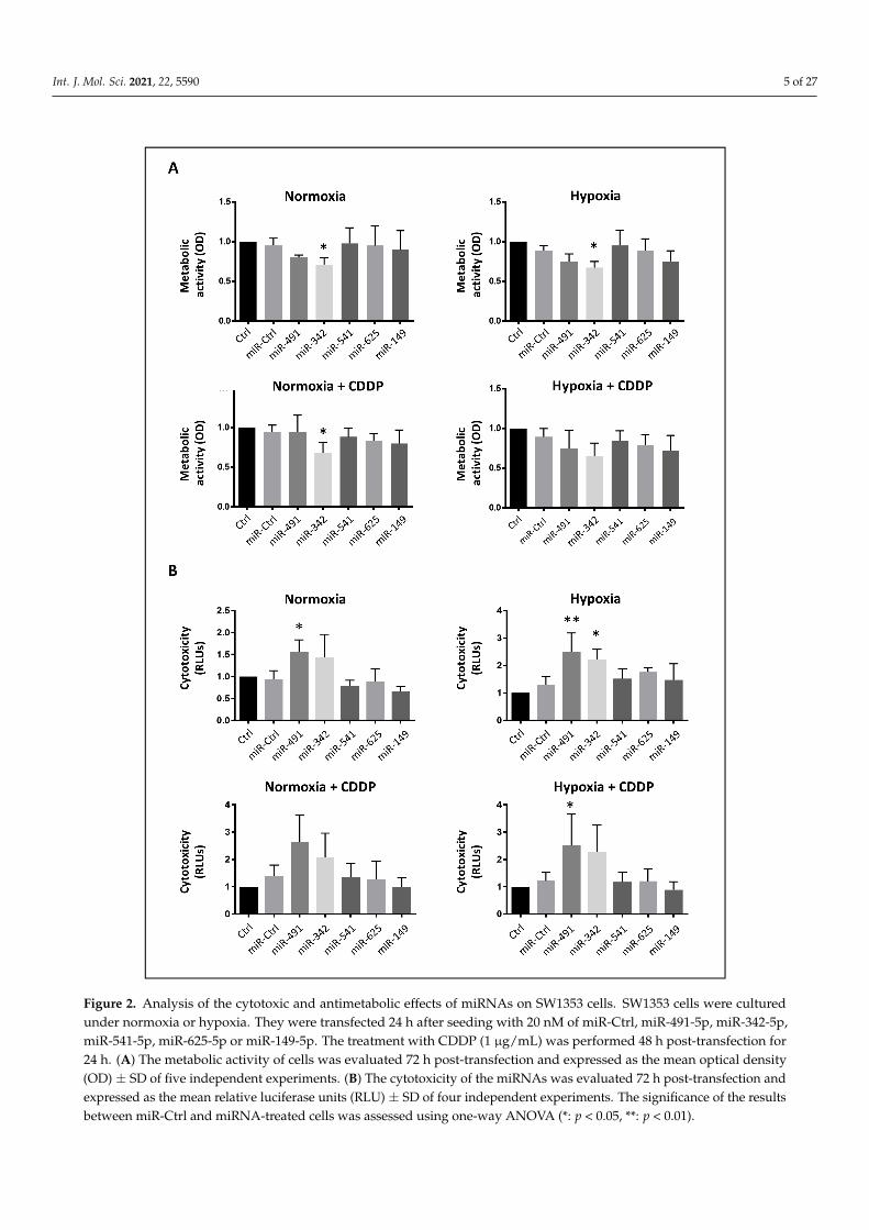
First, we carried out an XTT cell viability assay to measure the metabolic activityof the cells (Figure 2A). Only miR-342-5p significantly decreased cellular metabolismunder normoxia and hypoxia (0.7-fold decrease relative to miR-Ctrl, p < 0.05). MiR-491-5p seemed to reduce metabolic activity (0.8-fold decrease), but this effect was notstatistically significant.
Afterwards, we investigated the potential chemosensitizing effect of the five miRNAsof interest (Figure 2A). Upon sublethal exposure to CDDP, only the cells treated with miR-342-5p under normoxic conditions experienced a significant decrease in their metabolicactivity (0.75-fold decrease relative to miR-Ctrl/CDDP-treated cells, p < 0.05). In all cases,the metabolic activity did not further decrease with the combination of miRNAs and CDDP,compared with miRNAs alone.
Then, we investigated the potential cytotoxic effects of these miRNAs (Figure 2B). OnlymiR-491-5p and miR-342-5p elicited significant cytotoxic effects on the SW1353 cells witha 1.7- and 1.9-fold increase in cytotoxicity for miR-491-5p under normoxia and hypoxia,respectively, and a 1.7-fold increase for miR-342-5p under hypoxia, compared with theirrespective miR-Ctrl. In the presence of CDDP, these miRNAs increased cytotoxicity by thesame magnitude as without CDDP, suggesting no chemosensitizing effect. In this assay,we did not confirm the potential chemosensitizing effect of miR-342-5p and miR-541-5pexpected from the high-throughput screening using the xCELLigence system.
Int. J. Mol. Sci. 2021, 22, 5590 5 of 27Int. J. Mol. Sci. 2021, 22, x FOR PEER REVIEW 6 of 30
Figure 2. Analysis of the cytotoxic and antimetabolic effects of miRNAs on SW1353 cells. SW1353 cells were cultured under normoxia or hypoxia. They were transfected 24 h after seeding with 20 nM of miR-Ctrl, miR-491-5p, miR-342-5p, miR-541-5p, miR-625-5p or miR-149-5p. The treatment with CDDP (1 µg/mL) was performed 48 h post-transfection for 24 h. (A) The metabolic activity of cells was evaluated 72 h post-transfection and expressed as the mean optical density (OD) ± SD of five independent experiments. (B) The cytotoxicity of the miRNAs was evaluated 72 h post-transfection and ex-pressed as the mean relative luciferase units (RLU) ± SD of four independent experiments. The significance of the results between miR-Ctrl and miRNA-treated cells was assessed using one-way ANOVA (*: p < 0.05, **: p < 0.01).
Figure 2. Analysis of the cytotoxic and antimetabolic effects of miRNAs on SW1353 cells. SW1353 cells were culturedunder normoxia or hypoxia. They were transfected 24 h after seeding with 20 nM of miR-Ctrl, miR-491-5p, miR-342-5p,miR-541-5p, miR-625-5p or miR-149-5p. The treatment with CDDP (1 µg/mL) was performed 48 h post-transfection for24 h. (A) The metabolic activity of cells was evaluated 72 h post-transfection and expressed as the mean optical density(OD) ± SD of five independent experiments. (B) The cytotoxicity of the miRNAs was evaluated 72 h post-transfection andexpressed as the mean relative luciferase units (RLU) ± SD of four independent experiments. The significance of the resultsbetween miR-Ctrl and miRNA-treated cells was assessed using one-way ANOVA (*: p < 0.05, **: p < 0.01).

Int. J. Mol. Sci. 2021, 22, 5590 6 of 27
2.3. MiR-491-5p and miR-342-5p Induce Cell Death in Three Chondrosarcoma Cell Lines, But DoNot Affect the Cell Cycle of Healthy Human Articular Chondrocytes
We analyzed cell cycle progression using flow cytometry in the SW1353 chondrosar-coma cell line (Figure 3). None of the miRNAs under investigation induced a cell cycleblockade, but miR-491-5p and miR-342-5p induced a significant accumulation of cells inthe sub-G1 phase associated with the induction of cell death.
Int. J. Mol. Sci. 2021, 22, x FOR PEER REVIEW 8 of 30
Figure 3. Functional validation of cytotoxic or chemosensitizing effects of individual miRNAs on SW1353 cells. SW1353 cells were cultured under normoxia (A,C,E) or hypoxia (B,D,F). They were transfected 24 h after seeding with 20 nM of miR-Ctrl, miR-491-5p, miR-342-5p, miR-541-5p, miR-625-5p or miR-149-5p. An additional treatment with cisplatin (CDDP) (1 µg/mL) was performed 48 h post-transfection (C and D). Analyses were carried out 72 h post-transfection. (A–D) Cell cycle phase distribution was analyzed using flow cytometry. The histograms represent the analysis of five independent experiments (mean ± SD) with the different phases of the cycle. Statistically significant differences in the percentage of sub-G1 events between miR-Ctrl and miRNA-treated cells were determined using one-way ANOVA (*: p < 0.05, ***: p < 0.001). (E,F) The left panels show the cell morphology obtained under photonic microscopy at the end of the experiment. The middle panels show nuclear morphology obtained after DAPI staining as described in the Materials and Methods section. White arrowheads show cells with condensed and/or fragmented chromatin and cellular debris. The right panels

Int. J. Mol. Sci. 2021, 22, 5590 7 of 27
Figure 3. Functional validation of cytotoxic or chemosensitizing effects of individual miRNAs on SW1353 cells. SW1353 cellswere cultured under normoxia (A,C,E) or hypoxia (B,D,F). They were transfected 24 h after seeding with 20 nM of miR-Ctrl,miR-491-5p, miR-342-5p, miR-541-5p, miR-625-5p or miR-149-5p. An additional treatment with cisplatin (CDDP) (1 µg/mL)was performed 48 h post-transfection (C and D). Analyses were carried out 72 h post-transfection. (A–D) Cell cycle phasedistribution was analyzed using flow cytometry. The histograms represent the analysis of five independent experiments(mean ± SD) with the different phases of the cycle. Statistically significant differences in the percentage of sub-G1 eventsbetween miR-Ctrl and miRNA-treated cells were determined using one-way ANOVA (*: p < 0.05, ***: p < 0.001). (E,F)The left panels show the cell morphology obtained under photonic microscopy at the end of the experiment. The middlepanels show nuclear morphology obtained after DAPI staining as described in the Materials and Methods section. Whitearrowheads show cells with condensed and/or fragmented chromatin and cellular debris. The right panels show DNAcontent histograms obtained using flow cytometry. Images shown are representative of five independent experiments.
Regardless of the oxic conditions, miR-491-5p and miR-342-5p increased the percent-age of sub-G1 events, by 2.8-fold average increase under normoxia and by 3.4-fold averageincrease under hypoxia (p < 0.05 and p < 0.001 relative to miR-Ctrl-treated cells in eachgroup; Figure 3A,B). There was also a decrease in cellular density, more cellular debris andcondensed and/or fragmented chromatin—typical of apoptotic cells—after miR-491-5pand miR-342-5p transfection (Figure 3E,F).
The efficiency of the transfection of miR-541-5p, miR-625-5p and miR-149-5p wassimilar in extent to that observed for miR-491-5p and miR-342-5p (overexpression of atleast 3000-fold, p < 0.001; Figure S2). However, their overexpression did not induce celldeath (Figure 3A,B), the cells reached confluency and no cellular debris was detected (datanot shown).
Treatment with a sublethal dose of CDDP increased the percentage of events in theS and G2/M phases at the expense of the G0/G1 phase (Figures 3C,D and S3). MiR-491-5p and miR-342-5p increased the percentage of sub-G1 events when combined withCDDP, with significance reached only under normoxia (by 2.5-fold with p < 0.05 and by4.1-fold with p < 0.01, respectively, relative to miR-Ctrl/CDDP-treated cells). The miRNA-induced increases in sub-G1 peaks were comparable with or without CDDP, suggesting nochemosensitizing effect for any of the miRNAs. From these results, we conclude that theantiproliferative effects of miR-491-5p and miR-342-5p were due to the induction of celldeath in the SW1353 cell line, without any chemosensitizing effect.
We thus focused subsequent investigations on these two miRNAs and assessed theireffects on three other chondrosarcoma cell lines (OUMS-27, CH2879 and L835) and onprimary human articular chondrocytes (HACs) as healthy controls (Figure 4). In OUMS-27cells, miR-491-5p increased the percentage of sub-G1 events under normoxia and hypoxia(by 1.6- and 1.5-fold, respectively, but this increase was not significant; Figure 4A). MiR-342-5p induced a 2.2-fold increase (p < 0.05) in the sub-G1 peak compared with miR-Ctrl-treatedcells, under both oxic conditions. In CH2879 cells, miR-491-5p significantly increased thepercentage of sub-G1 events (by 3.6-fold under normoxia (p < 0.001) and 1.9-fold underhypoxia (p < 0.01); Figure 4B). MiR-342-5p also raised the percentage of sub-G1 events (by3.2-fold under normoxia (p < 0.001) and 1.9-fold under hypoxia (p < 0.01)). The increase insub-G1 peaks with miR-491-5p and miR-342-5p, indicating induction of cell death in bothcell lines, was supported by the extensive cellular debris and numerous apoptotic cellsobserved under fluorescence microscopy on DAPI-stained slides (Figure S4). In L835 cells,miR-491-5p and miR-342-5p did not induce any cell cycle arrest, nor did they significantlyalter the sub-G1 peak, regardless of the oxic conditions (Figures 4C and S5A). Therefore,these two miRNAs did not induce cell death in this chondrosarcoma cell line. We observedsimilar results for primary HACs transfected with miR-491-5p and miR-342-5p, in both oxicconditions, suggesting that these miRNAs have no cytotoxic effects on healthy cartilagecells (Figures 4D and S5B).

Int. J. Mol. Sci. 2021, 22, 5590 8 of 27Int. J. Mol. Sci. 2021, 22, x FOR PEER REVIEW 10 of 30
Figure 4. Analysis of the cytotoxic effects of miR-491-5p and miR-342-5p on chondrosarcoma cells and on HACs. OUMS-27 cells (A), CH2879 cells (B), L835 cells (C), and HACs (D) were cultured under normoxia or hypoxia. Cell lines were transfected 24 h after seeding with 20 nM of miR-Ctrl, miR-491-5p or miR-342-5p. Primary HACs were transfected with 20 nM miRNAs 5 days after seeding. Cell cycle phase distribution was analyzed using flow cytometry 72 h post-transfec-tion. The histograms represent the analyses of the different phases of the cell cycle of at least four independent experiments (mean ± SD). Statistically significant differences in the percentage of sub-G1 events between miR-Ctrl and miRNA-treated cells were determined using one-way ANOVA (*: p < 0.05, **: p < 0.01, ***: p < 0.001).
Figure 4. Analysis of the cytotoxic effects of miR-491-5p and miR-342-5p on chondrosarcoma cells and on HACs. OUMS-27cells (A), CH2879 cells (B), L835 cells (C), and HACs (D) were cultured under normoxia or hypoxia. Cell lines weretransfected 24 h after seeding with 20 nM of miR-Ctrl, miR-491-5p or miR-342-5p. Primary HACs were transfected with20 nM miRNAs 5 days after seeding. Cell cycle phase distribution was analyzed using flow cytometry 72 h post-transfection.The histograms represent the analyses of the different phases of the cell cycle of at least four independent experiments(mean ± SD). Statistically significant differences in the percentage of sub-G1 events between miR-Ctrl and miRNA-treatedcells were determined using one-way ANOVA (*: p < 0.05, **: p < 0.01, ***: p < 0.001).
In OUMS-27 and CH2879 cells, the treatment with a sublethal dose of CDDP signif-icantly increased the percentage of events in the S phase under normoxia and hypoxia(from 1.5- to 2-fold; Figures S6A and S6B). In L835 cells, CDDP treatment increased the per-

Int. J. Mol. Sci. 2021, 22, 5590 9 of 27
centage of G2/M events: 1.9-fold in normoxia (p < 0.05) and 2.7-fold in hypoxia (p < 0.001)(Figure S6C). As in SW1353 cells, the miR-491-5p- and miR-342-5p-induced increases insub-G1 peaks were similar with or without CDDP, suggesting no chemosensitizing effectof these miRNAs.
Overall, miR-491-5p and miR-342-5p seem to be apoptomiRs in SW1353, OUMS-27and CH2879 chondrosarcoma cell lines, but do not appear to chemosensitize the cells toCDDP activity.
2.4. MiR-491-5p and miR-342-5p Activate the Apoptosis Pathway under Normoxia and Hypoxiain Chondrosarcoma Cells
We then focused our investigations on the induction of apoptosis. Regardless ofthe oxygen level, miR-491-5p and miR-342-5p induced the cleavage of Poly(ADP-Ribose)Polymerase (PARP), with more marked effects of miR-342-5p on SW1353 cells (Figure 5A),which is in accordance with our previous results on cell death (Figure 3). PARP was alsocleaved in OUMS-27 and CH2879 cell lines, in both oxic conditions (Figure S7A). In addition,we evaluated caspase-3/7 activity in SW1353 cells by measuring green fluorescence in thenuclei, using the Incucyte® Caspase-3/7 Green apoptosis assay system.
Int. J. Mol. Sci. 2021, 22, x FOR PEER REVIEW 11 of 30
2.4. MiR-491-5p and miR-342-5p Activate the Apoptosis Pathway under Normoxia and Hypoxia in Chondrosarcoma Cells
We then focused our investigations on the induction of apoptosis. Regardless of the oxygen level, miR-491-5p and miR-342-5p induced the cleavage of Poly(ADP-Ribose) Pol-ymerase (PARP), with more marked effects of miR-342-5p on SW1353 cells (Figure 5A), which is in accordance with our previous results on cell death (Figure 3). PARP was also cleaved in OUMS-27 and CH2879 cell lines, in both oxic conditions (Figure S7A). In addi-tion, we evaluated caspase-3/7 activity in SW1353 cells by measuring green fluorescence in the nuclei, using the Incucyte® Caspase-3/7 Green apoptosis assay system.
Both miRNAs increased caspase-3/7 activity in SW1353 cells, with higher significance observed 96 h post-transfection (p < 0.001, Figure 5B). Because PARP cleavage by activated caspases is also a hallmark of apoptosis, these two results imply that miR-491-5p and miR-342-5p induce apoptosis in chondrosarcoma cells.
Figure 5. Analysis of the apoptotic effects of miR-491-5p and miR-342-5p on SW1353 cells. In parts (A,C,D), SW1353 cells were cultured under normoxia or hypoxia. They were transfected 24 h after seeding with 20 nM of miR-Ctrl, miR-491-5p or miR-342-5p. Analyses were carried out 72 h post-transfection. Protein extracts were analyzed with Western blots to evaluate PARP, Bcl-2, Bcl-xL and McL-1 levels versus a β-tubulin (β-Tub) loading control. Representative blots of three independent experiments are shown. Protein expressions are indicated at the bottom of the blots. They were normalized to β-tubulin and to the corresponding miR-Ctrl for each oxic condition. (B) SW1353 cells were cultured under normoxia and transfected 24 h after seeding as before. Caspase-3/7 activity was then assessed in real time for 96 h, as described in the Materials and Methods section. The images shown are representative of the experimental results obtained. Scale bar: 400 µm. The graph provides the analysis of fluorescence of four independent experiments (mean RFU ± SD). The signifi-cance of the results between miR-Ctrl and miRNA-treated cells was assessed using two-way ANOVA (*: p < 0.05, ***: p < 0.001).
Members of the anti-apoptotic Bcl-2 family are among the predicted targets for both miRNAs (Bcl-xL (validated) and McL-1 for miR-491-5p; Bcl-2, Bcl-xL (validated) and McL-
Figure 5. Analysis of the apoptotic effects of miR-491-5p and miR-342-5p on SW1353 cells. In parts (A,C,D), SW1353 cellswere cultured under normoxia or hypoxia. They were transfected 24 h after seeding with 20 nM of miR-Ctrl, miR-491-5p ormiR-342-5p. Analyses were carried out 72 h post-transfection. Protein extracts were analyzed with Western blots to evaluatePARP, Bcl-2, Bcl-xL and McL-1 levels versus a β-tubulin (β-Tub) loading control. Representative blots of three independentexperiments are shown. Protein expressions are indicated at the bottom of the blots. They were normalized to β-tubulinand to the corresponding miR-Ctrl for each oxic condition. (B) SW1353 cells were cultured under normoxia and transfected24 h after seeding as before. Caspase-3/7 activity was then assessed in real time for 96 h, as described in the Materials andMethods section. The images shown are representative of the experimental results obtained. Scale bar: 400 µm. The graphprovides the analysis of fluorescence of four independent experiments (mean RFU ± SD). The significance of the resultsbetween miR-Ctrl and miRNA-treated cells was assessed using two-way ANOVA (*: p < 0.05, ***: p < 0.001).

Int. J. Mol. Sci. 2021, 22, 5590 10 of 27
Both miRNAs increased caspase-3/7 activity in SW1353 cells, with higher significanceobserved 96 h post-transfection (p < 0.001, Figure 5B). Because PARP cleavage by activatedcaspases is also a hallmark of apoptosis, these two results imply that miR-491-5p andmiR-342-5p induce apoptosis in chondrosarcoma cells.
Members of the anti-apoptotic Bcl-2 family are among the predicted targets for bothmiRNAs (Bcl-xL (validated) and McL-1 for miR-491-5p; Bcl-2, Bcl-xL (validated) and McL-1for miR-342-5p, Tables S1 and S2). We therefore evaluated their expression to assess theirinvolvement in the induction of apoptosis by these miRNAs in chondrosarcoma cell lines.
Bcl-2 protein expression greatly decreased upon miR-342-5p overexpression in SW1353cells (by 2-fold under normoxia and 3.3-fold under hypoxia compared with their respectivemiR-Ctrl, Figure 5A). We obtained comparable results for OUMS-27 and CH2879 cells(Figure S7B). In contrast, miR-491-5p did not modulate the expression of Bcl-2 in SW1353(Figure 5A) or OUMS-27 cells (Figure S7B), independently of the oxic conditions, whereasit decreased Bcl-2 protein levels by approximately 2-fold in CH2879 cells under hypoxia(Figure S7B). MiR-491-5p and miR-342-5p decreased Bcl-xL protein expression in SW1353cells under both oxic conditions (from 1.3- to 1.7-fold, Figure 5C). Both miRNAs alsoreduced Bcl-xL protein levels in OUMS-27 and CH2879 cells (Figure S7C). Although McL-1may be a potential target of these two miRNAs, they had no effect on its expression ineither SW1353 (Figure 5D) or OUMS-27 cells (Figure S7D). On the contrary, they tended toincrease McL-1 protein levels in CH2879 cells (in particular with miR-491-5p, Figure S7D).
In summary, miR-342-5p seems able to limit the expression of Bcl-2 and Bcl-xL, andmiR-491-5p seems to be a negative regulator of Bcl-xL, effects that underlie their pro-apoptotic activity in chondrosarcoma cells.
2.5. MiR-342-5p Targets BCL2L1 and BCL2 mRNAs, Whereas miR-491-5p Targets Only BCL2L1mRNA in the SW1353 Chondrosarcoma Cell Line
To verify that miR-491-5p and miR-342-5p effects on Bcl-2 (BCL2 gene) or BCL-xL(BCL2L1 gene) were through direct binding to their respective mRNAs, we co-transfectedluciferase-reporter vectors containing the 3′Untranslated Region (UTR) sequence of thesemRNAs along with either miR-491-5p or miR-342-5p mimics or their corresponding hairpininhibitors in SW1353 cells.
In silico analysis identified a potential binding site for miR-342-5p between nucleotides818 and 824 in the 3′UTR of BCL2 (BCL2-3’UTR; Figure 6A). Only the transfection of miR-342-5p significantly decreased the luciferase activity (RLU) of the reporter vector containingthe wild-type (WT) BCL2-3′UTR (−23% relative to miR-Ctrl, p < 0.01, Figure 6B). In contrast,the co-transfection with anti-miR-342-5p significantly increased RLU (by 22%), which isconsistent with an inhibitory effect of miR-342-5p. Mutation in the binding site for miR-342-5p in the 3′UTR of BCL2 prevented the inhibitory effect of this miRNA. As previously,co-transfection with anti-miR-342-5p increased RLU (+30%, p < 0.01).
In silico analysis did not identify any potential binding site for miR-491-5p in the3′UTR of BCL2 mRNA. As expected, miR-491-5p did not modulate the luciferase activity ofthe WT-BCL2-3′UTR reporter construct (Figure 6B). Therefore, in SW1353 cells, only miR-342-5p can downregulate Bcl-2 protein expression through direct binding to the 818–824region in the 3′UTR of BCL2 mRNA.
We identified two putative binding sites for miR-342-5p in the 3′UTR of BCL2L1 at po-sitions 679–686 and 1407–1413. We thus made constructs in which these sites were mutatedseparately (MUT1 and MUT2 respectively, Figure 6C). Transfection of miR-342-5p led to asignificant decrease in luciferase activity only when co-transfected with WT-BCL2L1-3′UTRor MUT2-BCL2L1-3′UTR (−45% and −47%, p < 0.01 and p < 0.001, respectively; Figure 6D).In comparison, the overexpression of this miRNA decreased reporter activity by only 13%(p < 0.05) for the MUT1-BCL2L1-3′UTR construct and by 14% (NS) for the double mutantMUT1/MUT2-BCL2L1-3′UTR construct. In parallel, co-transfection of anti-miR-342-5pwith all the constructs led to a slight increase in luciferase activities. Therefore, in SW1353cells, miR-342-5p downregulates Bcl-XL protein expression through preferential and directbinding to the 679–686 region in the 3′UTR of BCL2L1 mRNA.

Int. J. Mol. Sci. 2021, 22, 5590 11 of 27
Int. J. Mol. Sci. 2021, 22, x FOR PEER REVIEW 13 of 30
repression was specifically abolished with anti-miR-491-5p. Therefore, as previously re-ported in other cancer cells, BCL2L1 mRNA is also a direct target of miR-491-5p in the SW1353 chondrosarcoma cell line.
Figure 6. Inhibition of both BCL2 and BCL2L1 mRNAs by miR-342-5p and of BCL2L1 mRNA by miR-491-5p. (A) The 3’UTR sequence of BCL2 was aligned with the conserved sequence complementary to hsa-miR-342-5p. Wild (WT) and mutant (MUT1) luciferase vectors of BCL2-3’UTR are shown. (B) WT and MUT1 reporter vectors of BCL2-3’UTR (0.25 ng/µL) were co-transfected with the indicated miR or anti-miR (20 nM) in SW1353 cells. (C) The 3’UTR sequences of BCL2L1 were aligned with the conserved sequence targeted by hsa-miR-342-5p. WT and mutants (MUT1, MUT2) luciferase vectors including BCL2L1-3’UTR are shown. The MUT1/MUT2-BCL2L1-3’UTR vector contains both mutations 1 and 2. (D) WT
Figure 6. Inhibition of both BCL2 and BCL2L1 mRNAs by miR-342-5p and of BCL2L1 mRNA by miR-491-5p. (A) The 3’UTRsequence of BCL2 was aligned with the conserved sequence complementary to hsa-miR-342-5p. Wild (WT) and mutant(MUT1) luciferase vectors of BCL2-3’UTR are shown. (B) WT and MUT1 reporter vectors of BCL2-3’UTR (0.25 ng/µL)were co-transfected with the indicated miR or anti-miR (20 nM) in SW1353 cells. (C) The 3’UTR sequences of BCL2L1 werealigned with the conserved sequence targeted by hsa-miR-342-5p. WT and mutants (MUT1, MUT2) luciferase vectorsincluding BCL2L1-3’UTR are shown. The MUT1/MUT2-BCL2L1-3’UTR vector contains both mutations 1 and 2. (D) WTand mutant vectors of BCL2L1-3’UTR (1 ng/µL) were co-transfected with the indicated miR or anti-miR (20 nM) in SW1353cells. (B,D) SEAP and GLUC luciferase activities were measured 24 h post-transfection. GLUC activity was normalizedto SEAP activity and expressed as RLU. The data show a representative experiment (mean ± SD) of four independentexperiments performed in triplicate. The significance of the results between miR-Ctrl and miRNA- or anti-miRNA-treatedcells was evaluated by two-tailed Student’s t-test (*: p < 0.05, **: p < 0.01, ***: p < 0.001).

Int. J. Mol. Sci. 2021, 22, 5590 12 of 27
Functional binding sites for miR-491-5p have previously been identified in the 3′UTRof BCL2L1 mRNA [19–21]. Accordingly, co-transfection of miR-491-5p and WT-BCL2L1-3′UTR, bearing three binding sites for miR-491-5p, led to a significant decrease in theluciferase activity of the reporter vector (−34% relative to miR-Ctrl, p < 0.01, Figure 6D).This repression was specifically abolished with anti-miR-491-5p. Therefore, as previouslyreported in other cancer cells, BCL2L1 mRNA is also a direct target of miR-491-5p in theSW1353 chondrosarcoma cell line.
2.6. MiR-342-5p Increases Autophagy in the SW1353 Chondrosarcoma Cell Line
To further investigate the mechanisms of action of miR-342-5p and miR-491-5p, wethen investigated their ability to regulate autophagy, which can also contribute to celldeath. We quantified the number of autophagosomes per nucleus using the specificautophagosome green marker of the Cell MeterTM autophagy fluorescence imaging kit andDAPI staining of the nuclei (Figure 7).
Int. J. Mol. Sci. 2021, 22, x FOR PEER REVIEW 14 of 30
and mutant vectors of BCL2L1-3’UTR (1 ng/µL) were co-transfected with the indicated miR or anti-miR (20 nM) in SW1353 cells. (B,D) SEAP and GLUC luciferase activities were measured 24 h post-transfection. GLUC activity was normalized to SEAP activity and expressed as RLU. The data show a representative experiment (mean ± SD) of four independent exper-iments performed in triplicate. The significance of the results between miR-Ctrl and miRNA- or anti-miRNA-treated cells was evaluated by two-tailed Student’s t-test (*: p < 0.05, **: p < 0.01, ***: p < 0.001).
2.6. MiR-342-5p Increases Autophagy in the SW1353 Chondrosarcoma Cell Line To further investigate the mechanisms of action of miR-342-5p and miR-491-5p, we
then investigated their ability to regulate autophagy, which can also contribute to cell death. We quantified the number of autophagosomes per nucleus using the specific au-tophagosome green marker of the Cell MeterTM autophagy fluorescence imaging kit and DAPI staining of the nuclei (Figure 7).
Figure 7. Effects of miR-491-5p and miR-342-5p on the autophagic activity of SW1353 cells. SW1353 cells were cultured and transfected as described in Figure 5, with an additional positive control (5% FCS in PBS for the last 24 h). Analyses were carried out 72 h post-transfection. The autophagosomes (green) and the nuclei (blue) were labeled as described in the Materials and Methods section. (A) The images shown are representative of the experimental results obtained. Scale bar: 100 µm. (B) The graph gives the number of autophagosomes normalized to the number of nuclei (mean ± SD of three independent experiments). The significance of the results between miR-Ctrl and miRNA-treated cells was assessed using one-way ANOVA (*: p < 0.05, **: p < 0.01).
Figure 7. Effects of miR-491-5p and miR-342-5p on the autophagic activity of SW1353 cells. SW1353 cells were culturedand transfected as described in Figure 5, with an additional positive control (5% FCS in PBS for the last 24 h). Analyseswere carried out 72 h post-transfection. The autophagosomes (green) and the nuclei (blue) were labeled as described inthe Materials and Methods section. (A) The images shown are representative of the experimental results obtained. Scalebar: 100 µm. (B) The graph gives the number of autophagosomes normalized to the number of nuclei (mean ± SD of threeindependent experiments). The significance of the results between miR-Ctrl and miRNA-treated cells was assessed usingone-way ANOVA (*: p < 0.05, **: p < 0.01).

Int. J. Mol. Sci. 2021, 22, 5590 13 of 27
Whatever the oxic conditions, miR-491-5p did not significantly modulate the numberof marked autophagosomes (Figure 7). MiR-342-5p significantly increased the number ofautophagosomes under normoxia (by 7.4-fold relative to miR-Ctrl-treated cells, p < 0.01),and to a lesser extent under hypoxia (by 2.8-fold, p < 0.05). Therefore, miR-342 increasesthe autophagic activity of SW1353 cells, which may participate in its tumor-suppressiveactivity.
2.7. MiR-491-5p and miR-342-5p Modulate the Expression of Numerous Proteins Related toProliferation or Apoptosis and Affect Mitogenic Signaling Pathways in Chondrosarcoma Cells
To gain further insight into the mechanisms underlying the activity of these twomiRNAs, we looked at the expression of some of their predicted, or validated, targets(Tables S1 and S2), with a special focus on those involved in cell survival/death and/or insignaling pathways commonly altered in cancer.
Although in silico analysis showed that CCND1 mRNA was a validated target ofmiR-342-5p (Table S2), the transfection of this miRNA did not clearly affect CCND1 proteinexpression in chondrosarcoma cell lines. In SW1353 cells, miR-491-5p and miR-342-5pled to a slight decrease in cyclin D1 protein expression under normoxia, whereas theytended to increase its levels under hypoxia (Figure 8A). We did not detect any reproduciblealteration in cyclin D1 expression in the CCND1 mRNA steady-state levels (data not shown).In CH2879 and OUMS-27 cells, there was no significant change in cyclin D1 expressionindependently of the oxic condition (Figure S8A).
We then measured the expression of EGFR, a direct target of miR-491-5p in glioblas-toma and ovarian cancer cells [19,22] (Table S1), and a predicted target of miR-342-5p (TableS2). In SW1353 cells, miR-491-5p repressed the expression of EFGR at the mRNA level by2.5-fold (p < 0.01) under both oxic conditions, whereas miR-342-5p increased its expressionby 1.6-fold (p < 0.05) only under normoxia (Figure 8B). At the protein level, miR-491-5pdecreased EGFR expression from 1.4- to 2-fold depending on the oxic condition (Figure 8A).This effect on EGFR protein was more marked in OUMS-27 and CH2879 chondrosarcomacell lines than in SW1353 cells (Figure S8B). MiR-342-5p did not have any significant effecton EGFR protein expression in any of the three chondrosarcoma cell lines.
Caspase-9 was also identified as a direct target of miR-342-5p ([23], Table S2). MiR-342-5p did not significantly modulate caspase-9 mRNA level in SW1353 cells (data notshown). However, in the three chondrosarcoma cell lines, it caused a strong decrease (atleast 3-fold) in pro-caspase-9 protein expression (47 kDa), without induction of the largecleaved caspase-9 fragments (37 and 35 kDa) (Figures 8A and S8B). MiR-491-5p had noeffect on pro-caspase-9 protein expression and it did not increase the cleavage of caspase-9.
The pro-apoptotic proteins Bcl-2-associated X (Bax) and Bcl-2 homologous antagonistkiller (Bak) are essential for mitochondrial outer membrane permeabilization and inductionof cell death. None of the considered miRNAs affected Bax protein expression (data noshown). Bak protein was upregulated when exposing the three chondrosarcoma cell linesto miR-491-5p, especially under hypoxia, whereas miR-342-5p tended to decrease Bakexpression (from 1.3-fold to 3-fold increase with miR-491-5p and approximately 1.2-folddecrease with miR-342-5p, Figures 8A and S8C).
Finally, we analyzed the effects of these miRNAs on two essential mitogenic signalingpathways involved in cell survival (Mitogen Activated Protein Kinase/Extracellular signal-Regulated Kinase (MAPK/ERK) and Protein Kinase B/AKT (PKB/AKT). In the ERKpathway, the effects of these miRNAs were quite variable from one cell line to another,and depended on the oxic conditions. In SW1353 cells, miR-491-5p decreased activatedand total ERK proteins under normoxia (by approximately 1.5-fold, Figure 8C). Mir-491-5phad no marked effect on the ERK pathway in OUMS-27 cells (Figure S8D). It decreasedphosphorylated or total ERK proteins levels under normoxia and hypoxia in CH2879 cells(maximum decrease of 1.6-fold for phosphorylated ERK under normoxia; Figure S8D).These results suggest that miR-491-5p can decrease the expression of ERK proteins and theiractivated/phosphorylated forms in chondrosarcoma cells. In SW1353 cells, miR-342-5pdid not alter total ERK protein expression (Figure 8C). However, it increased the levels of

Int. J. Mol. Sci. 2021, 22, 5590 14 of 27
phosphorylated ERKs under normoxia. In OUMS-27 and CH2879 cells, miR-342-5p didnot seem to significantly influence total or phosphorylated ERK proteins levels (FigureS8D). Therefore, miR-342-5p does not significantly influence ERK signaling pathway inchondrosarcoma cells.
Int. J. Mol. Sci. 2021, 22, x FOR PEER REVIEW 16 of 30
Figure 8. Analysis of some survival/death proteins and mitogenic signaling pathways affected by miR-491-5p and miR-342-5p. SW1353 cells were cultured and transfected as described in Figure 5. Analyses were carried out 72 h post-transfec-tion. (A,C) Protein extracts were analyzed with Western blots to evaluate the levels of Cyclin D1, EGFR, Caspase-9, Bak, P-Thr202/Tyr204-ERK, ERK, P-Ser473-AKT and AKT versus β-tubulin (β-Tub) or GAPDH. Representative blots of three independent experiments are shown. Protein expressions are indicated at the bottom of the blots. They were normalized to β-tubulin or GAPDH, and to the corresponding miR-Ctrl for each oxic condition. (B) Total RNA was extracted as de-scribed in the Materials and Methods section. Relative expression of EGFR was determined by RT-quantitative PCR and normalized to both PPIA1 and GAPDH levels and to Ctrl. Data are expressed as mean of triplicate samples ± SD. The results are representative of three independent experiments. The significance of the results was evaluated by two-tailed Student’s t-test compared with the corresponding miR-Ctrl (*: p < 0.05, **: p < 0.01).
Caspase-9 was also identified as a direct target of miR-342-5p ([23], Table S2). MiR-342-5p did not significantly modulate caspase-9 mRNA level in SW1353 cells (data not shown). However, in the three chondrosarcoma cell lines, it caused a strong decrease (at least 3-fold) in pro-caspase-9 protein expression (47 kDa), without induction of the large cleaved caspase-9 fragments (37 and 35 kDa) (Figures 8A and S8B). MiR-491-5p had no effect on pro-caspase-9 protein expression and it did not increase the cleavage of caspase-9.
Figure 8. Analysis of some survival/death proteins and mitogenic signaling pathways affected by miR-491-5p and miR-342-5p. SW1353 cells were cultured and transfected as described in Figure 5. Analyses were carried out 72 h post-transfection.(A,C) Protein extracts were analyzed with Western blots to evaluate the levels of Cyclin D1, EGFR, Caspase-9, Bak, P-Thr202/Tyr204-ERK, ERK, P-Ser473-AKT and AKT versus β-tubulin (β-Tub) or GAPDH. Representative blots of threeindependent experiments are shown. Protein expressions are indicated at the bottom of the blots. They were normalized toβ-tubulin or GAPDH, and to the corresponding miR-Ctrl for each oxic condition. (B) Total RNA was extracted as describedin the Materials and Methods section. Relative expression of EGFR was determined by RT-quantitative PCR and normalizedto both PPIA1 and GAPDH levels and to Ctrl. Data are expressed as mean of triplicate samples ± SD. The results arerepresentative of three independent experiments. The significance of the results was evaluated by two-tailed Student’s t-testcompared with the corresponding miR-Ctrl (*: p < 0.05, **: p < 0.01).
The effects of miRNAs on the AKT signaling pathway were also heterogeneous amongthe chondrosarcoma cell lines. In SW1353 cells, miR-491-5p decreased total AKT proteinonly under normoxia (by 2-fold, relative to miR-Ctrl-treated cells; Figure 8C), withoutclearly inhibiting AKT activation. MiR-342-5p downregulated the expression of total AKTprotein under both normoxia and hypoxia (by approximately 1.5-fold), with subsequentinhibition of AKT phosphorylation only under hypoxia (by 1.4-fold). In OUMS-27 cells,there was no significant variation in phosphorylated or total AKT proteins levels between

Int. J. Mol. Sci. 2021, 22, 5590 15 of 27
either miRNA or under either oxic condition (Figure S8E). In CH2879 cells, whereas bothmiR-342-5p and miR-491-5p decreased total AKT protein expression (from 1.5- to 5-fold),they also increased the level of phosphorylated AKT under normoxia and hypoxia (FigureS8E). Consequently, these results suggest that the effects of miR-491-5p and miR-342-5p onAKT signaling depend on the chondrosarcoma cell line. They can decrease AKT expression,but at the same time participate in its activation.
2.8. Only miR-342-5p Induces Cell Death in a 3D Organoid Chondrosarcoma Model
Finally, we tested the anti-tumor activity of miR-342-5p and miR-491-5p on SW1353cells cultured in 3D, a model that mimics the in vivo physiopathological microenvironmentmore accurately. SW1353 cells were transfected with either 20 or 50 nM of miRNA mimics.Cells were cultured under normoxia or hypoxia with the same kinetics as described above(Figure 9).
Int. J. Mol. Sci. 2021, 22, x FOR PEER REVIEW 18 of 30
Figure 9. Effects of miR-491-5p and miR-342-5p on SW1353 cells cultured in 3D. SW1353 cells were cultured in collagen sponge scaffolds under normoxia or hypoxia. They were transfected 24 h after seeding with 20 nM or 50 nM of miR-Ctrl, miR-491-5p and miR-342-5p. Analyses were carried out 72 h post-transfection. Cell cycle phase distribution was analyzed using flow cytometry. The histograms represent the analysis of the different phases of the cell cycle of four independent experiments (mean ± SD). Statistically significant differences in the percentage of sub-G1 events between miR-Ctrl and miRNA-treated cells were determined using one-way ANOVA (**: p < 0.01).
3. Discussion The resistance of chondrosarcomas to conventional radiotherapy and chemotherapy
calls for the development of new treatments. The use of therapeutic miRNAs offers the advantage of a multi-target approach and can help to discover new therapeutic targets for clinical applications [9,11]. In previous studies, the functions of about 20 miRNAs have been reported in chondrosarcomas [9,24]. Based on high-throughput screening of a hu-man library of 1200 miRNAs, we found numerous miRNAs able to decrease cell prolifer-ation in the SW1353 chondrosarcoma cell line. Among these, we identified five miRNAs with putative pro-apoptotic activity because they potentially target anti-apoptotic pro-
Figure 9. Effects of miR-491-5p and miR-342-5p on SW1353 cells cultured in 3D. SW1353 cells were cultured in collagensponge scaffolds under normoxia or hypoxia. They were transfected 24 h after seeding with 20 nM or 50 nM of miR-Ctrl,miR-491-5p and miR-342-5p. Analyses were carried out 72 h post-transfection. Cell cycle phase distribution was analyzedusing flow cytometry. The histograms represent the analysis of the different phases of the cell cycle of four independentexperiments (mean ± SD). Statistically significant differences in the percentage of sub-G1 events between miR-Ctrl andmiRNA-treated cells were determined using one-way ANOVA (**: p < 0.01).

Int. J. Mol. Sci. 2021, 22, 5590 16 of 27
Compared with miR-Ctrl, miR-491-5p did not significantly increased the percentage ofsub-G1 events under normoxia and hypoxia (by 1.1- and 1.2-fold, respectively, regardless ofits concentration. In contrast, miR-342-5p increased the number of sub-G1 peaks, with thesame efficiency at 20 as at 50 nM (approximately 1.5-fold relative to the respective miR-Ctrl,only significant under hypoxia at p < 0.01). More cellular debris and apoptotic cells wereobserved under fluorescence microscopy on DAPI-stained slides with miR-342-5p thanwith miR-491-5p (Figure S9). In conclusion, miR-342-5p, but not miR-491-5p, significantlyinduces cell death in our organoid chondrosarcoma culture model, especially in hypoxia.
3. Discussion
The resistance of chondrosarcomas to conventional radiotherapy and chemotherapycalls for the development of new treatments. The use of therapeutic miRNAs offers theadvantage of a multi-target approach and can help to discover new therapeutic targetsfor clinical applications [9,11]. In previous studies, the functions of about 20 miRNAshave been reported in chondrosarcomas [9,24]. Based on high-throughput screening of ahuman library of 1200 miRNAs, we found numerous miRNAs able to decrease cell prolifer-ation in the SW1353 chondrosarcoma cell line. Among these, we identified five miRNAswith putative pro-apoptotic activity because they potentially target anti-apoptotic proteinssuch as Bcl-xL and Bcl-2, which are thought to play a critical role in the chemoresistanceof chondrosarcoma chondrocytes [15,16,25]. Moreover, two miRNAs (miR-342-5p andmiR-541-5p) also seemed to exert chemosensitizing effects when combined with CDDP.Real-time cell analyses and endpoint morphological analyses are frequently performed inglass culture plates under normoxia, but chondrosarcoma cells live in a hypoxic environ-ment. Therefore, we carried out functional analyses in standard plastic culture plates tovalidate our approach and to unambiguously identify miRNAs with realistic cytotoxic andchemosensitizing effects. Considering the role of hypoxia in the resistance of tumor cells toconventional treatments [4,26], we performed functional analyses under both normoxia(21% O2) and hypoxia (3% O2). We validated the tumor-suppressive effects of miR-491-5pand miR-342-5p on three chondrosarcoma cell lines cultured in monolayer. Their effectshave already been described in other cancers, but to date, our work is the first to identifymiRNAs acting as tumor suppressors in a hypoxic microenvironment. Moreover, becausethe traditional 2D cell culture does not reflect the in situ physiopathological microenviron-ment, we used for the first time a 3D organoid culture model to further confirm the effectsof miRNAs on SW1353 cells.
We used four cell lines derived from central conventional chondrosarcomas of gradeII (SW1353) and grade III (OUMS-27, CH2879 and L835). They are all resistant to CDDP:CH2879 and OUMS-27 are the least resistant (inhibitory concentration to reach 50% reduc-tion in cell viability (IC50), 40 µM and 50 µM, respectively), L835 has an IC50 value of atleast 200 µM and SW1353 is the most resistant with an IC50 of at least 400 µM [15]. Mutationanalysis performed on TP53 and on isocitrate dehydrogenase (IDH) 1 or 2 has revealeddistinct characteristics: SW1353 harbors both IDH2 and TP53 mutations, OUMS-27 harborsa TP53 mutation, CH2879 is wild type for IDH and TP53 mutations and L835 harbors anIDH1 mutation [27,28]. Only the L835 cell line did not respond to miR-491-5p and miR-342-5p, and harbors an IDH1 mutation. Gain-of-function mutations of IDH1/2 are foundin 38–70% of primary central chondrosarcomas and only lead to DNA hypermethylation.Moreover, direct inhibition of the IDH1 mutant enzyme does not change the tumorigenicproperties of chondrosarcoma cell lines in vitro [29]. The four cell lines express Bcl-2 andBcl-xL proteins at different levels [15,25]. These latter two studies showed that L835 cellsexpress higher levels of Bcl-xL protein than Bcl-2 protein. Moreover, inhibition of Bcl-xLand Bcl-2 with the ABT-737 drug reduces cell viability and induces apoptosis of L835 cellsafter 72 h of incubation [15]. Inhibition of Bcl-xL by both miR-491-5p and miR-342-5pshould therefore have resulted in cell death by apoptosis. Complementary experimentswould be necessary to verify inhibition of Bcl-xL by both miR-491-5p and miR-342-5p onL835 cells.

Int. J. Mol. Sci. 2021, 22, 5590 17 of 27
Unlike miR-541-3p [30,31], miR-541-5p has never been studied in cancers. MiR-625-5p and miR-149-5p have been previously reported to have tumor suppressive roles invarious cancers, alone or combined with various chemotherapeutic agents [32–36]. Evenif miR-149-5p seems to play a dual role in cancer [37], both miR-625-5p and miR-149-5p can induce apoptosis. Our screening showed that these three miRNAs likely haveantiproliferative effects, but we could not confirm any significant antimetabolic, cytotoxicor killer effects on the SW1353 chondrosarcoma cell line. Furthermore, we did not validatethe chemosensitizing effect of miR-541-5p 72 h post-transfection. Because cells growingon glass are more sensitive than cells growing on plastic, it is possible that killer effectsrequire a longer incubation period in standard cell culture conditions, possibly more than96 h post-transfection.
MiR-491-5p has already been described as a tumor suppressor in various cancers andseveral of its direct targets have been identified and include Bcl-xL in colorectal cancer [21],Bcl-xL and EGFR in ovarian cancer [19], Bcl-xL and TP53 in pancreatic cancer [20], Bcl-xL,EGFR and CDK6 in glioblastoma [22], Pyruvate Kinase M2 (PKM2) [38] and Forkheadbox Protein P4 (FOXP4) in osteosarcoma [39], the most common type of bone cancers.In our study, miR-491-5p was first used as a positive control of cytotoxicity for the high-throughput screening. Given its high therapeutic potential, we next wanted to confirm itstumor suppressive effect on chondrosarcoma cell line, which has never been studied. Wefound it was able to induce cell death in SW1353, OUMS-27 and CH2879 chondrosarcomacell lines cultured in monolayer under normoxia and hypoxia.
EGFR is constitutively activated in high-grade chondrosarcoma tumors [40]. Regard-less of the oxygen level, miR-491-5p inhibited EGFR at the protein and mRNA levels. Thisinhibition suggests a possible downstream inhibition of the AKT and MAPK signaling path-ways, leading to a decrease in chondrosarcoma cell proliferation and migration, as shownin other studies investigating the inhibition or silencing of EGFR in chondrosarcomas [40].Previous studies have shown that the downstream inhibition of MAPK and AKT activityby miR-491-5p depends on the type of cancer or the cell line. In the SW1990 pancreatic cellline, miR-491-5p inhibits PI3K/AKT, but not RAS/MAPK [20]. This miRNA also inhibitsAKT and MAPK signaling pathways in IGROV1-R10 ovarian cancer cells, whereas SKOV3,another ovarian cell line, maintains its AKT and MAPK activity [19]. In the present study,the effect of miR-491-5p on AKT and MAPK signaling pathways was heterogeneous amongthe chondrosarcoma cell lines and also depended on the oxic conditions.
MiR-491-5p did not influence the expression of the Bcl-2 anti-apoptotic protein inany of the three investigated chondrosarcoma cell lines sensitive to miR-491-5p. Similarly,although the anti-apoptotic McL-1 may be a potential target of miR-491-5p, this miRNAdid not influence McL-1 protein expression in SW1353 and OUMS-27 cell lines, and rathertended to increase McL-1 expression in CH2879 cells, as observed in the IGROV1-R10ovarian cell line [19]. Like others, we found that miR-491-5p directly targets BCL2L1mRNA [19,20,22]. Consequently, miR-491-5p decreased the expression of the anti-apoptoticprotein Bcl-xL in the three chondrosarcoma cell lines responding to miR-491-5p under bothoxic conditions. It also increased the expression of the Bak pro-apoptotic protein. Boththese events may therefore contribute to its apoptotic activity in chondrosarcoma cells.
The miR-342-5p mechanism of action has been less investigated than that of miR-342-3p and, to our knowledge, never in chondrosarcomas. It has been identified as a tumorsuppressor in neuroblastomas [30]. It inhibits colon cancer tumorigenesis through directtargeting of the N-a-acetyl transferase 10 protein (NAA10) and also promotes apoptosis [41].In osteosarcomas, miR-342-5p inhibits cell growth, migration and invasion, and restoressensitivity to doxorubicin through direct targeting of Wnt member 7B (WnT7b) [42]. Inthe present study, high-throughput screening revealed that miR-342-5p has some antipro-liferative effects on SW1353 cells. However, miR-342-5p did not significantly affect AKTand ERK mitogenic signaling pathways as previously reported for breast cancer cells [43],or AKT mRNA in inflammatory macrophages [44]. We observed AKT inhibition at theprotein level in two out of the three cell lines (SW1353, CH2879), but at the same time

Int. J. Mol. Sci. 2021, 22, 5590 18 of 27
the phosphorylation of AKT increased in CH2879 cells. There is evidence in the litera-ture that miR-342-5p increases the level of phosphorylated forms of AKT via targeting aphosphatase on cardiomyocytes [23]. Others have shown that miR-342-5p attenuates theprotein level of total AKT, whereas its phosphorylated protein level is stable in neural stemcells [45]. In view of our results, we cannot assume that the antiproliferative effects ofmiR-342-5p are linked to AKT and ERK pathways, but are likely linked to its pro-apoptoticeffects. Indeed, miR-342-5p induced cell death and apoptosis in SW1353, OUMS-27 andCH2879 cells, independently of the oxygen level. In the three chondrosarcoma cell lines,miR-342-5p inhibited the expression of the anti-apoptotic proteins Bcl-2 and Bcl-xL, butnot that of McL-1, with a more sustained effect under hypoxia. We also reported for thefirst time that miR-342-5p downregulates Bcl-2 protein expression by directly bindingto sequence 818–824 of BCL2-3′UTR. Like Soriano et al. [30], we found that miR-342-5pdirectly targets the BCL2L1 gene and in addition, we identified its binding site at position679–686 of BCL2L1-3′UTR. Soriano et al. also identified the CCND1 gene as a direct targetof miR-342-5p in neuroblastomas, but in our experiments on chondrosarcoma cells lines,cyclin D1 protein expression was not affected by miR-342-5p.
Surprisingly, in the three chondrosarcoma cell lines, miR-342-5p also had anti-apoptoticeffects, because it tended to decrease (by 1.2-fold) the protein expression of the Bak pro-apoptotic protein, and that of the pro-caspase-9 (at least 3-fold). Our data are in line withthose of Hou et al., who identified exosomal miR-342-5p as a key cardioprotective moleculeinhibiting apoptotic signaling via direct targeting of caspase-9 and Jun Kinase 2 (JNK2)in cardiomyocytes [23]. Caspase-9 activation is stimulated by dimerization instead ofcleavage within the apoptosome. Dimerization facilitates autocatalytic cleavage, whichresults in the stabilization of the dimer [46]. Caspase-9 showed complete activity in itsuncleaved form [47]. This caspase initiates apoptosis by cleaving and thereby activatingexecutioner caspases-3, -6 and -7. In our study, cleaved caspase-9 was not clearly observedin all chondrosarcoma cell lines, even with miR-491-5p, whose activation of the intrinsicapoptosis pathway has already been demonstrated [19,20]. Despite anti-apoptotic potential,miR-342-5p clearly induced apoptosis as demonstrated by the induction of cleaved PARPand caspase-3/7 activity.
Autophagy is a lysosomal degradation pathway that protects cells from deleterious cy-toplasmic components. Autophagy can also be associated with cell death [48,49]. Here, wefound that miR-342-5p induced autophagy in SW1353 cells. The effect was less significantin hypoxia, probably because autophagy was already activated in a hypoxic environment.We also report the inhibition of Bcl-2 and Bcl-xL by miR-342-5p, which may contributeto the activation of autophagy. Indeed, Bcl-2 family proteins can inhibit autophagy bytargeting and inhibiting Atg6/Beclin 1, which has an essential role in the formation ofautophagosomes [50–52]. Inhibition of Bcl-xL by miR-491-5p did not, however, appearto be enough to induce autophagy. We cannot rule out the possibility that miR-342-5pcan directly target autophagy-related components, but we identified, for the first time,miR-342-5p as an autophagy-regulating miRNA.
In a previous study on SW1353, OUMS-27, CH2879 and L835 cells, inhibition of Bcl-xLand Bcl-2 with ABT-737 restored the chemosensitivity to doxorubicin and CDDP (5 µM),with the more marked inhibition in cell viability after 72 h [15]. This study also used 10 µMCDDP in combination with WEHI-539, a selective inhibitor of Bcl-xL, to induce apoptosisof chondrosarcoma cells after 72 h of treatment [25]. In contrast, in our study, miR-491-5p,which inhibits Bcl-xL, and miR-342-5p, which downregulates both Bcl-2 and Bcl-xL, didnot chemosensitize chondrosarcoma cells to CDDP. We used sublethal doses of CDDP toinduce cell cycle stalling at the S and/or G2/M phases (0.33 µM for OUMS-27, 1.65 µM forL835 and CH2879 and 3.3 µM for SW1353). We performed functional analysis 24 h afterCDDP treatment. Therefore, the chosen kinetics and these sub-lethal doses of CDDP maynot be adequate to reveal any chemosensitizing effect of these miRNAs. Other experimentsare required to explore this hypothesis.

Int. J. Mol. Sci. 2021, 22, 5590 19 of 27
We showed that miR-491-5p and miR-342-5p do not affect the healthy primary HACcell cycle, nor do they cause any cell death, indicating their biosafety on non-cancerouschondrocytes. Finally, we investigated their effects on a 3D organoid chondrosarcomamodel that mimics the in vivo microenvironment, rather than using a classic xenograftmodel. We previously used collagen sponge scaffolds to successfully re-differentiatededifferentiated HACs, or to differentiate mesenchymal stem cells into chondrocytes, withthe combination of growth factors/siRNAs/hypoxia [53–55]. These collagen spongesscaffolds have also been used to study the impact of irradiations in a 3D chondrosarcomamodel [56]. As previously described [56], this 3D organoid chondrosarcoma model mimicsan intermediated grade chondrosarcoma tissue cellularity with a homogeneous distributionof SW1353 cells into the 3D scaffold. A higher Ki67 proliferation index (33% ± 4%),measured by immunochemistry staining, was also determined after 7 days of culture ofSW1353 cells into the collagen scaffold [56]. Compared with the conventional subcutaneousxenograft implantation in nude mice, our 3D culture model makes it possible to controloxygen tension during experimentations. The effects of the miRNAs were thus studiedunder hypoxia to closely mimic the in situ microenvironment of chondrosarcomas. In thismodel, we found that miR-491-5p was not able to induce cell death, whereas miR-342-5pprovoked cell death in normoxia and hypoxia, with a higher level of significance in themore biologically relevant hypoxia. This miRNA would therefore be the most effective ifconsidering miRNAs as a therapeutic avenue for the treatment of chondrosarcomas.
4. Materials and Methods4.1. Cell Culture
The chondrosarcoma cell line SW1353 (ATCC® HTB-94) was grown and treated inhigh glucose-Dulbecco’s modified Eagle’s medium (HG-DMEM, Biowest, Nuaillé, France)supplemented with 10% fetal calf serum (FCS) (Eurobio Scientific, Courtaboeuf, France),3 µg/mL ciprofloxacin (Sigma-Aldrich, Saint-Louis, MO, USA) and 0.5 µg/mL ampho-tericin (Eurobio Scientific, Courtaboeuf, France). The chondrosarcoma cell lines OUMS-27,L835 and CH2879 were kindly provided by Y. Saintigny (LARIA, Caen, France) and theyinitially originated from J.V.M.G Bovée’s laboratory (Department of Pathology, Leiden,the Netherlands). They were grown and treated in RPMI 1640 medium (Eurobio Scien-tific, Courtaboeuf, France) with 10% FCS and a mixture of penicillin (100 IU/mL) andstreptomycin (100 µg/mL) (Eurobio Scientific, Courtaboeuf, France).
Human articular chondrocytes (HACs), obtained with appropriate ethical approval,were prepared from macroscopically healthy zones of femoral heads obtained from patientsundergoing joint arthroplasty, as previously described [57]. The study was performedin full accordance with local ethics committee guidelines and all the cartilage sampleswere collected after written and informed consent of the donors according to Frenchlegislation. All the experimental protocols were approved by the French Ministry of HigherEducation and Research (Ethics Committee for Research on Human Samples CODECOH:DC 2014–2325). Chondrocytes were seeded at 4 × 10⁴ cells/cm2 in plastic dishes, witha medium consisting of HG-DMEM supplemented with 10% FCS and a mixture of 100IU/mL penicillin, 100 mg/mL erythromycin and 0.25 mg/mL fungizone (Eurobio Scientific,Courtaboeuf, France).
All cells were certified mycoplasma-free with PCR analysis. They were maintained ina humidified atmosphere containing 5% CO2 at 37 ◦C. Treatments were performed undernormoxia (21% O2) and hypoxia (3% O2). Hypoxic cultures, including any handling, wereexclusively performed in a sealed chamber, with a controlled rate of oxygenation [57].
For 3D experiments, SW1353 cells were grown in collagen scaffolds manufactured bySymatèse Biomatériaux (Chaponost, France), as described previously for the redifferentia-tion of HACs [53] or to study the impact of radiation in a 3D chondrosarcoma model [56]with some modifications. The collagen sponge scaffolds were composed of native type Icollagen (90–95%) and type III collagen (5–10%) from calf skin. These sponges, 2 mm thickand 5 mm in diameter, were cross-linked with glutaraldehyde to increase their stability
Int. J. Mol. Sci. 2021, 22, 5590 20 of 27
and sterilized using β-radiation. Briefly, SW1353 cells were seeded at 2x10⁵ cells/spongein 96-well culture plates and incubated at 37 ◦C and 5% CO2 in HG-DMEM supplementedwith 10% FCS and antibiotics. After 1 h, each cell construct was transferred to 24-wellplates and incubated in the same medium pre-equilibrated with 3% O2 by bubbling forculture under hypoxia or without O2 pre-equilibration for culture under normoxia.
4.2. Drug and miRNAs
Cisplatin (Cis-diaminedichloroplatinum or CDDP) was purchased from Mylan (MerckSanté SAS, Lyon, France). Sublethal doses of CDDP were used during the transfectionof miRNA as follows: 1 µg/mL (3.3 µM) for SW1353, 0.5 µg/mL (1.65 µM) for L835and CH2879 cells and 0.1 µg/mL (3.3 µM) for OUMS-27 cells. All miRNA mimics werepurchased from Dharmacon (Horizon Discovery, Cambridge, UK). MiRNA-Control (miR-Ctrl, MIMAT0000039), was based on a C. elegans miRNA sequence that has been confirmedto have minimal sequence identify with human miRNAs. Human miRNAs hsa-miR-491-5p (MIMAT0002807, noted miR-491), hsa-miR-342-5p (miR-342, MIMAT0004694), hsa-miR-541-5p (miR-541, MIMAT0004919), hsa-miR-625-5p (miR-625, MIMAT0003294) andhsa-miR-149-5p (miR-149, MIMAT0000450) were also used in this study. MiRNA hairpininhibitors were purchased from Dharmacon: hsa-miRNA-491-5p-hairpin inhibitor (anti-miR-491, IH-300751-06), hsa-miRNA-342-5p-hairpin inhibitor (anti-miR-342, IH-301083-02)and miRNA hairpin inhibitor Negative Control (anti-miR-ctrl, IN-001005-01).
4.3. Real-Time Cell Analysis (xCELLigence)
MiRNA- and CDDP-mediated cytotoxicity were initially monitored using high-throughputscreening with the Real-Time Cell Analyzer (RTCA) multi-plate instrument (xCELLigenceSystem; ACEA, Ozyme, Saint-Quentin-en-Yvelines, France) under normoxia. This system mon-itors cellular events in real-time by measuring electrical impedance across interdigitatedmicro-electrodes integrated into the bottom surfaces of 96-well E-plates VIEW (Ozyme,Saint-Quentin-en-Yvelines, Trapp, France). RTCA 2.1.0 software calculates the cell index(CI) based on impedance. CI correlates with the area of cells attached to the bottom ofthe plate as described elsewhere [58]. Briefly, 3.5 × 10³ SW1353 cells/well were platedin 96-well E-plates VIEW. They were left to grow for 24 h before transfection with 20 nMmiRNAs using InterferinTM (Polyplus-Transfection, Strasbourg, France). In each plate,miR-Ctrl was used as a negative control of cytotoxicity. The cells were treated with orwithout CDDP 24 h post-transfection. Impedance was continuously measured until theend of the treatment (i.e., 96 h post-transfection and 120 h after seeding). MiRNA effectwas established according to three criteria: the shape of the curve, the area under the curve(AUC) and the CI at the end of the experiment. Standard deviations of triplicates wereanalyzed with the RTCA software. Endpoint morphological analysis of cells was also donein a high-throughput cell imaging system (Cellavista; Roche, Basel, Switzerland) at the endof the experiment.
4.4. Transfection of miRNA and CDDP Treatment
For individual characterization and validation studies, exponentially growing cellswere seeded at 7.5× 10⁴ cells/cm2 (for OUMS-27), 1× 10⁴ cells/cm2 (for SW1353 cells) and2× 104 cells/cm2 (for L835 and CH2879 cells). Cell lines were transfected 24 h after seedingwith 20 nM of miRNA mimics using INTERFERinTM (Polyplus-Transfection, Strasbourg,France) according to the manufacturer’s instructions. An additional CDDP treatment wasrealized 48 h post-transfection with sublethal doses of CDDP.
For 3D experiments, SW1353 cells were transfected with 20 or 50 nM miRNA theday after seeding in the collagen sponge. HACs were transfected with 20 nM miRNA atpassage 0% and at 80% confluency. In all cases, cells and media were harvested at the endof the experiment (72 h post-transfection) for further analysis.
Int. J. Mol. Sci. 2021, 22, 5590 21 of 27
4.5. Metabolic Activity Analysis
SW1353 cells were seeded onto 96-well microplates at a density of 3.5 × 10³ cells/wellin triplicate. Transfection of 20 nM miRNA was performed 24 h later with or without anadditional CDDP treatment 48 h post-transfection, as described above. Cellular metabolicactivity was estimated 72 h post-transfection using the XTT assay (Roche, Basel, Switzer-land). This assay is based on the cleavage of the tetrazolium salt XTT to a soluble formazansalt only in viable cells. Therefore, the amount of formazan dye formed is directly correlatedwith the number of metabolically active cells in the culture. The medium was removed andthe cells were incubated with XTT working solution (per well: 100 µL of culture medium,50 µL of XTT and 1 µL of electron coupling reagent). Optical density (OD) was measuredat 450 nm and 600 nm with an absorbance microplate reader (Spark 10M; Tecan, Lyon,France) after 1 h of incubation.
4.6. Cytotoxicity Assay
In parallel with the metabolic activity analysis, a bioluminescence cytotoxicity assaykit (Interchim, Montluçon, France) was used to evaluate miRNA-induced cytotoxicity72 h post-transfection. This kit is based on the measurement of adenylate kinase (AK)rapidly released into the culture medium upon damage to the plasma membrane. AKdetection was carried out in the supernatant in a simple one-step procedure involving twochemical reactions. The first reaction converts ADP to ATP by AK. The second reaction usesluciferase to catalyze the formation of the light from ATP and luciferin. The emitted lightwas then measured with a luminescence microplate reader (Spark 10M, Tecan Lyon, France)and expressed as relative luciferase units (RLU), reflecting the number of dead cells.
4.7. Cell Cycle Analysis
Adherent cells were harvested by trypsinization. The supernatant, containing thedetached cells, and the trypsinized cells were pooled, centrifuged (1400 rpm, 10 min),rinsed with PBS, fixed in 70% ethanol, and stored at −20 ◦C until analysis. Fixed cells werethen centrifuged (3000 rpm, 10 min) and rinsed with PBS to remove all traces of ethanol.After centrifugation (2000 rpm, 5 min), the cells were resuspended and incubated for 30 minin a PBS solution containing 20 µg/mL propidium iodide (Sigma-Aldrich, Saint-Louis,MO, USA) and 100 µg/mL RNase (Fisher Scientifics SAS, Illkirch, France). Cell cyclephase distribution was analyzed by flow cytometry using the Cytoflex S Flow Cytometer(Beckman Coulter France SAS, Paris, France). Data were computed with the Cytexpert®
acquisition software (Beckman Coulter France SAS, Paris, France).
4.8. Analysis of Nuclear Morphology
Both detached and adherent cells were pooled and applied to a poly-L-lysine-coatedglass slide by cyto-centrifugation at 850 rpm for 5 min (Cytospin, Fisher Scientifics SAS,Illkirch, France). They were fixed with a solution of ethanol/chloroform/acetic acid (6:3:1)before DNA staining. Slides were then treated with the UltraCruz™ Mounting Mediumwith DAPI (4′,6-diamidino-2-phenylindole, dilactate) (Santa Cruz Biotechnology, Dallas,TX, USA). The observations were carried out under a fluorescence microscope (NikonEclipse Ti; Nikon Instruments Inc., Melville, NY, USA).
4.9. Western Blotting
Both detached and adherent cells were centrifuged at 1400 rpm for 5 min, washedwith PBS and stored at −20 ◦C until analysis. Total proteins were extracted using the RIPA-lysis buffer as previously described [59]. Protein concentration was assessed accordingto the Bradford colorimetric procedure (Biorad, Hercules, CA, USA). Then, 15 µg of totalproteins were separated in 10% or 12% polyacrylamide gels containing 0.1% SDS andtransferred to a polyvinylidene difluoride membrane (PVDF, Biorad, Hercules, CA, USA)using the Trans Blot®TurboTM Transfer system (BioRad, Hercules, CA, USA). Unspecificbinding sites of the membranes were blocked with 10% non-fat milk powder in Tris-

Int. J. Mol. Sci. 2021, 22, 5590 22 of 27
buffered saline with 0.1% Tween (TBST) for 1 h. Then, protein levels were analyzed byimmunoblotting with antibodies from Cell Signaling Technology (Ozyme, Saint-Quentin-en-Yvelines, France), according to the manufacturer’s instructions: EGFR (4267), PARP(9542), Caspase-9 (9502), Bcl-xL (2764), Cyclin D1 (2878), Bak (3814), phospho-p44/42MAPK (Thr202/Tyr204 of Erk1/2, 4370), p44/42 MAPK (Erk1/2, 9102), phospho-AKT(Ser473, 4060), AKT (9272). We also used McL-1 (S-19), GAPDH (FL-335) and β-Tubulin(D-10) antibodies from Santa Cruz Biotechnology (Dallas, TX, USA), and the Bcl-2 (M0887)antibody from DAKO. After incubation, membranes were washed in TBST, followedby an incubation with HRP-conjugated goat anti-rabbit or mouse IgG antibody (JacksonImmunoresearch, West Grove, PA, USA). Signals were visualized with a chemiluminescenceimaging system (ChemiDocTM Touch imaging system; Biorad, Hercules, CA, USA). Eachimmunoblot is representative of at least three distinct experiments. Protein expressionwas estimated by quantifying the density of immunoblots bands adjusted to β-Tubulin orGAPDH (ImageLab® software; Biorad, Hercules, CA, USA).
4.10. Real-Time Detection of Caspase-3/7 Mediated Apoptosis
Caspase-3/7 activity was assessed using the Incucyte® caspase-3/7 green apoptosisassay reagent (Essen BioScience, Ltd., Royston, UK). This reagent couples the activatedcaspase-3/7 recognition motif (DEVD) to NucView™488, a DNA intercalating dye. Thus,caspase-3/7 activity correlates with an increase in the green fluorescence in the nuclei.Briefly, 3.5 × 10³ SW1353 cells/well were grown in 96-well plates and monitored in theIncucyte® S3 acquiring images (objective × 10) every 3 h in two separate regions per wellafter transfection of 20 nM miRNA. Each experiment was done in triplicate and analyzedusing Incucyte® software.
4.11. RNA Isolation and PCR Assay
Total RNA (including miRNA) was isolated from transfected cells using the NucleoSpin®
miRNA kit (Macherey-Nagel SAS, Herdt, France) according to the manufacturer’s instructions.RNA quantity and quality were assessed using the NanoDropTM 2000 spectrophotometer(ThermoFisher Scientific, Waltham, MA, USA). Reverse transcription was performed to gen-erate cDNA from 1 µg of total RNA with the miScript II RT kit (Qiagen SAS, Courtaboeuf,France). PCR was performed on a CFX96 Touch real Time PCR (Biorad, Hercules, CA,USA). Data were analyzed using CFX manager software. For the detection of miRNAs,we used QuantiTect™ SYBR Green PCR master mix (Qiagen SAS, Courtaboeuf, France)with an universal miScript primer and specific miScript primers provided by Qiagen:Hs_miR-491-5p_1 (miR-491-5p), Hs_miR-342-5p_1 (miR-342-5p), Hs_miR-541*_1 (miR-541-5p), Hs_miR-625_3 (miR-625-5p), Hs_miR-149_1 (miR-149-5p), Hs_RNU6-2_11 (U6 smallnuclear 6 or RNU-6B) and Hs_miR-15a_1 (miR-15a). The cycling conditions were 15 minfor polymerase activation at 95 ◦C, followed by 40 cycles at 94 ◦C for 15 s, 55 ◦C for 30 sand 70 ◦C for 30 s. In addition, melting curves were performed to ensure specificity of PCRproducts. Expression values of miRNAs were normalized to RNU-6B and to miR-15a withCFX manager software. They were expressed as mean of triplicate samples ± SD.
For RNA transcript quantification, we used GoTaq®qPCR Master Mix (Promega,Charbonnières-les-Bains, France). The following primers were purchased from Euro-gentec SA (Angers, France): EGFR F-5′-TGGCATCTTTAAGGGCTCCA-3′, EGFR R-5′-TGGCTAGTCGGTGTAAACGT-3′, PPIA1 F-5′-CGGATTTGATCATTTGGTG-3′, PPIA1R-5′-CAGGGAATACGTAACCAG-3′, GAPDH F-5′-CCTGCACCACCAACTGCTTA-3′,GAPDH R-5′-GGCCAT CCACAGTCTTCTGGG-3′. GAPDH and PPIA1 were both usedas endogenous reference genes to normalize EGFR expression, expressed as the mean oftriplicate samples ± SD.
4.12. Luciferase miRNA Target Reporter Assay
Targetscan 7.2 2020 (http://www.targetscan.org, accessed on 24 May 2021) was usedfor miRNA target prediction. Wild type (WT) and mutant (MUT) vectors of BCL2L1-3′UTR

Int. J. Mol. Sci. 2021, 22, 5590 23 of 27
and BCL2-3′UTR were purchased from GeneCopoeia (Rockville, MD, USA). The full lengthBCL2L1-3’UTR (217HmiT108616-MT05; 1489 bp) and of the proximal part of BCL2-3’UTR(217HmiT016211a; 2761 bp) were inserted downstream of a Gaussia luciferase (GLUC)reporter gene in the pEZMX-MT05 vector. This vector also carries the secreted alkalinephosphatase (SEAP) reporter gene for normalization of GLUC luciferase activity, as a func-tion of transfection efficiency. SW1353 cells were seeded at 8 × 10⁴ cells/well (triplicates)in a 24-well plate the day before transfection. The cells were co-transfected with 20 nMmiRNA mimic or miRNA hairpin inhibitor and with BCL2-3′UTR vector (0.25 ng/µL) us-ing jetOPTIMUSTM DNA transfection reagent (Polyplus-Transfection, Strasbourg, France).BCL2L1-3′UTR vector (1 ng/µL) and miRNA were co-transfected with EndoFectin trans-fection reagent (Genecopoeia, Rockville, MD, USA). The culture medium was collected24 h later to measure luciferase activities with the secrete-pair dual luminescence assay kit(Genecopoeia, Rockville, MD, USA) and a luminescence microplate reader (Spark 10M,Tecan, Lyon, France). Luciferase activities were expressed as relative luciferase units (RLU)corresponding to GLUC/SEAP ratio.
4.13. Autophagy Assay
Cell MeterTM autophagy fluorescence imaging kit (AAT Bioquest®, Sunnyvale, CA,USA) was used to analyze the autophagic activity of the cells. The kit uses the AutophagyGreenTM reagent as a specific autophagosome marker. SW1353 cells were seeded onto96-well microplates at a density of 3.5 × 10³ cells/well in triplicates. They were transfected24 h later with 20 nM miRNA as described above, and incubated for 72 h. As positivecontrols, cells were incubated for the last 24 h in PBS with 5% FCS. Seventy-two hourspost-transfection, the medium was replaced by the Autophagy GreenTM working solution.The cells were incubated in a 37 ◦C, 5% CO2 incubator for 1 h. After washing with PBS,the nuclei were counterstained with DAPI (Santa Cruz Biotechnology, Dallas, TX, USA).The cells were then observed under a fluorescence microscope (Nikon Eclipse Ti; NikonInstruments Inc, Melville, NY, USA). Autophagosomes and nuclei were counted withImageJ software (National Institutes of Health, Bethesda, MD, USA).
4.14. Statistical Analysis
All experiments were repeated at least three times. Values are reported as means ±SD. Statistical significances of the mean of at least three independent experiments wereassessed using one-way ANOVA corrected for multiple comparisons using Dunnett’s test,or two-way ANOVA corrected for multiple comparisons using Sidak’s test. Alternatively,two-tailed unpaired student’s t-test with Welch’s correction was used to analyze statisticaldifferences within representative experiments performed in triplicate. Statistical analyseswere carried out using GraphPad Prism 7 software (San Diego, CA, USA). p-values of lessthan 0.05 were considered significant: *** p < 0.001, ** p < 0.01, and * p < 0.05.
5. Conclusions
Our study unambiguously demonstrated the antiproliferative, antimetabolic andcytotoxic effects of miR-491-5p and miR-342-5p on the SW1353 chondrosarcoma cell line.However, neither miRNA was able to induce chemosensitivity to CDDP in our experimen-tal conditions, but the tumor suppressive effects of these miRNAs were validated in threeout of four chondrosarcoma cell lines cultured under normoxia and hypoxia. MiR-491-5pinhibited the expression of EGFR, whose constitutive activation is associated with cellproliferation, survival and migration. Both miRNAs induced apoptosis and miR-342-5palso promoted autophagy. We identified Bcl-xL and Bcl-2 as direct targets of miR-342-5p,and Bcl-xL as a direct target of miR-491-5p in chondrosarcoma chondrocytes. This studytherefore provides additional evidence of the therapeutic potential to specifically targetBcl-2 family members in chondrosarcomas. Only miR-342-5p appeared to be effectivein a more relevant 3D culture model in hypoxia. The loss of efficiency of miR-491-5p

Int. J. Mol. Sci. 2021, 22, 5590 24 of 27
in the organoid culture system demonstrates the importance of considering the tumormicroenvironment in the development of new efficient therapeutic tools.
Supplementary Materials: The following are available online at https://www.mdpi.com/article/10.3390/ijms22115590/s1, Figure S1: Identification of miRNAs with cytotoxic and chemosensitizingeffects using a high-throughput cell imaging system. Figure S2: Determination of miRNA transfectionefficiency in SW1353 cells. Figure S3: Analysis of chemosensitizing effects of miRNA-491 and miRNA-342 on SW1353 cells. Figure S4: Effects of miR-491-5p and miR-342-5p on OUMS-27 and CH2879cells. Figure S5: Effects of miR-491-5p and miR-342-5p on L835 chondrosarcoma cells and onHACs. Figure S6: Analysis of the chemosensitizing effects of miR-491-5p and miR-342-5p on severalchondrosarcoma cell lines. Figure S7: Analysis of some apoptotic/antiapoptotic proteins in OUMS-27 and CH2879 cells. Figure S8: Study of some survival/death proteins and mitogenic signalingpathways in OUMS-27 and CH2879 cells. Figure S9: Effects of miR-491-5p and miR-342-5p on SW1353cells cultured in 3D. Table S1: Predicted and direct targets of miR-491-5p. Table S2: Predicted anddirect targets of miR-342-5p.
Author Contributions: Conceptualization: P.G., C.D., L.P. C.V., F.L.; Methodology: A.B., C.V., R.C.,A.D., E.B., Y.S., J.E.L., M.D., P.G., F.L.; Formal analysis: C.V., A.B., E.B., F.L.; Software: A.B., E.B.;Validation: C.V., F.L., P.G.; Investigation: C.V., A.B, R.C., A.D., E.B., F.L.; Resources: P.G., C.D., L.P.,M.D., J.E.L., Y.S.; Data Curation: E.B. and C.D.; Writing—Original Draft Preparation: C.V., F.L.;Writing—Review & Editing: F.L., P.G.; Visualization: C.V., F.L, M.D., A.B., C.D., P.G.; Supervision:F.L., P.G.; Project Administration: F.L., P.G.; Funding Acquisition: P.G., C.D., L.P., F.L. All authorshave read and agreed to the published version of the manuscript.
Funding: This work was supported by the “Regional Council of Normandy” through an “Emergence”program (to C.D., L.P. and P.G.) and a “RIN Recherche OncoThera” program (to L.P. and P.G.), bytwo grants to F.L. from the “Ligue Contre le Cancer” of Normandy confederation (Orne and SeineMaritime committees), by the French Government and the European Community (ERDF (EuropeanRegional Development Funds)) to P.G. The xCELLigence apparatus (Core Facility Federative structure4206 ICORE of Normandie University) was financed by the European Community (ERDF) and theComprehensive Cancer Center F. Baclesse (Caen). These funding sources had no involvement in thestudy design, collection, analysis and interpretation of data, in the writing of the manuscript, or inthe decision to submit the manuscript for publication.
Institutional Review Board Statement: The study was conducted according to the guidelines of theDeclaration of Helsinki, and approved by the French Ministry of Higher Education and Research(Ethics Committee for Research on Human Samples CODECOH: DC 2014–2325, 20 October 2015).
Informed Consent Statement: Informed consent was obtained from all subjects involved in thestudy.
Data Availability Statement: The data presented in this study are available in SupplementaryMaterials.
Acknowledgments: We thank Marilyne Duval (Flow cytometry Core Facility, Federative Structure4206 ICORE, Normandie University) for her technical help. C.V. is a recipient of a PhD scholar-ship from the French Ministry for Higher Education and Research. R.C. was supported by a PhDscholarship co-financed by the Fonds Eperon and the Regional Council of Normandy.
Conflicts of Interest: The authors declare no conflict of interest.
Abbreviations
AKT: Protein kinase B; Bak: Bcl-2 homologous antagonist killer; Bax: Bcl-2-associated X; Bcl-2: B-celllymphoma-2; Bcl-xL: Bcl-2 lymphoma-extra large; CDDP: Cis-diaminedichloroplatinum or cisplatin;EGFR: Epidermal Growth Factor Receptor; ERK: Extracellular signal-Regulated Kinase; HAC: Hu-man Articular Chondrocytes; Mcl-1: Myeloid cell leukemia-1; miR: MicroRNA or miRNA; UTR:Untranslated Region.

Int. J. Mol. Sci. 2021, 22, 5590 25 of 27
References1. Gelderblom, H.; Hogendoorn, P.C.; Dijkstra, S.D.; van Rijswijk, C.S.; Krol, A.D.; Taminiau, A.H.M.; Bovée, J.V.M.G. The Clinical
Approach Towards Chondrosarcoma. Oncologist 2008, 13, 320–329. [CrossRef]2. Nazeri, E.; Gouran Savadkoohi, M.; Majidzadeh-A, K.; Esmaeili, R. Chondrosarcoma: An overview of clinical behavior, molecular
mechanisms mediated drug resistance and potential therapeutic targets. Crit. Rev. Oncol. Hematol. 2018, 131, 102–109. [CrossRef][PubMed]
3. Bovée, J.V.M.G.; Hogendoorn, P.C.W.; Wunder, J.S.; Alman, B.A. Cartilage tumours and bone development: Molecular pathologyand possible therapeutic targets. Nat. Rev. Cancer 2010, 10, 481–488. [CrossRef] [PubMed]
4. Onishi, A.C.; Hincker, A.M.; Lee, F.Y. Surmounting chemotherapy and radioresistance in chondrosarcoma: Molecular mechanismsand therapeutic targets. Sarcoma 2011, 2011, 381564. [CrossRef] [PubMed]
5. Bartel, D.P. Metazoan MicroRNAs. Cell 2018, 173, 20–51. [CrossRef]6. Bartel, D.P. MicroRNAs: Target recognition and regulatory functions. Cell 2009, 136, 215–233. [CrossRef]7. Gebert, L.F.R.; MacRae, I.J. Regulation of microRNA function in animals. Nat. Rev. Mol. Cell Biol. 2019, 20, 21–37. [CrossRef]8. Yoshitaka, T.; Kawai, A.; Miyaki, S.; Numoto, K.; Kikuta, K.; Ozaki, T.; Lotz, M.; Asahara, H. Analysis of microRNAs expressions
in chondrosarcoma. J. Orthop. Res. 2013, 31, 1992–1998. [CrossRef]9. Palmini, G.; Marini, F.; Brandi, M.L. What Is New in the miRNA World Regarding Osteosarcoma and Chondrosarcoma? Molecules
2017, 22, 417. [CrossRef]10. Zhu, Z.; Wang, C.; Zhang, Y.; Nie, L. MicroRNA-100 resensitizes resistant chondrosarcoma cells to cisplatin through direct
targeting of mTOR. Asian Pac. J. Cancer Prev. 2014, 15, 917–923. [CrossRef]11. Pu, F.; Chen, F.; Shao, Z. MicroRNAs as biomarkers in the diagnosis and treatment of chondrosarcoma. Tumour Biol. 2016, 37,
15433–15436. [CrossRef] [PubMed]12. Jeong, W.; Kim, H.-J. Biomarkers of chondrosarcoma. J. Clin. Pathol. 2018, 71, 579–583. [CrossRef] [PubMed]13. Kim, D.W.; Seo, S.W.; Cho, S.K.; Chang, S.S.; Lee, H.W.; Lee, S.E.; Block, J.A.; Hei, T.K.; Lee, F.Y. Targeting of cell survival genes
using small interfering RNAs (siRNAs) enhances radiosensitivity of Grade II chondrosarcoma cells. J. Orthop. Res. 2007, 25,820–828. [CrossRef] [PubMed]
14. Lechler, P.; Renkawitz, T.; Campean, V.; Balakrishnan, S.; Tingart, M.; Grifka, J.; Schaumburger, J. The antiapoptotic gene survivinis highly expressed in human chondrosarcoma and promotes drug resistance in chondrosarcoma cells in vitro. BMC Cancer 2011,11, 120. [CrossRef] [PubMed]
15. Van Oosterwijk, J.G.; Herpers, B.; Meijer, D.; Briaire-de Bruijn, I.H.; Cleton-Jansen, A.M.; Gelderblom, H.; van de Water, B.; Bovée,J.V.M.G. Restoration of chemosensitivity for doxorubicin and cisplatin in chondrosarcoma in vitro: BCL-2 family members causechemoresistance. Ann. Oncol. Off. J. Eur. Soc. Med. Oncol. 2012, 23, 1617–1626. [CrossRef] [PubMed]
16. De Jong, Y.; van Maldegem, A.M.; Marino-Enriquez, A.; de Jong, D.; Suijker, J.; Briaire-de Bruijn, I.H.; Kruisselbrink, A.B.; Cleton-Jansen, A.-M.; Szuhai, K.; Gelderblom, H.; et al. Inhibition of Bcl-2 family members sensitizes mesenchymal chondrosarcomato conventional chemotherapy: Report on a novel mesenchymal chondrosarcoma cell line. Lab. Investig. 2016, 96, 1128–1137.[CrossRef] [PubMed]
17. Kubo, T.; Sugita, T.; Shimose, S.; Matsuo, T.; Arihiro, K.; Ochi, M. Expression of hypoxia-inducible factor-1alpha and itsrelationship to tumour angiogenesis and cell proliferation in cartilage tumours. J. Bone Jt. Surg. Br. 2008, 90, 364–370. [CrossRef]
18. Gozuacik, D.; Akkoc, Y.; Ozturk, D.G.; Kocak, M. Autophagy-Regulating microRNAs and Cancer. Front. Oncol. 2017, 7, 65.[CrossRef] [PubMed]
19. Denoyelle, C.; Lambert, B.; Meryet-Figuière, M.; Vigneron, N.; Brotin, E.; Lecerf, C.; Abeilard, E.; Giffard, F.; Louis, M.-H.;Gauduchon, P.; et al. miR-491-5p-induced apoptosis in ovarian carcinoma depends on the direct inhibition of both BCL-XL andEGFR leading to BIM activation. Cell Death Dis. 2014, 5, e1445. [CrossRef]
20. Guo, R.; Wang, Y.; Shi, W.-Y.; Liu, B.; Hou, S.-Q.; Liu, L. MicroRNA miR-491-5p targeting both TP53 and Bcl-XL induces cellapoptosis in SW1990 pancreatic cancer cells through mitochondria mediated pathway. Molecules 2012, 17, 14733–14747. [CrossRef]
21. Nakano, H.; Miyazawa, T.; Kinoshita, K.; Yamada, Y.; Yoshida, T. Functional screening identifies a microRNA, miR-491 thatinduces apoptosis by targeting Bcl-X(L) in colorectal cancer cells. Int. J. Cancer 2010, 127, 1072–1080. [CrossRef] [PubMed]
22. Li, X.; Liu, Y.; Granberg, K.J.; Wang, Q.; Moore, L.M.; Ji, P.; Gumin, J.; Sulman, E.P.; Calin, G.A.; Haapasalo, H.; et al. Two matureproducts of MIR-491 coordinate to suppress key cancer hallmarks in glioblastoma. Oncogene 2015, 34, 1619–1628. [CrossRef][PubMed]
23. Hou, Z.; Qin, X.; Hu, Y.; Zhang, X.; Li, G.; Wu, J.; Li, J.; Sha, J.; Chen, J.; Xia, J.; et al. Longterm Exercise-Derived ExosomalmiR-342-5p: A Novel Exerkine for Cardioprotection. Circ. Res. 2019, 124, 1386–1400. [CrossRef] [PubMed]
24. Chang, L.; Shrestha, S.; LaChaud, G.; Scott, M.A.; James, A.W. Review of microRNA in osteosarcoma and chondrosarcoma. Med.Oncol. 2015, 32, 613. [CrossRef] [PubMed]
25. De Jong, Y.; Monderer, D.; Brandinelli, E.; Monchanin, M.; van den Akker, B.E.; van Oosterwijk, J.G.; Blay, J.Y.; Dutour, A.; Bovée,J.V.M.G. Bcl-xl as the most promising Bcl-2 family member in targeted treatment of chondrosarcoma. Oncogenesis 2018, 7, 74.[CrossRef] [PubMed]
26. Harrison, L.; Blackwell, K. Hypoxia and anemia: Factors in decreased sensitivity to radiation therapy and chemotherapy?Oncologist 2004, 9 (Suppl. S5), 31–40. [CrossRef]

Int. J. Mol. Sci. 2021, 22, 5590 26 of 27
27. Van Oosterwijk, J.G.; de Jong, D.; van Ruler, M.A.J.H.; Hogendoorn, P.C.W.; Dijkstra, P.D.S.; van Rijswijk, C.S.P.; Machado,I.; Llombart-Bosch, A.; Szuhai, K.; Bovée, J.V.M.G. Three new chondrosarcoma cell lines: One grade III conventional centralchondrosarcoma and two dedifferentiated chondrosarcomas of bone. BMC Cancer 2012, 12, 375. [CrossRef]
28. Kunisada, T.; Miyazaki, M.; Mihara, K.; Gao, C.; Kawai, A.; Inoue, H.; Namba, M. A new human chondrosarcoma cell line(OUMS-27) that maintains chondrocytic differentiation. Int. J. Cancer 1998, 77, 854–859. [CrossRef]
29. Suijker, J.; Oosting, J.; Koornneef, A.; Struys, E.A.; Salomons, G.S.; Schaap, F.G.; Waaijer, C.J.F.; Wijers-Koster, P.M.; Briaire-deBruijn, I.H.; Haazen, L.; et al. Inhibition of mutant IDH1 decreases D-2-HG levels without affecting tumorigenic properties ofchondrosarcoma cell lines. Oncotarget 2015, 6, 12505–12519. [CrossRef]
30. Soriano, A.; Masanas, M.; Boloix, A.; Masiá, N.; París-Coderch, L.; Piskareva, O.; Jiménez, C.; Henrich, K.-O.; Roma, J.;Westermann, F.; et al. Functional high-throughput screening reveals miR-323a-5p and miR-342-5p as new tumor-suppressivemicroRNA for neuroblastoma. Cell. Mol. Life Sci. 2019, 76, 2231–2243. [CrossRef]
31. Xu, L.; Du, B.; Lu, Q.J.; Fan, X.W.; Tang, K.; Yang, L.; Liao, W.L. miR-541 suppresses proliferation and invasion of squamous celllung carcinoma cell lines via directly targeting high-mobility group AT-hook 2. Cancer Med. 2018, 7, 2581–2591. [CrossRef]
32. Gong, X.; Xu, B.; Zi, L.; Chen, X. miR-625 reverses multidrug resistance in gastric cancer cells by directly targeting ALDH1A1.Cancer Manag. Res. 2019, 11, 6615–6624. [CrossRef] [PubMed]
33. Dao, R.; Wudu, M.; Hui, L.; Jiang, J.; Xu, Y.; Ren, H.; Qiu, X. Knockdown of lncRNA MIR503HG suppresses proliferation andpromotes apoptosis of non-small cell lung cancer cells by regulating miR-489-3p and miR-625-5p. Pathol. Res. Pract. 2020, 216,152823. [CrossRef] [PubMed]
34. Zhang, Y.; Guo, X.; Xiong, L.; Yu, L.; Li, Z.; Guo, Q.; Li, Z.; Li, B.; Lin, N. Comprehensive analysis of microRNA-regulated proteininteraction network reveals the tumor suppressive role of microRNA-149 in human hepatocellular carcinoma via targetingAKT-mTOR pathway. Mol. Cancer 2014, 13, 253. [CrossRef] [PubMed]
35. Xu, R.-D.; Feng, F.; Yu, X.-S.; Liu, Z.-D.; Lao, L.-F. miR-149-5p inhibits cell growth by regulating TWEAK/Fn14/PI3K/AKTpathway and predicts favorable survival in human osteosarcoma. Int. J. Immunopathol. Pharmacol. 2018, 32, 2058738418786656.[CrossRef] [PubMed]
36. Sun, L.; Zhai, R.; Zhang, L.; Zhao, S. MicroRNA-149 suppresses the proliferation and increases the sensitivity of ovarian cancercells to cisplatin by targeting X-linked inhibitor of apoptosis. Oncol. Lett. 2018, 15, 7328–7334. [CrossRef]
37. He, Y.; Yu, D.; Zhu, L.; Zhong, S.; Zhao, J.; Tang, J. miR-149 in Human Cancer: A Systemic Review. J. Cancer 2018, 9, 375–388.[CrossRef]
38. Chen, T.; Li, Y.; Cao, W.; Liu, Y. miR-491-5p inhibits osteosarcoma cell proliferation by targeting PKM2. Oncol. Lett. 2018, 16,6472–6478. [CrossRef]
39. Yin, Z.; Ding, H.; He, E.; Chen, J.; Li, M. Up-regulation of microRNA-491-5p suppresses cell proliferation and promotes apoptosisby targeting FOXP4 in human osteosarcoma. Cell Prolif. 2017, 50, e12308. [CrossRef]
40. Qin, J.; Shaukat, I.; Mainard, D.; Netter, P.; Barré, L.; Ouzzine, M. Constitutive activation of EGFR is associated with tumorprogression and plays a prominent role in malignant phenotype of chondrosarcoma. Oncotarget 2019, 10, 3166–3182. [CrossRef]
41. Yang, H.; Li, Q.; Niu, J.; Li, B.; Jiang, D.; Wan, Z.; Yang, Q.; Jiang, F.; Wei, P.; Bai, S. microRNA-342-5p and miR-608 inhibit coloncancer tumorigenesis by targeting NAA10. Oncotarget 2016, 7, 2709–2720. [CrossRef] [PubMed]
42. Liu, Q.; Wang, Z.; Zhou, X.; Tang, M.; Tan, W.; Sun, T.; Deng, Y. miR-342-5p inhibits osteosarcoma cell growth, migration, invasion,and sensitivity to Doxorubicin through targeting Wnt7b. Cell Cycle 2019, 18, 3325–3336. [CrossRef]
43. Leivonen, S.-K.; Sahlberg, K.K.; Mäkelä, R.; Due, E.U.; Kallioniemi, O.; Børresen-Dale, A.-L.; Perälä, M. High-throughput screensidentify microRNAs essential for HER2 positive breast cancer cell growth. Mol. Oncol. 2014, 8, 93–104. [CrossRef] [PubMed]
44. Wei, Y.; Nazari-Jahantigh, M.; Chan, L.; Zhu, M.; Heyll, K.; Corbalán-Campos, J.; Hartmann, P.; Thiemann, A.; Weber, C.; Schober,A. The microRNA-342-5p fosters inflammatory macrophage activation through an Akt1- and microRNA-155-dependent pathwayduring atherosclerosis. Circulation 2013, 127, 1609–1619. [CrossRef]
45. Gao, F.; Zhang, Y.-F.; Zhang, Z.-P.; Fu, L.-A.; Cao, X.-L.; Zhang, Y.-Z.; Guo, C.-J.; Yan, X.-C.; Yang, Q.-C.; Hu, Y.-Y.; et al. miR-342-5pRegulates Neural Stem Cell Proliferation and Differentiation Downstream to Notch Signaling in Mice. Stem Cell Rep. 2017, 8,1032–1045. [CrossRef]
46. McIlwain, D.R.; Berger, T.; Mak, T.W. Caspase functions in cell death and disease. Cold Spring Harb. Perspect. Biol. 2013, 5, a008656.[CrossRef]
47. Li, P.; Zhou, L.; Zhao, T.; Liu, X.; Zhang, P.; Liu, Y.; Zheng, X.; Li, Q. Caspase-9: Structure, mechanisms and clinical application.Oncotarget 2017, 8, 23996–24008. [CrossRef]
48. Denton, D.; Kumar, S. Autophagy-dependent cell death. Cell Death Differ. 2019, 26, 605–616. [CrossRef]49. Levine, B.; Kroemer, G. Autophagy in the Pathogenesis of Disease. Cell 2008, 132, 27–42. [CrossRef]50. Levine, B. Cell biology: Autophagy and cancer. Nature 2007, 446, 745–747. [CrossRef]51. Levine, B.; Sinha, S.; Kroemer, G. Bcl-2 family members: Dual regulators of apoptosis and autophagy. Autophagy 2008, 4, 600–606.
[CrossRef]52. Maiuri, M.C.; Le Toumelin, G.; Criollo, A.; Rain, J.-C.; Gautier, F.; Juin, P.; Tasdemir, E.; Pierron, G.; Troulinaki, K.; Tavernarakis,
N.; et al. Functional and physical interaction between Bcl-XL and a BH3-like domain in Beclin-1. EMBO J. 2007, 26, 2527–2539.[CrossRef]

Int. J. Mol. Sci. 2021, 22, 5590 27 of 27
53. Legendre, F.; Ollitrault, D.; Hervieu, M.; Baugé, C.; Maneix, L.; Goux, D.; Chajra, H.; Mallein-Gerin, F.; Boumediene, K.; Galera, P.;et al. Enhanced Hyaline Cartilage Matrix Synthesis in Collagen Sponge Scaffolds by Using siRNA to Stabilize ChondrocytesPhenotype Cultured with Bone Morphogenetic Protein-2 under Hypoxia. Tissue Eng. Part C Methods 2013, 19, 550–557. [CrossRef]
54. Ollitrault, D.; Legendre, F.; Drougard, C.; Briand, M.; Benateau, H.; Goux, D.; Chajra, H.; Poulain, L.; Hartmann, D.; Vivien, D.;et al. BMP-2, hypoxia, and COL1A1/HtrA1 siRNAs favor neo-cartilage hyaline matrix formation in chondrocytes. Tissue Eng.Part C Methods 2015, 21, 133–147. [CrossRef]
55. Legendre, F.; Ollitrault, D.; Gomez-Leduc, T.; Bouyoucef, M.; Hervieu, M.; Gruchy, N.; Mallein-Gerin, F.; Leclercq, S.; Demoor,M.; Galéra, P. Enhanced chondrogenesis of bone marrow-derived stem cells by using a combinatory cell therapy strategy withBMP-2/TGF-β1, hypoxia, and COL1A1/HtrA1 siRNAs. Sci. Rep. 2017, 7, 3406. [CrossRef]
56. Hamdi, D.H.; Barbieri, S.; Chevalier, F.; Groetz, J.-E.; Legendre, F.; Demoor, M.; Galera, P.; Lefaix, J.-L.; Saintigny, Y. In vitroengineering of human 3D chondrosarcoma: A preclinical model relevant for investigations of radiation quality impact. BMCCancer 2015, 15, 579. [CrossRef]
57. Legendre, F.; Heuze, A.; Boukerrouche, K.; Leclercq, S.; Boumediene, K.; Galera, P.; Domagala, F.; Pujol, J.-P.; Ficheux, H.Rhein, the metabolite of diacerhein, reduces the proliferation of osteoarthritic chondrocytes and synoviocytes without inducingapoptosis. Scand. J. Rheumatol. 2009, 38, 104–111. [CrossRef]
58. Ke, N.; Wang, X.; Xu, X.; Abassi, Y.A. The xCELLigence system for real-time and label-free monitoring of cell viability. MethodsMol. Biol. 2011, 740, 33–43. [CrossRef]
59. Legendre, F.; Dudhia, J.; Pujol, J.-P.; Bogdanowicz, P. JAK/STAT but not ERK1/ERK2 pathway mediates interleukin (IL)-6/solubleIL-6R down-regulation of Type II collagen, aggrecan core, and link protein transcription in articular chondrocytes. Associationwith a down-regulation of SOX9 expression. J. Biol. Chem. 2003, 278, 2903–2912. [CrossRef]