TUBES AND OSTOMIES FOR ENTERAL ACCESS K. Boeykens …NG-tube 1. Placement method • Insertion...
56
ESPEN Congress Krakow 2019 Nutritional Access TUBES AND OSTOMIES FOR ENTERAL ACCESS K. Boeykens (BE)
Transcript of TUBES AND OSTOMIES FOR ENTERAL ACCESS K. Boeykens …NG-tube 1. Placement method • Insertion...

ESPEN Congress Krakow 2019
Nutritional Access
TUBES AND OSTOMIES FOR ENTERAL ACCESS
K. Boeykens (BE)

KURT BOEYKENS
CLINICAL NUTRITION
NURSE SPECIALIST
Belgium
TUBES AND OSTOMIES
FOR
ENTERAL ACCESS

Content
SELECTION!
• Short time enteral access
• NG-tube and complications
• Postpyloric enteral access
• Long-term enteral access
• PEG and some complications
2

Disclosures
• Invited lectures:
• BAXTER
• FRESENIUS
• NUTRICIA
• HALYARD (AVANOS)
• NESTLE
3

Learning objectives
• Know the elements, including duration
and risk of aspiration, behind the choice of
enteral access
• Know the most common methods of
enteral access insertion
• Know the most important complications
of enteral access placement
4
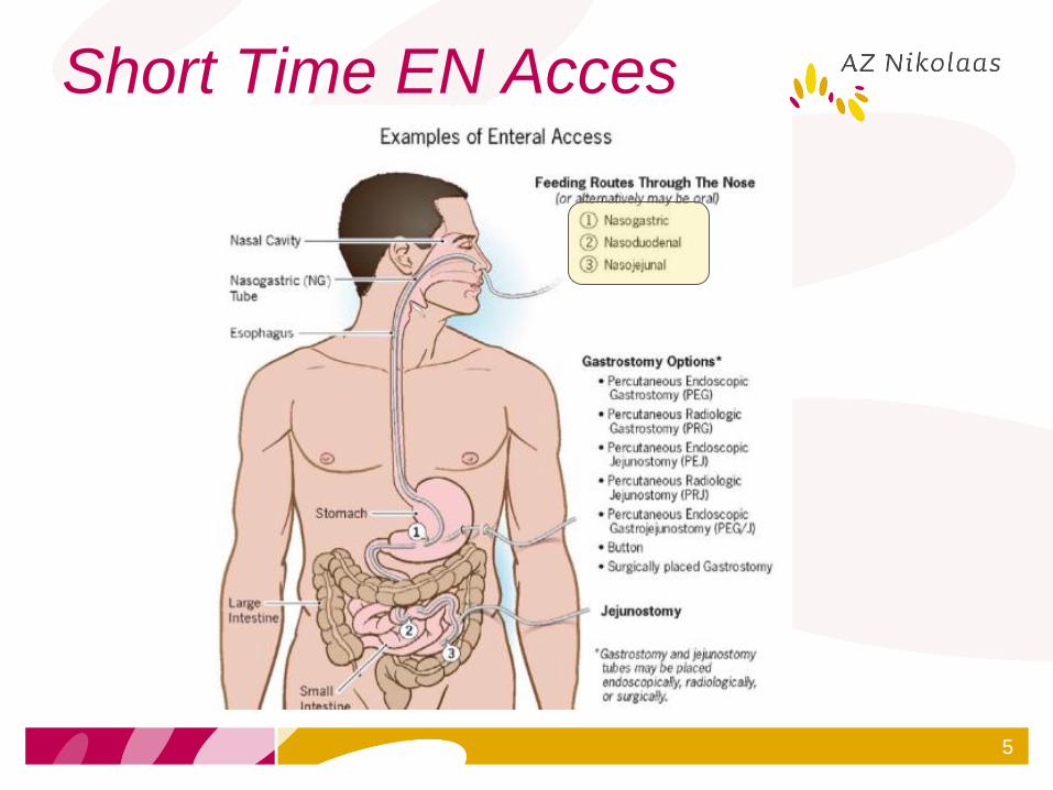
Short Time EN Acces
5

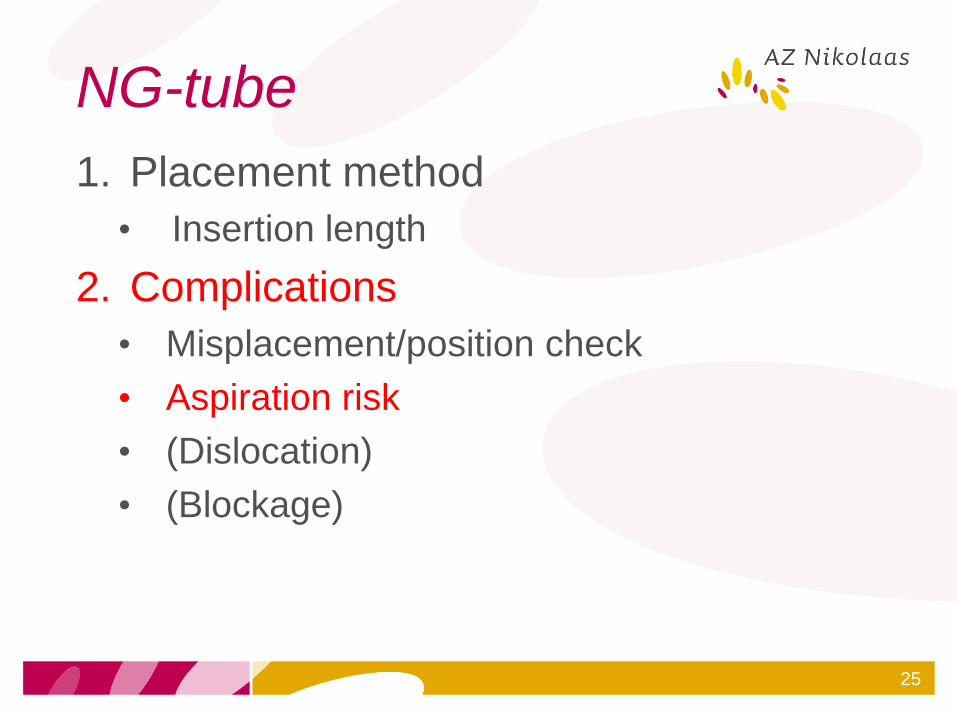
NG-tube
1. Placement method
• Insertion length
2. Complications
• Misplacement/position check
• Aspiration risk
• (Dislocation)
• (Blockage)
6

7
XEN NEX

• Four studies found that NEX was most
likely to result in a tube that is positioned
incorrectly, either ending in the
esophagus, in the stomach but too close to
the esophagus, or too far into the stomach
or duodenum.
8
2016

Hanson study 1979
• 99 cadavers
• 5 volunteers
• NEX: 72 % correct
• NEX + Formulas: 91,3% till 92,3%
correct
Hanson RL. Predictive Criteria for Length of Nasogastric Tube Insertion
for Tube Feeding. JPEN. 1979;3:160-63.
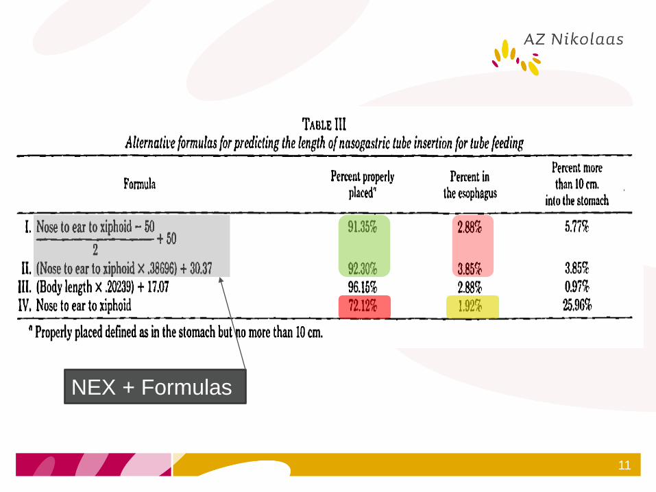
11
NEX + Formulas

12
NEX
2018
HANSON

Hanson + 6cmNEX
Hanson
NEX + 6
CONEX

14
Submitted for publication
CoNEX
78% aspirate

15

16
STEP 1: NEX
STEP 2: CONEX

NG-tube
1. Placement method
• Insertion length
2. Complications
• Misplacement/position check
• Aspiration risk
• (Dislocation)
• (Blockage)
17

Enteral feeding tube misplacements
• Pennsylvania (US)
• Report (2011-2016)
• 166 NGT misplacements
• 60-89 years: 68,7%
• 0-11 years: 6,6%
• 56% serious harm (2 deaths)
• 81 X-rays: 16 misread
18
2018

• 15 published case reports
• 4 children died
• The auscultatory method failed to detect
malpositioned tubes in all seven cases
were it was used
19
2014

Do not rely on the auscultatory method
alone to differentiate between gastric
and respiratory placement or between
gastric and small bowel placement.
20
2017

21

pH-method
22

• At 5.5, the pH test lacks sensitivity towards
oesophageal placements, a major risk
identified by feeding experts
• Under cut-off 5, respiratory feeding was
excluded; oesophageal feeding was kept to
a minimum
23
2018

24
NG-tube
1. Placement method
• Insertion length
2. Complications
• Misplacement/position check
• Aspiration risk
• (Dislocation)
• (Blockage)
25

• Prospective-multicentre
• Pepsin-positive tracheal secretions (a
proxy for the aspiration of gastric content)
• N = 6000 tracheal secretions
• 31,3 % positive
• Pneumonia on day 4 vs NOT:
• 42,2% vs 21,1% (P < 0,001) pepsine-pos!
26
2006

RISK ASPIRATION RISK PNEUMONIA
Low backrest elevation
(p = .024)
Low backrest elevation
(p = .018).
Vomiting
(p = .007)
Aspiration
(p < .001)
Gastric feedings
(p = .009)
Use of paralytic agents
(p = .002),
Glasgow Coma Scale score <9
(p = .021)
High sedation level
(p = .039).
Gastroesophageal reflux disease
(p = .033).
27

• 14 trials
• Gastric vs post-pyloric
• N = 1109
• Moderate-quality evidence of a 30% lower
rate of pneumonia
• No sufficient evidence to show that other
clinically important outcomes such as
duration of mechanical ventilation,
mortality and length of stay are influenced28
2015

• Aspiration as compared with the stomach:
• First portion duodenum: 11.6% lower
• Second/third part: 13.2% lower
• Fourth part and beyond: 18.0% lower
• Pneumonia occurred less often when feedings
were introduced at or beyond the second portion
of the duodenum
29
2011

Nasojejunal (duodenal)
• Blind
• During open surgery
• Endoscopically
• 1,2-3 lumens
• Electromagnetic-guidance
• (Cortrak®)
30

Nasojejunal (duodenal)
• Blind
• Endoscopically
• During open surgery
• Electromagnetic-guidance
• (Cortrak®)
31

• Bedside EM-guided placement of naso-enteral
feeding tubes appears to be as safe and effective
as fluoroscopic or endoscopic placement.
• Placed by nurses
32
2015
2016
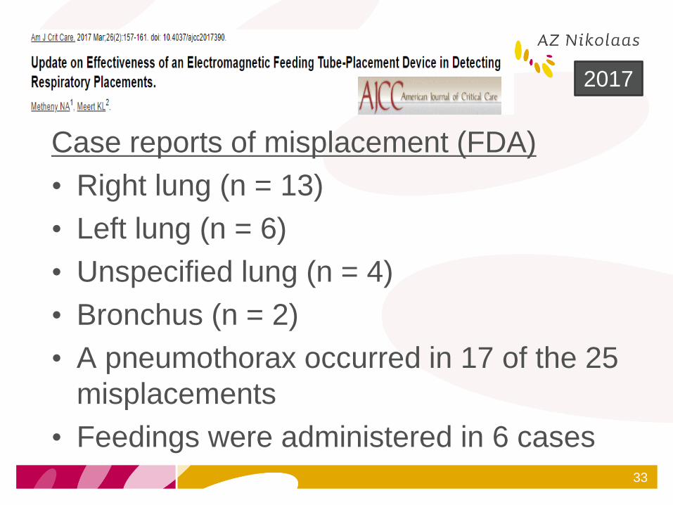
Case reports of misplacement (FDA)
• Right lung (n = 13)
• Left lung (n = 6)
• Unspecified lung (n = 4)
• Bronchus (n = 2)
• A pneumothorax occurred in 17 of the 25
misplacements
• Feedings were administered in 6 cases33
2017

• 2006-2016: 54 adverse events
• Lung placement: 98% (LL 46%)
• Pneumonitis: 21%
• Death in of 17% lung placements
• 89% of clinicians failed to detect
malpositioned insertion tracings reviewed
34
2017
EXPERIENCE-COMPETENCY!!

Long Time EN Acces
35

ESPEN HEN-guidelines
• A PEG should be preferred over a surgical
gastrostomy mainly due a lower complication
rate, cost-effectiveness and operating time.
Grade of recommendation B
• If a PEG if not suitable a percutaneous
laparosocpic assisted gastrostomy
(PLAG) may be a safe alternative.
Grade of recommendation 0
36

• N = 735 (11 RCT)
• PEG: less intervention failure
• No other significant differences in
important outcomes
• But scarce detailed patient characteristics
in studies (underlying disease, placement
technique,….)
37
2015
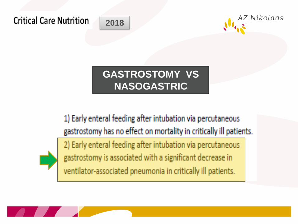
2018
GASTROSTOMY VS
NASOGASTRIC

QOL (NG vs PEG)
• Inconvenience,
• Discomfort
• Altered body image
• Social activities
39
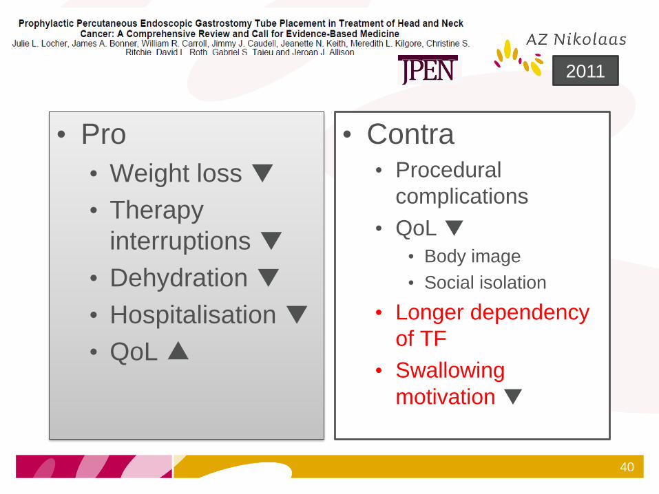
• Pro
• Weight loss ▼
• Therapy
interruptions ▼
• Dehydration ▼
• Hospitalisation ▼
• QoL▲
• Contra• Procedural
complications
• QoL▼• Body image
• Social isolation
• Longer dependency
of TF
• Swallowing
motivation ▼
40
2011

41
2003-2010
22 serious incidents:
- 11 patients died

Incidents reported
• 9 cases of leakage of feed into the peritoneal cavity
and/or peritonitis
• 2 of colonic puncture
• 1 related to haemorrhage
• 1 involving both haemorrhage and colonic puncture;
• 1 septic shock secondary to aspiration
• 1 of leakage of feed into thoracic cavity
• 1 of surgical emphysema
• 6 of unclear mechanism
42

43
2017
2007
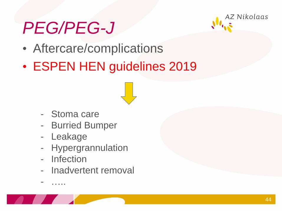
PEG/PEG-J • Aftercare/complications
• ESPEN HEN guidelines 2019
44
- Stoma care
- Burried Bumper
- Leakage
- Hypergrannulation
- Infection
- Inadvertent removal
- …..

Overgranulation
• Prolonged stimulation of fibrous tissue
and new blood vessels (angiogenesis)
• Excess friction/movement a/o moisture
• Infection

• Pain and bleeding
• Treatment: silver nitrate application

Treatment
• Dermal corticosteroid cream, ointment
• (With antibiotics)
• (Antimicrobial) aliginate,foam (+ silver)
dressing to keep modest pressure on the skin
around the stoma and minimize movement.

• Hypergranulation = oedematous tissue
• N = 8 (paediatric patients)
• Daily 1/3 of 5 ml teaspoon salt ‘sprinkled’ over
the tissue
• Treatment period: 3 days-2 months
• Positive result in all patients but 5/8 recurrence
but successfully repeated treatment afterwards
• 1 skin erosion: salt irrigated 10 minutes after
application
48
2013
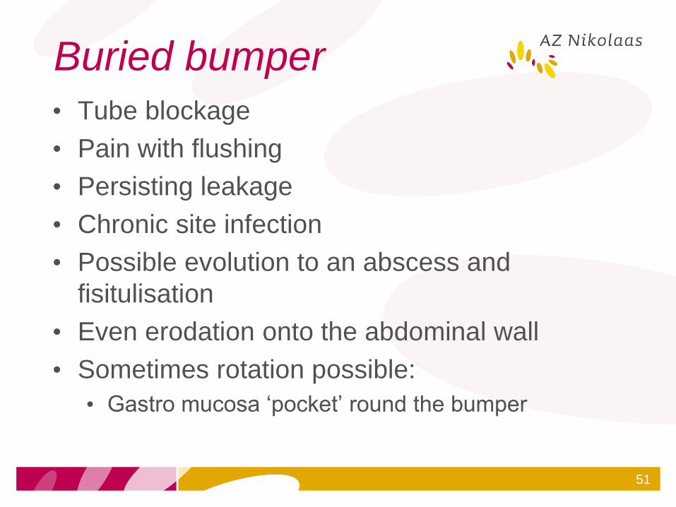
Buried bumper
• Tube blockage
• Pain with flushing
• Persisting leakage
• Chronic site infection
• Possible evolution to an abscess and
fisitulisation
• Even erodation onto the abdominal wall
• Sometimes rotation possible:
• Gastro mucosa ‘pocket’ round the bumper
51

Burried bumper
• Serious but preventable complication
• Normally a long-term complication
• Aftercare!
• Release external bumper
• Clean PEG-site
• Advance PEG into abdomen minimum 2-3 cm
but ideally 5 to even 10 cm (at least once a
week!)
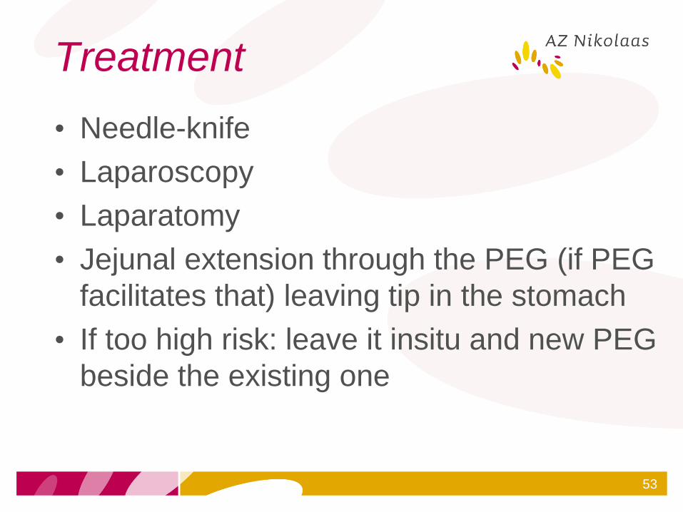
Treatment
• Needle-knife
• Laparoscopy
• Laparatomy
• Jejunal extension through the PEG (if PEG
facilitates that) leaving tip in the stomach
• If too high risk: leave it insitu and new PEG
beside the existing one
53
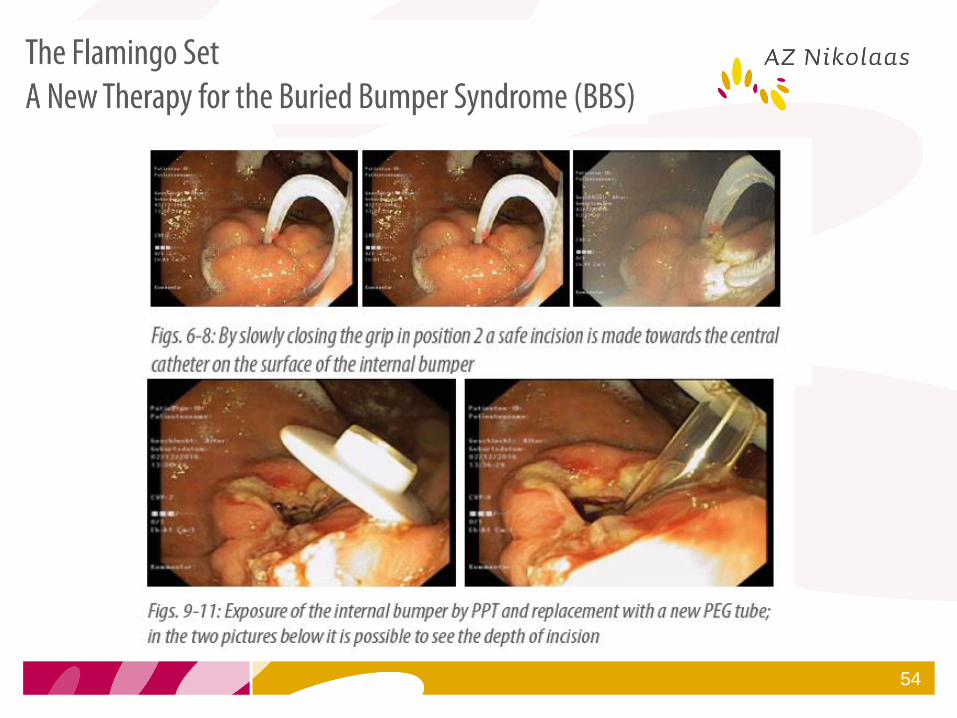
54

TO CLOSE

56
57

91