
Tuberculous Pleural Effusion - · PDF filecal processes involved in TB-pleuritis appears to...
26
Chapter 15 Tuberculous Pleural Effusion Wolfgang Frank Additional information is available at the end of the chapter http://dx.doi.org/10.5772/54955 1. Introduction Tuberculosis (TB) has traditionally been one of the major causes of pleural disease and until the earlier decades of the past century held as a principal paradigm of “pleuritis”. Indeed in the presence of a distinctly exudative effusion and a compatible clinical presentation the widely used term “pleuritis exudativa” insinuated a tuberculous aetiology and has therefore been understood to be synonymous with “pleuritis exudativa tuberculosa”. Whilst in the era of TB decline in the Western hemisphere the term “pleuritis exudativa” (which actually is a tautol‐ ogy!) has largely survived but should now describe exudative effusions in general, the full and precise term “pleuritis exudativa tuberculosa” is therefore suggested whenever the possibility of a tuberculous background is addressed. Otherwise the term “tuberculous pleurisy” or “tuberculous pleuritis” is used to describe this entity, in some countries also the term “specific pleurisy” is common. Apart from acute pleuritis exudative tuberculosa, TB of the pleura may however rarely present as a rather chronic disease state in terms of caseous pleurisy or specific (i. e. tuberculous) empyema, respectively. The following chapter reviews the different features and mechanisms of tuberculous pleural involvement as well as their diagnostic and thera‐ peutic implications. 2. Epidemiology In many regions of the world tuberculous effusion maintains its role as the leading inflam‐ matory pleural disease. With the worldwide unabated HIV epidemic and related immune deficiency syndrome this state of affairs is likely to continue or being even aggravated within least in certain high risk populations. On a global scale the current significance of human immunodeficiency virus (HIV)-co-infection may be illustrated by WHO data, indicating at a TB-prevalence of 1/3 of the world´s population – similar to the past decade – a HIV-association © 2013 Frank; licensee InTech. This is an open access article distributed under the terms of the Creative Commons Attribution License (http://creativecommons.org/licenses/by/3.0), which permits unrestricted use, distribution, and reproduction in any medium, provided the original work is properly cited.
Transcript of Tuberculous Pleural Effusion - · PDF filecal processes involved in TB-pleuritis appears to...
Chapter 15
Tuberculous Pleural Effusion
Wolfgang Frank
Additional information is available at the end of the chapter
http://dx.doi.org/10.5772/54955
1. Introduction
Tuberculosis (TB) has traditionally been one of the major causes of pleural disease and untilthe earlier decades of the past century held as a principal paradigm of “pleuritis”. Indeed inthe presence of a distinctly exudative effusion and a compatible clinical presentation the widelyused term “pleuritis exudativa” insinuated a tuberculous aetiology and has therefore beenunderstood to be synonymous with “pleuritis exudativa tuberculosa”. Whilst in the era of TBdecline in the Western hemisphere the term “pleuritis exudativa” (which actually is a tautol‐ogy!) has largely survived but should now describe exudative effusions in general, the full andprecise term “pleuritis exudativa tuberculosa” is therefore suggested whenever the possibilityof a tuberculous background is addressed. Otherwise the term “tuberculous pleurisy” or“tuberculous pleuritis” is used to describe this entity, in some countries also the term “specificpleurisy” is common. Apart from acute pleuritis exudative tuberculosa, TB of the pleura mayhowever rarely present as a rather chronic disease state in terms of caseous pleurisy or specific(i. e. tuberculous) empyema, respectively. The following chapter reviews the different featuresand mechanisms of tuberculous pleural involvement as well as their diagnostic and thera‐peutic implications.
2. Epidemiology
In many regions of the world tuberculous effusion maintains its role as the leading inflam‐matory pleural disease. With the worldwide unabated HIV epidemic and related immunedeficiency syndrome this state of affairs is likely to continue or being even aggravated withinleast in certain high risk populations. On a global scale the current significance of humanimmunodeficiency virus (HIV)-co-infection may be illustrated by WHO data, indicating at aTB-prevalence of 1/3 of the world´s population – similar to the past decade – a HIV-association
© 2013 Frank; licensee InTech. This is an open access article distributed under the terms of the CreativeCommons Attribution License (http://creativecommons.org/licenses/by/3.0), which permits unrestricted use,distribution, and reproduction in any medium, provided the original work is properly cited.

of approximately 13 % by the year 2009 [1, 2]. Conversely it is assumed, that 33 % to 50 % ofHIV infected individuals are co-infected with M. tuberculosis [2]. The MTB/HIV-associationhowever shows a huge intercontinental and regional variance, with the highest rate of HIV-pleural tuberculosis-coincidence being reported in Zimbabwe where 95 % of Patients withtuberculosis pleurisy were HIV positive [3]. In Burundi and Tansania a HIV-coinfection wasfound in 60 % of all cases of tuberculous pleurisy [4]. One of the lowest rates is reported fromSpain with 10 % [5]. Am example of the impact of a high HIV-endemic environment on theincidence of tuberculous pleurisy is also given in a series from Ruanda, where TB accountedfor as much as 86 % of all diagnosed pleural effusions [4]. Pleurisy incidence obviously andessentially parallels variability of global TB prevalence with an overwhelming share of 95 %occurring in developing countries. In TB-patients as a whole, pleural involvement variesbetween ~ 3-5 % in Western Europe and the USA vs. ~ 30 % in developing, HIV-high-preva‐lence-countries [6, 7, 8]. The differences clearly underline the modifying role of immunologicaldeterminants, stage and severity of the disease, general health status and nutritional factors.The effect of HIV on the occurrence of pleural involvement in a given TB-patient is illustratedby a study reporting a 38 % pleurisy incidence in AIDS-associated TB as compared with 20 %in matched HIV-negative TB patients [5]. On the basis of the presented data according to evenconservative WHO estimates the TB-pleurisy incidence throughout the current decade isexpected to remain grossly unchanged compared to the past decade, i. e. 18.2 – 62/100.000 inthe developing countries vs. 0.42-0.77/100.000 in Western countries [6, 7, 10]. When theepidemiology of pleurisy (or pleural effusion in general) is analysed in terms of the magnitudeof TB-contribution, a probably still valid estimate in Western countries is as low as 0.1 – 0.2 %and remains distinctly < 1 % even when referring to pleurisy in a strict sense (i. e. exudates)[11]. By comparison the previously reported percentages of 30-86 % in developing countriesare – and remain – indeed dramatically different.
3. Pathophysiology and natural history
3.1. Immunological and microbiological factors
MTB may affect the pleura at different stages of pulmonary or systemic disease and by anumber of different mechanisms. Thus pleural involvement occurs in primary, postprimaryand reactivated TB alike and is basically believed to arise directly from contiguous macroscopicor microscopic lung lesions or else lymphogenic or hematogenic spread, but probably also viaimmunogenic mechanisms. Pleuritis exudativa tuberculosa is by far the most common clinicalvariety and has been classically interpreted as an early delayed-hypersensitivity-type phe‐nomenon rather than direct organ involvement [12, 13]. Many clinical observations andexperimental findings are in favour of this hypothesis such as:
• its frequent association with known primary infection and a typical 6-12 weeks latency,
• an often striking absence of significant pulmonary or systemic TB-lesions,
• an often culturally negative or paucibacillary effusion [14],
Tuberculosis - Current Issues in Diagnosis and Management290

• the sometimes abundant isolation of specifically purified protein derivative (PPD)- proteinsensitized T-lymphocytes from pleural fluid [15] and
• more recently the inducible pleurisy in previously PPD-sensitized animals when exposedto intra-pleural mycobacterial protein.
Also the vigorous expression of inflammatory mediators interleukins (IL) like interferon (IFN)γ, IL-1 and IL-8 observed in this model (or conversely their suppression by antilymphocyteserum) support this view [16, 17].
On the other hand there is also strong evidence, that infectious invasion of the pleural spaceactually occurs at a substantial, albeit variable degree. At thoracoscopy, even with negativefluids studies, extensive inflammatory granuloma formation and fibrin deposits with unex‐pected abundant mycobacteria recovery are a common finding (see also section on invasiveendoscopic-bioptic studies) [18]. The increasingly emerging evidence of a preferred associationof TB-pleurisy with reactivated TB in Western populations clearly points to infectious as wellas immunological mechanisms being interrelated and operative in a complex manner. Directinfectious invasion however clearly prevails in chronic tuberculous involvement of the pleuraas in specific empyema.
According to present views and based on experimental evidence the sequence of immunologi‐cal processes involved in TB-pleuritis appears to follow a three stage pattern of cellular reactionsand granuloma formation as a topic variant of general interaction mechanisms between MTBand the human immune system. A schematic representation is given in figure 1 [19, 20].
Any trigger-mechanism that allows access of mycobacterial protein to the pleura will set off arapid mesothelial cell initiated and IL-8 mediated polymorphonuclear neutrophil (PMN)influx within a few hours [21]. In addition macrophages and blood-borne monocytes deter‐mine this IL-1, IL-6 and tumor necrosis factor (TNF)-α-orchestrated early stage reaction.
Within roughly 3 days, in the following intermediate stage lymphocyte subpopulations, mainlyof CD4+ helper cells but also a substantial CD8+ cytotoxic (natural killer cells) fractiondominate the scene resulting in a CD4+/CD8+-ratio of ~ 4.3 [22]. A minor contribution includesso-called T-cell receptor double negative (DN) αβ-T-cells and γδ-T-cells which appear to haveregulatory functions. More recently in tuberculous pleural fluid another unique CD4+CD25+T-cell-class could be demonstrated being specifically involved in the down-regulation of auto-reactive IFN-γ-producing T-cells, thus preventing inflammatory overshoot [23]. IFN-γ a strongpromoter of macrophage activation and granuloma formation (together with TNF-α) is thepredominant interleukin in this stage. IFN-γ-producing cells have been phenotypicallyindentified as CDW29+ subpopulation and make up a substantial portion of the granulomacore structure [24].
The late phase is characterised by an equilibrated and sustained CD4+/CD8+ cell-basedresponse with continued IFN-γ release and prolonged granuloma formation. Severalmodulating interleukins are involved in this process such as T-helper-cells (CD4+)-support‐ing IL-12 and counter-regulatory antiinflammarory cytokines like IL-10 and transforminggrowth factor (TGF-β).
Tuberculous Pleural Effusionhttp://dx.doi.org/10.5772/54955
291

Results of HIV and AIDS research also emphasize the importance of T-cell response. Severalworking groups have shown, that the prevalence of tuberculous pleurisy in HIV-infected patientswith TB is strikingly correlated with their CD4+ blood lymphocyte count. In one series pleuri‐sy prevalence in individuals with a count of > 200 cells/ml was 27 % as compared to 10 % in thosewith a count of < 200 cells/ml [25]. The data support the view, that the clinical expression ofexudative pleural effusion requires a largely intact cellular immune system and features pleurisyas a high activity response in a still immunocompetent individual. In epidemiologic terms onewould conclude that pleural effusion should be more frequent in the still immunocompetenthost than in patients with AIDS. In reality however in most HIV-high-prevalence countries likeSouth Africa, Uganda and Zimbabwe the percentage of thoracic TB-patients with pleural effusionis reportedly higher in HIV+ patients [26]. As an explanation the situation is probably blurredby a variable and poorly defined immune status in HIV+ individuals.
Figure 1. Mechanisms and immunogenesis of tuberculous pleurisy: the three stages of protective immune response.IFN: interferon; TNF: tumor necrosis factor; IL: interleukin; PMN: polymorphonuclear granulocyte; X: undefined cell;MΦ: macrophage, MTB: mycobacterium tuberculosis; PPD purified protein derivative
Tuberculosis - Current Issues in Diagnosis and Management292

3.2. Other factors
The mechanisms of fluid accumulation and of abundant protein leakage to the pleura withoften extensive fibrin deposits in tuberculous pleurisy have so far not been fully elucidated.In actual fact pleuritis exudativa tuberculosa generally presents with the highest protein levelscommonly seen in exudative conditions. The intensity of inflammation and a proportionatelyincreased vascular permeability would provide a satisfying explanation [25, 27] although atleast in animal models, no such significantly altered vascular permeability could be demon‐strated [12]. Current opinion holds that grossly impeded lymphatic protein clearance from thepleura due to altered parietal lymphatic channels is probably of tantamount importance.
Again the entry mechanism of mycobacteria to the pleura has remained unclear. It is usuallyassumed that the release of infectious material from a ruptured subpleural TB-lesion is themost common mechanism. While this is likely to occur in more or less extensive pulmonaryTB, it would not explain the frequent association of tuberculous pleuritis with an – at leastradiographically – unaffected lung. There are also no convincing data yet to quantify thecontribution of a purported hematogenous or lymphogenous contribution. One mightreasonably speculate that different patterns of pleural tuberculous involvement are operativewhich might correspond to the different clinical settings of primary, post-primary andreactivated TB.
Caseous tuberculous pleuritis or specific empyema is nowadays a rare condition which is believedto be the result of longstanding or chronic infection of the pleura, when either caseous materialgains access to the pleura or chronic pleuritis develops on the background of impaired localdefence such as pre-existing fibrous damage of the pleura or as a sequel and complication ofartificial pneumothorax, oleothorax or other TB-specific surgery dating back to the pre-chemotherapy era. Correspondingly there is usually an extremely long history often with aremarkable paucity or even absence of symptoms. Penetration to deeper chest wall structures(specific abscess) and ultimately transcutaneous discharge (empyema necessitans) or creationof a specific bronchopleural fistula, as not infrequently seen in the pre-chemotherapeutic era,may complicate this condition [27]. Putrid discharge from a thoracic mass or putrid expecto‐ration with or without haemoptysis may ultimately advert to the condition.
4. Clinical manifestations and natural course
Tuberculous pleurisy may occur as an acute, subacute or rather chronic disease. At times thecourse is also surprisingly oligosymtomatic. Therefore duration of symptoms or major illnessprior to hospital admission and diagnosis varies considerably from < 1 week (31 %) to < 1months(62 %) or even longer (7 %) [28]. These data refer to the pre-HIV era and would not apply for HIV-seropositive patients and elderly populations, which both tend to have a particularly longsymptomatic or else oligosymtomatic period. An infectious, i. e. febrile illness is nevertheless byfar the most common clinical presentation. As a general rule, an acute febrile illness is the morelikely to occur the younger and the more immunocompetent a given patient is. In developing,high-prevalence and high primary-TB-affected countries the age peak of incidence is in the mid
Tuberculous Pleural Effusionhttp://dx.doi.org/10.5772/54955
293
thirties, whereas in industrialized countries with a major contribution of reactivated TB it hasshifted to about 50 yrs [29]. But still the age-related incidence peak of tuberculous pleuritis isdistinctly lower than of parenchymal pulmonary TB which used to peak around 55 yrs [30].Implicitly by the same statement in Western populations TB-pleurisy was historically moresymptomatic than is currently the case. In a representative series from the 1960-1970s ~ 60 % ofpatients developed an acute illness mimicking bacterial (pleuro)-pneumonia with cough (70 %),chest pain (75 %) and low- to high-grade fever (86 %) as the most frequent symptoms [31, 32].Other symptoms include those commonly occurring in various TB disease states such as weightloss, malaise and night sweat. Severe or even live threatening disease, defined as persistent high-grade fever > 38,3°C over > 2 weeks or respiratory distress has been in reported in a more recentseries in only 7 % [31], whereas an oligosymptomatic or a febrile course is described in 14-33 %[32]. Tuberculous pleurisy usually involves one hemithorax only (90-95 %) and is of limited size(roughly up to one-half of the hemithorax volume). In a major series (n=254) effusions occupy‐ing more than 2/3 of a hemithorax were noted in only 18 % [33]. Rarely effusion will occupy theentire hemithorax and will almost never reveal compressive or displacing features [31]. Basicallythere are no specific clinical clues to tuberculous etiology in pleurisy unless some TB-contact isrevealed or suspected. An HIV-related background may be suspected in a compatible clinicaland history setting or when there is a long preclinical period, unusual additional symptoms likediarrhea and more hepato(spleno)-megaly or lymphadenopathy as might be attributed to thetuberculous condition. Untreated, lone pleuritis exudativa tuberculosa in the short term seemsto be a self-limited inflammatory process in most instances, terminating in complete or incom‐plete resolution within weeks or month. Frequently observed otherwise unexplained diaphrag‐matic adhesions may be a late sequel of clinical silent or oligosymtomatic TB pleurisy. Importantlyhowever progression or reactivation to active pleuropulmonary or extrapulmonary TB occursin an important fraction. In one follow-up study the recurrence rate within 1 year was 5 %, whereTB did not relapse earlier than 8 month after the onset of pleurisy. Within a 4-5 yr period howeverthe rate was dramatically higher and in initially culture positive and culture negative subjectswith 65 % and 60 % respectively roughly alike [27]. One major outcome determinant clearly isthe presence and the extent of pulmonary involvement. At a similar therapeutic intensity in avery recent major clinical study from Taiwan, 51 (24,9 %) out of 205 hospitalised patients havingbeen identified to have isolated (lone) pleuritis had a significantly better outcome, shorter hospitalstay and less comorbidity than the patients with pleuropulmonary disease [34].
5. Diagnosis
5.1. Clinical findings
5.1.1. Signs at physical examination
Physical examination clearly will provide only non-specific signs of pleural effusion in generalincluding dullness to percussion and the occasional demonstration of a pleural rub at auscul‐tation (“snow-ball-crunching sign”) in particular in the presence of chest pain. Signs of atrapped or loculated rather than free flowing fluid collection may suggest a tuberculous
Tuberculosis - Current Issues in Diagnosis and Management294

aetiology, but this observation holds also true for “plain” parapneumonic pleurisy. Usual signsof systemic infection, as mentioned above, that should be looked for, may alert to the possibilityof a HIV-related background.
5.1.2. Imaging studies
Imaging techniques are engaged in the evaluation of tuberculous pleurisy following generaldiagnostic pathway recommendations for effusion. Conventional chest radiography (CRX) requiresfluid amounts of at least 150 ml to become clearly detectable as blunting of the costodiaphrag‐matic angle in standard projections. Profuse effusion with opacification of an entire hemithor‐ax would rather favour differential diagnoses like malignancy in the elderly and afebrile patient[35]. Free flowing effusion may be easily identified, but one should look specifically for signs ofloculation, pleural thickening or adhesions and in profuse effusion for compressive signsinterfering with the respiratory performance. Apart from pleural changes pulmonary infil‐trates, nodules, lymphnodes and other suggestive signs of TB like encapsulated or cavitarylesions must be carefully looked for using routine CT-imaging. CT-based prevalence of lungperenchymal tuberculous lesions in mixed populations appears to be significantly higher thanpreviously assessed based on conventional radiography. In one recent series from Koreacomprising 106 patients with an age distribution from 16-89 yrs (mean 53) with 86% a remarka‐ble high rate of parenchymal changes was found, presumed to represent active tuberculosis in59 % [36]. Most of these lesions revealed features of reactivated rather than primary tuberculo‐sis. Sonography (Ultrasound, US) using innovative technical achievements like high frequency(5-7.5 MHz) – US and convex or sector scanners allow extended exploration of the chest wallstructures, the diaphragm and the anterior mediastinum up to a penetration depth of ~ 25 cm.Specific advantages of US are a more precise fluid volumetry than by CRX, precise localisationof septae, membranes and chambers as well as pleural thickening along with its particularversatility for bedside diagnosis. On demand guidance for interventions such as thoracentesisis a particular asset of US.. Examples are shown in figure 2, 3. Magnetic-resonance imaging (MRI)is a highly refined, not generally available technique, which will rarely be required but does havedifferential diagnostic merits in the analysis of critical borderline relationships i. e. distinguish‐ing between inflammatory- infiltrative and malignant-destructive pleural processes via differentT-weighted sequences [37]. Very recently a role of PET-CT has also been described. PET-imaging may indeed provide differently extensive focal and impressing laminar changes whichhowever remain indistinguishable from malignant lesions [38].
5.2. Immunologic tests
5.2.1. Tuberculin skin reaction
The tuberculin skin reaction is traditionally considered an indispensable tool in the diagnosisof tuberculosis in general and likewise in tuberculous pleurisy although it is less reliable thanin pulmonary TB. The rate of false negative reactions to PPD has been given as high 30 % ofcases but even figures up to < 41 % have been reported [31, 32, 33], the variability possiblyreflecting non-standardised test doses. Still however there remains an amazing false negativerate. There is no absolutely satisfying hypothesis to explain this paradoxon, let alone unequiv‐
Tuberculous Pleural Effusionhttp://dx.doi.org/10.5772/54955
295

Figure 3. Ultrasound detection of multiple chambers in pleuritis exudativa tuberculosa
Figure 2. Ultrasound detection of inflammatory visceral membranes and consecutively trapped lung in pleuritis exu‐dativa tuberculosa
Tuberculosis - Current Issues in Diagnosis and Management296

ocal experimental evidence. It appears a valid speculation to consider a local pooling ofsensitised T-lymphocytes at the site of infection responsible. Animal experiments and clinicalinvestigations have shown sequestration of PPD-sensitized lymphocytes to the pleuralcompartment actually to occur in the early phase of infection leading to their systemicdepletion [39]. As a presumptive additional mechanism the presence of adhering suppressorcells to blood lymphocytes has been demonstrated in PPD-anergic patients [39]. While theexplanatory evidence may remain scanty, it should be emphasised that in clinical practice thephenomenon appears to be transitory and restricted to the early phase of tuberculous infection.It might thus be associated with the pre-allergic phase of tuberculous infection, since conver‐sion of skin reactivity has been subsequently observed within a 6-8 weeks delay [27]. As areverse conclusion in the framework of discussed hypotheses the observation of a delayedPPD-conversion might be interpreted as a clue to primary infection to have occurred. Persistinganergy would then point to other immune-modulating factors like advanced age, certain druginterference or immune-compromising comorbidity.
5.2.2. Interferon-γ-release assays
In Europe commercially available Interferon-γ-release assays (IGRA) are the QuantiFERON-TB-Gold-Test and the T-Spot-TB-Test. Both use the MTB-RD1-region antigen sequences CFP10and ESAT 6 and measure the specific lymphocyte-induced quantitative IFN-γ-response or thesensitized IFN-γ-producing lymphocyte response, respectively. There has been elaborated abody of clinical data in practical use highlighting both the assets and pitfalls of the investiga‐tion. In summary and in general there is distinct superiority to the PPD-skin-test with an overallsensitivity of ~ 85 % and a high specificity well > 90 % [40]. The concordance of the PPD-testand IFN-γ-release assays is in the order of 60-85 % [41]. However in the identification of activeclinical tuberculosis blood-based IFN-γ-release assays also have revealed a considerable rateof false negative findings. In several studies including pulmonary a well as pleural tubercu‐losis, sensitivity was limited to 60-64 % [42, 43, 44]. There have also been a number of incon‐sistent and equivocal reports where the results obviously vary with different TB-prevalencesettings (i. e. pretest probability). In a number of studies the variability of sensitivity rangesbetween 96 % in low prevalence settings down to 58 % in studies featuring high prevalenceareas, also specificity setbacks are reported [45]. Blood-based IGRA´s therefore seem to sharethe limitations of PPD-testing. Since they cannot distinguish latent from active TB, in conclu‐sion, the diagnostic value for identification of tuberculous pleurisy in high prevalence settingsis very low and has even only limited value in industrialized countries.
5.3. Pleural fluid analysis
5.3.1. Biochemical parameters
When there is enough effusion to allow safe puncture and TB is suspected, thoracentesis is amandatory diagnostic step. The effusion will be invariably and markedly exudative with a(unless in tuberculous empyema) clear, straw- to amber-coloured appearance and a meanprotein content above 5.0 g/dl, in one series (n=83) it was 5.2 g/dl (range 3.5 – 7.0) [32]. Glucoseand pH-values have traditionally believed to be characteristically low in TB. It appearshowever, that on the basis of more recent data, as also confirmed in the author´s own experi‐
Tuberculous Pleural Effusionhttp://dx.doi.org/10.5772/54955
297

ence these values are not substantially different from exudates due to other aetiologies. SAHN[46] found pH-values < 7.29 and glucose values < 30 mg/dl in only 20% of patients and this hasbeen confirmed by others [47]. Interestingly however, if low values actually occur, they appearto correlate with the pleural bacillary load and are to some extent predictive of cultural results.In one thoracoscopic study positive pleural fluid culture yield was 59 % when the glucose levelwas < 50 mg/dl but only 25 % when the glucose values were > 50 mg/dl (p<0.005) [18]. Lacticdehydrogenase (LDH) is a non-specific marker of pleural inflammation, which may beexcessively elevated in tuberculous pleurisy, although with a mean value of 423 IU/ml (range43 – 1.575) as reported in a representative series again does not discriminate TB from para‐pneumonic and not even from malignant effusion [32]. Adenosine deaminase (ADA) has beena promising and much hailed semispecific biochemical parameter. ADA is an inflammatoryenzyme expressed predominantly by sensitized and activated T-lymphocytes. Isoenzymes(ADA2) in addition reflect to some extent monocyte/macrophage activation. Thus increasedADA-activity in general indicates various T cell/macrophage interactive inflammatoryprocesses like granulomatous disease but also empyema and collagen vascular disease. Itappears however particularly sensitive to TB. In a key study (n=129) in patients < 35 yrs areceiver operating characteristics (ROC) –derived cut-off level of 47 U/ml allowed distinctionof tuberculous effusion from empyema, rheumatic and neoplastic disease with a 100 %sensitivity and 87.5 specificity. When empyema was eliminated, specificity and the positivepredictive value even attained 100 % [48]. There are important limitations to the interpretationof these results and their clinical relevance:
• the data reflect the afore mentioned age group only, in more heterogenous groups bothsensitivity and specificity have to be (down)-corrected to 95 % and 90 % respectively [22,49, 50].
• the results strictly apply to high TB prevalence settings only and do not allow for differentpre-test prohabilities [3].
• also immune suppression like in AIDS endemic areas may interfere with inflammatoryADA-release and invalidate diagnostic conclusions [3, 51].
Nevertheless, based on the most accepted cut-off level of 40 IU/l and provided its critical usein areas of at least intermediate TB-prevalence ADA determination must be regarded as a truediagnostic enrichment. An era of successful ADA-use has been recently summarized andconfirmed by a large size metaanalysis (63 studies, 5297 tuberculous and non-tuberculouseffusions) resulting in a sensitivity and specificity of 92 % and 90 % respectively [52].
5.3.2. Cytological analysis
Based on the immunological processes involved, a marked lymphocytosis is the predicted andcharacteristic feature of TB-pleurisy along with significantly increased total white cell countsas reflected in one representative study with a mean count of 2.309/mm3 (range 30 – 24.009mm3) [32]. Usually 90 – 95 % of pleural fluid cells are T-lymphocytes, the remainder being B-lymphocytes and (mostly) activated mesothelial cells. Only exceptionally (in ~ 5 %) lympho‐cyte counts < 50 % may occur [27]. Thus when an 80 % lymphocyte reference line is chosen,
Tuberculosis - Current Issues in Diagnosis and Management298

pleuritis tuberculosa exudativa is by far the most frequent cause of pleural lymphocytosis [46].Rarely, in particular in the early phase of inflammation fluid cytology may reveal neutrophilleucocyte (PMN) predominance. Expansion of the eosinophil compartment would be anextremely unusual finding. In the presence of significant numbers of eosinophils (> 5 %)differential diagnoses should be considered.
5.3.3. Microbiological studies
The microbiological yield from diagnostic (low volume) thoracentesis as far the smear isconcerned is very low unless the whole effusion or large amounts are being centrifuged or thepatient has a tuberculous empyema [14, 29]. In HIV positive individuals, particularly in thosewith CD4 cell counts < 200 x 106/l significantly higher yields are being reported amounting inone study to 37 % vs. 0 % in non HIV-patients [53]. In a comprehensive study on microbiologicsmear findings in pleural fluid specimens in non-selected HIV negative out-patients, thepositive acid fast smear yield (n=232) again was actually zero [54]. Cultures should be obtainedboth from the sputum and pleural fluid. The positive cultural yield from pleural fluid has beengiven in collective reviews with 10 – 35 %, being ~ 25 % in the mean [14, 30]. In one of the largestseries (n=100] the sensitivity of pleural fluid culture was 28 % [18, 55]. The use of radiometricor non-radiometric liquid culture systems (BACTEC, MB/BacT, MGIT) will markedly acceler‐ate results and possibly lead to an enhanced yield (~ 50 %), when bedside instead of laboratoryinoculation is used [56]. The yield of sputum cultures in tuberculous effusion is expectedlylargely dependent on the extent and nature of pulmonary involvement and may mount up to~ 50 %. In the non-expectorating patient the use of induced sputum is advised [57]. The positiveyield is also believed to be higher in HIV-infected patients [53, 57]. In the complete absence ofpulmonary lesions according to most sources the sensitivity will be no more than 4-7 % [30].Only exceptionally a surprisingly high figure of 31 % for induced sputum has been reported[57].
5.3.4. Immunological and molecular studies
Immunological studies of pleural fluid in TB-pleurisy focus on the measurement and analysisof chemokins and interleukins that are characteristically associated with the tuberculousimmune response. TNFα and IFNγ revealed at a cut-off 140 pg/ml a sensitivity of 94 % and aspecificity of 85 % [58,60]. Similarly as for ADA the major confounders were bacterial empyemaand parapneumonic effusion respectively. Interestingly TNFα did not attain enough discrim‐inatory power to separate TB from various inflammatory conditions and is no more considereda valid option in the diagnosis of TB. More recent meta analysis-derived collective data from22 studies resulted in an overall sensitivity of 89 % at a 97 % specificity [61]. Thus at presentIFNγ-determination in pleural fluid – contrasting to systemic IGRA-application – wouldappear a useful diagnostic test with a sensitivity and discriminatory power comparable to thatof ADA-determination if one was to accept the significantly higher costs and disregard morepowerful diagnostic options as provided by subsequently discussed invasive biopsy techni‐ques.
Tuberculous Pleural Effusionhttp://dx.doi.org/10.5772/54955
299

Molecular mycobacterial identification methods employing a variety of nucleic acid amplifica‐tion techniques (NAAT) have been applied in TB pleurisy with considerable enthusiasm andexpectations ever since their first application in TB in 1989 [62]. The techniques that have beenused include target amplification (polymerase chain reaction, PCR), strand displacementamplification (SDA), transcription mediated amplification (TMA), probe/primer amplification(ligand chain reaction, LCR) and Q-Beta replicase amplification mostly with the IS 6110, 16Srecombinant ribonucleic acid (rRNA) and 65 XD target sequence [63-66]. So far published data,both biopsy- and pleural fluid-based have shown considerable variance of diagnostic yield,which ranged from 20-81 % as to sensitivity with an expectedly high specificity in the order of98-100 % (table 1). When analyzing the sources of this high variance, apart from technicalfactors like contamination-related “carry over” or amplification inhibitors, the most importantdeterminant appeared to be the number of bacilli in the pleura fluid or specimen sample [31].Although theoretically requiring the presence of merely one microorganism to triggeramplification, similar to sputum anlysis, failed to detect pleural MTB in particular when thepleurisy was paucibacillar, correlating with cultural negativity. In addition to fluid samplesnumerous studies have evaluated the value of various nucleic acid extraction and amplifica‐tions techniques in formalin-fixed and paraffin-embedded pleural tissue specimen [67-71].With the use of commercial kits of both DNA amplicons (ligand chain MTB assay, LCxMTBor AMPLICOR MTB) or RNA amplicons (amplified MTB direct test, AMTDT) according to thelatest currently available sources, the sensitivity of each single technique did not exceed 63.2% albeit at an expected 100 % specificity [71]. The so far largest meta-analysis including 40studies and featuring commercial as well as in-house (“home-brew”) tests, confirms a low andheterogeneous sensitivity (in the mean 62 %) and high specificity of 98 % [72]. Thus there isno convincing evidence, that generally and especially in the critical issue of paucibacillar(cultural negative) pleurisy, NAATs perform substantially better in tissue than in effusionspecimens (table 1). Although in-house assays have been reported to be slightly superior [73],there remain significant sensitivity set backs both in liquid- and tissue-derived specimen. Insummary NAATs may offer certain advantages like quick results within hours or addedspecificity. They may also improve sensitivity in combined and parallel use with conventionalmethods and multiple amplicors (diagnostic confirmation), but can certainly not replace orobviate the need for conventional tools in the diagnosis of TB pleurisy.
5.3.5. Invasive bioptic and endoscopic studies
Bioptic techniques in the evaluation of tuberculous effusions incorporate closed blind orimaging guided needle biopsy and medical (video)-thoracoscopy. Only exceptionally, if ever,surgical diagnostic efforts including video-assisted surgical thoracoscopy (VATS) wouldappear appropriate. Invasive techniques are indicated when clinical investigation and pleuralfluid analysis provide only ambiguous or conflicting results and this is particularly true ifrelevant differential diagnoses like malignancy need to be reliably excluded. Needle biopsymay be considered a first step. There are no clear preferences as to the type of needle to beused, although in the author´s opinion the Tru-Cut or Raja-system may be preferable to theolder Abrams- or Ramel-needle by providing a larger specimen along with easier han‐dling. It is recommended, that at least six biopsies are obtained, since they will not regular‐
Tuberculosis - Current Issues in Diagnosis and Management300

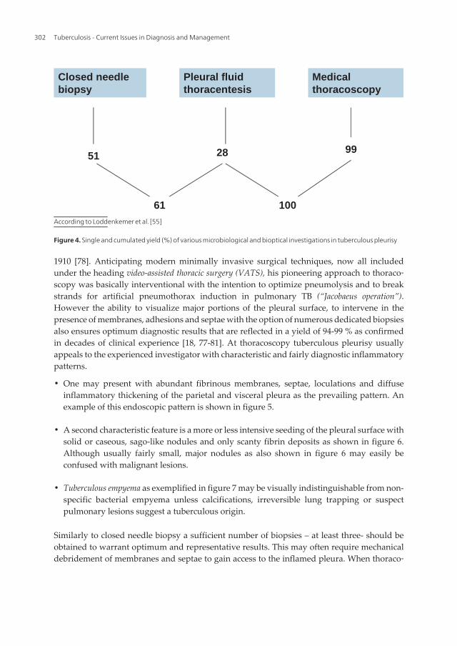
ly contain a representative parietal pleural sample [74]. With this premise and the expectedyield of at least two valid samples closed needle biopsy should be diagnostic in tubercu‐lous pleurisy in ~ 60 % of cases, when histology and tissue-, as well as fluid-culture are beingcombined. In a major series (n=100 %) a 61 % positive yield was composed of 51 % biopsyyield and 28 % positive fluid culture (figure 4) [18, 55]. Distinctly higher yields have alsobeen reported in the literature, leading in a collective review to an average sensitivity of 69% (range 28-88 %) [75]. The difference and wide range is likely to be due to technical disparitiesand inclusion of data originating from largely different prevalence areas. In one study froma high prevalence area (South Africa) comprising 51 patients with undiagnosed pleurisy thepositive closed needle yield in tuberculous pleurisy (histology+AFB-stain+culture) was 79%,when combined with pleural fluid ADA-determination and a lymphocyte/neutrophil ratio >0.75 sensitivity increased to 93% at a specificity of 100% [76]. Thus with the parallel use ofless invasive parameters needle biopsy approaches the diagnostic potency of more inva‐sive techniques and would appear the second best diagnostic option in areas with limitedmedical logistics and resources.
Medical thoracoscopy as a “window to the pleural space” [77] is the gold standard procedure in theevaluation of exudative pleural effusion, hence also pleural pleurisy. The current and futurerole of thoracoscopy needs to be redefined for its diagnostic and interventional efficacy in thelight of its close historical affiliation with TB. In fact tuberculosis was already a major focus ofmedical thoracoscopy or “pleuroscopy” as referred to and initiated by JACOBAEUS back in
Table 1. Role of Nucleic-Acid-Amplification-Techniques (NAAT) in the Diagnosis of Tuberculous Pleuritis
Tuberculous Pleural Effusionhttp://dx.doi.org/10.5772/54955
301
1910 [78]. Anticipating modern minimally invasive surgical techniques, now all includedunder the heading video-assisted thoracic surgery (VATS), his pioneering approach to thoraco‐scopy was basically interventional with the intention to optimize pneumolysis and to breakstrands for artificial pneumothorax induction in pulmonary TB (“Jacobaeus operation”).However the ability to visualize major portions of the pleural surface, to intervene in thepresence of membranes, adhesions and septae with the option of numerous dedicated biopsiesalso ensures optimum diagnostic results that are reflected in a yield of 94-99 % as confirmedin decades of clinical experience [18, 77-81]. At thoracoscopy tuberculous pleurisy usuallyappeals to the experienced investigator with characteristic and fairly diagnostic inflammatorypatterns.
• One may present with abundant fibrinous membranes, septae, loculations and diffuseinflammatory thickening of the parietal and visceral pleura as the prevailing pattern. Anexample of this endoscopic pattern is shown in figure 5.
• A second characteristic feature is a more or less intensive seeding of the pleural surface withsolid or caseous, sago-like nodules and only scanty fibrin deposits as shown in figure 6.Although usually fairly small, major nodules as also shown in figure 6 may easily beconfused with malignant lesions.
• Tuberculous empyema as exemplified in figure 7 may be visually indistinguishable from non-specific bacterial empyema unless calcifications, irreversible lung trapping or suspectpulmonary lesions suggest a tuberculous origin.
Similarly to closed needle biopsy a sufficient number of biopsies – at least three- should beobtained to warrant optimum and representative results. This may often require mechanicaldebridement of membranes and septae to gain access to the inflamed pleura. When thoraco‐
Figure 4: Single and cumulated yield (%) of various microbiological and bioptical investigations in tuberculous pleurisy
Closed needle biopsy
Pleural fluid thoracentesis
Medical thoracoscopy
51
61
28 99
100
According to Loddenkemer et al. Reference 55
According to Loddenkemer et al. [55]
Figure 4. Single and cumulated yield (%) of various microbiological and bioptical investigations in tuberculous pleurisy
Tuberculosis - Current Issues in Diagnosis and Management302

scopic results are combined with aforementioned techniques positive results may be aug‐mented to virtually 100 % (fig. 2) [18].
Thoracoscopy also provides a number of additional advantages:
• With the reasonable diagnostic certainty of visual findings combined with an immedi‐ate histological yield of > 90 % it allows instant implementation of antituberculouschemotherapy
• The percentage of positive TB-cultures obtained from biopsies and fibrous membranes maybe twice as high (78 %) as from needle biopsies and pleural fluid combined (39 %) [77]. Thisin turn provides superior opportunity for drug susceptibility testing.
• Complete removal and subsequent drainage of pleural fluid with pulmonary re-expansionprovides instant relief to the patients and warrants better healing and outcome options (seesection on therapy).
Male 48 years, pleuritis tuberculosa exsudativa
Figure 5. Typical thoracoscopic aspect of fibrin-type multi-loculated effusion including septae and chambers in tuber‐culous pleurisy
Tuberculous Pleural Effusionhttp://dx.doi.org/10.5772/54955
303

• In addition thoracoscopy may be easily expanded to an adjuvant therapeutic interventionby breaking adhesions and debridement of membranes as also discussed in the section ontherapy.
In the overall assessment of biopsy techniques the experienced investigator will thereforebypass closed needle biopsy and prefer thoracoscopy. Closed needle biopsy however willremain the second best alternative if
• there is no logistic option for thoracoscopy or
• in the presence of clinical obstacles such as contraindications or mal-detachment of the lungdue to adhesions or advanced obliteration of the pleural space.
In summary, for the diagnosis of tuberculous pleurisy it appears and remains a well-foundedclinical policy to push for the recovery of biopsy specimens whenever possible and to combinethese with less invasive test results to ensure optimum management of the condition.
Male 24 years, pleuritis exsudativa tuberculosa
Figure 6. Typical thoracoscopic aspect of sago-type disseminated small and larger nodules both of the parietal andvisceral pleura giving rise to confusion with malignancy
Tuberculosis - Current Issues in Diagnosis and Management304

6. Therapy options
6.1. Systemic therapy
Basically systemic therapy of tuberculous pleurisy in the moderately ill patient neither differsin intensity nor duration from antituberculous chemotherapy of pulmonary and other organtuberculosis in general. Current short term recommendations for non-complicated pulmonaryand extrapulmonary organ tuberculosis call for a quadruple drug therapy in the 2-month acutephase in the combination of 5 mg/kg Isoniacid (INH), 10 mg/kg Rifampicin (RMP), 30-35 mg/kg Pyrazinamid (PZA) and 20-25 mg/kg Ethambutol (EMB) or 15 mg/kg Streptomycin (SM),where daily alternation of the SM and EMB component may be preferable [82]. In the second4-month stabilizing phase a INH/RMP dual therapy is recommended. Until the 1990-years atriple therapy in the initial phase for Tb was considered safe enough in view of a low incidenceof primary drug resistance. The quadruple therapy recommendation is therefore an amend‐ment to a meanwhile globally changed drug susceptibility situation. Thus in an un-clarifiedclinical setting the extent of drug resistance expectation will modify treatment strategies. The
Male patient, 48 yrs
Figure 7. Typical thoracoscopic aspect of tuberculous („specific“) empyema showing putrid parietal coverings includ‐ing bioptic lesion and circumscript coin-like pleural thickening
Tuberculous Pleural Effusionhttp://dx.doi.org/10.5772/54955
305

current policy in the case of tuberculous pleurisy therefore holds, that in lone and paucibacillarpleurisy (without lung parenchymal lesions) after immediate quadruple therapy a down-grading to a historical triple scheme is safe enough, provided full drug susceptibility iswarranted. In the presence of lung parenchymal involvement the full standard scheme wouldhowever apply. The current average drug resistance probability is reflected in one major series(n=78) with a rate of 6.4 %, being probably representative for Middle Europe [55].
The addition of an oral or parenteral steroid regimen to antituberculous drug therapy has beendiscussed controversially. The rationale put forward for this approach focuses on
• the assumption of a shorter, attenuated clinical course in the severely ill patient,
• improved outcome by prevention of sequels in terms of pulmonary encasement andfibrothorax.
Three valid clinical studies employing a randomized, double-blind controlled design may beconsidered to have basically settled the issue [83, 84, 85]. These studies consistently showed,that a tapering steroid therapy for 4 and up to 12 weeks starting with 0.5, 0.75 and 1.0 mg/kg/day prednisone added to a standard antituberculous drug regimen, although mitigating andshortening the clinical course to a moderate extent in two studies, did not alter any of theoutcome endpoints (clinical status, effusion resolution, pleural sequelae, lung volume and gasexchange). In conclusion from these data, steroids would generally not appear indicated inTB-pleurisy, a reasonable practice however would be a temporary use in the presence of asevere febrile and consumptive clinical course. Their long term use for the prevention of fibroticsequels would appear obsolete.
6.2. Local therapy
Local therapy is an option, which is usually directly derived from a thoracoscopic approachto the management of the condition. First of all it needs to be emphasized that the (possiblycomplete) evacuation of pleural effusion is already an important topic treatment approach.While this can basically be achieved by non-endoscopic techniques like simple thoracentesisand small bore catheters as well, there is no doubt, that thoracoscopy will be disparately moreeffective due to the ability of visual guided optimum positioning and the use of large boredrains. In addition there is ample clinical evidence, that expert medical thoracoscopy can openintrapleural loculations and chambers, completely evacuate sequestrated effusion compart‐ments and also to some extent produce effective debridement of membranes. Although nocontrolled study has so far proven the value of such efforts, from the view of the expertendoscopist it would appear a straightforward and convincing approach. Together with theearly induction of antituberculous chemotherapy it might be responsible for the fact that inour institution over more than two decades of experience none of the patients needed decor‐tication subsequently.
Another more recently discussed approach in topical therapy would be, by a rationaleanalogue to non-specific bacterial empyema, the use of fibrinolysis (streptokinase) which evenneed not necessarily be linked to an endoscopic protocol. There is so far only scanty experience[85]. However one fairly comprehensive study from Taiwan using a non-endoscopic pigtail
Tuberculosis - Current Issues in Diagnosis and Management306

catheter technique and comparing a loculated streptokinase group (n=22) with a loculatednormal saline irrigation group (n=22), reported significantly better outcome both in clinicalterms of imaging and functional criteria [86]. Additional future evidence provided, this wouldseem an encouraging step towards further improvement in acute tuberculous pleurisymanagement.
Surgery in the era of antituberculous chemotherapy is only exceptionally required in themanagement of tuberculous involvement of the pleura. Remaining indications refer to rareinstances of previously mentioned tuberculous empyema and in particular its complications.Specific issues in this context would be excessive membrane formation with trapped lung andsignificant long term pulmonary encasement due to fibrothorax. Due to the scarcity ofpertinent cases and studies (at least in the western hemisphere) there are no generally acceptedsurgical guidelines for the management of these conditions. Surgical decisions must be createdin an individual case-determined approach. A reasonable policy would appear to perform loneor combined empyemectomy/pleurectomy, also termed early decortication in clinically severeand functionally disabling conditions refractory to medical efforts. These indications may beamenable to video-assisted thoracic surgery (VATS)-based interventions. Formal thoracotomywould however be required if it comes to additional lung parenchymal resection or thoraco‐plasty in rare complicated cases e.g. with persisting pyopneumothorax with or withouttrapped lung due to a large, medically intractable broncho-pleural fistula.
A different issue is severe, chronically trapped lung due to fibrothorax. A reserved approach tosurgical strategies is generally advised because unexpected long term remission of inflammato‐ry peels is sometimes impressing. Although decortication has been performed as early as 6 weeksafter the precipitating insult (empyema), the indication to late decortication is basically dis‐cussed in the context of definitely and irreversibly trapped lung (fibrothorax) i.e. when at least6 months have elapsed. With a focus on repair of lung function and prevention of chest deformitymost investigators agree that the indication requires a significant decrement of lung function(TLC < 60% pred., reduction of perfusion > 50%) and level of deformity in the absence of significantcalcifications (pleuritis calcarea). Even then with extensive fibrotic fusion of both pleural sheathsnot only will surgery be fraught with considerable technical problems but also the certainty andextent of functional improvement may not be predictable and warranted.
6.3. Sequels and prognosis
There are largely diverging data as to the prevalence of fibrothorax and permanent pleuralthickening as the most important sequel of pleural TB. In one source based on standardradiographs in pleuritis exudative tuberculosa a percentage as high as 49 % has been given[32]. With the strict definition of fibrothorax as a pleural membrane of at least 5 mm thicknessextending across major portions of the hemithorax and persisting > 8 weeks after initiation oftherapy a figure of ~ 5 % is a more likely and widely accepted rate of this complication. Theintensity of pleural inflammation expressed as interleukin levels and derangement of bio‐chemical parameters is assumed to be to some extent predictive for this complication. In onestudy residual pleural thickening was indeed significantly correlated with the magnitude ofthe intitial change of inflammatory glucose-, pH- and TNF-α-levels [87].
Tuberculous Pleural Effusionhttp://dx.doi.org/10.5772/54955
307

Caseous tubercus pleurisy and specific empyema respectively is in its natural course and inprognostic terms an entirely different entity. These patients will invariably and typicallydevelop an extensive calcified fibrothorax (pleuritis calcarea) with or without concomitantchest deformity. Also chronic non-specific lung disease (COPD) with or without bronchiecta‐sis, late TB-exacerbations and internal or external fistulisation (specific empyema necessitans)may develop. Anecdotical occurrence of non-HODGKIN lymphomas arising from long termsmouldering encasements has also been described.
Author details
Wolfgang Frank*
Address all correspondence to: [email protected]
Lungenklinik Amsee, Waren (Müritz), Germany
References
[1] Dolin, P. J, Raviglione, M. C, & Kochi, A. Global tuberculosis incidence and mortalityduring (1990). Bull WHO 1994; , 72, 213-220.
[2] Tomford JW wwwclevelandclinicmeded.com (2010).
[3] Riantawan, P, Chaowalit, P, Wongsangeiem, M, & Rojanaraweewong, P. Diagnosticvalue of pleural fluid adenosine deaminase in tuberculous pleuritis with reference toHI coinfection and a Bayesian analysis Chest (1999). , 116, 97-103.
[4] Batungwanayo, J, Taelman, H, Allen, S, et al. Pleural effusion, tuberculosis andHIV-1 infection in Kigali, Rwanda AIDS (1993). , 7, 73-79.
[5] Luzze, H, Elliott, A. M, Joloba, M. L, et al. Evaluation of suspected tuberculouspleurisy: clinical and diagnostic findings in HIV-positive and HIV-negative adults inUganda Internat J Tub Lung Dis (2001). , 5, 746-753.
[6] Seibert, A. F, Haynes, J, & Middleton, R. Bass JB Tuberculous pleural effusion: twen‐ty years experience Chest (1991). , 99, 883-886.
[7] Mlika-cabanne, N, Brauner, M, Magusi, F, et al. Radiographic abnormalities in tuber‐culosis and coexisting human immunodeficiency virus infection: results from DaresSalaam, Tanzania Am Rev Respir Crit Care Med (1995). , 152, 786-793.
[8] Saks, A. M, & Posner, R. Tuberculosis in HIV positiv patients in South Africa; A com‐parative radiological study with HIV negative patients Clin Radiol (1992). , 46,387-390.
Tuberculosis - Current Issues in Diagnosis and Management308

[9] Perez-Rodriguez, E, & Jimenez, D. Light RW Effusions from tuberculosis In: LightRW, Gary Lee YC eds. Textbook of Pleural Disease Arnold, (2003). , 329.
[10] Mehta, J. B, Dutt, A, & Harvill, L. Matthews KM Epidemiology of extrapulmonarytuberculosis Chest (1991).
[11] Light RW Approach to the patient In: Light RWed. Pleural Disease 3rd edn. BaltimoreWilliams and Wilinson (1995).
[12] Allen, J. C. Apicella MA Experimental pleural effusion as a manifestation of delayedhypersensivity to tuberculin PPD J Immunol (1968). , 101, 481-487.
[13] Yamamoto, S, & Dunn, C. J. Wolloughby DA Studies on delayed hypersensitivity topleural exudates in guinea pigs: II. The interrelationship of monocytic and lympho‐cytic cells with respect to migration activity Immunology (1976). , 30, 513-519.
[14] Bueno, C. E, Clemente, G, Castro, B. C, et al. Cytologic and bacteriologic analysis offluid and pleural biopsy specimens with Copes needle Arch Intern Med (1990). ,1190-1194.
[15] Fujiwara, H, & Tsuyuguchi, I. Frequency of tuberculine reactive T-lymphocytes inpleural fluid and blood from patients with tuberculous pleurisy Chest (1986). , 89,530-532.
[16] Leibowitz, S, & Kennedy, L. Lessof MH The tuberculin reaction in the pleural cavityand its suppression by antilymphocyte serum Br J Exp Pathol (1973). , 54, 481-487.
[17] Antony, V. B, Sahn, S. A, & Antony, A. C. Repine JE Bacillus Calmette-Guerin-stimu‐lated neutrophils release chemotaxins for monocytes in rabbit pleural space in vitroClin Invest (1985). , 76, 1414-1421.
[18] Loddenkemper, R, & Boutin, C. Thoracoscopy: Diagnostic and therapeutic Indica‐tions Eur Respir J (1993). , 6, 1544-1555.
[19] Kaufmann SHEKaplan G. Immunity to intracellular bacteria Editorial Overview ResImmunol (1996). , 487.
[20] Schluger, N. W. Rom WL The host immune response to tuberculosis. State of the ArtAm J Respir Crit Care Med (1998). , 157, 679-691.
[21] Antony, V. B, Hott, J. W, Kunkel, S. L, et al. Pleural mesothelial cell expression ov C-C (monocyte chemotactic peptide) and C-X-C (interleukin 8] chemokines Am J RespirCell Mol Biol (1995). , 12, 5812-588.
[22] Fontes Baganha MPego A., Lima MA et al. Serum and pleural adenosine deaminase:correlation with lymphocytic populations. Chest (1990). , 97, 605-610.
[23] Qin, X. J, Shi, H. Z, Liang, Q. I, et al. CD4+CD25+ regulatory T lymphocytes in tuber‐culous pleural effusion Chin Med J (2008). , 581-586.
Tuberculous Pleural Effusionhttp://dx.doi.org/10.5772/54955
309

[24] Barnes, P. F, Mistry, S. D, Cooper, C. L, et al. Compartimentalization of a CD4 T-lym‐phocyte subpopulation in tuberculous pleural effusions J Immunol (1989). , 142,1114-1119.
[25] Jones, B. E. Young SSMM, Antoniskis D. Relationship of the manifestations of tuber‐culosis to CD4 cell counts in patients with human immunodeficiency virus infectionAm Rev Respir Dis (1993). , 148, 1292-1297.
[26] Pozniak, A. L, Mcleod, G. A, Ndlovu, D, et al. Clinical and chest radiographic fea‐tures of tuberculosis associated with human immunodeficiency virus in ZimbabweAm J Respir Crit Care Med (1995). , 152, 1558-1561.
[27] Light RW Tuberculous pleural effusions In: Light RW edPleural Diseases 3rd edn.Baltimore Williams & Wilkinson (1995). , 154-166.
[28] Levine, H, & Szanto, P. B. Cugell DW Tuberculous pleurisy: an acute illness Arch In‐tern Med (1968). , 122, 329-332.
[29] Mougdil, H, & Stridhar, G. Leitch AG Reactivation disease: the commonest form oftuberculous pleural effusion in Edinburgh 1980-1991 Respir Med (1994). , 88, 301-304.
[30] Epstein, D. M, Kline, L. R, & Abelda, S. M. Miller WT Tuberculous pleural effusionsChest (1987). , 91, 106-109.
[31] Ferrer, L. Pleural tuberculosis Eur Respir J (1997). , 10, 942-947.
[32] Chan, C. H, Arnold, M, Chan, C. Y, et al. Clinical and pathological features of tuber‐culous pleural effusion and its long term consequences Respiration (1991). , 58,171-175.
[33] Valdes, L, & Alvarez, D. San Jose E. et al. Tuberculous pleurisy: a study of 254 pa‐tients Arch Intern Med (1998). , 158, 2017-2021.
[34] Shu, C. C, Wang, J. T, Wang, J. Y, et al. In hospital outcome of patients with culture-confirmed tuberculous pleurisy: clinical impact of pulmonary involvement BMC In‐fect Dis (2011). www.biomedcentral.com/
[35] Maher, G. G. Berger HW Massive pleural effusion: malignant and non-malignantcause in 46 patients. Am Rev Respir Dis (1972). , 105, 458-460.
[36] Hee, H. J, Lee, H. J, Kwon, S. Y, Ho, I. Y, et al. The prevalence of pulmonary paren‐chymal tuberculosis in patients with tuberculous Pleuritis Chest (2006). , 129,1253-1258.
[37] Bittner, R. C, Gürvit, Ö, & Felix, R. Magnetic Resonance (MR) Imaging of the Chest:State of the Art Eur Respir J (1998). , 11, 1392-1404.
[38] Elboga, U, Yilmaz, M, Uyar, M, et al. The role of FDG PET-CT in differential diagno‐sis of pleural changes Rev Esp Med Nucl (2011). article in press
Tuberculosis - Current Issues in Diagnosis and Management310
[39] Rossi GA; Balbi BManca F. Tuberculous pleural effusions: Evidence of selective pres‐ence of PPD-specific T-lymphocytes at the site of inflammation in the early phase ofinfection Am Rev Respir Dis (1987). , 136, 575-579.
[40] Pai, M, & Riley, L. W. Colford JM jr. Interferon-γ-assays in the immunodiagnosis oftuberculosis: a systematic review The Lancet Inf Dis (2004). , 4, 761-776.
[41] Mazurek, G. H. LoBue PA, Daley CL et al. Comparison of a whole-blood interferongamma assay with tuberculin skin testing for detecting latent mycobacterium tuber‐culosis infection JAMA (2001). , 286, 1740-1747.
[42] Chegou, N. N, Walzl, G, Bolliger, C. T, et al. Evaluation of adapted whole-blood in‐terferon-γ-release assays for the diagnostic of pleural tuberculosis Respiration(2008). , 76, 131-138.
[43] Hooper, C. Lee YCG, Maskell NA Interferon gamma release assays for the diagnosisof TB pleural effusion: hype or real Hope? Curr Opin Pulm Dis (2009). , 15, 358-365.
[44] Dewan, P. K, Grinsdale, J, & Kawamura, M. Low sensitivity of a whole-blood inter‐feron-γ-assay for detection of active Tuberculosis Clin Infect Dis (2007). , 44, 69-73.
[45] Greco, S, Girardi, E, Masciangelo, R, et al. Adenosine deaminase and interferon gam‐ma measurements for the diagnosis of tuberculous pleurisy: a meta-analysis Int. J.Tuberc. Lung Dis. (2003). , 7, 777-786.
[46] Sahn SA The diagnostic value of pleural fluid analysis Sem Respir Crit Care Med1995; 16:269-278
[47] Good JT JrRayle DA, Maulitz RM et al. The diagnostic value of pleural fluid pHChest (1980). , 78, 55-59.
[48] Valdes, L, & Alvarez, D. San Jose E. et al. Value of adenosine in the diagnosis of tu‐berculous pleural effusions in young patients in a region of high prevalence of tuber‐culosis Thorax (1995). , 50, 600-603.
[49] Petterson, T, & Ojala, K. Weber TH Adenosine deaminase in pleural fluids: test forthe diagnosis of tuberculous pleural effusions Acta Med Scand (1984). , 215, 299-304.
[50] Ungerer JPJ; Oosthuizen HMRetief JH Significance of adenosine deaminase activityand its isoenzymes in tuberculous effusions
[51] Hsu, W. H, & Chiang, C. D. Huang PL Diagnostic value of pleural adenosine deami‐nase in tuberculous effusions of immunocompromised hosts J Formosan Med Ass(1993). , 92, 668-670.
[52] Liang, Q. L, Shi, H. Z, Wang, K, et al. Diagnostic accuracy of adenosine deaminase intuberculous pleurisy: a meta analysis Respir Med (2008). , 102, 744-754.
[53] Heydermann, R. S, Makunike, R, Muza, T, et al. Pleural tuberculosis in Harare, Zim‐babwe: the relationship between Human immunodeficiency virus, CD4 lymphocyte
Tuberculous Pleural Effusionhttp://dx.doi.org/10.5772/54955
311

count, granuloma formation and disseminated disease Tropical Med Internat Health(1998). , 3(1), 14-20.
[54] Barnes, T. W, Olson, E. J, Morgenthaler, T. I, et al. Low yield of microbiological stud‐ies on pleural fluid specimens Chest (2005). , 127, 916-921.
[55] Loddenkemper, R, Grosser, H, Mai, J, et al. Diagnostik der tuberkulösen Pleuraer‐güsse: prospektiver Vergleich laborchemischer, bakteriologischer, zytologischer undhistologischer Untersuchungsergebnisse Prax Klein Pneumol (1983). , 37, 1153-1156.
[56] Maartens, G. Bateman ED Tuberculous pleural effusions: increased cukture yieldwith bedside inoculation of pleural fluid and poor diagnostic value of adenosine de‐aminase Thorax (1991). , 46, 96-99.
[57] Conde, M. B, Loivos, A. C, Rezende, V. M, et al. Yield of sputum induction in the di‐agnosis of pleural tuberculosis Am J Respir Crit Care Med (2003). , 167, 723-725.
[58] Valdes, L. San Jose E., Alvarez D. et al. Diagnosis of tuberculous pleurisy using thebiologic parameters adenosine deaminase, lysozyme and interferon-γ Chest (1993). ,103, 458-465.
[59] Ribera, F, & Ocana, I. Martinez-Vasquez JM High level of interferon gamma in tuber‐culous pleural effusion Chest (1988). , 93, 308-311.
[60] Söderblom, T, Nyberg, P, Teppo, A. M, et al. Pleural fluid interferon gamma and tu‐mor necrosis factor in tuberculous and rheumatoid pleurisy Eur Respir J (1996). , 9,1652-1655.
[61] Jiang, J, Shi, H. Z, Liang, Q. L, et al. Diagnostic value of interferon-γ in tuberculouspleurisy: a metaanalysis Chest (2007). , 131, 1133-1141.
[62] Brisson-noel, A, Gicquel, B, Lecossier, D, et al. Rapid diagnosis of tuberculosis byamplification of mycobacterial DNA in clinical samples Lancet (1989). , 4, 1069-1071.
[63] Roth, A, Schaberg, T, & Mauch, H. Molecular diagnosis of tuberculosis: current clini‐cal validity and future perspectives Eur Respir J (1997). , 10, 1877-1891.
[64] Lassence, A, Lecossier, D, Pierre, C, et al. Detection of mycobacterial DANN in pleu‐ral fluid from patients with tuberculous pleurisy by means of the polymerase chainreaction: comparison of protocols Thorax (1992). , 47, 265-269.
[65] De Wit, D, Maartens, G, & Steyn, L. A comparative study of the polymerase chainreaction and conventional prcedures fort the diagnosis of tuberculous pleural effu‐sion Tuber Lung Dis (1992). , 73, 262-267.
[66] Querol, J. M, & Minguez, J. Garcia Sanchez E. et al. Rapid diagnosis of pleural tuber‐culosis by polymerase chain reaction Am J Respir Crit Care Med (1995). , 152,1977-1981.
Tuberculosis - Current Issues in Diagnosis and Management312

[67] Salian, N. V, Rish, J. A, Eisenach, K. D, & Cave, M. D. Bates JH Polymerase chain re‐action to detect mycobacterium tuberculosis in histologic specimens Am J Respir CritCare Med (1998). , 148, 1150-1155.
[68] Marchetti, G, Gori, A, & Catozzi, L. Evaluation of PCR in detection of mycobacteri‐um tuberculosis from formalin-fixed paraffin-embedded tissues: comparison of fouramplification assays J Clin Microbiol (1998). , 36, 1512-1517.
[69] Gamboa, F, Fernandez, G, Padilla, E, et al. Comparative evaluation of initial and newversions of the gene-probe amplified mycobacterium tuberculosis in respiratory andnon-respiratory specimens J Clin Microiol (1998). , 36, 684-689.
[70] Palacios, J. J, Ferro, J, Ruiz-palma, N, et al. Comparison of the ligase chain reactionwith solid and liquid culture media for routine detection of mycobacterium tubercu‐losis in non-respiratory specimens Eur J Clin Microbiol Infect Dis (1998). , 17,767-772.
[71] Ruiz-manzano, J, Manterola, J. M, Gamboa, F, et al. Detection of mycobacterium tu‐berculosis in paraffin-embedde pleural biopsy specimens by commercial ribosomalRNA and DNA amplification kits Chest (2000). , 118, 648-655.
[72] Pai, M, Flores, L. L, Hubbard, A, et al. Nucleic Acid amplification tests in the diagno‐sis of tuberculous pleuritis: a systematic review and meta analyses BMC InfectiousDisease (2004). www.biomedcentral.com/
[73] Dinnes, J, Deeks, J, Kunst, H, et al. A systematic review of rapid diagnostic tests forthe detection of tuberculous infection Health Tecnol Assess (2007). , 11, 1-196.
[74] Kirsch, C. M, Kroe, D. M, Azzi, T. L, et al. The optimal number of pleural biopsyspecimens for a diagnosis of tuberculous pleurisy Chest (1997). , 112, 702-706.
[75] Loddenkemper, R, Mai, J, & Scheffler, N. Brandt HJ Wertigkeit bioptischer Verfahrenbeim Pleuraerguss: Individueller Vergleich zwischen Exsudatuntersuchung, Stanz‐biopsie und Thorakoskopie Prax Klin Pneumol (1978). , 32, 334-343.
[76] Diacon, A. H, & Van De, B. W. Wal, C. Wyser, JP Smedema, J. Bezuidenhout, CT Bol‐liger, G. Walzl Diagnostic tools in tuberculous pleurisy: a direct comparative studyEur Respir J (2003). , 22, 589-591.
[77] Colt HG Thoracoscopy: window to the pleural space Chest 1999; 107:1409-1415
[78] Jocabaeus HC Über die Möglichkeit die Zystoskopie bei der Untersuchung seröserHöhlen anzuwenden Münch Med Wschr [1910] 40:2090-2092
[79] Loddenkemper R Thoracoscopy: state of the Art Eur Respir J 1998; 11:213-221
[80] Mathur, P. N, Boutin, C, & Loddenkemper, R. Medical thoracoscopy: techniques andindications in pulmonary medicine J Bronchiol (1994). , 1, 1153-1156.
Tuberculous Pleural Effusionhttp://dx.doi.org/10.5772/54955
313

[81] Harris, R. J, & Kavuru, M. S. Mehta ACThe impact of thoracoscopy on the manage‐ment of pleural disease Chest (1996). , 107, 845-852.
[82] Blumberg, H. M, Burman, W. J, Chaisson, R. E, et al. American Thoracic Society/Centers for Disease Control and Prevention / Infectious Disease Society of America:treatment of tuberculosis Am J Respir Crit Care Med (2003). , 167, 603-662.
[83] Lee, C. H, & Wang, C. J. Lan RS Corticosteroids in the treatment of tuberculouspleurisy: a double-blind, placebo-controlled, randomised study Chest (1988). , 94,1256-1259.
[84] Wyser, C, & Walzl, G. Smedema JP Corticosteroids in the treatment of tuberculouspleurisy: a double-blind, placebo-controlled, randomised study. Chest (1996). , 110,333-338.
[85] Galarza, I, Canete, C, & Granados, A. Randomised trial of corticosteroids in the treat‐ment of tuberculous pleurisy Thorax (1999). , 50, 1305-1307.
[86] Chung, C. L, Chen, C. L, Yeh, C. Y, et al. Early effective drainage in the treatment ofloculated tuberculous pleurisy Eur Respir J (2008). , 31, 1261-1267.
[87] Pablo de AVillena V., Echave-Sustaeta L., Encuentra AL Are pleural fluid parametersrelated to the development of residual pleural thickening in tuberculosis? Chest(1997). , 112, 1293-1297.
[88] Iuchi K, Aozasa K, Yamamoto S et al. Non-Hodgkin´s lymphoma of the pleural cavi‐ty developing from longstanding pyothorax. Summary of clinical and pathologicalfindings in thirty-seven cases Jpn J Clin Oncol (1989) 19(3):249-57
Tuberculosis - Current Issues in Diagnosis and Management314


![Follow Sipi cantpancreatitis · tuberculous]Tuberculous 38. 2010167550 lymphaderioPathy [lymph Fallow Up: 4 Korea Republ.. 09-Sep- node 11. tuberculosis]Tuberculous Pleural effusion](https://static.fdocuments.us/doc/165x107/5f7d6a51d573d133e30b0217/follow-sipi-tuberculoustuberculous-38-2010167550-lymphaderiopathy-lymph-fallow.jpg)


