Treatment Planning Considerations for Breast...
37
Treatment Planning Considerations for Breast Cancer Jean M. Moran, Ph.D., DABMP, FAAPM Associate Professor The University of Michigan Department of Radiation Oncology August 6, 2013
Transcript of Treatment Planning Considerations for Breast...

Treatment Planning Considerations for Breast Cancer
Jean M. Moran, Ph.D., DABMP, FAAPM Associate Professor
The University of Michigan Department of Radiation Oncology
August 6, 2013
JMM 2
Disclosures
I receive research support from Blue
Cross Blue Shield of Michigan and
Varian Medical Systems.
Some of the work was funded by NIH
Grants R01 CA102435-01 and P01-
CA59827.

JMM 3
Objectives
• Describe needs for using advanced
beam treatment planning and
delivery technologies

JMM 4
Moving from conventional treatment
to advanced techniques
• What are the targets? How are they
defined?
• Advanced techniques such as IMRT
require contoured volumes
– Allows more control when using
optimization methods
– Need to consider margins
• Planning goals must be clearly
identified for planning

JMM 5
Considerations for IMRT/VMAT
• Impact of respiratory motion and target reproducibility
• Target: breast and lumpectomy cavity
– Planning Target Volume?
• Organs at risk
– Heart and sub-structures such as the left-anterior descending artery
– Lungs
– Contralateral breast
– Brachial plexus
• Determine beam arrangement
JMM 6
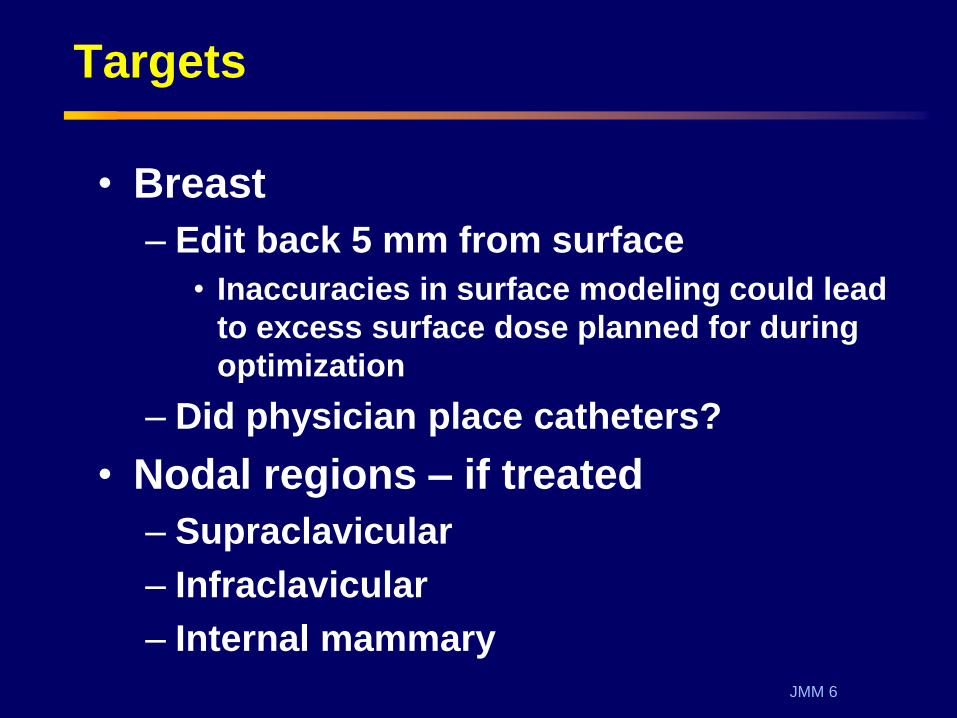
Targets
• Breast
– Edit back 5 mm from surface
• Inaccuracies in surface modeling could lead
to excess surface dose planned for during
optimization
– Did physician place catheters?
• Nodal regions – if treated
– Supraclavicular
– Infraclavicular
– Internal mammary

JMM 7
Contouring for Breast Cancer
• Contouring of structures is required for inverse planning which is still a change of practice at many centers
• There can be significant variability in the contours by practicioner

JMM 8
Organs at risk
• Heart
• Contralateral
breast
• Lungs
• Brachial Plexus
• Left anterior
descending artery
– Sensitive small
volume to help
push optimization
LAD

JMM 9
Radiation Therapy Oncology Group:
Breast Group
Li et al, IJROBP, 2009
Contours by 9 physicians from 8 institutions. Structure overlaps as
small as 10%. Volumes with standard deviations as high as 60%.

JMM 10
Additional Considerations
• Spectrum of techniques
– Simple IMRT (missing tissue
compensation) to beamlet IMRT to
VMAT
• Still need adequate flash
– Jaws should be open for flash
– Want intensity in air to be similar to
intensity over the breast
JMM 11
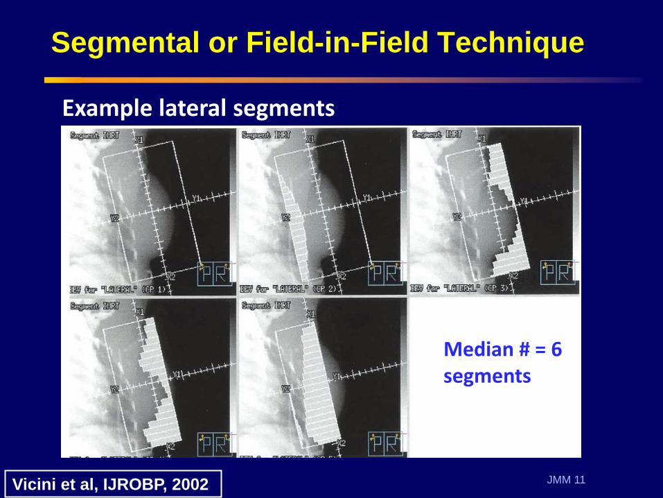
Segmental or Field-in-Field Technique
Vicini et al, IJROBP, 2002
Median # = 6 segments
Example lateral segments
JMM 12
Use of Deep Inspiration Breath hold
• Sixel et al IJROBP 2001
• Remouchamps et al 2003
• Dosimetric advantages when using
deep inspiration breath hold
– Move heart away from breast
– Decrease amount of lung in the field

JMM 13
Effect of breathing on heart position
40% 80%
Moran, ASTRO, 2004
JMM 14
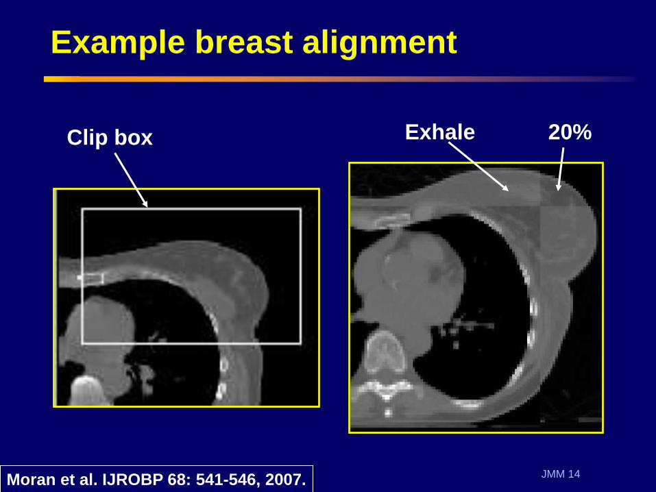
Example breast alignment
Exhale 20% Clip box
Moran et al. IJROBP 68: 541-546, 2007.
JMM 15
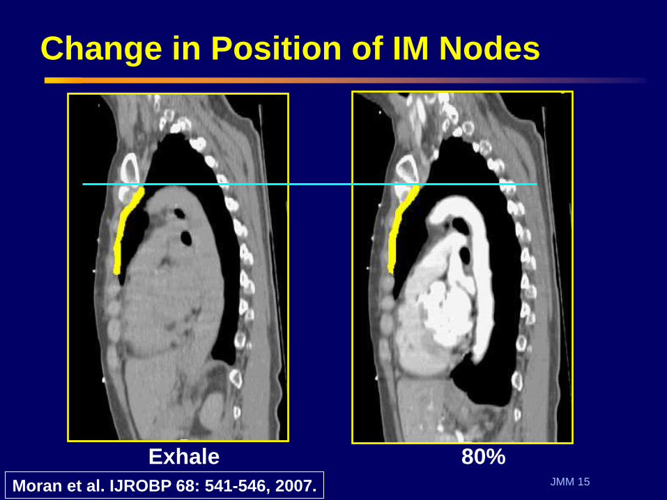
Exhale 80%
Change in Position of IM Nodes
Moran et al. IJROBP 68: 541-546, 2007.

JMM 16
Breast or Chestwall Motion
-1.4
-1.2
-1.0
-0.8
-0.6
-0.4
-0.2
0.0
0.2
0.4
20% 40% 60% 80%
Breathing State
Mo
tio
n (
cm
)
Left-Right
Ant-Pos
Inf-Sup
Adapted from Moran et al. IJROBP 68: 541-546, 2007.
JMM 17
Reproducibility of position with ABC
• Up to 0.8 cm movement anteriorly and
superiorly of breast/chestwall, ICV, and
IMN regions with respect to end exhale
• Individual patient variation was up to
1.3 cm
• The reproducibility with ABC (based
on 3 scan sessions) was on the order
of 3 mm for all breathing states and
directions
JMM 18
Treatment Planning Techniques
• IMRT and VMAT techniques have
been applied to:
– Whole breast
– Whole breast + nodal
– Accelerated partial breast
• Sequential or concurrent boost
• Electron beams can play a role when
needing to spare organs-at-risk such
as the heart and lungs

JMM 19
Whole breast and nodal irradiation
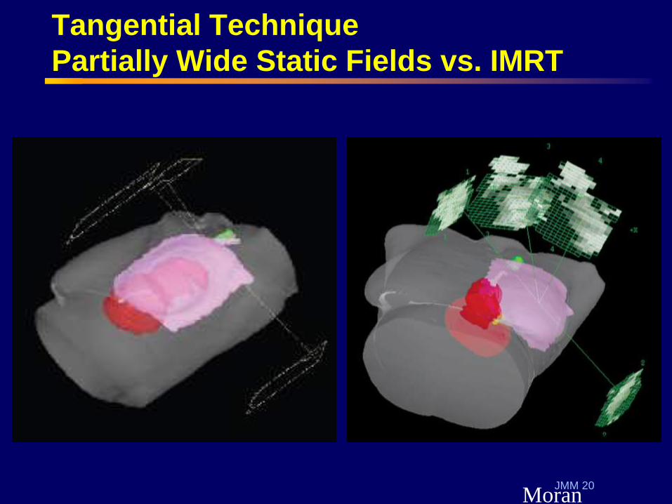
JMM 20
Tangential Technique
Partially Wide Static Fields vs. IMRT
Moran
09/07/2012 20

JMM 21
Objective Function for IMRT Plans
Structure Dose/Volume Costs
Breast, Nodal regions (ICV, SCV, IMN)
95% volume, dose ≥ 52.2 Gy
Min-Max Range: 49.6-60 Gy
Lumpectomy Cavity with margin
99% volume, dose ≥ 60 Gy
1% volume, dose ≤ 63 Gy
Heart and Left Anterior Descending Artery (LAD)
Mean dose ≤ 3 Gy
Maximum dose < 15 Gy
Ipsilateral lung <30% volume, dose ≥ 20Gy
Brachial plexus Minimize dose
Contralateral breast and lung
Minimize dose

JMM 22
Dose Distributions
6 MV photons
Electrons (6, 9,
or 12 MeV) used
as deemed
necessary
for normal
tissue sparing
or for nodal
coverage
Jagsi et al, IJROBP 78: 2010.
Clinical Practice at Our Center
9 field: Concerns re: dose
to other organs
JMM 23
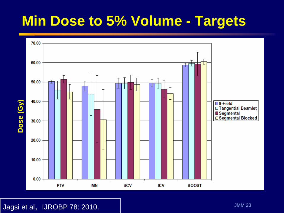
Min Dose to 5% Volume - Targets
Jagsi et al, IJROBP 78: 2010.
Do
se (
Gy)

JMM 24
Rotational Techniques
• Demonstrated
improved
minimum dose
to the target with
a TomoTherapy
technique
• Also static
gantry technique
Goddu et al, IJROBP 73: 1243-1251, 2009.

JMM 25
VMAT: Arc span + Field Considerations
Fig. 2 Popescu et al, IJROBP 289, 2010.
Two VMAT arcs of 190 deg:
CW: 300 to 130
CCW: 130-300
2 cm overlap to distribute dose
for arcs so no sharp gradient
or match

JMM 26
VMAT – Breast + Nodes
Popescu et al, IJROBP 287–295, 2010.
• Beware of increased dose to
contralateral breast and lung in
addition to heart and ipsilateral lung
• Partial arcs are typically used to keep
some sparing of tissues not normally
irradiated with tangential arcs

JMM 27
Accelerated Partial Breast Techniques

JMM 28
Volumes
• Expansion from Clinical Target
Volume (CTV) to Planning Target
Volume (PTV) depends on
– Immobilization
– Breath hold technique used
• Device or voluntary?
– Localization
– Concerns re: seroma cavity position
JMM 29
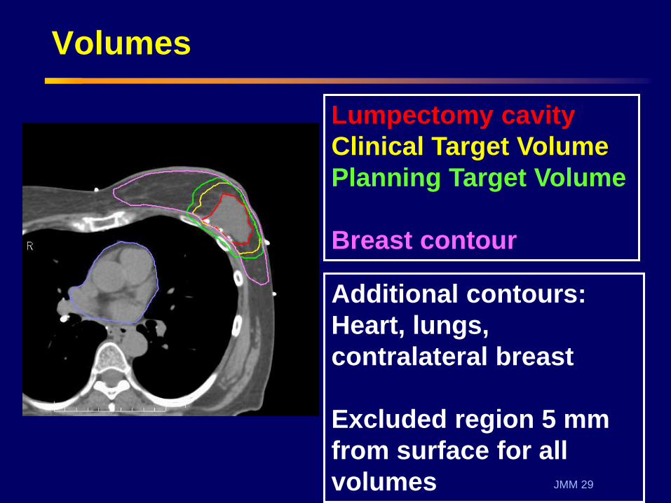
Volumes
Lumpectomy cavity
Clinical Target Volume
Planning Target Volume
Breast contour
Additional contours:
Heart, lungs,
contralateral breast
Excluded region 5 mm
from surface for all
volumes

JMM 30
Volumes – 10 patients
• Mean volume of the contoured
breast (cc): – FB: 722±389
– DIBH: 731±382
• Mean PTV volumes (cc) – FB: 202 cc
– DIBH: 185 cc
– Volumes are different because expansions are
different
Moran et al. IJROBP 75: 294-301, 2009.

JMM 31
Example beam arrangement
Contoured breast,
CTV, heart, LAD
Technique: 3 or 4 beams per patient
Mean PTV volumes in cc:
FB: 202 cc
DIBH: 185 cc

JMM 32
Cost Function for IMRT Plans: Treatment Planning Study
Structure Dose/Volume Costs
CTV 100% volume, dose ≥ 38.5 Gy
99% volume, dose ≤ 40.4 Gy
PTV 95% volume, dose ≥ 38.5 Gy
99% volume, dose ≤ 40.4 Gy
Heart and LAD Mean dose ≤ 3 Gy
Uninvolved
ipsilateral breast
Minimize dose
Lungs 90% volume, dose ≤ 5 Gy
Moran et al. IJROBP 75: 294-301, 2009.

JMM 33
Example Oblique Dose Distributions
WBRT
FB 3DCRT
FB
3DCRT
DIBH
IMRT
DIBH
38-42
34-38
31-34
27-31
23-27
20-23
16-20
12-16
8-12
Moran et al. IJROBP 75: 294-301, 2009.
JMM 34
PBI Technique Comparison
• Acceptable target coverage with
all PBI techniques – IMRT can be used improve dose
homogeneity to the PTV and reduce the
maximum dose
– The use of DIBH result in further dose
reductions of heart dose when
compared to free-breathing 3DCRT
• Dose to uninvolved left breast
can be reduced with IMRT

JMM 35
IMRT Techniques

JMM 36
Summary – Advanced Tx Planning
• Targets must be defined to use DVH
constraints
– Use RTOG atlas as a guide to improve
consistency of targets
• Beware when using beam arrangements that
involve irradiation of contralateral structures
– Limit arc range to reduce likelihood of extraneous
dose to contralateral structures
• When transitioning from previous techniques
the treatment team must work together
– Reproducibility of techniques, implementation of
breath hold or gating technology, margin
evaluation, assessment of patient changes

JMM 37
Acknowledgements
• Lori Pierce, MD
• Robin Marsh, CMD
• James Balter, PhD
• Kent Griffith, MS, MPH
• Reshma Jagsi, MD
• Jim Hayman, MD
• Chrissy Lockhart
NIH Grants R01 CA102435-01 and P01-CA59827