Blood sugar levels regulated by pancreatic hormones insulin and glucagon.
1
TREATMENT OF HYPERINSULINEMIA & INSULIN RESISTANCE AND
METABOLIC SYNDROME copywritten WiO Diet 2007, 2012©
BACKGROUND This paper reviews the findings of these clinical studies and the direct experience that we have recorded with thousands of patients. The findings of these studies and how these studies have already identified promising relief from this exploding epidemic that is washing over America and the rest of the world. The WiO Diet is a medically designed protocol that is the result of over 3 ½ years of researching over 600 clinical studies spanning 50(+) years and additional beta testing in 6 clinics with over 1,000 patients within 18 months. The complete name of the program is ‘Your Weight is Over – Your Last Diet’ WiO Diet for short. After intensive study of over 600 clinical studies, we created a data base of relative findings from the studies listing the correlating conclusions that were in harmony with each other. The areas of focus were: metabolic interaction in relationship with the symptoms of metabolic syndrome, primary organs involved, effects of macro & micro-nutrients, psychological impact & treatment for a term life style change. We then set up 6 separate beta test clinics in existing businesses in; General medicine, Chiropractic, Nutrition shoppe, Physical/ exercise training, Health Spa, Health counseling. We have designed a 4 phase protocol covering 18 weeks. Phase 1 is 4 meals per day, three of which are controlled by a meal-replacement formula provided by WiO. The patient is coming into the WiO clinic each week. Their physiological improvements are measured weekly. They receive nutritional education and personal coaching on how to emotionally establish a healthy relationship to food. As can be expected with any weight loss, certain physiological parameters will improve. Blood pressure, total cholesterol, and fasting glucose will be reduced. In the beginning of our research we expected these physiological improvements, but you could expect these improvements from any successful weight loss program. Within a few weeks and months we noticed other patterns that were pleasing but couldn’t be explained by weight loss alone. Dieters reported a greater amount of energy, were actually stronger, better digestion including GERD. Relief from inflammation issues, significant mood enhancement and even some cases of sleep apnea were resolved, or improved and several others. Dieters were reporting that they had better ability to concentrate and their skin and nails were better. We were amazed by one account of a 63 year old man that had suffered from type II diabetes for 12 years, his average daily glucose readings had dropped from 347 to 75 (-78.4%) in the first 7 days. This dieter did not have a weight issue which indicated that his blood sugar was affected by something other than weight loss. Equally satisfying, but puzzling was that this was occurring with just one week into the program. Many of our hypertensive dieters were reporting extreme dizziness in the first 7-10 days: their high blood pressures had dropped so fast, that their prescription had to be reduced. Weight loss alone could not explain these results – not with only losing a small percentage of their total body weight in a matter of days.
2
CONTENTS
Background The Primary Focus of This Dietary Approach ………………………………………………………………………………... 4
Metabolic Syndrome - Modern Day Health Apocalypse ……………………………………………………… 4 Recommend Pre-Testing …………………………………………………………………………………………………. 5 Food - Insulin and Glucagon ……………………………………………………..…………………………………….. 6 OBESITY: The Epidemic of the 21st Century ……………………………………………………………………… 7-9 What Metformin Does ……………………………….……………………………………………………………………… 9
The Relationship Between Insulin And Glucagon …………………………………………………………….…………… 9-20 What Is “Metabolic Syndrome” ………………………………………………………………………………………… 9 What Causes Hypertension ……………………………………………………………………………………………… 13 Coronary Artery Disease ………………………………………………………….………………………………………. 14 Obesity …………………………………………………………………………………………………………………………… 14 How Fat Is Created And Burned ………………………………………………………………………………………. 14 Regulating the “Flow of Fat” ……………………………………………………………………………………………. 14-15 What Causes High Cholesterol – And How to Control It …………………………………………………….. 16 How Cholesterol Levels Get Out Of Control ……………………………………………………………………….. 16-17 The Ebb And Flow Of Cholesterol - And The Three Main Players - VLDL – LDL – HDL………….. 17-18 How To Keep Cholesterol Off The Arterial Walls ………………………………………………………………. 18 Food Will Control Cholesterol - To Lower OR To Raise It …….…………………………………………….. 18-19 Keeping the Carbohydrates Low Changes the Biochemical Pathway…………………………………… 18-19
Fat Loss Or Water Weight? …………………………………………………………………………………………………………. 20-23 Diet Pills ……………………………………………………………………………………………………………………………………. 24 Ketogenesis (Healthy) Ketosis vs. ‘Ketoacidosis’ (Deadly) ….…………………………………………………..……. 25 Eicosanoids – How The Body Controls Inflammation ……………………………………………………………………. 26
What are Eicosanoids? Second Messengers ……………………………………………………………………….. 26-27 General Concepts about eicosanoids …………………………………………………………………………………. 28-30 Insulin and Glucagon: Regulators of the Eicosanoid Pathways …………………………………………… 31-32 Why Fish Oils Are Not Enough ………………………………………………………………………………………….. 32 Keeping Hormone’s Simple ……………………………………………………………………………………………… 32 How We Control Your Hormones ……………………………………………………………………………………... 32 PGE1 (Prostaglandin EI) ………………………………………………………………………………………………….. 33 Why A Special Omega 3-6-9 Blend …………………………………………………………………………………… 33 If We Need More Omega-3 – Why Don’t We Just Take Flax Seed Oil? …………………………………. 33-34 Fish Oil – Isn’t it Healthy? ………………………………………………………………………………………………… 34 Fish Oil Processing …………………………………………………………………………………………………………… 35 Did You Know? Fun Facts About Eicosanoids ……………………………………………………………………. 36-38
Problems of Too Much Protein; Controversies …………………………………………………………………………….. 39-49 Liver Health …………………………………………………………………………………………………………………….. 39-41 Balanced Nitrogen …………………………………………………………………………………………………………... 41-42 Kidney Function ……………………………………………………………………………………………………………… 42 Bone Health ……………………………………………………………………………………………………………………. 42-43 Metabolic Acidosis ………………………………………………………………………………………………………….. 43 Colon Cancer/Heart Disease/Overall Health …………………………………………………………………….. 43-44 Protein Digestion/Quality ………………………………………………………………………………………………… 44 The Steps of Digestion …………………………………………………………………………………………………….. 44-45 Medication That Reduce Digestive Quality ……………………………………………………………………….. 45-46 True Digestibility ……………………………………………………………………………………………………………. 46-47

3
Vitamins & Minerals Digestibility ……………………………………………………………………………………. 47 Vitamins & Minerals In Our Food? ………………………………………..…………………………………………... 48 From the Cradle to Your Plate ………………………………………………..………………………………………... 48 If It Can’t Take The Heat – Get Out Of The Kitchen ………………………………………………………….. 48 The Power of Your Digestive System ……………………………………………………………………………….. 49 Risks of Dieting ………………………………………………………………………………………………………………. 49
Acid Balancing – Importance Of Protein & Alkaline Minerals ………………………………………………………… 49 A Brief Review of the Digestive Process ……………………………………………………………………………. 49-50 SECRETIN – Gastric Acid And Bicarbonate Production ………………………………………………………. 51 Why Does GERD Improve On The WiO Protocol? ……………………………………………………………… 51 Importance Of Protein In Balancing Acid/Base ………………………………………………………………… 51-52 Effects Of Drinking Soda – (Pop) ……………………………………………………………………………………… 52 How Bicarbonate Buffers The Blood ………………………………………………………………………………… 52 Bicarbonate Levels Drop As We Age ………………………………………………………………………………… 53 How To Maintain Healthy Bicarbonate Levels ………………………………………………………………….. 54
Body Intelligence – An Intuitive Way To Eating …………………………………………………………………………… 54-59 The Spirit Is Willing – But The Flesh Is Weak …………………………………………………………………... 54 A Health Relationship With Food And Eating ………………………………………………………………….. 54-55 Tuning In To Your ‘Body Intelligence’ …………………………………………………………………………….. 55 Diets should not be used to ‘lose’ something – A Diet is the food you give your Body to feel healthy ……………………………………………………….. 56 Control Your Hunger by Feeding It ………………………………………………………………………………….. 56 Make Food an Ally ………………………………………………………………………………………………………….. 56 Turn Deft to the Whistle Blowers …………………………………………………………………………………….. 56-57 Calibrate your Full Meter ………………………………………………………………………………………………... 57 Feel the Joy in Eating ………………………………………………………………………………………………………. 57 Don’t Feed Emotions ………………………………………………………………………………………………………. 57-58 Love your Body with Respect ………………………………………………………………………………………….. 58 Exercise is Movement Not Just the Gym …………………………………………………………………………… 58 Give Thanks to Health – By Being Healthy ………………………………………………………………………… 58-59
References …………………………………………………………………………………………………………………………………. 60-61
4
HYPERINSULINEMIA & INSULIN RESISTANCE THE BALANCE BETWEEN
MASTER HORMONS INSULIN & GLUCAGON IN THE PATHOGENESIS OF “METABOLIC SYNDROME”
THE PRIMARY FOCUS OF THIS DIETARY APPROACH The goal of the protocol is to aid in overcoming the symptoms of Metabolic Syndrome (MSx). The approach is to treat directly the causes of hyperinsulinemia and insulin resistance, this is accomplished by treating the dysfunctions of the:
1) Pancreas 2) Liver 3) Digestion 4) Life Style (weekly education in our relationship to food(s))
The CDC (Center for Disease Control) and the WHO (World Health Organization) reports the following: persons in the USA suffer from the health risks:
*Overweight 79.8% (obesity is over 40%) NAFLD 75% in obese persons up to 25% in general public (Non-Alcohol Fatty Liver Disease) Digestive complications 47% (day-to-day issues) *High blood pressure 31% *High cholesterol 27% *Diabetes 12%.
METABOLIC SYNDROME - Modern Day Health Apocalypse The CDC estimates that over 1,550,000 (3 each minute) people die each year from the metabolic conditions listed above, directly or indirectly. There is no other disease that claims more lives and is completely preventable. We could only think of one thing when thinking of these thieves of health; the apocalypse. We have coined the phrase “The Four Horsemen – the Modern Day Health Apocalypse”. Hundreds of clinical research supports that not only are these conditions preventable but reversible (not necessarily including Type I diabetes, however some research shows promise). Currently having any two of these four* health risks listed above is defined as having Metabolic Syndrome ie: syndrome X, insulin resistance syndrome, Reaven's syndrome or MSx. Many experts are lobbing to include NAFLD as one of the original four*. Because research is finding that in nearly every case NAFLD (fatty liver) preceded each of the symptoms of metabolic syndrome. Researchers are also discovering that with the advent of one of these symptoms, if left untreated it is a matter of time before other symptoms are adopted. Some studies estimate the prevalence of metabolic syndrome in the USA to be up to 34% of the population.[60] Metabolic syndrome affects 54% of the U.S. population older than age 50. With respect to that demographic, the percentage of women having the syndrome is higher than that of men.[31]
RECOMMEND PRE-TESTING When beginning the protocol we always obtain a blood sample and perform a HRV (Heart Rate Variability) test. A benchmark was established with each new dieter, measuring; Triglycerides, LDL, HDL, cholesterol ratios, glucose, blood pressure, body fat percentage, body measurements, and weight. Patient’s HDL levels increased markedly and LDL level decreased, but more importantly their ratios were at or below 4 (ratio HDL/LDL).

5
One 40 year old man looked basically healthy however, his biomarkers were telling another story. After just 41 days on the WiO Protocol his total cholesterol was dropped from 304 to 204, HDL increased from 33 to 56. Triglycerides went from 261 to 74 (-74%), followed by a decrease in his LDL of 219 to 132 (-39.7%). More importantly his ratio improved an impressive 9.2 to 3.6 (+60.9). A 41 year old man who had been diagnosed as a Type II diabetic 5 years previous reported from his cardiologist said that he was no longer a diabetic after just 3 weeks. The reports continued to pour in ranging from inflammation issues improving to digestive disorders being resolved. Again it was clear that something more than just losing weight was happening. One of the areas that this protocol focuses on is the pancreas hyper-secretion of insulin as a result of a high carbohydrate diet (over 250g daily). Thus this protocol addresses hyperinsulinemia in individuals. After refreshing your knowledge of the metabolic effects of insulin and insulin resistance, many of these unexplainable benefits our dieters were experiencing began to make sense. The proper balance between insulin and its counterpart glucagon and the notion of “insulin dominance and glucagon dominance” made its impact. The book “Protein Power” by Michael and Mary Dan Eades [16] (both M.D.’s) explains how the two master hormones (insulin & glucagon) are likened to the brake pedal and the gas pedal of a car; you need both throughout the day as you drive. However, the type of road (or metabolic path) you are traveling on at any particular time largely dictates which pedal will be used more. Driving on a freeway you’ll use the gas pedal more, in the city, you use the brake more. In our body, it’s our food choices that determine which hormone is used more.
Food - Insulin and Glucagon
Table 1[48] shows the effects of different combinations of macronutrients on our body’s production of insulin and glucagon. The goal is to strive for a balance between glucagon and insulin, a diet with a little more protein and fat (EFA balance) with fewer carbohydrates would seem to be required. After review it is clear that the food combinations are more interesting. A meal of high carbohydrates and fat, with little protein, will likely produce a veritable flood of insulin and very little, if any, glucagon. Consider your menus at home, those in restaurants and in the schools we send our children to. Our children’s favorite foods; soda, even juice, macaroni and cheese, pizza, peanut butter and jelly, cheese and crackers, just to name a few of their favorites. All of these are high in carbohydrates and fat and have very little, if any, protein, thus, they promote a dominance of insulin rather than a balance of both hormones.
TABLE 1
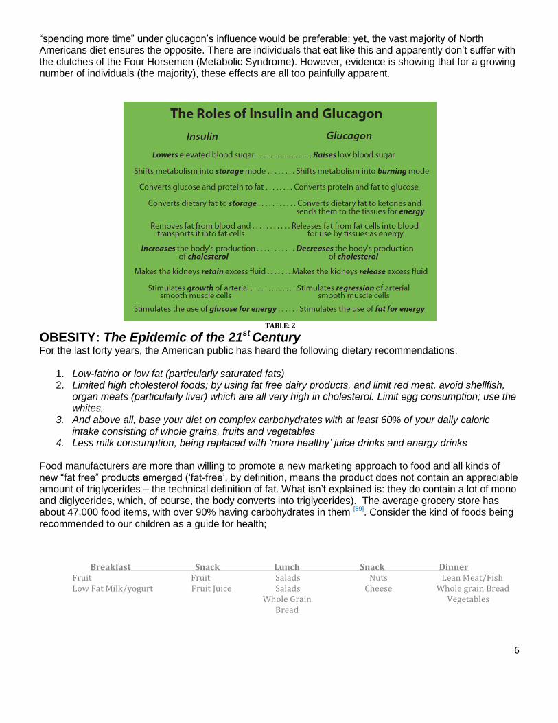
The Roles of Insulin and Glucagon TABLE: 2 INSULIN - GLUCAGON
Table 2[48] lists the effects insulin and glucagon have on our physiological processes. It is pretty obvious that
6
“spending more time” under glucagon’s influence would be preferable; yet, the vast majority of North Americans diet ensures the opposite. There are individuals that eat like this and apparently don’t suffer with the clutches of the Four Horsemen (Metabolic Syndrome). However, evidence is showing that for a growing number of individuals (the majority), these effects are all too painfully apparent.
TABLE: 2
OBESITY: The Epidemic of the 21st Century
For the last forty years, the American public has heard the following dietary recommendations:
1. Low-fat/no or low fat (particularly saturated fats) 2. Limited high cholesterol foods; by using fat free dairy products, and limit red meat, avoid shellfish,
organ meats (particularly liver) which are all very high in cholesterol. Limit egg consumption; use the whites.
3. And above all, base your diet on complex carbohydrates with at least 60% of your daily caloric intake consisting of whole grains, fruits and vegetables
4. Less milk consumption, being replaced with ‘more healthy’ juice drinks and energy drinks Food manufacturers are more than willing to promote a new marketing approach to food and all kinds of new “fat free” products emerged (‘fat-free’, by definition, means the product does not contain an appreciable amount of triglycerides – the technical definition of fat. What isn’t explained is: they do contain a lot of mono and diglycerides, which, of course, the body converts into triglycerides). The average grocery store has about 47,000 food items, with over 90% having carbohydrates in them [89]. Consider the kind of foods being recommended to our children as a guide for health;
Breakfast Snack Lunch Snack Dinner Fruit Fruit Salads Nuts Lean Meat/Fish Low Fat Milk/yogurt Fruit Juice Salads Cheese Whole grain Bread Whole Grain Vegetables Bread

7
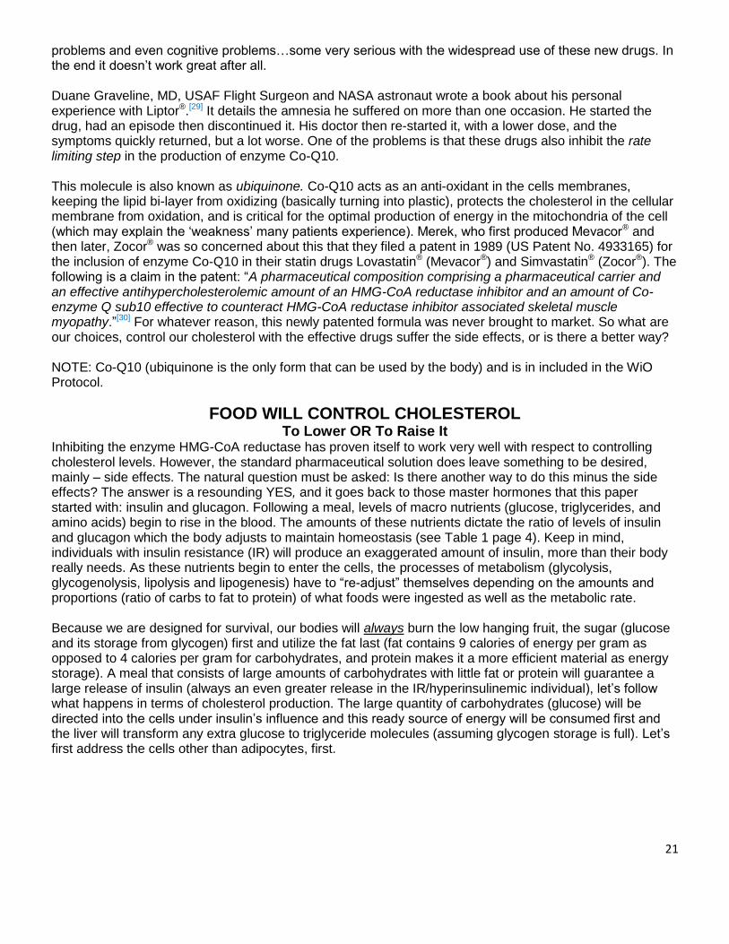
These foods recommendations come from the newest food pyramid which came out in February of 2011, these recommendations are made up of 65% carbohydrates 5-10% protein 2% sugars and 1% from fat (Fig. 2). What the new food pyramid does not make clear is the increase in fruits up to 14% which is made up of nearly 100% of sugar. Review the increase of carbohydrates from the food pyramid from its first introduction in 1978. If you compare the level of persons overweight in 1978 (32%) to the most recent report from the WHO (World Health Origination), Americans that are overweight as of 2010 were 79.8% of the population. Match that increase to the level of carbohydrates being consumed for the same period and a startling conclusion must be addressed. From 1978 to 2011 carbohydrate intake increased 260%. During the same period the increase of overweight persons was 247%, another frightening fact was that the obesity rate increased 322% for the same period. The pancreas interprets this level of carbohydrates regardless of its origin; it doesn’t matter if the carbohydrates are coming from healthy whole wheat bread, from fruit or from soda. This recommendation will produce the same glycemic impact as consuming 2 1/4 cups of white sugar consumed daily. It is easy to see why our society is plagued with the highest levels of Diabetes, Hypertension, Hypercholesterolemia, and Obesity our nation has ever seen. And the fact that it is estimated that 33% of our children will develop type II diabetics in their adulthood is inexcusable. Hyper consumption of carbohydrates is the culprit.
1978 2011
Fig. 1 Fig. 2 20-25% Carbohydrates 65% Carbohydrates
Today’s healthy breakfast (not counting Pop Tarts or Toaster Strudel) might be a whole wheat bagel with a glass of fresh, organic orange juice;
Total carbohydrates Wheat Bagel: 38 grams Fresh OJ: 26 grams Total: 74 grams …not counting any jam or spread on the bagel
A child’s size serving of cereal (1 oz.), eight ounces of low-fat milk and a glass of “OJ” would yield Total carbohydrates Cereal: 25 grams Low Fat Milk: 12 grams OJ: 26 grams Total: 63 grams
8
If we subtract the grams of dietary fiber (about 3 in each case) we have two breakfasts containing 71 and 60 grams of total “impact” carbohydrates. Metabolically speaking, that is the equivalent of 6 and 5 TABLESPOONS of pure sugar respectively. We must realize every four grams of carbohydrates (less grams of fiber) has the same net effect of a teaspoonful of sugar in our bodies …sometimes quickly, sometimes a little slower depending on the source, but that is its metabolic destiny. Dr. Eades states that 2,200 Kcal daily diet containing 60% carbohydrates is the equivalent of two full cups of sugar. Even if we say a 2,500 Kcal (amply allowing for extra fiber) contains a little more than two cups of sugar, the fact is startling just the same. School lunch menus (balanced by dieticians) usually contain the persistent favorites such as macaroni and cheese, peanut butter and jelly, grilled cheese sandwiches, and pizza. Drink choices still include milk. But juices, sweet teas, sodas and Gatorades are still more popular. Ask yourself “How many ‘fat kids’ do we see in our schools today”? Based on clinical research we must conclude that we are making them fat! The National Institutes of Health’s newsletter (NIH NEWS) and The New England Journal of Medicine both published a study in March of 2005 [61] that warned, for the first time in history, that this generation may have a shorter life expectancy than the preceding one, one of the primary reasons is the staggering rate at which obesity is occurring. I recall a resent occasion of being in church and witnessing a mother giving what I imagine was her daughter’s lunch. She was about 14 months 22-25 lbs. she was eating Silk Plus® yogurt and a slice of whole wheat bread. In that snack the glycemic impact was the same as feeding her 7.5 TABLESPOONS of sugar. No well meaning parent would ever believe that giving that much sugar would be healthy; people are simply ill-informed. I whispered to my wife that I bet the child’s name is ‘Princess Di - Obe’ she gave me a puzzled looked and asked how I knew what they named her? “Because of what they are feeding her I said, it’s her nick name for what she will sadly become… Diabetic and Obese” She gave me a look that promptly told me to be quiet and listen to the service. But this experience explains this report (and many others) in newspapers on childhood obesity. Stating “Although the rest of the nation is much heavier too, among those ages 6 to 19 the rate of obesity has not just doubled, as with their parents and grandparents, but has more than tripled.” [4] Such alarming statistics well might be expected, giving rise to theories and studies. Genetic predisposition is a big focus, especially since we have decoded the genome. Although this no doubt may play some role, such a dramatic change in one generation would not be scientifically congruent to support such a genetic shift. A large focus is now being concentrated to hyperglycemia during pregnancy but at levels lower than the diagnostic criteria for diabetes. Two studies recently published in another issue of The New England Journal of Medicine recently explored this. In the first, (The HAPO Study – Hyperglycemia and Adverse Pregnancy Outcomes) [25], 505 pregnant women underwent a 75 gram glucose tolerance test at 24 to 32 weeks of gestation. Data remained blinded if the fasting plasma glucose was 105mg/dl or less and the 2 hour plasma glucose was 200mg/dl or less. Their conclusions were summarized: “Our results indicate strong, continuous associations of maternal glucose levels below those diagnostic of diabetes with increased birth weight and increased cord-blood serum C-peptide levels”. The second study involved 751 women diagnosed with gestational diabetes and in the same gestational stage as the above study. They were randomized to be treated with metformin (and insulin if needed) or just insulin alone. The object of the study was to judge the safety and efficacy of metformin compared to the traditional insulin alone therapy and to see if there was any effect on the composite outcomes of babies compared to those whose mothers received the insulin alone therapy. The conclusions stated “In women with gestational diabetes mellitus, metformin (alone or with insulin) is not associated with increased prenatal complications as compared with insulin. The women preferred Metformin to insulin treatment.” [51] It would logically follow that the next study would be to treat a group of pregnant women with elevated plasma glucose, but at levels below what would be considered diagnostic of gestational diabetes, with metformin/insulin versus an untreated control group. The composite outcomes of the neonates would be
9
compared, and we would see if an indication for treatment of such a population is warranted. In fact, Donald R. Coustan, MD, Professor and chair of Obstetrics and Gynecology at the Warren Alpert Medical School of Brown University (and one of the authors of the HAPO Study), announced recently that conferences will be held to discuss the pro’s and con’s of treating elevated glycemia in pregnancy. He stated: “For now, doctors will still use the glucose threshold that they’re currently using” [52]. This line of reasoning is rather disturbing in that we (general healthcare) are focusing on treating symptoms and ignoring the physiological underpinnings. Hyperglycemia, may be subclinical for a diagnosis of diabetes, is a symptom of what? I would suggest the most likely cause would be insulin resistance brought about by constantly elevated levels of insulin (hyperinsulinemia) due to a diet too rich in carbohydrates. Not once did any of the researchers look at maternal insulin levels nor did they discuss the maternal diet. If that in fact is the case, we’ll know the mothers will be getting a diet largely based on complex carbohydrates and low in fat and cholesterol (usually with very little serving of protein). This is a diet that will ensure an abundant secretion of insulin! It would have been interesting to have had insulin levels drawn in addition to the plasma glucose – both in the fasting state and 2 hours post glucose challenge. In our clinics, a fasting insulin level above 10 MU/ml or a 2 hour post-glucose challenge level above 30 MU/ml would have automatically caused us to look at the diet. If appropriate, the carbohydrate content would have been decreased by half, making up the calorie reduction with protein and “good” fats (EFA) and re-testing the patient in one week. In the above example, they are contemplating administering insulin – are they serious! The very fact that metformin is effective (cases where added insulin was not required) in tempering their hyperglycemia should be a diagnostic criterion all by itself that these women are insulin resistant. The liver releases glycogen when blood glucose becomes low, as normal levels are reached insulin is secreted and this inhibits the further release of glycogen. In an insulin resistant individual, the liver does not respond to the proper level of insulin and it continues to release glycogen and blood sugar continues to rise. Metformin is used to block the release of glycogen by the liver. Which requires the patient to increase blood sugar by consuming more carbohydrates which will secrete even more insulin and the cycle repeats exasperating the problem. Now let’s turn our attention to the fetus in all of these ‘experiments’. The little human is developing in a virtual “sea of insulin” due to the mother’s hyperinsulinemia. What physiological consequences will be caused? Certainly large birth weight, increased cord-blood C-peptide and hypoglycemia at birth would be consistent with this, and these are exactly the types of babies we are seeing being born today. These children, due to the maternal environment, are being born, maybe not genetically, but certainly environmentally, predisposition to developing insulin resistance and diabetes (51 ‘new’ adolescent are diagnosed daily[53]) at an early age. Following weaning, smashed bananas and rice cereal are some of the first “sweet” foods given to children. All carbohydrates or worse, carbohydrates and fat, the very combination guaranteed to produce the most insulin. Then they graduate to the “Happy Meals” and the “Juicy Juice” and here we go! This, I truly believe, is the root cause of the explosion in childhood obesity we have witnessed in the last 15 years or so. Mark my words, if these pre-diabetic, hyperglycemic “moms-to-be” are treated with insulin during their pregnancies, the situation will worsen rapidly; that is just the physiological/biochemical fact of the matter.
What Metformin Does
Metformin inhibits the liver's production of glucose. There is some scholarly debate about what exactly it is that Metformin does, but most researchers agree that in most people Metformin suppresses the production of glucose and the release of glycogen. If you'll remember, it is the liver's tendency to dump additional glucose into the blood stream when insulin response is weak or missing, which will cause blood sugar to shoot up after a meal. The liver may also dump glucose in the blood stream early in the morning when fasting insulin levels are low. Metformin may lower fasting blood sugar by limiting the liver's production of
10
glucose rather than by making cells more sensitive to insulin, which is a more preferred reaction. A mouse study published on May 15, 2009[54] suggests that Metformin works to lower blood sugar by directly stimulating a gene in the liver which is how it shuts off glucose production. Rather than by improving insulin sensitivity, it bypasses the broken insulin signal.
THE RELATIONSHIP BETWEEN INSULIN AND GLUCAGON
The following paper is an examination of “Metabolic Syndrome” from the perspective of “glucagon versus insulin dominance”. The biochemistry and cellular physiology described herein directly out of current medical school textbooks. Other references cited are from prestigious, peer-reviewed professional journals. Keep in mind, we will be discussing the pathological condition of “Metabolic Syndrome” so many of the dietary recommendations may seem moot or ‘not applicable’ if we view them from a normal physiologic state. A WiO Diet has a medically designed, precise protocol. As with any other treatment plan, there is a separate protocol for the treatment of acute conditions including a maintenance phase after the dysfunctions have been corrected. Be prepared that they are in contrast to USDA and RDA guidelines. In America, and the rest of the world is not far behind, we are facing a healthcare crisis of unparalleled scope. “Metabolic Syndrome” and all of its co-morbidities have spiraled out of control and continue to get worse not better every year. We cannot afford to keep doing the same things with the same mindset and expect a different outcome (definition of ‘insanity’?), in other words we must not be content to merely treat the symptoms while ignoring the underlying pathophysiology of the cause.
THE RELATIONSHIP BETWEEN INSULIN AND GLUCAGON IN THE PATHOGENESIS OF “METABOLIC SYNDROME”
By: Michael P. Ciell, R.Ph. WHAT IS “METABOLIC SYNDROME” A commonly accepted definition “Metabolic Syndrome” might be a generalized disorder whose four hallmark symptoms are hyperglycemia, hyperlipidemia, hypertension and obesity. Presenting with two of the above is generally considered the diagnostic criteria for this disorder. Gerald Reaven, MD (Professor Emeritus - Active of Medicine at Stanford University) was the first to use the term in 1988, saying he preferred it to names like “Metabolic Syndrome” or the “Deadly Quartet”. He said “many of the manifestations of the disorder might not be considered ‘metabolic’ (i.e. increases in plasminogen activator inhibitor –1 (PAI-1) a factor regulating the process of fibrinolysis), and the “Deadly Quartet” implies obesity is an essential component while many very obese persons may have nothing resembling the syndrome (Sumo wrestlers may be an example)”. Semantics aside, the real significance of Dr. Reaven’s work was to establish, for the first time, the link between insulin resistance (primarily with regard to insulin stimulated glucose disposal by muscle and insulin regulation of lipolysis in adipose tissue) and the four hallmark symptoms of this syndrome. He reasoned that insulin’s first function will always be to mediate glucose uptake by the muscles. If glucose levels remain elevated (due to the muscles’ insulin resistance) the pancreas will continue to produce more insulin in an attempt to control the high glycemia. Complications now appear because many of the other tissues/organs still retain their sensitivity to insulin. The kidney is a good example. Insulin stimulates sodium retention by the kidney, thus contributing to water retention and hypertension. Dr. Reaven cites polycystic ovary syndrome (hypersecretion of androgens from the ovary) as another example of insulin sensitive organs being affected [1]. Basically the ovary, being constantly exposed to higher than normal levels of insulin, increases its testosterone production accordingly. Thus, the insulin resistance of one tissue with the compensatory hyperinsulinemia that ensues will lead to many other insulin sensitive tissues being affected and complicating the entire physiological picture of that individual. Our complete understanding of this principle is necessary so that a protocol addressing the cause of the problem may be designed, instead of
11
merely treating the symptoms as isolated and unrelated pathologies. According to Dr. Reaven, “The manifestations of Metabolic Syndrome” can be divided into six major categories:
1. Glucose intolerance: Individuals with Metabolic Syndrome don’t have diabetes, by definition, but their plasma glucose concentration is higher than those individuals who don’t have Metabolic Syndrome.
2. Dyslipidemia: The characteristic findings are high plasma triglycerides and low HDL-cholesterol. The insulin resistance and compensatory hyperinsulinemia cause the liver to produce more triglyceride rich VLDL, thus increasing the plasma triglyceride concentration. Cholesterol ester transfer protein (CETP) transfers cholesterol from HDL to VLDL, exchanging it for triglycerides. Therefore, the HDL cholesterol falls. The increased VLDL also reduces the ability to remove postprandial newly absorbed chylomicrons. In Metabolic Syndrome, VLD, chylomicrons and their metabolic remnants (chylomicron and VLDL remnants) are removed more slowly from the plasma by virtue of their increased concentrations, resulting in increased postprandial lipemia. In addition, there is a shift in the LDL particle diameter to smaller and denser LDL particles.
3. Uric acid metabolism: There is a tendency to increased serum uric acid concentration. There is a decrease in the ability of the kidney to excrete uric acid; therefore, renal uric acid clearance is decreased.
4. Kidney manifestation: There is an increased salt retention. It appears that half the patients with hypertension are insulin resistant. From population-based studies, the best predictor of hypertension developing has been hyperinsulinemia as a surrogate measure of insulin resistance.
5. Hemodynamic manifestations: There is evidence that the sympathetic nervous system activity is increased in insulin resistant individuals. This is another example of other tissues reacting to the hyperinsulinemia.
6. Fibrinolytic changes: There is an increase in PAI-1, with a resultant decrease in fibrinolysis. The increase in fibrinogen tends to increase coagulation. All of these manifestations can have some role in the development of coronary heart disease. [2]. As Dr. Reaven points out; the insulin resistant/hyperinsulinemic patient is at a greatly increased risk for developing CHD. Let’s briefly look at insulin’s role in the mechanisms involved in the etiology of hypertension and CHD. Michael P. Ciell, R.Ph.
COACHES NOTES: Before starting a patient on The WiO Diet, it is helpful to have some base-line labs done. This will allow the coach and the patient to monitor their progress and serve as a benchmark for evaluating this protocol against any other dietary intervention or weight loss program. Suggested tests may include: 1. Complete Metabolic Profile: Fasting glucose in the mid to upper 90’s indicates insulin resistance may already be occurring. Potassium levels in the low-normal range may indicate a larger supplemental amount than what is standard with the protocol. Uric Acid levels above 6 generally indicate insulin resistance. HbA1c should be below 6, repeat in 3 months and note the improvement.
Fasting Glucose 70-85 Potassium Uric Acid A1c

12
2. Fasting Insulin or Insulin, glucose challenge (75g glucose): Draw blood fasting and at 1 & 2 hour intervals (draw both glucose and insulin levels). Note insulin samples (tubes) must be frozen immediately and processed with 24 hours. Insulin levels should be 5 or less fasting and not above 30 (mU/ml) at one or two hours. Glucose should be less than 90 fasting and not more than 150 after one or two hours.
Fasting Insulin <5 mU/ml Fasting Glucose <90 - <150 1-2 hours later
3. Fasting Lipids: Total Cholesterol should be less than 200. HDL should be at least 40. HDL may be less than if on statin therapy. Retest 4 to 6 weeks to evaluate need for continuing medication. Triglycerides should be 150 or less. LDL should be below 130. Non LDL should be lower than 130. And most importantly the (TC/HDL) Ratio should be at or below 4.0 (these levels, if elevated, usually normalize within the first half of Phase 1).
Total Cholesterol <200 HDL >40 Triglycerides <150 LDL <130 Non-LDL <100 Ratio <4.0
4. High sensitivity C-reactive protein: Should be less than <1.0. It is a marker for inflammation and can be both a cause and a result of insulin resistance.
C-reactive protein <1.0 5. Fibrinogen: A clotting risk factor and often associated with insulin resistance. The level should be less than 300.
Fiberinogen <300 6. C-peptide: This test should be ordered for Type II diabetic patients and those not diagnosed as diabetic but who are on insulin therapy (insulin levels will be meaningless here). If the test shows positive, the pancreas is still producing insulin and there is a good possibility they may be able to decrease or eliminate the insulin. The levels should be equal to insulin levels should be less than 10 IU/mL is ideal.
C-peptide <10 IU/mL 7. Kidney Function: Severe Kidney damage is an absolute contra-indication for this protocol. However, those with somewhat compromised renal function (GFR 35 – 50 health range) may still participate providing they take no more than the minimum amount of protein recommended. Test should be repeated in 6 to 8 weeks and an improvement should be seen (at least it should be no worse). If the re-test indicates a worsening, the program should be discontinued. (see page )
GRF >30 (35-50 healthy) 8. Liver Function: Insulin resistance often causes certain enzymes to be elevated. Unless severe,
the program may be started and tests repeated every 8 weeks. An elevation in alkaline phosphatase
may be indicative of gallstones. An ultrasound maybe ordered to rule this out. Deficiencies of
protein, vitamin B-6, zinc, folate and vitamin C can cause a lower than normal alkaline phosphatase.
13
Phosphatase 44 to 140 IU/L normal range Urea nitrogen ‘BUN’ 7 to 21 mg per 100 ml (7–21 mg/dL).
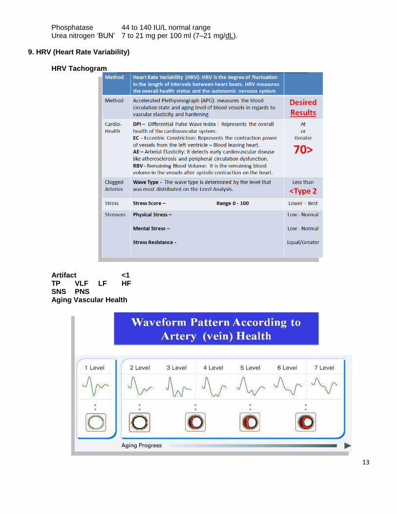
9. HRV (Heart Rate Variability)
HRV Tachogram
Artifact <1 TP VLF LF HF SNS PNS Aging Vascular Health
14
WHAT CAUSES HYPERTENSION (High Blood Pressure) • Insulin stimulates the kidneys to retain sodium and, therefore, water. Glucagon produces the opposite effect (think of a Type I diabetic - there is no insulin and the individual continually urinates). The kidney is one of the last organs to become insulin resistant; therefore, most insulin resistant (IR), hyperinsulinemic patients will be hypertensive with an increased fluid load. In fact, Dr. Reaven reports that as many as 50% of hypertensive patients will show as IR/hyperinsulinemic. [3] • Insulin facilitates cellular magnesium uptake. In IR patients or Type II diabetics, intracellular magnesium concentrations are significantly lower compared to normal individuals. Magnesium is necessary for proper insulin receptor function; therefore as magnesium levels decline, insulin sensitivity decreases further, and their condition worsens. Magnesium ads in cells being more elastic and has a dilatory effect on smooth muscle (opposing calcium’s tonic effect). Lower magnesium levels therefore contribute to increased peripheral resistance (force against blood flow). [4, 5] • Insulin strongly stimulates the release (or gene expression) of vascular endothelial growth factor (VEGF) [6]
substance made by cells that stimulates new blood vessel formation. This causes increase production of the smooth muscle cells of the arteries and arterioles making them less elastic and decreasing the lumen diameter; pressure increases and the heart works harder. VEGF expression is also strongly implicated in tumor angiogenesis (tumor formation). Administration of ‘insulin receptor sensitizers’ (i.e. thiazolidendiones: piglitazone (Actos®) or Rosiglitazone (Avandia®) can worsen this condition. [7, 8, 9, 10] This is particularly dangerous if used in conjunction with insulin therapy. Troglitazone (Rezulin®) was approved in 1997 by the FDA with the indication for use with Type II diabetic patients currently receiving 30 or more units of insulin daily, but whose hyperglycemia was still inadequately controlled (HbA1c greater than 8.5%). In March 2000, Rezulin® was recalled from the market due to concerns (increased deaths) from liver toxicity and implications of heart failure and ‘adverse cardiovascular events’ this class of drugs illustrates a point. When we increase a cells receptors’ sensitivity, we increase the sensitivity of ALL of those receptors not just the ones concerned with the drug’s main effect. In this case, we want the glucose disposing effect of insulin magnified on the muscle cells which have lost their sensitivity to insulin; but, at the same time, we increase insulin’s side-effect profile, maybe dangerously on other tissues which also respond to insulin but have retained their “original sensitivity” to the hormone. • Insulin resistance and hyperinsulinemia were strongly correlated with increased levels of aldosterone*, rennin*, and sympathetic hyperactivity (precedes hypertension) in two recent Italian studies and one German study. [11, 12, 13] Increases in aldosterone would lend to potassium wasting, another observation cited, and may in part explain insulin’s sodium sparing effects on the kidney. Conclusions were that these factors may contribute to the cause and maintenance of hypertension in insulin-resistant subjects.
*(AL·do·ste·rone) A hormone made by the outer portion (cortex) of the adrenal gland that regulates the balance of salt and water in the body. Is secreted in response to low salt levels. Aldosterone then activates the MR, which in turn, stimulates the kidney to reabsorb and retain salt, thereby retaining water. *(Ren-nin) An enzyme produced in the kidneys that controls the activation of the hormone angiotensin (causes blood vessels to constrict, and drives blood pressure up), which stimulates the adrenal glands to produce aldosterone. A protein-digesting enzyme that is released by the kidneys and that catalyzes the hydrolysis of angiotensinogen (a serum α2-globulin secreted in the liver which, on hydrolysis by rennin, gives rise to angiotensin) to angiotensin.
• It has been shown that direct pressure (i.e. fat mass) on the kidney is sufficient to markedly increase blood pressure. Therefore, it has been accepted that the abdominal fat frequently associated with Metabolic Syndrome may, in itself, be a direct contributing factor in the cause of hypertension in the patient population.

15
COACHES’S NOTES: When beginning the WiO Protocol, the hypertensive patient should be instructed to monitor his/her blood pressure and report any dizziness or orthostatic hypotension (head rush or a dizzy spell, is a form of hypotension which a person's blood pressure suddenly falls when the person stands up. The decrease is typically greater than 20/10 mm Hg, and may be most pronounced after resting. This incidence increases with age). More often than not, these patients will undergo a pronounced dieresis (excessive urine production) within one week – some within 4 days. The decreased levels of insulin secreted (due to low carbohydrate consumption) seems to have an immediate effect on the kidney which now will function normally and cease to retain sodium. Adjustments (downward) in doses of anti-hypertensive medications may have to be contemplated (by the prescribing physician). For patients requiring continued anti-hypertensive therapy until an effective weight loss occurs, these modifications have proven beneficial in most cases:
1.) D/C thiazide type diuretics (sulfonylureas [an anti-diabetic drug] such as: trade names Glyburide, Diabeta, Glynase, Glucovance, Glibomet and Micronase and Tolbutamide (potassium channel blocker – treatment of type II diabetics) trade name: Orinase. Tolbutamide stimulates the secretion of insulin by the pancreas. Is structurally similar thiazides (treatment of hypertension) can compromise fatty acid oxidation in the mitochondria by inhibiting the enzyme carnitine-palmitoyl transferase I. Thus, the full benefit of the “fat-loss” program as well as reduction in plasma triglycerides may not be as pronounced as we would expect.
2.) ACE inhibitors are fine, but it is recommended not use single tablet combinations with a thiazide (ex. Enalapril is fine, Enalapril/HCTZ should be discontinued).
3.) If a diuretic is still needed, consider a low-dose loop-type diuretic (ex. 10 mg Furosemide or 0.25 to 0.5 mg Bumetanide).
4.) If an ACE inhibitor or an ARB (Angiotensin receptor blocker (antagonist), a medication for treating high blood pressure) is not on the patient’s regimen, consider adding one temporarily OR use a combination of a low-dose loop diuretic with spironolactone 12.5 to 25 mg QD. 5.) Hypertensive patients (as well as all patients on this protocol) should be advised to watch for signs of potassium deficiency (muscle weakness, muscle cramping or fatigue). [5]
CORONARY ARTERY DISEASE
• As previously stated, insulin stimulates the growth of smooth muscle cells in the walls of the arteries. Glucagon will inhibit this. • Insulin contributes to an increased oxidation of the LDL particle and, in the IR state, a higher average blood sugar level. Both of these result in a greater degree of LDL damage by glycation (the attachment of glucose molecules to the lipoprotein molecule). All of this increases the probability that the altered LDL will become “misdirected” into the arterial wall. Once in the intima (innermost layer of an artery or vein) of the artery, these damaged LDL particles will attract macrophages (contribute to the formation of artery plaques – atherosclerosis). These cells will phagocytize (to ingest by phagocytosis; engulfing of microorganisms or other cells and foreign particles) the particles, inflammation will occur and ultimately incorporating this damaged cholesterol into the forming plaque (atherosclerosis - thickening and hardening of the walls of arteries). • Insulin increases the production of fibrinogen (a soluble plasma glycoprotein, synthesised by the liver that is converted by thrombin into fibrin during blood coagulation. Too much makes the blood too thick – too little
16
and a cut won’t stop bleeding), the substance that begins the process of clot formation. This material forms web-like strands that trap RBCs (Red Blood Cell), WBCs (White Blood Cell) and platlets as they flow by thickening the blood and thus making it more prone to clot. Coupled with this is the fact that insulin resistance increases expression of PAI-1 [Plasminogen activator inhibitor-1] (an inhibitor of tPA and uPA/urokinase – so called “clot busters”). In fact, a study published in 2006 concludes ominously that “insulin resistance induced accumulation of PAI-1 in the heart, particularly in the zones of infraction. Such increases may contribute to fibrosis and diastolic dysfunction typical late after infraction in patients with insulin resistance.” [14] Its worth mentioning a common blood thinner Warfarin (Coumadin, [Plavix works in a similar way as aspirin see ‘eicosanoids’ page 36]) can react to vitamin K. This vitamin can decrease the effects of Warfarin. To help Warfarin work effectively, it is important to keep your vitamin K intake as consistent as possible. Sudden increases in vitamin K intake may decrease the effect of Warfarin (Coumadin). On the other hand, greatly lowering your vitamin K intake could increase the effect of Warfarin. • Glycation, mentioned above, is not just confined to lipoproteins. The term refers to the attachment of glucose to any protein forming so called “AGEs” (advanced glycated endproducts) and has become a common topic in the area of anti aging medicine. Thus, glucose may attach to other proteins in the blood making it thicker and “stickier”. Actually this is the basis for the HBA1c test which determines glucose control over a 3 month period (how much glucose was attached to the hemoglobin). Taking all of the above in consideration, it can easily be seen why insulin resistance/hyperinsulinemia poses such a great risk of coronary artery disease in such patients. • Insulin drives the kidneys to waste magnesium and potassium, which in time, can lead to electrolyte imbalances within cardiac cells and predispose a patient to abnormal cardiac rhythms (artifacts). An Italian study, published in 2006, looked at electrolytes of a cohort of patients and found that those who later suffered a stroke showed “significantly higher plasma glucose and insulin concentrations, higher creatinine and a modified serum electrolyte pattern characterized by significantly lower potassium and magnesium levels, and by hypercalcemia (is an elevated calcium level in the blood) and 6 hyperphosphatemia (is an electrolyte disturbance in which there is an abnormally elevated level of phosphate in the blood). This pattern is the physiological consequence of the attendant compensatory (damages) hyperinsulinemia.” [15]
OBESITY Obesity is the abnormal accumulation of excess body fat and is almost always linked to excessive caloric intake (in the beginning), but the actual storage of fat is more directly linked to the many physiological effects of the hormones insulin and glucagon. Of course, the extreme example is the Type I diabetic, who in the absence of insulin, can eat continually and still lose weight. As Dr. Eades states, “it’s not a matter of how much is consumed but the result of a complicated interplay among insulin, glucagon and what and how much is consumed.” [16]
HOW FAT IS CREATED AND BURNED
We burn food via one pathway and store it via another. Both processes can occur simultaneously, but usually one is the predominant metabolic pathway, just as is the case with insulin and glucagon. What is important after time is the net direction of the flow of fat. If the ‘burning pathway’ is predominate (glucagon predomination), you will lose fat. Conversely, if the storage pathway is dominant, you will store fat (insulin predominate). This flow of fat arises from three sources: the fat you eat, the fat released from storage by the adipocytes (fat cells), and the fat you make – mostly from excess carbohydrates and the consequent release of insulin. The fat either goes to the adipocytes for storage or to the muscles and other tissues to be oxidized for energy. The good news, and one of pillars of the WiO Protocol, is that you can regulate which biochemical pathway the fat goes down simply by your choice of foods. Your food choices will determine if you are insulin dominant or glucagon dominant. [17]
17
Regulating the ‘Flow of Fat’ • Fat moves through the blood as triacylglycerols (triglycerides) which are composed of 3 molecules of fatty acids attached to a glycerol molecule. • At the cellular surfaces of muscle cells, heart cells, liver cells and other tissues, there are enzymes that break-off the fatty acids from the glycerol; and the free fatty acid can now enter the cell’s cytoplasm (part of a cell that is enclosed within the cell membrane). • Once in the cytoplasm, they can enter the mitochondria to be oxidized (burned) for energy, but it is here that they encounter the first hormonal regulation point: the outer mitochondrial membrane. • To enter the mitochondria, they need L-carnitine (a molecule that acts as a ‘shuttle’ to carry the fatty acids across the membrane). The ‘shuttle’ is an enzyme called carnitine-palmitoyl transferase.[18] Each serving of MRP shake has 2g of L-carnitine. • Insulin inhibits this enzyme (‘the shuttle system’) and the fatty acids cannot enter the mitochondria. Basically they are re-routed to the adipocytes (fat cell) for storage via the bloodstream after first being reconstituted to triglycerides. • Glucagon, as might be expected, has the opposite effect. It mobilizes stored energy so that it is readily available for ‘cellular fuel’. Not only does glucagon cause the release of glycogen from the muscles and liver, but it also enhances the activity of CPT-1 (the L-carnitine shuttle) thus greatly increasing the rate at which the free fatty acids can enter the mitochondria. Therefore under glucagon’s influence, the ‘flow of fat’ is directed to the mitochondria for energy production and away from the fat storage of the adipocytes. • The physiology of the fat cell (the adipocyte) is a little different. These are merely storage vats for fat globules. Again, at the surface of these cells, enzymes are present – exquisitely regulated by insulin and glucagon. Their function is to control the flow of fat either into the adipocyte for storage or release stored fuel (fat) into the circulation so that it can be available as an energy source. Lipoprotein-lipase (enzyme) causes fatty acids to enter the fat cell and keeps them there. Two other enzymes, Hormone-sensitive lipase (HSL) and the recently discovered Adipose-triglyceride Lipase (ATGL) do the exact opposite: they release fat from the adipocyte. Insulin enhances the action of Lipoprotein lipase (storage) and glucagon inhibits its action. Likewise, glucagon stimulates the activities of HSL and ATGL, while insulin inhibits these two enzymes. A study published in the Journal of Chemical Endocrinology and Metabolism (June 2007) [19]
showed that the activity of HSL and ATGL was greatly suppressed in the obese, insulin resistant state. • Due to a particularly “cruel little twist” of physiology, the very act of losing weight, increases the activity of the ‘fat storing’ Lipoprotein lipase and tries to keep it at high levels of activity for several months (perhaps a evolutionary survival mechanism).[20] If more insulin is added (consume more carbohydrates) to this already “ramped-up” enzyme (which will increase its activity further) it becomes easy to understand why 95% of people who have successfully lost weight regain it, plus an additional 6 pounds of fat within 12 months [55]. These poor souls, usually acting under the advice of well meaning professionals, rely on a “healthy balanced diet” usually consisting of a diet based on complex carbohydrates and very low amounts of fat-the very food combination that assures a profuse secretion of insulin. Because of these guidelines, the medical community as a whole has greater success treating cancer than it does treating obesity/Metabolic Syndrome. As Dr. Eades point out, “it’s amazing that even 5% of successful dieters manage to keep it off – but that may correlate with the percentage of overweight people who don’t have hyperinsulinemia and IR.” [21]

18
WHAT CAUSES HIGH CHOLESTEROL – And How To Control It
Elevated total cholesterol (TC) with an elevated LDL fraction and lower than desirable HDL fraction are a ‘given’ of “Metabolic Syndrome” or more precisely IR/hyperinsulinemia. Your patients will routinely have TC/HDL ‘ratios’ of much greater than 4.0 (undesirable) and LDL/HDL ratios of greater than 3.0 – also undesirable. It is important to understand that dietary sources of cholesterol have little effect on the patient’s plasma cholesterol levels, contributing at best to 10-20% of the body’s total cholesterol (perhaps that is why the addition of Zetia® to a statin didn’t really show any added benefit). 80-90 percent of total cholesterol is synthesized (made) by the body, primarily in the liver although the intestines, the skin and some other tissues also contribute. The cells of the body require a certain amount of cholesterol at any given time, and if there is an insufficient amount available from dietary sources, the cells will simply make more. Conversely, the more that is available from our food, the less the cells need to make. This is particularly interesting with hyperinsulinemic/IR individuals. In 2003, a study in Finland compared the rates of cholesterol synthesis and absorption between insulin sensitive men and insulin resistant/hyperinsulinemic men. The authors of the study found insulin resistant men synthesized more cholesterol and absorbed less than their insulin sensitive counterparts. They reported:
“Fasting insulin was more strongly correlated with cholesterol synthesis than were BMI or the rates of
WBGU (whole blood glucose uptake), and no association of peripheral FFA levels with cholesterol metabolism was observed. These findings imply that the regulation of cholesterol metabolism by hyperinsulinemia, itself or as a marker of hepatic insulin resistance, is the link between insulin
resistance and cholesterol metabolism.” [26]
This is very interesting and should give us pause to consider recommending high carbohydrate/low fat diets for these patients. Carbohydrates (although usually cholesterol free) will cause a surge of insulin in these individuals leading to increased cholesterol synthesis, and remember 80-90% of our total cholesterol is synthesized in vivo. On the other hand, recommending a diet low in carbohydrates and higher in fat and protein will reduce insulin levels; and because of this patient population’s decreased absorption of cholesterol, the amounts associated with common protein/fat foods (i.e. eggs, dairy and meat) should be of minor concern. Dr. Eades states that “the key to lowering cholesterol levels is not in the restriction of dietary cholesterol or fat but in the dietary manipulation of the internal cholesterol regulatory system {controlling the level of insulin}.” [27]
HOW CHOLESTEROL LEVELS GET OUT OF CONTROL
Cholesterol is a very important compound in human physiology and the body requires a lot of it. Cholesterol is the substrate for all of the sex hormones, all of the adrenal corticoids, keeps the skin ‘water-proof’, and when sunlight strikes the skin, the cholesterol is transformed into vitamin D3. Cholesterol is important in wound healing and is the major component of scar tissue. It comprises the bulk of the nerves myelin sheath and gives structure to our cell membranes, also helping control the flow of nutrients into the cell and the egress of metabolic wastes. In addition, when it is conjugated (at least one of the components is a lipid) into bile acids, it aids in the digestion of fats and the absorption of oil soluble vitamins. Sufficient bile acids are also required to keep free cholesterol (in the liver and gall bladder) from precipitating and forming stones. In fact, the only negative about cholesterol, albeit a big negative, is when there are excess amounts and it ends up being deposited in the walls of the blood vessels. Why does this occur, why does our make more than we need, more than is healthy? As we have seen, our cells require a lot of cholesterol to fulfill all of the fore-mentioned tasks and, therefore, needs a steady supply of it. Our cells receive cholesterol from two sources: either they “pull it out of “the bloodstream or

19
they make it themselves – or both. Problems arise due to a little ‘quirk’ in our ‘micro-anatomy’. Since the interior of the cell is where “cholesterol processing” takes place, it is here where the cholesterol ‘sensors’ are located. These are called SSDs or Sterol Sensing Domains, and they are located on the endoplasmic reticulum (ER) of the cell (also located on the ER are the proteins (enzymes) HMG-CoA reductase and SREBP (Sterol Regulated Element Binding Protein). If the level of cholesterol becomes insufficient, these sensors send signals to increase the supply – either make more or get some from the blood. It is by this means that the cell (primarily liver cells) can ensure an adequate supply of cholesterol when it requires it. The “quirk” is that there are no sensors in the blood vessels, so there cannot be a signal sent back to control the production of cholesterol. The cells never get “gummed up” with excess cholesterol, because they can sense the levels inside and make adjustments accordingly. This is not the case with the walls of the arteries. Because the sensors are located within the cells, they have no way of knowing the levels of cholesterol outside the cell (i.e. in the blood stream); of course, this can cause problems. Fortunately, there is a way around this “anatomical quirk”.
THE EBB AND FLOW OF CHOLESTEROL And The Three Main Players
VLDL – LDL - HDL
Our society’s preoccupation with cholesterol has spawned an enormous industry or perhaps it is vice-a-versa, the mission it would seem, is to devise all manners and means to lower our levels of this substance at all costs … physiologically and financially. We have drugs, fiber supplements, herbs, teas, garlic, cereals, unsaturated oils, red wine, etc. all promising to lower your cholesterol. In sorting out all of this from a clinical and therapeutic perspective, it helps to keep the focus on the “three major players” of the cholesterol transport system. This, by the way, is an excellent way to convey a working understanding of a complex system to your patients. Because cholesterol is a waxy substance, it cannot be transported (by its self) in a water-based blood stream. To make them water soluble, these substances must be joined to proteins (which act as ‘carriers’). There are the VLDL (very low density lipoprotein) molecules, the LDL (low density lipoprotein) molecules, and the HDL (high density lipoprotein) molecules – the heaviest and densest of the lot. These proteins can be thought of as “bus-lines”. The VLDL and the LDL ‘bus-lines’ carry passengers (triglycerides and cholesterol) to the various cells of the body, and the HDL line carries excess or unused cholesterol back to the liver (bus station) to be conjugated into bile acids for elimination from circulation. A ‘trip’ on the bus-lines may go as follows: The VLDL bus leaves the liver carrying mainly triglycerides and a little bit of cholesterol. As it moves through the blood stream, it “drops off” the TGs to various cells either to be used as fuel or to be stored as fat. When these have been dropped off, the VLDL picks up more cholesterol and the bus “changes” into a LDL ‘bus’ carrying only cholesterol to all the tissues of the body. There are three “stops” where the cholesterol can get off. First Stop: they can be summoned by cells in need of cholesterol by way of the cell’s LDL receptors
(these basically pull the cholesterol off the LDL bus and into the cell).
Second Stop: the cholesterol may get returned to the liver and be eliminated from the circulation (this is known as RCT or reverse cholesterol transport in biochemical parlance).
Third Stop: Lastly, and most unfortunately, they can be deposited in the walls of the arteries. The HDL
line “picks up” or scavenges excess cholesterol from the tissues of the body –including the lining of the arteries. The HDL bus then transfers these “passengers” to a VLDL bus, turning it into an LDL bus which then carries the excess cholesterol back to the bus station (the liver) for disposal. These “buses” run all the time, and how much cholesterol is deposited in the tissues is greatly influenced by the ratio of LDL to HDL (or clinically LDL / HDL).
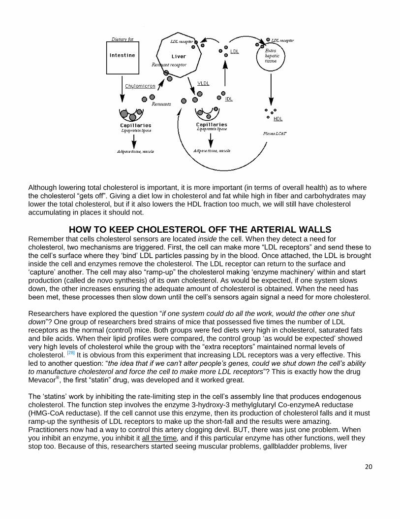
20
Although lowering total cholesterol is important, it is more important (in terms of overall health) as to where the cholesterol “gets off”. Giving a diet low in cholesterol and fat while high in fiber and carbohydrates may lower the total cholesterol, but if it also lowers the HDL fraction too much, we will still have cholesterol accumulating in places it should not.
HOW TO KEEP CHOLESTEROL OFF THE ARTERIAL WALLS
Remember that cells cholesterol sensors are located inside the cell. When they detect a need for cholesterol, two mechanisms are triggered. First, the cell can make more “LDL receptors” and send these to the cell’s surface where they ‘bind’ LDL particles passing by in the blood. Once attached, the LDL is brought inside the cell and enzymes remove the cholesterol. The LDL receptor can return to the surface and ‘capture’ another. The cell may also “ramp-up” the cholesterol making ‘enzyme machinery’ within and start production (called de novo synthesis) of its own cholesterol. As would be expected, if one system slows down, the other increases ensuring the adequate amount of cholesterol is obtained. When the need has been met, these processes then slow down until the cell’s sensors again signal a need for more cholesterol. Researchers have explored the question “if one system could do all the work, would the other one shut down”? One group of researchers bred strains of mice that possessed five times the number of LDL receptors as the normal (control) mice. Both groups were fed diets very high in cholesterol, saturated fats and bile acids. When their lipid profiles were compared, the control group ‘as would be expected’ showed very high levels of cholesterol while the group with the “extra receptors” maintained normal levels of cholesterol. [28] It is obvious from this experiment that increasing LDL receptors was a very effective. This led to another question: “the idea that if we can’t alter people’s genes, could we shut down the cell’s ability to manufacture cholesterol and force the cell to make more LDL receptors”? This is exactly how the drug Mevacor®, the first “statin” drug, was developed and it worked great. The ‘statins’ work by inhibiting the rate-limiting step in the cell’s assembly line that produces endogenous cholesterol. The function step involves the enzyme 3-hydroxy-3 methylglutaryl Co-enzymeA reductase (HMG-CoA reductase). If the cell cannot use this enzyme, then its production of cholesterol falls and it must ramp-up the synthesis of LDL receptors to make up the short-fall and the results were amazing. Practitioners now had a way to control this artery clogging devil. BUT, there was just one problem. When you inhibit an enzyme, you inhibit it all the time, and if this particular enzyme has other functions, well they stop too. Because of this, researchers started seeing muscular problems, gallbladder problems, liver
21
problems and even cognitive problems…some very serious with the widespread use of these new drugs. In the end it doesn’t work great after all. Duane Graveline, MD, USAF Flight Surgeon and NASA astronaut wrote a book about his personal experience with Liptor®.[29] It details the amnesia he suffered on more than one occasion. He started the drug, had an episode then discontinued it. His doctor then re-started it, with a lower dose, and the symptoms quickly returned, but a lot worse. One of the problems is that these drugs also inhibit the rate limiting step in the production of enzyme Co-Q10. This molecule is also known as ubiquinone. Co-Q10 acts as an anti-oxidant in the cells membranes, keeping the lipid bi-layer from oxidizing (basically turning into plastic), protects the cholesterol in the cellular membrane from oxidation, and is critical for the optimal production of energy in the mitochondria of the cell (which may explain the ‘weakness’ many patients experience). Merek, who first produced Mevacor® and then later, Zocor® was so concerned about this that they filed a patent in 1989 (US Patent No. 4933165) for the inclusion of enzyme Co-Q10 in their statin drugs Lovastatin® (Mevacor®) and Simvastatin® (Zocor®). The following is a claim in the patent: “A pharmaceutical composition comprising a pharmaceutical carrier and an effective antihypercholesterolemic amount of an HMG-CoA reductase inhibitor and an amount of Co-enzyme Q sub10 effective to counteract HMG-CoA reductase inhibitor associated skeletal muscle myopathy.”[30] For whatever reason, this newly patented formula was never brought to market. So what are our choices, control our cholesterol with the effective drugs suffer the side effects, or is there a better way? NOTE: Co-Q10 (ubiquinone is the only form that can be used by the body) and is in included in the WiO Protocol.
FOOD WILL CONTROL CHOLESTEROL To Lower OR To Raise It
Inhibiting the enzyme HMG-CoA reductase has proven itself to work very well with respect to controlling cholesterol levels. However, the standard pharmaceutical solution does leave something to be desired, mainly – side effects. The natural question must be asked: Is there another way to do this minus the side effects? The answer is a resounding YES, and it goes back to those master hormones that this paper started with: insulin and glucagon. Following a meal, levels of macro nutrients (glucose, triglycerides, and amino acids) begin to rise in the blood. The amounts of these nutrients dictate the ratio of levels of insulin and glucagon which the body adjusts to maintain homeostasis (see Table 1 page 4). Keep in mind, individuals with insulin resistance (IR) will produce an exaggerated amount of insulin, more than their body really needs. As these nutrients begin to enter the cells, the processes of metabolism (glycolysis, glycogenolysis, lipolysis and lipogenesis) have to “re-adjust” themselves depending on the amounts and proportions (ratio of carbs to fat to protein) of what foods were ingested as well as the metabolic rate. Because we are designed for survival, our bodies will always burn the low hanging fruit, the sugar (glucose and its storage from glycogen) first and utilize the fat last (fat contains 9 calories of energy per gram as opposed to 4 calories per gram for carbohydrates, and protein makes it a more efficient material as energy storage). A meal that consists of large amounts of carbohydrates with little fat or protein will guarantee a large release of insulin (always an even greater release in the IR/hyperinsulinemic individual), let’s follow what happens in terms of cholesterol production. The large quantity of carbohydrates (glucose) will be directed into the cells under insulin’s influence and this ready source of energy will be consumed first and the liver will transform any extra glucose to triglyceride molecules (assuming glycogen storage is full). Let’s first address the cells other than adipocytes, first.
22
• As the triglyceride (TG) comes into contact with the cellular membrane, enzymes divide it into free fatty acids (FFAs) and glycerol. The FFAs now enter the cytoplasm of the cell.
• These FFAs are activated by ATP and an enzyme called acyl-CoA synthetase (or thiokinase) to molecules of Acyl-CoA.
• Now, here’s the determining step in the fate of the original TG (note - it can enter the mitochondria to be used as fuel or it can be ‘rerouted’ to the adipocytes for storage as fat). If glucagon were ‘dominant’ at this point, it would activate the enzyme carnitine-palmitoyl tranferase I (CPT-1) and the acyl-CoA would be ‘hooked up’ to the CTP-1 ‘shuttle’ and be carried into the mitochondria’s “furnace” to be burned for fuel. But in the case of a high carbohydrate diet (meal), insulin will be the dominant metabolic hormone (IR individual combined with ‘high insulin producing meal combination’), the shuttle enzyme (CPT-1) is inhibited by insulin and the acyl-CoA is re-routed. But it doesn’t leave the cell just yet.
• Because insulin is now directing the body to store fat, it must prepare the adipocytes to accommodate the incoming volume; and because that entails the cell membrane expanding and cholesterol is an essential component of the membrane, the cell will require more. Insulin will simultaneously activate the enzyme lipoprotein-lipase to open the “gates” of the adipocyte for TG storage.
• Now, the cholesterol ‘sensors’ we mentioned earlier are activated and signal our cell go and get some cholesterol. It can either make more LDL receptors, or make some more ‘de novo’. Again, because insulin is the ruling hormone and calling the shots, it activates the enzyme HMG-CoA synthetase (located on the ER of the cell). This enzyme joins units of acyl-CoA together to form HMG-CoA. This intermediary product is acted on by another “ER” enzyme, HMG-CoA reductase (the rate-limiting step of cholesterol synthesis and the enzyme which the statin-class drugs inhibit) and the process of making more cholesterol is up and running full steam.[31, 32]
The lesson here is this: Telling insulin resistant/hyperinsulinemic patients to base their diets on the standard “Food Pyramid Guidelines” of 60-65% carbohydrates and little fat will ONLY SET THEM UP TO STORE MORE FAT (and likely lose muscle) AND MAKE MORE CHOLESTEROL. TRIGLYCERIDE LEVELS WILL CONTINUE TO WORSEN AS WILL THEIR CHOLESTEROL. AT THIS JUNCTURE, PRESCRIBING A STATIN MAY BE THE ONLY CLINICAL RECOURSE – But, as clear shown a better option is available.
Keeping the Carbohydrates Low Changes the Biochemical Pathway Now, let’s observe how the metabolic pathways are altered simply by changing the ratio of macronutrients (Fat – Protein – Carbohydrates) in the diet. Again referring to Table 1 (page 4), we will see that if we keep the carbohydrates low and increase the amount of fat and protein; we will have a huge effect on the ratio of insulin to glucagon. Following a meal, the blood glucose rises and insulin is secreted. The glucose begins to enter the muscle cells; however due to the consumption of few carbohydrates, the ready supply of glucose in the blood soon begins to decrease. This is particularly profound in IR /hyperinsulinemic individuals and
23
the phenomenon is called “reactive hypoglycemia”. When the blood glucose falls to a certain level, the pancreas will instead secrete glucagon and the adrenals secrete epinephrine (also known as adrenaline), norepinephrine and cortisol (stress hormones – each serving of MRP has Ashwagandha which has clinically been shown to reduce cortisol by 31%) as it attempts to maintain homeostasis with respect to blood glucose levels. The first effect of glucagon is to immediately stop the secretion of insulin. The next effect is to cause the liver and skeletal muscles (glycogen storage 80-90% liver 10-20% muscle) to release some glycogen, which will be converted into glucose. More importantly, the triglycerides now become a source of energy. Let’s go back to the previous scenario and take note of how things have changed:
• Again, the TGs come into contact with the liver cells membranes and are split into FFAs and glycerol. The FFAs enter the cell and are activated to molecules of Acyl-CoA by the action of thiokinase.
• Now with the changing of the guards and because glucagon is the dominant metabolic hormone, the ‘shuttle’ (enzyme CTP-1) is activated – NOT inhibited as it was with insulin – and the acyl CoA molecules can now enter the mitochondria – to be used as an energy source. Simultaneously at the adipocytes, glucagon along with epinephrine and norepinephrine has inhibited the “fat-storing enzyme” lipoprotein lipase and has activated the enzymes HSL and ATGL causing the adipocytes to release stored TG’s (stored fat).
• If at this point the body requires cholesterol, a different mechanism comes into play. Again the sensors, the SSDs, send out the signal cholesterol is needed, but glucagon has shutdown the important ‘cholesterol making enzyme’ HMG-CoA reductase (just like the ‘statins’ do). Therefore, the cell cannot use the ‘de novo’ pathway.
• As a recourse, SREBP (Sterol Regulated Element Binding Protein) is activated which directs the protein manufacturing machinery of the endoplasmic reticulum to produce new LDL receptors (the final ‘touches’ are put on these new receptors in the golgi apparatus of the cells by a process called glycosylation). [33] These new LDL receptors now go to the cell’s surface and “capture cholesterol filled LDL particles” and bring (pulling out of the blood stream) the cholesterol back inside the cell.
• The net result is a ‘flow of fat’ out of storage and its mobilization for an energy source. The’ de novo’ synthesis of cholesterol is inhibited, and the body is forced to use the cholesterol present in the blood stream, thus lowing cholesterol to healthy levels.
FAT LOSS OR WATER WEIGHT?
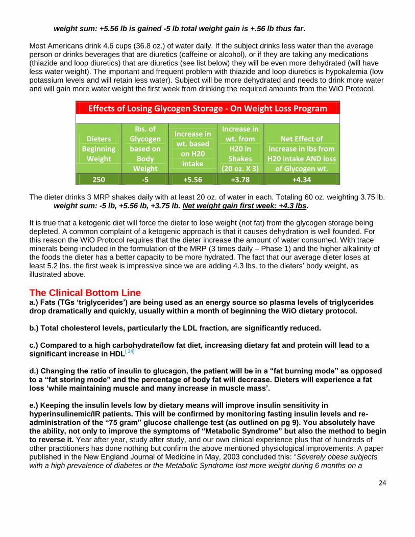
Some critics make the claim that the weight that is lost in the first few weeks is all glycogen (water weight) and not fat. Where it is true that the glycogen is dumped the first week (except in those that are very large-it can take 10-14 days) it does not explain the pounds that are lost in the first few weeks. Let’s dive into the numbers and find an explanation. We are going to use a male weighing 250 lbs. as an example: Studies show that a man has approximately .08 oz. of glycogen per pound of body weight [56]. Each molecule of glycogen is bonded to four (4) water molecules. Thus, consuming 94g of carbohydrates will increase body weight by one (1) lb. within 24 hours. A 250 lbs. man has approximately 5 lbs. of glycogen held in storage in the liver and muscles. When a dieter begins the WiO Protocol the carbohydrate consumption is drastically reduced which forces the liver and muscles to release glycogen into the blood stream. After 3-4 days the glycogen storage is depleted and will result in a reduction in body weight of five (5) pounds.
weight sum: -5 pounds is lost thus far To maintain proper hydration the dieter begins drinking half their body weight (in ounces) per day, in this example 125 oz. of water weight increases the dieters body weight 5.56 lbs.
24
weight sum: +5.56 lb is gained -5 lb total weight gain is +.56 lb thus far. Most Americans drink 4.6 cups (36.8 oz.) of water daily. If the subject drinks less water than the average person or drinks beverages that are diuretics (caffeine or alcohol), or if they are taking any medications (thiazide and loop diuretics) that are diuretics (see list below) they will be even more dehydrated (will have less water weight). The important and frequent problem with thiazide and loop diuretics is hypokalemia (low potassium levels and will retain less water). Subject will be more dehydrated and needs to drink more water and will gain more water weight the first week from drinking the required amounts from the WiO Protocol.
Effects of Losing Glycogen Storage - On Weight Loss Program 0.08 20
Dieters Beginning
Weight
lbs. of Glycogen based on
Body Weight
Increase in wt. based
on H20 intake
Increase in wt. from
H20 in Shakes
(20 oz. X 3)
Net Effect of increase in lbs from H20 intake AND loss
of Glycogen wt.
250 -5 +5.56 +3.78 +4.34 The dieter drinks 3 MRP shakes daily with at least 20 oz. of water in each. Totaling 60 oz. weighting 3.75 lb.
weight sum: -5 lb, +5.56 lb, +3.75 lb. Net weight gain first week: +4.3 lbs. It is true that a ketogenic diet will force the dieter to lose weight (not fat) from the glycogen storage being depleted. A common complaint of a ketogenic approach is that it causes dehydration is well founded. For this reason the WiO Protocol requires that the dieter increase the amount of water consumed. With trace minerals being included in the formulation of the MRP (3 times daily – Phase 1) and the higher alkalinity of the foods the dieter has a better capacity to be more hydrated. The fact that our average dieter loses at least 5.2 lbs. the first week is impressive since we are adding 4.3 lbs. to the dieters’ body weight, as illustrated above.
The Clinical Bottom Line
a.) Fats (TGs ‘triglycerides’) are being used as an energy source so plasma levels of triglycerides drop dramatically and quickly, usually within a month of beginning the WiO dietary protocol. b.) Total cholesterol levels, particularly the LDL fraction, are significantly reduced. c.) Compared to a high carbohydrate/low fat diet, increasing dietary fat and protein will lead to a significant increase in HDL[ 34] d.) Changing the ratio of insulin to glucagon, the patient will be in a “fat burning mode” as opposed to a “fat storing mode” and the percentage of body fat will decrease. Dieters will experience a fat loss ‘while maintaining muscle and many increase in muscle mass’. e.) Keeping the insulin levels low by dietary means will improve insulin sensitivity in hyperinsulinemic/IR patients. This will be confirmed by monitoring fasting insulin levels and re-administration of the “75 gram” glucose challenge test (as outlined on pg 9). You absolutely have the ability, not only to improve the symptoms of “Metabolic Syndrome” but also the method to begin to reverse it. Year after year, study after study, and our own clinical experience plus that of hundreds of other practitioners has done nothing but confirm the above mentioned physiological improvements. A paper published in the New England Journal of Medicine in May, 2003 concluded this: “Severely obese subjects with a high prevalence of diabetes or the Metabolic Syndrome lost more weight during 6 months on a

25
carbohydrate-restricted diet than on a calorie and fat-restricted diet, with a relative improvement in insulin sensitivity and triglyceride levels, even after adjustment for the amount of weight lost.” [35] f). If dieter is taking any diuretics it is possible for hyperkalemia (low potassium) to manifest (see charts below).
Conclusions from a study published in the Annals of Internal Medicine in May 2004 echoed the same opinion: “Compared with a low-fat diet, a low-carbohydrate diet program had better participant retention and greater weight loss. During active weight loss, serum triglyceride levels decreased more and high-density lipoprotein cholesterol levels increased more with a low-carbohydrate diet than with a low-fat diet.” [36] Gerald M. Reaven, MD (the one who first coined the term “Metabolic Syndrome” more commonly known as Metabolic Syndrome) summed up nicely his experience with hyperinsulinemic/IR patients on a high carbohydrate/low fat diet versus a low carbohydrate/high fat diet in a 2001 article published in San Francisco Medicine.[37] Dr. Reaven states that “the most dramatic improvements in the manifestations of Metabolic Syndrome occur in overweight, insulin resistant/hyperinsulinemic individuals when they lose weight. However, there appears to be little or no evidence, as long as the energy content is kept constant, that low fat/high carbohydrate diets will directly improve insulin sensitivity. On the other hand, there is considerable evidence that isocaloric diets low in fat and enriched in carbohydrates will accentuate the manifestations of Metabolic Syndrome. The more insulin resistant an individual, the greater is the amount of insulin that must be secreted in response to a carbohydrate-enriched diet in order to maintain glucose homeostasis. Thus, the inevitable and consistently replicated effect of replacing saturated fat with carbohydrates in insulin resistant individuals is to increase the concentration of triglyceride-rich lipoproteins, both fasting and postprandial. The increase in the ambient TG-rich lipoproteins seen following low fat/high carbohydrate diets is associated with a decrease in HDL-cholesterol concentration; and more recently, it appears that such diets will convert the LDL to VLDL in half the individuals who had either high LDL or an intermediate pattern at the outset. Given the evidence that low fat/ high carbohydrate diets do not modify the basic defects in Metabolic Syndrome (insulin resistance) and accentuates all of its metabolic manifestations, there seems to be little rationale for substituting saturated fat with carbohydrates This is particularly true in light of the multiple observations that replacing saturated fat with mono-saturated or polyunsaturated fat, or both, will lead to the same decrease in LDL cholesterol without any of the adverse metabolic effects seen with low fat carbohydrate diets.” [38, 39] Insulin Resistance, Coronary Heart Disease and Diet Gerald M. Reaven, MD “The most dramatic improvements in manifestations of Syndrome X occur in overweight, insulin resistant/hyperinsulinemic individuals, when they lose weight. However, there appears to be little or no evidence, as long as the energy content is kept constant, that low fat/high (CHO – high carbohydrate) diets will directly improve insulin sensitivity. On the other hand, there is considerable evidence that isocaloric diets, low in fat and enriched in CHO, will accentuate the manifestations of Syndrome X. The more insulin resistant an individual, the greater is the amount of insulin that must be secreted in response to a CHO-enriched diet in order to maintain glucose homeostasis. Thus, the inevitable and consistently replicated effect of replacing SF with CHO in insulin-resistant individuals is to increase in concentration of TG-rich lipoproteins, both fasting and postprandial. The increase in the ambient TG-rich lipoproteins seen following low fat/high CHO diets is associated with a decrease in HDL-cholesterol concentration and, more recently it appears that such diets will convert the LDL to VLDL in half of the individuals who had either high VDL or an intermediate pattern at the outset. Given the evidence that low fat/ high CHO diets do not modify the basic defects in Syndrome X (insulin resistance), and accentuate all of its metabolic manifestations, there seems to be little rationale for substituting saturated fat (SF) with CHO. This is particularly true in light of the multiple observations that replacing SF with monounsaturated fat (MUF) or polyunsaturated fat (PUF), or both, will lead to the same fall in LDL-cholesterol, without any of the adverse metabolic effects seen with low fat CHO diets.
(14, 15) At
best, low fat/high CHO diets will only adversely affect CHD risk
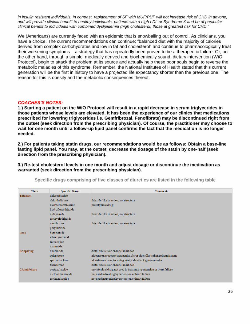
26
in insulin resistant individuals. In contrast, replacement of SF with MUF/PUF will not increase risk of CHD in anyone, and will provide clinical benefit to healthy individuals, patients with a high LDL or Syndrome X and be of particular clinical benefit to individuals with combined dyslipidemia (high cholesterol) those at greatest risk for CHD.”
We (Americans) are currently faced with an epidemic that is snowballing out of control. As clinicians, you have a choice. The current recommendations can continue; “balanced diet with the majority of calories derived from complex carbohydrates and low in fat and cholesterol” and continue to pharmacologically treat their worsening symptoms – a strategy that has repeatedly been proven to be a therapeutic failure. Or, on the other hand, through a simple, medically derived and biochemically sound, dietary intervention (WiO Protocol), begin to attack the problem at its source and actually help these poor souls begin to reverse the metabolic maladies of this syndrome. Remember, the National Institutes of Health stated that this current generation will be the first in history to have a projected life expectancy shorter than the previous one. The reason for this is obesity and the metabolic consequences thereof. COACHES’S NOTES: 1.) Starting a patient on the WiO Protocol will result in a rapid decrease in serum triglycerides in those patients whose levels are elevated. It has been the experience of our clinics that medications prescribed for lowering triglycerides i.e. Gemfribrozal, Fenofibrate) may be discontinued right from the outset (seek direction from the prescribing physician). Of course, the practitioner may choose to wait for one month until a follow-up lipid panel confirms the fact that the medication is no longer needed. 2.) For patients taking statin drugs, our recommendations would be as follows: Obtain a base-line fasting lipid panel. You may, at the outset, decrease the dosage of the statin by one-half (seek direction from the prescribing physician). 3.) Re-test cholesterol levels in one month and adjust dosage or discontinue the medication as warranted (seek direction from the prescribing physician).
Specific drugs comprising of five classes of diuretics are listed in the following table
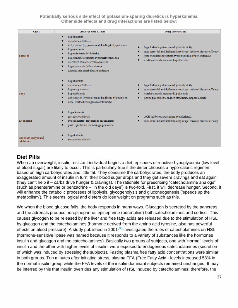
27
Potentially serious side effect of potassium-sparing diuretics is hyperkalemia. Other side effects and drug interactions are listed below:
Diet Pills
When an overweight, insulin resistant individual begins a diet, episodes of reactive hypoglycemia (low level of blood sugar) are likely to occur. This is particularly true if the dieter chooses a hypo-caloric regimen based on high carbohydrates and little fat. They consume the carbohydrates, the body produces an exaggerated amount of insulin in turn, their blood sugar drops and they get severe cravings and eat again (they can’t help it – carbs drive hunger & cravings). The rationale for prescribing “catecholamine analogs” (such as phenteramine or benzadrine – ‘in the old days’) is two-fold. First, it will decrease hunger. Second, it will enhance the catabolic processes of lipolysis, glycogenolysis and gluconeogenesis (‘speeds up the metabolism’). This seems logical and dieters do lose weight on programs such as this. We when the blood glucose falls, the body responds in many ways. Glucagon is secreted by the pancreas
and the adrenals produce norepinephrine, epinephrine (adrenaline) both catecholamines and cortisol. This
causes glycogen to be released by the liver and free fatty acids are released due to the stimulation of HSL
by glucagon and the catecholamines (hormone derived from the amino acid tyrosine, also has powerful
effects on blood pressure). A study published in 2001[22] investigated the roles of catecholamines on HSL
(hormone-sensitive lipase was named because it responds to a variety of substances like the hormones
insulin and glucagon and the catecholamines). Basically two groups of subjects, one with ‘normal’ levels of
insulin and the other with higher levels of insulin, were exposed to endogenous catecholamines (secretion
of which was induced by stressing the subjects). Fasting plasma free fatty acid concentrations were similar
in both groups. Ten minutes after initiating stress, plasma FFA (Free Fatty Acid - levels increased 53% in
the normal insulin group while the FFA levels of the insulin dominant subjects remained unchanged. It may
be inferred by this that insulin overrides any stimulation of HSL induced by catecholamines; therefore, the
28
weight loss most affected by using “diet drugs” appears to be more of lean muscle mass (via glycogenolysis
and gluconeogenesis) rather than the loss of fat. Losing muscle mass in the WiO Protocol is unacceptable
and is deadly. One of the longest studies conducted on weight loss and the importance of maintaining
muscle concluded; that if you lose muscle when losing weight (fat) mortality rates increased by 39%. Weight
loss was associated with higher death rates. BUT – if you maintain muscle while losing FAT the mortality
rates improved by 17%. [55]. Losing muscle will also lower the metabolic rate so maintaining continued fat
loss will be even more difficult. The cardiovascular risks previously described for hyperinsulinemic
individuals makes giving stimulants a “dicey” business. The “Phen-Fen” protocol is a great example of how
risky this can be…remember, the heart is a muscle, and this protocol attacks muscle… any muscle.
COACHES’S NOTES: The dieter should never be hungry on the WiO Protocol (after the first week). Hunger usually is a result of too many carbohydrates. Check the weekly food diary for hidden carbs AND amounts of food eaten. Make sure no meals were skipped (or more than 4-5 hours ‘a-wake hours’ between meals) they consumed the required water, salads and vegetables.
Ketogenesis (Healthy) Ketosis vs. ‘Ketoacidosis’ (Deadly) Ketogenesis and Ketoacidosis are NOT the same thing and must not be confused with each other. The WiO Protocol does not nor is it possible to put someone into a state of Ketoacidosis (see Type I diabetes). One must keep in mind, that although fats are both oxidized to acetyl-CoA and synthesized from acetyl Co-A, they are not simply the reverse of the same biochemical reaction. These processes take place in two different compartments of the cell:
(1) Fats are made in the cytosol and oxidized in the mitochondria. (2) Fats get ‘burnt’ under glucagon’s influence and created under insulin’s influence.
Under certain conditions such as starvation, untreated Type I diabetes, or going on a carbohydrate restricted diet, the body is forced to utilize stored or dietary fat and possibly protein (dietary or catabolized muscle ‘catabolic’) as its primary sources of energy. Because of the increased rate of oxidation of fatty acids, intermediary products called ketone bodies can build up in the Liver. Ketones are acidic substances, and if enough of them build up, they can precipitate a dangerous condition called ketoacidosis, which can quickly become fatal (basically the acidic condition of the blood prevents it from carrying sufficient oxygen). This can only occur in individuals with Type I diabetes or individuals with severe liver or kidney disease, severe alcoholics. In a ‘normal’, healthy person, the pathological state of ketoacidosis cannot occur.
The reason for this is simple. They produce insulin and the two organs responsible for disposing/metabolizing the ketonic bodies are functional. While the body is burning fat, it is also creating glucose through gluconeogenesis (protein catabolism is possible); and to a smaller extent utilizing the glycerol molecules cleaved off of the triglycerides when the free fatty acids are ‘liberated’ as additional substrates in this process. As glucose levels rise, insulin is secreted. When the concentration in the blood reaches a certain level, the process of ketogenesis is inhibited.
Gluconeogenesis (synthesis of glucose from molecules that are not carbohydrates) occurs in the liver and, unknown by many people, also in the kidneys [23] which actually produce about 54% of the glucose generated from the entire gluconeogenic process. [24] Thus, the process of ketogenesis is a component of normal metabolism (and is perfectly safe) – it would only be considered pathological in certain disease states. The body also has another ‘built-in safety mechanism’ to prevent ketoacidosis. While in the ketogenic state, the liver and kidneys are engaged in gluconeogenesis, or the production of glucose as described above. This must occur simultaneously with ketogenesis as some of the cells of the brain, the nucleated blood cells and cells of the adrenal medulla (there may be a few others) must have glucose as their sole source of energy (most of the other cells of the body actually do quite well using ketonic bodies as
29
their fuel). As these processes occur, nitrogen-based wastes accumulate and ammonia is produced. When the body becomes acidic, ammonia can “pick-up” a hydrogen ion, forming the ammonium ion and decreasing the level of acid as a result. The ammonium ion then reacts with the ‘alpha-amino nitrogen’ of aspartate and by a series of five catalytic reactions; urea is ultimately produced and excreted [25] .It must be emphasized that the WiO Protocol is a very alkaline protocol (discussed in acid/base balancing). Each MRP shake is providing sufficient alkaline minerals helps the body maintain the proper concentration of the bicarbonate ion in the blood (an important acid/base buffer) and represents another way the body’s physiology copes with an acid overload. Again, the standard WiO Protocol is contra-indicated for Type I diabetics and those suffering from liver or kidney disease (Use the Alternative WiO Protocol for these persons). For all others, it is not only perfectly safe, but it represents a normal metabolism albeit using a metabolic pathway that is not frequently required given today’s standard high carbohydrate based diet. Remember, there is a correction phase of this protocol (Phase 1) and a maintenance phase (Phase 4) and they are two entirely different metabolic formats. COACHES’S NOTES: They are becoming less popular but you may experience a patient who comes in and is concerned ‘that the program isn’t working!’ They explain that they are not in ketosis because they used Keto-stix and report that it didn’t turn purple. Explain to them that “ketosis” is not an ‘all or none’ proposition. During the protocol, they will be deriving much of their energy from ketogenesis, but the body is never in ketogenesis 100%. Keto-stix is a test used to detect the presence of ketone bodies in the urine that the body was unable to use for fuel. As dieter progress through the first 2 – 3 weeks of the protocol the body has had enough time to get all of the necessary enzymes synthesized so that it may burn more of these ketone bodies more efficiently – thus fewer will be excreted through the urine. If they are exercising, they may never “spill’ any Ketones into the urine. Also excess ketones may be eliminated via the lungs. This can explain why dieters will have bad-breath (acetone or ketone breath) and/or in the feces. For all of these reasons, the “dip-stick test” is not a reliable method by which to measure the level of ketosis occurring in the body. The weekly food diary, if they are being totally honest, is the ‘gold standard’ by which to gauge compliance. Progress on the program MUST consist of other methods besides the scale only. The weekly 10 point body measurements and body fat/muscle and hydration measurements are key (Body Fat impedance equipment). After a few weeks muscle mass is going to increase and this will conflict with the readings on the scale. It is important to note that muscle is nearly twice as dense as fat. Because most people are not either, consuming enough or not digesting [see digestion section] enough protein to maintain their muscle mass they are slowing losing muscle mass (decreasing metabolic rate) and have been for years. This would explain why they complain of feeling like they have less energy and strength than they did just five or ten years previous. This equipment will show lean muscle mass, body fat %, cellular hydration, and more. This is an excellent clinical tool, and is required to incorporate it into your WiO Clinic. We recommend not using “Keto-Stix” by your patients or staff.

30
EICOSANOIDS - HOW THE BODY CONTROLS INFLAMMATION
“There are few biological pathways that are more central to overall…cardiovascular disease than the eicosanoid pathway”. “Regulation of Eicosanoid Pathways: A Pathway to Health and Development” Mark E. Cook Animal Sciences Department, University of Wisconsin “You can view eicosanoids as the biological glue that holds together the human body. In that regard they are the most powerful agents known to man, yet they are totally controlled by the diet.” Dr. Barry Sears A gang of a least 100 powerful hormone like substances that control virtually all physiological actions in the body is an elite group called eicosanoids. The most important thing about eicosanoids is to keep them in balance. If you have too little of one, too little of another, eicosanoids can send he body hurtling down the slippery slope of biochemical evil towards arthritis, blood clots and dozens of other dangerous conditions. “In fact, they play a major role in most diseases, including heart disease and cancer”. [16] It is impossible to discuss the health benefits of essential Fatty Acids more commonly known as Omega-3, 6, and 9 without discussing "eicosanoids." Chances are you have controlled these powerful hormones, by taking a special little white pill. In 1971, it was discovered that aspirin actually works by changing the level of eicosanoids. Eicosanoids' mission as an autocrine hormone is that they are secreted by the cell to test the external environment and then report back to the cell what is going on just outside the cell by interacting with its receptor on the cell surface, similar to how cells determine the level of cholesterol and if more is needed. Using this information, the cell can take the appropriate biological action to respond to any change in its environment. They do that by a system called a “second messenger” – which I will talk about in just a minute. Eicosanoids can send your body hurtling down the slippery slope of a biochemical evil towards: arthritis, blood clots, arterial constriction, depression, heart disease, dry skin - rashes, allergies, headaches, splitting nails, asthma, hormonal disorder and literally dozens of other dangerous conditions. Studies show that they play a major role in most diseases like cancer and heart disease. But when they are balanced you body runs along as it was designed …in perfect health.
What are Eicosanoids?
Eicosanoids are micro-hormones derived from long-chain essential fatty acids, they are called ‘micro’ because they do all their business INSIDE the cells and act in a fraction of a second. They are also some of the most powerful hormones, since they affect the synthesis of every other hormone in your body. In a sense, eicosanoids can be considered as “super-hormones” acting under the guidance of the Master Hormones – Insulin and Glucagon. They are capable of great health benefits (“good” eicosanoids) or great harm (“bad” eicosanoids), like the green and red light analogy I mentioned earlier. This balance depends on which eicosanoid a cell produces. Many doctors are not familiar with eicosanoids but that you may be aware of prostaglandins, leukotrienes, thromboxanes, interferons, and interleukins known collectively as eicosanoids. If your patient has ever experienced a headache, menstrual cramps, abdominal discomfort from ulcers, swelling or inflammation or rash and you prescribed aspirin (or any other large number of drugs) this drug(s) specifically interferes with the formation of prostaglandins and other eicosanoids. Eicosanoids are produced inside the cells, act inside the cells, and vanish in fractions of seconds, much too quickly to be detected easily, which is the reason they were just discovered a few decades ago. 1982 Nobel Prize Unlike typical hormones that are produced by a particular gland (i.e. pancreas , liver), every cell in your body is capable of producing eicosanoids. In essence, you have about sixty trillion eicosanoid glands, and by flowing the WIO Protocol you will maintain an appropriate balance of these molecular building blocks of both “good” and “bad” eicosanoids in each cell.
31
The terms “good” and “bad” eicosanoids are simply operational terms, terms that describe very powerful, but opposite physiological actions generated by different eicosanoids. Just keep in mind that the patient needs a balance of “good” and “bad” eicosanoids for optimal health. Just as we need both good and bad cholesterol if your patient had no “bad” cholesterol, their cells would literally fall apart and he/she would die. What patients need, though, is an appropriate balance between “good” and “bad” cholesterol to help reduce the risk of heart disease. You can think of eicosanoids in a similar fashion, but realize that they’re vastly more important than cholesterol in terms of their impact on your overall health, in spite of the attention given to cholesterol. You can see from the list of “bad” eicosanoids appear to have very few redeeming characteristics, since many chronic diseases can be viewed as an excess of “bad” eicosanoid production. Here are some examples of chronic diseases that result from an excess production of “bad” eicosanoids.
• Alzheimer’s • Arthritis • Cancer • Depression • Heart attack • Hypertension • Stroke
You may be asking “Why not just eliminate all the “bad” eicosanoids so that you would never get a heart attack or cancer”? It’s not quite that simple. Let’s take the example of a heart attack. If you didn’t have enough “bad” eicosanoids, you would probably bleed to death, since you need some “bad” eicosanoids that cause your blood to form a clot that stops you from bleeding to death. Of course, if you are producing too many “bad” eicosanoids your platelets will clot at the wrong time to stop blood flow and will result in a stroke or heart attack. The same is true of high blood pressure, cancer, pain, immune disorders, and neurological diseases. Doesn’t this sound familiar to the importance of balancing the amount of insulin in our bodies? What the patient needs is an improved balance of “good” and “bad” eicosanoids, since most chronic diseases stem from an imbalance of eicosanoids, not a deficiency of them. For many of your patients/dieters they only care about losing their weight or perhaps their goal is to lower their cholesterol and blood pressure. Many diabetics will be attracted to the WIO Protocol and they many never ask you about how the WIO diet will help them with their eicosanoids, but it is important that you know we have designed the formulation and the protocol regimen to balance these master micro-hormone as well as the other hormones in the body. You will learn that our attention to these details will make your clients health and happy, and that is why they will continue to refer people to your clinic and why they will continue to purchase the WiO products from you and benefit from your expertise.
Second Messengers These are the ultimate key to hormonal action and act through the following sequence of events:
1. A hormone like insulin docks on a cell receptor 2. The receptor which spans the membrane of the cell undergoes a change that is transmitted to the interior of the cell 3. Depending on the receptor and the hormone that has activated, a molecule is synthesized within the cell that completes the message 4. These new molecules are called second messengers of which there are two primary kinds: cAMP, which is considered the "green light" for cells
"Good" eicosanoids interact with receptors that produce this second messenger; Inositol triphosphate/diaclglycerol (IPx/DAG) which is equivalent to the "red light" (see illustration below) for the cell

32
and usually has a physiological action opposite to that of cAMP. Both insulin and "bad" eicosanoids use this pathway. Once your green lights and red lights are balanced and working smoothly your results are health and wellness. If you have an excess of red lights, then your traffic signals are out of balance, just as they would be in the city where are too many red lights at the same time causing major grid lock. This imbalance of red lights will result is the development of chronic disease.
General Concepts about Eicosanoids The goal is to increase the production of cAMP (Cyclic adenosine monophosphate / Cyclic AMP) are your key to maintaining wellness because cAMP is used as a second messenger and used by a number of endocrine hormones in the body to transmit their biological information to the appropriate target cell throughout the body. Thus, an eicosanoid's effect on second messengers becomes the definition of whether it will be a "good" or "bad" eicosanoid. A good eicosanoid will increase the levels of cAMP in a cell, whereas a "bad" eicosanoid will decrease the level of cAMP. Wellness really is a function of balance between "good" and "bad" eicosanoids. The AA/EPA ration (Arachidonic Acid, Eicosapentaenoic Acid both essential fatty acids [Omga-3, 6 fats]) in the blood will indicate where you stand in terms of having a balance between good and bad eicosanoids. This balancing act will be achieved and maintained by following the WiO Protocol and consuming the required amounts of Dr. Udo’s 3-6-9 Oil. I will explain later in greater detail why we use Dr. Udo’s Oil rather than Fish Oils or other brands. Pharmacists and physicians understand the concept of maintaining drugs within a therapeutic zone is well known to medical community. If the prescribed drug falls below that designated therapeutic zone, the drug is ineffective, and above that therapeutic zone, the drug is toxic. That same concept must be applied to the
blood pressure
triglycerides lower
improved
Omega-3derived
eicosanoids
rheumatoid arthritis pain
inflammation
major depression
Clinical Implications
sudden cardiac death
coronary artery disease
platelet aggregation
Omega-3Omega-6Eicosapentanoic acid (EPA)
Omega 6derived
Series 3
Prostanoids
Series 5
Leukotrienes
Prostaglandins
Leukotrienes
Thromboxanes
The Omega-6 Omega-3 Balacning Act
Production of:
more
higher
higher
Production of:
Anti-inflammatory
mood elevation
mood stabilization
vasoconstriction
increased
eicosanoids
Prostaglandins
Leukotrienes
Thromboxanes
Series 4
Leukotrienes
decreased risk
decreased risk
decreased
less
lower
Arachidonic acid (AA)
Pro-inflammatory
Series 2
Prostanoids
increased risk
increased risk
less
less
more
more
worse
more
more
less
less
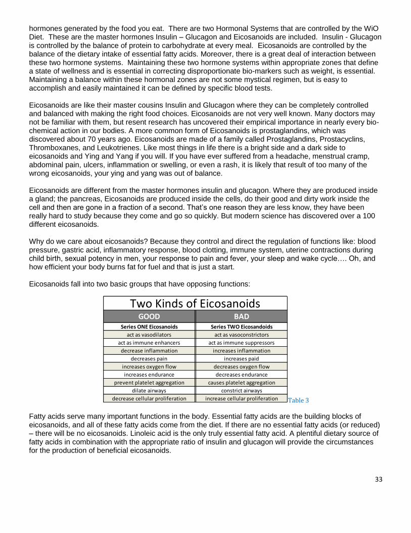
33
hormones generated by the food you eat. There are two Hormonal Systems that are controlled by the WiO Diet. These are the master hormones Insulin – Glucagon and Eicosanoids are included. Insulin - Glucagon is controlled by the balance of protein to carbohydrate at every meal. Eicosanoids are controlled by the balance of the dietary intake of essential fatty acids. Moreover, there is a great deal of interaction between these two hormone systems. Maintaining these two hormone systems within appropriate zones that define a state of wellness and is essential in correcting disproportionate bio-markers such as weight, is essential. Maintaining a balance within these hormonal zones are not some mystical regimen, but is easy to accomplish and easily maintained it can be defined by specific blood tests. Eicosanoids are like their master cousins Insulin and Glucagon where they can be completely controlled and balanced with making the right food choices. Eicosanoids are not very well known. Many doctors may not be familiar with them, but resent research has uncovered their empirical importance in nearly every bio-chemical action in our bodies. A more common form of Eicosanoids is prostaglandins, which was discovered about 70 years ago. Eicosanoids are made of a family called Prostaglandins, Prostacyclins, Thromboxanes, and Leukotrienes. Like most things in life there is a bright side and a dark side to eicosanoids and Ying and Yang if you will. If you have ever suffered from a headache, menstrual cramp, abdominal pain, ulcers, inflammation or swelling, or even a rash, it is likely that result of too many of the wrong eicosanoids, your ying and yang was out of balance. Eicosanoids are different from the master hormones insulin and glucagon. Where they are produced inside a gland; the pancreas, Eicosanoids are produced inside the cells, do their good and dirty work inside the cell and then are gone in a fraction of a second. That’s one reason they are less know, they have been really hard to study because they come and go so quickly. But modern science has discovered over a 100 different eicosanoids. Why do we care about eicosanoids? Because they control and direct the regulation of functions like: blood pressure, gastric acid, inflammatory response, blood clotting, immune system, uterine contractions during child birth, sexual potency in men, your response to pain and fever, your sleep and wake cycle…. Oh, and how efficient your body burns fat for fuel and that is just a start. Eicosanoids fall into two basic groups that have opposing functions:
Table 3
Fatty acids serve many important functions in the body. Essential fatty acids are the building blocks of eicosanoids, and all of these fatty acids come from the diet. If there are no essential fatty acids (or reduced) – there will be no eicosanoids. Linoleic acid is the only truly essential fatty acid. A plentiful dietary source of fatty acids in combination with the appropriate ratio of insulin and glucagon will provide the circumstances for the production of beneficial eicosanoids.
GOOD BADSeries ONE Eicosanoids Series TWO Eicosandoids
act as vasodilators act as vasoconstrictors
act as immune enhancers act as immune suppressors
decrease inflammation increases inflammation
decreases pain increases paid
increases oxygen flow decreases oxygen flow
increases endurance decreases endurance
prevent platelet aggregation causes platelet aggregation
dilate airways constrict airways
decrease cellular proliferation increase cellular proliferation
Two Kinds of Eicosanoids

34
There are three points along the eicosanoids synthesis pathway where we can exert dietary influence over the eicosanoids end products.
The First control point: is at the start of the process, where Linoleic acid enters the system. Linoleic acid is the raw material – insulin and glucagon is the processors, and eicosanoids the finished products. The Second control point: is by altering the synthesis process itself in a way that results in the production of predominantly “good” eicosanoids. The Third: is by restricting the dietary intake of arachidonic acid, a precursor of many of the “bad” eicosanoids. The figure below shows the representation of the eicosanoids synthesis pathway, and shows how our protocol affects these control points.
As we have discussed, they can be “re-converted” to triglycerides and stored as fat, they can be utilized as a rich source of energy, they can be transformed into phospholipids and become integral parts of the cellular membranes, and they can be converted into cholesterol; however, there are two “special types” of fatty acids that, in addition to playing roles in all of the above mentioned physiological processes, have other unique properties. Humans cannot synthesize these molecules and must obtain them from dietary sources (hence the name ‘essential fatty acids’), these are the “Omega-3 oils”, “Omega-6 oils” and “Omega-9 oils”. The “omega nomenclature” simply means that the first carbon-to-carbon double bond occurs at carbon #3 from the ‘omega-end’ (i.e. the non-acid end) of the fatty acid molecule. Due to the American diet and appetite in the most part created by commercial production of our foods, we consume dangerously too much omega-6. To obtain optimum health and for an ideal transformation of phospholipids, a balance of omega-3 at a 2:1 ratio to omega-6 and the addition of omega-9 must be maintained. Omega-6 fatty acid would have the first carbon double band at carbon #6 from the omega-end and omega-9 at the carbon #9 from the omega-end. Omega-6 oils are most commonly derived from plant sources. Most vegetable oils, as well as most ‘seed oils’, contain predominately omega-6 fatty acids. Flax
Linoletic Acid
Delta 6 Desaturase
Inibited by:
Trans Fatty Acids
Alpha Linolenic Acid
High Carbohydrate
diet
Eicosainods
Synthesis
Process
Delta 5 Desaturase
Activated by Insulin
Inhibied by Gluacogon
& EPA
Series One
Eicosanoids
Arachidonic
Acid
Series Two
Eicosanoid
Intake of Arachidonic Acid
Precursor of BAD Eicosanoids

35
seed oil (an exception) contains a high percentage (57%) of alpha linolenic acid, omega-3 oil, and canola oil also contains some omega-3s, although only about 10%. Common sources of omega-3 oils are the “marine oils” such as fish oil, cod liver oil and krill oil, not necessarily the ideal source but the most common. Although the body may use omega-3 and the omega-6 fatty acids like other fatty acids, these two groups have another unique and very important physiological function. They are the building blocks or substrates (particularly the omega-6 oils such as linoleic acid) for a class of compounds referred to as eicosanoids. The word is derived from the Greek word meaning twenty (eikosi). These are a family of at least 100 compounds all containing 20 carbon atoms. Arguably these substances, even though they may only exist for seconds or milli-seconds before being degraded, (and that is why we do not have standard laboratory tests to ascertain their concentrations) may be some of the most powerful substances in the body in terms of orchestrating profound physiological effects. These ‘biochemical controllers” work not only inside the cell, but also serve as “mini-hormones’ in that they signal adjacent cells to perform specific tasks. Eicosanoids control coagulation and anti-coagulation of the blood, they can control dilation and constriction of the bronchioles, and they are responsible for the degree of inflammation associated with the immune response (i.e. how much fever, how much pain, how much swelling is produced during a “counter-attack” on an invading pathogen). In layman’s terms, they ‘send out the cavalry AND call it back’. In other words, eicosanoids instigate the inflammatory processes and calm them – they modulate the degree and intensity of the immune response ensuring the minimum amount of ‘collateral damage’ is done). Pharmacologically, we can only increase the immune response or suppress it…..we cannot modulate it! How important is all of this in your daily clinical practice? How many anti-inflammatory drugs do you prescribe, both steroidal and non-steroidal? If you are questioning the profound impact these short-lived “little things” have on human physiology, look at the records of Bextra® and Vioxx® and look at the side-effect profile of the corticosteroids. Obviously as clinicians we want to alleviate the pain, inflammation and collateral tissue destruction associated with increased levels of certain eicosanoids such as leukotrienes, thromboxanes, interferons, interleukines and prostaglandins. In conditions such as rheumatoid arthritis, Crohn’s disease, pelvic inflammatory disease, colitis or any “itis”, there is an exaggerated immune response. So logically, we prescribe ‘immunosupressants’ in an attempt to ameliorate the symptoms. However, there is only so far we can go along this path until side-effects, usually manifestations of a suppressed immune system, come back to haunt us and we then attempt to counter-act these problems by prescribing immuno-stimulants (such as Epogen® for instance). Let’s consider for a moment the impact our protocol diet plays in all of this. (Table 3) lists the two general classes of eicosanoids and their physiological effects. Notice how insulin and glucagon influence the expression of the two classes. It is apparent that one would probably prefer to live the majority of one’s life under the direction of the series on eicosanoids, although at times, the series two eicosanoids are very important. If you have an infection, you need an inflammatory response – but not too much. If you are bleeding, platelets need to coagulate, but you wouldn’t want ‘sticky blood’ all the time. Optimal health depends on a balance between the two types and many chronic disease states arise when the eicosanoids are constantly out of balance. A huge part of the pharmaceutical industry concerns itself with the production of anti-inflammatory compounds – both steroidal and non-steroidal (NSAIDs) medications. These drugs inhibit the synthesis of many of these compounds, such as prostaglandins, leukotrienes or thromboxanes in order to suppress symptoms. The problem is that it is very difficult, or impossible, to exquisitely control their balance pharmacologically. Aspirin, for example, will inhibit platelet aggregation, decrease pain, inflammation, and has anti-pyretic properties; however, it will also decrease production of the prostaglandins that protect the stomach from the acid it produces, hence making G.I. bleeds a common side-effect. The ultimate 21 therapeutic tool would be to channel the majority of eicosanoid synthesis so that ‘we spend most of our time’ under the influence of the beneficial series one eicosanoids, yet do not inhibit the body from producing the series two eicosanoids when necessary. In other words, we do not want to stimulate or inhibit these two types of compounds, like drugs do, but rather we

36
want to modulate them, that is let the body remain in control. Fantastic request, but how do we accomplish this?
Two Classes of Eicosanoids Glucagon Dominant Insulin Dominant
Series One Eicosanoids Series Two Eicosanoids Act as vasodilators………………………………… Act as vasoconstrictors
Act as immune enhancers………………............ Act as immune suppressors Decreases inflammation………………………….Increases inflammation
Decreases pain……………………………………… Increases pain Increases oxygen flow……………………………. Decreases oxygen flow Increases endurances………………….…………. Decreases endurances
Prevents platelet aggregation……………........ Causes platelet aggregation Dilate airways…………………………..................... Constrict airways
Decrease cellular proliferation……………….. Increase cellular proliferation 22
Insulin and Glucagon: Regulators of the Eicosanoid Pathways One of the most important things a person can do to influence the types of eicosanoids produced is to balance his or her levels of insulin and glucagon. These two master hormones have a profound effect on eicosanoid synthesis and once again we will see the benefit of living life “on the glucagon-dominant side of the street”. The WIO Protocol has yielded wonderful benefits not only with regard to weight control and diabetes, but also with those who suffer from such diseases as hypertension, asthma, COPD, acid reflux and immune disorders to name a few. Of course for the IR/hyperinsulinemic individual, the program can be a God-send. Let’s return to the discussion of ‘the flow of fat’ with the free fatty acid inside the cytoplasm of the cell (other than a fat cell). Let’s remember that FFA can be directed into the mitochondria where it is oxidized for energy or will be incorporated into the cellular membrane. These functions happen under glucagon’s influence. If insulin is dominant, the FFA may be directed to the adipocyte for storage or may be used in the de novo synthesis of cholesterol should the cell require that; but if this FFA happens to be a molecule of linoleic acid (LA), the most common omega-6 oil in the American diet, it may be used in the synthesis of eicosanoids. This process begins with the body activating the enzyme delta 6 desaturase (D6D) which is the initiating step of eicosanoid production and, by the way, requires a lot of energy. Factors such as disease, aging, stress and a diet high in trans-fats (basically metabolic poisons) or a diet high in carbohydrates will hinder this first step. Conversely, a diet containing an adequate supply of a quality protein will enhance the activity of this important first step and ensure a good flow of LA into the eicosanoid production line.41 The WIO Protocol does a marvelous job in this respect: very low in carbohydrates, no trans-fats and high quality, non-GMO easily absorbable protein. The molecule of linoleic acid (LA) now begins its biochemical transformation into an eicosanoid. There are a few preliminary steps (called elongation) which involve the attachment of additional carbon atoms to the original LA molecule to bring the total number of carbon atoms to twenty. At this stage, another enzyme, delta 5 desaturase or D5D, may act on our ‘blossom-ecosanoid’. If D5D does in fact react with this fatty acid, it will soon be transformed to arachidonic aAcid (AA), and will be on the way to becoming one of the “undesirable series two eicosanoids”. Insulin, very strongly, forces this metabolic pathway. However if glucagon is present, this enzyme is suppressed, and the fatty acid will be directed to become a series one eicosanoid. Remember, we do not want to inhibit either of these biochemical pathways (Bextra® and Vioxx® are great examples of this-an idea that ‘looked good on paper but had disastrous clinical

37
results), but we do want to influence which is the predominant pathway. In your practice, you will encounter patients who are, of course elderly, suffering from a chronic condition or who are excessively ‘insulin-dominant’. These factors will no doubt impede the entry of LA into the ‘eicosanoid production line’ and the full benefits of the “WIO Protocol” will not be realized. There is a solution for such problems. If we add an omega-3 oil supplement to their diet, we can affect a neat biochemical “trick”. The enzyme D5D preferentially binds to the omega-3 oils rather than the omega-6 oils (like LA). So in these cases, where insulin is predominant and is directing this enzyme to attach to LA, some of the enzyme available binds to the omega-3 oils and less arachidonic acid (the precursor to the series two eicosanoids will be produced and more of the series one eicosanoids ‘the good guys’) will be made. Incidentally when D5D attaches to an omega-3 oil, a subclass of eicosanoids (the series three eicosanoids) are formed. These are neither pro or anti-inflammatory but rather modulate the degree to which the series one or series two eicosanoids express themselves. These are also very beneficial in terms of clinical outcomes. In your practice, you may occasionally encounter a patient who, despite very good success with the WIO Protocol (good weight loss, improved blood lipids and glucose levels), may not appear to be doing as well as other patients in other respects. For instance, their blood pressure, although improved due to the weight loss and reduction in insulin levels may not as be as good as other patients on the same protocol. This small sub-class of patients may be extra sensitive to arachidonic acid. The Eades42 advise you to watch for these main symptoms often associated with high levels of arachidonic acid or show sensitivity to it:
●Chronic fatigue ●Poor or restless sleep ●Difficulty awakening or grogginess upon awakening ●Constipation ●Brittle hair and/or thin, brittle nails ●Dry, flaking skin ●Minor rashes
Eliminating dietary sources of AA may prove very beneficial to these individuals. AA is found in all meats, particularly red meats and organ meats. It is also found in egg yolks. Having these patients use only egg whites (or only one whole egg and remainder of the dish of just egg whites) will help reduce AA levels. Substituting wild game (if available) instead of grain-fed, commercially raised livestock and instructing them to trim off the fat will also help decrease dietary sources of AA. The fat of grain–fed beef will always contain more AA than free-range, grass-grazed animals, as the diet of grain will raise insulin levels in these animals contributing to a greater synthesis of AA in vivo. Using coconut oil or clarified butter in sautéing as opposed to seed or vegetable oils will also prove beneficial.
WHY FISH OILS ARE NOT ENOUGH If you simply take fish oil, you would automatically alter the balance of the traffic signals in your body. By changing the levels of these second messengers, you can ultimately control cellular function moment by moment. Once you learn to control your hormones, you have a "magical natural drug" – called your diet – and it can help prevent heart disease, reverse cancer cell production, reduce pain and inflammation, treat neurological disease, lower elevated bio-markers and burn fat like never before. But if you can treat those diseases with this "natural-food-drug," then this same "drug" can also slow or even possibly reverse the aging process, make you smarter, make you thinner, improve your physical and athletic performance and even improve your relationships with others by improving your mood. As we have discussed, controlling your insulin levels is obtained by limiting your intake of carbohydrates and repairing a dysfunctional pancreas, liver, and digestion. That is why balancing your carbohydrate intake, is key to the entire process of producing ‘good’ and ‘bad’ eicosanoids. Because the western diet is typically deficient in omega-3 fatty acids, and have as much as 28 times too much omega-6, we must

38
supplement our diet with a proper balance of omegas, again the reason for Dr. Udo’s Oil special formulation.
KEEPING HORMONES SIMPLE As a basic re-fresher; a hormone is any chemical that can transmit information - essentially hormones are messengers. There are 1,966,514,816 internet users in the world (as of 6-30-2010 www.internetworldstats.com). Within our bodies lies a biological Internet that is vastly more complex and numerous. To put it in perspective you would have to have over 30,000 planet earths all on the internet to equal the some sixty trillion cells needed to maintain constant communication with one another. Our body’s communication system is controlled by hormones, which are and can be modified by the diet. There are three distinct classes of hormones:
1. Endocrine - these are like microwave towers that send telephone conversation into the air. Endocrine can communicate with any ‘targeted’ cell in the body. With no dead zones, no dropped calls…ever – You WISH Verizon!
2. Paracrine - these hormones don't travel randomly in the bloodstream. They are cell-to-cell regulators that have very defined constraints on how far they can travel. They are more like the old fashion physical telephone wires that come directly into your house, talking to just the cells in the neighborhood.
3. Autocrine - These hormones are sent out from the individual cell to test the immediate surrounding environment and then come back to report to that cell what is just outside the cell. These hormones are like the Walkie-Talkies you received as a kid. The most important autocrine hormones are the eicosanoids, because they ultimately drive your biological Internet, just as electrons drive the Internet.
HOW WE CONTROL YOUR HORMONES Ultimately we are showing your patient/client how to repair, balance and control this ultra complex bio-chemical network with a very simple solution, by consuming 3 - MRP’s shakes per day in Phase 1 in the WiO Protocol. Here’s what we have learned in our clinics and from clinical studies about controlling our hormones:
1. Balancing protein and carbohydrate at every meal - this will control insulin levels 2. Calorie restriction without hunger or deprivation - this is a proven way to increase longevity 3. Supplementation with Dr. Udo’s Omega oil - this alters eicosanoid production
The reality is that your diet does three things:
1. Controls eicosanoid formation 2. Alters eicosanoid balance in the body 3. Determines how eicosanoids become central players in your health. The proper balance of “Good” and “Bad”.
PGE1 (Prostaglandin EI) When your diet is properly balanced, the primary "good" eicosanoid produced is called "PGE1 (Prostaglandin EI) which has the following effects on the body. PGE1 is a(an):
1. Powerful vasodilator 2. Inhibitor of platelet aggregation 3. Reduces the secretion of insulin from the pancreas
39
4. Increases the synthesis of a wide variety of hormones that normally decease during the aging process such as those from the thyroid, adrenal and pituitary glands - including human growth hormone
You may be interested to know that the synthesized version of PGE1 is used in drugs for erectile dysfunction, say hello to my ‘little blue friend’ - Viagra. A unique benefit about PGE1 is that it has been shown to increase sexual arousal in women, another reason someone may want to balance their hormones …naturally. Dr. Udo’s Choice® 3-6-9 Oil Blend was formulated by Vancouver nutrition expert, Dr. Udo Erasmus, Ph.D. in Nutrition, Masters in Genetics, Biochemistry and Counseling Psychology, Bachelors in Zoology and Psychology author of Fats that Heal, Fats that Kill. The primary purpose of Dr. Udo’s Choice® 3-6-9 Oil Blend, from its beginning in 1994, has been to provide foundational fats as a healthy alternative to the processing-damaged cooking oils in common use. Dr. Udo’s Choice® 3-6-9 Oil Blend contains everything we need for healthy foundational fats with none of the “bad fats” that we should avoid. We recommend the Dr. Udo’s brand because their oil is:
- derived from organically grown plants - the optimum balance of omega-3 and -6 essential fatty acids, and omega-9 - oils made with health rather than the shelf life of the product in mind (no preservatives) - free of pesticides, PCBs, and dioxins - unrefined and not damaged in processing in any way - packaged in glass to prevent plastics from being leached into the oil
WHY A SPECIAL n-3, n-6, n-9 BLEND You may have heard some negative news about Omega-6 (n-6) and that we get too much of it and can be blamed for much of the obesity problem in America. Most of the problems blamed on omega-6 should be blamed on the damage done to them by processing when being manufactured. Omega-6, undamaged by processing and balanced by sufficient omega-3 (n-3), is not a health problem, but healthy and your body requires it. In North America, most people are omega-3 deficient because of our over-processed diets. 99.9% people get enough omega-6, however is damaged, but are deficient in omega-3. Another important point is the disproportionate balance of omega-6 and omega-3 which will create an imbalance in the eicosanoids production. Which means that the body will have too many red lights and not enough green ones (see graph on pg 41).
IF WE NEED MORE OMEGA-3 – WHY DON’T WE JUST TAKE FLAX SEED OIL If we are currently getting over a 100 times more of omega-6, why don’t we just take a lot more omega-3? The first oil that Dr. Dr. Udo developed with health in mind was flax oil, but he soon realized that flax oil is only a partial, incomplete solution to human fat needs for health. Flax seed oil has a lot of omega-3, but not enough of undamaged omega-6. Within a few months of using flax oil as his main source of oils in the diet, Dr. Udo became omega-6 deficient by taking flax oil. He experienced dry eyes, skipped heartbeats, arthritis-like pains in finger joints and thin, papery skin. These are classic omega-6 deficiency symptoms. Some people, even trained professionals, advise that we should just take flax oil and forget about omega-6, since they are already plentiful in our normal diet. The problem is that the omega-6 oils in our diet are the kind that is process damaged. Let’s put the seriousness of this imbalance into perspective. Because we
40
consume so much fat in the U.S. diet, we get way too much omega-6. And ALL of it is damaged (because of processing), which will give us more than a million damaged molecules for every cell in our body. All of this damage in just one single tablespoon of regular omega-6! That’s when Dr. Udo discovered the importance of the balance between omega-3 and omega-6. This led to the development of Dr. Udo’s Choice® 3-6-9 Oil Blend as a complete solution for our daily fat needs for optimal health.
FISH OIL – ISN’T IT HEALTHY? A good amount of your dieters will be taking fish oil as a supplement. We chose to empower the client to better health by educating them, not selling them how much better our products or methods are than the ones they are using. So, if you are taking or recommending fish oil we are only educating you on why we recommend Dr. Udo’s Oil instead. A typical fish oil supplement at recommended doses makes up only 1 to 5 grams of oil daily, but the daily recommended amount of fat is 100 grams. We put fat into two groups we call Foundation Fats and Supplement Fats. Foundation fats are the fats we get in the foods we eat; from seeds, nuts, meat, dairy products, eggs, and seed/nut oils. It would be ideal if Foundation Fats could make 95 to 99% of all fats consumed each and every day. As you have guessed Foundation Fats should be made up of the healthy ones. Supplement Fats usually come from fish oils. But there are other, cleaner (better because they are less damaged) sources of supplement fats, including CO2-extracted fish oils, krill oil (intensely smelly), and unrefined algae oil (the newest, cleanest, undamaged, plant-based) exclusive to Dr. Udo’s. Generally speaking people outside of the WiO Protocol taking fish oil as a supplement alone cannot provide enough healthy fats in the diet. In a normal diet, the total daily intake of oil is between 75 and 125 grams. If we use 100 grams as our total oil intake per day as an example for most of the fish oil consumers, a maximum of 5 grams of the 100 grams of oil per day will come from fish/algae oil. While the other 95 grams of oil are foundation fats obtained from our foods and cooking oils. It is more important to get the foundation oils correct than to get the source of their oil supplement correct, because all of us need that foundation. Supplements like fish oil may be beneficial, but healthy foundational fats are far more important. Remember, the reason America is fat is NOT because we eat TOO much fat. But rather, we eat too many carbs WITH fat and the fats we eat are the wrong kind. If 5 grams per day of supplement were all the oil in our diet, it would be too little and would kill us. That would be like; eating vitamins and no food. Don’t forget that as important as vitamins and minerals are they are micro-nutrients. And we need macro-nutrients to get any energy to power our cells. When talking about Fish and Seed oils, they should not be in competition with each other. These two oils have different roles and different benefits. Each makes up a different part of the fat intake that leads to optimum health. The challenge with Fish oil is getting a good quality in light of the environmental challenges we face, The damage that occurs to the oil is during processing.
FISH OIL PROCESSING Few people love the taste of fish oil. In order to make fish oil taste palatable, producers use chemical processing, resulting in damage to the oil. Damaged oil means causing a cascading event of health issues to our body. It’s true that processing will give you better taste, but will damage the molecules. In the end you end up with oil that tastes better, but is not as good for us as the bad-tasting stuff, which was already damaged enough due to environmental realities.
41
If your dieters are sold on the benefits of fish oil then offer them Dr. Udo’s DHA 3-6-9 oil blend. It will give them everything that is fish oil provides, but none of the damaged oil molecules. It is a vegetarian formula made with specially cultivated algae. COACHES’S NOTES: During phase 1 and 2 of the WiO Protocol, the dieter starts out with a ½ teaspoon of Dr. Udo Oil per MRP shake (omega oils can provide a cleansing effect). The dose increases up to 1 tablespoon (or more as needed) per MRP. If the dieter suffers from any inflammatory condition, (such as arthritis) Dr. Udo’s DHA Oil may be substituted for olive oil. If the dieter suffers from elevated blood pressure, elevated cholesterol, marked fluid retention, or inflammatory conditions such as arthritis, bursitis, asthma, allergies, or skin rashes, you may want to consider limiting the intake of red meat and egg yolk (egg yolk is the most concentrated source of AA [arachidonic acid]) somewhat for three weeks and review if syndromes improve. Then have the dieter eat two hearty meals in a-row, of the restricted foods to see if symptoms improve or worsen. The restriction is not because of the cholesterol content but because these foods are also rich in arachidonic acid ‘AA’, one of the fatty acids that lead directly to the production of the “bad” eicosanoids that promote or worsen these conditions. To prepare the shake with the Dr. Udo Oil, pour the oil on top of the water (in the shaker) then add the MRP powder. The oil will bind with the protein and will become water-soluble. This enhances absorption and prevents the drink from seeming “oily”. Patients should notice an improvement in their symptoms within a week providing they do this every day. Advise the patient to always store the bottle of Dr. Udo Oil in the refrigerator and gently shake the bottle before each use.

42
Did You Know? Fun Facts About Eicosanoids By Tom Brock, Ph.D.
For the average student of the sciences, there’s an overwhelming mountain of information to assimilate. No wonder, then, that textbooks keep the eicosanoid biosynthetic and signaling pathways simple. However, you, the reader of Cayman’s catalogs, are not an average student of the sciences. You’ve mastered the basic information and seek more. Comfortable with your “inner scientist”, you enjoy reading detailed information about eicosanoids. This article’s for you.
ABCs of Lipid Export
Roy Soberman demonstrated clearly, some two decades ago, that LTB4
1 and LTC42
are exported from cells by a carrier-mediated process. The LTC4 export process was pursued deeper by cancer researchers when they discovered that drug resistance, at the cellular level, resulted from a transporter which exported LTC4 as well as drugs. These transporters were originally called “multidrug resistance Figure 1. Transcellular metabolism and autocrine signaling of eicosanoids Neutrophils
proteins” and variably generate or abbreviated as ‘MDR’ abundant LTA4. While some is metabolized in
‘MRP’ but have since neutrophils to LTB4, much is secreted, presumably through ABC transporters like ABCC1.
been placed in the larger Neighboring cells, like endothelial cells, can import LTA4, using cell-specific downstream
family of ATP-binding enzymes to complete LT synthesis. Exported LTs, like LTC4, can activate nearby cells
cassette (ABC) transporters. or, as shown here, bind CysLT1 on the source cell. Receptor activation leads to cPLA2
The ABC transporters are a activity, with release and export of AA.
_______________________________________________________________________
large and ancient family of transmembrane proteins that are found in prokaryotes and eukaryotes. They transport a wide variety of molecules unidirectionally across different membranes, with sometimes broad substrate specificity. For example, ABCC1 (ABC sub-family C member 1) exports LTC4 and numerous drugs, but not LTB4 or PGE2, through the plasma membrane. ABCC4 exports PGE2
3 as well as LTB4 and LTC4
4 and can be inhibited by some NSAIDs.3 Obviously, much work needs to be done to identify other proteins involved in the export of eicosanoids from cells, how they are regulated, and how dysregulation contributes to disease.
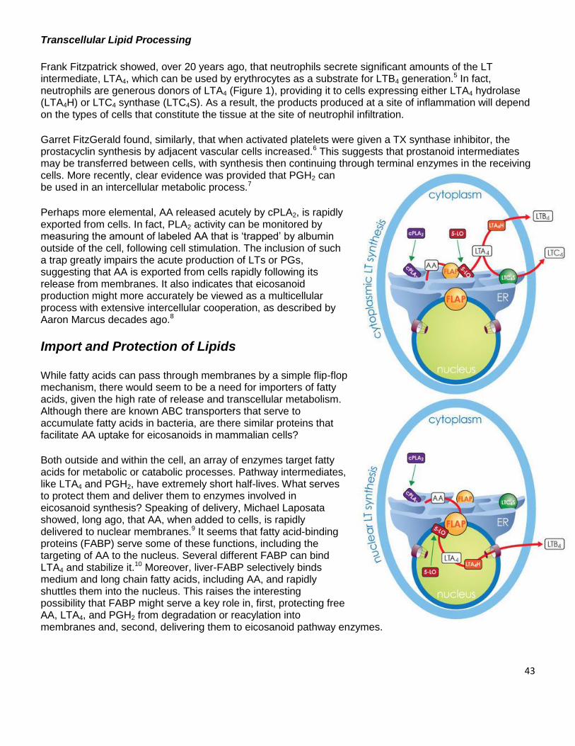
43
Transcellular Lipid Processing
Frank Fitzpatrick showed, over 20 years ago, that neutrophils secrete significant amounts of the LT intermediate, LTA4, which can be used by erythrocytes as a substrate for LTB4 generation.5 In fact, neutrophils are generous donors of LTA4 (Figure 1), providing it to cells expressing either LTA4 hydrolase (LTA4H) or LTC4 synthase (LTC4S). As a result, the products produced at a site of inflammation will depend on the types of cells that constitute the tissue at the site of neutrophil infiltration.
Garret FitzGerald found, similarly, that when activated platelets were given a TX synthase inhibitor, the prostacyclin synthesis by adjacent vascular cells increased.6 This suggests that prostanoid intermediates may be transferred between cells, with synthesis then continuing through terminal enzymes in the receiving cells. More recently, clear evidence was provided that PGH2 can be used in an intercellular metabolic process.7
Perhaps more elemental, AA released acutely by cPLA2, is rapidly exported from cells. In fact, PLA2 activity can be monitored by measuring the amount of labeled AA that is ‘trapped’ by albumin outside of the cell, following cell stimulation. The inclusion of such a trap greatly impairs the acute production of LTs or PGs, suggesting that AA is exported from cells rapidly following its release from membranes. It also indicates that eicosanoid production might more accurately be viewed as a multicellular process with extensive intercellular cooperation, as described by Aaron Marcus decades ago.8
Import and Protection of Lipids
While fatty acids can pass through membranes by a simple flip-flop mechanism, there would seem to be a need for importers of fatty acids, given the high rate of release and transcellular metabolism. Although there are known ABC transporters that serve to accumulate fatty acids in bacteria, are there similar proteins that facilitate AA uptake for eicosanoids in mammalian cells?
Both outside and within the cell, an array of enzymes target fatty acids for metabolic or catabolic processes. Pathway intermediates, like LTA4 and PGH2, have extremely short half-lives. What serves to protect them and deliver them to enzymes involved in eicosanoid synthesis? Speaking of delivery, Michael Laposata showed, long ago, that AA, when added to cells, is rapidly delivered to nuclear membranes.9 It seems that fatty acid-binding proteins (FABP) serve some of these functions, including the targeting of AA to the nucleus. Several different FABP can bind LTA4 and stabilize it.10 Moreover, liver-FABP selectively binds medium and long chain fatty acids, including AA, and rapidly shuttles them into the nucleus. This raises the interesting possibility that FABP might serve a key role in, first, protecting free AA, LTA4, and PGH2 from degradation or reacylation into membranes and, second, delivering them to eicosanoid pathway enzymes.
44
Enzymes on the Move
The transport of lipids to their pathway enzymes would be straightforward if the enzymes were consistently localized in one place within the cell. However, their positioning is dynamic and regulated. The best-studied example is 5-LO, which is cytoplasmic in circulating leukocytes but accumulates in the nucleoplasm, within the nucleus, when leukocytes adhere and migrate into sites of inflammation (Figure 2).11 Moreover, when cAMP levels rise in response to adenylate cyclase activation, as happens when leukocytes are exposed to PGE2, 5-LO becomes phosphorylated and returns to the cytoplasm.12 These changes in localization greatly affect LT biosynthesis, at least in part because of availability of AA. LTA4H, which completes the synthesis of LTB4, likewise moves into the nucleus in a regulated fashion.13 This means that its substrate, LTA4, must be appropriately trafficked to the nucleus. Similar to 5-LO, certain types of 12-LO and 15-LO are reportedly cytoplasmic in some situations and intranuclear in others.
An interesting difference in distribution of COX and mPGES proteins may also be argued. As mentioned above, when AA is added to cells, it is known to first move to nucleus-associated membranes. With time, AA becomes dispersed to all membranes through various transport mechanisms. Similarly, when membrane-bound proteins are first synthesized, this occurs on the endoplasmic reticulum. Over time, these proteins are redistributed to specific sites through various processes. This would suggest that COX-2 and mPGES-1, which are normally not expressed but are induced by inflammatory stimuli, would be translated on the endoplasmic reticulum. Given their short half-lives, they would not have time to be redistributed to other membranes. The constitutively-expressed COX-1 and mPGES-2, on the other hand, would be expected to be broadly distributed on perinuclear membranes, including the contiguous nuclear envelope. Conceivably, exogenous AA might be transported preferentially to membranes rich in COX-2 and mPGES-1. Similarly, AA that is released from different membranes by various PLA2 enzymes might be more accessible to one COX form over the other. While the subcellular localization of COX-1 and COX-2 protein and enzymatic activity have been examined (with conflicting results), temporal changes in distribution, as well as utilization of different AA pools, remain open to investigation.
Altered Gene Expression
One could, with some justification, paint a picture of eicosanoids as relatively short-lived mediators, acting locally, producing rapid effects. Perhaps this is entirely accurate for how eicosanoids work in healthy individuals. However, the persistence of LTE4, now known to be an important mediator (see related story on page 54), reveals that LTs are not always short-lived. The fact that eicosanoids can be measured in the circulation and sputum from the airways suggests that parts of the body can be awash with these lipids. And often overlooked is the impact that different LTs and PGs have on gene expression. For example, PGE2 induces multiple genes in human leukocytes and synoviocytes while inhibiting the expression of those induced by IFN-γ, primarily through EP4.
14 LTD4 induces 37 genes over 2.5-fold in human endothelial cells via CysLT2.
15 The net effect of LTD4 activity is the conversion of endothelial cells to a pro-inflammatory phenotype.
The effects of lipid mediators on gene expression can cascade, prolonging the effects of transient eicosanoid production. For example, LTD4 induces several transcription factors,15 making endothelial cells subsequently susceptible to additional agonists. In addition, induced genes may lead to the production of extracellular signaling proteins. Thus, LTB4 triggers the production of monocyte chemotactic protein (MCP-1) and colony-stimulating factor (CSF-1) in rat basophilic leukemia cells.16
Interestingly, the 5-LO pathway activates NF-κB in many ways. First, reactive oxygen species, produced during 5-LO catalysis, initiate the rapid phosphorylation of IκB through a non-canonical pathway and initiate nuclear import of NF-κB. Second, LTB4 and CysLTs activate NF-κB, via the canonical pathway, through their specific GPCRs, leading to persistent NF-κB signaling. Finally, LTs induce the expression of IL-1β and
45
TNF-α, further targeting the NF-κB pathway. Pharmacological inhibition or genetic deficiency of 5-LO severely diminishes NF-κB activation in response to TNF-α, underscoring the importance of 5-LO in this inflammatory pathway.
References 1. Lam, B.K., Gagnon, L., Austen, K.F. et al. J. Biol. Chem. 265, 13438-13441 (1990). 2. Lam, B.K., Owen, W.F., Austen, K.F. et al. J. Biol. Chem. 264, 12885-12889 (1989). 3. Reid, G., Wielinga, P., Zelcer, N., et al. Proc. Natl. Acad. Sci. USA 100, 9244-9249 (2003).4. Rius, M., Hummel-Eisenbeiss, J. and Keppler, D. J. Pharmacol. Exp. Ther. 324, 86-94 (2008). 5. McGee, J.E. and Fitzpatrick, F.A. Proc. Natl. Acad. Sci. USA 83, 1349-1353 (1986). 6. Nowak, J. and FitzGerald, G.A. J. Clin. Invest. 83, 380-385 (1989). 7. Salvado, M.D., Alfranca, A., Escolano, A., et al. Arterioscler. Thromb. Vasc. Biol. 29, 1131-1137 (2009). 8. Marcus, A.J. Am. J. Med. 78, 805-810 (1985). 9. Capriotti, A.M., Furth, E.E., Arrasmith, M.E. et al. J. Biol. Chem. 263, 10029-10034 (1988). 10. Zimmer, J.S.D., Dyckes, D.F., Bernlohr, D.A. et al. J. Lipid Res. 45, 2138-2144 (2004). 11. Luo, M., Flamand, N., and Brock, T.G. Biochim. Biophys. Acta 1761, 618-625 (2006). 12. Luo, M., Jones, S.M., Flamand, N., et al. J. Biol. Chem. 280, 40609-40616 (2005). 13. Brock, T.G., Lee, Y.J., Maydanski, E., et al. Am. J. Physiol. Lung Cell Mol. Physiol. 289, L224-L232 (2005). 14. Mathieu, M.C., Lord-Dufour, S., Bernier, V., et al. Eur. J. Immunol. 38, 1900-1912 (2008). 15. Uzonyi, B., Lotzer, K., Jahn, S., et al. Proc. Natl. Acad. Sci. USA 103, 6326-6331 (2006). 16. Subbarao, K., Jala, V.R., Mathis, S., et al. Arterioscler. Thromb. Vasc. Biol. 24, 369-375 (2003).
_______________________________________________________________________
46
Problems of Too Much Protein; Controversies Some of the most common controversies that tend to surround high protein consumption is liver stress, kidney function, bone health, and heart disease and colon cancer, most all of these claims are scientifically unfounded. First, let’s set the record straight that the WiO Diet Protocol is NOT a ‘high’ protein diet by scientific definition. This section is designed to indentify the following:
1. How much protein is considered ‘too much’ 2. To determine if high protein diets present harmful results 3. Confirm that the WiO Diet is not a high protein diet 4. Clarify that ‘too little’ protein in the diet is harmful, even deadly.
1. How much protein is considered ‘too much’ The RDA recommends 50g of protein daily based on 2,000 calorie diet which is 10% of total calories. OR .8g per kg of body weight/.5g per lbs. of lean body mass (LBM). The WiO Diet is based on providing .5g of high digestible protein per pound of LBM. When exercising, it is understandable that more protein will be needed for overall health. Requirements will depend on the type of exercise and level of intensity, ranging from 1.7-3.0g per kg of body weight per day
[67]. It is interesting to note that burn victims, will be placed on a diet ensuring 4 grams of protein per kilogram of lean body mass. In the WiO Protocol the general protein amount is 63g daily not including self prepared meals (one daily – Phase 1). Thus, the general daily amount of protein is suited for a person weighing 183 lbs. The amounts are adjusted accordingly for more or less muscle dense individuals. These amounts are in line with RDA and clinical research data. Clearly the WiO Protocol is NOT a high protein diet. When people state that a high protein diet is dangerous or un-healthy they are partially right. One of the biggest concerns of getting too much protein is because of the imbalance or elevated levels of nitrogen produced in the body.
2. To determine if high protein diets present harmful results
Liver Health
NAFLD (Non-Alcoholic Fatty Liver Disease) effects up to 24-31% of the American population, much higher (75%) for those with metabolic syndrome or are overweight. NAFLD is one of the most difficult to diagnose progressive liver failure conditions. The condition begins with steatosis (NAFLD) as the first stage, and if not attended progressing to steatohepatitis (NASH - fat inside the liver plus inflammation) is the next stage, liver cirrhosis (profound dysfunctional fibrous liver) and hepatocellular carcinoma often follow. In the first two stages you may not have biomarkers of liver damage, e.g. elevated liver enzymes, elevated blood triglycerides, but the NAFLD maybe manifesting in such conditions as pre-diabetes, weight gain (fat), elevated blood pressure. Untreated NAFLD will manifest itself with as insulin resistance, metabolic syndrome, diabetes and obesity. Basically all mentioned metabolic conditions would be secondary to NAFLD, and NAFLD would be the cause of these comorbid conditions occurring simultaneously. Thus, if a person is 30 lbs over their ideal weight there is a 75% that that they have fatty liver (over 5%). And/or if they are pre-diabetes, diabetic, are insulin resistance, have elevated tryglisterides they are likely to be in at least the first stages of NAFLD. With growing understanding of the role NAFLD plays in metabolic health we see a focus on having a healthy liver.
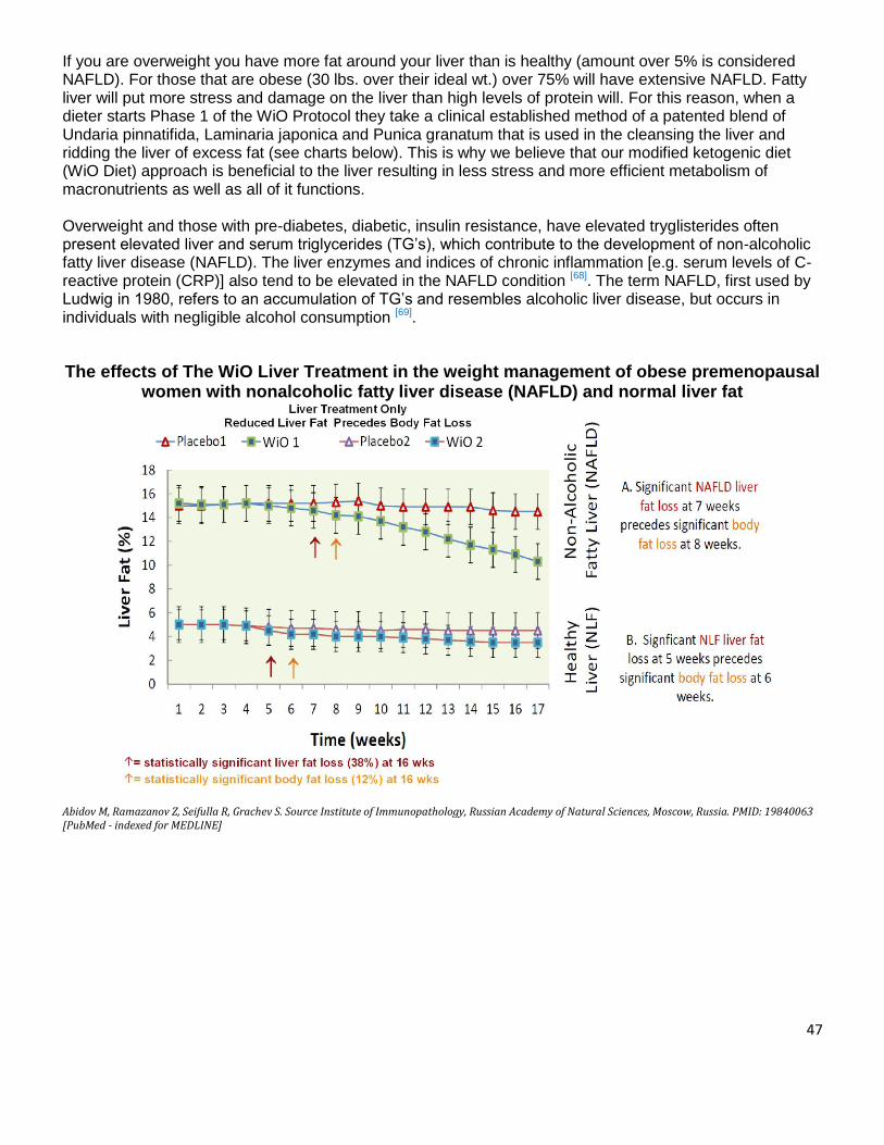
47
If you are overweight you have more fat around your liver than is healthy (amount over 5% is considered NAFLD). For those that are obese (30 lbs. over their ideal wt.) over 75% will have extensive NAFLD. Fatty liver will put more stress and damage on the liver than high levels of protein will. For this reason, when a dieter starts Phase 1 of the WiO Protocol they take a clinical established method of a patented blend of Undaria pinnatifida, Laminaria japonica and Punica granatum that is used in the cleansing the liver and ridding the liver of excess fat (see charts below). This is why we believe that our modified ketogenic diet (WiO Diet) approach is beneficial to the liver resulting in less stress and more efficient metabolism of macronutrients as well as all of it functions. Overweight and those with pre-diabetes, diabetic, insulin resistance, have elevated tryglisterides often present elevated liver and serum triglycerides (TG’s), which contribute to the development of non-alcoholic fatty liver disease (NAFLD). The liver enzymes and indices of chronic inflammation [e.g. serum levels of C-reactive protein (CRP)] also tend to be elevated in the NAFLD condition [68]. The term NAFLD, first used by Ludwig in 1980, refers to an accumulation of TG’s and resembles alcoholic liver disease, but occurs in individuals with negligible alcohol consumption [69].
The effects of The WiO Liver Treatment in the weight management of obese premenopausal women with nonalcoholic fatty liver disease (NAFLD) and normal liver fat
Abidov M, Ramazanov Z, Seifulla R, Grachev S. Source Institute of Immunopathology, Russian Academy of Natural Sciences, Moscow, Russia. PMID: 19840063 [PubMed - indexed for MEDLINE]

48
(ALAT) normal ranges 7 - 56 IU/L (ASAT) normal range 5 - 40 IU/L (GGT) Gamma glutamyl transpeptidase normal range Women 0 - 35 men 0 - 50 IU/L (ALT) Alanine transaminase also called Serum Glutamic Pyruvate Transaminase (AST) Aspartate transaminase also called Serum Glutamic Oxaloacetic Transaminase (SGOT) or aspartate aminotransferase (SGPT) or Alanine aminotransferase Abidov M, Ramazanov Z, Seifulla R, Grachev S. Source Institute of Immunopathology, Russian Academy of Natural Sciences, Moscow, Russia. PMID: 19840063 [PubMed - indexed for MEDLINE]
It is clear that having a healthy liver is required for good health. It is also evident that many people have livers that are above 5% fat content. For short term success and positive long term results free from the symptoms of metabolic syndrome, a liver cleanse is essential.
Balanced Nitrogen
Proteins are broken down by the metabolism of proteins in the liver. The liver produces urea nitrogen, the kidneys in turn will remove urea nitrogen from the blood, basically this is how your body maintains the proper level of nitrogen. Perhaps a better question should be: How much nitrogen is dangerous or un-healthy? And how much nitrogen is produced when I eat chicken nuggets? (We don’t recommend chicken nuggets). Normal human adult blood should contain between 7 to 21 mg of urea nitrogen per 100 ml (7–21 mg/dL). Nitrogen is measured by blood urea nitrogen (BUN) this test measures the amount of nitrogen in the blood in the form of urea, and a measurement of renal function. The liver produces urea in the urea cycle as a waste product of the digestion of protein. Urea is a by-product from metabolism of proteins by the liver [59]. The figure below, from Brooks et al. (2005), shows a graph relating nitrogen balance and protein intake. A nitrogen balance of zero is a state in which body protein mass is stable; that is, it is neither increasing nor decreasing. The graph was taken from this classic study by Meredith et al (1989). The participants in the
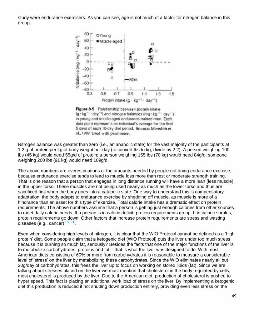
49
study were endurance exercisers. As you can see, age is not much of a factor for nitrogen balance in this group.
Nitrogen balance was greater than zero (i.e., an anabolic state) for the vast majority of the participants at 1.2 g of protein per kg of body weight per day (to convert lbs to kg, divide by 2.2). A person weighing 100 lbs (45 kg) would need 55g/d of protein; a person weighing 155 lbs (70 kg) would need 84g/d; someone weighing 200 lbs (91 kg) would need 109g/d. The above numbers are overestimations of the amounts needed by people not doing endurance exercise, because endurance exercise tends to lead to muscle loss more than rest or moderate strength training. That is one reason that a person that engages in long distance running will have a more lean (less muscle) in the upper torso. These muscles are not being used nearly as much as the lower torso and thus are sacrificed first when the body goes into a catabolic state. One way to understand this is compensatory adaptation; the body adapts to endurance exercise by shedding off muscle, as muscle is more of a hindrance than an asset for this type of exercise. Total calorie intake has a dramatic effect on protein requirements. The above numbers assume that a person is getting just enough calories from other sources to meet daily caloric needs. If a person is in caloric deficit, protein requirements go up. If in caloric surplus, protein requirements go down. Other factors that increase protein requirements are stress and wasting diseases (e.g., cancer) [70, 71]. Even when considering high levels of nitrogen, it is clear that the WiO Protocol cannot be defined as a ‘high protein’ diet. Some people claim that a ketogenic diet (WiO Protocol) puts the liver under too much stress because it is burning so much fat, seriously? Besides the facts that one of the major functions of the liver is to metabolize carbohydrates, proteins and fat – that is what the liver was designed to do. With most American diets consisting of 60% or more from carbohydrates it is reasonable to measure a considerable level of ‘stress’ on the liver by metabolizing these carbohydrates. Since the WiO eliminates nearly all but 20g/day of carbohydrates, this frees the liver up to focus on working on stored lipids (fat). Since we are talking about stresses placed on the liver we must mention that cholesterol in the body regulated by cells, most cholesterol is produced by the liver. Due to the American diet, production of cholesterol is pushed to hyper speed. This fact is placing an additional work load of stress on the liver. By implementing a ketogenic diet this production is reduced if not shutting down production entirely, providing even less stress on the
50
liver. Giving the liver an additional shot in the arm is EFA’s (essential Fatty Acids). Our WiO Protocol provides 45mL of omega-3-6-9 (EFA – Essential Fatty Acids) each day which has been shown to benefit all organs not only the liver [71]. On page 26, 27 we outline how effective the WiO Liver Cleansing System is in removing excessive fat from the liver.
Kidney Function
Another common criticism of high protein diets is the concern that they are damaging to the kidneys. This belief seems to stem from the fact that, in individuals with preexisting kidney damage, persons with this issue often has to reduce protein intake to prevent further development of the disease and we agree. However, it is important to repeat it is vital to correctly identify the definition of what level of protein is ‘high’, which is above 3.2 g of protein per pounds of body weight per day. Incorrectly, this has been turned around to suggest that high-protein intakes are damaging to the kidneys [72]. At best a weak case can be made for a risk of high protein intakes on kidney function; interestingly in fact, some research suggesting a beneficial effect of higher protein intakes on kidney function [73]. Often, simple and normal occurring changes to kidney function are often cited as a strain or damage is more likely to be normal adaptive effects of varying protein intake [71]. Presently, little research has directly examined the impact of high protein intake on kidney function in athletes. One study examined the impact of 2.8 g/kg (1.3g/lb - 260% more than is prescribed in the WiO Protocol) protein on the kidney function of bodybuilders, and no negative effect was seen [74]. Currently, we have not found if higher intakes have been studied. It is a given and worth considering that athletes are known for consuming large amounts of protein for over 4 decades without any reported increase in the incidence of kidney problems. If problems were going to occur, it seems likely that it would have shown up by now. While this certainly doesn’t prove that high protein intakes aren’t potentially detrimental to kidney function, the data in support of that idea would seem to be lacking both from a scientific and real-world point of view. The most important point is that in either case, the WiO Protocol is NOT a high protein diet and does not pose a risk to the kidneys. It is true that protein metabolism does require more hydration (water intake). And when the body is in a ketogenic state, dehydration can develop which can strain the kidneys. Not only do the kidneys suffer, but kidney stones may also develop. Other symptoms of dehydration while in a ketogenic state include headaches, dizziness, nausea, and confusion. For this reason it is mandatory that each dieter drink at least .5 oz. of water per pound of body weight. This is in addition to the water used to mix the powdered meal-replacement shakes (MRP), which will add an additional minimum of 60 oz. of water daily. Consumption of the MRP’s will provide 300% of the RDA recommended amounts of minerals and trace minerals which will balance the electrolyte levels for the dieter. Following this protocol guideline will ensure that dehydration does not occur.
Bone Health
One of the most persistent criticisms of high protein intakes has to do with the impact of protein on bone health and calcium status. This claim dates back to an old argument based on early nutritional studies where the researchers gave ‘purified’ protein diets and saw a loss of calcium from the body. More recent studies, using whole food proteins (like the proteins found in the WiO MRP) found very different effects. Frankly, the early studies on this topic are irrelevant to normal human nutrition since the consumption of protein in the total absence of other nutrients would be extremely rare; all whole food proteins and protein powders contain micronutrients. The impact of protein on overall calcium status is more complex than having a simple positive or negative effect as dietary protein can impact on; calcium excretion as well as calcium absorption and utilization. It is the combined effect of these processes which determines the end result in terms of bone health.
51
Epidemiological studies show that a high intake of animal protein increases the risk of bone fractures; as well, a high ratio of animal to vegetable protein intake has also been associated with an increased risk of bone loss [75]. In contrast, high intakes of protein improve bone healing, following a fracture for example. This is mediated both by increased calcium absorption as well increased levels of insulin-like growth factor 1 (IGF-1), a hormone involved in tissue growth [76]. Sounds like I’m switching sides on the argument that high protein is bad for calcium. Frankly, it’s too simplistic to look at protein intake in isolation in terms of its effects on bone health as the protein content of food interacts with other nutrients in that food or in the total diet [77]. For example, recent studies suggest an interaction between protein and calcium intake. When calcium intake is low, high protein intakes appear to have negative effects on bone health. In contrast, when calcium (100% RDA in each MRP shake) and vitamin D (300% RDA in each MRP shake) intake are sufficient, protein intake has a beneficial effect on bone health [78]. Allow me a shameless plug for the reason that the WiO protocol has sufficient levels of ALL micronutrients in each MRP (meal). This suggests that ensuring adequate calcium intake is crucial for bone health when a high protein intake is being consumed. This should provide the remedy to the above contradiction. In the studies where dietary protein intake was found to have a negative impact on bone health, there were other dietary factors playing a role. Calcium or Vitamin D intake may have been insufficient causing an overall negative effect. However, when sufficient calcium and Vitamin D are provided (as they typically are following bone injury), dietary protein has a beneficial impact. But hold on we are not quite finished with the ‘healthy bone’ issue, keep reading.
Metabolic Acidosis
Let’s review a concept referred to as net renal acid load (NRAL). When foods are consumed, they have the potential to produce either a net acidic or net alkaline (basic) effect, which the body, primarily the kidneys has to deal with. NRAL refers to the total amount of acid produced that the kidneys have to process. Generally, protein foods tend to increase the net renal acid load, as does a high intake of sodium relative to potassium (potassium to sodium intake should be 2:1). In contrast, fruits and vegetables, along with foods high in potassium, tend to buffer this net acid load and have an overall alkalizing effect on the body. With an excess of acid forming foods in the diet relative to the number of base producing foods, a metabolic acidosis can occur. Since fruits are omitted in Phase 1, 2 of the WiO Protocol we have formulated the MRP to be slightly alkaline (namely; calcium, magnesium, potassium, copper, iron, manganese). Don’t forget that when the pancreas secretes glucagon rather than insulin this hormonal change aids in maintaining an alkaline physiology. The modern diet, with its high reliance on animal proteins and high intake of sodium, along with a low intake of fruits, vegetables is thought to generate a sub-clinical metabolic acidosis [79]. Even a slight increase in the overall acid status of the body can have a number of negative health effects, not the least of which is an impact on hormones important to the average person but especially athletes [80]. Normally ensuring sufficient intake of healthy foods including fruits and vegetables will to balance out the acid produced from a high protein intake and avoiding this problem. From both a bone health and performance standpoint, any athlete consuming a high protein diet must ensure sufficient intake of other foods including plenty of fruits and vegetables to buffer any potential negative effects. The same is true when consuming the WiO MRP’s [80]. For this reason we have ample amounts of alkalizing minerals to provide the proper sodium/potassium balance. Take note that the major cause of elevated sodium levels has less to do with the amount of sodium consumed but rather the fact that the average American eats a high carbohydrate diet and too little potassium. This type of diet forces hyper

52
levels of insulin secretion which in turn signals the kidneys to retain sodium thus, driving sodium concentration levels up and adding to the acidic problem.
Colon Cancer/Heart Disease/Overall Health A large meat intake, especially red meat, is often claimed to be involved in the development of a number of diseases, especially heart disease and colon cancer. A great deal of this research is based on observational work where individuals consuming a meat-based diet are more likely to get such diseases. As well, there is ample evidence to suggest health benefits with vegetarian diets [82]. However, as with the protein and bone health issue, you can’t simply isolate protein/meat intake from other aspects of the diet. This is important when looking at the research as most of it tends to be epidemiological in nature, because they look at large populations of individuals and tries to draws correlations between different measured variables. This can lead researchers to draw incorrect conclusions. For example, modern meat based diets are also typically very high in un-healthy types of fats with typical cuts of red meat being high in saturated fat, including a high percentage of the diet being made up of carbohydrates in the form of mostly highly refined carbohydrates and simple carbs. This deadly combination is a known risk factor for various diseases. In contrast, lean red meats, trimmed of visible fat, and lower carbohydrate meals have a drastically different impact on the risk of cardiac disease [83]. As well, unprocessed lean red meat alone doesn’t increase markers of inflammation or oxidation [84]. In addition to potential cancer promoting factors, meat also contains a number of cancer preventing factors [84]. Replacement of carbohydrate with lean red meat has also been shown to lower of blood pressure [85]. The key here, of course, is that lean red meat, as opposed to the fattier cuts commonly consumed were studied. Diets high in meat are often low in fruits and vegetables (meaning a low intake of important micronutrients as well as fiber) and research suggests that it is the lack of those foods (fruits, vegetables) and a disproportionate level of carbohydrates in the presence of red meat that is responsible for any increased cancer risk [86]. High fat (not essential fatty acids - omega fats) intakes have also been associated with low food variety and low intakes of fruits and vegetables [87]; this would further contribute to the apparent link between consuming fatty meat and health risk. Put differently, there is going to be a fairly large difference in the overall impact of a diet that is high in animal protein, high in carbohydrate, high in fat, low in fruits and vegetables (and thus low in fiber and other important nutrients) which may be accompanied with other health risks such as inactivity, being obese, and metabolic syndrome, etc. This would be held in complete contrast to an athletic diet containing large amounts of lean meats along with a high fruit and vegetable intake, high levels of activity, maintenance of a low level of body fat, etc. As I mentioned above with regards to bone health any diet high in animal protein must be accompanied by a high intake of water, fruits and vegetables. As well, leaner cuts of meat (especially red meat) should be chosen whenever possible.
Protein Digestion/Quality
The intent of this section is to outline the definitive definition of the digestion of food, not just proteins. Digestion is; The quality and quantity of macro and micro-nutrients that is delivered to human cells. Needless to say not all proteins are created equal and the quality of an individual’s digestion will vary from person to person. Basically the bottom line is - the best proteins are those that have the greatest biological uptake. Studies have shown that it is not uncommon for a poor digestion to reduce the digestion of any/all macro and micro-nutrient by 47% [49]. Some researchers have concluded that the digestive prowess of the average

53
American has been decreased by 9-20% [49]. As you know, the breakdown of protein (and all macronutrients) begins in the mouth through the act of chewing. Seems too simple, but it is generally accepted that most people do not properly chew their food, therefore placing more work on the other five steps of digestion listed below:
The Steps of Digestion: 1. The mouth- Digestion begins when food enters the mouth. Chewing begins breaking down the food. Saliva begins breaking down the carbohydrates. Interesting Tip: Try chewing a saltine cracker for a full minute or two. You will begin to taste sweetness. That is an indication that the saliva is breaking down the carbohydrates into simple sugars. 2. The esophagus- After the food is swallowed it enters the esophagus. The esophagus is a muscular tube that helps move the food along to the stomach. 3. The Stomach- In the stomach the enzyme pepsin is excreted and mixes with the hydrochloric acid (HCL) present in the stomach to begin the digestion of proteins. Once food is passed through the stomach is referred to as chyme. Interesting Tip: Often it is mistaken, that too much stomach acid causes acid reflux and heart burn (see ). Too much stomach acid is a very rare condition, too little acid affect 15% of the population. By age 40, 40% of the population is affected, and by age 60, 50%. 4. The intestines (small & large)- In the duodenum, the food is exposed to enzymes produced by the pancreas which will aid in breaking down proteins, carbohydrates and fats. In the small intestines the nutrients are absorbed through the microscopic finger like villi on the lining of the small intestines. Vitamins and minerals pair up with fats, proteins and carbohydrates to be absorbed. Interesting Tip: If vitamins and minerals are not present in the chyme (food), then they have to be extracted from the body. This can cause vitamin and mineral deficiencies which occur in 90% of women and 71% in men [65]. 5. The Liver and the gallbladder- The presence of fat in a meal induces the gallbladder to release bile into the duodenum, which will aid in the emulsification of fats. Bile, produced by the liver, are transported through specific ducts and stored in the gallbladder. The liver excretes bile into the small intestines to emulsify the fats in the chyme, making it easier for the body to absorb. The left over bile is recycled into the gallbladder where it will be filtered by the liver and reused. 70% of all nutrient absorption takes place in the small intestine starting in the duodenum and progressing through the jejunum to the ileum. Interesting Tip: The liver provides several functions in the body besides filtering toxins. To reduce stress on the liver, avoid non EFA foods (high fat), alcohol, and toxic chemicals. Many experts support that 1 tablespoon of properly balanced EFA (n-3, n-6, n-9 ‘2:1, 1:1’) per 50 lbs. of body weight daily will prevent a host of many illnesses including gallstones [64]. 6. The Large intestines- Whatever is not absorbed in the small intestines moves on to the large intestines to be eliminated. This is largely fiber and hard starches. Good bacteria are also present in the large intestines; they feed on soluble fiber and produce vitamin K for the body. The primary reason for these deficiencies is the foods that we eat and medications that are taken. The following are examples of some of the classes of pharmaceutical medications have various effects upon the
54
nutritional status of the user. Over time, these effects can become very significant as to the comfort level and even the survival of the person taking them.
MEDICATIONS THAT REDUCE DIGESTIVE QUALITY
Loop diuretics (furosemide) - Excretion of sodium, chloride, potassium, hydrogen ions, calcium, magnesium, ammonium bicarbonate, and possibly phosphate is enhanced. After 4 weeks of furosemide use, thiamin concentrations and transketolase activity were significantly reduced. Thiazide diuretics (hydrochlorthiazide) - Excretion of sodium, chloride, potassium, bicarbonate, magnesium, phosphate, and iodine are enhanced. Calcium excretion is decreased. Triamterene-containing diuretics (Dyazide, Dyrenium, Maxzide) - Triamterene is potassium-sparing; supplementation could result in potassium overload. Folic acid deficiency is possible. Histamine H2 antagonists (Tagamet, Zantac, Pepcid, Axid) - Reduction of gastric acid secretion, resulting in poor digestion of protein. Decreased vitamin B12. Gastric acid is required for B12 absorption. Tagamet inhibits cytochrome P-450 pathways. Biquanides (Metforman) - Interferes with glucose absorption. Decreases absorption of B12. Potassium Chloride - Interferes with the absorption of B12. Sulfasalazine - Interferes with folic acid metabolism. Oral contraceptives - Oral contraceptives have significantly increased plasma Vitamin A levels. This is thought to be mediated by steroid-induced alterations in the rate of retinal-binding protein synthesis and release; depletion of reserves may result. Vitamin B6 deficiencies due to alteration in B6 and tryptophan metabolism. Interference with folate absorption. Reduced serum B12 levels. Increased serum copper as a result of increased plasma ceruloplasm; clinical importance has not been determined. Increased serum iron and increased total iron-binding capacity, along with increased incidence of iron deficiency anemia. Increased serum magnesium and zinc; clinical importance has not been determined. Corticosteroids (hyrocortisone, prednisone, dexamethasone, etc.) - Corticosteroids increase the rate of Vitamin A transport from the liver, resulting in elevated serum levels and depletion of reserves. Negative nitrogen balance due to increased protein catabolism. Increased calcium excretion (increased catabolism). Sodium retention (mineralocorticoid activity). Increased potassium excretion (sodium is exchanged for potassium). May deplete Vitamins B6, B12, and folic acid. May deplete Vitamin D3. Bile acid sequestrants (Questran) - Interference with absorption of fats and fat-soluble vitamins. Enhanced absorption of chloride ions in exchange for bicarbonate ions, which may lead to acidosis. Increased urinary calcium excretion. Increased urinary magnesium excretion. Altered absorption of phosphate and nitrogen. Vitamin K deficiency. Reduced folic acid absorption. Reduced absorption of Vitamin E and iron are possible. HMG-CoA reductase inhibitors (Zocor, Mevacor, Pravachol) - Block the biosynthesis of Coenzyme Q-10. Levodopa - Pyridoxine reverses the effects of levodopa, although this does not occur when levodopa is given with carbadopa. (Pyridoxine stimulates decarboxylation of levodopa in the periphery; carbadopa inhibits decarboxylation.)
55
Phenytoin (Dilantin) - Folate deficiency -- Increased folate catabolism or utilization as a result of enzyme induction is considered to be the mechanism. However, supplementation may decrease the effectiveness of the phenytoin. Interference with Vitamin D metabolism. Folic acid analogs (methotrexate, pyrimethamine, trimethoprin) - These antagonists inhibit the enzyme dihydrofolate reductase, which can lead to a functional folate deficiency. Supplementation can antagonize the effects of these drugs. NSAIDS (Motrin, Naprosyn, Tylenol, ASA, etc.) - Reduce night-time melatonin secretion (related to prostaglandin inhibition). Isoniazid - Increases excretion of pyridoxine into the urine, resulting in deficiency. Inhibits the tryptophan-to-niacin pathway, resulting in increased need for niacin and tryptophan [66]. Another focus must be made on the vital organs that play a pivotal role in digestion referred to as ‘incompetence’. Pancreatic Incompetence The pancreas is, as the acid producing parietal cells of the stomach also are, especially sensitive to toxins. One of the toxins to which the pancreas is especially sensitive is alcohol. As discussed many people are unable to fully digest their food, because the pancreas is not producing sufficient amylase, lipase and proteinase. Liver Incompetence When the liver is damaged, it ceases to put out a healthy complement of bile salts, and this causes a failure of emulsification of fats leading to poor digestion of fats. Colonic Incompetence The frequently overlooked colon is equally important to health as any of the other organs of digestion. With age, a low fiber diet and low intake of water, it may slow down and stasis (standing still) of food occurs, thereby allowing unfriendly bacteria to multiply, producing toxic material which leads to fatigue, headache, anxiety, insomnia, etc. Companies make claims of how their protein formulation has achieved 100% digestibility. Especially sport nutrition companies will add supplements containing protein based hormones such as Growth Hormone (GH), Insulin-Like Growth Factor 1 (IGF-1). The human digestion of protein just doesn’t work this way, such peptide hormones will simply be digested in the gut and lose their biological availability. Major pharmaceutical companies have been trying to make oral insulin (protein based hormone) for diabetic treatment and have given up on it; because the human body doesn’t digest in such a manner. If the big drug companies haven’t figured out how to do it, neither has the protein powder company with much smaller budgets for R&D, regardless of the claims made in their ads. Besides, even the most efficient human digestive system will not operate at 100% digestibility. It must be understood that to optimize the absorption of nutrients of protein (foods) the following areas must be considered:
The quality of the protein, (BV and PDCAAS) The addition of vitamins and minerals The quality of the individuals’ digestion.

56
The key areas of poor digestion can be identified as inadequate digestive enzymes effecting most Americans, too little stomach acid (hydrochloric acid-HCL) is affecting 15% of the public, [57] too much acid is rare. Stomach aches or poor digestion is the number #1 health complaint to healthcare professionals in the United States. As many as 47% of the American public reports that they suffer from poor digestive issues on a day-to-day basis [57].
True Digestibility
Essentially the bottom line is; if the body can’t digest it and deliver it to the cells it is of no use to the human body. For this very reason, WiO Protocol focuses so much effort on improving the quality of the individual’s digestion. In fact, WiO Diet is not a weight lost company at all, the loss of weight is simply a reward achieved. The heart of the WiO Protocol is to attempt to restore health to the pancreas, liver, and the digestion. By doing so all the symptoms associated to metabolic syndrome are improved or eradicated. At WiO we utilize both canola and whey proteins; a special patented formulated canola protein is uniquely enriched with cysteine and sulfur amino acids. Whey protein is recognized for its high cysteine content. Canola has nearly twice as much as whey. Canola could very well inhibit the onset of metabolic syndrome more any other protein (58).
In this section we are focusing on protein digestion but this discussion can be applied to all macro and micro-nutrients. Before we explore protein quality we need to make clear that the MOST important determining factor of ANY protein is the digestion of it – meaning “what is the quality (how good is) the individuals (the person consuming the protein) digestion”? In other words, how much of the protein will the dieter be able to digest and deliver to their cells?
Food Source Protein Digestibility
(%)
Egg / Canola protein 98
Milk and Cheese 97
Mixed US Diet 96
Peanut Butter 95
Meat and Fish 94
Whole Wheat 86
Oatmeal 86
Soybeans 78
Rice 76 Source: National Research Council. Recommended Dietary Allowances, 10th ed. National Academy Press, 1989.
When looking at the quality of protein it is customary to consider the percentage level of protein ie: egg and canola are at 98% and whey will vary from 80-91% depending on concentrate or isolate formulations. Biological Value (BV) is one of the more common methods of measuring protein quality and tends to be the one that is seen the most. BV is simply a measure of how much of the protein actually entering the bloodstream is retained in the body (in a perfect world of a properly working digestive system), with a healthy digestive system as discussed above. Protein Digestibility Corrected Amino Acid Score (PDCAAS) is the newest method of scoring protein quality and is the one most in common use today. It compares the amino acid profile to some reference protein; as well it takes into account digestion.
57
However, the most striking fact of the chart above is what the general public interrupts from the information on the chart. I will offer a reminder, that the digestibility’s of any protein is determined more on the power of the individual’s digestion than anything else. Looking at the chart above, two major things stand out. The first is that, contrary to the occasional vegetarian claim, vegetable source proteins have a significantly lower digestibility than animal source proteins. This actually has relevance for an issue beyond the scope of this article: protein requirements. Because they provide less available protein from consumption, a larger amount of vegetable proteins have to be consumed to meet human (or athletic) requirements. The second is that commonly available animal-source food source proteins have extremely high digestibility’s, 94-97%. This means that for every 100 grams of protein consumed, 94-97 grams are being digested and assimilated by the gut.
Vitamins & Minerals Digestibility Much has been written on the value and need for vitamins and minerals. Any trained healthcare professional that understands micro-biology will not argue the necessity of these macro-nutrients. Without them we will suffer deficiencies and eventually die. Most of the debate about vitamins & minerals surrounds these topics;
The need for supplementation
Dosage requirements
The form - the human body’s preferred digestible formula. Some patients will confess that their doctor has told them that they ‘don’t believe in vitamins’. After review we have found that what the doctor is likely saying “I don’t believe that taking extra vitamins as a supplements is necessary because you are getting what you need in you the foods you eat.” Doctors do believe in vitamins, they know that they exist and are required for good health. Some simply believe that people may be wasting their money in taking supplements because Mother Nature has put the vitamins in the food for you. We believe that this belief is based on what the doctors are not ‘aware’ of rather than what they ‘know’ to be true. According to the Max Planck Institute of Molecular Cell Biology and Genetics. “Less than 6% of graduating physicians in the U.S.A receive any formal training in nutrition”[90].
Vitamins & Minerals In Our Food? The facts are that over 90% of the foods (meats, fruits, vegetables and canned goods) that are available in the grocery stores are controlled by less than 10 muti-national conglomerates. They dictate the quality of the foods we eat that includes what minerals are put into the soil. Over 50% of the U.S.A. space is reserved for cultivating food and 30% of all crops grown are corn which is in 90% of all food products contributing to the metabolic syndrome epidemic. In the last 100 years the crop yield has increase from 20 bushels of corn per acre to over 200. One of the reasons for this increase in production is fertilization, which universally is nitrogen, phosphorous and potassium (NPK) in commercial farming. Our soil needs all 52 minerals in order to produce healthy foods, if it isn’t in the soil it won’t be in the food. According to WHO mortality data, around 1.6 million deaths (3% of the total) can be attributed to iron and vitamin A deficiency each year [91] and that is just one mineral and a vitamin. It’s not that the farmers are evil, but rather all about being able to compete and profits! And it is not as profitable to make sure these foods are fortified with these macro-nutrients. A study of over 50,000 Americans was conducted by the USDA over twenty years ago found that not one (1) person was receiving 100% of the recommend vitamins & minerals from the foods that they ate! The level of nutrients in our food is determined on 4 factors.
1. The level of vitamins and minerals in our food is based on the level of these macro-nutrients being present in the soil in which it is grown.
2. How long does it take for the food to travel from the farm (source) to your table? 3. Is the food being cooked? 4. What is the quality of the digestives system of the person eating the food?
58
Why are our foods ‘fortified’? We have all read on the label of the foods we eat that it has been fortified. As yourself ‘why’? Why are they putting some vitamins and minerals in the food? Are they just being nice and want us to have an extra boost to get us through the day? When foods are processed they can lose the effectiveness of vitamins, minerals and enzymes, often 100% of enzymes are depleted [90]. Food manufactures will put some back in because they destroyed them while it was being processed. However, the form (quality) that they put in is not the same as what was destroyed. And ‘why not’ you ask, because it costs more. For example there are as many as 9 different form of vitamin C (L-ascorbic acid). Not all forms are bio-available (digested) equally; some have a higher quality than others. Powered ascorbic acid is the least expensive and is generally what you will find as an additive in foods and supplements. It is worth noting that vitamins and minerals work hand and hand. Without combining their counterpart minerals with a vitamin and the effectiveness is greatly diminished.
From the Cradle to Your Plate “Nearly every food that you find in shops in the city have been processed until the nutrients have been depleted or eliminated by the time it gets on the plate, we use and use the soil [without restoring ] the nutrients, they are the center of our soils, they are becoming a desert worldwide” [92]. It’s not just that our foods don’t have the nutrients that they used to but if they have any nutrients in them when they are pulled from the ground or plucked from the tree, bush, or vine they are lost by the time they hit our plate. It takes at least a week and travels 1,500-2,000 miles to get to your local store [93]. The food is treated and sprayed with ethane to turn color so they ‘look’ like they ripened on the vine… but didn’t.
If It Can’t Take The Heat – Get It Out Of The Kitchen As if nutrient depleted soil, our food sitting in a warehouse or traveling thousands of miles wasn’t enough; heat will destroy a good portion of the remaining nutrients. At least 50% of protein is destroyed when it is cooked [90]. All enzymes in the food can be eliminated even when seaming your vegetables [94]. Digestive Leukocytosis is the result of not eating enough raw vegetables and eating too high of a cooked vegetables ratio. Digestive Leukocytosis an increase in the number of white blood cells after eating foods heated to a certain temperature, whereas raw, unheated foods did not have this effect. Leukocytosis refers to an increase in the total number of white blood cells from any cause. It indicates that the body is under attack and these leukocytes (white blood cells) are sent to the site of the attack to help defend it, thus putting the immune system under needless stress. The study also found similar results with cured, salted, canned, cooked meats brought on a violent reaction, equivalent to the leukocytosis seen in poisoning. It is recommended to eat at least 10% by volume of raw vegetables if cooking is required or desired. According to Kouchakoff [1937, pp. 330-332], foods heated below their critical temperature for less than 30 minutes don't induce digestive leukocytosis. Critical temperatures vary between 87°C (189°F) and 97°C (207°F). The message here, is that since much our food is cooked, over cooked and reheated again, supplementing enzymes back into our diet is essential – which makes a case as to why WiO’s MRP is fortified with enzymes including pre and pro-biotic’s.
The Power of Your Digestive System As discussed in depth above, a poor digestion can minimize even the best quality food source. Our cells can only get the benefits of the foods we eat if our digestive system can break it down and deliver it.
Risks of Dieting In the world’s largest study (totally 19,000 people) of weight loss has shown that diets do not work for the vast majority of dieters and are even putting their lives at risk and increasing their mortality rate by 39% (55). The study estimates that the average woman has lost and gained 357 pounds during their lifetime. Most people would be alarmed of such a large number as 357 lbs. The most frightening statistic is the increase of death by 39%. The study outlined that 140 more pounds of fat was gained that total weight lost during the life time of the average dieter. Researchers concluded that in spite the risks of heart disease, diabetics, and

59
cardiovascular issues ect., it was better that they not diet at all. The turning point for the researchers conclusion was the fact that when a dieter losses muscle mass while dieting posed an increased risk of mortality, of 39%. However, if the dieter maintained their muscle mass while losing fat, their mortally rates actually decreased 17% and improving the quality of life and adding years to it. The study went on to reveal that over 90% of dieters gain the weight lost within 1 year, plus 6 lbs. of fat. After 5 years they gained 12 lbs. of fat and 66% gained 22 lbs. after 15 years. Repeated weight gain and loss associated with dieting can double the risk of death from heart disease, including heart attacks and the risk of premature death in general (55). Such yo-yo weight loss is also linked to stroke and shown to suppress the immune system, making the body more vulnerable to infection (55).
Summary A number of health risks have been attributed to the consumption of high protein intakes; this includes potential problems with the kidneys, bone health, metabolic acidosis and certain types of cancers. For the most part, these risks tend to be extremely overstated. While high protein intakes may cause problems when there is pre-existing kidney disease, no research suggests that high protein intakes cause liver or kidney damage. While there is potential for high protein intakes to cause body calcium loss, this appears to only occur when calcium intake is insufficient in the first place; high protein intakes with high calcium intakes improves bone health (100% RDA of calcium is provided daily each in the MRP). Ensuring sufficient vegetable (4 daily servings are required in the WiO Protocol) and alkalizing minerals (included in each MRP) intake along with a high protein intake is a key aspect not only to bone health but to preventing a small metabolic acidosis which may occur when large amounts (.5g per lb of body weight is not defined as a ‘large amount’) of protein are consumed by themselves. Concerns over heart disease and cancer are more related to the high fat (non-omega fats) content of many cuts of meat, along with other nutritional factors such as insufficient fruit and vegetable intake that contributes. Other lifestyle factors that typically accompany the consumption of higher fat cuts of meat are also a likely contributor to the overall health risk. The consumption of lean cuts of meat has actually been shown to improve overall health; both athletic and diets for general health should ideally contain plenty of fruits and vegetables for this reason.
ACID BALANCING – IMPORTANCE OF PROTEIN & ALKALINE MINERALS As was mentioned in the introduction, one of the first clinical improvements patients reported after beginning the WiO Diet Protocol was the rapid resolution of their symptoms of gastro-esophageal reflux disease. Although, eliminating the “junk foods” from the diet will improve anyone’s health from a GI perspective, a medical explanation is warranted. First, most over-weight individuals’ skipped meals, usually breakfast and even lunch in many cases. As a group, they preferred to “graze” on snacks (usually high-carb and fat) and consumed drinks such as soda, juice, energy drinks and coffee. With the USDA nutritional guidelines that states approximately 22 to 25% of the total caloric intake in a 2,000 calories (K) per day diet should be of protein. 25% of 2,000K would be 500K derived from protein sources. Protein contains 4K of energy per gram so that would be about 125 grams of protein from whole food sources per day. On average, we absorb only about 60% of the protein from meat, fish, poultry, etc. Studies have shown that a poor digestion will reduce your body’s ability to absorb any/all macro and micro-nutrient by 47% [49]. So in terms of actual protein, we would be talking about roughly 60% of 125 grams or about 75 grams of absorbable protein for a person weighing about 150 lbs (lean body weight). Remember, this is the recommended minimal amount. An athlete in training would

60
be advised to consume about twice that amount. These patients were getting considerably less on a daily basis. It seemed rational that a poor diet related to their GERD (we knew their poor diet would obviously impact the “classic symptoms of Metabolic Syndrome”). After reviewing some basic texts on gastrointestinal physiology, we discovered some interesting corollaries.
A Brief Review of the Digestive Process
When the stomach is empty the ‘gastric juice’ is not highly acidic (about 3.5 on the pH scale). Consuming a meal dilutes this acid further thus causing the pH to rise. When the pH of the gastric fluid reaches approximately 4.5 and coupled with the mechanical distention of the stomach wall, the secretion of gastrin is triggered. This hormone stimulates receptors on the parietal cells and the production of HCL (Hydrochloric acid) is started (these same cells also produce intrinsic factor). This process requires an enormous amount of energy (underscored by the fact that these cells possess the most mitochondria of any cell in the body, save only cardiac muscle cells). These parietal cells take water (H2O), salt (either sodium chloride and/or potassium chloride – NaCl or KCl) and a waste product, carbon dioxide (CO2) and transform these reactants into hydrochloric acid (HCL) and sodium and/or potassium bicarbonate (Na HCO3 or KHCO3 - see Figure 1[43].
Figure 1
The “proton-pump” channels the acid into the cavity of the stomach and the bicarbonate is released into the mucus layer of the stomach (protecting it from the now very acidic conditions) and out to the blood stream. This is an extremely important physiological process and is called “The Alkaline Tide”. While this is occurring, the chief cells release pepsinogen which the strong acid environment converts to the active proteolytic enzyme pepsin. To summarize thus far, hydrochloric acid and sodium and/or potassium bicarbonate are produced (an acid and a base), the acid being pumped into the stomach, the bicarbonate into the blood stream and many calories of energy are expended. At this point, the contents of the stomach
61
are very acidic – below a pH of 3.0. Pepsin and the acid work together to unfold, or denature (breaking down into shorter chain amino acid), any protein that was consumed during the meal. These two substances attack the amide linkages that join amino acids together in the protein macromolecule. As these bonds break (or are hydrolyzed) polypeptides are formed and a lot of the stomach’s acid is “consumed” in the process. Soon the lower pyloric sphincter opens and this acidic chyme begins to enter the duodenum (the first section of the small intestine).
This triggers a number of physiological events. First, any glucose in the chyme (semi-fluid mass of partly digested food expelled by the stomach into the duodenum) stimulates the release of a group of very short-lived (half-life is about two minutes) but powerful substances called incretins. The two main ones are GIP (glucose-dependant insulinotropic peptide) and GLP-1 (glucagon-like peptide). Their presents increase insulin secretion and inhibit glucagon secretion, increase beta cell mass and insulin gene expression in the pancreas, and to inhibit acid secretion and gastric emptying in the stomach. In addition, GIP is thought to have effects on fatty acid metabolism through the stimulation of lipoprotein lipase activity in the adipocyte. The relatively short half-lives of these substances are due to their rapid degradation by the enzyme DPP-4 (dipeptidyl Peptidase-4). A new class of diabetic medications, DPP-4 inhibitors which includes sitagliptin (Januvia®), prolongs the effects of these incretins thus increasing insulin production. These incretins are also thought to play a role in satiety. Whether or not these new DPP-4 inhibitors may be useful in helping obese, diabetic patients control their weight remains to be seen. However, increasing insulin levels and increasing lipoprotein lipase activity would seem counter-productive in terms of a benefit for weight loss.
SECRETIN – GASTRIC ACID and BICARBONATE PRODUCTION
In addition to causing the release of the incretins, the entry of the chyme into the duodenum triggers the release of another powerful hormone called secretin. Secretin signals the pancreas (and the liver to some degree) to begin the same reaction the parietal cells (“G” cells) of the stomach have been doing – the production of HCL and bicarbonate. This hormone also inhibits further production of gastric acid (it was previously thought that GIP was responsible for this inhibition, but now we know GIP only inhibits this in pharmacological amounts not physiological). In this process, bicarbonate (or pancreatic juice) is delivered to the duodenum via the pancreatic duct along with the various pancreatic enzymes. The epithelial cells of the biliary ducts also produce bicarbonate in preparation for the release of bile. The pH of the intestine must be slightly alkaline for the pancreatic enzymes (the lipases, proteases and amylases) to be maximally effective. Therefore, the body must neutralize all of the acid ‘sent down’ by the stomach, but this presents a potential problem. The pancreas cannot produce bicarbonate without producing hydrochloric acid. The resultant acid is delivered to the blood stream and will have a very serious effect on the blood’s pH if the parietal cells did not deliver sufficient bicarbonate during the stomach’s phase of digestion. Balancing this system, again through what we eat and drink, is crucial for optimal health. Many consequences of poor ‘dietary acid/base balancing’ are all too common in the Metabolic Syndrome population.
WHY DOES GERD IMPORVE ON THE WiO PROTOCOL The symptoms of GERD significantly improve or is totally resolved within 7 - 10 days of starting the protocol is not unusual. A study published in 2006 [44] reported the same finding within six days of initiating a diet containing 20 grams or less of carbohydrates per day. The authors, although impressed with the results, could offer no mechanism by which the improvements occur. We believe that it may be the protein that is consumed with a meal that may be the key factor in the observed clinical improvements. When protein is acted upon by HCL and pepsin in the stomach, much of the acid is ‘consumed’ or neutralized. In contrast, a meal containing large amounts of carbohydrates and fat with very little protein would not significantly reduce the acid present. Perhaps these higher levels of acid over a period of time may exacerbate symptoms associated with GERD. The protein blends used in the WiO Protocol contain sufficient amounts of protein along with pre-biotic, pro-biotic and digestive enzymes. These are pure proteins are highly absorbable and would facilitate digestion in that pepsin and HCL may more easily react with them as opposed to whole protein such as meat. More complete digestion ensures more of the acid is consumed.
62
IMPORTANCE PROTIEN IN BALANCING ACID/BASE The physiological pH of the arterial blood must be maintained between 7.3 and 7.4. This narrow range is exquisitely controlled by many mechanisms and involves the lungs, the kidneys, the stomach and the pancreas. For example, the lungs through hyperventilation may expel more carbon dioxide which would exert an alkalizing effect on the blood. The kidneys may adjust the pH of the urine to assist in maintaining acid/base equilibrium. As a rule, these processes are largely autonomic, and we can do relatively little to effect their functioning. It is a different case with the stomach and pancreas. Here we can greatly influence the ability to buffer our blood through our choice of foods. When a meal is consumed, the stomach is stimulated to produce hydrochloric acid (HCL) and sodium and/or potassium bicarbonate (NaHCO3 or KHCO3). These products are produced in a one to one ratio; that is for every one molecule of acid, there is one molecule of bicarbonate produced. To illustrate how proper acid/base balance should work, it is helpful to assign some fictitious values. Let us say the stomach produces 10 molecules of HCL. Then, it would also produce 10 molecules of bicarbonate which would be released in the blood stream. In this example, let’s assume our “patient” has eaten a meal which contains an adequate amount of protein and 5 molecules (of the 10) of acid are ‘used up’ in the portion of the digestive process. Thus, as the stomach begins to empty into the duodenum, the remaining 5 molecules of “unused” acid will also enter. The pancreas must neutralize this acid and adjust the pH to be slightly alkaline. To accomplish this, it must produce at least 6 molecules of bicarbonate (5 to neutralize the acid and 1 to slightly alkalinize the environment). To produce 6 molecules of bicarbonate, it must also release 6 molecules of acid into the blood stream (the pancreas must secrete both at the same time). Because the stomach introduced the original 10 molecules of bicarbonate into the blood, the addition of 6 molecules of acid from the pancreas present no problem. Six molecules of the bicarbonate would neutralize the acid and we would be left with 4 molecules of bicarbonate in the blood –or a positive bicarbonate balance is maintained – keeping the blood in the sweet spot of 7.3-7.4 pH. If the meal, on the other hand, contained less than an adequate amount of protein, gastric acid levels would remain much higher – possibly contributing to GERD and, of course, would contribute to a smaller bicarbonate balance in the blood.
EFFECTS OF DRINKING SODA – (POP) We are often asked if diet sodas are permitted on the WiO Protocol, Diet Coke® being the most popular brand. The patient reasons that if the soda doesn’t contain any sugar or calories it must be fine. The answer is it is not fine. Soda drinks are very acidic and the “brown-colored” ones are the most acidic (root beer, Dr. Pepper®, Coca Cola® and Pepsi® for example). These drinks range from 3.2 to 2.5 on the pH scale and will have a very negative effect of acid/base balance. Recall that the stimulus for the parietal cells’ production of HCL/bicarbonate is the raising of the gastric pH to approximately 4.5 (coupled with the mechanical distention of the stomach wall). If the patient has a “Diet Coke®” or two with his/her meal (pH 2.5), the production of HCL/bicarbonate will be severely compromised. Here’s why, when all of the acid from the soda enters the duodenum, the pancreas must neutralize it with bicarbonate, releasing acid into the blood as a consequence. But if the patient made very little, if any, bicarbonate during the stomach’s phase of the digestive process, we will now have a large amount of “unopposed acid” entering the blood stream (a negative bicarbonate balance) and the pH of the blood will begin to fall requiring the body to resort to another means of correcting this imbalance. It is easy to imagine the ‘metabolic havoc’ caused by a meal of “large fries and a big soda”. High glycemic carbohydrates cause an outpouring of insulin with trans-fats leading to the production of the series two eicosanoids and a large influx of acid, with hardly any protein. Eating like this on a regular basis is a prescription for chronic disease.
HOW BICARBONATE BUFFERS THE BLOOD Maintaining the proper concentration of bicarbonate in the blood is hugely important in acid/base homeostasis. The bicarbonate ion is a buffer, a substance that resists changes in pH both increases and
63
decreases. In the course of normal metabolism, the cells of the body produce acidic wastes (which will be generically represented here by HCL). As these wastes enter the blood stream, they react with sodium/potassium bicarbonate (“the alkaline tide”) so that they exert no effect on the pH level of the blood. In these reactions, a water soluble salt (NaCl or KCl) is produced along with a weak acid –carbonic acid (H2CO3). Carbonic acid is relatively short-lived and one of two metabolic fates awaits it. First an enzyme, alpha carbonic anhydrase (along with catalytic amounts of zinc) may break down this molecule to water (H2O) and carbon dioxide (CO2) which is exhaled by the lungs. However, if the blood should become too alkaline, the carbonic acid reacts with the alkaline agent (represented here by OH-) forming water and regenerating the original bicarbonate buffer. This is a beautifully balanced system. These reactions can be written as the following chemical equations:
1) HCl + KHCO3 = KCl + H2CO3 (this carbonic acid may then react as follows)
2a) H2CO3 + a-carbonic anhydrase/ Zinc = H2O + CO2 (exhaled via the lungs) OR, if the blood becomes too alkaline, this reaction occurs:
2b) KOH + H2CO3 = KHCO3 + H2O (The alkalinity is reduced by producing water and the original bicarbonate is regenerated)
BICARBONATE LEVELS DROP AS WE AGE Lynda Frassetto, MD (a nephrologists from the University of California, San Francisco) tracked the serum bicarbonate levels of over four thousand patients. She found that bicarbonate levels steadily declined, by age 90 they were 18% of what they were at age 45. This is very interesting in that it is precisely in this age group (over 45) where certain chronic conditions begin to appear; osteoporosis (thinning of the bones) for one. As the body’s bicarbonate levels decline, its buffering capacity for acidic wastes also declines. This would cause a slight decline in the pH of the blood. In a study published in February 2008, Arnett showed that osteoclastic (multinucleate cells that reabsorbs bony tissue) activity is directly related to pH. He stated that “we discovered that bone resorption by cultured osteoclasts are stimulated directly by acid. The stimulatory is near-maximal at pH 7.0; whereas above pH 7.4 resorption is switched off. In bone organ cultures, H+ stimulated bone mineral release is almost entirely osteoclast-mediated with a negligible physiochemical component.” [47] This is profound: As the body struggles to maintain its physiological pH of 7.3 to 7.4, if there is an insufficient amount of bicarbonate to buffer acids, the body will use alkaline minerals from its bones to compensate. Dr. Arnett concludes that “Diets or drugs that shift acid–base balance in the alkaline direction may provide useful treatments for bone loss disorders.” [48] This is a simple explanation of how the WiO Diet aids in maintaining and even building bone density. You can see why we don’t coin ourselves as a weight-loss program … the protocol provides so much more.
HOW TO MAINTAIN HEALTHY BICARBONATE LEVELS Pharmacy students are taught to ‘never tell an ulcer patient to drink milk or to take antacids for stomach pain, although they may give a temporary relief, acid rebound will occur and their symptoms will quickly worsen.’ This is a clinical illustration of how the stomach produces acid. Alkaline minerals, perhaps in the form of an antacid are consumed. The pH level of the gastric juice increases (the stomach becomes less acidic), and the patient experiences a relief from the burning sensation; however as the pH continues to rise (to about 4.5), gastrin is secreted and the stomach starts to produce HCL (more acid – to bring the pH in balance), and the burning returns – but this time more severely. Remember as we produce acid, we also are putting bicarbonate in the blood stream and thereby maintaining our blood’s buffering capacity. The WiO Protocol is a very alkaline program. Many of the foods are made from isolates and concentrate proteins. Whey and Canola is a unique protein in that it is considered an “alkalizing food” as opposed to most proteins which are considered acidic. People attempting other protein diets using whole food sources of protein must keep this in mind. Failure to maintain proper acid/base balance can lead to consequences such as gout or kidney stones (many people following “Atkins type” diets had these experiences). In the WiO Protocol, patients receive alkaline mineral supplements
64
containing calcium, magnesium, and potassium right in the MRP powder. These minerals are balanced with the foods they are eating to ensure a proper amount of these minerals – never a hyper amount! The patients are also required to consume two-four cups of fresh vegetables per day, contributing additional alkalizing minerals and anti-oxidants. WiO participants are further required to use Redmonds Real Salt®, pink color instead of commercially bleached table salt on their foods, which provide a rich source of alkaline minerals, and they are educated on the perils of highly acidic soft drinks. Finally, and perhaps most significant, is the fact that so many people suffering from GERD have been able to discontinue their proton-pump inhibitors. These drugs inhibit the Na+/K+ “pump” which effectively shuts down the stomach’s ability to produce acid and consequently bicarbonate. How long term use of these pharmaceuticals impact the overall physiology has not been studied; however, it has been an observation, both in our clinic and in the pharmacy, that many patients taking these medications are also taking a biphosphonate (such as Fosmax®, Actonel®, or Boniva®). If the body becomes too acidic, perhaps due to insufficient bicarbonate buffering, it must draw on the alkaline mineral reserves of the bones. Is inhibiting this process by drugs rather than food the best long-term course? What metabolic consequences might arise? We believe that a nutritional approach via the WiO Protocol may well represent a therapeutic alternative for medication intolerant patients. COACHES’S NOTES: Although the vast majority of patients suffering from GERD notice a rapid improvement of symptoms, occasionally some symptoms may persist. We recommend based on our experience, switching from a proton-pump inhibitor to a H2 antagonist may represent a better therapeutic option. In less severe cases, the use of liquid antacids on a PRN basis may be prudent. Of course, if these changes are not effective or bring a worsening of symptoms, the proton-pump inhibitor may certainly be re-started.
65
BODY INTELLIGENCE – AN INTUITIVE WAY TO EATING
THE SPIRIT IS WILLNG – BUT THE FLESH IS WEAK The prior section was dedicated to the research and information and is centered in the physiological realities and requirements needed to correct dysfunctions associated and those that are causing the symptoms of Metabolic Syndrome i.e. 1) Pancreas, 2) Liver, 3) Digestion. Thus, the flesh will no longer be weak. This is the foundation, the vehicle if you will, for a life time of success and well-being. As important as this foundation is, it MUST be accompanied with a healthy emotional and physiological relationship with food and eating discovered in section 4) Life Style “weekly education in our relationship to food(s)”. This healthy relationship will be the driving force for a true lifestyle change. As powerful as the WiO Protocol is, even the successes achieved through it will likely be undermined without this relationship change. Life Style (weekly education in our relationship to food(s))
The principles outlined below are a brief summary of what each client receives to ensure the life-long success that we all seek.
1. Diets should not be used to ‘lose’ something – A Diet is the food you give your Body to feel healthy 2. Control Your Hunger by Feeding It 3. Make Food an Ally 4. Turn Deft to the Whistle Blowers 5. Calibrate your Full Meter 6. Feel the Joy in Eating 7. Don’t Feed Emotions 8. Love your Body with Respect 9. Exercise is Movement Not Just the Gym 10. Give Thanks to Health – By Being Healthy
A HEALTHY RELATIONSHIP WITH FOOD AND EATING Anorexia, Bulimia, and Binge eating are the most common forms of an eating disorder [62]. However, if a person becomes overweight, over eats, skips meals, develops Type II diabetes (for example but not exclusively) or eating food that has a poor nutritional value are all simpler forms of eating disorders, this statement can be made by the fact that these issues like the symptoms of metabolic syndrome are all caused by what we eat, or WHY we eat – we create these conditions within us by the foods we put in us and the reasons we eat them. We call this “having an inappropriate relationship with food”. The WiO Diet Protocol call can be split into two distinct categories in the treatment of metabolic syndrome; overcoming physiological disorders / overcoming the emotional and intellectual relationship with food. For years there have been many programs that focus on these two categories individually, that are not unique. The challenge is that each category has powerful influence over the other. They are distinctive in how they are treated but cannot be separated while trying to treat each. Allow me to illiterate: Mary is 55 and overweight, she is starting to exhibit symptoms of hypertension and higher blood sugar levels (diabetes). She has tried nearly every mainstream diet program since she was 14 years old, she will total 293 diets in her lifetime [63]. She will spend 31years 1of her life being on a diet [63]. Mary knows more about dieting than most doctors or the counselors she has been treated by. Today, Mary is starting a new diet, she knows that she will have to give up (deprive herself) of some food items, amount of food, time she can eat, and have a positive attitude about all of it, even though she will quit her diet within 5.5 weeks [63]. Fifty percent of the reason she will quit is because of lacking willpower, twenty five percent of the reason will come from being depressed and moody [63]. Allow me to put this issue into perceptive:
66
You’re looking for a job and applied and have an interview with ABC Company and they said “Sure – we will hire you”. But after 5.5 weeks you get fired or quit. After a short time, you realize you need more money so you go back to ABC Company and they say “Sure – we will hire you”. And after 5.5 weeks you get fired or quit because you don’t have the will power to do what the job requires or because your job makes you moody or depressed. And you repeat this cycle over and over again, every 5.5 weeks for 31 years. How would you feel about your ability to keep the job the next time you go back to apply. Or how much of a desire do you believe you will have to KEEP the job? Can you imagine the emotional imprint left on Mary after a lifetime of hundreds of failed attempts? It’s not hard to imagine that Mary doesn’t have much will power left because of her many failed diets. She KNOWS that in the next diet she will be deprived of the foods she needs and is driven by cravings that she has, regardless of whether her body needs the food that she is craving or not. And each time she tries a new diet she knows she has to tap into her will power to get her through it. But her will power is definitely depleted from her many failures! For Mary it is not just a matter of will power, but she truly feels depressed when she starts a new diet, and swears it’s not just in her head. She doesn’t feel happy when she is on the diet. And Mary, like 66% of her friends, isn’t happy with her body [63] when she isn’t on a diet. Until Mary overcomes the physiological disorders and her emotional and intellectual relationship with food – ANY and ALL diets will continue to fail her. With the WiO Diet approach, Mary doesn’t have to have nearly as much will power because she won’t feel deprived. Her body won’t crave what she NEEDS and after the first 7-10 days of starting the protocol, the cravings from sugar, caffeine and un-healthy fats will not be an issue. For the first time, Mary’s BODY from the physiological perspective will be on her side. Each week Mary receives the education and support to ‘reboot’ her emotional and intellectual perspective by learning what a healthy relationship with food, feels like (10 principles below).
TUNING IN TO YOUR ‘BODY INTELLIGENCE’ Listening to the signals that your body sends you is defined as ‘Body Intelligence’. It is the cornerstone to a life-long lifestyle change. These signals pertain to what and when you should eat as much as exercise and how you should move your body. Being able to interpret your body’s signals came naturally when you were toddler. But your translation of those signals and the signals themselves became distorted after years of bombardment from the media, advertising, entertainment, and social influence. And perhaps most importantly, the chemical impact those foods choices have had on your body. There are two kinds of cravings: 1) Biological and 2) Emotional or ‘memory’ cravings. There is a lot of truth in the saying ‘the devil made me do it’ as you reach for that third helping of chocolate cake with ice cream on top. Except this ‘devil’ wasn’t holding a pitch fork and had horns on his head. It was the footprint left behind by the symptoms of metabolic syndrome – called biological cravings. The devil in this case is the chemical and biological signals being sent because of the imbalances in your body. Like most things in life there are healthy cravings and un-healthy ones. Correct those imbalances and you will be AMAZED at how much will power you have, and you will be able to decode the messages that your body is intending to tell you, giving directions to your journey through health. We call this your Body Intelligence, which is an intuitive way to eat. An Emotional or ‘memory’ craving is embedded in all of us. We all have some emotional connection with a particular food. I, for example, used to feel compelled to eat popcorn while at the movies. For me it is a tradition and a connection. I used to feel that if I couldn’t have popcorn, I couldn’t enjoy the movie as much. Therefore, the quality of the movies experience was controlled or influenced by ‘popcorn’. As if the actors’ skill was diminished by the presences or absence of a kernel of corn. We make statements like “you can’t have a birthday party without CAKE”. We have traditions in our society that harbor around food. A newly married couple commemorates their union by sharing the first bite of their wedding cake. Hot dogs at a baseball game, cotton candy at the fair, barbeques on the 4th of July are just a few of other traditions. I
67
do not make the claim that these traditions are bad and should be abandoned in order to have a healthy relationship with food. But I am saying that all of us have some emotional relationship with food and some of them are un-healthy. We may be an emotional eater, we eat when we are mad, sad, lonely, depressed, happy and the list goes on. Food should not be used to comfort or cover an emotion. Food is good enough to be eaten on its own. We should eat food for the sake of eating good foods and how we feel when and after we eat it. There are four classes of eating personalities: The Careful Eater, the Professional Dieter, the Unconscious Eater, and the Intuitive Eater [88]. It is possible to have a combination of these eating personalities. It is important to identify what eating personality(s) you fit into, before overcoming it. The WiO Protocol will help identify your personally and guide you to become an Intuitive eater. The WiO Diet Protocol is a four phase program. Phase 1 lasts 12 weeks. This is where the physiological and biological dysfunctions are corrected and recalibrated (reset-rebooted). Phase 2 and 3 ease you back into Phase 4, a healthy lifestyle where you basically eat whatever you want – an intuitive eater listening to their body intelligence. One of the secret weapons of the WiO lifestyle is simple - the things you want to eat now (before the WiO Protocol), will not be the things you WANT to eat after you graduate from the WiO Protocol. Each week while we are recording your physical progress, we will be teaching you how to listen to your body’s intelligence. You will receive clinical education about food and nutrition; in a nut-shell you will know more than the average doctor and nutritionist on how to overcome the issues of metabolic syndrome. And you will also receive an emotional ‘reboot’ that will help you break the un-healthy bonds to emotional eating. One of the ten principles (listed below) of body intelligence will be given to you weekly. This is how you will never have to ‘diet’ again. And this is how this will be “Your Last Diet”.
Diets should not be used to ‘lose’ something – A Diet is the food you give your Body to feel healthy The proper definition of a diet is the foods what we choose to eat, be it healthy or un-healthy. Right now, in this instant, I ask that you change what the word ‘diet’ has meant to you. It no longer means ‘a program that will help you lose ‘something’ i.e. fat, getting off medication, get over an ailment or illness’ TODAY, RIGHT NOW. The word diet means ‘any and ALL food you put into your mouth’ – including pizza, chocolate, and ice cream. Take every diet book, article and video and put them it a box or throw them away. The old definition of diet meant – ‘depravity’. Your NEW definition of diet means - ‘abundant fulfillment’ Most every diet gives you false hope of a quick, easy, and simple solution to your health issue i.e. weight, diabetes, hypertension, ect. You have permission to get mad at the volumes of lies, which have led you to feel like YOU are the reason (failure) the ‘other’ diets didn’t work. After your body has corrected any physiological dysfunction (Phase 1-3), you will be able to and should eat anything you want. The amazing fact is that the foods that you will want to eat will be the foods that your body needs, you will be ONE with your body again, what you want will be what you need. This will truly be your last diet.
Control your Hunger by Feeding It Most diets tell you to ‘pull up your boot straps’ and gather your ‘will power’ and ignore your hunger, ‘no pain - no gain’. Instead, you will learn to honor your hunger. Hunger is a primal drive and is a natural healthy signal when your body needs FOOD, if it doesn’t get it – bad things can happen. You shouldn’t ignore it. By meeting your biological needs, there won’t be any primal drive or desire to overeat or to eat foods that will be bad for you. Generally speaking there is a difference between hunger and cravings. They are not the same thing. There are however, two kinds of cravings: 1 - Biological, your body signaling you it NEEDS something, i.e. vitamins, water, or protein. 2 - Memory or Intellectual cravings, i.e. popcorn at a movie, because that’s the way you had it as a kid, or ice cream after a date just because it is a tradition. Neither is really driven by ‘true’ hunger. Biological cravings is driven by a NEED, memory or intellectual cravings is driven by a WANT that connected to an emotion. It is important to separate the two.

68
Make Food an Ally No more fighting – call a truce. Food is your friend and is absolutely required in you achieving your goals. Since you need food, make it your ally not the fulfillment of an addiction from a craving. Temporarily there is a short list of foods you are going to avoid in Phase 1 and that is only for 18 weeks. The average woman will spend 31 years of her life on a diet. Wouldn’t you give up 18 weeks in trade for 31 years of not having to give up food choices for the rest of your life? Seems like a small price to pay. That is the reward that the WiO Protocol offers you. After your body has been ‘rebooted’ you can eat anything you want. You don’t need me to account of the many studies done that confirm that deprivation of food only builds to uncontrollable cravings, overeating and bingeing. You have experienced similar situations for yourself. If you have thoughts that you can’t eat a particular food because of the WiO Protocol, then intense feelings of deprivation will settle in until you give-in. Instead make a decision that you are choosing to eat something else for a short time only. Each day you decide what clothes to wear. By picking the blue shirt and the tan pants does NOT mean that you can never wear the other clothes in your wardrobe. You’re simply choosing one option for another, and that is only for a short term. Soon you will be able to be around all the foods you love all day and not be tempted to eat more than your body needs. And it won’t be because we will teach you a trick that ‘ramps-up’ your will power to super human strength. It will be because of YOU! Because you have become ONE with your body and both want the same things.
Turn Deft to the Whistle Blowers A nibble here – and alarms go off – a bite there and sirens start screaming. Of course they are all in your head, and the whistle blower is YOU. If you pat yourself on the head for refusing all the foods that you COULD have had but didn’t, you’re sending the wrong signal. This kind of signal is suggestion that getting ‘less’ is the reward – getting LESS is not the reward. On other day, you slap yourself on the hand as being ‘bad’ because you smuggled a bite of chocolate cake in to your mouth, this mental punishment only reinforces the idea that eating the foods you like makes you BAD. It doesn’t. If you and a friend witnessed two boys playing at the playground and the bigger boy pushes the smaller one down and takes the ball out of his hands, would you be accurate in calling the bigger boy a ‘bully’? Or would it be MORE accurate to say “the bigger boy pushed the smaller one down and took the ball”? The first example is a belief that the action defines the event. The second one simply describes what happened. Here is another illustration. If aliens visited earth and were required to make a report of the weather conditions, which of the following would be accurate? 1 – When they arrived to earth, they observed that is was raining even snowing, cold and windy. 2 – Perhaps the day they came it was record breaking heat, dry and no wind at all. Which report would be correct? The earth is a cold, wet, and very windy planet OR the earth is a really hot, dry with no wind planet? The answer – neither! Just as the weather conditions (events or happenings) on earth does NOT define what kind of planet earth is, neither does eating certain foods make you bad. We have become a world of ‘labelers’. We put labels on everything. You can call labels the ‘meaning’ that we put on things that happen (happenings). By pushing someone down doesn’t ‘mean’ that the boy is a bully any more than you telling a small (or big) lie ‘makes’ you a liar! I’m sure you have told at least one lie in your life and if you have, doesn’t that mean that you are a liar? Or does it simply mean that you are a person that once said something that wasn’t accurate or true. Eating some cake doesn’t ‘make’ you BAD. It simply means you put some cake into your mouth. It is true that it will have biological consequences that may take you in a different direction from your goal. But it definitely doesn’t ‘make’ you bad, weak, undisciplined, lazy, dumb, fat, uneducated, or ugly. It doesn’t ‘make’ or turn you into anything. Throw away the whistle you have been blowing on yourself. If you eat something that isn’t on the protocol during Phase
69
1, observe how you feel, how it tastes, then make a decision to stop doing it for a little while. Then make a decision to continue eating it (later) and for now you choose to say ‘no thanks’ and keep on your journey.
Calibrate your “Fullness” Meter When your stomach begins to fill with food, it sends signals to your brain telling you to stop eating. You will learn how to listen to those signs and you will have feelings of being comfortably full. Have you ever been really hungry or knew that you were about to have one of your absolute favorite meals? When you sat down to eat, you ate quickly and ignored the signals that your body was sending that the hunger is gone? You ate so quickly that the signals of being full didn’t have time to register and all of a sudden you realized that you ate too much – way too much. This is called being absent from your eating experience, a perfect example of a ‘unconscious eater. You are so full that you are uncomfortable and even have pain around your stomach. You try to position yourself to be more comfortable with little success. You make a promise to yourself that you will never eat again …or at least that much. You hate this feeling; you may even start to hate yourself for doing this to yourself… again. Through the WiO Protocol you will learn to pause a few times during the meal and ask yourself how the food tastes and to gauge how full you are. You will not starve on the WiO Protocol, you will get the signal that you are hungry, and that is when it is time to eat. On the protocol you will have a meal every 4-5 hours, with snacks in-between, if needed. Being hungry is not a problem on the WiO Protocol.
Feel the Joy in Eating If you don’t love it, don’t eat it, and if you love it, savor it. We would like you to adopt that motto. How many times have you said “it was ok – but not worth the calories”? Some other cultures seem to have the wisdom of enjoying life and its many pleasures, more than Americans. Food is one of those pleasures that should be filled with joy. The saying ‘eat to live – don’t – live to eat’, is wise wisdom. The latter puts a whole different meaning on why we are eating and what we are willing to put into our mouths. When you eat what you really want, it will be in harmony with what you really need. The WiO Protocol will help you learn the difference between NEEDS and WANTS (cravings). That alone will bring you pleasure and you will feel content. Remember not to put ‘labels’ on WANTS as being bad. They are just a choice. You will have the capacity of not being driven by WANTS. You will enjoy them, not NEED them. When it is time to eat, take time for it, don’t watch T.V. or work at your desk while you are eating. This will present the action of an unconscious eater. Savor the moment and enjoy it. Through this wisdom you will learn that it takes much less food for you to say ‘I have had enough and I am content’.
Don’t Feed Emotions At one point in our lives we have all had at least one challenging emotion …and you will have them again. Some of us eat when we are bored, angry, sad, happy, stressed, scared ect., it really doesn’t matter what emotion it is that triggers you to eat. The lesson is to not feed your emotions. Food won’t solve the issue that caused the emotion. Eating, at this time, will offer a temporary relief or will only be a distraction from the emotion. And after you have eaten for the wrong reason, you may now be left with another emotion – regret, and then perhaps disappointment, maybe ‘self loathing’ will follow, get the idea? Eating is good! You don’t need the excuse of an emotion to eat. When you allow an emotion to control you, to eat is the first step to other inappropriate relationships with food. Seldom is the case when a person has just one emotional trigger. One will grow into two and can spread from there. Emotional eating is living to eat. A healthy lifestyle is eating to live.
Love your Body with Respect Your body doesn’t look like mine. The truth is there is no “Perfect Body”. Body types are like art. They come in many beautiful forms. Studies have proven that the person you think is ‘perfect’ and has the body that you wish you had, has something that they dis-like and even hate about their body. You’re different from your brother or sister even your parents and you came from the same gene pool. You’re not a carbon copy of anyone – you’re unique!
70
Accept your genetic blueprint. Respect your body and you will feel better about who you are. Respect the progress and the results that your body is providing for you. Don’t measure your success by the number on a scale. Don’t stand in front of the mirror and criticize the things you hate about your body, be amazed at what your body has done, is doing and will continue to provide for you. If you are 5’ 2” you’re never going to be a world class pole vaulter. If you’re over six feet tall, you’re not going to fit into a size six shoe. And that is OK. There is no perfect size. Your food needs are dependent on your body type. Don’t feel like you should eat like someone else – eat for yourself, eat to live. Don’t eat just to copy someone else. This is not to say that you should not strive to be the ‘best – healthiest’ you. But base your goal on you, not on your neighbor.
Exercise is “Movement” Not Just the Gym Exercise for the sake and enjoyment of ‘moving’. Don’t exercise to burn ‘calories or fat’. Take pleasure in the feeling you get when you simply move and stretch your body. Focusing on how you feel from working-out, such as the energized feeling you get from being present in the activity can make all the difference between springing out of bed for a brisk walk or slamming the snooze alarm for a few more minutes of sleep. You may not enjoy traditional exercise. If you connect exercise and the WiO Protocol together as one, you may quit both because they seem connected to you. Since you don’t like one you will not like the other because they are one ...to you. Don’t connect them, but use them separately as forms of enjoyment. To be successful on the protocol you don’t NEED to exercise. The physiological ‘rebooting’ of your pancreas, liver and digestive system will happen with or without exercise. HOWEVER, exercise will complement what the protocol is doing for you. And you will receive additional benefits from the movement and activity that only exercise can provide. Honor the benefit of your body’s ‘movement’ just as you honor your body’s hunger. You were designed and created to MOVE. If the reason you are exercising is to lose weight or to get healthier you will likely exercise for so long. Allow exercise to stand on its own, on its own merits. Make the reward from exercise more immediate. Make it the feeling you get while being present in the exercise in the moment, rather than the delayed benefit of losing weight. Enjoy exercise for its self not as a tool to get you something else.
Give Thanks to Health – By Being Healthy We talk a lot about ‘honor’ and honoring your hunger, your body. Here is another reason to honor and give thanks – for Good Health regardless of the level that you are presently. The concept of ‘honor’ is not a destination or a fix state of mind. As you progress through the phases of the protocol, your body is going to literally change, both inside and out. If you decide to only honor your body when you reach your ‘end’ goal, perhaps your goal is a pants size or a level of body fat, you will be denying yourself the benefits of the journey to your destination. You have heard of sayings like: ‘enjoy the view’ – ‘stop and smell the roses’. Imagine you were going out to dinner on a special occasion. And you were driving up a beautiful canyon to your favorite restaurant, but you didn’t allow yourself to start enjoying the evening until you arrive at the destination. Can you visualize all the beauty and rewards you ignored on your way to the restaurant? By looking past the trees, and rocks, the shapes and color of nature, you miss out on the ‘whole’ experience. Enjoy every part of the journey, the feel of food in your mouth, the taste and texture of it. How you feel after you have eaten? The great feeling of being able to ‘move’ your body the way you can. Honor the feeling you have as you ’feel’ your body giving thanks for the nutrients it has received. You’re not going to have a perfect life-long diet – you will eat foods that are not the ‘healthiest’ things in the world. Eating them respectfully (in moderation) will not cause you to gain weight, have a heart attack, or get cancer. Your body was created to defend its self. It is what you eat consistently over time that matters most. Progress, is not being perfect, consistency is what really counts.
___________________________________________________________________________________
71
WiO provides “in-house” training for you and your staff at no charge as well as on-going advanced trainings. Our Area Directors and Regional Sales Managers will help you set up a “turn-key” operation. Hopefully, you see the many benefits and health benefits of this protocol and that it goes far beyond mere weight loss. At WiO, we strive to live by our motto: “Your Last Diet”
Material contained in this paper is copywritten 2009, 2011© Written permission to use any content must be
obtained by WiO Diet.
For information of how to set up a WiO Clinic – follow the link below http://www.wiodiet.com/becomeclinic.php
or call Jason Whitney at 801-631-4886 – [email protected]
REFERENCES [1, 2, 3] Reaven, Gerald. “Syndrome X: The Risks of Insulin Resistance.” Canadian Association of Cardiac Rehabilitation Newsletter Sept. 2000. Barry Sears, PH., author of The Zone, 1982 Nobel Prize for Physiology/Medicine; biochemists, Sune Bergstro¨m, Bengt Samuelsson and John Vane. [4] Barbagalio, M., et al.. “Role of Magnesium in Insulin action, diabetes and cardiometabolic Syndrome X. Molecular Aspects of Medicine Vol.24 (Feb. 2003): 39-52. [5] Barbaglio, M., et al.. “Magnesium Metabolism in Hypertension and Type 2 Diabetes Melitus.” American Journal of Therapeutics Vol. 14 No. 4 (Jul-Aug., 2007): 375-85. [6] Miele, C., et al.. “Insulin and Insulin-like Growth Factor-1 Induce Vascular Endothelial Growth Factor mRna Expression via Different Signaling Pathways.” Journal of Biological Chemistry Vol. 275 No. 28 (July 14, 2000): 21695-21702. [7] Baba, T., et al.. “The Oral Insulin Sensitizer, Thiazolidinedione, Increases Plasma Vascular Endolthelial Growth Factor in Type 2 Diabetic Patients .” Diabetes Care Vol. 24 (2001): 935-954. [8] FDA News. “FDA Issues Safety Alert on Avandia.” (May 21, 2007): P07-88. [9] Nissen, M., Wolski, K. “Effect of Rosiglitazone on the Risk of Myocardial Infraction and Death from Cardiovascular Causes.” The New England Journal of Medicine Vol. 356 No.24 (July 14, 2007): 2457-2471. [10] Pignone, M.. “Rosiglitazone Appears to Be Associated With an Increased Risk of Cardiovascular Events.” Clinical Diabetes Vol. 25 (2007): 23-24. [11] Perciaccante, A., et al.. “Insulin Resistance and Hyperinsulinemia Are Related to Plasma Aldosterone Levels in Hypertensive Patients.” Diabetes Care Vol. 30 (2007): 1176. [12] Colussi, G., et al.. “Insulin Resistance and Hyperinsulinemia are Related to Plasma Aldosterone Levels in Hypertensive Patients.” Diabetes Care Vol.30 No.9 (2007): 2349-2354. [13] Krug, A., and M. Ehrhart-Bornstein. “Aldosterone and Metabolic Syndrome: Is Increased Aldosterone in Metabolic Syndrome Patients an Additional Risk Factor?” Hypertension Vol.51 No.5 (May 2008): 1252-1258. 34 [14] Sobel, B.E., et al.. “Insulin Resistance Increases PAI-1 in the Heart.” Biochem Biophys Res Commun Vol.346 No.1 (July 21, 2006): 102-107. [15] Solini, A. et al. “Acute Vascular Events and Electrolytes Variations in Elderly Patients.” Horm Metab Res. Vol.38 (2006): 197-202. [16] Eades, Michael and Mary D. Eades, MD. Protein Power New York: ©Creative Paradox, L.L.C. (1996): 302. [17] Ibid: 303-306. [18] Murray R., D. Granner, and V. Rodwell. Harper’s Illustrated Biochemistry 27th Ed. McGraw-Hill Companies, Inc. (2006): 188. [19] Jocken JW, et al. “Adipose Triglyceride Lipase and Hormone-sensitive Lipase Protein Expression is Decreased in the Obese Insulin-Resistant State.” J Clin Endocrinol Metab Vol. 92 No.6 (June 2007): 2292-9. [20] Eades, Michael and Mary D. Eades, MD. Protein Power New York:
©Creative Paradox, L.L.C. (1996): 305. [21] Ibid: 305. [22] Meijssen S,
et al., “Insulin Mediated Inhibition of Hormone-sensitive Lipase activity in Vivo in Relation to Endogenous
72
Catecholamines in Healthy Subjects.” J Clin Endocri Metab Vol. 86 No.9 (2001): 4193-4197. [23] Murray, R., D. Granner, and V. Rodwell. Harper’s Illustrated Biochemistry 27th Ed. McGraw-Hill Companies, Inc. (2006): 169. [24] Gerich JE, et al.. “Renal Gluconeogenesis.” Diabetes Care Vol.24 (2001): 382-391. [25] Murray, R., D. Granner, and V. Rodwell. Harper’s Illustrated Biochemistry 27th Ed. McGraw-Hill Companies, Inc. (2006): 249-250. [26] Pihlajamäki J,et al.. “Insulin Resistance is Associated with Increased Cholesterol Synthesis and Decreased Cholesterol Absorption in Normoglycemic Men.” J Lipid Research Vol.45 (March 2004): 507-512. 35 [27] Eades, Michael and Mary D. Eades, MD. Protein Power New York: ©Creative Paradox, L.L.C. (1996): 364. [28] Yokode M, et al.. “Diet-Induced Hypercholestrolemia in Mice: Prevention by Overexpression of LDL Receptors.” Science Vol.250 (Nov.30, 1990): 1273-75. [29] Graveline, D. Lipitor®, Thief of Memory. Haverford, PA: Infinity Press (2004). [30] US Patent No. 4933165. [31] Ness, GC, et al.. “Insulin and Glucagon Modulate Hepactic 3-hydroxy-3-methylglutaryl –coenzyme A Reductase Activity by Affecting Immunoreactive Protein Levels.” J.Biol. Chem. Vol. 269 No. 46 (Nov.1994): 29168-29172. [32] Spence, JT, et al.. “Role of Protein Synthesis in the Carbohydrate-Induced Changes in the Activities of Acetyl-CoA Carboxylase and Hydroxymethylglutaryl-CoA Reductase in Cultured Rat Hepatocytes.” Biochem J. Vol.227 No. 3 (May 1, 1985): 939-47. [33] Murray, R., D. Granner, and V. Rodwell. Harper’s Illustrated Biochemistry 27th Ed. McGraw-Hill Companies, Inc. (2006): 231, 519. [34] Hayek T, et al.. “Dietary Fat Increases High Density Lipoprotein (HDL) Levels Both by Increasing the Transport Rates and Decreasing the Fractional Catabolic Rates of HDL-cholesterol ester and apolipoprotein (Apo) A-1. Presentation of a New Animal Model and Mechanistic Studies in Human Apo A-1 Transgenic and Control Mice.” J Clin Invest. 91 (4) (1993): 1665-1671. [35] Samaha, FF, et al. “A Low-carbohydrate as Compared with a Low-fat Diet in Severe Obesity.” New England Journal of Medicine 348(21) (2003 May 22): 2074-81. [36] Yancy, W.S, et al.. “A Low-Carbohydrate, Ketogenic Diet versus a Low-Fat Diet to Treat Obesity and Hyperlipidemia.” Annals of Internal Medicine Vol. 140 No. 10 (May 18, 2004): 769-777. [37, 38, 39] Reaven, G.M.. “Insulin Resistance, Coronary Heart Disease and Diet.” Journal of the Medical Society of San Francisco Vol.74 No.2 (Feb 2001): 26-29. [40] Eades, Michael and Mary D. Eades, MD. Protein Power New York:
©Creative
Paradox, L.L.C. (1996): 335. 36 [41] Ibid: 340. [42] Ibid: 350. [43] Ciell, M. and J. Young, MD.. “I’m Staying Young: Ask Me How.”
©Matol Botanical International (2007). [44] Austin, GL, et al.. “A Very Low-Carbohydrate Diet Improves
Gastroesophageal Reflux and Its Symptoms.” Digestive Diseases and Sciences Vol.51 No.8 (Aug 2006): 1307-12. [45] Ciell, M. and J. Young, MD.. “I’m Staying Young: Ask Me How.”
©Matol Botanical International (2007): 29. [46]
Frassetto, L., et al. Journal of Gerontology Vol.51A No.1 (1996): B91-99. [47] Arnett, T. “Extracellular pH Regulates Bone Cell Function.” The Journal of Nutrition Vol. 138 (Feb 2008): 415S-418S. [48] Ibid. 37 [49] Joint FOOD AND AGRICULTURE ORGANIZATION OF THE UNITED NATIONS (FAO), WORLD HEALTH ORGANIZATION (WHO), THE UNITED NATIONS UNIVERSITY (UNU);. ESN: FAO/WHO/UNU; EPR/81/32; August 81 Expert Consultation on Energy and Protein Requirements Rome, 5 - 17 October 1981 PROTEIN DIGESTIBILITY AND ABSORPTION: EFFECTS OF FIBRE, AND THE EXTENT OF INDIVIDUAL VARIATION by N.A. Blackburn, D.A.T. Southgate A.R.C. Food Research Institute, Norwich, UK. INCLUDING References: McCance, R.A. and Widdowson, E.M. (1947). J. Hyg. Camb., 45, 59. - McCance, R.A. and Walsham, C.M. (1948) Br. J. Nutr., 2, 26. - Heupke, W. (1943) Deut. Z. Verdauungs-Stoffwechselkrankh, 7, 49. - Southgate, D.A.T. and Durnin, J.V.G.A. (1970) Br. J. Nutr., 24, 517. - Walker, A.R.P. (1975) Am. J. Clin. Nutr., 28, 1161. - Spiller, G.A., Saperstein, S., Beigler, M.A., Amen, R.J. (1975) Am. J. Clin. Nutr., 28, 502. - Reinhold, J.G., Faradji, B., Abadi, P., Ismail-Beigi, F. (1976) J. Nutr., 106, 493. - Calloway, D.H., Kretsch, M.J., (1978) Am. J. Clin. Nutr., 31, 1118. - Kelsay, J.L., Behall, K.M., Prather, E.S. (1978) Am. J. Clin. Nutr., 31, 1149. - Farrel, D.J., Girle, L., Arthur, J. (1978) Aust. J. Exp. Biol. Med. Sci., 56, 469. - Stephen, A.M., Cummings, J.H. (1979) Proc. Nutr. Soc., 38, 141A. - Keim, K., Kies, C. (1979) Cereal Chem., 56, 73. - Hove, E.L., King, S., (1979) J. Nutr., 109, 1274. - Slavin, J.L., Marlett, J.A. (1980) J. Nutr., 110, 2020. - Mickelson, O., Makdani, D.D., Cotton, R.M., Titcomb, S.T., Colney, J.C., Gatty, R. (1979) Am. J. Clin. Nutr., 32, 1703. - Kies, C., Fox, H.M. (1977) J. Food Sci., 42, 440. - Hsu, H.W., Vavak, D.L., Satterlee, L.D., Miller, G.A. (1977) J. Food Sci., 42, 1269. - Cummings, J.H., Southgate, D.A.T., Branch, W.J., Wiggins, H.S., Houston, H., Jenkins, D.J.A., Jivraj, T., Hill, M.J. (1979) Br. J. Nutr., 41, 477. - Sauer, W.C., Eggum, B.O., Jacobsen, I. (1979) Arch. Tierernährung 29, 533. - Harmuth-Hoene, A.E., Jakubick, V., Schelenz, R. (1978) Nutr. Metab. 22, 32. - Harmuth-Hoene, A.E., Schwerdtfeger, E. (1979) Nutr. Metab. 23, 399. - Prynne, C.J., Southgate, D.A.T. (1979) Br. J. Nutr. 41, 495. - McCance, R.A., Glaser, E.M. (1948) Br. J. Nutr. 2, 221. - Sandberg, A.S., Andersson, H., Hallgren, B., Hasselblad, K., Isaksson, B., Hultén L. (1981) Br. J. Nutr. 45, 283. - Oômen, H.A.P.C., Corden, M.W. (1970) Metabolic Studies in New Guineans ; Nitrogen Metabolism in sweet potato eaters.Technical Paper No. 163.South Pacific Commission, Noumea, New Caledonia. - Rich, N., Satterlee, L.D., Smith, J.L. (1980) Nutr. Rep. Int., 21, (2), 285. [50] Olshansky, S.,Jay, et al. “A Potential Decline in Life Expectancy in the United States in the 21st Century.” N Engl J Med. Vol.352 No.11 (March 17, 2005): 1138-1145. Levine, S, et al.. “Surge in Childhood Obesity May Threaten a Generation.” The St. Petersburg Times (as published in The Washington Post). (20,May 2008): 1. Metzger, B.E., et al. “Hyperglycemia and Adverse Pregnancy Outcomes.” N Engl J Med Vol.358 No.19 (May 8, 2008): 1191-2002. Vi. [51]
Rowan, J.,A., et al. “Metformin versus Insulin for the Treatment of
Gestational Diabetes.” N Engl J Med Vol.358 No.19 (May 8, 2008): 2003-2005. [52] Gordan, S. “High Blood Sugar Tied to Pregnancy Complications.” HealthDay News (May 2008): http://www.healthday.com vii. [53] National Diabetes
73
Information Clearinghouse (NDIC); National Diabetes Statistics, 2011. [54] Metformin and Insulin Suppress Hepatic Gluconeogenesis through Phosphorylation of CREB Binding Protein Ling He et al,, Cell Volume 137, Issue 4, 635-646, 15 May 2009. doi:10.1016/j.cell.2009.03.016. [55].Healthy Weight Journal 2000:14:4;51/ Korkeila M, Rissanen A, Kaprio J, et al. Weight-loss attempts and risk of major weight gain. Am J Clin Nutr 1999;70:965-973) (Allison DB, Zannolli R, Faith MS, et al. Weight loss increases and fat loss decreases all-cause mortality rate: results from two independent cohort studies. I J Obesity 1999;23:603-611.) [56] Lyle McDonald, “The Ketogenic Diet: A complete guide for the Dieter and Practitioner” 1998., [57] Young, V 1987 McCollum lecture. Kinetics of human amino acid metabolism: nutritional implications and some lessons. Am J Clin Nutr (1987) 46:709-725. INCLUDING - Tipton KD and RR Wolfe. Protein and amino acids for athletes. J Sports Sci. (2004) 22 (1):65-79. Appl Physiol. 1989 Jun;66(6):2850-6. - Brooks, G.A., Fahey, T.D., & Baldwin, K.M. (2005). Exercise physiology: Human bioenergetics and its applications. Boston, MA: McGraw-Hill. - Wilson, J., & Wilson, G.J. (2006). Contemporary issues in protein requirements and consumption for resistance trained athletes. Journal of the International Society of Sports Nutrition, 3(1), 7-27. - Dietary protein requirements and body protein metabolism in endurance-trained men. Meredith CN, Zackin MJ, Frontera WR, Evans WJ - Martin WF et. al. Dietary protein intake and renal function. Nutr Metab (2005) 2: 25. - Omega-3 And Liver Disease: Ned Tijdschr Geneeskd. 2010;154:A2316. [Omega-3 fatty acids in parenteral nutrition: are they what makes the difference?]. Universitair Medisch Centrum St. Radboud, Darmfalen Unit, afd. Maag-, Darm-, en Leverziekten, Nijmegen, the Netherlands. - Millward DJ. Optimal intakes of protein in the human diet. Proc Nutr Soc. (1999) 58(2): 403-13. - Poortmans JR and Dellalieux O. Do regular high protein diets have potential health risks on kidney function in athletes? Int J Sport Nutr Exerc Metab. (2000) 10(1):28-38. - Dawson-Hughes B. Calcium and protein in bone health. Proc Nutr Soc. (2003) 62(2): 505-9. - Bonjour JP. Dietary protein: an essential nutrient for bone health. J Am Coll Nutr. (2005) 24(6 Suppl): 526S-36S. - Massey LK. Dietary animal and plant protein and human bone health: a whole foods approach. J Nutr. (2003) 133(3):862S-865S. - Dawson-Hughes B. Interaction of dietary calcium and protein in bone health in humans. J Nutr. (2003) 133(3):852S-854S. [15] Frassetto L et. al. Diet, evolution and aging–the pathophysiologic effects of the post-agricultural inversion of the potassium-to-sodium and base-to-chloride ratios in the human diet. Eur J Nutr. (2001) 40(5):200-13. [16] Wiederkehr M, Krapf R. Metabolic and endocrine effects of metabolic acidosis in humans. Swiss Med Wkly. (2001) 131(9-10):127-32. [17] Barzel US and LK Massey Excess dietary protein can adversely affect bone. J Nutr. (1998) 128(6):1051-3. [18] Sabate J. The contribution of vegetarian diets to human health. Forum Nutr. (2003) 56:218-20. 13. Li D et. al. Lean meat and heart health. Asia Pac J Clin Nutr. (2005) 14(2):113-9. [19] Hodgson JM et. al. Increased lean red meat intake does not elevate markers of oxidative stress and inflammation in humans. J Nutr. (2007) 197(2):363-7. [20] Biesalski HK.Meat and cancer: meat as a component of a healthy diet. [21] Eur J Clin Nutr. (2002) 56 Suppl 1:S2-11. [22] Hodgson JM et. al. Partial substitution of carbohydrate intake with protein intake from lean red meat lowers blood pressure in hypertensive persons. Am J Clin Nutr. (2006) 83(4):780-7. [23] Hill M. Meat, cancer and dietary advice to the public. Eur J Clin Nutr. (2002) 56 Suppl 1:S36-41 [24] Elmadfa I, Freisling H. Fat intake, diet variety and health promotion. Forum Nutr. (2005) (57):1-10. [25] Heilbronn LK, Noakes M, Clifton MP. Energy restriction and weight loss on very-low-fat diets reduce C-reactive protein concentrations in obese, healthy women. Atheroscler Thromb Vasc Biol 2001; 21: 968–970. [26] Ludwig J, Viggiano TR, McGill DB, Oh BJ. Nonalcoholic steatohepatitis: Mayo Clinic experiences with a hitherto unnamed disease. Mayo Clin Proc 1980; 55: 434–438. [58] Mariotti F. Hermier D. Sarrat C. Magne J, Fenart E., Evrard J, et al, British Journal of nutrition. 2008 No; 100(5):984-91.). [59] J Appl Physiol. 1989 Jun;66(6):2850-6. [4] Brooks, G.A., Fahey, T.D., & Baldwin, K.M. (2005). Exercise physiology: Human bioenergetics and its applications. Boston, MA: McGraw-Hill. [60] Ford ES, Giles WH, Dietz WH (2002). Prevalence of metabolic syndrome among US adults: findings from the third National Health and Nutrition Examination Survey. JAMA 287. [61] The New England Journal of Medicine March 2005:356-359. PMID 11790215. [62] Hudson, JI; Hiripi, E; Pope Jr, HG; Kessler, RC (2007). "The Prevalence and Correlates of Eating Disorders in the National Comorbidity Survey Replication". Biological Psychiatry 61 (3): 348–58. doi:10.1016/j.biopsych.2006.03.040. PMC 1892232. PMID 16815322.
[63] http://www.dailymail.co.uk/health/article-430913/Average-woman-spends-31-years-diet-
researchers-say.html. [64] Dr. Dr. Udo Erasmus, B.S., MA.Psy, Ph.D. “Fats that Heal Fats that Kill” Alive Books, 1993. [65] NHANES II, 1976-1980. Kant AK, et al. 1990. Common Digestive Disorders – Hypochlorhydria, Ron Kennedy, M.D., Santa Rosa, CA INCLUDING REFERENCES- Wright JV Dr. Wright's Guide To Healing Nutrition Keats Publishing Inc., New Canaan Connecticut;1990:31-41. ISBN 0-87983-530-3. - Hartfall SJ Achlorhydria: a review of 336 cases. Guy's Hospital Report, vol. 82;1932:13-39. - Oliver TH, Wilkinson JF Critical review achlorhydria Quarterly Journal of Medicine, vol. 2;1933: 431-455. - Schiff L, Tahl T The effects of dessicated hog's stomach in chlorhydria Amer J Diges Dis vol. 1;1934-35:543-548. - Williams RH The Adrenals in Textbook of Endocrinology, 5th ed., WB Saunders;1974:271. - Dotevall G, Walan A Gastric secretion of acid and intrinsic factor in patients with hyper- and hypothyroidism Acta Med Scan, vol. 186;1969:529-533. - Matthews DM, linnell JC Vitamin B12: an area of darkness Brit Med Jour, Sept. 1, 1979:533-535. - Jacobs A, Rhodes J, Eakins JD Gastric factors influencing iron absorption in anemic patients Scan Jour Haematology, vol. 4, 1967:105-110. - Gillespie M, Hypochlorhydria in asthma with special reference to the age incidence Quar Jour Med, vol. 4;1935:397-405. - Ruddell WSJ et.al. Gastric juice nitrite Lancet,

74
Nov. 13;1976: 1037-1039. - Rabinowitch IM Achlorhydria and its clinical significance in diabetes mellitus Amer Jour of Diges Dis, Sept. 1949:322-332. - Gianelli RA Broitman SA Zamcheck N Influence of gastric acidity on bacterial, and parasitic enteric infections Ann of Int Med, vol. 78;1973:271-276. - DeWitte TJ et.al. Hypochlorhydria and Hypergastrinemia in rheumatoid arthritis Ann of the Rheu Dis, vol. 38;1979:14-17. - Ryel JA et.al. Gastric analysis in acne rosacea Lancet, Dec. 11, 1920:1195-1196. [66] 1003. “The Effects of Drugs on Nutrition”, Dr. Theresa MacLean [67] Ford ES, Giles WH, Dietz WH (2002). Prevalence of metabolic syndrome among US adults: findings from the third National Health and Nutrition Examination Survey. JAMA 287(3):356-359. PMID 11790215. [68] Heilbronn LK, Noakes M, Clifton MP. Energy restriction and weight loss on very-low-fat diets reduce C-reactive protein concentrations in obese, healthy women. Atheroscler Thromb Vasc Biol 2001; 21: 968–970. [69] Ludwig J, Viggiano TR, McGill DB, Oh BJ. Nonalcoholic steatohepatitis: Mayo Clinic experiences with a hitherto unnamed disease. Mayo Clin Proc 1980; 55: 434–438. [70] Wilson, J., & Wilson, G.J. (2006). Contemporary issues in protein requirements and consumption for resistance trained athletes. Journal of the International Society of Sports Nutrition, 3(1), 7-27. [71] Dietary protein requirements and body protein metabolism in endurance-trained men. Meredith CN, Zackin MJ, Frontera WR, Evans WJ [7] Martin WF et. al. Dietary protein intake and renal function. Nutr Metab (2005) 2: 25. [72] Omega-3 And Liver Disease: Ned Tijdschr Geneeskd. 2010;154:A2316. [Omega-3 fatty acids in parenteral nutrition: are they what makes the difference?]. Universitair Medisch Centrum St. Radboud, Darmfalen Unit, afd. Maag-, Darm-, en Leverziekten, Nijmegen, the Netherlands. [73] Millward DJ. Optimal intakes of protein in the human diet. Proc Nutr Soc. (1999) 58(2): 403-13. [74] Poortmans JR and Dellalieux O. Do regular high protein diets have potential health risks on kidney function in athletes? Int J Sport Nutr Exerc Metab. (2000) 10(1):28-38. [75] Dawson-Hughes B. Calcium and protein in bone health. Proc Nutr Soc. (2003) 62(2): 505-9. [76] Bonjour JP. Dietary protein: an essential nutrient for bone health. J Am Coll Nutr. (2005) 24(6 Suppl): 526S-36S. [77] Massey LK. Dietary animal and plant protein and human bone health: a whole foods approach. J Nutr. (2003) 133(3):862S-865S. [78] Dawson-Hughes B. Interaction of dietary calcium and protein in bone health in humans. J Nutr. (2003) 133(3):852S-854S. [79] Frassetto L et. al. Diet, evolution and aging–the pathophysiologic effects of the post-agricultural inversion of the potassium-to-sodium and base-to-chloride ratios in the human diet. Eur J Nutr. (2001) 40(5):200-13. [80] Wiederkehr M, Krapf R. Metabolic and endocrine effects of metabolic acidosis in humans. Swiss Med Wkly. (2001) 131(9-10):127-32. [81] Barzel US and LK Massey Excess dietary protein can adversely affect bone. J Nutr. (1998) 128(6):1051-3. [82] Sabate J. The contribution of vegetarian diets to human health. Forum Nutr. (2003) 56:218-20. 13. Li D et. al. Lean meat and heart health. Asia Pac J Clin Nutr. (2005) 14(2):113-9. [83] Hodgson JM et. al. Increased lean red meat intake does not elevate markers of oxidative stress and inflammation in humans. J Nutr. (2007) 197(2):363-7. [84] Biesalski HK.Meat and cancer: meat as a component of a healthy diet. [21] Eur J Clin Nutr. (2002) 56 Suppl 1:S2-11. [86] Hodgson JM et. al. Partial substitution of carbohydrate intake with protein intake from lean red meat lowers blood pressure in hypertensive persons. Am J Clin Nutr. (2006) 83(4):780-7. [87] Hill M. Meat, cancer and dietary advice to the public. Eur J Clin Nutr. (2002) 56 Suppl 1:S36-41 [24] Elmadfa I, Freisling H. Fat intake, diet variety and health promotion. Forum Nutr. (2005) (57):1-10. [88] Evelyn Tribole, M.S., R.D. – Elyse Resch, M.S., R.D., F.A.D.A. “Intuitive Eating” St. Martin’s Griffin, New York 1995, 2003. [89] Food Inc. TakePart is the Social Action Network™ of Participant Media © 2008- 2011 TakePart, LLC. [90] Max Planck Institute of Molecular Cell Biology and Genetics. [91]
World Health
Organization & Food and Agricultural Organization of the United Nations; Guidelines on food fortification with micronutrients; Lindsay Allen, University of California., Bruno de Benoist World Health Organization, Geneva, Switzerland, Omar Dary, A2Z Outreach – The USAID Micronutrient Leadership and Support and Child Blindness Activity, Washington, DC, United States of America, Richard Hurrell, Swiss Federal Institute of Technology, Zurich, Switzerland. [93] Ian Brighthon M.D., M.B.B.S., D.AG.SCI., Professor of Nutritional and Environmental Medicine, FAC, NEM, FACHM. [93] Dr. Victor Zeimes ” How is Your Food”, Food Matters. [94] Paul Kouchakoff M.D. Institute of Clinic Chemistry, Lausanne, Switzerland, “The Influence of Foods Cooking On The Blood Formulas of Man”, Paris 1930.