
Treatment of Extraction Sockets - Geistlich BioBrief · 6 Treatment of Extraction Sockets A new...
6
Treatment of Extraction Sockets A new classification Indication Sheet 1 TYPE I soft tissue height intact (CEJ) hard tissue intact Inflammation damage limited to apical area Special attention to immediate implant placement is possible, fill-the-gap using biomaterials is necessary TYPE II soft tissue height intact (CEJ) hard tissue loss of 1 wall either buccal or lingual Inflammation damage buccal/lingual wall destruction Special attention to mostly buccal wall destruction TYPE III soft tissue height recession hard tissue buccal defect Inflammation damage mostly buccal wall destruction Special attention to ridge augmentation combined with simultaneous soft tissue augmentation needed TYPE IV-A soft tissue height intact (CEJ) hard tissue bucco-lingual destruction ≤ 50% Inflammation damage mostly buccal and lingual wall destruction Special attention to the flap serves as barrier to preserve and augment simultaneously TYPE IV-B soft tissue height intact (CEJ) hard tissue bucco-lingual destruction > 50% Inflammation damage mostly buccal and lingual wall destruction Special attention to the flap serves as barrier, more augmentation than preservation due to destruction TYPE V soft tissue height recession hard tissue bucco-lingual destruction Inflammation damage mostly buccal and lingual wall destruction Special attention to additional vertical augmentation may be necessary to achieve desired crestal height Prof. Ki-Tae Koo, Dr. Jung-Ju Kim and Dr. Heithem Ben Amara Seoul National University, Korea
Transcript of Treatment of Extraction Sockets - Geistlich BioBrief · 6 Treatment of Extraction Sockets A new...

6
Treatment of Extraction Sockets A new classification
Indication Sheet
15
Product range
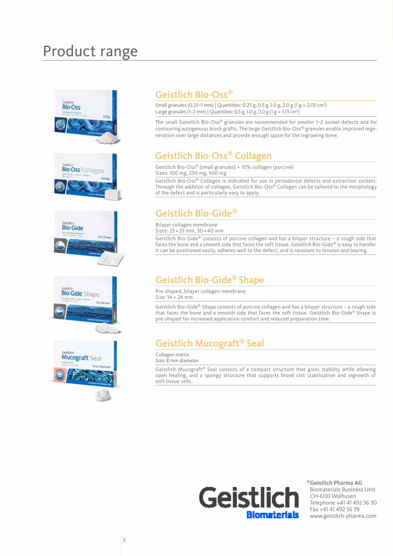
Geistlich Bio-Oss® (small granules) + 10% collagen (porcine) Sizes: 100 mg, 250 mg, 500 mg Geistlich Bio-Oss® Collagen is indicated for use in periodontal defects and extraction sockets. Through the addition of collagen, Geistlich Bio-Oss® Collagen can be tailored to the morphology of the defect and is particularly easy to apply.
Geistlich Bio-Oss® Collagen
Bilayer collagen membrane Sizes: 25 × 25 mm, 30 × 40 mmGeistlich Bio-Gide® consists of porcine collagen and has a bilayer structure – a rough side that faces the bone and a smooth side that faces the soft tissue. Geistlich Bio-Gide® is easy to handle: it can be positioned easily, adheres well to the defect, and is resistant to tension and tearing.
Geistlich Bio-Gide®
Collagen matrix Size: 8 mm diameter
Geistlich Mucograft® Seal consists of a compact structure that gives stability while allowing open healing, and a spongy structure that supports blood clot stabilisation and ingrowth of soft-tissue cells.
Geistlich Mucograft® Seal
Geistlich Bio-Oss®
Small granules (0.25–1 mm) | Quantities: 0.25 g, 0.5 g, 1.0 g, 2.0 g (1 g ≈ 2.05 cm3) Large granules (1–2 mm) | Quantities: 0.5 g, 1.0 g, 2.0 g (1 g ≈ 3.13 cm3)
The small Geistlich Bio-Oss® granules are recommended for smaller 1–2 socket defects and for contouring auto genous block grafts. The large Geistlich Bio-Oss® granules enable improved rege-neration over large distances and provide enough space for the ingrowing bone.
Background informationVarious classification systems for extraction sockets have been proposed thus far1–5 mostly focusing on the anterior teeth.1–3 Classifying different types of sockets should not solely be based on defect location. Information regarding as to soft and hard tissue breakdown and the number of remaining walls of the socket is crucial for determining the appropriate treatment protocol.
In the following extraction sockets impaired due to chronic pathology are classified into 5 types depending on the residual bone morphology and the level of soft tissue. Mesial and distal proximal bone peaks of single tooth extraction sockets are assumed to be intact if not described otherwise in the case.
Surgical Procedure
Literature references1 Funato A, Salama MA, Ishikawa T, Garber DA, Salama H. Timing, positioning, and sequential staging in esthetic implant therapy: a four-dimen-
sional perspective. Int J Periodontics Restorative Dent 2007;27:313-323.2 Elian N, Cho SC, Froum S, Smith RB, Tarnow DP. A simplified socket classification and repair technique. Pract Proced Aesthet Dent 2007;19:99-
104.3 Caplanis N, Lozada JL, Kan JY. Extraction defect assessment, classification, and management. J Calif Dent Assoc 2005;33:853-863.4 Smith RB, Tarnow DP. Classification of molar extraction sites for immediate dental implant placement: technical Note. Int J Oral Maxillofac
Implants 2013;28:911-916. 5 Al-Shabeeb MS, Al-Askar M, Al-Rasheed A, Babay N, Javed F, Wang HL, Al-Hezaimi K. Alveolar Bone remodeling around immediate implants
placed in accordance with the extraction socket classification: A three-dimensional microcomputed tomography analysis. J Periodontol 2012;83:981-987.
6 Crespi R, Capparè P, Gherlone E. Fresh-socket implants in periapical infected sites in humans. J Periodontol 2010;81:378-383.7 Park JC, Koo KT, Lim HC. The hidden X suture: a technical note on a novel suture technique for alveolar ridge preservation. J Periodontal Im-
plant Sci. 2016;46(6):415-425.8 Barone A, Toti P, Piattelli A, Iezzi G, Derchi G, Covani U. Extraction socket healing in humans after ridge preservation techniques: comparison
between flapless and flapped procedures in a randomized clinical trial. J Periodontol. 2014;85(1):14-239 Barone A, Borgia V, Covani U, Ricci M, Piattelli A, Iezzi G. Flap versus flapless procedure for ridge preservation in alveolar extraction sockets: a
histological evaluation in a randomized clinical trial. Clin Oral Implants Res. 2015;26(7):806-13.10 Cardaropoli D, Tamagnone L, Roffredo A, Gaveglio L, Cardaropoli G. Socket preservation using bovine bone mineral and collagen membrane: a
randomized controlled clinical trial with histologic analysis. Int J Periodontics Restorative Dent. 2012;32(4):421-30.
Further Indication Sheets> To receive these by mail free of charge, please contact: www.geistlich-biomaterials.com
> If you do not wish to collect indication sheets any more, please unsubscribe at your local distribution partner.
Geistlich Bio-Gide® ShapePre-shaped, bilayer collagen membrane Size: 14 × 24 mm
Geistlich Bio-Gide® Shape consists of porcine collagen and has a bilayer structure – a rough side that faces the bone and a smooth side that faces the soft tissue. Geistlich Bio-Gide® Shape is pre-shaped for increased application comfort and reduced preparation time.
© Geistlich Pharma AG Biomaterials Business Unit CH-6110 Wolhusen Telephone +41 41 492 56 30 Fax +41 41 492 56 39 www.geistlich-pharma.com
6016
79/1
706/
e
TYPE Isoft tissue height intact (CEJ) hard tissue intactInflammation damage limited to apical area Special attention to immediate implant placement is possible,
fill-the-gap using biomaterials is necessary
TYPE IIsoft tissue height intact (CEJ)hard tissue loss of 1 wall either buccal or lingualInflammation damage buccal/lingual wall destruction Special attention to mostly buccal wall destruction
TYPE IIIsoft tissue height recessionhard tissue buccal defectInflammation damage mostly buccal wall destruction Special attention to ridge augmentation combined with simultaneous
soft tissue augmentation needed
TYPE IV-Asoft tissue height intact (CEJ)hard tissue bucco-lingual destruction ≤ 50%Inflammation damage mostly buccal and lingual wall destruction Special attention to the flap serves as barrier to preserve and augment
simultaneously
TYPE IV-Bsoft tissue height intact (CEJ)hard tissue bucco-lingual destruction > 50%Inflammation damage mostly buccal and lingual wall destruction Special attention to the flap serves as barrier, more augmentation than
preservation due to destruction
TYPE Vsoft tissue height recessionhard tissue bucco-lingual destructionInflammation damage mostly buccal and lingual wall destruction Special attention to additional vertical augmentation may be necessary
to achieve desired crestal height
Prof. Ki-Tae Koo, Dr. Jung-Ju Kim and Dr. Heithem Ben Amara
Seoul National University, Korea
Contacts
> Prof. Ki-Tae Koo; E-mail: [email protected]
> Dr. Jung-Ju Kim; E-mail: [email protected]
> Dr. Heithem Ben Amara; E-mail: [email protected]
Fig. 1 Atraumatic tooth extraction followed by meticulous debridement is mandatory.6 Inflammatory tissues must be completely removed using the surgical curette or the he-mostat. Inflammatory tissues adhering to the soft tissue should be removed with a blade.
Fig. 2 Bone replacement material with slow resorption rate and good biofunctionality (Geistlich Bio-Oss® or Geistlich Bio-Oss® Collagen) is used to fill the socket to coun-teract bone resorption/increase the bone volume.
Fig. 3 A bilayer collagen membrane or colla-gen matrix with good biofunctionality is used to cover the augmented area for bone graft protection, graft stabilization and supporting early wound healing. The membrane can be applied in single or double layer fashion, the matrix should be applied in one layer.
Fig. 4 Suturing the defect without tension to avoid moving the mucogingival line which may narrow the keratinized zone. Hidden X sutures and healing by secondary intention can be used to prevent loss of keratinized tissue.7–10

2 3 4
Fig. 1 Classical type I defect in region 21. The pre-dictability of the treatment is good.
Fig. 4 The surgery site shows good healing by se-condary intention at 2 weeks post-op.
Fig. 2 Application of Geistlich Bio-Oss® Collagen after debridement.
Fig. 5 Placement of the implant at 4 months, the bony width of the ridge is preserved.
Fig. 3 Geistlich Mucograft® Seal is applied and secu-red with a cross-suture.
Fig. 6 Follow-up at 1 year after prosthesis delivery shows good esthetic results. Radiographic control 1 year after prosthesis delivery shows stable bony situation.
Type I
Defect configuration 4-wall defect / No loss of soft tissue
Pathology Endodontic origin / Fracture / Severe caries
Bone graft Geistlich Bio-Oss® or Geistlich Bio-Oss® Collagen
Membrane / matrix Geistlich Mucograft® Seal
Time of implant placement Immediate or delayed
Predictability Highly predictable
Ridge preservation potential Good
Fig. 1 Type 3 defect in region 14 with both soft and hard tissue deficiency. Radiographic view of the de-fect prior to extraction.
Fig. 4 Adaptation of Geistlich Bio-Gide® over Geist-lich Bio-Oss® Collagen.
Fig. 2 Situation after atraumatic extraction of tooth 14.
Fig. 5 A connective tissue graft from the palate is added to the defect to compensate for the soft tis-sue loss.
Fig. 3 Application of Geistlich Bio-Oss® Collagen after debridement. Geistlich Bio-Oss® Collagen is trimmed to shape to fit the defect.
Fig. 6 Stable clinical situation and radiographic view at 1 year follow-up after loading.
Type III
Defect configuration 3-wall defect / Loss of soft tissue
Pathology Periodontal origin / Periodontal-Endodontic combined
Bone graft Geistlich Bio-Oss® Collagen
Membrane / matrix Geistlich Bio-Gide®
Time of implant placement Delayed
Predictability Highly compromised, soft tissue augmentation necessary
Ridge preservation potential Poor
Fig. 1 Radiographic view in region 37. Clinical situa-tion after tooth extraction. Buccal and lingual bone walls are missing, only the soft tissue remains in normal position
Fig. 4 Good soft tissue healing at 3 months post-op.
Fig. 2 Application of Geistlich Bio-Oss® Collagen after debridement of the socket.
Fig. 5 Clinical and radiographic control during im-plant placement at 4 months show sufficient crestal ridge width.
Fig. 3 Fixation of the Geistlich Bio-Gide® with cross-suture and healing with secondary intention.
Fig. 6 Stable clinical situation 1 year after prosthesis delivery.
Type IV-B
Defect configuration 2-wall defect / No loss of soft tissue
Pathology Periodontal origin / Periodontal-Endodontic combined
Bone graft Geistlich Bio-Oss® or Geistlich Bio-Oss® Collagen
Membrane / matrix Geistlich Bio-Gide®
Time of implant placement Delayed
Predictability Highly compromised, vertical augmentation might be necessary
Ridge preservation potential Very poor
Fig. 1 Radiographic and clinical situation of the defect. Main medical conditions include swelling, acute pain and pus formation in region 36.
Fig. 4 Good healing by secondary intention at 2 weeks.
Fig. 2 Situation after extraction. Note the buccal bone resorption and granulation tissue at the bot-tom of the socket. Application of Geistlich Bio-Oss® Collagen after debridement of the socket.
Fig. 5 Situation prior to implant placement at 9 months shows good bony situation.
Fig. 3 Covering of the grafted area with Geistlich Bio-Gide® in two layers. The flap margin is fixed across the membrane without tension.
Fig. 6 Stable clinical/radiographic situation at 3 years after loading.
Type II
Defect configuration 3-wall defect / No loss of soft tissue
Pathology Periodontal origin/ Periodontal-Endodontic combined
Bone graft Geistlich Bio-Oss® or Geistlich Bio-Oss® Collagen
Membrane / matrix Geistlich Bio-Gide® or Geistlich Bio-Gide® Shape
Time of implant placement Immediate or delayed
Predictability Slightly compromised
Ridge preservation potential Good
Fig. 1 Type IV-A defect in region 26. Radiographic view of region 26 prior to extraction displays exten-ded bone defect.
Fig. 4 Slightly resorbed ridge contour with healed soft tissue situation after 3 months of healing.
Fig. 2 Extraction site immediately after tooth ext-raction.
Fig. 5 Sufficient ridge width for implant placement after 4 months of healing.
Fig. 3 Application of Geistlich Bio-Oss® Collagen after debridement. The augmented site is covered with a Geistlich Bio-Gide® membrane.
Fig. 6 Prosthesis delivery 4 months after implant placement. Stable radiographic situation after 1 year of loading.
Type IV-A
Defect configuration 4-wall defect / No loss of soft tissue
Pathology Periodontal origin / Periodontal-Endodontic combined
Bone graft Geistlich Bio-Oss® or Geistlich Bio-Oss® Collagen
Membrane / matrix Geistlich Bio-Gide®
Time of implant placement Delayed
Predictability Compromised
Ridge preservation potential Poor
Fig. 1 Clinical situation of type V defect in region 16.
Fig. 4 Radiographic view at 4 months after extrac-tion. The ridge preservation is limited to the height of the surrounding soft and hard tissue at the time of extraction. Additional vertical augmentation is indicated to increase the crestal height.
Fig. 2 Radiographic view prior to extraction docu-ments extended loss of bone.
Fig. 5 Clinical view at 1 year after implant loading.
Fig. 3 After extraction and debridement the defect is filled with Geistlich Bio-Oss® Collagen. A Geistlich Bio-Gide® is used to protect the grafted area.
Fig. 6 The radiographic view at 1 year after implant loading documents a stable bony situation.
Type V
Defect configuration 4- or 2-wall defect / Loss of soft tissue
Pathology Periodontal origin / Periodontal-Endodontic combined
Bone graft Geistlich Bio-Oss® or Geistlich Bio-Oss® Collagen
Membrane / matrix Geistlich Bio-Gide®
Time of implant placement Delayed
Predictability Compromised, vertical augmentation necessary
Ridge preservation potential Poor
6
Treatment of Extraction Sockets A new classification
Indication Sheet
15
Product range
Geistlich Bio-Oss® (small granules) + 10% collagen (porcine) Sizes: 100 mg, 250 mg, 500 mg Geistlich Bio-Oss® Collagen is indicated for use in periodontal defects and extraction sockets. Through the addition of collagen, Geistlich Bio-Oss® Collagen can be tailored to the morphology of the defect and is particularly easy to apply.
Geistlich Bio-Oss® Collagen
Bilayer collagen membrane Sizes: 25 × 25 mm, 30 × 40 mmGeistlich Bio-Gide® consists of porcine collagen and has a bilayer structure – a rough side that faces the bone and a smooth side that faces the soft tissue. Geistlich Bio-Gide® is easy to handle: it can be positioned easily, adheres well to the defect, and is resistant to tension and tearing.
Geistlich Bio-Gide®
Collagen matrix Size: 8 mm diameter
Geistlich Mucograft® Seal consists of a compact structure that gives stability while allowing open healing, and a spongy structure that supports blood clot stabilisation and ingrowth of soft-tissue cells.
Geistlich Mucograft® Seal
Geistlich Bio-Oss®
Small granules (0.25–1 mm) | Quantities: 0.25 g, 0.5 g, 1.0 g, 2.0 g (1 g ≈ 2.05 cm3) Large granules (1–2 mm) | Quantities: 0.5 g, 1.0 g, 2.0 g (1 g ≈ 3.13 cm3)
The small Geistlich Bio-Oss® granules are recommended for smaller 1–2 socket defects and for contouring auto genous block grafts. The large Geistlich Bio-Oss® granules enable improved rege-neration over large distances and provide enough space for the ingrowing bone.
Background informationVarious classification systems for extraction sockets have been proposed thus far1–5 mostly focusing on the anterior teeth.1–3 Classifying different types of sockets should not solely be based on defect location. Information regarding as to soft and hard tissue breakdown and the number of remaining walls of the socket is crucial for determining the appropriate treatment protocol.
In the following extraction sockets impaired due to chronic pathology are classified into 5 types depending on the residual bone morphology and the level of soft tissue. Mesial and distal proximal bone peaks of single tooth extraction sockets are assumed to be intact if not described otherwise in the case.
Surgical Procedure
Literature references1 Funato A, Salama MA, Ishikawa T, Garber DA, Salama H. Timing, positioning, and sequential staging in esthetic implant therapy: a four-dimen-
sional perspective. Int J Periodontics Restorative Dent 2007;27:313-323.2 Elian N, Cho SC, Froum S, Smith RB, Tarnow DP. A simplified socket classification and repair technique. Pract Proced Aesthet Dent 2007;19:99-
104.3 Caplanis N, Lozada JL, Kan JY. Extraction defect assessment, classification, and management. J Calif Dent Assoc 2005;33:853-863.4 Smith RB, Tarnow DP. Classification of molar extraction sites for immediate dental implant placement: technical Note. Int J Oral Maxillofac
Implants 2013;28:911-916. 5 Al-Shabeeb MS, Al-Askar M, Al-Rasheed A, Babay N, Javed F, Wang HL, Al-Hezaimi K. Alveolar Bone remodeling around immediate implants
placed in accordance with the extraction socket classification: A three-dimensional microcomputed tomography analysis. J Periodontol 2012;83:981-987.
6 Crespi R, Capparè P, Gherlone E. Fresh-socket implants in periapical infected sites in humans. J Periodontol 2010;81:378-383.7 Park JC, Koo KT, Lim HC. The hidden X suture: a technical note on a novel suture technique for alveolar ridge preservation. J Periodontal Im-
plant Sci. 2016;46(6):415-425.8 Barone A, Toti P, Piattelli A, Iezzi G, Derchi G, Covani U. Extraction socket healing in humans after ridge preservation techniques: comparison
between flapless and flapped procedures in a randomized clinical trial. J Periodontol. 2014;85(1):14-239 Barone A, Borgia V, Covani U, Ricci M, Piattelli A, Iezzi G. Flap versus flapless procedure for ridge preservation in alveolar extraction sockets: a
histological evaluation in a randomized clinical trial. Clin Oral Implants Res. 2015;26(7):806-13.10 Cardaropoli D, Tamagnone L, Roffredo A, Gaveglio L, Cardaropoli G. Socket preservation using bovine bone mineral and collagen membrane: a
randomized controlled clinical trial with histologic analysis. Int J Periodontics Restorative Dent. 2012;32(4):421-30.
Further Indication Sheets> To receive these by mail free of charge, please contact: www.geistlich-biomaterials.com
> If you do not wish to collect indication sheets any more, please unsubscribe at your local distribution partner.
Geistlich Bio-Gide® ShapePre-shaped, bilayer collagen membrane Size: 14 × 24 mm
Geistlich Bio-Gide® Shape consists of porcine collagen and has a bilayer structure – a rough side that faces the bone and a smooth side that faces the soft tissue. Geistlich Bio-Gide® Shape is pre-shaped for increased application comfort and reduced preparation time.
© Geistlich Pharma AG Biomaterials Business Unit CH-6110 Wolhusen Telephone +41 41 492 56 30 Fax +41 41 492 56 39 www.geistlich-pharma.com
6016
79/1
706/
e
TYPE Isoft tissue height intact (CEJ) hard tissue intactInflammation damage limited to apical area Special attention to immediate implant placement is possible,
fill-the-gap using biomaterials is necessary
TYPE IIsoft tissue height intact (CEJ)hard tissue loss of 1 wall either buccal or lingualInflammation damage buccal/lingual wall destruction Special attention to mostly buccal wall destruction
TYPE IIIsoft tissue height recessionhard tissue buccal defectInflammation damage mostly buccal wall destruction Special attention to ridge augmentation combined with simultaneous
soft tissue augmentation needed
TYPE IV-Asoft tissue height intact (CEJ)hard tissue bucco-lingual destruction ≤ 50%Inflammation damage mostly buccal and lingual wall destruction Special attention to the flap serves as barrier to preserve and augment
simultaneously
TYPE IV-Bsoft tissue height intact (CEJ)hard tissue bucco-lingual destruction > 50%Inflammation damage mostly buccal and lingual wall destruction Special attention to the flap serves as barrier, more augmentation than
preservation due to destruction
TYPE Vsoft tissue height recessionhard tissue bucco-lingual destructionInflammation damage mostly buccal and lingual wall destruction Special attention to additional vertical augmentation may be necessary
to achieve desired crestal height
Prof. Ki-Tae Koo, Dr. Jung-Ju Kim and Dr. Heithem Ben Amara
Seoul National University, Korea
Contacts
> Prof. Ki-Tae Koo; E-mail: [email protected]
> Dr. Jung-Ju Kim; E-mail: [email protected]
> Dr. Heithem Ben Amara; E-mail: [email protected]
Fig. 1 Atraumatic tooth extraction followed by meticulous debridement is mandatory.6 Inflammatory tissues must be completely removed using the surgical curette or the he-mostat. Inflammatory tissues adhering to the soft tissue should be removed with a blade.
Fig. 2 Bone replacement material with slow resorption rate and good biofunctionality (Geistlich Bio-Oss® or Geistlich Bio-Oss® Collagen) is used to fill the socket to coun-teract bone resorption/increase the bone volume.
Fig. 3 A bilayer collagen membrane or colla-gen matrix with good biofunctionality is used to cover the augmented area for bone graft protection, graft stabilization and supporting early wound healing. The membrane can be applied in single or double layer fashion, the matrix should be applied in one layer.
Fig. 4 Suturing the defect without tension to avoid moving the mucogingival line which may narrow the keratinized zone. Hidden X sutures and healing by secondary intention can be used to prevent loss of keratinized tissue.7–10


![Morphological Classification of Extraction Sockets and Clinical … · 2019. 9. 9. · end up with epithelized closure over the bone filled socket [5-7]. The healing of the extraction](https://static.fdocuments.us/doc/165x107/603a37eee6585d7ce66b5981/morphological-classification-of-extraction-sockets-and-clinical-2019-9-9-end.jpg)


