
Traumatic Brain Injury (TBI)
59
Traumatic Brain Injury (TBI) 1 Adult Health II Traumatic Brain Injury—Part 2 Jerry Carley RN, MA, MSN, CNE Summer 2010
description
Traumatic Brain Injury (TBI). Adult Health II Traumatic Brain Injury—Part 2. Jerry Carley RN, MA, MSN, CNE Summer 2010. Concept Map: Selected Topics in Neurological Nursing. PATHOPHYSIOLOGY Traumatic Brain Injury Spinal Cord Injury Specific Disease Entities : - PowerPoint PPT Presentation
Transcript of Traumatic Brain Injury (TBI)
Traumatic Brain Injury (TBI) 1
Adult Health IITraumatic Brain Injury—Part 2
Jerry Carley RN, MA, MSN, CNESummer 2010
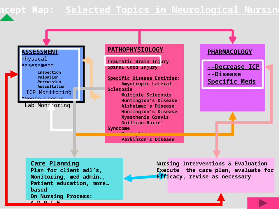
Concept Map: Selected Topics in Neurological Nursing
PATHOPHYSIOLOGY
Traumatic Brain InjurySpinal Cord Injury
Specific Disease Entities: Amyotropic Lateral Sclerosis Multiple Sclerosis Huntington’s Disease Alzheimer’s Disease Huntington’s Disease Myasthenia Gravis Guillian-Barre’ Syndrome Meningitis Parkinson’s Disease
PHARMACOLOGY
--Decrease ICP--Disease Specific Meds
ASSESSMENTPhysical Assessment Inspection Palpation Percussion Auscultation
ICP Monitoring“Neuro Checks” Lab Monitoring
Care PlanningPlan for client adl’s, Monitoring, med admin.,Patient education, more…basedOn Nursing Process: A_D_P_I_E
Nursing Interventions & EvaluationExecute the care plan, evaluate for Efficacy, revise as necessary

Objectives3
Recall anatomy and physiology of the brain & cranial nerves
Explain pathophysiology of various brain (head) injuries
Detail signs, symptoms and prevention of Increased Intracranial Pressure (ICP)
Demonstrate effective use of Glasgow Coma Scale
Discuss medical & nursing management of brain injuries
4
Prevent Secondary Injury !!!
Meaningful recovery of function after head injury is possible IF
secondary injuries are prevented or minimized
Secondary Brain Injury5
Any physiological event that can occur within minutes, hours, or days after the initial injury and leads to further damage of nervous tissue
Secondary Injury is mostly due to Increased ICP caused by hypotension, hypoxia, intracranial bleeding, seizures
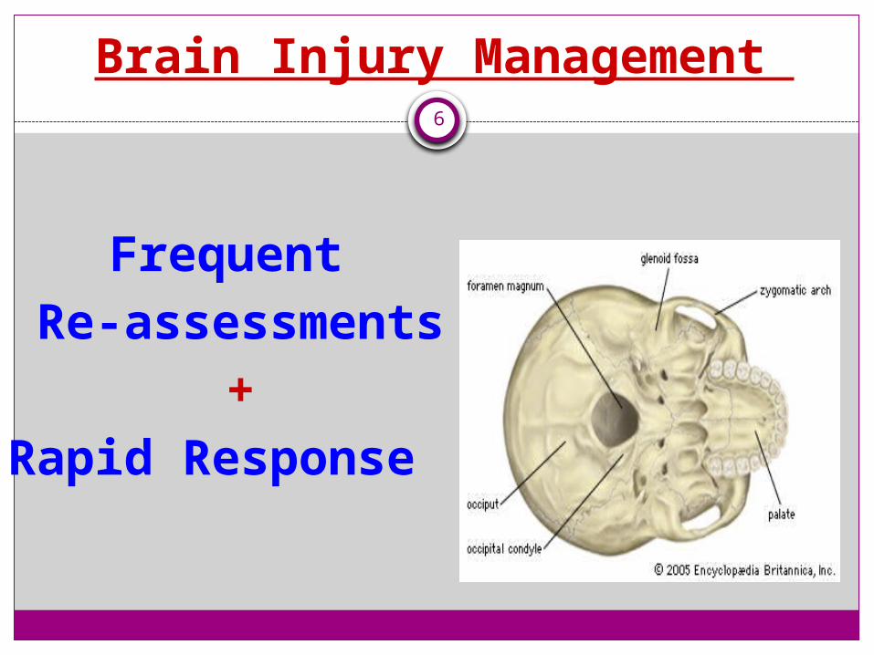
Brain Injury Management 6
Frequent Re-assessments
+Rapid Response

Be Vigilant for Increased ICP !7
To understand intracranial pressure, think of the skull as a rigid box. After brain injury, the skull may become overfilled with swollen brain tissue, blood, or CSF.
The skull will not stretch like
skin to deal with these changes. The skull may become too full and increase the pressure on the brain tissue. This is called increased intracranial pressure.
ICP Peaks 48 – 72 hours after injury
Foramen Magnum
8
Vital Signs Q15 minutes
Glasgow Coma Score Q15 minutes
Monitor: Neuro Checks q 15 minutes

9
Expanded Neuro
Assessment Tool
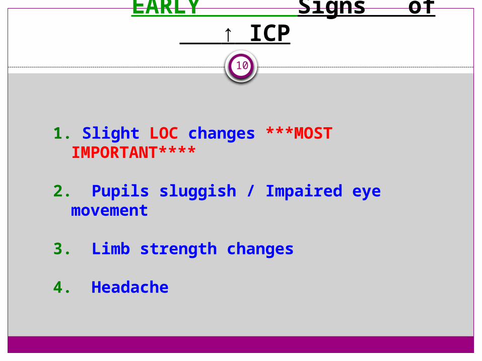
EARLY Signs of ↑ ICP
10
1. Slight LOC changes ***MOST IMPORTANT****
2. Pupils sluggish / Impaired eye movement
3. Limb strength changes
4. Headache

11
Change in
Level Of Consciousness (LOC)
***MOST IMPORTANT**** +
EARLIEST
Indicator of neurological deterioration
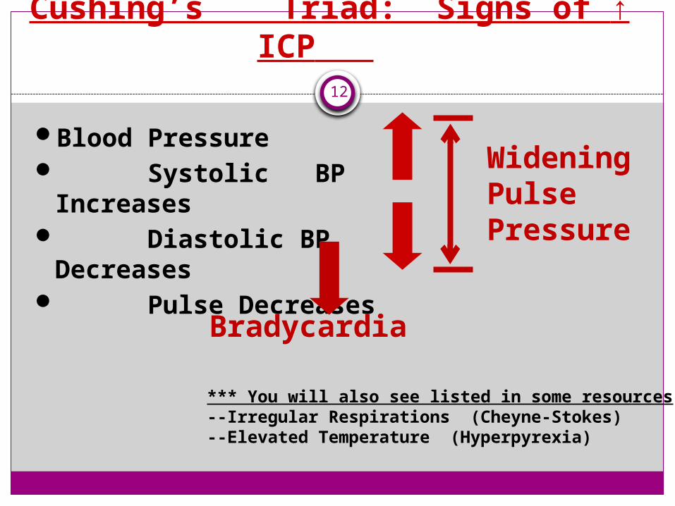
Cushing’s Triad: Signs of ↑ ICP
12
Blood Pressure Systolic BP
Increases Diastolic BP
Decreases Pulse Decreases
WideningPulsePressure
Bradycardia
*** You will also see listed in some resources:--Irregular Respirations (Cheyne-Stokes)--Elevated Temperature (Hyperpyrexia)

13
TREND Re-Assessment Data
+
COMPARE
to Baseline Assessment Data
Temp
BP
Pulse
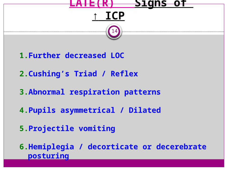
LATE(R) Signs of ↑ ICP
14
1. Further decreased LOC
2. Cushing’s Triad / Reflex
3. Abnormal respiration patterns
4. Pupils asymmetrical / Dilated
5. Projectile vomiting
6. Hemiplegia / decorticate or decerebrate posturing

Decerebrate Rigidity15

16
Brain Herniation occurs when a part of the brain pushes downward inside the skull through the opening that leads into the neck
(Foramen Magnum)
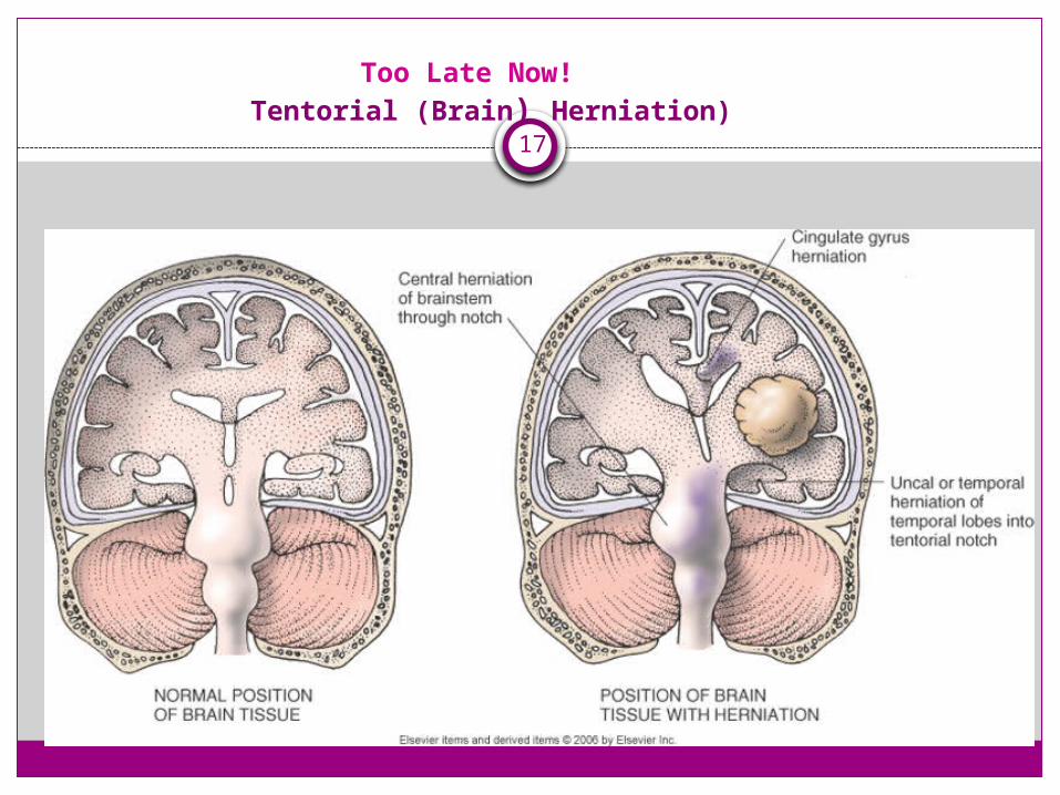
Too Late Now! Tentorial (Brain) Herniation)
17
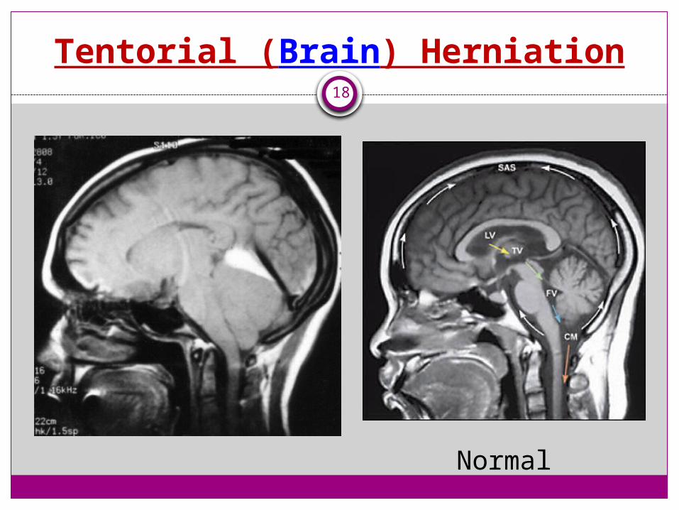
Tentorial (Brain) Herniation18
Normal
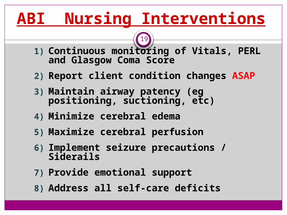
ABI Nursing Interventions19
1) Continuous monitoring of Vitals, PERL and Glasgow Coma Score
2) Report client condition changes ASAP
3) Maintain airway patency (eg positioning, suctioning, etc)
4) Minimize cerebral edema
5) Maximize cerebral perfusion
6) Implement seizure precautions / Siderails
7) Provide emotional support
8) Address all self-care deficits
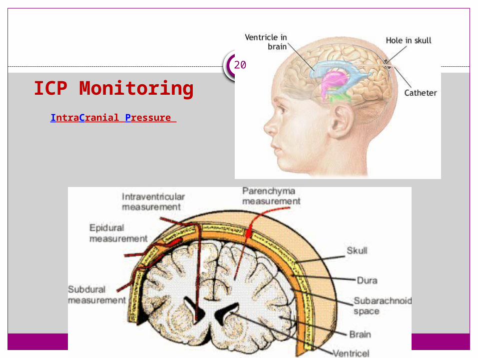
ICP MonitoringIntraCranial Pressure
20

Neurosurgeon drilling prior to placing an intracranial pressure monitor
21
Normal ICP for adults:
22
10 to 15 mm Hg
ABI Priority Nursing GOALS 23
* Minimize cerebral edema
* Maximize cerebral perfusion

ABI Nursing Interventions24
Continuous monitoring of Vitals, PERL and Glasgow Coma Score
Report client condition changes ASAP
Maintain airway patency BUT…
Avoid suctioning or Hyperventilate with 100% O2 FIRST
ABI Nursing Interventions25
Implement seizure precautions / Siderails
Phenytoin (Dilantin) (prevent / treat Sz)
Maintain head midline (neutral position)
HOB > 30 degrees

ABI Nursing Interventions26
Address all self-care deficits…BUT
Avoid clustering activities
Provide emotional support
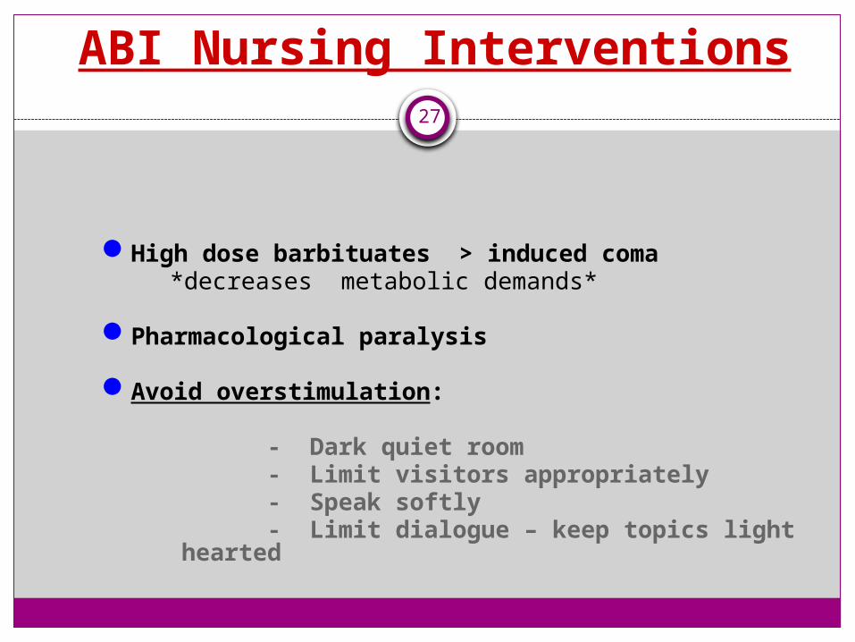
ABI Nursing Interventions27
High dose barbituates > induced coma *decreases metabolic demands*
Pharmacological paralysis
Avoid overstimulation:
- Dark quiet room- Limit visitors appropriately- Speak softly- Limit dialogue – keep topics
light hearted
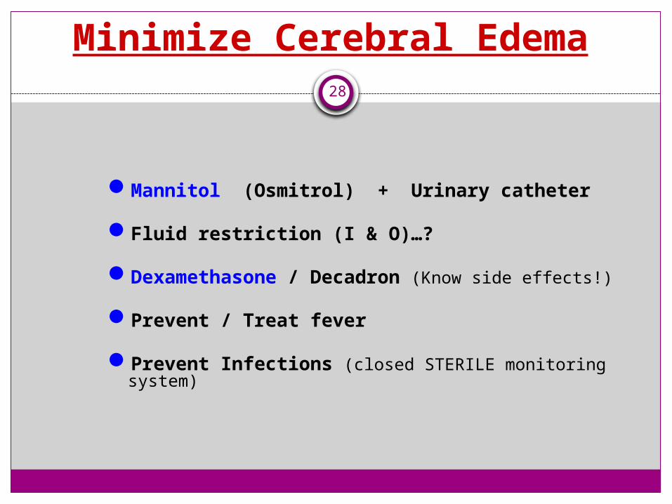
Minimize Cerebral Edema28
Mannitol (Osmitrol) + Urinary catheter
Fluid restriction (I & O)…?
Dexamethasone / Decadron (Know side effects!)
Prevent / Treat fever
Prevent Infections (closed STERILE monitoring system)
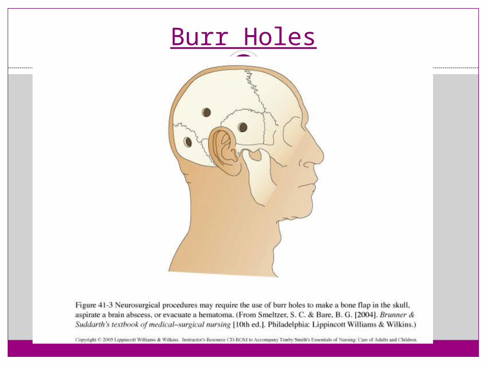
Burr Holes29

Minimize Cerebral Edema30
Maintain
Cerebral perfusion pressure MAP of 50 – 70 mm Hg
Prevents Hypoxia (Hypercarbia)

If BP too low…then O2 perfusion is poor…and Brain Can’t Function
31
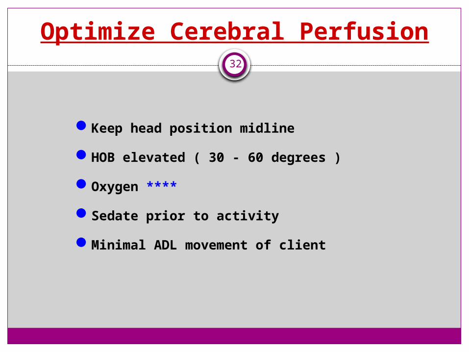
Optimize Cerebral Perfusion32
Keep head position midline
HOB elevated ( 30 - 60 degrees )
Oxygen ****
Sedate prior to activity
Minimal ADL movement of client
Teach Client / Family33
• Minimal stimulation environment
• No coughing, no straining, no hard laughing
• Head midline + Bedrest + HOB elevated
• S & S to report to nurse ASAP (Headache, drainage, etc)
• Purpose + frequency of neuro checks
• Medication regime (Narcotics, diuretics, stool softeners, etc)
• Medical interventions (Tests, traction, logrolling, surgery, etc)
Cerebral Concussion34
A ‘concussion’ is a relatively mild form of traumatic brain injury that results in temporary neurological changes
No apparent structural damage
Usually involves unconsciousness for a few seconds or minutes
Frontal lobe = bizarre irrational behavior
Temporal lobe = amnesia or disorientation

Discharge ….35
Mild concussion & neurological stability = usually will not require hospital admission
However !!! Must be observed by a reliable companion for at least 12 hours
No alcohol for several days
No pain medications stronger than Tylenol
Cerebral Contusion36
More severe
Brain bruised
Possible surface hemorrhage
Initially appears like shock
Can have B & B incontinence
Can be aroused…briefly
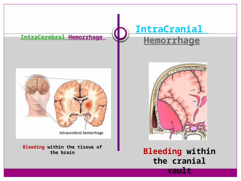
IntraCerebral Hemorrhage
Bleeding within the tissue of the brain
IntraCranial Hemorrhage
Bleeding within the cranial vault

IntraCranial Hemorrhage38
Bleeding within the cranial vault

Intracranial Epidural / Extradural Hematoma
39
- Between skull and dura- Extreme emergency- Mostly arterial

Epidural / Extradural Hematoma 40
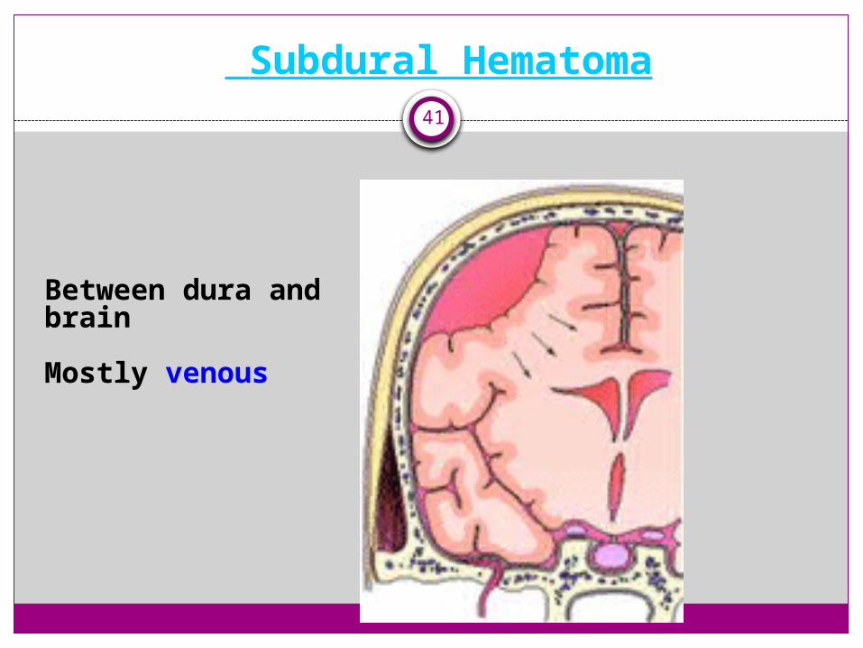
Subdural Hematoma41
Between dura and brain
Mostly venous
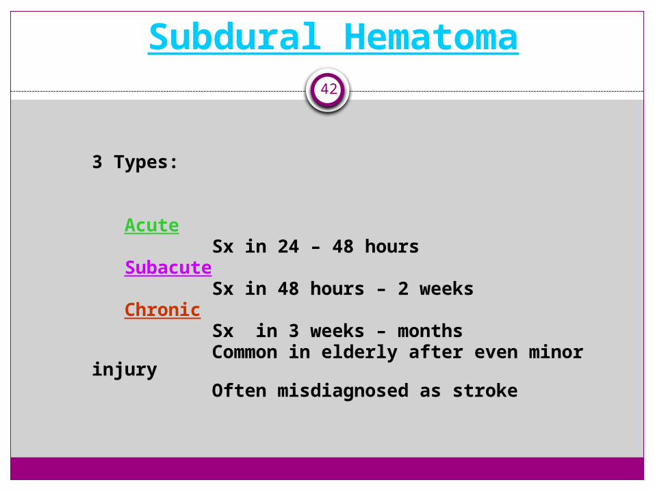
Subdural Hematoma42
3 Types:
Acute Sx in 24 – 48 hours
Subacute Sx in 48 hours – 2 weeks
Chronic Sx in 3 weeks – months Common in elderly after even
minor injury Often misdiagnosed as stroke
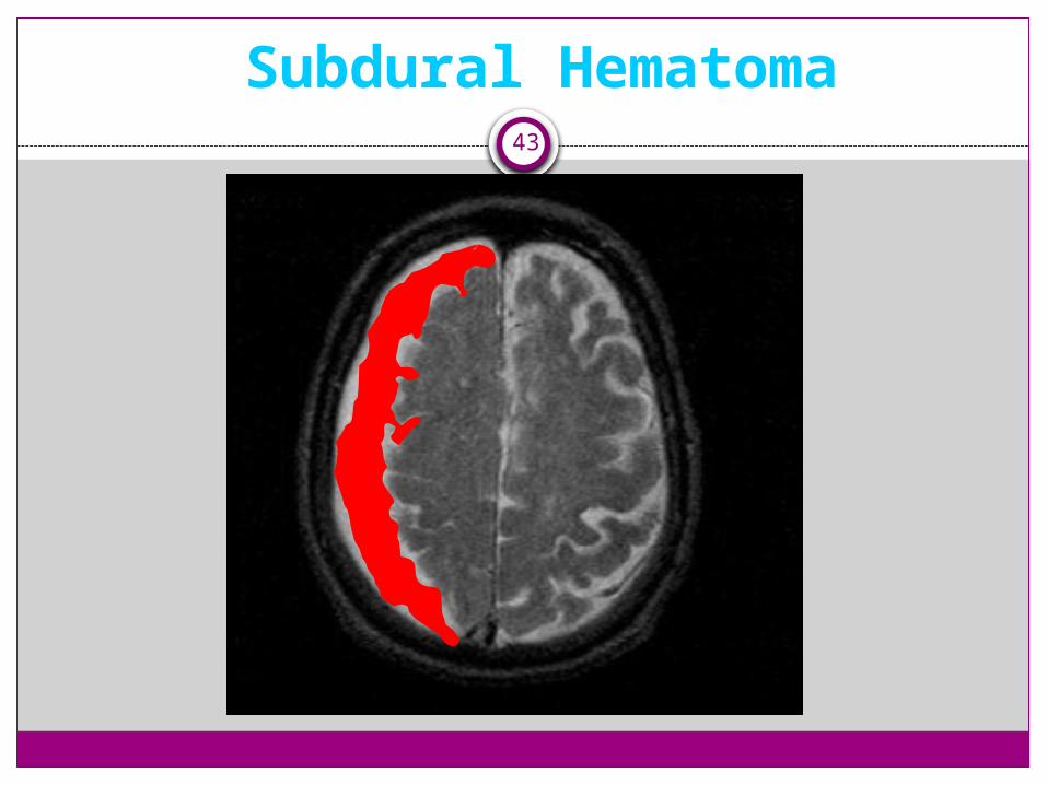
Subdural Hematoma43

Head trauma leading to subdural hematoma and intracranial hypertension
44
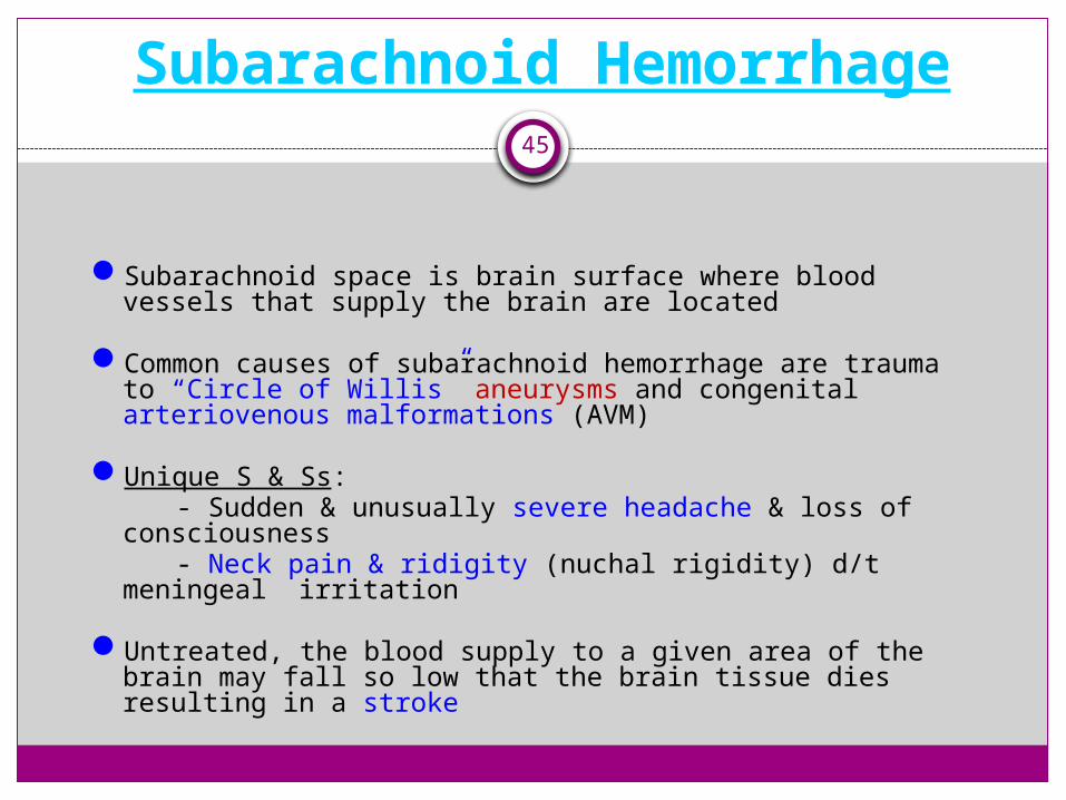
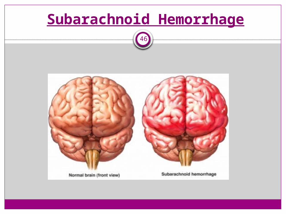
Subarachnoid Hemorrhage45
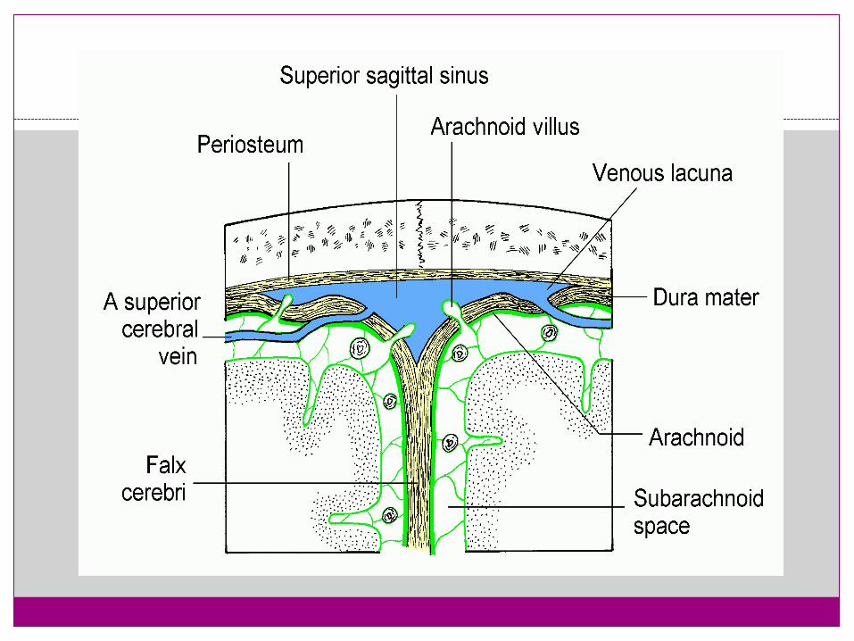
Subarachnoid space is brain surface where blood vessels that supply the brain are located
Common causes of subarachnoid hemorrhage are trauma to “Circle of Willis” aneurysms and congenital arteriovenous malformations (AVM)
Unique S & Ss: - Sudden & unusually severe headache & loss of
consciousness- Neck pain & ridigity (nuchal rigidity) d/t meningeal
irritation
Untreated, the blood supply to a given area of the brain may fall so low that the brain tissue dies resulting in a stroke
Subarachnoid Hemorrhage46

47
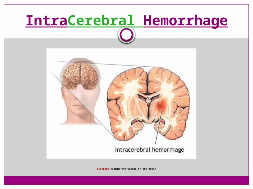
IntraCerebral Hemorrhage
Bleeding within the tissue of the brain
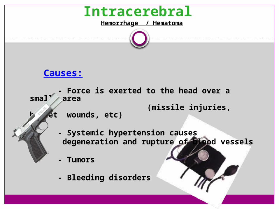
Intracerebral Hemorrhage / Hematoma
Causes:
- Force is exerted to the head over a small area
(missile injuries, bullet wounds, etc)
- Systemic hypertension causes degeneration and rupture of blood
vessels
- Tumors
- Bleeding disorders
Gunshot Wounds (GSW)
51
Suicides, homicides or accidental shootings
GSWs to the head are the most lethal of all firearm injuries
Estimated that greater than 90% fatality rate and at least two thirds of the victims die before ever reaching a hospital
Because of the high mortality associated with gunshot wounds to the head, they account for only approximately 10% of all traumatic brain injury patients who survive
GSW to the Head 52
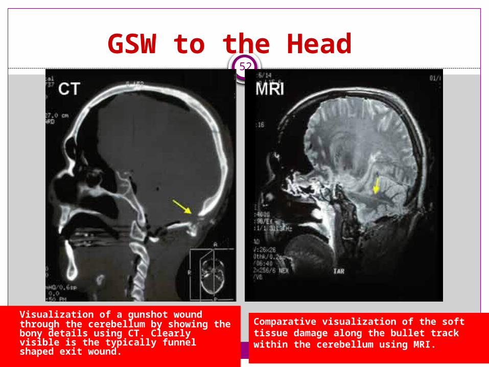
Visualization of a gunshot wound through the cerebellum by showing the bony details using CT. Clearly visible is the typically funnel shaped exit wound.
Comparative visualization of the soft tissue damage along the bullet track within the cerebellum using MRI.

Outcomes53
The predictors of poor neurological outcome or death after a gunshot wound to the head include:
- Initial Glasgow Coma Scale score- Older age- Presence of low blood pressure or inadequate oxygenation
early after injury- Dilated non-reactive pupils
Bullet trajectory through the brain has major significance. Bullets that traverse the brainstem, multiple lobes of the brain, or the ventricular system (chambers where cerebrospinal fluid is located) are particularly lethal
Many initial survivors develop uncontrollable intracranial pressure and subsequently succumb

ALL Cranial Injury Tx 54
ATLS evaluation & intervention (ABCs / Foley / NG / oxygen / Maintain traction)
Constant Monitoring
Diagnosis:
- CT scan (FAST!)- MRI - PET Scan (brain function assessment)
Medical interventions depend on severity:
- Endotracheal intubation / hyperventilation- Sedation- Diuresis- Rapid surgical evacuation
Surgical Outcomes55
Normal pupil reactivity prior to surgery is associated with a favorable outcome in 84 -100% of patients
When both pupils are dilated a poor outcome or death occurs in the great majority of individuals
Postoperative seizures are relatively common in these patients
In general, a favorable (functional) outcome is more likely in those patients who are treated very soon after injury, those who are younger adults, those with a higher GCS (above GCS of 6 or 7), those with reactive pupils, those without multiple cerebral contusions and those who do not develop difficult to control raised intracranial pressure

Head Injury Recovery56
Despite very severe initial injuries, some patients make dramatic recoveries within several months to a year after injury
Despite intensive intervention, long-term disability occurs in a large portion of the survivors
Patients with significant neuro-cognitive impairment are best managed at a comprehensive rehabilitation unit for several weeks or months after they leave the hospital
Recovery of function from the time of discharge to 6 months post-injury can be dramatic, even in some deeply comatose individuals
Improvement generally begins to plateau at 6 months post-injury and is typically maximal by one year to 18 months

Continued….57
Every brain injury is unique. Severity and types of impairments depend on the area and extent of the damage to the brain
Rehabilitation and support provided to a person who has received an injury has a major impact on the person’s recovery
ABI is known as an Invisible Disability due to the invisible nature of changes that may occur following an injury to the brain, such as memory loss, cognitive impairments, challenging behaviours and personality changes
People with ABI usually retain previous IQ, past memories, skills and interests. Their ability to use this knowledge can be lost to varying degrees
ABI is not an Intellectual or Psychiatric disability and therefore the needs of a person with an ABI are different from the needs of people with an intellectual or psychiatric disability
58
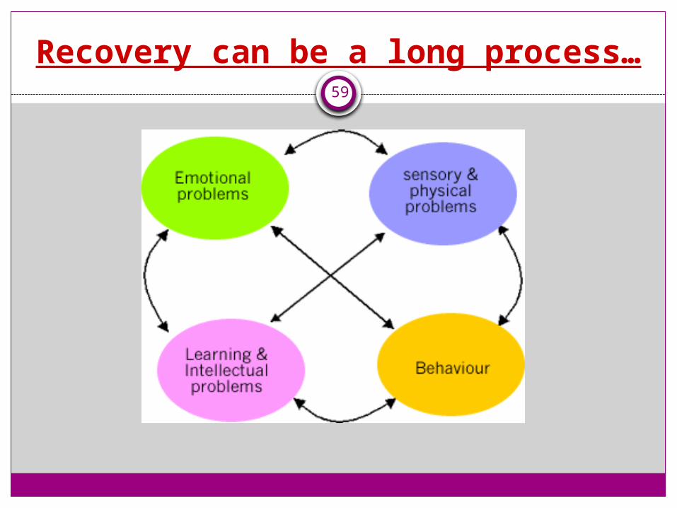
Recovery can be a long process…59


