Trauma: Spinal Cord Injury - CBC · Spine Spine trauma Spinal cord injury Spinal cord syndromes...
15
Trauma: Spinal Cord Injury Matthew J. Eckert, MD a, *, Matthew J. Martin, MD a,b INTRODUCTION Traumatic spine and spinal cord injury (SCI) occurred in roughly 17,000 US citizens in 2016, with an estimated prevalence of approximately 280,000 injured persons. 1 Although the injury has historically been a disease of younger adult men, a progressive increase in SCI incidence among the elderly has been reported over the last few de- cades. 2 Upwards of 70% of SCI patients suffer multiple injuries concomitant with spi- nal cord trauma, contributing to the high rates of associated complications during the acute and long-term phases of care. 3 SCI is associated with significant reductions in life expectancy across the spectrum of injury and age at time of insult. 1 Patients who survive the initial injury face significant risks of medical complications throughout the rest of their lives. More than half of all SCI patients will develop com- plications during the initial hospital stay, with higher rates corresponding to increased a Department of Surgery, General Surgery, Madigan Army Medical Center, 9040-A Jackson Avenue, Tacoma, WA 98431, USA; b Trauma and Emergency Surgery Service, Legacy Emanuel Medical Center, 501 N. Graham Street, #580, Portland OR 97227, USA * Corresponding author. E-mail address: [email protected] KEYWORDS Spine Spine trauma Spinal cord injury Spinal cord syndromes Spinal shock Spine immobilization KEY POINTS Hypotension following trauma should be considered secondary to hemorrhage until proven otherwise, even in patients with early suspicion of spinal injury. Neurogenic shock and spinal shock are separate, important entities that must be understood. Hypoxia and hypotension should be aggressively corrected because they lead to second- ary spinal cord injury, analogous to traumatic brain injury. Critical care support of multiple organ systems is frequently required early after injury. Early spinal decompression may lead to improved neurologic outcomes in select spinal cord injuries, and prompt consultation with spine surgeons is recommended. Computed tomography (CT) is the gold-standard screening study for evaluation of the spine after trauma and has significantly greater sensitivity and specificity compared with plain radiographs. High-quality CT imaging without evidence of cervical spine injury may be adequate for removal of the cervical immobilization collar in obtunded patients. Surg Clin N Am 97 (2017) 1031–1045 http://dx.doi.org/10.1016/j.suc.2017.06.008 surgical.theclinics.com 0039-6109/17/Published by Elsevier Inc.
Transcript of Trauma: Spinal Cord Injury - CBC · Spine Spine trauma Spinal cord injury Spinal cord syndromes...
Trauma: Spinal Cord Injury
Matthew J. Eckert, MDa,*, Matthew J. Martin, MDa,b
KEYWORDS
� Spine � Spine trauma � Spinal cord injury � Spinal cord syndromes � Spinal shock� Spine immobilization
KEY POINTS
� Hypotension following trauma should be considered secondary to hemorrhage untilproven otherwise, even in patients with early suspicion of spinal injury. Neurogenic shockand spinal shock are separate, important entities that must be understood.
� Hypoxia and hypotension should be aggressively corrected because they lead to second-ary spinal cord injury, analogous to traumatic brain injury. Critical care support of multipleorgan systems is frequently required early after injury.
� Early spinal decompression may lead to improved neurologic outcomes in select spinalcord injuries, and prompt consultation with spine surgeons is recommended.
� Computed tomography (CT) is the gold-standard screening study for evaluation of thespine after trauma and has significantly greater sensitivity and specificity comparedwith plain radiographs.
� High-quality CT imaging without evidence of cervical spine injury may be adequate forremoval of the cervical immobilization collar in obtunded patients.
INTRODUCTION
Traumatic spine and spinal cord injury (SCI) occurred in roughly 17,000 US citizens in2016, with an estimated prevalence of approximately 280,000 injured persons.1
Although the injury has historically been a disease of younger adult men, a progressiveincrease in SCI incidence among the elderly has been reported over the last few de-cades.2 Upwards of 70% of SCI patients suffer multiple injuries concomitant with spi-nal cord trauma, contributing to the high rates of associated complications during theacute and long-term phases of care.3 SCI is associated with significant reductions inlife expectancy across the spectrum of injury and age at time of insult.1
Patients who survive the initial injury face significant risks of medical complicationsthroughout the rest of their lives. More than half of all SCI patients will develop com-plications during the initial hospital stay, with higher rates corresponding to increased
a Department of Surgery, General Surgery, Madigan Army Medical Center, 9040-A JacksonAvenue, Tacoma, WA 98431, USA; b Trauma and Emergency Surgery Service, Legacy EmanuelMedical Center, 501 N. Graham Street, #580, Portland OR 97227, USA* Corresponding author.E-mail address: [email protected]
Surg Clin N Am 97 (2017) 1031–1045http://dx.doi.org/10.1016/j.suc.2017.06.008 surgical.theclinics.com0039-6109/17/Published by Elsevier Inc.
Eckert & Martin1032
injury severity, the presence of associated traumatic brain injury, and cerebrovasculardamage occurring with cervical spine injury.4,5 SCI may result in numerous multi-system complications particularly during the acute phase of care with long-term com-plications often related to infectious morbidity (Table 1). For the trauma/acute caresurgeon managing SCI patients during the acute phase of injury, respiratory compro-mise and shock are of primary concern, as discussed later.During the initial presentation and evaluation of SCI patients, almost all other injuries
should take precedent in both evaluation and management, unless the SCI isimpeding the airway (cervical spine) or the hemodynamics (neurogenic shock). Noemergent imaging of the spine is required before a laparotomy or other life-saving sur-gical intervention. Spinal immobilization is adequate for initial prevention of furtherinjury while addressing sources of hemorrhage. Prevention of hypotension and hypox-ia is also critical to mitigating further neurologic injury. Even if one of the rare spinalcord emergencies is encountered, such as a progressively worsening examinationwith cord compression that requires surgical decompression, your job will be to sta-bilize the patient and address any other life-threatening injury before intervention by aspine surgeon. Hypotension in the setting of a suspected acute traumatic SCI shouldalways be first assumed to be due to hypovolemia/hemorrhage, until ruled out.
ASSESSMENT OF SPINAL CORD INJURIES
The critical step in early evaluation of patients with possible SCI is recognition of pa-tients at risk and a focused, yet thorough neurologic examination. Too often the
Table 1Organ system complications following spinal cord injury
Organ System Complications
Cardiovascular Bradycardia/dysrhythmiaCardiac arrestCardiogenic pulmonary edema
Pulmonary Hypoventilation/respiratory failurePoor secretion controlAcute respiratory distress syndromeAspirationPneumonia
Gastrointestinal Gastric dysmotilityAdynamic ileusGastritis and ulcerationPancreatitis
Hematologic Venothromboembolism
Neurologic Neurogenic shockDepressionPosttraumatic stress disorderAnxietyAutonomic dysreflexia
Genitourinary Bladder dysfunctionUrinary tract infectionPriapism
Integument Pressure ulceration
Adapted from Stricsek G, Ghobrial G, Wilson J, et al. Complications in the management of patientswith spine trauma. Neurosurg Clin N Am 2017;28:150–2; with permission.
Trauma: Spinal Cord Injury 1033
steps of physical examination are deferred to the all-knowing computed tomo-graphic (CT) scanner. This delay can slow recognition of SCI and establishmentof baseline function and delay consultation of appropriate specialists and the initia-tion of preventive measures avoiding further secondary injury. The performance ofsuch an examination is frequently overlooked in busy trauma bays. The main pointsare that in addition to global neurologic disability (Glasgow Coma Scale and pupilexamination), the secondary survey of the patient should include sensory and motortesting in upper and lower extremity muscle groups as well as an anorectal exam-ination for tone and sensation. Table 2 lists the key muscle groups and their corre-sponding motor level that should be checked to determine the motor level of theinjury. Both upper and lower extremities and the right and left sides should betested, because certain syndromes may cause “skip” patterns or have unilateralasymmetric deficits (eg, central cord syndrome, Brown-Sequard). Both the motorand the sensory level should be clearly documented in the neurologic portion ofthe admission history and physical examination as well as communicated to anyconsulting spine provider.The distinction of complete and incomplete cord injury should be made because
this may influence operative decision making such as decompressive laminectomyor removal of bone fragments that are compressing the spinal cord. A “complete”injury is an injury pattern in which there is absolutely no spine-mediated neurologicfunction below the level of the injury. An “incomplete” injury is one in which there isany function below the level of injury, typically in the form of intact sensation (suchas perineal) or slight distal motor function. Sacral root sparing, which may allowsome residual anal sphincter function or sensation or slight movement of a greattoe, is an indication that the injury is incomplete and carries a better prognosis for re-covery of some degree of neurologic function.The American Spinal Injury Association (ASIA) International Standards for Neurolog-
ical Classification of SCI is the most frequently used and studied spinal injury severityassessment score.6 ASIA injury scoring is dependent on the presence or absence ofmotor function and sacral nerve root sparing below the level of injury (Fig. 1). Knowl-edge of which muscle group is fired by each spinal cord level and a more thoroughchecking for neurologic function distal to the apparent spinal cord level (including
Table 2Muscle function tested on physical examination and the corresponding motor level
Motor Level Muscle Function
C5 Elbow flexion
C6 Wrist extension
C7 Elbow extension
C8 Finger flexion
T1 Finger abduction
L2 Hip flexion
L3 Knee extension
L4 Ankle dorsiflexion
L5 Great toe extension
S1 Ankle dorsiflexion
Adapted from Branco F, Cardenas DD, Svircev JN. Spinal cord injury: a comprehensive review. PhysMed Rehabil Clin N Am 2007;18(4):651–79, v; with permission.
Fig. 1. ASIA SCI evaluation and scoring sheet. (From the American Spinal Injury Association(ASIA). Available at: www.asia-spinalinjury.org/information/downloads. Accessed February1, 2017; with permission.)
Eckert & Martin1034
Trauma: Spinal Cord Injury 1035
rectal examination) can help identify patients with incomplete injuries who may benefitfrom more urgent operative intervention. In addition, such an examination may helpidentify the possible injury location in order to better immobilize and prevent iatrogenicextension of the neurologic deficit. If spinal shock is present, it cannot be determinedwhether the injury is complete or incomplete until 24 to 48 hours after the shock periodresolves.Although imaging of the spine is often thought of as being primarily about the bony
structures, it is important to remember that complete assessment for spine injury andspinal stability requires assessment of the bone, the ligaments, and the spinal cord it-self. Traditional plain radiograph imaging of the spine only evaluates the bony compo-nent and has essentially been replaced by the widespread availability and efficiency ofmodern multidetector CT imaging. Although the utility of plain radiograph screening ofthe cervical spine in select low-risk patients has been extensively studied, numerousstudies have demonstrated the superiority of CT versus plain radiograph for screeningacutely injured patients with suspected or at risk for spine injury.7–9 Negative predic-tive values approaching 100% have been reported in several trials, particularly withrelation to the presence of a clinically unstable injury or injury requiring interven-tion.10–12 Although CT is ideal for screening identification of bony injury and suggestionof alignment abnormalities, the diagnostic accuracy of CT for spinal cord, ligamen-tous, and soft tissue injury is exceeded by MRI.13,14 Patients with deficits on examina-tion, CT findings suggestive of injury to the cord, disks, ligaments, or nerve roots, orunexplained neurologic examination may benefit from early MRI for diagnostic andtherapeutic decisions.
SPINAL SHOCK VERSUS NEUROGENIC SHOCK
The terms “spinal shock” and “neurogenic shock” are often both used inappropriatelyor incorrectly, or are confused for one another in the clinical setting. Neurogenic shockis the hemodynamic consequence of the SCI, classically characterized by hypoten-sion due to vasodilation and increased perfusion of the lower extremities (also knownas “warm shock”). In cases of higher SCI (cervical spine), hypotension may often beaccompanied by paradoxical bradycardia. This pattern is a relatively unique and spe-cific hemodynamic pattern to SCI and should prompt immediate evaluation and inter-ventions. Cervical spine and high thoracic spine injuries may result in loss ofsympathetic cardiac stimulation (bradycardia) and vasomotor tone in the lower body(hypotension) that will benefit from early initiation of vasopressor medication alongwith standard trauma resuscitation to restore intravascular volume status.Although neurogenic shock refers to a hemodynamic pattern, spinal shock refers to
the neurologic examination findings that may be seen after an acute SCI. The diagnosisof spinal shock is made in the presence of complete loss of reflexes below the level ofinjury, including the monosynaptic pathways. If spinal shock is present, this means thatit is not yet known what the ultimate amount of functional recovery will be. You will haveto wait until the spinal shock period is over. If spinal shock is not present, or it hasresolved, then whatever neurologic deficits you have at that time are likely to be fixedand permanent. Thus, for the patient presenting with paralysis in spinal shock, an un-known amount of functional recoverymay still occur. Spinal shock is diagnosed throughevaluation of the bulbocavernosus and/or cremasteric reflexes. If these reflexes are ab-sent, then the patient is in spinal shock, and when they return, the shock period hasended.15 Once the period of spinal shock has ended and the bulbocavernosus and/orcremasteric reflexes have returned, then the neurologic examination at that time likelyreflects what the permanent level and degree of deficits will be.
Eckert & Martin1036
MANAGEMENT OF ACUTE SPINAL CORD INJURY
The management of SCI begins with spine precautions (logrolling, cervical collar) andprotection from further injury. Spinal immobilization precautions do not mean lying flatand motionless once the initial trauma evaluation has been completed. Reverse Tren-delenburg position up to 30� will greatly benefit, and participatory pulmonary toiletshould begin if they are not intubated. Ensure adequate pain control to maximize tidalvolumes. Have a low threshold for nasogastric decompression because gastric ileusoften accompanies SCI with paraplegia or quadriplegia. Similarly, bladder dysfunctionis common, and a urinary catheter should be placed if not already present. Begin man-agement of pressure points with padding and frequent patient repositioning immedi-ately for paralyzed patients. Do not forget the psychological and emotional aspects ofthese injuries, particularly in young acutely injured patients. Early mental health profes-sional and/or chaplain consultation to begin helping the patient deal with the almostuniform depression and grieving over the loss of bodily function that accompaniesthese injuries is essential.Similar to studies of traumatic brain injury, avoidance of hypotension (systolic blood
pressure <90 mm Hg) and hypoxia is critical to avoiding further secondary SCI.16,17
Neurogenic shock may manifest as hypotension, which is poorly responsive to fluidresuscitation but responds briskly to vasopressor agents. In pure neurogenic shock,there is no associated tachycardia; the extremities may be warm and dry ratherthan cold and clammy, and typically, the patient has a significant cervical SCI. Treat-ment involves judicious volume resuscitation and the use of vasopressors to supportblood pressure. A purely peripheral vasoconstrictor such as Neo-Synephrine is oftenused, but in the multitrauma patient or the patient with associated bradycardia, abalanced vasopressor such as norepinephrine is a better choice. Maintaining themean arterial pressure greater than 85 mm Hg for up to 7 days after injury has beenassociated with improved ASIA scores and is recommended in current guidelines.18,19
The concept of spinal cord perfusion pressure monitoring through direct measure ofintraspinal pressure (ISP) after placement of an intradural catheter has demonstratedsignificant promise. Small trials have demonstrated safety and efficacy of monitoringISP at the site of maximal cord edema for up to 1 week after injury.20–22
Controversy persists over the utility of high-dose systemic steroids following bluntSCI. The series of National Acute Spinal Cord Injury Studies (NASCIS) I-III trials failedto demonstrate any difference in neurologic outcomes after high-dose methylprednis-olone.23–25 Post hoc subgroup analyses in the later NASCIS trials showed a 5-point(ASIA) motor scoring improvement across 15 muscle groups but did not correlatewith any measure of functional improvement or effect upon disability. In contrast, pa-tients receiving high-dose steroids consistently suffered increased rates of infectiouscomplications, gastric ulceration, venothromboembolic events, wound complications,and trended toward increased mortality related to pulmonary complications.Numerous additional trials confirmed the NASCIS outcomes with similar complicationprofiles.26,27 The use of high-dose steroids after penetrating SCI has consistentlyshown poor outcomes.28–30 Previous guidelines had recommended intravenous ste-roid bolus and infusion for all patients with blunt SCI and no contraindication to treat-ment. However, this approach has now been widely abandoned because of the highlyquestionable clinical benefit identified in the NASCIS trials as well as more recent dataindicating no benefit to steroid use. Recent evidence-based management guidelinesrecommend against the use of high-dose steroids for all acute SCI.31 There may bea role for steroid use in highly select or atypical types of spinal cord trauma and injury,and this decision should be at the discretion of the managing spine surgeon.
Trauma: Spinal Cord Injury 1037
Not infrequently, the examination of an injured patient will suggest neurologic deficitwithout any obvious supporting radiographic findings. These SCI without radiographicabnormalities (SCIWORA) represent a challenging group because limited evidenceand support for treatment have been established. Unfortunately, the ultimate neuro-logic outcome in SCIWORA patients appears correlated with the initial deficits on ex-amination similar to those patients with radiographic abnormalities and neurologicdeficits.32 MRI appears particularly useful in identifying subtle intraneural and extra-neural abnormalities that may be associated with the neurologic deficits and correlatewith outcomes.33 Supportive care is similar to that described previously for those pa-tients with neurologic injury and radiographic abnormalities. Despite increased reportson populations of SCIWORA patients, limited high-quality data are available, and norandomized controlled trials of management have been conducted.The timing of surgical spinal decompression in the setting of compressive phenom-
ena, such as epidural hematoma, cord edema or hemorrhage, or impinging bony frag-ments and foreign bodies, has been extensively studied. Preclinical models havedemonstrated that the extent and duration of cord compression correlate with ultimateneurologic deficit.34,35 Human series have suggested that early decompression in thefirst 8 to 24 hours after injury in those patients with incomplete SCI is associated withimproved neurologic outcomes. In the Surgical Timing in Acute Spinal Cord InjuryStudy, patients with cervical SCI that underwent decompression in the first 24 hoursafter injury had a significant improvement in ASIA grade.36 Additional studieshave confirmed the benefits of early decompression for these incomplete patients;however, not all trials of time to decompression have shown neurologic improvementwith early intervention.37 Early surgical decompression is also associated withdecreased pulmonary morbidity and duration of mechanical ventilation as well asdecreased intensive care unit and hospital length of stay.38,39 Unfortunately, surgicaldecompression in patients presenting with ASIA A complete injury has not shown sig-nificant improvements in neurologic outcomes.40,41
As previously noted, one of the early measures in the assessment and stabilizationof a suspected SCI patient is the prevention of future mechanical injury due to insta-bility of the spinal column. Although logroll precautions, maintenance of axial align-ment, pressure point management, and attentive nursing care are generallyadequate for thoracolumbar spinal protection, the cervical spine is generally protectedwith specialized cervical immobilization devices, or “c-collars.” Controversy persistsregarding the actual effectiveness and necessity of these devices to maintain properalignment and prevent skeletal motion.42–45 In addition, these hard devices may beassociated with development of pressure ulceration in upwards of 30% of patients.46
For the obtunded trauma patient with associated brain injury or critical illness in whomthe cervical spine cannot be cleared by standard imaging and physical examination, aparticular challenge arises. Traditionally, these patients were left in a hard cervical col-lar until examinable or until an MRI or other imaging adjunct demonstrated no evi-dence of injury. Recent studies from multiple centers have demonstrated the safetyof cervical collar removal after a negative high-quality CT given negative predictivevalues of 100% for unstable injuries.12 As a result of these studies, practice patternsare changing with expected decreased utilization of MRI resources and prevention ofcomplications of hard cervical immobilization devices.
MANAGEMENT OF PENETRATING SPINE TRAUMA
Penetrating SCI is most commonly secondary to gunshot wounds and typically resultsin complete SCI due to direct trauma to the cord and associated blast effect as well as
Eckert & Martin1038
secondary hemorrhage and ischemia.47 In civilian trauma centers, the thoracolumbarspine is the region most frequently injured. The management options for open spinetrauma are not much different than those for closed spine trauma, even in patientswith open vertebral column fractures. The wound must be managed with irrigationand debridement of all nonviable tissues and early antibiotic coverage. The choice ofantibiotics is generally the same as for patients with open extremity fractures, and thereare no good data on recommended duration of therapy (ranges of 48 hours to 10 daysreported). Patients with associated intestinal injuries, particularly if those injuriescommunicate with the spinal column injury, may require broader coverage. Thesedevastating injuries will obviously require multidisciplinary care for optimal outcomes.Incomplete SCI, cauda equina syndrome, or evidence of cord compression after
penetrating injury due to hematoma or bony fragments may benefit from surgicaldecompression.48 In addition, surgical stabilization of unstable vertebral column in-juries is necessary in any patient likely to survive the acute phase of care. Explorationof the injuries may be required for debridement, neurologic deterioration due to cordcompression, or persistent cerebrospinal fluid leak. Wound infection, spinal columninstability, and cerebrospinal fluid leaks were the most common complications in a se-ries of penetrating SCI.49 These penetrating injuries are frequently managed conserva-tively as outcomes have been shown to be equivocal or even more favorable innonoperative management.50
AIRWAY MANAGEMENT
The need for appropriate airway management is of particular importance for patientswith cervical SCIs. Most patients with high cervical SCIs will present with quadriplegiaand respiratory distress or arrest and clearly require intubation. The difficult patient pop-ulation is the lower cervical spine injury (C5-C7) and upper thoracic spine (T1-T6), whofrequently present with no obvious respiratory distress due to the ability to continueshallow breathing. Be wary of these patients: previous reports have demonstratedthat up to 50% will slowly decompensate and require a delayed emergent airway inter-vention.51,52 This can result in secondary SCI due to hypoxia and trauma from manipu-lation during emergent intubation attempts. Over a period of several hours to days, theshallow breathing will result in progressive atelectasis, pulmonary consolidation orpneumonia, and finally, acute hypoxic decompensation. The insidious airway collapsein this setting can be severely harmful or even fatal and should be anticipated. A lowthreshold for intubating these patients semi-electively for the initial hospital period orbefore transfer may be prudent, because up to 30% will require intubation in the first24 hours after cervical SCI.53 Factors suggestive of early intubation include higher levelof injury (above C5), complete paralysis, the presence of associated injuries (particularlychest wall or intrathoracic), and low lung volumes on chest radiograph. If you have thecapability tomeasure and follow vital capacity, then this may be a useful adjunct to iden-tify the patient progressing to respiratory failure.Numerous publications discuss the methods for intubation in the patient with a cer-
vical spine injury and the potential impact on spinal mobility. Direct laryngoscopy withmanual in-line stabilization of the cervical spine during the procedure has been shownto be safe and effective in 2 large studies.54,55 If available, fiberoptic intubation is safeand avoids significant spinal motion but requires manual stabilization during the pro-cedure as well if the collar is released. Finally, a surgical airway is always an option andmay be necessary for patients who will require long-term mechanical ventilation and/or pulmonary toilet. Consideration should be given to potential incision location foranterior cervical spine surgical stabilization if indicated. Although the definitions of
Trauma: Spinal Cord Injury 1039
what constitutes “early” tracheostomy vary in the literature, those patients who requireor are projected to require mechanical ventilation for more than 2 weeks after injurycertainly may benefit from tracheostomy. Improved secretion control, enhanced facil-itation of mechanical ventilation weaning, patient comfort, ability to participate in reha-bilitation, and possibly reduced risks of pneumonia and resource utilization have beenassociated with tracheostomy in SCI patients.56–58
SPINAL CORD SYNDROMES
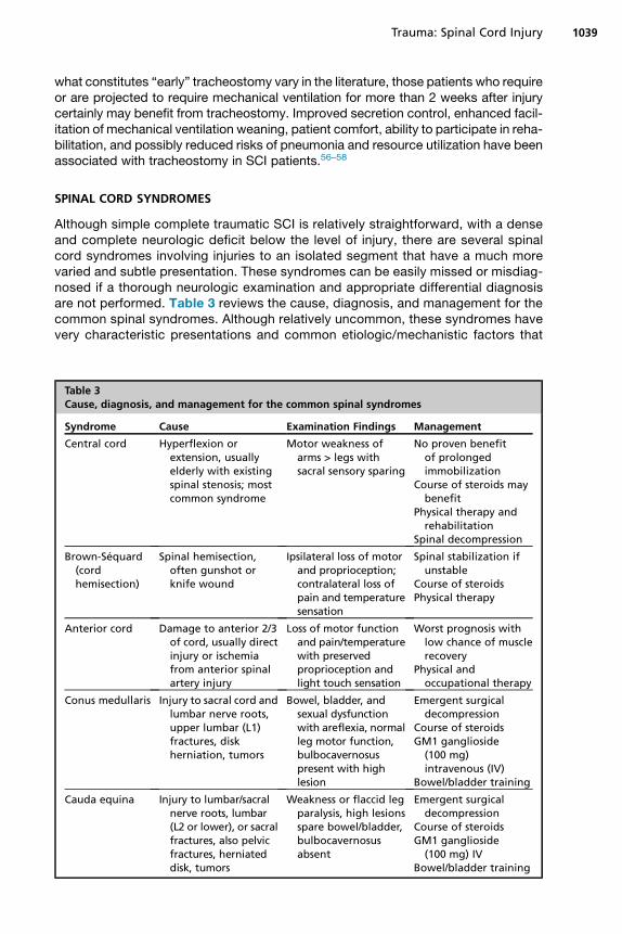
Although simple complete traumatic SCI is relatively straightforward, with a denseand complete neurologic deficit below the level of injury, there are several spinalcord syndromes involving injuries to an isolated segment that have a much morevaried and subtle presentation. These syndromes can be easily missed or misdiag-nosed if a thorough neurologic examination and appropriate differential diagnosisare not performed. Table 3 reviews the cause, diagnosis, and management for thecommon spinal syndromes. Although relatively uncommon, these syndromes havevery characteristic presentations and common etiologic/mechanistic factors that
Table 3Cause, diagnosis, and management for the common spinal syndromes
Syndrome Cause Examination Findings Management
Central cord Hyperflexion orextension, usuallyelderly with existingspinal stenosis; mostcommon syndrome
Motor weakness ofarms > legs withsacral sensory sparing
No proven benefitof prolongedimmobilization
Course of steroids maybenefit
Physical therapy andrehabilitation
Spinal decompression
Brown-Sequard(cordhemisection)
Spinal hemisection,often gunshot orknife wound
Ipsilateral loss of motorand proprioception;contralateral loss ofpain and temperaturesensation
Spinal stabilization ifunstable
Course of steroidsPhysical therapy
Anterior cord Damage to anterior 2/3of cord, usually directinjury or ischemiafrom anterior spinalartery injury
Loss of motor functionand pain/temperaturewith preservedproprioception andlight touch sensation
Worst prognosis withlow chance of musclerecovery
Physical andoccupational therapy
Conus medullaris Injury to sacral cord andlumbar nerve roots,upper lumbar (L1)fractures, diskherniation, tumors
Bowel, bladder, andsexual dysfunctionwith areflexia, normalleg motor function,bulbocavernosuspresent with highlesion
Emergent surgicaldecompression
Course of steroidsGM1 ganglioside(100 mg)intravenous (IV)
Bowel/bladder training
Cauda equina Injury to lumbar/sacralnerve roots, lumbar(L2 or lower), or sacralfractures, also pelvicfractures, herniateddisk, tumors
Weakness or flaccid legparalysis, high lesionsspare bowel/bladder,bulbocavernosusabsent
Emergent surgicaldecompression
Course of steroidsGM1 ganglioside(100 mg) IV
Bowel/bladder training
Eckert & Martin1040
the managing physician should be aware of. Central cord syndrome is almost al-ways a flexion/extension injury in an elderly patient with preexisting spinal steno-sis. Although spinal immobilization is often maintained in these patients becauseof the presence of neurologic deficits, it is typically not an unstable spine injury,and there is no proven benefit of immobilization with a cervical collar. Brown-Sequard (or “cord hemisection”) is extremely uncommon and typically only seenafter direct penetrating injury to the spinal cord that results in different unilateraland contralateral deficits. Anterior cord syndrome is typically a vascular causerelated to injury or interruption of flow through the anterior spinal artery. For allof the spinal cord syndromes presenting with fixed and established defects,management is usually expectant and aimed at treating symptoms and pain.59,60
However, for any patient with a progressively worsening neurologic deficit, emer-gent consultation with a spine surgeon for possible decompression should be apriority.
VENOUS THROMBOEMBOLISM AFTER SPINAL CORD INJURY
Venous thromboembolism (VTE) events and appropriate prophylaxis are major con-cerns in the acute and long-term management of SCI patients. A recent large popula-tion study of roughly 48,000 SCI patients found an approximately 2.5-fold increasedrisk of deep venous thrombosis (DVT) and 1.6-fold increased risk of pulmonary embo-lism compared with controls. The risks were greatest within the first 3 monthsfollowing injury and with increasing age.61 Additional studies confirm the heightenedrisk in the early period after injury and with increased patient age, pointing to the sig-nificance of falls in older patients.62,63 Overall incidence of VTE after SCI in recent pub-lications suggests an incidence of approximately 3% to 5%, with associated earlymortality of 7.5%, but up to 20% in patients greater than 70 years of age.2,63 For un-clear reasons, the location of SCI along the spine may also be associated with VTErisk. Maung and colleagues64 found upper thoracic SCI to be associated with a higherrate of VTE compared with other spinal level injury. Variation in reported incidence ofVTE is likely due to the timing and modality of screening methods. Duplex and Dopplerultrasonography are the most frequently and conveniently used screening modalitiestoday.65 There is no clear consensus on the timing or schedule for screening afteracute SCI; however, the systematic review by Furlan and Fehlings66 suggests thatweekly screening for asymptomatic DVT in the early high-risk period following SCImay be appropriate.In recognizing the increased risk of VTE following SCI, optimal preventive measures
are an important facet of care for these patients. Until recently, controversy persistedregarding the optimal treatment with mechanical and/or chemoprophylaxis. A seriesof studies has confirmed the superior efficacy of low-molecular-weight heparin(LMWH) versus unfractionated subcutaneous heparin for the prevention of VTE and alower associated bleeding risk.67,68 Optimal dosing of LMWH remains controversial.Studies suggest that standard daily or twice daily enoxaparin dosing may not achievetherapeutic anti-Xa levels in trauma patients; however, no consensus for dose adjust-ment currently exists.69,70 Combined preventive therapywith gradient elastic stockingsor sequential pneumatic compression devices of the lower extremities and LMWHmayoffer an even greater reduction in VTE risk after SCI.71 Early initiation of chemoprophy-laxis (<72 hours from injury) has been associatedwith a significant reduction in the inci-dence of VTE after SCI (2% vs 26%) and recommended in at least one consensusguideline.72,73 For patients with contraindications to chemoprophylaxis or VTE despitetreatment, retrievable vena cava filter placementmaybeappropriate. However, outside
Trauma: Spinal Cord Injury 1041
these unique situations, studies suggest empiric filter placement offers no benefit overroutine mechanical/chemoprophylaxis for acute SCI.74–76
SUMMARY
The impact of a SCI in any trauma patient can range from a minor nuisance to devas-tating paralysis, and unfortunately, the full spectrum of these injuries is frequently seenafter trauma. Although much of the damage is done at the time of presentation andirreversible immediately, adherence to comprehensive supportive care aimed at treat-ing the injury and preventing secondary injury may make a significant difference in thepatient’s ultimate functional outcome. Every physician should be able to perform aquick but thorough neurologic examination and understand the implications of signif-icant examination findings such as spinal shock.
REFERENCES
1. National Spinal Cord Injury Statistical Center. Facts and figures at a glance.Birmingham (AL): University of Alabama; 2016.
2. Jain NB, Ayers GD, Peterson EN, et al. Traumatic spinal cord injury in the UnitedStates, 1993-2012. JAMA 2015;313(22):2236–43.
3. Herbert JS, Burnham RS. The effect of polytrauma in persons with traumaticspine injury. A prospective database of spine fractures. Spine (Phila Pa 1976)2000;25(1):55–60.
4. Kushner DS, Alvarez G. Dual diagnosis: traumatic brain injury with spinal cordinjury. Phys Med Rehabil Clin N Am 2014;25(3):681–96.
5. Silva Santos E, Santos Filho W, Possatti L, et al. Clinical complications in patientswith severe cervical spine trauma: a ten-year prospective study. Arq Neuropsi-quiatr 2012;70(7):524–8.
6. Kirshblum SC, Burns SP, Biering-Sorensen F, et al. International standards forneurological classification of spinal cord injury (revised 2011). J Spinal CordMed 2011;34(6):535–46.
7. Ryken TC, Hadley MN, Walters BC, et al. Radiographic assessment. Neurosur-gery 2013;72(Suppl 2):54–72.
8. Holmes JF, Akkinepalli R. Computed tomography versus plain radiography toscreen for cervical spine injury: a meta-analysis. J Trauma 2005;58(5):902–5.
9. Bailitz J, Starr F, Beecroft M, et al. CT should replace three-view radiographs asthe initial screening test in patients at high, moderate, and low risk for blunt cer-vical spine injury: a prospective comparison. J Trauma 2009;66(6):1605–9.
10. Hogan GJ, Mirvis SE, Shanmuganathan K, et al. Exclusion of unstable cervicalspine injury in obtunded patients with blunt trauma: is MR imaging neededwhen multi-detector row CT findings are normal? Radiology 2005;237(1):106–12.
11. Adams JM, Cockburn MI, Difazio LT, et al. Spinal clearance in the difficult traumapatient: a role or screening MRI of the spine. Am Surg 2006;72(1):101–5.
12. Patel MB, Humble SS, Cullinane DC, et al. Cervical spine collar clearance in theobtunded adult blunt trauma patient: a systematic review and practice manage-ment guideline from the Eastern Association for the Surgery of Trauma. J TraumaAcute Care Surg 2015;78(2):430–41.
13. Bozzo A, Marcoux J, Radhakrishna M, et al. The role of magnetic resonance im-aging in the management of acute spinal cord injury. J Neurotrauma 2011;28(8):1401–11.
14. Beers GJ, Raque GH, Wagner GG, et al. MR imaging in acute cervical spinetrauma. J Comput Assist Tomogr 1988;12(5):755–61.
Eckert & Martin1042
15. Ditunno JF, Little JW, Tessler A, et al. Spinal shock revisited: a four-phase model.Spinal Cord 2004;42(7):383–95.
16. Vale F, Burns J, Jackson A, et al. Combined medical and surgical treatment afteracute spinal cord injury: results of a prospective pilot study to assess the meritsof aggressive medical resuscitation and blood pressure management.J Neurosurg 1997;87:239–46.
17. Stevens R, Bhardwaj A, Kirsh J. Critical care and perioperative management intraumatic spinal cord injury. J Neurosurg Anesthesiol 2003;15:215–29.
18. Hamryluk G, Whetstone W, Saigal R, et al. Mean arterial blood pressure corre-lates with neurologic recovery after human spinal cord injury. J Neurotrauma2015;32(24):1958–67.
19. Ryken T, Hurlbert R, Hadley M, et al. The acute cardiopulmonary management ofpatients with cervical spinal cord injuries. Neurosurgery 2013;77:84–92.
20. Werndle MC, Saadoun S, Phang I, et al. Monitoring of spinal cord perfusion pres-sure in acute spinal cord injury: initial findings of the injured spinal cord pressureevaluation study. Crit Care Med 2014;42(3):646–55.
21. Phang I, Zoumprouli A, Saadoun S, et al. Safety profile and probe placement ac-curacy of intraspinal pressure monitoring for traumatic spinal cord injury: injuredspinal cord pressure evaluation study. J Neurosurg Spine 2016;25(3):398–405.
22. Varsos GV, Werndle MC, Czosnyka ZH, et al. Intraspinal pressure and spinal cordperfusion pressure after spinal cord injury: an observational study. J NeurosurgSpine 2015;23(6):763–71.
23. Bracken MB, Shepard MJ, Hellenberg KG, et al. Methylprednisolone and neuro-logical function 1 year after spinal cord injury. Results of the National Acute SpinalCord Injury Study. J Neurosurg 1985;63:704–13.
24. Bracken MB, Shepard MJ, Collins WF, et al. A randomized controlled trial of meth-ylprednisolone or naloxone in the treatment of acute spinal-cord injury. Results ofthe Second National Acute Spinal Cord Injury Study. N Engl J Med 1990;322:1405–11.
25. Bracken MH, Shepard MJ, Holford TR, et al. Administration of methylpredniso-lone for 24 or 48 hours or tirilazad mesylate for 48 hours in the treatment of acutespinal cord injury. Results of the Third National Acute Spinal Cord Injury Random-ized Controlled Trial. National Acute Spinal Cord Injury Study. JAMA 1997;277:1597–604.
26. Otani K, AbeH, Kadoya S, et al. Beneficial effect ofmethylprednisolone sodium suc-cinate in the treatment of acute spinal cord injury. Sekitsui Sekizui 1994;7:633–47.
27. Petitjean ME, Pointillart V, Dixmerias F, et al. Medical treatment of spinal cordinjury in the acute stage. Ann Fr Anesth Reanim 1998;17:114–22.
28. Prendergast MR, Saxe JM, Ledgerwood AM, et al. Massive steroids do notreduce the zone of injury after penetrating spinal cord injury. J Trauma 1994;37:576–9.
29. Levy ML, Gans W, Wijesinghe HS, et al. Use of methylprednisolone as an adjunctin the management of patients with penetrating spinal cord injury: outcome anal-ysis. Neurosurgery 1996;39:1141–8.
30. Heary RF, Vaccaro AR, Mesa JJ, et al. Steroids and gunshot wounds to the spine.Neurosurgery 1997;41:576–83.
31. Hurlbert RJ, Hadley MN, walters BC, et al. Pharmacological therapy for acute spi-nal cord injury. Neurosurgery 2015;76(Suppl 1):S71–83.
32. Wilson JR, Cadotte DW, Fehlings MG. Clinical predictors of neurologic outcome,functional status, and survival after traumatic spinal cord injury: a systematic re-view. J Neurosurg Spine 2012;17(Suppl 1):S11–26.
Trauma: Spinal Cord Injury 1043
33. Boese CK, Lechler P. Spinal cord injury without radiographic abnormalities inadults: a systematic review. J Trauma Acute Care Surg 2013;75:320–30.
34. Dimar JR, Glassman SD, Raque GH, et al. The influence of spinal canal narrowingand timing of decompression on neurologic recovery after spinal cord contusionin a rat model. Spine (Phila Pa 1976) 1999;24(16):1623–33.
35. Batchelor PE, Willis TE, Skeers P, et al. Meta-analysis of pre-clinical studies ofearly decompression in acute spinal cord injury: a battle of time and pressure.PLoS One 2013;8(8):e72659.
36. Fehlings MG, Vaccaro A, Wilson JR, et al. Early versus delayed decompressionfor traumatic cervical spinal cord injury: results of the surgical timing in acute spi-nal cord injury study (STASCIS). PLoS One 2012;7(2):e32037.
37. Wilson JR, Singh A, Craven C, et al. Early versus late surgery for traumatic spinalcord injury: the results of a prospective Canadian cohort study. Spinal Cord 2012;50(11):840–3.
38. Bourassa-Moreau E, Mac-Thiong JM, Feldman DE, et al. Complications in acutephase hospitalization of traumatic spinal cord injury: does surgical timing matter?J Trauma Acute Care Surg 2013;74(3):849–54.
39. Liu JM, Long XH, Zhou Y, et al. Is urgent decompression superior to delayed sur-gery for traumatic spinal cord injury? A meta-analysis. World Neurosurg 2016;87:124–31.
40. Petitjean ME, Mousselard H, Pointillart V, et al. Thoracic spinal trauma and asso-ciated injuries: should early spinal decompression be considered? J Trauma1995;39(2):368–72.
41. Bourassa-Moreau E, Mac-Thiong JM, Li A, et al. Do patients with complete spinalcord injury benefit from early surgical decompression? Analysis of neurologicalimprovement in a prospective cohort study. J Neurotrauma 2016;33(3):301–6.
42. Clemency BM, Bart JA, Malhotra A, et al. Patient immobilized with a long spineboard rarely have unstable thoracolumbar injuries. Prehosp Emerg Care 2016;20(2):266–72.
43. Wampler DA, Pineda C, Polk J, et al. The long spine board does not reducelateral spine motion during transport- a randomized healthy volunteer crossovertrial. Am J Emerg Med 2016;34(4):717–21.
44. Horodyski M, DiPaola CP, Conrad BP, et al. Cervical collars are insufficient for im-mobilizing an unstable cervical spine injury. J Emerg Med 2011;41:513–9.
45. Lador R, Ben-Galim P, Hippa JA. Motion within the unstable cervical spine duringpatient maneuvering: the neck-pivot-shift phenomenon. J Trauma 2011;70:247–50.
46. Ham W, Schoonhoven L, Schuurmans MJ, et al. Pressure ulcers from spinalimmobilization in trauma patients: a systematic review. J Trauma Acute CareSurg 2014;76(4):1131–41.
47. Rosenfeld JV, Bell RS, Armonda R. Current concepts in penetrating and blastinjury to the central nervous system. World J Surg 2015;39(6):1352–62.
48. Klimo P, Ragel BT, Rosner M, et al. Can surgery improve neurologic function inpenetrating spinal injury? A review of the military and civilian literature andtreatment recommendations for military neurosrgeons. Neurosurg Focus 2010;28(5):E4.
49. Simpson RK, Venger BH, Narayan RK. Treatment of acute penetrating injuries ofthe spine: a retrospective analysis. J Trauma 1989;29(1):42–6.
50. Sidu GS, Ghag A, Prokuski V, et al. Civilian gunshot injuries of the spinal cord: asystematic review of the current literature. Clin Orthop Relat Res 2013;471:3945–55.
Eckert & Martin1044
51. Jackson AB, Grommers TE. Incidence of respiratory complications following SCI.Arch Phys Med Rehabil 1994;75:270–5.
52. Cotton BA, Pryor JP, Chinwilla I, et al. Respiratory complications and mortality riskassociated with thoracic spine injury. J Trauma 2005;59:1400–9.
53. Gardner BP, Watt JW, Krishnan KR. The artificial ventilation of acute spinal corddamaged patients: a retrospective study of forty-four patients. Paraplegia1986;24(4):208–20.
54. Grande CM, Barton CR, Stene JK. Appropriate techniques for the airway man-agement of emergency patients with suspected spinal cord injury. Anesth Analg1988;67(7):714–5.
55. Shatney CH, Brunner RD, Nguyen TQ. The safety of orotracheal intubation in pa-tients with unstable cervical spine fracture or high spinal cord injury. Am J Surg1995;170(6):676–9.
56. Como JJ, Sutton ER, McCunn M, et al. Characterizing the need for mechanicalventilation following cervical spinal cord injury with neurologic deficit. J Trauma2005;59:912–6.
57. Harrop J, Sharan A, Scheid E, et al. Tracheostomy placement in patients withcomplete cervical spinal cord injuries: American Spinal Injury Association GradeA. J Neurosurg 2004;100:20–3.
58. Jaeger J, Littlewood K, Durbin C. The role of tracheostomy in weaning from me-chanical ventilation. Respir Care 2002;47(4):469–80.
59. Brooks NP. Central cord syndrome. Neurosurg Clin N Am 2017;28(1):41–7.
60. Radcliff KE, Kepler CK, Delasota LA, et al. Current management review of thora-columbar cord syndromes. Spine J 2011;11(9):884–92.
61. Chung WS, Lin CL, Chang SN, et al. Increased risk of deep vein thrombosis andpulmonary thromboembolism in patients with spinal cord injury: a nationwidecohort prospective study. Thromb Res 2014;133(4):579–84.
62. Giorgi PM, Donadini MP, Dentali F, et al. The short- and long-term risk of venousthromboembolism in patients with acute spinal cord injury: a prospective cohortstudy. Thromb Haemost 2013;109(1):34–8.
63. Jones T, Ugalde V, Franks P, et al. Venous thromboembolism after spinal cordinjury: incidence, time course, and associated risk factors in 16,240 adults andchildren. Arch Phys Med Rehabil 2005;86(12):2240–7.
64. Maung AA, Schuster KM, Kaplan LJ, et al. Risk of venous thromboembolism afterspinal cord injury: not all levels are the same. J Trauma Acute Care Surg 2011;71(5):1241–5.
65. Zierler BK. Ultrasonography and diagnosis of venous thromboembolism. Circula-tion 2004;109(12 Suppl 1):I–9.
66. Furlan JC, Fehlings MG. Role of screening tests for deep venous thrombosis inasymptomatic adults with acute spinal cord injury: an evidence-based analysis.Spine (Phila Pa 1976) 2007;32(17):1908–16.
67. Spinal Cord Injury Thromboprophylaxis Investigators. Prevention of venousthromboembolism in the acute treatment phase after spinal cord injury: a ran-domized, multicenter trial comparing low-dose heparin plus intermittent pneu-matic compression with enoxaparin. J Trauma Acute Care Surg 2003;54(6):1116–24.
68. Teasell RW, Hsieh JT, Aubut JA, et al. Spinal cord injury rehabilitation evidencereview research team. Venous thromboembolism after spinal cord injury. ArchPhys Med Rehabil 2009;90(2):232–45.
Trauma: Spinal Cord Injury 1045
69. Rutherford EJ, Schooler WG, Sredzienski E, et al. Optimal dose of enoxaparin incritically ill trauma and surgical patients. J Trauma Acute Care Surg 2005;58(6):1167–70.
70. Costantini TW, Min E, Box K, et al. Dose adjusting enoxaparin is necessary toachieve adequate venous thromboembolism prophylaxis in trauma patients.J Trauma Acute Care Surg 2013;74(1):128.
71. Aito S, Pieri A, D’Andrea M, et al. Primary prevention of deep venous thrombosisand pulmonary embolism in acute spinal cord injured patients. Spinal Cord 2002;40(6):300.
72. Christie S, Thibault-Halman G, Casha S. Acute pharmacological DVT prophylaxisafter spinal cord injury. J Neurotrauma 2011;28(8):1509–14.
73. Harrop JS, Tetreault L, Aarabi B, et al. Guidelines for the management of patientswith spinal cord injury: efficacy, safety and timing of anticoagulation prophylaxis.Spine J 2016;16(10):S214.
74. Gorman PH, Qadri SF, Rao-Patel A. Prophylactic inferior vena cava (IVC) filterplacement may increase the relative risk of deep venous thrombosis after acutespinal cord injury. J Trauma Acute Care Surg 2009;66(3):707–12.
75. Maxwell RA, Chavarria-Aguilar M, Cockerham WT, et al. Routine prophylacticvena cava filtration is not indicated after acute spinal cord injury. J Trauma AcuteCare Surg 2002;52(5):902–6.
76. Cook AD, Gross BW, Osler TM, et al. Vena cava filter use in trauma and rates ofpulmonary embolism, 2003-2015. JAMA Surg 2017. [Epub ahead of print].