trauma in pregnanacy
46
Trauma in Pregnancy
-
Upload
garima-prakash -
Category
Health & Medicine
-
view
109 -
download
0
Transcript of trauma in pregnanacy
Trauma in Pregnancy

• Trauma is not just something that happens to other people. Trauma is a disease that could affect anyone, but it is more importantly it is something that we can all prevent.

• Complicates 6-7% of
pregnancies.
• Leading cause of non- obstetric
death.
• Maternal death is the common
cause of fetal death.

Relative Frequency of Trauma
Severity and Mortality
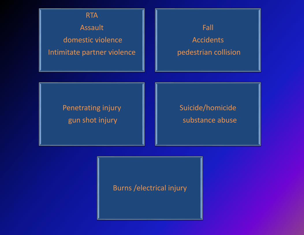
Causes
RTA
Assault
domestic violence
Intimitate partner violence
Fall
Accidents
pedestrian collision
Penetrating injury
gun shot injury
Suicide/homicide
substance abuse
Burns /electrical injury

Relative Frequency of Trauma

• Shift in the centre of gravity as the pregnancy advances makes the woman prone to falls and accidents.


UNIQUE CHALLENGE
CARE OF TWO PATIENTS
ALTERED PHYSIOLOGY
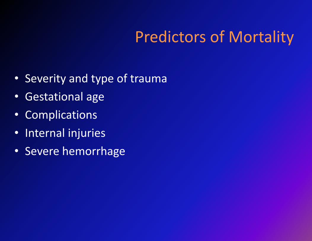
Predictors of Mortality
• Severity and type of trauma
• Gestational age
• Complications
• Internal injuries
• Severe hemorrhage

Keel et al reported the major killers in polytrauma
head injury (66%).
hemorrhagic shock (21%).
sepsis and multiorgan failure (13%)
coagulopathies (dilutional and consumption).

ANATOMICAL & PHYSIOLOGICAL CHANGES
Plasma volume increases by 45-50% Reduce maternal resistance to limited blood flow
Red cell mass Increases by 30% Dilutional anemia
Cardiac output Increases by 30-50% Relative maternal resistance to limited blood loss
Uteroplacental blood flow 20-30% shunt Uterine injury may predispose to increased blood loss, increase vascularity
Uterine size Dramatic increase Change in position of abdominal contents, supine hypotension
Minute ventilation Increases by 25-30% Diminished Paco2Diminished buffering capacity
Functional residual capacity decreased Predisposition to atelectasis and hypoxemia
Gastric emptying delayed Predisposition to aspiration

Injury specific
considerations

BLUNT TRAUMA
• 2/3 cases of all trauma in pregnancy.
• CAUSES
- Motor vehicular collisions
- Assault
- Falls
• Especially in 2nd and 3rd trimester.
• PELVIC FRACTURES – engaged head
• Haemorrhage from dilated retroperitoneal veins can cause massive hemorrhagic shock and death

• MVA - Passenger restraint system decreases maternal/fetal injury.
• Crosby & Costilee
- 33% maternal mortality with no restraints
- 5% using seat belts
Penetrating trauma
• Primarily -stabbing or gunshot wounds
• The gravid uterus in 2nd & 3rd trimester provides protection to maternal internal organs.
• Maternal mortality lower than the non-pregnant women – 3.9% vs 12.5%
• Awwad and colleagues, observed fetal death rates 70-90%- direct injury to uterus and 38% for injuries above uterus.

BURNS• BURNS -6.8% to 7.8% of all pregnancies
• Fetal loss is 56% -if 15-25% of body surface area(BSA) involved.
63% - If 25-50% BSA involved.
100%- If >50% BSA involved.
• Maternal and fetal deaths are often a result of inadequate fluid resuscitation, prolonged hypotension, shock, hypoxia, septicemia and hyponatremia.
• Potential for carbon monoxide poisoning
Assessing and managing the
pregnant patient with trauma

MULTIDISCIPLINARY APPROACH
Trauma Surgeon
Obstetrician
Anaesthesiologist
Neonatologist


Primary Survey
Maternal assessment
„ Fetal age assessment & presence of life
If CPR unsuccessful consider Perimortem CS
Minimize effect of uterine compression on maternal
resuscitation
Fetal resuscitation

Maternal Resuscitation
The main principle guidingtherapy must be that
resuscitating the mother will
resuscitate the fetus.

PRIMARY SURVEY

CIRCULATION• Position- 30 degrees to left.
• Volume resuscitation.
Crystalloid- 3:1 replacement
Blood transfusion
• Maternal B.P,H.R- not a reliable indicator of maternal and fetal well being.
• Uterus not critical organ- after acute blood loss- uterine blood flow decreased to maintain normal maternal B.P.
• When signs of shock appear- fetal compromise far advanced

Supine hypotension syndrome
GOALS OF INITIAL RESUSCITATION
• Systolic blood pressure - 80 to 100 mmHg.
• PaCO2 > 90%
• Hematocrit - 25% to 36%.
• Platelet count >50,000/cu mm.
• Normal serum calcium.
• Core body temperature > 35°C.
• Avoiding an increase in serum lactate level and metabolic acidosis.
• Adequate analgesia

SECONDARY SURVEY• ‘Top to bottom’ physical assessment .
• More extensive fetal evaluation ; specific fetal evaluation
• Identify - vaginal bleeding
- ruptured fetal membranes
- abruption
- PTL
- Direct uterine injury or fetal injury
- fetal distress
• Assess the extent of feto-maternal hemorrhage.
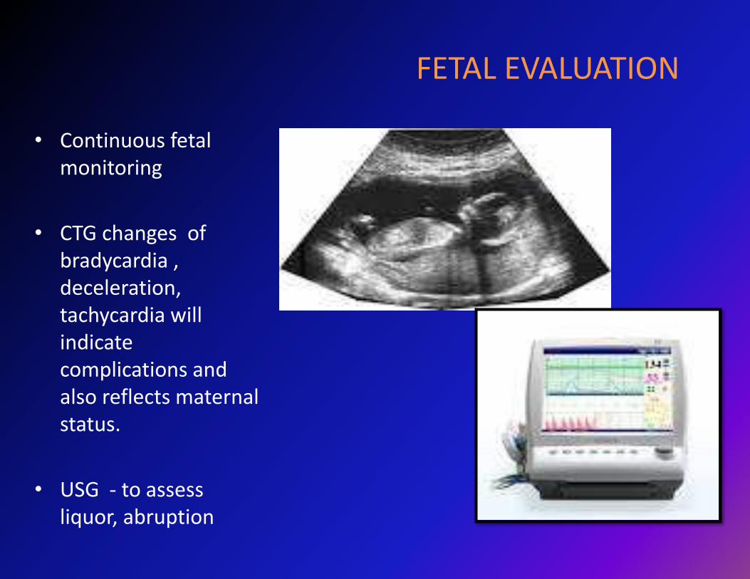
FETAL EVALUATION
• Continuous fetal monitoring
• CTG changes of bradycardia , deceleration, tachycardia will indicate complications and also reflects maternal status.
• USG - to assess liquor, abruption

Laboratory
• CBC
• Serum electrolytes, blood sugar
• Blood group &Type and Cross match,
• PT/aPTT, fibrinogen,
• Kleihauer-Betke(KB)
• urinalysis (and HCG if needed).
• ABG
Diagnostic Imaging
Investigations During
Pregnancy

IMAGING STUDIES
• Do not avoid or delay necessary exams due to concerns about fetal radiation exposure.
• Fetal adverse effects are unlikely if radiation dose less than 5 rads or distance more than 10 cm.
• Relative risk of childhood cancer greatest before 8 weeks.
• Lesser than 1% of trauma patients are exposed to more than 3 rads.
• Fetal effects of radiation depend upon gestational age at the time of exposure.

• Ultrasound (US)
- simultaneous assessment of mother and fetus.
- Fluid or air collections in the abdomen
- ultrasound has a sensitivity of only 50% in detecting abruption .

FAST (focused assessment with sonography in trauma)
• Reduces the need for x-ray or CT scan.
• shortens the time to surgery.
• 96% of gravid trauma patients required no tests using ionizing radiation
• sensitivity of 61% to 83% & specificity of 94% to 100%.
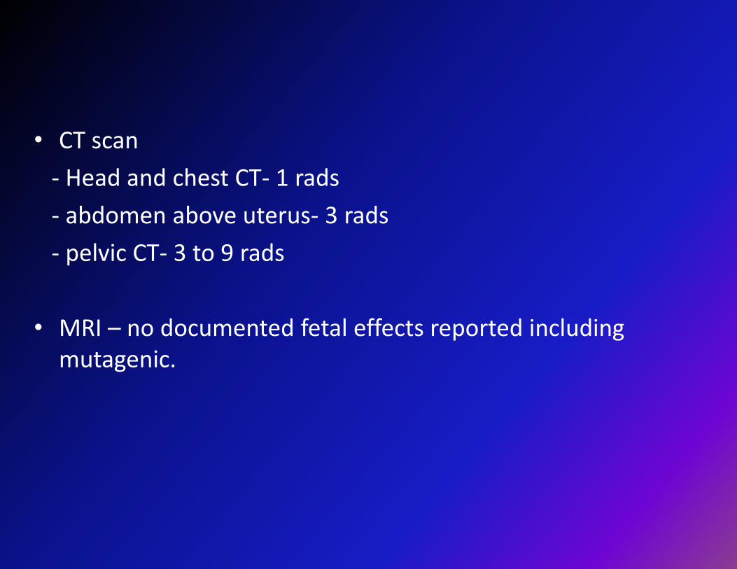
• CT scan
- Head and chest CT- 1 rads
- abdomen above uterus- 3 rads
- pelvic CT- 3 to 9 rads
• MRI – no documented fetal effects reported including mutagenic.

DIAGNOSTIC PERITONEAL LAVAGE
• DPL is an invasive, rapid, and highly accurate test for evaluating intraperitoneal haemorrhage or a ruptured hollow viscus.
• Performed less frequently; replaced by FAST and helical computed tomography (CT).
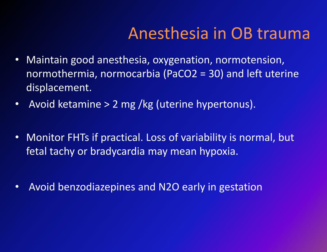
Anesthesia in OB trauma
• Maintain good anesthesia, oxygenation, normotension, normothermia, normocarbia (PaCO2 = 30) and left uterine displacement.
• Avoid ketamine > 2 mg /kg (uterine hypertonus).
• Monitor FHTs if practical. Loss of variability is normal, but fetal tachy or bradycardia may mean hypoxia.
• Avoid benzodiazepines and N2O early in gestation

MOTHER STABLE, FETUS STABLE
• Once mother is stabilized, focus on fetus.
• Direct impact – not necessary for feto placental pathology.
• No obvious abdominal trauma- still needs monitoring.
• 4hr- CTG monitoring recommended.(Pearlman et al.)

MOTHER STABLE, FETUS UNSTABLE
• CESAREAN SECTION
- Fetal distress despite optimizing mother.
- uterine rupture.
- placental rupture
- Fetal malpresentation during preterm labor.
- uterus mechanically limits maternal repair

MOTHER UNSTABLE FETUS UNSTABLE
• Primary repair of maternal injuries- best course
• Even in fetal distress, as critically injured mother will not withstand cesarean section.
• Early restoration of maternal physiology – best initial action for fetus.
• If mother can withstand- cesarean section can be performed

When to Salvage Fetus ?

PERIMORTEM CS
• Maternal resuscitation as per ACLS guidelines.
• If no response- decision for perimortem cesarean section.
• No return of spontaneous circulation after resuscitation for four minutes.
• Delivery within 5 min carries the best chance of fetal and maternal survival.


CONCLUSION
• Most diagnostic and therapeutic modalities relating to trauma care should not be modified or avoided during pregnancy.
• Co-management /multidisciplinary approach , function to insure appropriate care of the trauma victim and her fetus.