Transmigration of Mandibular Canines - Clinical...
5
Transmigration of Mandibular Canines - Clinical Implications Preema Melani Pinto 1 , Vijay R Naik 2 , Vikram Pai 3 , Kalpana S Jadhav 4 , Siddharth Revankar 5 ABSTRACT: Transmigration of a mandibular canine across the midline is a rare condition with obscure etiology. Bilateral transmigration is found to be extremely rare. Transmigrated canines have been found to be associated with dental anomalies and pathologies such as cystic lesions and root resorption of the adjacent teeth. The treatment of transmigrated mandibular canine demands clinical expertise. The presence of mandibular canal, mental foramen and the symphyseal bone complicates the surgical removal of these impacted canines. The treatment options vary according to the needs of the individual case and whether the tooth is symptomatic or not. The success of orthodontic treatment relies on the position, bone density and mechanics involved. Key words: impacted canine; mandibular canine; transmigration. CASE REPORT doi: 10.5866/2015.7.10212 1 Consultant Orthodontist Belgaum, India 2 Professor & Head 3&5 Reader Department Of Orthodontics and Dentofacial Orthopaedics, Maratha Mandal's Institute pf Dental Sciences, Belgaum, Karnataka, India. 4 Specialist Orthodontist United Arab Emirates Article Info: Received: July 7, 2015 Review Completed: August 6, 2015 Accepted: September 9, 2015 Available Online: July, 2015 (www.nacd.in) © NAD, 2015 - All rights reserved Email for correspondence: [email protected] Introduction: The maxillary canines are the commonly impacted teeth in the human dentition next to the third molars. Impaction of mandibular canines is quite rare in comparison to maxillary canines as described by Rohrer, who concluded that impacted canines are 20 times more frequent in maxilla than in the mandible. 1 In few instances, the mandibular canines have been found to have migrated across the midline to the other side of the mandible and INDIAN JOURNAL OF DENTAL ADVANCEMENTS Journal homepage: www. nacd. in are described as being transmigrated. In cases where these teeth tend to erupt, they are usually transposed. Transmigration of a tooth, in general refers to the event of migration of a tooth away from its site of origin. As is evident from the literature, varying definitions have been attributed to the transmigration of mandibular canine depending on the status of the affected tooth in relation to the midline. 2-4 Quick Response Code Indian J Dent Adv 2015; 7(3): 212-216
Transcript of Transmigration of Mandibular Canines - Clinical...
Transmigration of MandibularCanines - Clinical Implications
Preema Melani Pinto1, Vijay R Naik2, Vikram Pai3,Kalpana S Jadhav4, Siddharth Revankar5
ABSTRACT:
Transmigration of a mandibular canine across the midline is a
rare condition with obscure etiology. Bilateral transmigration
is found to be extremely rare. Transmigrated canines have been
found to be associated with dental anomalies and pathologies
such as cystic lesions and root resorption of the adjacent teeth.
The treatment of transmigrated mandibular canine demands
clinical expertise. The presence of mandibular canal, mental
foramen and the symphyseal bone complicates the surgical
removal of these impacted canines. The treatment options vary
according to the needs of the individual case and whether the
tooth is symptomatic or not. The success of orthodontic
treatment relies on the position, bone density and mechanics
involved.
Key words: impacted canine; mandibular canine;
transmigration.
C A S E R E P O R T
doi: 10.5866/2015.7.10212
1Consultant OrthodontistBelgaum, India2Professor & Head3&5ReaderDepartment Of Orthodontics and DentofacialOrthopaedics, Maratha Mandal's Institute pf DentalSciences, Belgaum, Karnataka, India.4Specialist OrthodontistUnited Arab Emirates
Article Info:
Received: July 7, 2015Review Completed: August 6, 2015Accepted: September 9, 2015Available Online: July, 2015 (www.nacd.in)© NAD, 2015 - All rights reserved
Email for correspondence:[email protected]
Introduction:
The maxillary canines are the commonlyimpacted teeth in the human dentition next to thethird molars. Impaction of mandibular canines isquite rare in comparison to maxillary canines asdescribed by Rohrer, who concluded that impactedcanines are 20 times more frequent in maxilla thanin the mandible.1 In few instances, the mandibularcanines have been found to have migrated acrossthe midline to the other side of the mandible and
INDIAN JOURNAL OF DENTAL ADVANCEMENTS
Jour nal homepage: www. nacd. in
are described as being transmigrated. In cases wherethese teeth tend to erupt, they are usuallytransposed.
Transmigration of a tooth, in general refers tothe event of migration of a tooth away from its siteof origin. As is evident from the literature, varyingdefinitions have been attributed to thetransmigration of mandibular canine depending onthe status of the affected tooth in relation to themidline.2-4
Quick Response Code
Indian J Dent Adv 2015; 7(3): 212-216

Numerous theories speculating the aetiologyhave been put forth but none of which do accuratelydefine the cause of this unique phenomenon. Theyinclude heredity, abnormal displacement of thedental lamina in the embryonic life, axial inclinationof erupting canines, retention or premature loss ofdeciduous canine, inadequate eruption space,supernumerary teeth, excessive length of crown,endocrine disorders, trauma, diet and intrauterinedefects.5-9
The scope for treatment of these transmigratedcanines is very limited. The treatment optionsinclude preventive and interceptive treatment,surgical exposure and orthodontic treatment,autotransplantation, surgical removal andradiographic monitoring.10-14
The unpredictably unique manifestation ofthese canines renders the treatment difficult if notimpossible. Each treatment possibility presents withits own merits and demerits. The literature doesn’tgive any insight into the intricacies of the treatmenttechniques. The purpose of this article is to discussthe possible clinical implications while treating thetransmigration. Four cases of transmigration arepresented in this context.
Case reports:
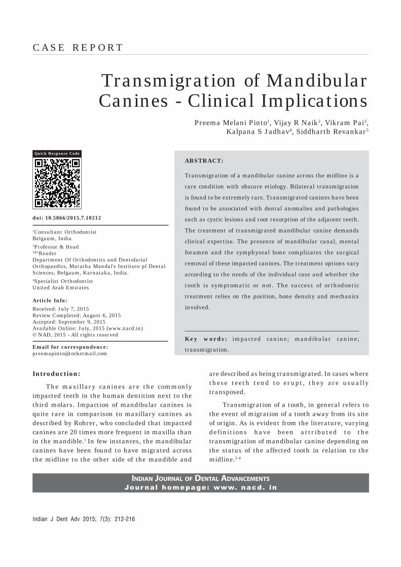
Case 1: A 16 year old male patient reportedwith the chief complaint of forwardly placed upperfront teeth (Figure 1 and 2). Medical history wasnon contributory. The patient had a dental historyof undergoing orthodontic treatment previouslywhich he discontinued midway. Clinicalexamination revealed that all permanent teeth haderupted except the two mandibular canines andmandibular left third molar. The patient had aAngle’s Class II division 1 malocclusion with a 100%deep bite, an overjet of 10 mm and posterior scissorbite. The right lateral incisor had a talon’s cusp. Thepanoramic radiograph showed the mandibular rightand left canines lying horizontally beneath theapices of mandibular anterior teeth. The crowns ofthe impacted canines were pointing towards thecontra-lateral mental foramina (Figure 3). Thelateral cephalogram depicted a small radiopaque
mass in the symphysis region (Figure 4). Nopathologic finding was associated with thetransmigrated teeth. The mandibular left thirdmolar was horizontally impacted.
Case 2: Clinical examination of a 19 year oldmale patient who presented with the chief complaintof irregularly placed lower front teeth revealed classI molar relation and a retained mandibular leftdeciduous canine. The permanent left canine wasupright and erupted in the midline, labial to themandibular right central incisor (Figure 5 and 6).No pathologic finding was associated with thetransmigrated tooth.
Case 3: A 30 year old male patient presentedwith the chief complaint of missing upper frontteeth. On clinical examination, he was found to havea class II molar relation, 100% deep bite, scissor biteon left side, congenitally missing maxillary lateralincisors and over retained mandibular rightdeciduous canine (Figure 7). Panoramic radiographrevealed obliquely impacted mandibular rightpermanent canine at the midline (Figure 8). Nopathologic finding was associated with thetransmigrated tooth
Case 4: A 15 year old male patient presentedwith the chief complaint of irregularly placed lowerfront teeth. On clinical examination, he had a classI molar relation, transmigrated and transposedmandibular canines. The left permanent mandibularcanine had erupted in the midline, with the otherpermanent canine lying adjacent to it on the rightside (Figure 9 and 10). In the strict sense, the rightcanine couldn’t be termed as transmigrated. Nopathologic finding was associated with thetransmigrated tooth.
Discussion:
Mupparapu classified mandibular caninetransmigration depending on its path of deviationinto five types. Type 1: Canine positioned mesio-angularly across the midline within the jaw bone,labial or lingual to anterior teeth, and the crownportion of the tooth crossing the midline (45.6%).Type 2: Canine horizontally impacted near theinferior border of the mandible below the apices of
Transmigration of Mandibular Canines - Clinical Implications Preema Melani Pinto, et, al.
Indian J Dent Adv 2015; 7(3): 212-216
Figure 1: Extra-oral frontalview of case 1.
Figure 2: Extra-oral frontalsmiling view of case 1.
Figure 3: OPG showing transmigrated canines impactedhorizontally in the midline of case 1.
Figure 4: LateralCephalogram showing thetransmigrated canines at
the symphysis as aradiopaque mass of case 1.
Figure 5: Intra-oral photographshowing transmigrated
permanent left mandibularcanine of case 2.
Figure 6: OPG showing 33 overlapping theimage of 41 in case 2.
Figure 7: Intra-oral frontal view of case 3.
Figure 8: OPG showing obliquely placed 43 with the crowncrossing the mandibular midline in case 3.
Figure 9: Intra-oral view of the mandibular canines lyingadjacent to each other in case 4.
Figure 10: Mandibular occlusal view of case 4.
Transmigration of Mandibular Canines - Clinical Implications Preema Melani Pinto, et, al.
Indian J Dent Adv 2015; 7(3): 212-216
the incisors (20%). Type 3: Canine erupting eithermesial or distal to the opposite canine (14%). Type4: Canine horizontally impacted near the inferiorborder of the mandible below the apices of eitherpremolars or molars on the opposite side (17%). Type5: Canine positioned vertically in the midline (thelong axis of the tooth crossing the midline)irrespective of eruption status (1.5%).15
The first case reported in this paper belongedto Mupparapu classification type 2, whose positionis unfavourable to attempt orthodontic traction(Figure 3). Surgical removal leaves a temporarylarge bony defect and is associated with risks ofiatrogenic fracture of the mandible and injury to theelements of mandibular canal. Since the teeth areasymptomatic, periodic radiographic monitoring isbeing carried out in conjunction with thecomprehensive orthodontic treatment that thepatient is receiving for his malocclusion.Furthermore, the significance of periodicobservation is emphasized by the fact that theaffected canines lie in close proximity to the roots ofmandibular incisors because of which, duringorthodontic tooth movement, there may bepossibilities of root resorption of mandibular incisorsand/or pathologic changes in relation to the sleepingcanines. As the patient presents with 100% deepbite, the possible treatment options are intrusion ofthe maxillary or mandibular incisors. Intrusion ofmaxillary incisors could be carried out but at theexpense of soft tissue balance since the patient haszero incisor exposure at rest and the intrusion ofmandibular incisors is complicated by the precariouslocation of the transmigrated canines.
The second case belongs to the Mupparapuclassification type 5. This transmigration is welltreated by extraction of 33. Space discrepancyrenders the alignment of the canine quite difficult.Orthodontic alignment, if possible, has to be donefollowing carefully planned extractions. However,this mode of treatment seems to be cumbersomegiven the ectopic position of the transmigratedcanine which in itself warrants its extraction.Furthermore, since the canine needs to be alignedat the midline itself, transmigrated canine crown
should be sequentially contoured to resemblemandibular central incisor. Conversion of canineinto an incisor requires removal of a substantialamount of crown structure which may necessitateintentional root canal therapy followed by a crown.Another major drawback is that a canine guidedocclusion cannot be established. Also, the canine isvulnerable for root dehiscence because of the narrowdistance between the labial and lingual corticalplate. In a similar case treated by Brezniak, Yehudaand Shapira gingival recession was found on thelabial side of the treated canine.16
The third case belongs to the Mupparapuclassification type 1. The root of the affected caninelies in a favourable position but the success oforthodontic traction to get the tooth into the arch isquestionable due to two factors. Firstly the exactposition of the canine in the sagittal plane needs tobe determined. A labially positioned tooth isamenable for surgical exposure and bondingattachments. Secondly the proximity of thetransmigrated canine to the roots of lower centralincisors creates complications as discussed earlier.Surgical removal may be considered.
The fourth case belongs to the Mupparapuclassification type 5. According to the classification,the mandibular left canine alone can be termed astransmigrated. The migration of mandibular rightcanine seems to have been obstructed by the leftcanine. Considering the space deficiency in themandibular arch, extraction of both the canines wascarried out and the first premolars were convertedinto canines. In a scenario where there was adequatespace in the lower arch, orthodontic traction of theright canine could have been an option since it waspositioned at a shorter distance without actuallyhaving crossed the midline. The left canine couldhave been treated similar to case number 2.However, this approach would require longertreatment duration and lighter forces as the toothis moved through dense cortical bone.
Conclusion:
In recent years, many cases of transmigrationof mandibular canines have been documented. Thecorrective treatment of these canines is debatable
Transmigration of Mandibular Canines - Clinical Implications Preema Melani Pinto, et, al.
Indian J Dent Adv 2015; 7(3): 212-216
and quite challenging. When the impacted teetharen’t removed, care has to be taken not to inducepathologic changes. Maintaining the health of theperiodontium of the transmigrated tooth as well asthe adjacent teeth is critical while attemptingorthodontic traction. Root torqueing in specific casesshould be planned prior to initiation of orthodontictreatment to place the root in trabecular bone.Iatrogenic damage can be best avoided by carefultreatment planning with due consideration to theclinical implications of the particular case. Thoughearly detection and treatment is suggested in theliterature, evidence is lacking in support ofinterceptive measures.
References
1. Rohrer A. Displaced and impacted canines. Orthod OralSurg Int J 1929; 15:1002-1004.
2. Tarsitano JJ, Wooten JW, Burditt JT. Transmigration ofnon erupted mandibular canines: report of cases. J Am DentAssoc. 1971; 82:1395-1397.
3. Javid B. Transmigration of impacted mandibular cuspids.Int J Oral Surg 1985; 14:547-549.
4. Joshi MR. Transmigrant mandibular canines: a record of28 cases and a retrospective review of the literature. AngleOrthod. 2001; 71:12-22.
5. Nodine AM. Aberrant teeth, their history, causes andtreatment. Dent Items Interest. 1943; 65:440-451.
6. Shapira Y, Mischler WA, Kuftinec MM. The displacedmandibular canine. ASDC J Dent Child 1982; 49(5):362-364.
7. Pippi R, Kaitsas R. Mandibular canine transmigration:aethiopathogenetic aspects and six new reported cases. OralSurg 2008; 1:78-83.
8. Ryan FS, Batra P, Witherow H, Calvert M. Transmigrationof a maxillary canine. A case report. Prim Dent Care 2005;12(2):70-72.
9. Mitchell L. Displacement of a mandibular canine followingfracture of the mandible. Br Dent J 1993; 174(11):417-418.
10. Taguchi Y, Kurol J, Kobayashi H, Noda T. Eruptiondisturbances of mandibular permanent canines in Japanesechildren. International Journal of Paediatric Dentistry 2001;11(2): 98-102.
11. Vichi M, Franchi L. The transmigration of the permanentlower canine. Minerva Stomatol. 1991; 40:579-589.
12. Wertz RA. Treatment of transmigrated mandibular canines.Am J Orthod Dentofacial Orthop 1994; 106:419-427.
13. Kumar S, Urala AS, Kamath AT, Jayaswal. P, Valiathan.A. Unusual intraosseous transmigration of impacted tooth.Imaging Science in Dentistry 2012; 42: 47-54.
14. Howard RD. The anomalous mandibular canine. Br J Orthod1976; 3(2):117-121.
15. Mupparapu M. Patterns of intra-osseous transmigration andectopic eruption of mandibular canines: review of literatureand report of nine additional cases. Dentomaxillofac Radiol2002; 31(6):355-360.
16. Brezniak N, Ben-Yehuda A, Shapira Y. Unusual mandibularcanine transposition: a case report. Am J Orthod DentofacialOrthop 1993; 104:91-94.
Gain quick access to our journal online
View our journal at
www.nacd.in
Transmigration of Mandibular Canines - Clinical Implications Preema Melani Pinto, et, al.
Indian J Dent Adv 2015; 7(3): 212-216