Transient protein–protein interactionshome.ku.edu.tr/~okeskin/AcunerOzbabacanPEDS.pdf · kinetics...
14
Transient protein–protein interactions Saliha Ece Acuner Ozbabacan, Hatice Billur Engin, Attila Gursoy 1 and Ozlem Keskin 1 Center for Computational Biology and Bioinformatics, College of Engineering, Koc University, Rumelifeneri Yolu, 34450 Sariyer Istanbul, Turkey 1 To whom correspondence should be addressed. E-mail: [email protected] (O.K.) and [email protected] (A.G.) Received February 24, 2010; revised May 11, 2011; accepted May 17, 2011 Edited By Frederic Rousseau Transient complexes are crucial for diverse biological processes such as biochemical pathways and signaling cascades in the cell. Here, we give an overview of the transient interactions; the importance of transient inter- actions as drug targets; and the structural characteriz- ation of transient protein–protein complexes based on the geometrical and physicochemical features of the transient complexes’ interfaces. To better understand and even- tually design transient protein – protein interactions (TPPIs), a molecular perspective of the protein–protein interfaces is necessary. Obtaining high-quality structures of protein – protein interactions could be one way of achieving this goal. After introducing the association kinetics of TPPIs, we elaborate on the experimental techniques detecting TPPIs in combination with the com- putational methods which classify transient and/or non- obligate complexes. In this review, currently available databases and servers that can be used to identify and predict TPPIs are also compiled. Keywords: protein interface/protein interaction types/ transient protein2protein interactions/transient protein–drug interactions Introduction Transient complexes are essential, especially in the regu- lation of biochemical pathways and signaling cascades in the cells. A wide range of biological processes, such as hormone–receptor binding, signal transduction, allostery of enzymes, inhibition of proteases and correction of misfolded proteins by chaperones contain transient interactions between proteins (Valdar and Thornton, 2001; Schreiber et al., 2009). These interactions are quite frequent in signaling pathways as they provide a mechanism for the cell to quickly respond to extracellular stimuli. They also function in the secretory pathways in eukaryotes by controlling the transport- competent proteins (Nyfeler et al., 2005). Our review mainly focuses on transient complexes. First, we introduce types of protein–protein interactions (PPIs) emphasizing the transient ones. We then describe some key points and case studies about transient protein and drug interactions. Next, we elaborate on the structural characterization of transient and non-obligate complexes, based on the geometrical and physicochemical properties of their interfaces. Kinetic models of transient PPIs (TPPIs) are explained in the pro- ceeding section. We then continue with the introduction of experimental techniques that are used for detecting TPPIs in combination with the computational methods which aim to classify transient and/or non-obligate complexes among other types of PPIs. Furthermore, we list the databases and servers which are utilized to identify and predict TPPIs, and then conclude our review by stating the key points about transient complexes. Types of protein–protein interactions Both metabolic and regulatory networks are driven by PPIs; however, different types of complexes with specific functions are observed: large macromolecular complexes, such as the ribosome, are highly stable and permanent whereas dynamic and transient interactions are key components in signaling and regulatory networks (Bhattacharyya et al., 2006; Stein et al., 2009a; Bashor et al., 2010). Protein –protein inter- actions can be classified based on their composition, affinity and life time (Nooren and Thornton, 2003a; Park et al., 2009) as: (i) homo- and hetero-oligomeric complexes, (ii) non-obligate and obligate complexes (Fig. 1) and (iii) transi- ent and permanent complexes (Fig. 1), respectively. Homo-oligomeric and hetero-oligomeric complexes These groups of complexes are differentiated based on their compositions such that if a PPI occurs between identical chains, it is said to form a homo-oligomer whereas if the PPI takes place among non-identical chains then it forms a hetero-oligomer complex. Homo-oligomers are symmetric and provide a good scaffold for stable macromolecules. For example, a chaperonin protein is formed by seven GroEL proteins associating as a homo-heptamer to form a cylinder and seven GroES proteins cap one side of this cylinder (Braig et al., 1994). The cylindrical region is an example of a homo-oligomer, whereas the GroEL/GroES complex is a supramolecule of hetero-oligomers. The stability of hetero- oligomers can vary and form a basis to gather different pro- teins that cooperate in a single macromolecule. For example, a/b tubulins form a stable dimer and these dimers form long protofilaments, which are constituents of microtubules (Lowe et al., 2001). Obligate and non-obligate complexes The key point for differentiation between these two groups is affinity. If the constituents ( protomers, monomers) of a complex are unstable on their own in vivo then this is an obligate interaction whereas the components of non-obligate interactions can exist independently. As an obligate complex # The Author 2011. Published by Oxford University Press. All rights reserved. For Permissions, please e-mail: [email protected] Page 1 of 14 Protein Engineering, Design & Selection pp. 1–14, 2011 doi:10.1093/protein/gzr025 PEDS Advance Access published June 15, 2011 at Koc University on June 15, 2011 peds.oxfordjournals.org Downloaded from
Transcript of Transient protein–protein interactionshome.ku.edu.tr/~okeskin/AcunerOzbabacanPEDS.pdf · kinetics...
Transient protein–protein interactions
Saliha Ece Acuner Ozbabacan, Hatice Billur Engin,Attila Gursoy1 and Ozlem Keskin1
Center for Computational Biology and Bioinformatics, College ofEngineering, Koc University, Rumelifeneri Yolu, 34450 Sariyer Istanbul,Turkey
1To whom correspondence should be addressed.E-mail: [email protected] (O.K.) and [email protected] (A.G.)
Received February 24, 2010; revised May 11, 2011;accepted May 17, 2011
Edited By Frederic Rousseau
Transient complexes are crucial for diverse biologicalprocesses such as biochemical pathways and signalingcascades in the cell. Here, we give an overview of thetransient interactions; the importance of transient inter-actions as drug targets; and the structural characteriz-ation of transient protein–protein complexes based on thegeometrical and physicochemical features of the transientcomplexes’ interfaces. To better understand and even-tually design transient protein–protein interactions(TPPIs), a molecular perspective of the protein–proteininterfaces is necessary. Obtaining high-quality structuresof protein–protein interactions could be one way ofachieving this goal. After introducing the associationkinetics of TPPIs, we elaborate on the experimentaltechniques detecting TPPIs in combination with the com-putational methods which classify transient and/or non-obligate complexes. In this review, currently availabledatabases and servers that can be used to identify andpredict TPPIs are also compiled.Keywords: protein interface/protein interaction types/transient protein2protein interactions/transient protein–druginteractions
Introduction
Transient complexes are essential, especially in the regu-lation of biochemical pathways and signaling cascades inthe cells. A wide range of biological processes, such ashormone–receptor binding, signal transduction, allostery ofenzymes, inhibition of proteases and correction of misfoldedproteins by chaperones contain transient interactions betweenproteins (Valdar and Thornton, 2001; Schreiber et al., 2009).These interactions are quite frequent in signaling pathways asthey provide a mechanism for the cell to quickly respond toextracellular stimuli. They also function in the secretorypathways in eukaryotes by controlling the transport-competent proteins (Nyfeler et al., 2005). Our review mainlyfocuses on transient complexes. First, we introduce types ofprotein–protein interactions (PPIs) emphasizing the transientones. We then describe some key points and case studies
about transient protein and drug interactions. Next, weelaborate on the structural characterization of transient andnon-obligate complexes, based on the geometrical andphysicochemical properties of their interfaces. Kineticmodels of transient PPIs (TPPIs) are explained in the pro-ceeding section. We then continue with the introduction ofexperimental techniques that are used for detecting TPPIs incombination with the computational methods which aim toclassify transient and/or non-obligate complexes among othertypes of PPIs. Furthermore, we list the databases and serverswhich are utilized to identify and predict TPPIs, and thenconclude our review by stating the key points about transientcomplexes.
Types of protein–protein interactions
Both metabolic and regulatory networks are driven by PPIs;however, different types of complexes with specific functionsare observed: large macromolecular complexes, such as theribosome, are highly stable and permanent whereas dynamicand transient interactions are key components in signalingand regulatory networks (Bhattacharyya et al., 2006; Steinet al., 2009a; Bashor et al., 2010). Protein–protein inter-actions can be classified based on their composition, affinityand life time (Nooren and Thornton, 2003a; Park et al.,2009) as: (i) homo- and hetero-oligomeric complexes, (ii)non-obligate and obligate complexes (Fig. 1) and (iii) transi-ent and permanent complexes (Fig. 1), respectively.
Homo-oligomeric and hetero-oligomeric complexesThese groups of complexes are differentiated based on theircompositions such that if a PPI occurs between identicalchains, it is said to form a homo-oligomer whereas if the PPItakes place among non-identical chains then it forms ahetero-oligomer complex. Homo-oligomers are symmetricand provide a good scaffold for stable macromolecules. Forexample, a chaperonin protein is formed by seven GroELproteins associating as a homo-heptamer to form a cylinderand seven GroES proteins cap one side of this cylinder(Braig et al., 1994). The cylindrical region is an example ofa homo-oligomer, whereas the GroEL/GroES complex is asupramolecule of hetero-oligomers. The stability of hetero-oligomers can vary and form a basis to gather different pro-teins that cooperate in a single macromolecule. For example,a/b tubulins form a stable dimer and these dimers form longprotofilaments, which are constituents of microtubules (Loweet al., 2001).
Obligate and non-obligate complexesThe key point for differentiation between these two groups isaffinity. If the constituents (protomers, monomers) of acomplex are unstable on their own in vivo then this is anobligate interaction whereas the components of non-obligateinteractions can exist independently. As an obligate complex
# The Author 2011. Published by Oxford University Press. All rights reserved.
For Permissions, please e-mail: [email protected]
Page 1 of 14
Protein Engineering, Design & Selection pp. 1–14, 2011doi:10.1093/protein/gzr025
PEDS Advance Access published June 15, 2011 at K
oc University on June 15, 2011
peds.oxfordjournals.orgD
ownloaded from

example, Ku proteins, which are involved in DNA repair, areshown to bind DNA as obligate homodimers (Krishna andAravind, 2010). On the other hand, signaling protein com-plexes are good non-obligate interaction examples, due totheir transient nature. After contributing to the propagationof a signal, they are dissociated into the stable constituentproteins. For example, H-Ras protein, which is a G protein,has a key role in controlling the cell growth and differen-tiation signaling pathways. It interchangeably forms non-obligate complexes with guanosine triphosphatase (GTPase)activating proteins (GAPs) (acceleration of GDP-bound stateof H-Ras—switch OFF) and guanine nucleotide-exchangefactors (acceleration of GTP-bound state of H-Ras—switchON), when the cell is resting and when activated in responseto stimuli, respectively (Vetter and Wittinghofer, 2001).
Transient and permanent complexesThese groups of interaction types are discriminated based onthe lifetime (or stability) of the complex. Permanent inter-actions are usually very stable and irreversible (e.g. IL-5cytokine dimer (PDB ID: 3b5k)) (Nooren and Thornton,2003b; (Fig. 2a). However, the components of the transientinteractions associate and dissociate temporarily in vivo(Mintseris and Weng, 2003; Nooren and Thornton, 2003a;Nooren and Thornton, 2003b; Block et al., 2006; Janin et al.,2008; Levy and Pereira-Leal, 2008). The a/b tubulin dimer isan example of an obligate/permanent complex, whereas thedimers of a/b dimers are transient and non-obligatory pro-viding the dynamic nature to microtubules in cell division,cargo transportation and cytoskeleton (Hyams and Lloyd,1993). Non-obligate interactions are predominantly transient(Janin et al., 2008), with a few examples of permanent(Fig. 1), but obligate interactions are usually permanent innature (Nooren and Thornton, 2003a). It should be noted thatpermanent and obligate terms are used interchangeably inthe literature.
Transient complexes, depending on their functional rolesin the cell, have a wide range of affinities and lifetimes andhence can be further classified as strong and weak (Noorenand Thornton, 2003a; Nyfeler et al., 2005) (see Fig. 2b and c)based on the stability of their oligomeric equilibrium. Thestrong transient interactions (e.g. heterotrimeric G protein(PDB ID: 1got)) shift equilibrium of association/dissociationunder certain disturbances (Nooren and Thornton, 2003a).G proteins, which are crucial in signaling pathways, are
examples for strong transient interactions. These membrane-bound proteins are transient in nature and they get activatedby G-protein-coupled-receptors (GPCRs) in order to activatea target in the plasma membrane causing a cascade of othersignaling events (Alberts et al., 2009). G proteins are com-posed of three subunits, namely a, b and g, and are inactivewhen a subunit is bound to GDP. When a subunit is acti-vated by a ligand-bound GPCR, it loses the affinity for GDPand exchanges it for a molecule of GTP so that the detach-ment of GTP-bound a subunit from the bg complex is trig-gered (see Fig. 2b). However, the weak transient interactions(such as dimers of abalone sperm lysin (PDB ID: 2lyn)) arebroken and formed continuously (Nooren and Thornton,2003a) (see Fig. 2c). The lysin dimer is used to make a holein the vitelline envelope (VE), which is protective, so thatthe sperm swims and fuses with the egg (Kresge et al.,2000). For this process to take place, lysin is released fromthe sperm and binds to the VE receptor lysin (VERL) to dis-solve the VE. It was found that the lysin monomer is activeduring the binding process to VERL, whereas it is observedto be a dimer when it contacts the egg (Kresge et al., 2000).
Domain–domain and domain–peptide complexesProtein–protein interactions can also be classified based ontheir folds as domain–domain and domain–peptide inter-actions (Aloy and Russell, 2006) (see Fig. 2). The complexesbelonging to the latter group have mostly transient natures asthey are formed by the recognition of a globular domain, ashort linear motif (LM) and the small interface on which theinteraction takes place (e.g. SH3 domain of tyrosine kinaseFyn—proline-rich peptide (PDB ID: 1fyn) (Stein et al.,2009a; London et al., 2010). These domain–peptide inter-actions are also called transient peptide-mediated inter-actions. Indeed, special interaction domains (such as PDZ,SH2, SH3, WW, etc.) provide an elegant mechanism in sig-naling by making use of transient interactions. Thesemodular interaction domains usually recognize and bindspecific motifs of peptides (either at the termini or disorderedregions of partner proteins). These are like ready-to-bindinteraction domains as they do not undergo large confor-mational changes on binding and are frequently used. Forexample, in homo sapiens, there are 223 SH3, 234 PDZand 91 WW domains (Bhattacharyya et al., 2006). Thesedomains can be used to assemble constituent proteins intolarge complexes, bringing together different combinations ofcatalytic domains with regulatory domains. Each complexwith a different combination of domains will then have adifferent function leading to a different signal in the cell.
Transient protein and drug interactions
Cellular processes, such as the cell cycle, which are involvedin disease-related pathways, are regulated via transient inter-actions. Hence, understanding the details of TPPIs by usinga systematic wide range approach may enlighten the discov-ery and development of inhibitors, which can serve as thera-peutics for such diseases (Rudolph, 2007; Kar et al., 2010;Ozbabacan et al., 2010). Until recently, TPPI inhibitors werenot widely studied as they were considered to bind targetswith low specificity, low efficiency, low amount and weredifficult to screen and analyze (Rudolph, 2007; Ohlson,2008). However, experimental and computational advances
Fig. 1. Relation of protein–protein interaction types based on affinity andstability. Non-obligate interactions are transient but there are some examplesof permanent non-obligate interactions such as enzyme2inhibitorinteractions (e.g. thrombin–rhodnin inhibitor interaction).
S.E. Acuner Ozbabacan et al.
Page 2 of 14
at Koc U
niversity on June 15, 2011peds.oxfordjournals.org
Dow
nloaded from

have significantly improved the knowledge of protein inter-actions and the inhibitors against them (Rudolph, 2007).Some specific examples of experimental techniques, whichuse a wide range of proteomic methods, are yeast two-hybridscreens (Uetz et al., 2000), systematic RNA interference(Kamath et al., 2001), mass spectrometry (Ho et al., 2002)and the intracellular localization of proteins with fluor-escence markers (Lippincott-Schwartz and Patterson, 2003).
Transient interactions might be important in drug mechan-isms in two ways: the drugs that (i) act on TPPIs and that(ii) act transiently on their multiple targets (Rudolph, 2007;Ohlson, 2008). A cancer-related example for the former typeof drugs is nutlins (Vassilev et al., 2004). Vassilev et al.identified these small and selective inhibitors, whichuniquely target the interaction between murine doubleminute 2 (MDM2) and tumor suppressor p53 in order tostabilize p53, and they developed a novel strategy for cancertherapy. When the interaction between MDM2 and p53 takesplace transiently; p53 changes its conformation so that itcannot bind DNA (Wawrzynow et al., 2007) and becomedegraded. Nutlins inhibit this transient interaction by compe-titively binding to the site on MDM2, which is the bindingsite for p53, so that p53 accumulates and becomes activated(Jiang et al., 2007). Another example of a drug which targetsTPPIs is brefeldin A (BFA) (Robineau et al., 2000). Thisinhibitor uncompetitively attacks macromolecular complexeswhen they are in action; in other words, it attacks when thecomplex is in a transition state being structurally and energe-tically unbalanced, so that its hotspots which are targets fordrug binding are exposed (Pommier and Cherfils, 2005) (seeFig. 3a). This type of drug mechanism is called ‘interfacialinhibition’ (Renault et al., 2003). Colchicine is another can-didate interfacial inhibitor as it stabilizes the dimer of
a-tubulin and b-tubulin by acting on their interface andhence blocking the polymerization (Ravelli et al., 2004) (seeFig. 3b).
A new hypothesis suggests that the multi-target drugapproach, which aims at several targets simultaneously, canmaximize the efficiency of a drug (Frantz, 2005; Morphy andRankovic, 2005; Hopkins et al., 2006; Morphy and Rankovic,2007). The reason to consider multi-target drugs, which maytransiently bind to their targets, is the fact that diseases suchas cancer, depression, inflammatory and cardiovascular dis-eases are caused not by a single molecular defect but by dys-functions that are combined in a complex manner (Ohlson,2008). These drugs are advantageous in the treatment of neu-rodegenerative diseases and cancer as they minimize adverseeffects, such as cell degeneration, by weakly binding to thereceptors and not totally blocking them (Ohlson, 2008). Forexample, the multi-target anti-cancer agent Gleevecw isfound to be promising in leukemia treatment (Frantz, 2005).Furthermore, multi-target drugs are also represented as aform of combinatorial therapy and are more frequently usedfor the treatment of diseases such as AIDS, cancer and ather-osclerosis (Huang, 2002; Borisy et al., 2003; Kaelin, 2004).A few examples of transiently binding drugs are alcohol(ethanol) (Siggins et al., 2005), non-steroidal anti-inflammatory drugs (NSAIDs) namely aspirin, naproxen andibuprofen (Cryer and Feldman, 1998), weak enzyme inhibi-tors such as valproic acid and butyric acid (Ohlson, 2008),salicylate and metformin (Csermely et al., 2005). The transi-ent nature of alcohol stems from the fact that it binds todifferent receptors with low affinity and NSAIDs bind recep-tors and enzymes with an affinity higher than 1 mM.Memantine is another example of transiently acting drugsand is effective against the group of neurodegenerative
Fig. 2. Classification of protein–protein interaction types based on stability and fold. The mechanisms of association and dissociation processes are shown forstrong and weak transient protein–protein complexes, along with the structures of example cases. (a) Permanent protein–protein interaction: Components arestable only in complex form, e.g. IL-5 cytokine dimer (PDB ID: 3b5k). (b) Strong transient protein–protein interaction: association/dissociation takes placeunder certain triggers such as chemical modification, conformational change and colocalization; dissociation constant (Kd) is in nanomolar range, e.g.Heterotrimeric G protein (PDB ID: 1got), in which Ga is in complex with guanosine diphosphate (GDP) and interacts transiently with Gbg, dissociates intoGa (PDB ID: 3ffb) and Gbg (PDB ID: 1tbg) subunits upon guanosine triphosphate (GTP) (PDB ID: 3ffb) binding. (c) Weak transient protein–proteininteraction: Complexes are broken and formed continuously and Kd is in micromolar range, e.g. Red abalone lysin dimer (PDB ID: 2lyn) dissociates into Redabalone lysin monomer (PDB ID: 2lis). (d) Domain–peptide interaction: a globular domain recognizes a short linear motif, e.g. SH3 domain of tyrosinekinase Fyn in complex with a proline-rich peptide (PDB ID: 1fyn).
Transient protein–protein interactions
Page 3 of 14
at Koc U
niversity on June 15, 2011peds.oxfordjournals.org
Dow
nloaded from

diseases called dementia, the most common form of which isAlzheimer’s disease (Rogawski, 2000; Lipton, 2004).
Structural characterization of transientprotein–protein complexes
A PPI takes place through an interface formed by the inter-acting pair of proteins and the interface consists of the resi-dues in contact that belong to the chains of proteins on eachside. The binding interfaces have been subject to many struc-tural analyses (Jones and Thornton, 1996; Keskin et al.,2008; Tuncbag et al., 2008), as they have key roles not onlyin the comparison of interaction types but also in the predic-tion of new PPIs.
With the ever-increasing information on binary PPIs, weknow that some proteins bind to 10s or even 100s of otherproteins, acting as hubs and these interactions are transientby nature. These hub proteins should have evolved tobalance between their specificity and promiscuity (Humphrisand Kortemme, 2007; Cukuroglu et al., 2010). Three-dimensional (3D) structures and interfaces of hub proteinswith many different partners combined with available kineticdata will surely enlighten our understanding how transientcomplexes achieve their high specificity and how enzymesbind their inhibitors with high selectivity (e.g. protease-inhibitor complexes) (Meenan et al., 2010). For the structuralcomparison of permanent and transient interactions, geo-metrical and physicochemical properties at the interfaces canbe considered (Jones and Thornton, 1996). These propertiesare the change in accessible surface area (DASA; as acontact area measure) and planarity as size and shape proper-ties; gap volume index as a measure for complementarity;polarity (Nooren and Thornton, 2003a, Nooren andThornton, 2003b; Jones and Thornton, 1996), hydrophobicityand mean number of hydrogen bonds; the number of discon-tinuous segments in the interface for measuring segmentationand the portions of secondary structures; and the extent ofconformation change on binding.
In transient domain–peptide interactions, usually the glob-ular recognition domain of a protein (50–150 residues long)interacts with a linear extended peptide (common consensusmotif of 3–10 residues) (Stein and Aloy, 2008). The consen-sus motifs characterizing the peptides are found in loops or
unstructured (disordered) regions of proteins. The interfacesof domain–peptide complexes (200–500 A2) are smallerthan the domain–domain complexes (�2000 A2)(Chakrabarti and Janin, 2002; London et al., 2010), andtherefore they are characterized as transient (Stein and Aloy,2008). Usually, the contact area of transient interactions issmaller (than 1500 A2) compared with the permanent com-plexes, which have larger and more twisted interfaces (withcontact areas ranging from 1500 to 10000 A2) (Nooren andThornton, 2003a; Nooren and Thornton, 2003b; De et al.,2005; Block et al., 2006; Zhu et al., 2006; Levy andPereira-Leal, 2008; Park et al., 2009). The limited surfacearea of the transient (recognition) complex interface is due tothe intrinsic physical requirement of the components to foldindependently and to exist in solution without aggregating(Mintseris and Weng, 2003). When planarity is considered,the interfaces of heterocomplexes (which may be both transi-ent and permanent) are found to be more planar with respectto homodimers (permanent complexes) (Jones and Thornton,1996). In terms of polarity as the interfaces of transient com-plexes resemble the exterior surface of a protein, they havemore polar and charged groups (Jones and Thornton, 1997;Lo Conte et al., 1999; Nooren and Thornton, 2003a; Ansariand Helms, 2005). Since permanent complexes are stable,their interfaces are hydrophobic similar to the interior of anaverage globular protein (Lo Conte et al., 1999; Nooren andThornton, 2003a; Block et al., 2006; Park et al., 2009). Onthe other hand, as the components of transient complexesshould be stable on their own, their interfaces are less hydro-phobic (Jones and Thornton, 1996) and also consist ofsolvent-exposed amino acids (Tsai et al., 1997). This is intui-tive, since transient complexes need to be soluble whendissociated (Block et al., 2006). In terms of residue propensi-ties, obligate (permanent) interactions embody hydrophobicresidues such as Leu, Ala, while non-obligate (usually transi-ent) interactions include polar residues such as Ser and Gly(Park et al., 2009). Additionally, both types of interactionsinclude charged residues such as Glu, Asp, Lys (Park et al.,2009) and Arg (De et al., 2005; Park et al., 2009). Mainlynon-polar residues such as Ile and Met are observed on thecenter of obligatory interfaces whereas non-polar residuessuch as Leu and Val and aromatic residues such as Tyr areincluded in the core of non-obligatory interfaces (De et al.,
Fig. 3. Examples of drugs acting on transient protein–protein interactions. (a) Structure of ARF1-GDP bound to SEC7 domain complexed with Brefeldin A(BFA) (PDB ID: 1re0). (b) Structure of tubulin a and tubulin b dimer complexed with colchicine (PDB ID: 1sa0)
S.E. Acuner Ozbabacan et al.
Page 4 of 14
at Koc U
niversity on June 15, 2011peds.oxfordjournals.org
Dow
nloaded from

2005). These non-polar residues supply the required strengthand specificity to these non-obligate interfaces (De et al.,2005). Although the findings of Park et al. (2009) and Deet al. (2005) agree that the centers of both interface types aremainly non-polar, their findings about residue propensities ofnon-obligate interactions are contradictory. Trp residue isseen in both types of interfaces both at the center and periph-ery, with a larger propensity in non-obligatory interfaces(De et al., 2005). This is explained by the fact that Trp isfavored as an interaction hotspot (Samanta and Chakrabarti,2001; Keskin et al., 2005; Ma and Nussinov, 2007).
When the interfaces of obligatory and non-obligatory com-plexes are compared based on their secondary structures,irregular secondary structure regions such as turns are morefrequently involved in non-obligatory interfaces becausethese regions provide the flexibility required for dissociationunder certain conditions (De et al., 2005). Additionally,b-sheet formation is not observed in non-obligatory inter-faces because it provides stability (De et al., 2005). For bothtypes of complexes, interactions between two helices areobserved. Hence, types of interactions can be characterizedby the involvement of regular secondary structures (De et al.,2005; Keskin and Nussinov, 2005).
Salt-bridges and hydrogen bonds take place more often forstabilization of transient complexes (Lo Conte et al., 1999)whereas the association of permanent PPIs may sometimesoccur through covalent disulphide bridges between the inter-acting components (De et al., 2005). Both obligate and tran-sient complexes are close-packed and complementary fromthe geometric and electrostatic standpoint (Lawrence andColman, 1993; De et al., 2005; Jones and Thornton, 1995;McCoy et al., 1997; Lo Conte et al., 1999). If an interfacebelongs to a permanent complex and the conformationalrearrangements occurring upon binding take place moreoften; then the interface size and hydrophobicity are stated tobe, respectively, higher (Lo Conte et al., 1999; Nooren andThornton, 2003a; Nooren and Thornton, 2003b; Levy et al.,2005). Table I summarizes the main points discussed aboutthe structural and kinetic comparison of protein–proteincomplex types.
The structural interface properties are strongly related withthe evolution of complexes. From the evolutionary stand-point, TPPI interfaces are more affected by their localenvironments or physiological conditions than permanentones, since they associate or dissociate at least once during
their cellular process (Cho et al., 2006). When the interfaceresidues of transient and obligate complexes are comparedevolutionarily, their conservations are considerably different.Also, the interface residues of obligate complexes are morerobustly dependent on each other than those of transientcomplexes (Mintseris and Weng, 2005). This is becauseunder high pressure, the coevolution of obligate complexeswith their interacting partners enables their interface residuesto evolve slowly and at similar rates. Conversely, transientinteractions require the fast adaptation of mutations at theinterface of the interacting partner causing the correlatedmutations to be undetectable (Mintseris and Weng, 2005).
Another evolutionary aspect for comparison of complexesis specificity. The transient interactions between recognitiondomains and peptides are known to be highly specific invivo, even though the interface consists of a few residues.Also it was found that the binding specificity is dependenton the single-point mutations in arbitrary residues of the con-sensus motif of a peptide. These facts imply that the transientpeptide-mediated interactions evolve in order to maximizespecificity (Stein et al., 2009a).
Disordered regions of transient protein–protein complexesProteins having disordered regions, which are also calledintrinsically unfolded proteins (IUPs) or intrinsically disor-dered proteins (IDPs), evolve in higher organisms due totheir advantageous regulatory strategies. Mutations in theseproteins are also directly related to important pathologies incomplex organisms such as cancer (Iakoucheva et al., 2002;Kim et al., 2008; Stein et al., 2009a; Uversky et al., 2009).Furthermore, disordered regions of complexes may affect thedegree of motion between domains, cover binding sites, actas the targets of post-translational modifications (PTMs), andenable transient binding of different binding partners (Mittaget al., 2010).
Mittag et al. (2010) listed some biophysical and evolution-ary advantages of IUPs. They mentioned the ‘polyelectro-static’ effect that supplies multiple charges on disorderedproteins to affect binding affinity through long-range electro-static effects causing a ‘net charge’ or ‘mean field’. Plasticityand malleability of proteins were stated to expand with thedisorder that helped the binding of the same proteinsequences with several binding partners. Additionally, sincedisordered regions show higher rates of mutation they mayfacilitate restriction of protein size, inhibit molecular crowd-ing inside cells and also limit cell size.
In TPPIs, disordered regions (Dunker et al., 2005; Ekmanet al., 2006; Singh et al., 2007) and high content of repeatingdomains (Bjorklund et al., 2006; Ekman et al., 2006) areimportant. These properties maintain TPPIs’ large surfacesfor flexible binding, letting them contain many interactionsin vivo (Bjorklund et al., 2008). In order to test this hypoth-esis and describe multi-partnered proteins, hub proteins wereclassified into two groups as sociable (transient hub proteins)and non-sociable proteins (Higurashi et al., 2008). Since it isnow widely accepted that hub proteins tend to have manydisordered regions, which will eventually form the bindinginterfaces (Liu et al., 2002; Wright and Dyson, 1999), themain difference between those two groups is the abundanceof disordered regions. However, the most distinctive featureof sociable proteins was found to be the overall structural
Table I. Structural and kinetic characterization of types of protein–protein
complexes
Transient\non-obligate Permanent\obligate
Interface contact area DASA(A2)
,1500 1500–10000
Secondary structures Helix and turns Helix and b-sheetInterface polarity High LowConformational changesupon binding
Low High
Residue propensity Polar, charged Hydrophobic,charged
Shape and electrostaticcomplementarity
High High
Equilibrium dissociationconstant (Kd)
.1026 M(micromolar, mM)
,1026 M(micromolar, mM)
Transient protein–protein interactions
Page 5 of 14
at Koc U
niversity on June 15, 2011peds.oxfordjournals.org
Dow
nloaded from
flexibility of the proteins, not the disordered regions(Higurashi et al., 2008).
Another relevance of disorder and transient interactions isthat transient interactions are likely to be mediated by linearmotifs, which are short sequence patterns related to a particu-lar function and generally fall into locally disorderedregions. Linear motifs are often found in signaling pathwaysas consensus sites of PTM or recognition elements in transi-ent complexes, and are identified by local flexibility (Neduvaand Russell, 2005; Fuxreiter et al., 2007).
Specific PTMs can take place, in order for the bindingsites of the recognition domains to be formed in transientpeptide-mediated interactions. The dynamic regulation ofcomplex cellular processes is achieved by these often revers-ible PTMs (Perkins et al., 2010) (while our work was underrevision, this concurrent review which covers other aspectsof TPPIs, also appeared) and their fast attachment andremoval kinetics. Post-translational modifications of aminoacids can occur in many ways such as: addition of simplechemical groups (phosphorylation, methylation and hydroxy-lation) and the attachment of small proteins (sumoylationand ubiquitylation) (Stein et al., 2009a).
Kinetics of transient protein–protein interactions
Protein complexes have different affinities depending ontheir functions (Wallis et al., 1995). If the assembly and dis-assembly of proteins take place in seconds, it is a dynamicprocess (Levy and Pereira-Leal, 2008). Many complexes inthe cell do not last for a long time and dissociate frequently;hence, constituents of these complexes are in equilibriumwith the complex form during this dynamic association/dis-sociation process. The binding affinity of a complex, or inother words the strength of a PPI, may be measured with theequilibrium dissociation constant (Kd). Equilibrium dis-sociation constant Kd is defined as the ratio between rate con-stant of the complex dissociation reaction (off rate: koff or kd)and that of the association reaction (on rate: kon or ka) (seeScheme 1); and is used to describe the strength of a PPI(Creighton, 1993; Phizicky and Fields, 1995). Kd has thedimension of a concentration and is expressed by molar perliter (also noted as M). As its name suggests, if dissociationconstant is high, the reaction tends to proceed in the reversedirection; i.e. the complex tends to dissociate and has a lowaffinity with a low ratio of bound to free form. Thus, theequilibrium dissociation constants of the transient complexesmay be observed in the range of millimolar (1023 M) tomicromolar (1026 M), as their constituents associate and dis-sociate rapidly, whereas the constants of permanent com-plexes may be in the range of micromolar to femtomolar(10215 M) (Wallis et al., 1995). The knowledge of strongPPIs (Kd , 1026 M) improved considerably over time whiletransient and/or weak PPIs, especially the ones with Kd .
1024 M, are still poorly understood, although they are knownto have essential functions in various cellular processes(Vaynberg et al., 2005). Determination of the high-resolution3D structures of transient interactions would enlighten themolecular knowledge of specificity and binding mechanismsof weak PPIs. In addition to the two-state kinetics (seeScheme 1) for protein association introduced above;three-state and four-state association kinetics are alsoobserved for transient interactions.
Three-state kinetics assumes that the process of associationgoes through an intermediate state (A*B) called transientcomplex (Zhou et al., 1997; Alsallaq and Zhou, 2008) orsometimes encounter complex (Gabdoulline and Wade,1997). The transient complex can either dissociate into itscomponents or form the final native state (AB) (seeScheme 2, Alsallaq and Zhou, 2008). The overall rate con-stant of association (ka, M21s21) is dependent on diffusion-controlled rate constant (kD), dissociation rate constant of thetransient complex (k2D) and conformational rearrangementrate constant (kc) (Scheme 2). The observed protein associ-ation rate constants (ka’s) range from 103 to 109 M21s21
(Schreiber et al., 2009). Four-state kinetic models are alsoused to describe the association of a protein complex(Schreiber, 2002). According to this model (Scheme 3), Aand B proteins form an unstable encounter complex (AB*)initially and then it evolves into the intermediate (AB**),which will finally form the final complex (AB). The associ-ation rates are usually between 105 and 106 M21s21 butsometimes it can exceed 109 M21s21 for interactions inwhich speed is important (Schreiber, 2002).
Aþ B�ka! �kd
AB; Kd ;kd
ka
ð1Þ
Aþ B�kD! �k�D
A � B �kc! AB; ka ¼kDkc
k�D þ kc
ð2Þ
Aþ B�k1! �k�1
AB� �k2! �k�2
AB�� �k3! �k�3
AB ð3Þ
Non-obligate and obligate complexes have been designatedalternative names based on their functions. For instance, thekey roles of non-obligate proteins in signaling pathwayscause them to also be called recognition complexes whereasobligate ones are known as folding complexes for beingformed as a result of protein biosynthesis (Block et al.,2006). Obligate and non-obligate complexes are also desig-nated as two-state and three-state complexes, respectively(Tsai et al., 1998; Xu et al., 1998; Mintseris and Weng,2003). Obligate complexes are called two-state as they func-tion in processes where inseparable binding and folding takeplace, whereas the name of the latter type comes from thefact that components of three-state (non-obligate) complexesfold independently and then associate with each other(Mintseris and Weng, 2003). Some authors also address theobligate and non-obligate complexes, based on their specif-ities, as cognate and noncognate, respectively (Wallis et al.,1995; Meenan et al., 2010). Cognate complexes are regardedas high-affinity complexes, whose interacting partners arespecific to each other such as the complexes of colicin endo-nucleases (DNases) with immunity (Im) proteins (Meenanet al., 2010). Colicins are protein antibiotics that targetEscherichia coli cells and the Group E colicins, through theirDNase domain, degrade the bacterial genome (Meenan et al.,2010). The organism can protect itself only by a specific Improtein, which binds to the incoming colicin. ColicinDNase–Im complexes are of special importance in terms ofspecificity, as their equilibrium dissociation constant valuescover a wide range of stabilities (Meenan et al., 2010). Onlya specific Im can bind to colicin, which is an incoming cyto-toxin, in order to protect the organism. On the other hand, anon-cognate complex is composed of weakly associated
S.E. Acuner Ozbabacan et al.
Page 6 of 14
at Koc U
niversity on June 15, 2011peds.oxfordjournals.org
Dow
nloaded from

proteins, and can be observed between DNase and Imcouples that are not specific to each other (Meenan et al.,2010). The binding affinity of such complexes might bemuch weaker than the cognate complexes. For example, theauthors found that the binding affinity of the cognate colicinE9 endonuclease (E9 DNase)–immunity protein 9 (Im9)interaction is seven orders of magnitude higher than the non-cognate E9 DNase–Im2 complex, yet they observed similartypes of hotspots and conserved interfacial water interactionsin both complexes. These hotspot interactions are so favor-able that they can even tolerate some other unfavorable inter-actions leading to selectivity for different partner proteins.
Experimental detection of transient protein–proteininteractions
Identification of TPPIs, which occur instantaneously, is tech-nically challenging because they produce an insufficientamount of complexes and cannot be easily recognized invitro or in vivo by traditional approaches. Classical methodsused in biotechnology may lead to picking the most robustcomplexes, while weakly bound and transient complexesmight be ignored. Identification and analysis of TPPIsrequire sensitive and high-resolution experimental techniques(Sali et al., 2003). Some of the high-resolution analysistechniques, which detect TPPIs directly, are listed below.Advantages and disadvantages of the experimental methodsdescribed in this section are summarized in Table II.
Nuclear magnetic resonance (NMR) spectroscopy is oneof the most useful tools for investigating weak protein2
target interactions at physiological conditions (Qin et al.,2001; Walters et al., 2001; Zuiderweg, 2002; Gao et al.,2004), and it is effective for investigating the weak PPIs atatomic levels (Vaynberg and Qin, 2006). Nuclear magneticresonance effect is observed when magnetic nuclei take inand diffuse electromagnetic energy in a magnetic field. Itwas first described by Isidor Rabi in 1938 (Rabi et al.,1992). Paramagnetic relaxation enhancement (PRE) is one ofthe NMR approaches. It maintains a method for directly
investigating the presence and the nature of low population,transient intermediates under equilibrium conditions(Iwahara and Clore, 2006). Data on complexes in the fastexchange regime, obtained from PRE, supplies useful infor-mation about intermediates. These observations reveal boththe structural features and the presence of intermediate states.Another NMR procedure is 2D transferred nuclearOverhauser effect spectroscopy (TRNOESY), which is also aquick assay for identifying weak PPIs. Since no isotopelabeling is a necessity, this method is known to be economic.One handicap of the system is that the target protein masseshave to be large enough (Vaynberg and Qin, 2006).Kobayashi et al. used this method to study the interaction insolution between minichaperone GroEL (193–335) and asynthetic peptide (Rho) (Kobayashi et al., 1999).
Disulfide trapping is an effective method of obtainingfurther structural information about weak interactions in theguidance of NMR docking. This approach was used on anon-cognate complex between the colicin E9 endonuclease(E9 DNase) and immunity protein 2 (Im2) (Meenanet al., 2010).
Another suitable method for the identification and analysisof transient interactions is fluorescence resonance energytransfer (FRET). This method detects the interactions basedon physical distance, in which energy can be transferredfrom an excited molecular flurophore (the donor) to anotherfluorophore (the acceptor) via intermolecular long-rangedipole–dipole coupling (Sekar and Periasamy, 2003).Fluorescence resonance energy transfer enables experimen-talists to follow the transient interactions with precisemeasurements with respect to time and high resolution insingle cells. Measurements yielded by this method supply anon-invasive procedure to visualize the spatiotemporaldynamics of interactions between protein partners in vivo(Sullivan and Kay, 1999; Phizicky et al., 2003). As a toolthat enables detection of inter- and intramolecular inter-actions of fluorescent proteins, FRET has a major role inmodern fluorescence microscopy (Gertler et al., 2005)(Fig. 4a).
Table II. Comparison of experimental methods for detection of transient protein–protein interactions
Method Advantages Disadvantages
Y2H Scalable, eligible of analyzing many interactions Low covering of detected interactions between differentexperiments (Bader and Chant, 2006). High false-positive (falsenegative) rate
Mammaliantwo hybrid
Highly complementary to Y2H with regard to the subset ofinteractions they are able to detect (Lievens et al., 2009)
Much less responsive to high-throughput analyses than yeasttechnologies such as Y2H (Lievens et al., 2009)
NMR Investigating weak PPIs at atomic levels (Vaynberg and Qin, 2006) Abundance of data obtained from a systemTRNOESY No isotope labeling is a necessity (Vaynberg and Qin, 2006) The target protein masses have to be large enough (Vaynberg and
Qin, 2006)TAP-TAG High probability of detecting actual protein partners quantitatively in
vivo (Collins and Choudhary, 2008)Transient interactions are believed to be lost throughout longpurification time (Collins and Choudhary, 2008)
FRET Following transient interactions precisely with respect to time andhigh resolution in single cells (Phizicky et al., 2003)
When it fails, there will be uncertainty about the cause since itmay be due to false location, free fluorophores or proteins’distance to each other
YFP-PCA Direct visualization of protein–protein interactions in their normalcompartmental environment of living cells (Nyfeler et al., 2005)
Increased solubility of YFP fragments (Nyfeler et al., 2005)
BIFC A simple and sensitive method due to the stability of the reconstitutedYFP complexes (Hu et al., 2002; Ohad et al., 2007)
Detection of non-specific interactions when expression levels ofthe split YFP fragments are high (Hu et al., 2002; Ohad et al.,2007)
SPR Examining quantity of formed complex in the presence of freematerial. Does not require a washing process before quantization(Rich and Myszka, 2007)
Not efficient for high-throughput assays. Not very reliable foranalyzing small molecules
Transient protein–protein interactions
Page 7 of 14
at Koc U
niversity on June 15, 2011peds.oxfordjournals.org
Dow
nloaded from

The bimolecular fluorescence complementation (BIFC)method can be used as an alternative method to FRET. Greenfluorescent protein (GFP) family is used for interaction trap-ping. It is shown to be appropriate primarily for protein inter-action identification in bacteria (Ghosh et al., 2000) and inmammalian cells (Hu and Kerppola, 2003). The use of theBIFC method for direct visualization of weak intracellularprotein interactions is emphasized by Morell et al. (2007). Theworking principle of this method is the binding of split yellowfluorescent protein/green fluorescent protein (YFP/GFP) var-iants in order to construct a functional fluorophore (Ohadet al., 2007). Morell et al. (2007) used SH3 domain withnatural and designed binding partners as a test case (Fig. 4b).
Nyfeler et al. (2005) applied a YFP-based protein frag-ment complementation assay (PCA) to secretory pathway ofliving cells for identifying PPIs. Secretory pathway is a chal-lenging focus due to the transient nature of the interactions itcontains; such as the interaction between proteins of theendoplasmic reticulum quality control machinery and theirsubstrates or the interaction between cargo and cargo recep-tors. Detection of low-affinity interactions was achieved byfixing the complex by the reconstituted YFP. Yellow
fluorescent protein PCA could visualize weak, transientprotein interactions that may escape interest by coimmuno-precipitation and chemical cross-linking.
Surface plasmon resonance (SPR) which makes use of anoptical event, was first practiced in biology in 1983 (Liedberget al., 1983). It is a useful method for screening transientlybinding proteins in real time. The main advantage of SPR bio-sensor technology is its ability to examine the quantity offormed complex in the presence of free material. It also doesnot require a washing process before quantitation. That is whytransient interactions can be characterized with this method(Rich and Myszka, 2007). In addition, SPR maintains a greatvariety of information about molecular interactions, such asspecificity and the kinetic details on binding such as strength,affinity and the rate of association. On the other hand, thismechanism is not very reliable for analyzing small molecules(Pattnaik, 2005). Further, one should keep in mind that SPRdata may give different rate constants from the ones obtainedin solution (Schreiber et al., 2009).
Apart from the methods explained above, some of theexperimental techniques that detect TPPIs indirectly arepresented below.
Fig. 4. Experimental methods used for the detection of transient protein-protein interactions. (a) Fluorescence resonance energy transfer: this procedure, inwhich energy is transferred from an excited molecular flurophore (the donor) to another fluorophore (the acceptor) via intermolecular long-range dipole–dipolecoupling, detects the interactions based on the physical distance (Sekar and Periasamy, 2003). (b) Bimolecular fluorescence complementation: in the BFCmethod, N and C terminals of YFP/GFP are fused to two distinct proteins; in the case of meeting again, they fluorophore. (c) Yeast two-hybrid method: thismethod is based on the molecular dissection of transcription activators. Since a particular structural contact is not required between DNA binding andtranscriptional activation domains, the physical connection among them can be substituted with a non-covalent interaction settled by interacting proteins(Seraphin, 2002). (d) Chemical cross-linking: with the help of formaldehyde, chemical cross-linking is used to stabilize interactions through covalent bondconstructions in weak and/or transient native cells or tissues, during the purification processes.
S.E. Acuner Ozbabacan et al.
Page 8 of 14
at Koc U
niversity on June 15, 2011peds.oxfordjournals.org
Dow
nloaded from
The yeast two-hybrid (Y2H) system, which is being per-formed in many laboratories, is one of the most popularmethods for detecting weak PPIs. This system was originallydeveloped in 1989 by Fields and Song (1989) and thepremise behind it was based on the molecular dissection oftranscription activators (Fig. 4c). Since a particular chemicalbond is not required between DNA binding and transcrip-tional activation domains, the physical connection betweenthem can be substituted with a non-covalent interactionsettled by interacting proteins (Seraphin, 2002). Because thegenetic reporter gene strategy concludes with remarkablesignal amplification (Estojak et al., 1995), transient andweak interactions are usually identified by this procedure(Berggard et al., 2007). The major handicap in forming inter-actome maps with the data obtained from different Y2Hexperiments is the low overlap of detected interactionsbetween different experiments (Bader and Chant, 2006).Performing consecutive Y2H screening is proposed as a sol-ution for handling this problem (Venkatesan et al., 2009;Vinayagam et al., 2010). This procedure captures transientinteractions that cannot be detected in a single Y2H exper-iment and increases the overlap between different datasets. Inorder to detect 90% of all Y2H detectable interactions, atleast six repeated Y2H screens are needed (Venkatesan et al.,2009). Vinayagam et al. (2010) found that singletons, whichare interactions found in a single experiment, are transient innature. Despite being widely used, the Y2H system is knownto produce a high number of false positives (and false nega-tives). Consequently, there is a validation requirement for theinteractions discovered by Y2H.
The mammalian two hybrid system, which is also capableof detecting transient and weak interactions, is known as acomplementary method to the Y2H system. This method wasfirst defined by Rossi et al. (1997), who monitored PPIs inintact eukaryotic cells by b–galactosidase complementation.While the sensitivity of the method is comparable to Y2H,these two methods are mostly complementary to each otherin terms of the interactions they detect (Lievens et al., 2009).In a mammalian host, mammalian proteins are likely to pre-serve their native conformation, which permits PPIs to berecorded as a function of time, space and physiologicalcontext (Fu and Liang, 2002; Lievens et al., 2009).
Affinity chromatography-based methods, which are com-monly used for separating biochemical mixtures in many lab-oratories, may not be sufficient for transient proteininteractions since they tend to bias toward high-affinity inter-actions and slow kinetics of dissociation, particularly whenstringent rinsing processes are performed. The fact that thedilute buffers used in laboratories and intracellular environ-ments are very different from each other may be one expla-nation for this bias. In fact, PPIs in vivo take place in ahighly condensed macromolecular mixture. This intracellularprotein concentration affects the diffusion rate of moleculesand cause competition for water. As a result, the connectionof two proteins may have much higher affinity in a loadedenvironment like the milieu inside a cell than a buffer(Berggard et al., 2007).
Parallel to those processes, transient interactions arebelieved to be lost throughout the long purification timeneeded for the Tandem Affinity Purification method, whereassingle-step purifications could preserve them relatively.Labeling of low abundance transient interactions may be
handicapped because of the reduced purity of single-stepmethods (Collins and Choudhary, 2008). That is why massspectrometry analysis is unable to characterize transient com-plexes when coupled with affinity purification strategies.Nevertheless, with the help of cross-linking in vivo, transientinteractions may be frozen through covalent-bond formationbefore affinity purification. Formaldehyde is commonly usedas a cross-linker for this purpose (Orlando et al., 1997;Ethier et al., 2006). The quantitative analysis of thetandem-affinity-purified in vivo cross-linked protein com-plexes (QTAX) method (Guerrero et al., 2006; Guerreroet al., 2008) is an example of integrated mass spectrometrybased procedures. With the help of formaldehyde, chemicalcross-linking is used to stabilize interactions through covalentbond constructions in weak and/or transient native cells ortissues, during purification processes (Fig. 4d). Other thanQTAX, a number of techniques have recently been developedfor differentiation of static and dynamic interactions basedon the tandem affinity purification method. Two of thesemethods are time course-purification after mixing-SILAC(Tc-PAM-SILAC) and mixing after purification-SILAC(MAPSILAC) (Mousson et al., 2008; Wang andHuang, 2008).
The phage display is an alternative method for detectingPPIs. It is a display method of polypeptides or proteins viafusion to phage coat proteins, and it uses bacteriophages forlinking proteins with the genetic information encoding them(Smith, 1985). Like the tandem-affinity-purification method,this technique is not suitable for identifying transient proteininteractions.
Computational studies on transient protein–proteininteractions
The previous section listed some of the experimentalmethods to detect transient and non-obligate PPIs. Recently,many researchers have also focused on diverse computationalmethods for the prediction and classification of PPI types.Computational methods combined with the experimentaldetection results improve the overall understanding aboutTPPIs.
Distinguishing obligate complexes from non-obligate onesStructural interface properties of complexes are widely usedto distinguish between types such as obligate and non-obligate complexes. The following computational studies arebased on differentiating between types of complexes byusing their interfacial properties, and hence they requireinitial knowledge of 3D structures.
In 2003, Nooren and Thornton (2003b) compared weaktransient homodimers with obligate heterodimers via structu-rally analyzing their interface properties. Results suggestedthat although identification of interaction types based solelyon interface properties is difficult, the ASA and polarity ofthe interface are critical parameters in distinguishing transi-ent complexes from the more stable and obligate ones.
De et al. (2005) focused on classification of proteins intotwo sets as obligatory and non-obligatory (or transient) com-plexes. They statistically analyzed the structural descriptorsof interfaces, such as area and polarity, on the chain level forrecognition of those two classes. Statistical tests on differentfeatures pointed out that only some of the features are
Transient protein–protein interactions
Page 9 of 14
at Koc U
niversity on June 15, 2011peds.oxfordjournals.org
Dow
nloaded from
significant. So, they benefited from the cumulative effect ofknown specifications—like obligatory interfaces havinglarger interface areas and being non-polar, and involving sec-ondary structural elements across the interface—in the classi-fication step. They also found out that the stability of acomplex can be judged by the information about the inter-face properties.
A machine learning-based classification (support vectormachine) by Zhu et al. (2006) differentiated biological inter-actions from crystal packing contacts, and differentiated obli-gate interactions from non-obligate ones. Their algorithm,called NOXclass, uses six attributes, namely interface area,ratio of interface area to protein surface area, amino acidcomposition of the interface, correlation between amino acidcompositions of interface and protein surface, interface shapecomplementarity and conservation of the interface. Using theleave one-out cross-validation procedure, NOXclass obtains91.8% accurate classifications. Additionally, NOXclassempowers the prediction and evaluation of protein quaternarystructures and gives clues about the features of PPIs whenexperimental data are unavailable.
Atomic contact vectors (ACV), introduced by Mintserisand Weng (2003), are representations of atomic contacts ininterfaces and are used to compare the interfaces of transientrecognition and permanent oligomeric complexes. Afterfinding all protein–protein interfaces available in PDB(Berman et al., 2000), the researchers were able to dis-tinguish these two types of complexes with 91% accuracy byusing ACVs. Later, they also compared these two classes ofcomplexes on an evolutionary basis (Mintseris and Weng,2005). They concluded that the transient complexes aremembers of larger families compared with obligate ones andthe number of paralogs per represented species is higher intransient complexes.
Distinguishing permanent complexes from transient onesDistinguishing via structural interface properties. Similarly,structural interface properties are used in the literature forevaluating whether a complex is transient or permanent.Gunasekaran et al. (2004) analyzed the interface and surfaceproperties, such as ASA, residue composition and polarity, ofseveral types of complexes, consisting of natively unstruc-tured proteins, ribosomal proteins, two-state and three-statecomplexes and crystal dimers. Starting from known 3D struc-tures, they centered their research on whether ordered and dis-ordered monomers are dissimilar in their structural properties,existing in their complexed form. Results revealed thattwo-state (permanent) complexes coincide with disorderedproteins whereas the three-state (transient) ones coincide withcrystal-packing dimers. Ordered proteins’ per residue inter-face and surface areas were found to be considerably smallerthan the disordered proteins. Making use of this fact, a simplescale that evaluates whether a protein in its complex form canexist as a stable monomer, was provided (Gunasekaran et al.,2004). They also introduced a scheme to classify whether theproteins in complexes are ordered (stable) or disordered whenseparated from their partners.
Using Minsteris and Weng’s (Mintseris and Weng, 2003)structurally known interface dataset, Block et al. (2006)classified transient and permanent complexes. The physicaland chemical properties of those complexes’ interfaces wererepresented at the atomic level. By using machine learning
algorithms as a means of classification, their work obtained93.6% accuracy. Results highlighted the significance of thecontact area as a discriminating property between permanentand transient complexes. They also acquired 76% accuratepredictions by just focusing on the sizes of the interfaces.
Additionally, depending on residue–residue preferencesand sequence properties, such as amino acid composition, aninformation theory-based method for differentiating betweendifferent types of interactions, including transient and perma-nent interactions, was developed by Ofran and Rost (2003).Without any prior knowledge of protein complex structuresand based only on the amino acid composition, they wereable to statistically predict the class of an interface correctlyin 63–100% of the cases.
Recently, Park et al. (2009) used pattern discovery of theinteraction sites for the classification of a set of structurallyknown complexes, which represent four different interactiontypes. These interaction types are obligate permanent inter-actions consisting of homo or hetero obligomers and non-obligate transient interactions consisting of enzyme inhibitoror non-enzyme inhibitor. They also reinforced the predictionof PPI types using association rule-based classification(ARBC). Their results revealed that prediction efficiency ofclassification models may considerably benefit from theselective capability of association rules. Additionally, thiswork showed that structural domain information and second-ary structure content may improve classification accuracy.
Distinguishing via expression data. Some computationalmethods consider expression levels as a classification criterionfor complexes and do not require a solved protein structure.An example for comparing transient and permanent com-plexes was the work of Jansen et al. (2002). They studied therelationship between mRNA expression levels and the type ofPPIs via computationally clustering and inter-relating theexpression levels of different data sources for yeast. Twodifferent types of expression measurements were used: absol-ute expression levels in vegetative yeast cells (SAGE or genechip experiments) and ratio-type expression data from micro-array experiments. They pointed out a strong correlationbetween expression levels and permanent protein complexeswhereas transient complexes were found to be weakly corre-lated with expression data. Based on the fact that permanentcomplexes are known to be coexpressed, whereas the coex-pression of transient complexes is lower, transient interactionsare harder to identify with coexpression data.
Tirosh and Barkai (2005) introduced a verification tool forPPIs, depending on the coexpression of orthologs of interact-ing partners. They proved that the expression data from mul-tiple organisms can lead to increased confidence ofhypothetical PPIs by analyzing coexpression of orthologs ofthe presumed interacting partners. Making use of this concept,coexpression of orthologs was shown to be especially usefulfor identification of transient interactions.
Another method of detecting candidate PPIs based ongene expression data was published by Zanivan et al. (2007).In this work they claimed that interacting pairs belonging toa multi-protein complex could be more easily detectedbecause correlations in expression data show much highersignal-to-noise ratio when multiple correlations are con-sidered at the same time. An additional outcome of thisstudy was the combination of the standard Pearson-based
S.E. Acuner Ozbabacan et al.
Page 10 of 14
at Koc U
niversity on June 15, 2011peds.oxfordjournals.org
Dow
nloaded from

method, which is capable of permanent interaction determi-nation, with the analyses of synchronous peaks of expression.This combined method was more suitable for TPPI predic-tions since the expression peaks method can detect the func-tionally important changes in gene expression although thelevel of change is small.
Overall, machine learning approaches are widely used indifferentiating transient interactions from permanent ones.Among interface attributes, ASA seems to be the majordiscriminator. Other than interface properties, microarrayexpression data were also shown to be useful in identificationof transient interactions.
Databases and servers for identificationand prediction of TPPIs
As explained in the previous sections, capturing TPPIsexperimentally or computationally is a tedious and hardassignment. Thus, our comprehension of these short-timeinteractions is limited. As an attempt for advancing ourunderstanding about the issue, knowledge obtained by pre-dictions and experiments have been collected in databasesand servers, some of which are listed below. A list of thesedatabases and servers for identification and prediction ofPPIs are given in Table III.
Eukaryotic LM (ELM) database (Puntervoll et al., 2003)which is available at http://elm.eu.org/, contains manypeptide-mediated transient interactions of eukaryotic pro-teins. Motifs in this database are usually between 4 and 11residues long, which might not be noticed in high-throughputexperiments (Pawson and Linding, 2005) but ELM supplies aliterature-originated cluster of motifs and their interactionpartners (Stein and Aloy, 2008). Although it is the largestlinear motif database, there are many other linear motifservers such as PROSITE (Bairoch, 1993) and Scansite(Obenauer et al., 2003).
The Adan database (Encinar et al., 2009) is built for pre-dictive analysis of modular domains settled by linear motifs.It combines different modular protein domains (SH2, SH3,PDZ and WW) and has a subset name Prediadan, whichmaintains position-specific scoring matrices for PPIs and pre-dictions of optimum ligands and candidate binding partners.The Adan database is accessible at http://adan-embl.ibmc.umh.es/ or http://adan.crg.es/.
Sppider (Porollo and Meller, 2007) is a tool for PPI siterecognition (available at http://sppider.cchmc.org/), whichcombines enhanced relative solvent accessibility predictionswith high-detailed structural data. Porollo and Meller usedthe NT86 dataset of transient complexes (Nooren andThornton, 2003b), to test the success of their tool. Sppidercan be classified as a useful prediction tool for transientcomplexes, with 74% accuracy.
The PPI classification tool of Park et al. (2009), as intro-duced in the previous chapter, is based on association rulesand has a web application available at http://bioinfo.ssu.ac.kr/~shpark/picasso/. This tool identifies different PPI typessuch as obligate, permanent and non-obligate transientinteractions.
NOXclass (Zhu et al., 2006) server, which was alsodescribed in the previous section, is used for classifying obli-gate, non-obligate and crystal packing interactions. Thisserver is available at http://noxclass.bioinf.mpiinf.mpg.de/.
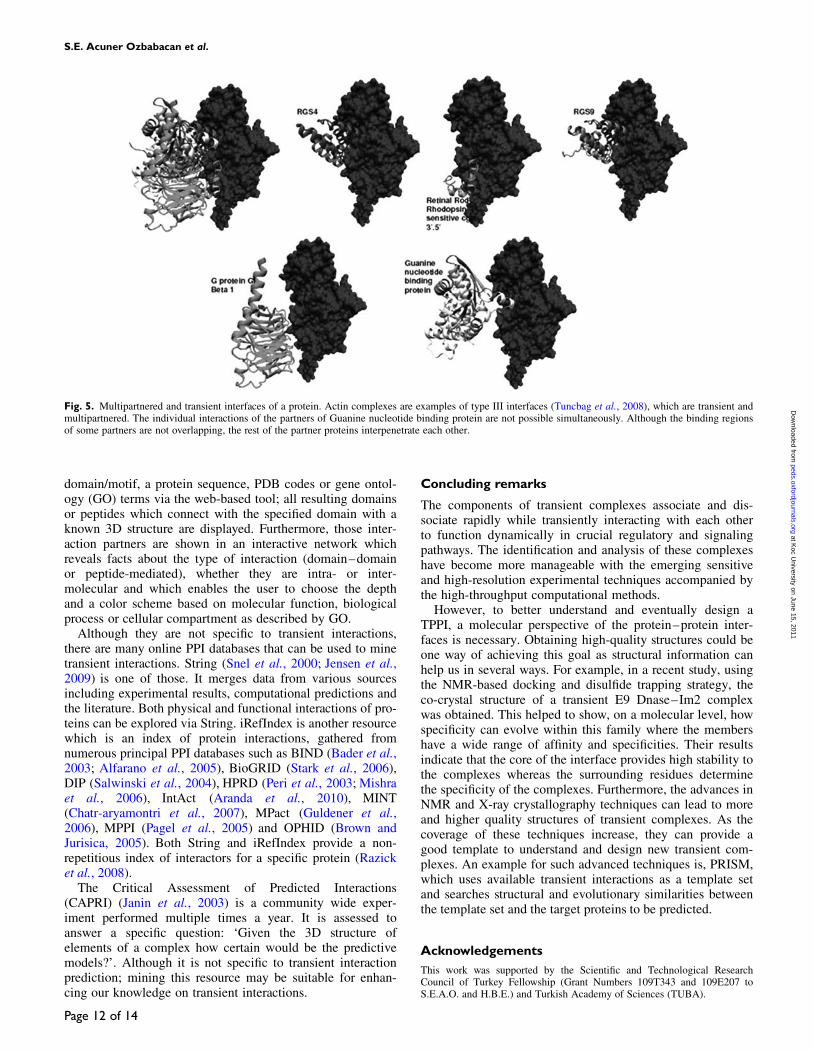
Tuncbag et al. (2008) published a dataset (PRINT) consist-ing of 8205 interface clusters, each standing for a separateinterface structure. In this dataset, which is available fromhttp://prism.ccbb.ku.edu.tr/, there are 14 501 obligate and2709 non-obligate PPIs, classified with NOXclass (Zhu et al.,2006) filter. Those interfaces are classified into three types astype I clusters; similar interface architectures, similar globalfolds, type II clusters; similar interface architectures, dissimi-lar global folds, type III clusters; one side structurally similarinterfaces (Keskin et al., 2004). Type III cluster consists ofmultipartnered and transient interfaces (Fig. 5).
PRISM is a novel algorithm for predicting PPIs based onstructure conservation in protein interfaces (Aytuna et al.,2005; Ogmen et al., 2005). PRISM bases its predictions ontemplate interface datasets. When a subset of the non-obligate template dataset, containing 158 TPPIs is used, onecan get predictions for other potential transient interactions.This method, which is now being studied by many others,was the first to make PPI predictions based on the structuralsimilarity of the binding sites. It is publicly available fromhttp://prism.ccbb.ku.edu.tr/.
3did is a database which uses structural data in order toexplain how protein interactions arise at the molecular levels(Stein et al., 2005) and is available at http://3did.irbbarcelona.org/. It contains both globular domain interactions and ahand-curated set of transient peptide-mediated interactions(Stein et al., 2009b). If 3did is queried with a specific
Table III. Databases and servers for identification and prediction of transient protein–protein interactions
Server/database Explanation Web site
ELM Largest linear motif server http://elm.eu.org/PROSITE Linear motif server http://expasy.org/prosite/SCANSITE Linear motif server http://scansite.mit.edu/ADAN Predictive analysis of modular domains settled by linear motifs http://adan.crg.es/SPIDER PPI site recognition http://sppider.cchmc.org/NOXCLASS Classification of obligate, non-obligate and crystal packing interactions http://noxclass.bioinf.mpiinf.mpg.de/PRISM Protein interaction prediction by structural matching http://prism.ccbb.ku.edu.tr/3DID Structural explanation of how protein interactions arise in molecular level http://3did.irbbarcelona.org/Minimotif Miner Queries for the presence of short functional motifs http://mnm.engr.uconn.edu/QuasiMotiFinder Identification of signatures and signature-like patterns in protein sequences http://quasimotifinder.tau.ac.il/AutoMotifServer Identification of post-translational modification sites in proteins http://ams2.bioinfo.pl/SIRW Search protein/nucleotide databases with a sequence motif http://sirw.embl.de/DILIMOT Finding linear motifs, in a set of protein http://dilimot.embl.de/SLiMFinder Linear Motif Server http://bioinformatics.ucd.ie/shields/software/slimfinder/
Transient protein–protein interactions
Page 11 of 14
at Koc U
niversity on June 15, 2011peds.oxfordjournals.org
Dow
nloaded from
domain/motif, a protein sequence, PDB codes or gene ontol-ogy (GO) terms via the web-based tool; all resulting domainsor peptides which connect with the specified domain with aknown 3D structure are displayed. Furthermore, those inter-action partners are shown in an interactive network whichreveals facts about the type of interaction (domain–domainor peptide-mediated), whether they are intra- or inter-molecular and which enables the user to choose the depthand a color scheme based on molecular function, biologicalprocess or cellular compartment as described by GO.
Although they are not specific to transient interactions,there are many online PPI databases that can be used to minetransient interactions. String (Snel et al., 2000; Jensen et al.,2009) is one of those. It merges data from various sourcesincluding experimental results, computational predictions andthe literature. Both physical and functional interactions of pro-teins can be explored via String. iRefIndex is another resourcewhich is an index of protein interactions, gathered fromnumerous principal PPI databases such as BIND (Bader et al.,2003; Alfarano et al., 2005), BioGRID (Stark et al., 2006),DIP (Salwinski et al., 2004), HPRD (Peri et al., 2003; Mishraet al., 2006), IntAct (Aranda et al., 2010), MINT(Chatr-aryamontri et al., 2007), MPact (Guldener et al.,2006), MPPI (Pagel et al., 2005) and OPHID (Brown andJurisica, 2005). Both String and iRefIndex provide a non-repetitious index of interactors for a specific protein (Razicket al., 2008).
The Critical Assessment of Predicted Interactions(CAPRI) (Janin et al., 2003) is a community wide exper-iment performed multiple times a year. It is assessed toanswer a specific question: ‘Given the 3D structure ofelements of a complex how certain would be the predictivemodels?’. Although it is not specific to transient interactionprediction; mining this resource may be suitable for enhan-cing our knowledge on transient interactions.
Concluding remarks
The components of transient complexes associate and dis-sociate rapidly while transiently interacting with each otherto function dynamically in crucial regulatory and signalingpathways. The identification and analysis of these complexeshave become more manageable with the emerging sensitiveand high-resolution experimental techniques accompanied bythe high-throughput computational methods.
However, to better understand and eventually design aTPPI, a molecular perspective of the protein–protein inter-faces is necessary. Obtaining high-quality structures could beone way of achieving this goal as structural information canhelp us in several ways. For example, in a recent study, usingthe NMR-based docking and disulfide trapping strategy, theco-crystal structure of a transient E9 Dnase–Im2 complexwas obtained. This helped to show, on a molecular level, howspecificity can evolve within this family where the membershave a wide range of affinity and specificities. Their resultsindicate that the core of the interface provides high stability tothe complexes whereas the surrounding residues determinethe specificity of the complexes. Furthermore, the advances inNMR and X-ray crystallography techniques can lead to moreand higher quality structures of transient complexes. As thecoverage of these techniques increase, they can provide agood template to understand and design new transient com-plexes. An example for such advanced techniques is, PRISM,which uses available transient interactions as a template setand searches structural and evolutionary similarities betweenthe template set and the target proteins to be predicted.
Acknowledgements
This work was supported by the Scientific and Technological ResearchCouncil of Turkey Fellowship (Grant Numbers 109T343 and 109E207 toS.E.A.O. and H.B.E.) and Turkish Academy of Sciences (TUBA).
Fig. 5. Multipartnered and transient interfaces of a protein. Actin complexes are examples of type III interfaces (Tuncbag et al., 2008), which are transient andmultipartnered. The individual interactions of the partners of Guanine nucleotide binding protein are not possible simultaneously. Although the binding regionsof some partners are not overlapping, the rest of the partner proteins interpenetrate each other.
S.E. Acuner Ozbabacan et al.
Page 12 of 14
at Koc U
niversity on June 15, 2011peds.oxfordjournals.org
Dow
nloaded from
ReferencesAlberts,B., Bray,D., Hopkin,K., Johnson,A., Lewis,J., Raff,M., Roberts,K.
and Walter,P. (2009) Essential Cell Biology. 3 edn, Garland Science,New York.
Alfarano,C., Andrade,C.E., Anthony,K. et al. (2005) Nucleic Acids Res., 33,D418–D424.
Aloy,P. and Russell,R.B. (2006) Nat. Rev. Mol. Cell Biol., 7, 188–197.Alsallaq,R. and Zhou,H.X. (2008) Proteins, 71, 320–335.Ansari,S. and Helms,V. (2005) Proteins, 61, 344–355.Aranda,B., Achuthan,P., Alam-Faruque,Y. et al. (2010) Nucleic Acids Res.
38, D525–D531.Aytuna,A.S., Gursoy,A. and Keskin,O. (2005) Bioinformatics, 21,
2850–2855.Bader,G.D., Betel,D. and Hogue,C.W. (2003) Nucleic Acids Res., 31,
248–250.Bader,J.S. and Chant,J. (2006) Science, 311, 187–188.Bairoch,A. (1993) Nucleic Acids Res., 21, 3097–3103.Bashor,C.J., Horwitz,A.A., Peisajovich,S.G. and Lim,W.A. (2010) Annu.
Rev. Biophys., 39, 515–537.Berggard,T., Linse,S. and James,P. (2007) Proteomics, 7, 2833–2842.Berman,H.M., Westbrook,J., Feng,Z., Gilliland,G., Bhat,T.N., Weissig,H.,
Shindyalov,I.N. and Bourne,P.E. (2000) Nucleic Acids Res., 28, 235–242.Bhattacharyya,R.P., Remenyi,A., Yeh,B.J. and Lim,W.A. (2006) Annu. Rev.
Biochem., 75, 655–680.Bjorklund,A.K., Ekman,D. and Elofsson,A. (2006) PLoS Comput. Biol., 2,
e114.Bjorklund,A.K., Light,S., Hedin,L. and Elofsson,A. (2008) Proteomics, 8,
4657–4667.Block,P., Paern,J., Hullermeier,E., Sanschagrin,P., Sotriffer,C.A. and
Klebe,G. (2006) Proteins, 65, 607–622.Borisy,A.A., Elliott,P.J., Hurst,N.W. et al. (2003) Proc. Natl Acad. Sci. USA,
100, 7977–7982.Braig,K., Otwinowski,Z., Hegde,R., Boisvert,D.C., Joachimiak,A.,
Horwich,A.L. and Sigler,P.B. (1994) Nature, 371, 578–586.Brown,K.R. and Jurisica,I. (2005) Bioinformatics, 21, 2076–2082.Chakrabarti,P. and Janin,J. (2002) Proteins, 47, 334–343.Chatr-aryamontri,A., Ceol,A., Palazzi,L.M., Nardelli,G., Schneider,M.V.,
Castagnoli,L. and Cesareni,G. (2007) Nucleic Acids Res., 35, D572–574.Cho,K.I., Lee,K., Lee,K.H., Kim,D. and Lee,D. (2006) Proteins, 65,
593–606.Collins,M.O. and Choudhary,J.S. (2008) Curr. Opin. Biotechnol., 19,
324–330.Creighton,T.E. (1993) Proteins: Structures and Molecular Properties. 2nd
edn, W.H. Freeman, New York.Cryer,B. and Feldman,M. (1998) Am. J. Med., 104, 413–421.Csermely,P., Agoston,V. and Pongor,S. (2005) Trends Pharmacol. Sci., 26,
178–182.Cukuroglu,E., Gursoy,A. and Keskin,O. (2010) Ann. Biomed. Eng., 38,
2068–2078.De,S., Krishnadev,O., Srinivasan,N. and Rekha,N. (2005) BMC Struct. Biol.,
5, 15.Dunker,A.K., Cortese,M.S., Romero,P., Iakoucheva,L.M. and Uversky,V.N.
(2005) FEBS J., 272, 5129–5148.Ekman,D., Light,S., Bjorklund,A.K. and Elofsson,A. (2006) Genome Biol.,
7, R45.Encinar,J.A., Fernandez-Ballester,G., Sanchez,I.E., Hurtado-Gomez,E.,
Stricher,F., Beltrao,P. and Serrano,L. (2009) Bioinformatics, 25,2418–2424.
Estojak,J., Brent,R. and Golemis,E.A. (1995) Mol. Cell Biol., 15,5820–5829.
Ethier,M., Lambert,J.P., Vasilescu,J. and Figeys,D. (2006) Anal. Chim.Acta., 564, 10–18.
Fields,S. and Song,O. (1989) Nature, 340, 245–246.Frantz,S. (2005) Nature, 437, 942–943.Fu,L. and Liang,J.J. (2002) J. Biol. Chem., 277, 4255–4260.Fuxreiter,M., Tompa,P. and Simon,I. (2007) Bioinformatics, 23, 950–956.Gabdoulline,R.R. and Wade,R.C. (1997) Biophys. J., 72, 1917–1929.Gao,G., Williams,J.G. and Campbell,S.L. (2004) Methods Mol. Biol., 261,
79–92.Gertler,A., Biener,E., Ramanujan,K.V., Djiane,J. and Herman,B. (2005)
J. Dairy Res., 72 Spec No., 14–19.Ghosh,I., Hamilton,A.D. and Regan,L. (2000) J. Am. Chem. Soc., 122,
5658–5659.Guerrero,C., Milenkovic,T., Przulj,N., Kaiser,P. and Huang,L. (2008) Proc.
Natl Acad. Sci. USA, 105, 13333–13338.
Guerrero,C., Tagwerker,C., Kaiser,P. and Huang,L. (2006) Mol. CellProteomics, 5, 366–378.
Guldener,U., Munsterkotter,M., Oesterheld,M., Pagel,P., Ruepp,A.,Mewes,H.W. and Stumpflen,V. (2006) Nucleic Acids Res., 34, D436–441.
Gunasekaran,K., Tsai,C.J. and Nussinov,R. (2004) J. Mol. Biol., 341,1327–1341.
Higurashi,M., Ishida,T. and Kinoshita,K. (2008) Protein Sci., 17, 72–78.Ho,Y., Gruhler,A., Heilbut,A. et al. (2002) Nature, 415, 180–183.Hopkins,A.L., Mason,J.S. and Overington,J.P. (2006) Curr. Opin. Struct.
Biol., 16, 127–136.Hu,C.D., Chinenov,Y. and Kerppola,T.K. (2002) Mol. Cell., 9, 789–798.Hu,C.D. and Kerppola,T.K. (2003) Nat. Biotechnol., 21, 539–545.Huang,S. (2002) Drug Discov. Today, 7, S163–S169.Humphris,E.L. and Kortemme,T. (2007) PLoS Comput. Biol., 3, e164.Hyams,J.S. and Lloyd,C.W. (1993) Microtubules, Modern Cell Biology.
Wiley-Liss, New York.Iakoucheva,L.M., Brown,C.J., Lawson,J.D., Obradovic,Z. and Dunker,A.K.
(2002) J. Mol. Biol., 323, 573–584.Iwahara,J. and Clore,G.M. (2006) Nature, 440, 1227–1230.Janin,J., Bahadur,R.P. and Chakrabarti,P. (2008) Q Rev. Biophys., 41,
133–180.Janin,J., Henrick,K., Moult,J., Eyck,L.T., Sternberg,M.J., Vajda,S., Vakser,I.
and Wodak,S.J. (2003) Proteins, 52, 2–9.Jansen,R., Greenbaum,D. and Gerstein,M. (2002) Genome Res., 12, 37–46.Jensen,L.J., Kuhn,M., Stark,M. et al. (2009) Nucleic Acids Res., 37,
D412–416.Jiang,M., Pabla,N., Murphy,R.F., Yang,T., Yin,X.M., Degenhardt,K.,
White,E. and Dong,Z. (2007) J. Biol. Chem., 282, 2636–2645.Jones,S. and Thornton,J.M. (1995) Prog. Biophys. Mol. Biol., 63, 31–65.Jones,S. and Thornton,J.M. (1996) Proc. Natl Acad. Sci. USA, 93, 13–20.Jones,S. and Thornton,J.M. (1997) J. Mol. Biol., 272, 121–132.Kaelin,W.G., Jr. (2004) Sci. STKE, 2004, pe12.Kamath,R.S., Martinez-Campos,M., Zipperlen,P., Fraser,A.G. and
Ahringer,J. (2001) Genome Biol., 2, RESEARCH0002.Kar,G., Keskin,O., Gursoy,A. and Nussinov,R. (2010) Curr. Opin.
Pharmacol., 10, 715–722.Keskin,O., Gursoy,A., Ma,B. and Nussinov,R. (2008) Chem. Rev., 108,
1225–1244.Keskin,O., Ma,B. and Nussinov,R. (2005) J. Mol. Biol., 345, 1281–1294.Keskin,O. and Nussinov,R. (2005) Protein Eng. Des. Sel., 18, 11–24.Keskin,O., Tsai,C.J., Wolfson,H. and Nussinov,R. (2004) Protein Sci., 13,
1043–1055.Kim,P.M., Sboner,A., Xia,Y. and Gerstein,M. (2008) Mol. Syst. Biol., 4, 179.Kobayashi,N., Freund,S.M., Chatellier,J., Zahn,R. and Fersht,A.R. (1999)
J. Mol. Biol., 292, 181–190.Kresge,N., Vacquier,V.D. and Stout,C.D. (2000) Acta. Crystallogr. D Biol.
Crystallogr., 56, 34–41.Krishna,S.S. and Aravind,L. (2010) J. Struct. Biol., 172, 294–299.Lawrence,M.C. and Colman,P.M. (1993) J. Mol. Biol., 234, 946–950.Levy,E.D. and Pereira-Leal,J.B. (2008) Curr. Opin. Struct. Biol., 18,
349–357.Levy,Y., Cho,S.S., Onuchic,J.N. and Wolynes,P.G. (2005) J. Mol. Biol., 346,
1121–1145.Liedberg,B.O., Nylander,C. and Lundstrom,I. (1983) Sensors and Actuators,
4, 299–304.Lievens,S., Lemmens,I. and Tavernier,J. (2009) Trends Biochem. Sci., 34,
579–588.Lippincott-Schwartz,J. and Patterson,G.H. (2003) Science, 300, 87–91.Lipton,S.A. (2004) Nature, 428, 473.Liu,J., Tan,H. and Rost,B. (2002) J. Mol. Biol., 322, 53–64.Lo Conte,L., Chothia,C. and Janin,J. (1999) J. Mol. Biol., 285, 2177–2198.London,N., Movshovitz-Attias,D. and Schueler-Furman,O. (2010) Structure,
18, 188–199.Lowe,J., Li,H., Downing,K.H. and Nogales,E. (2001) J. Mol. Biol., 313,
1045–1057.Ma,B. and Nussinov,R. (2007) Curr. Top Med. Chem., 7, 999–1005.McCoy,A.J., Chandana Epa,V. and Colman,P.M. (1997) J. Mol. Biol., 268,
570–584.Meenan,N.A., Sharma,A., Fleishman,S.J., Macdonald,C.J., Morel,B.,
Boetzel,R., Moore,G.R., Baker,D. and Kleanthous,C. (2010) Proc. NatlAcad. Sci. USA, 107, 10080–10085.
Mintseris,J. and Weng,Z. (2003) Proteins, 53, 629–639.Mintseris,J. and Weng,Z. (2005) Proc. Natl Acad. Sci. USA, 102,
10930–10935.Mishra,G.R., Suresh,M., Kumaran,K. et al. (2006) Nucleic Acids Res., 34,
D411–414.
Transient protein–protein interactions
Page 13 of 14
at Koc U
niversity on June 15, 2011peds.oxfordjournals.org
Dow
nloaded from

Mittag,T., Kay,L.E. and Forman-Kay,J.D. (2010) J. Mol. Recognit., 23,105–116.
Morell,M., Espargaro,A., Aviles,F.X. and Ventura,S. (2007) Proteomics, 7,1023–1036.
Morphy,R. and Rankovic,Z. (2005) J. Med. Chem., 48, 6523–6543.Morphy,R. and Rankovic,Z. (2007) Drug Discov. Today, 12, 156–160.Mousson,F., Kolkman,A., Pijnappel,W.W., Timmers,H.T. and Heck,A.J.
(2008) Mol. Cell Proteomics, 7, 845–852.Neduva,V. and Russell,R.B. (2005) FEBS Lett., 579, 3342–3345.Nooren,I.M. and Thornton,J.M. (2003a) EMBO J., 22, 3486–3492.Nooren,I.M. and Thornton,J.M. (2003b) J. Mol. Biol., 325, 991–1018.Nyfeler,B., Michnick,S.W. and Hauri,H.P. (2005) Proc. Natl Acad. Sci.
USA, 102, 6350–6355.Obenauer,J.C., Cantley,L.C. and Yaffe,M.B. (2003) Nucleic Acids Res., 31,
3635–3641.Ofran,Y. and Rost,B. (2003) J. Mol. Biol., 325, 377–387.Ogmen,U., Keskin,O., Aytuna,A.S., Nussinov,R. and Gursoy,A. (2005)
Nucleic Acids Res., 33, W331–W336.Ohad,N., Shichrur,K. and Yalovsky,S. (2007) Plant Physiol., 145,
1090–1099.Ohlson,S. (2008) Drug Discov. Today, 13, 433–439.Orlando,V., Strutt,H. and Paro,R. (1997) Methods, 11, 205–214.Ozbabacan,S.E., Gursoy,A., Keskin,O. and Nussinov,R. (2010) Curr. Opin.
Drug Discov. Dev., 13, 527–537.Pagel,P., Kovac,S., Oesterheld,M. et al. (2005) Bioinformatics, 21, 832–834.Park,S.H., Reyes,J.A., Gilbert,D.R., Kim,J.W. and Kim,S. (2009) BMC
Bioinform., 10, 36.Pattnaik,P. (2005) Appl. Biochem. Biotechnol., 126, 79–92.Pawson,T. and Linding,R. (2005) FEBS Lett., 579, 1808–1814.Peri,S., Navarro,J.D., Amanchy,R. et al. (2003) Genome Res., 13,
2363–2371.Perkins,J.R., Diboun,I., Dessailly,B.H., Lees,J.G. and Orengo,C. (2010)
Structure, 18, 1233–1243.Phizicky,E., Bastiaens,P.I., Zhu,H., Snyder,M. and Fields,S. (2003) Nature,
422, 208–215.Phizicky,E.M. and Fields,S. (1995) Microbiol. Rev., 59, 94–123.Pommier,Y. and Cherfils,J. (2005) Trends Pharmacol. Sci., 26, 138–145.Porollo,A. and Meller,J. (2007) Proteins, 66, 630–645.Puntervoll,P., Linding,R., Gemund,C. et al. (2003) Nucleic Acids Res., 31,
3625–3630.Qin,J., Vinogradova,O. and Gronenborn,A.M. (2001) Methods Enzymol.,
339, 377–389.Rabi,II., Zacharias,J.R., Millman,S. and Kusch,P. (1992) J. Magn. Reson.
Imaging, 2, 131–133.Ravelli,R.B., Gigant,B., Curmi,P.A., Jourdain,I., Lachkar,S., Sobel,A. and
Knossow,M. (2004) Nature, 428, 198–202.Razick,S., Magklaras,G. and Donaldson,I.M. (2008) BMC Bioinform., 9,
405.Renault,L., Guibert,B. and Cherfils,J. (2003) Nature, 426, 525–530.Rich,R.L. and Myszka,D.G. (2007) Anal. Biochem., 361, 1–6.Robineau,S., Chabre,M. and Antonny,B. (2000) Proc. Natl Acad. Sci. USA,
97, 9913–9918.Rogawski,M.A. (2000) Amino Acids, 19, 133–149.Rossi,F., Charlton,C.A. and Blau,H.M. (1997) Proc. Natl Acad. Sci. USA,
94, 8405–8410.Rudolph,J. (2007) Nat. Rev. Cancer, 7, 202–211.Sali,A., Glaeser,R., Earnest,T. and Baumeister,W. (2003) Nature, 422,
216–225.Salwinski,L., Miller,C.S., Smith,A.J., Pettit,F.K., Bowie,J.U. and
Eisenberg,D. (2004) Nucleic Acids Res., 32, D449–451.Samanta,U. and Chakrabarti,P. (2001) Protein Eng., 14, 7–15.Schreiber,G. (2002) Curr. Opin. Struct. Biol., 12, 41–47.Schreiber,G., Haran,G. and Zhou,H.X. (2009) Chem. Rev., 109, 839–860.Sekar,R.B. and Periasamy,A. (2003) J. Cell Biol., 160, 629–633.Seraphin,B. (2002) Adv. Protein Chem., 61, 99–117.Siggins,G.R., Roberto,M. and Nie,Z. (2005) Pharmacol. Ther., 107, 80–98.Singh,G.P., Ganapathi,M. and Dash,D. (2007) Proteins, 66, 761–765.Smith,G.P. (1985) Science, 228, 1315–1317.Snel,B., Lehmann,G., Bork,P. and Huynen,M.A. (2000) Nucleic Acids Res.,
28, 3442–3444.Stark,C., Breitkreutz,B.J., Reguly,T., Boucher,L., Breitkreutz,A. and
Tyers,M. (2006) Nucleic Acids Res., 34, D535–539.Stein,A. and Aloy,P. (2008) PLoS One, 3, e2524.Stein,A., Pache,R.A., Bernado,P., Pons,M. and Aloy,P. (2009a) FEBS J.,
276, 5390–5405.Stein,A., Panjkovich,A. and Aloy,P. (2009b) Nucleic Acids Res., 37,
D300–D304.
Stein,A., Russell,R.B. and Aloy,P. (2005) Nucleic Acids Res., 33,D413–D417.
Sullivan,K.F. and Kay,S.A. (1999) Cytoskeletal Dynamics in Yeast. InWilson,L. and Matsudaira,P. (eds.), Green Fluorescent Proteins.Academic Press, San Diego.
Tirosh,I. and Barkai,N. (2005) BMC Bioinform., 6, 40.Tsai,C.J., Lin,S.L., Wolfson,H.J. and Nussinov,R. (1997) Protein Sci., 6,
53–64.Tsai,C.J., Xu,D. and Nussinov,R. (1998) Fold Des., 3, R71–R80.Tuncbag,N., Gursoy,A., Guney,E., Nussinov,R. and Keskin,O. (2008)
J. Mol. Biol., 381, 785–802.Uetz,P., Giot,L., Cagney,G. et al. (2000) Nature, 403, 623–627.Uversky,V.N., Oldfield,C.J., Midic,U., Xie,H., Xue,B., Vucetic,S.,
Iakoucheva,L.M., Obradovic,Z. and Dunker,A.K. (2009) BMC Genomics,10(Suppl 1), S7.
Valdar,W.S. and Thornton,J.M. (2001) Proteins, 42, 108–124.Vassilev,L.T., Vu,B.T., Graves,B. et al. (2004) Science, 303, 844–848.Vaynberg,J., Fukuda,T., Chen,K., Vinogradova,O., Velyvis,A., Tu,Y., Ng,L.,
Wu,C. and Qin,J. (2005) Mol. Cell, 17, 513–523.Vaynberg,J. and Qin,J. (2006) Trends Biotechnol., 24, 22–27.Venkatesan,K., Rual,J.F., Vazquez,A. et al. (2009) Nat. Methods, 6, 83–90.Vetter,I.R. and Wittinghofer,A. (2001) Science, 294, 1299–1304.Vinayagam,A., Stelzl,U. and Wanker,E.E. (2010) Theor. Chem. Acc.., 125,
613–619.Wallis,R., Leung,K.Y., Pommer,A.J., Videler,H., Moore,G.R., James,R. and
Kleanthous,C. (1995) Biochemistry, 34, 13751–13759.Walters,K.J., Ferentz,A.E., Hare,B.J., Hidalgo,P., Jasanoff,A., Matsuo,H. and
Wagner,G. (2001) Methods Enzymol., 339, 238–258.Wang,X. and Huang,L. (2008) Mol. Cell Proteomics, 7, 46–57.Wawrzynow,B., Zylicz,A., Wallace,M., Hupp,T. and Zylicz,M. (2007)
J. Biol. Chem., 282, 32603–32612.Wright,P.E. and Dyson,H.J. (1999) J. Mol. Biol., 293, 321–331.Xu,D., Tsai,C.J. and Nussinov,R. (1998) Protein Sci., 7, 533–544.Zanivan,S., Cascone,I., Peyron,C., Molineris,I., Marchio,S., Caselle,M. and
Bussolino,F. (2007) Genome Biol., 8, R256.Zhou,H.X., Wong,K.Y. and Vijayakumar,M. (1997) Proc. Natl Acad. Sci.
USA, 94, 12372–12377.Zhu,H., Domingues,F.S., Sommer,I. and Lengauer,T. (2006) BMC
Bioinform., 7, 27.Zuiderweg,E.R. (2002) Biochemistry, 41, 1–7.
S.E. Acuner Ozbabacan et al.
Page 14 of 14
at Koc U
niversity on June 15, 2011peds.oxfordjournals.org
Dow
nloaded from