Transcriptomic analysis of shell repair and ...
14
RESEARCH ARTICLE Open Access Transcriptomic analysis of shell repair and biomineralization in the blue mussel, Mytilus edulis Tejaswi Yarra 1,2 , Kirti Ramesh 3 , Mark Blaxter 4 , Anne Hüning 3 , Frank Melzner 3 and Melody S. Clark 2* Abstract Background: Biomineralization by molluscs involves regulated deposition of calcium carbonate crystals within a protein framework to produce complex biocomposite structures. Effective biomineralization is a key trait for aquaculture, and animal resilience under future climate change. While many enzymes and structural proteins have been identified from the shell and in mantle tissue, understanding biomieralization is impeded by a lack of fundamental knowledge of the genes and pathways involved. In adult bivalves, shells are secreted by the mantle tissue during growth, maintenance and repair, with the repair process, in particular, amenable to experimental dissection at the transcriptomic level in individual animals. Results: Gene expression dynamics were explored in the adult blue mussel, Mytilus edulis, during experimentally induced shell repair, using the two valves of each animal as a matched treatment-control pair. Gene expression was assessed using high-resolution RNA-Seq against a de novo assembled database of functionally annotated transcripts. A large number of differentially expressed transcripts were identified in the repair process. Analysis focused on genes encoding proteins and domains identified in shell biology, using a new database of proteins and domains previously implicated in biomineralization in mussels and other molluscs. The genes implicated in repair included many otherwise novel transcripts that encoded proteins with domains found in other shell matrix proteins, as well as genes previously associated with primary shell formation in larvae. Genes with roles in intracellular signalling and maintenance of membrane resting potential were among the loci implicated in the repair process. While haemocytes have been proposed to be actively involved in repair, no evidence was found for this in the M. edulis data. Conclusions: The shell repair experimental model and a newly developed shell protein domain database efficiently identified transcripts involved in M. edulis shell production. In particular, the matched pair analysis allowed factoring out of much of the inherent high level of variability between individual mussels. This snapshot of the damage repair process identified a large number of genes putatively involved in biomineralization from initial signalling, through calcium mobilization to shell construction, providing many novel transcripts for future in-depth functional analyses. Keywords: Mollusc, Bivalve, Shell matrix proteins, Haemocytes, Calcium © The Author(s). 2021 Open Access This article is licensed under a Creative Commons Attribution 4.0 International License, which permits use, sharing, adaptation, distribution and reproduction in any medium or format, as long as you give appropriate credit to the original author(s) and the source, provide a link to the Creative Commons licence, and indicate if changes were made. The images or other third party material in this article are included in the article's Creative Commons licence, unless indicated otherwise in a credit line to the material. If material is not included in the article's Creative Commons licence and your intended use is not permitted by statutory regulation or exceeds the permitted use, you will need to obtain permission directly from the copyright holder. To view a copy of this licence, visit http://creativecommons.org/licenses/by/4.0/. The Creative Commons Public Domain Dedication waiver (http://creativecommons.org/publicdomain/zero/1.0/) applies to the data made available in this article, unless otherwise stated in a credit line to the data. * Correspondence: [email protected] 2 British Antarctic Survey, Natural Environment Research Council, High Cross, Madingley Road, CB3 0ET Cambridge, UK Full list of author information is available at the end of the article Yarra et al. BMC Genomics (2021) 22:437 https://doi.org/10.1186/s12864-021-07751-7
Transcript of Transcriptomic analysis of shell repair and ...

RESEARCH ARTICLE Open Access
Transcriptomic analysis of shell repair andbiomineralization in the blue mussel,Mytilus edulisTejaswi Yarra1,2, Kirti Ramesh3, Mark Blaxter4, Anne Hüning3, Frank Melzner3 and Melody S. Clark2*
Abstract
Background: Biomineralization by molluscs involves regulated deposition of calcium carbonate crystals within aprotein framework to produce complex biocomposite structures. Effective biomineralization is a key trait foraquaculture, and animal resilience under future climate change. While many enzymes and structural proteins havebeen identified from the shell and in mantle tissue, understanding biomieralization is impeded by a lack offundamental knowledge of the genes and pathways involved. In adult bivalves, shells are secreted by the mantletissue during growth, maintenance and repair, with the repair process, in particular, amenable to experimentaldissection at the transcriptomic level in individual animals.
Results: Gene expression dynamics were explored in the adult blue mussel, Mytilus edulis, during experimentallyinduced shell repair, using the two valves of each animal as a matched treatment-control pair. Gene expression wasassessed using high-resolution RNA-Seq against a de novo assembled database of functionally annotated transcripts.A large number of differentially expressed transcripts were identified in the repair process. Analysis focused ongenes encoding proteins and domains identified in shell biology, using a new database of proteins and domainspreviously implicated in biomineralization in mussels and other molluscs. The genes implicated in repair includedmany otherwise novel transcripts that encoded proteins with domains found in other shell matrix proteins, as wellas genes previously associated with primary shell formation in larvae. Genes with roles in intracellular signalling andmaintenance of membrane resting potential were among the loci implicated in the repair process. Whilehaemocytes have been proposed to be actively involved in repair, no evidence was found for this in the M. edulisdata.
Conclusions: The shell repair experimental model and a newly developed shell protein domain database efficientlyidentified transcripts involved in M. edulis shell production. In particular, the matched pair analysis allowed factoringout of much of the inherent high level of variability between individual mussels. This snapshot of the damagerepair process identified a large number of genes putatively involved in biomineralization from initial signalling,through calcium mobilization to shell construction, providing many novel transcripts for future in-depth functionalanalyses.
Keywords: Mollusc, Bivalve, Shell matrix proteins, Haemocytes, Calcium
© The Author(s). 2021 Open Access This article is licensed under a Creative Commons Attribution 4.0 International License,which permits use, sharing, adaptation, distribution and reproduction in any medium or format, as long as you giveappropriate credit to the original author(s) and the source, provide a link to the Creative Commons licence, and indicate ifchanges were made. The images or other third party material in this article are included in the article's Creative Commonslicence, unless indicated otherwise in a credit line to the material. If material is not included in the article's Creative Commonslicence and your intended use is not permitted by statutory regulation or exceeds the permitted use, you will need to obtainpermission directly from the copyright holder. To view a copy of this licence, visit http://creativecommons.org/licenses/by/4.0/.The Creative Commons Public Domain Dedication waiver (http://creativecommons.org/publicdomain/zero/1.0/) applies to thedata made available in this article, unless otherwise stated in a credit line to the data.
* Correspondence: [email protected] Antarctic Survey, Natural Environment Research Council, High Cross,Madingley Road, CB3 0ET Cambridge, UKFull list of author information is available at the end of the article
Yarra et al. BMC Genomics (2021) 22:437 https://doi.org/10.1186/s12864-021-07751-7

BackgroundKThe molluscan shell is composed of varying proportionsof organic components (largely proteins, acidic polysac-charides and chitin) and the calcium carbonate poly-morphs: calcite and aragonite. Combined, these give theshell of each mollusc species their unique physical andchemical properties. During shell formation, calciumcarbonate is produced from the reaction of calcium ionswith bicarbonate ions, and evidence suggests that theproteins (shell matrix proteins or SMPs) determine themineral polymorph and are involved with the nucleation,growth and termination of the calcium carbonate crys-tals [1]. SMPs are secreted by the mantle, a layer of tis-sue between the shell and the rest of the organs itencloses, into the extrapallial fluid, where they are incor-porated into the growing edge of the shell along withthe calcium carbonate crystals [1]. Hence, the processesof the production of crystal lattices and proteinaceousextracellular matrix are intimately linked in molluscanbiomineralization.SMPs have been identified and characterized in mul-
tiple proteomic studies via the extraction of proteins dir-ectly from shells. SMPs have been described fromseveral molluscan genera, which have been collated inan in-house SMP database (https://doi.org/10/cz2w [2]).This database contains protein sequences of both puta-tive and known SMPs identified in Uniprot using key-word searches related to molluscan biomineralization(full details in methods). Complementary to these prote-omic data, transcriptomic data have been generatedfrom mantle tissue and putative biomineralization lociidentified through sequence similarity to already identi-fied SMPs. Transcriptome data have also been deployedto propose source proteins for proteomic mass spec-trometry data [3]. The specific roles of SMPs in biomin-eralization have been explored through functionalexperimentation. For example, RNA interference medi-ated knock-down of Pif and PfN23 genes in the mantledisrupted nacre formation in Pinctada imbricata fucata[4, 5], while knockdown of the Shematrin gene resultedin disordered foliate structures in Chlamys farreri [6]. Invitro studies on the effects of SMPs on calcium carbon-ate crystal formation revealed functional specificity. Pifinduced calcium carbonate crystal growth and PfN23and p10 accelerated crystal growth in P. imbricatafucata [4, 5, 7]. In contrast, perlinhibin and perlwapinfrom Haliotis laevigata, prismalin-14 from P. imbricatafucata and caspartin from Pinna nobilis were found toinhibit crystal growth [4, 8–10]. Although SMPs andmantle transcripts from multiple molluscan species havebeen identified, there are still many unknowns in thebiomineralization process.Shell matrix proteomics can only identify proteins that
are incorporated into the shells and cannot report on
enzymic or other upstream processes. Similarly, whilemantle transcriptomes have been used to identify puta-tive biomineralization related transcripts, this has largelybeen based on sequence similarity to previously knownSMPs. Importantly, mantle tissue is made up of multipledifferent cell types with different origins and roles in-cluding ectodermal and mesodermal components in-volved in sensory and muscular functions as well asepidermal and secretory tissue involved in shell forma-tion. This makes it hard to ascertain whether a transcriptis involved in biomineralization or in multiple otherfunctions. Species-specific adaptations may also obscureshared biology. For example, it has been proposed thathaemocytes, found in the open circulatory system ofmolluscs, may play an active role during shell formationby carrying amorphous calcium, calcium crystals orSMPs to the site of shell formation [11–13]. However,the involvement of haemocytes in mollusc biominerali-zation may be species-specific, as they were associatedwith immune processes in Crastostrea gigas, but withion regulation and calcium transport in C. virginica [14].While in vivo and in vitro experiments have identifiedSMPs as integral to shell production, the molecularplayers in other shell formation processes such as theuptake, mobilisation and storage of calcium and bicar-bonate ions, are unclear [15]. Molluscs are proficient atrepairing shell damage [16]. Repair of experimentally-induced shell damage has been used in several species toexplore the dynamics of the repair process and the genesand proteins involved in biomineralization [17–22].These previous studies used either pooled individuals orseparate controls and treated animals. Therefore part ofthe aim of this study was to validate the matched pairdesign using individuals via Illumina RNA-Seq.The blue mussel Mytilus edulis is endemic to Euro-
pean and West Atlantic waters, and is an important spe-cies in commercial aquaculture (http://www.fao.org/fishery/species/2688/en). M. edulis shells are composedof an outermost organic layer of periostracum, a middlelayer consisting of calcite based prismatic structures, andan innermost layer of aragonite based laminar structurecalled nacre [23]. In this study, samples generated aspart of a previously published M. edulis shell regener-ation experiment [20] were used to measure gene ex-pression changes consequent on damage and repair ofadult shells using RNA-Seq transcriptomics. Importantlythe experimental model, using within-individual controlsenabled identification of differences in gene expressionpatterns due to the systemic effects of injury and thegenetic difference between individuals from those associ-ated with the processes occurring at the wound site. Dueto financial constraints and the need for a (relatively)high level of replication (n = 5) and to sequence four tis-sues per animal, this study focused on the time point
Yarra et al. BMC Genomics (2021) 22:437 Page 2 of 14
with the most distinct and homogenous calcification re-sponse. A database of genes, proteins and protein do-mains previously identified as SMPs or associated withSMPs was generated to explore the involvement of thesecandidates in the shell repair process through time. Inaddition, comparisons were carried out against M. edulishaemocyte expressed sequence tag (EST) datasets to as-sess the contribution of haemocytes to shell repair andagainst transcriptome data from M. edulis larvae duringthe synthesis of the first larval shell to validate novelSMPs.
ResultsStudy designThe tissue samples from five 5 individuals analysed inthis study were generated during a longitudinal study ofshell repair in adult M. edulis [20]. Recent studies haveshown that most Mytilus populations in Europe are hy-brids of M. edulis, M. galloprovincialis and M. trossulus,with varying degrees of admixture [24]. Kiel animals arecharacterized by a high proportion of Mytilus edulis al-leles (ca. 80 %) and admixture of M. trossulus (ca. 20 %)alleles [25] (Stuckas, Melzner et al. unpublished). A Kielhybrid transcriptome was assembled and sequencedreads were mapped on this hybrid transcriptome. Sincewe utilized five replicate animals, we expect that ourstatistical analyses captured at least the essential tran-scriptomic signatures related to shell repair. Details ofthe experimental procedures are given in the originalpublication, but the salient features are reviewed here.
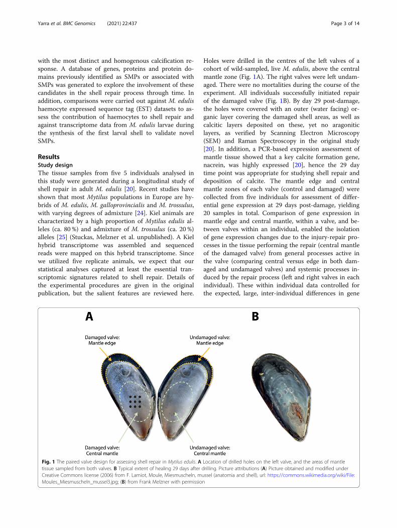
Holes were drilled in the centres of the left valves of acohort of wild-sampled, live M. edulis, above the centralmantle zone (Fig. 1A). The right valves were left undam-aged. There were no mortalities during the course of theexperiment. All individuals successfully initiated repairof the damaged valve (Fig. 1B). By day 29 post-damage,the holes were covered with an outer (water facing) or-ganic layer covering the damaged shell areas, as well ascalcitic layers deposited on these, yet no aragoniticlayers, as verified by Scanning Electron Microscopy(SEM) and Raman Spectroscopy in the original study[20]. In addition, a PCR-based expression assessment ofmantle tissue showed that a key calcite formation gene,nacrein, was highly expressed [20], hence the 29 daytime point was appropriate for studying shell repair anddeposition of calcite. The mantle edge and centralmantle zones of each valve (control and damaged) werecollected from five individuals for assessment of differ-ential gene expression at 29 days post-damage, yielding20 samples in total. Comparison of gene expression inmantle edge and central mantle, within a valve, and be-tween valves within an individual, enabled the isolationof gene expression changes due to the injury-repair pro-cesses in the tissue performing the repair (central mantleof the damaged valve) from general processes active inthe valve (comparing central versus edge in both dam-aged and undamaged valves) and systemic processes in-duced by the repair process (left and right valves in eachindividual). These within individual data controlled forthe expected, large, inter-individual differences in gene
Fig. 1 The paired valve design for assessing shell repair in Mytilus edulis. A Location of drilled holes on the left valve, and the areas of mantletissue sampled from both valves. B Typical extent of healing 29 days after drilling. Picture attributions (A) Picture obtained and modified underCreative Commons license (2006) from F. Lamiot, Moule, Miesmuscheln, mussel (anatomia and shell), url: https://commons.wikimedia.org/wiki/File:Moules_Miesmuscheln_mussel3.jpg; (B) from Frank Melzner with permission
Yarra et al. BMC Genomics (2021) 22:437 Page 3 of 14

expression profiles in Mytilus species, which are all out-breeders and highly heterozygous [26, 27].
Transcriptome assembly, filtering and annotationTranscriptomic analysis (Illumina RNA-Seq) generated714 million raw read pairs in total, with 601 million readpairs remaining after adapter trimming and quality andlength filtering. Because of the high genetic variability be-tween M. edulis individuals and haplotypes, and thus poormapping of reads from individuals in this study to previ-ously generated transcriptomic and genomics data, a denovo transcriptome was assembled to act as reference.The pooled, cleaned read set was down-sampled to 31 mil-lion read pairs by in silico normalization. These were as-sembled using the Trinity pipeline into 560,776 putativegenes with 874,699 transcript fragments (likely isoforms).Filtering of the assembly to eliminate expression noise (in-cluding putative genes only if they had more than 1mapped read per million mapped reads in at least 10 li-braries) yielded 30,822 putative genes, with 158,880 tran-script fragments (Table 1). These data are similar inmagnitude to a recently produced M. edulis transcrip-tome, which also sourced animals from the Baltic [28].Reads were aligned from each sample to this filtered refer-ence and gene expression was assessed by summing thecounts of mapped read pairs per putative gene.
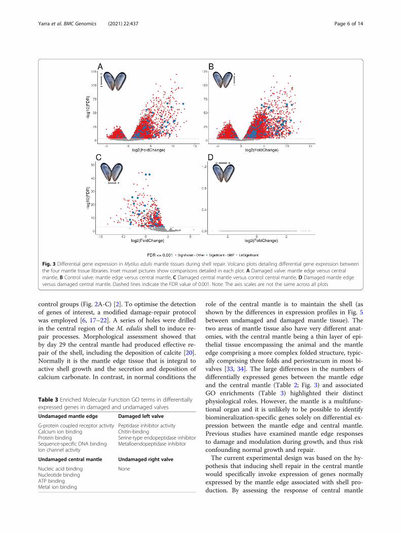
Differential gene expressionMultidimensional scaling (MDS) plots of the digitalexpression levels showed separation between mantleedge and central mantle tissues in dimension 1, with di-mension 2 roughly corresponding to different individuals(Fig. 2A). There was a significant difference in expres-sion levels in the central mantle both between damagedand undamaged valves and between individuals (Fig. 2B).Although the expression levels of mantle edge libraries
also showed separation between different individuals,there was no significant difference between the damagedand undamaged valves (Fig. 2C). Four pairwise compari-sons were made for differential gene expression betweenthe tissues and valves (Table 2; Fig. 3). In both the dam-aged and undamaged valves, many putative genes werefound to be differentially expressed between the mantleedge and the central mantle (Fig. 3A,B). When themantle of the damaged and undamaged (control) valveswere compared, 653 transcripts were highly expressed inthe central mantle of the damaged valve during shell re-pair, with 54 of these transcripts having sequencesimilarity with SMPs (Fig. 3C, Table 2). No putativegenes were identified as differentially expressed betweenthe mantle edge tissues of damaged and control valves(Fig. 3D).
Annotation of transcripts associated with damage-repairFurther in-depth analysis was restricted to the 653 puta-tive genes associated with the comparison of damagedand control central mantle tissues (Fig. 3C, Table 2), asthese were most likely to be involved in damage-repair.All 653 genes were upregulated in the damaged valveundergoing repair. Gene ontology analysis of these 653genes showed enrichment, compared to the total putativegene translation dataset of several molecular processes as-sociated with protease inhibition (including serine-typeendopeptidase inhibition), chitin-binding and metalloen-dopep tidase activity (Table 3). Sequence similaritysearches identified specific transmembrane transporters,proteases and protease inhibitors, signalling moleculesand tyrosinases in this gene set (Figs. 4 and 5). Just over8 % (54 of 653) of these putative genes had sequence simi-larity with known SMPs or domains associated with SMPs(Fig. 4). In addition to identification of homologues of pre-viously described SMPs, we identified a number of puta-tive genes that had no strong sequence similarity toknown SMPs but contained SMP-associated domainssuch as VWA (chitin-binding), EF-hand, FAMeT, Kazal,and TIMP (Fig. 4).The initial stages of embryonic shell formation in M.
edulis are characterised by the deposition of aragonite,while the adult shell has both calcite and aragonite mi-crostructures. However, analyses in other species such asthe gastropod Lymnaea stagnalis and the oysters P.imbricata fucata and Crassostrea gigas have revealedsimilarities in gene expression repertoires between adultand larval shells [29, 30]. Many of the differentiallyexpressed genes with SMP annotations identified in thisstudy were also differentially expressed in the tran-scriptomics dataset from the prodissoconch I stage of M.edulis developing larvae (Fig. 4) [31] (Fig. 4). Further-more, to identify whether haemocytes could be involvedin shell repair processes, 2,194 sequences from a Mytilus
Table 1 Mantle transcriptome assembly metrics
Main assembly
Trinity genes 560,776
Trinity transcripts 874,699
Filtered assembly (> 1 CPM in ≥ 10 libraries)
Trinity genes 30,822
Trinity transcripts 158,889
Protein sequences (ORF≥ 100 amino acids) 81,456
Filtered assembly features
% GC 33.54
N50 (bp) 1,602
Minimum length (bp) 201
Maximum length (bp) 26,467
Total assembled bases (Mbp) 181
Yarra et al. BMC Genomics (2021) 22:437 Page 4 of 14

haemocyte EST dataset were extracted from MytiBase[32] and compared with the current dataset. Only onesequence with one of the SMP-associated domains(C1Q) was identified in both datasets. Thus evidence forhaemocyte involved in damage repair is limited in M.edulis. Interestingly, transcripts highly expressed in thecentral mantle of the damaged valve during shell repairwere also present in the mantle edge transcriptomes andwith similar expression levels, suggesting a general simi-larity in function (Fig. 5).
DiscussionBiomineralization is a complex process, and subject todevelopmental and environmental control. Using a
carefully internally-controlled gene expression analysis,this study identified a large number of putative genesthat may be involved in coordinating and carrying outshell repair in M. edulis, an important ecosystem andaquaculture species. Importantly the experimental designcontrolled for the known high genetic variation in M.edulis [2, 26, 27] by exploiting the bivalve condition andusing a matched pair analysis, whereby the control andtreated (damaged) samples were taken from the same in-dividual (Fig. 1) [20]. The sampling regime minimisedindividual effects (both genetic and environmental) onsignal discovery, as confirmed by the MDS plots, inwhich the variability between individuals was much lar-ger than the difference between experimental and
Fig. 2 Multidimensional scaling identifies significant contributions of individual variation to gene expression differences in shell repair in Mytilusedulis. MDS plots of expression counts for the filtered set of putative genes in (A) All libraries: Central mantle – left/damaged valve; Centralmantle – right undamaged (control) valve: Mantle edge – left/damaged valve; Mantle edge – right undamaged (control) valve, B Central mantlelibraries only (C) Mantle edge libraries only
Table 2 Number of differentially expressed contigs between mantle tissue sections and annotation levels
Comparison Differential expression Annotationb
Tissue in which genes are more highlyexpressed
FDRa <=0.001
SwissProt Trembl SMPdatabase
Undamaged valve: mantle edge vs. centralmantle
Mantle edge 8,955 2,293 4,001 220
Central mantle 7,221 3,128 3,780 23
Damaged valve: mantle edge vs. centralmantle
Mantle edge 7,340 2,039 3,484 155
Central mantle 6,229 2,614 3,144 28
Central mantle: Damaged vs. undamagedvalve
Damaged valve 653 131 236 54
Undamaged valve 0 0 0 0
Mantle edge: Damaged vs. undamaged valve Damaged valve 0 0 0 0
Undamaged valve 0 0 0 0aFDR False Discovery Ratebnumber of putative genes with annotation derived through sequence similarity searches of the stated databases
Yarra et al. BMC Genomics (2021) 22:437 Page 5 of 14
control groups (Fig. 2A-C) [2]. To optimise the detectionof genes of interest, a modified damage-repair protocolwas employed [6, 17–22]. A series of holes were drilledin the central region of the M. edulis shell to induce re-pair processes. Morphological assessment showed thatby day 29 the central mantle had produced effective re-pair of the shell, including the deposition of calcite [20].Normally it is the mantle edge tissue that is integral toactive shell growth and the secretion and deposition ofcalcium carbonate. In contrast, in normal conditions the
role of the central mantle is to maintain the shell (asshown by the differences in expression profiles in Fig. 5between undamaged and damaged mantle tissue). Thetwo areas of mantle tissue also have very different anat-omies, with the central mantle being a thin layer of epi-thelial tissue encompassing the animal and the mantleedge comprising a more complex folded structure, typic-ally comprising three folds and periostracum in most bi-valves [33, 34]. The large differences in the numbers ofdifferentially expressed genes between the mantle edgeand the central mantle (Table 2; Fig. 3) and associatedGO enrichments (Table 3) highlighted their distinctphysiological roles. However, the mantle is a multifunc-tional organ and it is unlikely to be possible to identifybiomineralization-specific genes solely on differential ex-pression between the mantle edge and central mantle.Previous studies have examined mantle edge responsesto damage and modulation during growth, and thus riskconfounding normal growth and repair.The current experimental design was based on the hy-
pothesis that inducing shell repair in the central mantlewould specifically invoke expression of genes normallyexpressed by the mantle edge associated with shell pro-duction. By assessing the response of central mantle
Fig. 3 Differential gene expression in Mytilus edulis mantle tissues during shell repair. Volcano plots detailing differential gene expression betweenthe four mantle tissue libraries. Inset mussel pictures show comparisons detailed in each plot. A Damaged valve: mantle edge versus centralmantle, B Control valve: mantle edge versus central mantle, C Damaged central mantle versus control central mantle, D Damaged mantle edgeversus damaged central mantle. Dashed lines indicate the FDR value of 0.001. Note: The axis scales are not the same across all plots
Table 3 Enriched Molecular Function GO terms in differentiallyexpressed genes in damaged and undamaged valves
Undamaged mantle edge Damaged left valve
G-protein coupled receptor activityCalcium ion bindingProtein bindingSequence-specific DNA bindingIon channel activity
Peptidase inhibitor activityChitin-bindingSerine-type endopeptidase inhibitorMetalloendopeptidase inhibitor
Undamaged central mantle Undamaged right valve
Nucleic acid bindingNucleotide bindingATP bindingMetal ion binding
None
Yarra et al. BMC Genomics (2021) 22:437 Page 6 of 14

tissue to damage, switching from a low level of mainten-ance to active repair and reconstruction, it was possibleto identify signals specific to the biomineralizationprocess. Similarly, as wounding may induce whole-organism stress and immune processes, the undamagedvalve was used as a within-individual control to removesystemic gene expression responses (Fig. 5) [2]. All sam-pled M. edulis were healthy and active at the time ofsampling, suggesting that the experimental damage hadnot resulted in major systemic infection or necrosis.Multidimensional scaling analysis identified inter-individual variation as a major component describing ex-pression level variation, and inter-individual variationwas much larger than the difference between experimen-tal and control valves (Fig. 2A-C) [2]. This approachshould also be effective in analysis of other traits in thisand other species of bivalve.In mollusc damage-repair experiments, the level of re-
sponse in the mantle tissue can depend heavily on wherethe damage was caused relative to where the mantle tis-sue was sampled [22]. In the current experiment geneexpression of the mantle edge in the damaged valve wasnot affected during repair (Fig. 3D) suggesting that atthis late stage of the repair process, gene expression ef-fects were localized to tissue at the area of damage. Thisdoes not mean that mantle edge tissue did not respondto damage or was not involved in repair, but that the re-pair occurring in the this region of the mantle did notresult in changes in gene expression over the normalbiomineralization programmes active in this tissue.Many genes that were highly expressed in the centralmantle of the damaged valve during shell repair also hadhigh expression in the mantle edge (Fig. 5). Thus, thefunctions of the central mantle can transition to resem-ble those of the mantle edge during shell healing, inkeeping with observations of altered mantle tissue ultra-structure during shell repair in bivalves [35, 36]. Asthickening and repair of central shell parts occur in adultM. edulis, for example in response to high predatordensities, shell boring polychaetes such as Polydora
Fig. 4 Shell matrix protein homologues identified in Mytilusedulis shell proteomes, transcriptomes, and differential geneexpression. For each identified protein or protein domain thecolumns indicate: Shell proteome: Previously identified shellproteome sequences; Mantle transcriptome: Transcripts previouslyidentified in mantle transcriptome studies; DGE: CM vs. ME:Differential gene expression (DGE) identified in the central mantle(CM) versus the mantle edge (ME); DGE: shell repair in CM: Trajectoryof DGE in the central mantle (UP = up-regulation; P = putative shellproteins with no strong sequence similarity to, but with similarfunctional domains to known SMPs); DGE: Prodissoconch I: Genesdifferentially expressed in the prodissoconch I in transcriptomicanalysis of development. The haemocyte dataset has not beenincluded, as only one domain (C1Q) in common was identified
Yarra et al. BMC Genomics (2021) 22:437 Page 7 of 14

species, or in response to specific local habitat condi-tions [37–39], this phenotypic plasticity is of adaptivesignificance.The hypothesis of critical involvement of haemocytes
in repair-associated biomineralization [11–14] was notsupported in M. edulis. Cross comparison of the geneshighly expressed in the central mantle during shell repair
with an EST dataset generated from Mytilus haemocytesidentified very few shared genes, highlighting their dif-ferent functional repertoires. Molluscs have an open cir-culatory system, where the haemocytes are not confinedto the haemolymph and are free to move into surround-ing tissues and mantle cavity [40]. At a general func-tional level, only three domains (C1Q, tumour necrosis
Fig. 5 Expression of selected sets of differentially expressed genes in central mantle during shell repair in Mytilus edulis. For each differentiallyexpressed gene set (rows) four sets of five columns show the fold expression change in each of the five individuals (001–005). The sets ofcolumns from left to right are: Damaged central mantle, Control central mantle, Damaged mantle edge, and Control mantle edge. Thedifferentially expressed gene sets are grouped and colour coded: Blue: DE genes with sequence similarity to SMPs, ordered by SMP name; Green:DE genes with domains found in SMPs, but no sequence similarity to known SMPs, ordered by domain name; Orange: DE genes containingtransmembrane domains; Grey: Non-DE genes with sequence similarity to ATPases of interest
Yarra et al. BMC Genomics (2021) 22:437 Page 8 of 14
factor-like (TNF) and FN3) were found in proteinsexpressed in both haemocytes and adult mollusc SMPs.These domains are associated with proteins involved inthe mollusc, and other non-vertebrate, immune re-sponses [41–43]. Genes encoding these domains werehighly expressed in the mantle edge compared to thecentral mantle in the control valve, suggesting a higherlevel of haemocyte activity in the mantle edge comparedto the central mantle (Fig. 4). This is consistent with thepositions of these tissues in the animal and their differ-ent functions. The mantle edge faces the external envir-onment and therefore would be expected to requireincreased levels of immune defence compared with in-ternal tissues. The identification of immune-related do-mains within shell proteomes has led to the suggestionthat shells are not only structurally protective, but mayalso play a role in biochemical defence [44].Previous analyses of diverse mollusc gene sets has
shown that some genes involved in biomineralizationcan be highly divergent between species [45], and genesinvolved in shell production can be members of lineage-restricted protein families or unique adaptations of con-served genes through the acquisition of new domainsand domain shuffling [46]. In this study a dataset of ex-perimentally determined SMPs, and the protein func-tional domains within those proteins was produced andis openly available at https://doi.org/10/cz2w. Screeningthe M. edulis mantle transcriptomes for sequences hom-ologous to these genes or containing these domains pro-vided a primary list of several hundred candidate SMPs,and this candidate set was further refined through differ-ential expression analysis. In the 653 putative genes (2 %of all putative genes) whose expression was specificallymodulated following to damage to the shell, half (325)had significant similarity to previously determined pro-tein sequences, including SMPs. Among the 328 putativegenes that had no significant similarity to other proteins,an additional 10 % were detected with similarity to pro-tein domains previously associated with shell formation.Many of these unknowns encoded predicted proteinswith secretory leader peptides (39 sequences), coiled do-mains (16 sequences) and natively disordered regions(91 sequences, 14 % of all differentially expressed genes).Natively disordered regions are characteristic of repeti-tive low complexity domain proteins (RLCDs), which areoften present in shell proteomes and transcriptomes inhigh numbers as a result of species-specific expansions[47–49]. The identification of 91 such domains in thisdataset (almost 14 % of damage-repair differentiallyexpressed sequences) indicated that similar expansionsof RLCD families have also occurred in Mytilus.Many of the repair-upregulated genes had functional
annotations previously indicated as important in biomin-eralization, but this study identified further annotations
that extend this model. Many repair-upregulated geneshad annotations associated with carbohydrate-binding:C-type lectin, beta-hexosaminidase, glycosyl hydrolase,chitinase and chitin-binding. Of particular interest wasthe identification of chitin-binding, which was also oneof the GO terms enriched in the central mantle duringrepair (Table 3). Support for a role of chitin in the shellcomes from experiments examining the effects of chiti-nase inhibitors on adults and larvae of the freshwatergastropod, Lymnaea stagnalis and the mussel Mytilusgalloprovincialis. Treatment resulted in thinner shellsand malformations [50, 51]. Chitin-binding domains arealso found in the SMPs Pif97 and blue mussel shell pro-tein (BMSP). These two proteins also have conservedvon Willebrand factor A domain (vWA) domains [52],and vWA domains were found in several additional M.edulis repair-upregulated genes. Other protein-proteininteraction domains found in SMPs such as epidermalgrowth factor (EGF), fibronectin type III (FN3) and wheyacidic protein repeats (WAP) were also found in other-wise novel repair-upregulated genes (Fig. 4). VWF, alongwith FN3 is involved in cell adhesion and wound healing[53, 54]. Epidermal growth factor (EGF) domains arefound in gigasin-2 and other EGF-like proteins and is acommon domain in secreted or membrane bound pro-teins [23, 55]. Tyrosinase proteins were also up-regulated during repair. These proteins are critically in-volved in the formation of the periostracum, the initialorganic layer integral to calcium carbonate deposition[56]. It was perhaps, not surprising to identify three do-mains (chitin-binding, vWA and tyrosinase) along withcarbonic anhydrase (another domain expressed in thisdamage-repair study), in the up-regulated gene set.These are all members of a proposed universal mol-luscan biomineralization tool kit, a core set of proteindomains shared between all bivalves irrespective ofcalcium carbonate polymorph and microstructure[44]. Other SMPs, highly expressed during shell repairincluded the SCP domain, first identified in Lottiaand Gigasin 3a from Crassostrea [23, 57]. These twodomains were also identified in the Mytilus prodisso-conch I transcriptome [31].GO analysis identified other processes active during
M. edulis shell repair and deposition. Peptidase in-hibitor activity, serine-type endopeptidase inhibitionand metalloendopep tidase activity were enriched inthe central mantle (Table 3) [58]. These GO termsare associated with known SMPs such as perlwapin,BPTI/kunitz, alpha-2-macroglobulin, kazal and WAP-type ‘four-disulfide core’ domains, tissue inhibitor ofmetalloproteinase (TIMP) and serine protease inhibi-tors (Serpins). Furthermore, these domains are allgenerally found in proteins with proteinase inhibitoractivity. Proteases and protease inhibitors were shown
Yarra et al. BMC Genomics (2021) 22:437 Page 9 of 14
to be directly involved in the nucleation and, or,growth and termination of crystal calcification, re-spectively. For example, serine proteases promotemineralization in vertebrates and bacteria, with serineprotease inhibitors controlling this mineralization [59,60]. In addition, metallopeptidases have been shownto assist in enamel calcification in humans [61] andperlwapin inhibits growth of nacre crystals [9].Genes with potential enzymatic functions found to be
upregulated during repair included several known bio-mineralization enzymes such as carbonic anhydrase andtyrosinase, but some repair-upregulated genes were an-notated with functions not previously strongly associatedwith biomineralization (Fig. 4). The rediscovery ofknown biomineralization genes supported the assertionthat the novel genes are very likely also biomineralisa-tion toolkit loci. Genes predicted to encode proteinswith a farnesoic acid O-methyltransferase (FaMeT) do-main were upregulated in the repairing tissue. FaMeTcatalyzes the formation of methyl farnesoate from farne-soic acid. Methyl farnesoate is an important hormoneprotein in crustaceans, with possible roles in moulting[62]. The FaMeT domain was previously identified inSMPs from the gastropod Haliotis [47] and these find-ings in a bivalve suggest that FaMeT involvement in bio-mineralisation process may be more widespread inmolluscs. An amine oxidase (AO) was upregulated inthe repairing tissue. AO was implicated in shell produc-tion during larval growth of the pearl oyster P. fucada[63] and this finding in M. edulis suggests that AO in-volvement in biomineralisation may be more general.To orchestrate the expression of structural and enzym-
atic proteins for shell repair, the mollusc must modulatepathways of intra- and inter-cellular signalling and ionbalance, but these will not necessarily be evident in SMPanalyses. In this study, a number of genes were identifiedwith annotations associated with intra- and inter-cellularsignalling in the repair-upregulated set, including arhodopsin-like G-protein coupled receptor (GPCR),frizzled-like domain and serine-threonine and tyrosinekinases. Whilst GPCRs have previously been identifiedin shell transcriptomes and have a known role in verte-brate calcium metabolism [64], specific involvement ofrhodopsin-like GPCRs and frizzled domains have notpreviously been established in biomineralization experi-ments. Serine-threonine kinases are important in bio-mineralization of teeth and bones in vertebrates [65] andtyrosine kinases are important in phosphorylation ofproteins secreted to the extracellular space [66]. Hence,there is the suggestion from vertebrate studies that theirroles may be, at least partially, conserved ininvertebrates.Mantle tissue is responsible for calcium turnover and
calcium deposition in the shell of molluscs [67] and this
process requires active ion transport against environ-mental gradients and between cells. In the oyster C.gigas treatment of mantle tissue in vitro with the calciumchannel inhibitor verapamil identified some of the entryinto the outer mantle through L-type and T-typevoltage-gated calcium channels located in the basolateralmembrane [68, 69]. However, as verapamil only reducedcalcium transport by 20 %, other calcium transportingproteins are likely to be involved. Secretion of Ca2+ ionsinto the extrapallial space across the apical membranewas demonstrated via calcium ATPases and Na+/Ca2+-exchangers [68, 69]. Previous mantle transcriptomestudies have shown that Na+/K+ ATPase and bicarbon-ate transporters are upregulated during shell production[29, 58]. In the experiment reported here, calcium trans-porting ATPases and sodium-potassium transportingATPases were highly expressed in both repairing andcontrol mantle edge tissue, but were not significantlyoverexpressed in repairing central mantle (Fig. 5). Solutecarrier 4 bicarbonate transporters (SLC4 family mem-bers), sodium neurotransmitter symporters (SNSs) andinwardly rectifying potassium channels (Kirs) were iden-tified in the repair-upregulated gene set. SNS belong tothe solute carrier 6 gene family and are found in theplasma membrane of neuronal or neuroglial cells, wherethey are involved in the removal of neurotransmittersfrom the extracellular space, deriving energy for the up-take from the co-transport of Na+ ions along the con-centration gradient [70]. Kirs selectively mediatemovement of K+ ions from the extracellular space intothe cell, against a K+ gradient [71]. Kir channels areexpressed in epithelial cells during osteoblastogenesis inhumans [72], and in the freshwater ramshorn snailPlanobarius corneus neuronal Kir channels maintain theresting potential of membrane in steady state andperturbation conditions [73]. Upregulation of expressionof SNS and Kir loci suggests active neural involvementin repair, possibly maintaining membrane potential inthe face of the considerable movement of charged ionsrequired during shell repair.
ConclusionsUsing a shell damage-repair model and a newly devel-oped SMP and SMP-associated domain database,novel loci were identified with likely roles in biomin-eralization in the important bivalve M. edulis. Amatched pair analysis to reduce the inherent highlevel of variability between individuals greatly facili-tated the identification of genes that were differen-tially expressed during shell repair, identifying a largenumber of genes putatively involved in biomineraliza-tion, including several previously identified shellmatrix proteins. Importantly this study extended theanalysis of biomineralisation from the enzymatic and
Yarra et al. BMC Genomics (2021) 22:437 Page 10 of 14

structural players in the shell matrix depositionprocess itself to loci likely to be involved in associ-ated ion balance and signalling pathways. Our studyprovides new candidates for functional genomic andreverse-genetic analysis of mollusc biomineralization.
MethodsExperimental designThe shell damage-repair experiment is described in de-tail in a previous study [20] and comprised a total of 45blue mussels (Mytilus edulis) sampled under differentexperimental conditions. In summary, M. edulis were ac-quired from the Kiel Fjord, Germany (54°19.8’N,10°9.0’E)between April 7–12 2011. Nine holes of 1mm in diam-eter were drilled (using drill N62/E, Proxxon, Germany)into the central area of the left valve while ensuring theanimal soft tissue inside the shell was not harmed. Thedrilled mussels were suspended in Kiel Fjord in netcages (mesh diameter: 15mm) in 2 m depth, thus ensur-ing sufficient supply with planktonic food. Temperaturesclose to the cages rose from ca. 5–12 °C during the re-generation period (April – May 2011), pH (> 8.1–8.3),but salinity (13–16) fluctuated randomly (see Figure S1in [20]). Mantle tissue was sampled 29 days after drilling.Mantle tissue from the edge and central areas of bothvalves was collected separately for RNA extraction andsequencing (Fig. 1A).
RNA extraction and sequencingTotal RNA from the mantle tissues (n = 5 individuals, 4tissue sections each: damaged valve mantle edge andcentral mantle and control valve mantle edge and centralmantle) was extracted according to [74]. ComplementaryDNA (cDNA) was synthesized using the SMART cDNAsynthesis kit (Clontech Laboratories, Mountain View,USA) with quality control performed using the ExperionAutomated Electrophoresis System (Bio-Rad,Hercules,USA) and the Nanodrop spectrophotometer (ThermoScientific, Waltham, USA) for RNA as well as using theBioanalyzer 2100 (Agilent Technologies, Santa Clara,USA) for cDNA. Non-stranded libraries were preparedusing the TruSeq RNA Library Prep Kit (includingpolyA selection; Illumina, San Diego, USA). The indexedlibraries from each sample were pooled at equimolarconcentrations and sequenced on three HiSeq2000 lanes(Illumina, USA) following a 2 × 125 bp paired-end proto-col at the University of Kiel Sequencing Facility at theInstitute of Clinical Molecular Biology (IKMB) [26].
Bioinformatics analysisAll bioinformatic analyses were carried out usingdefault software parameters unless otherwise specified.Adapters were trimmed from raw reads using Trimmo-matic v.0.33 [75] and quality- and length-based
trimming was performed using Fastq-mcf v.1.04.636[76], setting the Phred quality score to 30 and mini-mum read length to 80 b. Cleaned reads were normal-ized in silico with a coverage value of 30 (-max cov)and assembled using Trinity v.2.2.0 [77] with the maxkmer cut-off value set to 2. Non-normalized cleanedreads were then aligned to the de novo transcript as-sembly with Bowtie v.1.1.1 [78] and expression level es-timation of putative genes was calculated using RSEM(RNA-Seq by Expectation-Maximization) v.1.2.20) [79].Raw counts, and counts normalized using trimmedmean of maximum-values (TMM) and transcripts permillion (TPM) were generated [80]. Differential geneexpression analysis was performed using edgeR v.3.12.1[81]. Raw counts were used for differential expressionassessment, as edgeR performs its own samplenormalization. Putative genes from the Trinity assem-bly that had fewer than 1 counts per million (CPM)read mapping values in at least 10 libraries were re-moved prior to analysis, as very low count values inter-fere with statistical approximations and exaggeratefold-change calculations [81]. Differential gene expres-sion was assessed using an additive model to accountfor the paired experimental design (individual and tis-sue), and only results with an FDR of at least 0.001were considered.Contigs based on CPM-filtered putative genes were
translated into putative protein sequences using Trans-decoder in the Trinity pipeline. Translations shorterthan 100 amino acids were discarded. The transcriptsand protein sequences were annotated using multipletools, including sequence similarity searches usingBLAST (blastx or tblastx) v.2.2.30 [82] and domainsearches using Interproscan v.5.25-64.0 [83]. BLASTsearches were performed with an E-value cut off of lessthan 1e-10 against both protein (SwissProt, Trembl, ourin-house SMP database (https://doi.org/10/cz2w)) andnucleotide (haemocyte expressed sequenced tags; [30],Mytilus larval transcriptome [29]) databases. BLASTmatches postfiltered to excclude matches that coveredless than 40 % of the database entry. Domains and motifswere identified in the translated protein sequences andgene ontology (GO) terms assigned using Interproscanand Interpro [83, 84]. Enrichment of GO terms wasassessed using the Trinity Trinotate and GOSeq, with aFDR value of 0.001. TMM normalized TPM countvalues were used to generate heatmaps.
Shell Matrix Proteins databaseAn in-house molluscan Shell Matrix Proteins (SMP)database was developed to aid annotation (https://doi.org/10/cz2w [2]). SMPs of multiple species were down-loaded from Uniprot (http://www.uniprot.org/) usingkeywords related to molluscan biomineralization
Yarra et al. BMC Genomics (2021) 22:437 Page 11 of 14

(molluscs, shell, bivalve, aragonite, calcite, prismatic, foli-ated, mantle, mantle edge, central mantle, pallialmantle). The SMP dataset was manually curated byreviewing the publication related to each protein entry,and only entries that were validated to be present inmolluscan shell matrices were retained. Sequences thatwere initially selected because they were only mantle-specific were not included. The SMP database contains327 SMPs from molluscan genera. Domains found in theproteins in the SMP database were annotated Interpros-can v.5.25-64.0 [83]. SMP database entries were groupedby functional domain, to reconcile differing naming con-ventions in previous studies.
AbbreviationsAO: Amine oxidase; BMSP: Blue mussel shell protein; cDNA: ComplementaryDNA; CPM: Counts per million; EGF: Epidermal growth factor; EST: Expressedsequence tag; FDR: False discovery rate; FN3: Fibronectin type III domain;GO: Gene ontology; Kir: Inwardly rectifying potassium channel;MDS: Multidimensional scale; RLCD: Repetitive low complexity domainprotein; SEM: Scanning electron microscopy; SMP: Shell matrix protein;SNS: Sodium neurotransmitter symporter; TMM: Trimmed mean ofmaximum-values; vWA: Von Willebrand factor A domain; WAP: Whey acidicprotein
AcknowledgementsThe authors would like to thank Jamie Oliver (BAS) for his help withproducing the figures and Ulrike Panknin (GEOMAR) for help with tissuesampling and RNA extraction.
Authors’ contributionsTY conducted all the bioinformatics analyses and data interpretation andwrote the first draft of the manuscript; FM, AH, KR and TY were involved inthe design and implementation of the experiments. KR supplied the Mytiluslarval datasets. MB, FM and MSC were involved in data interpretation. Allauthors were involved in writing of the manuscript. The author(s) read andapproved the final manuscript.
FundingThis manuscript was funded by the European Union Seventh FrameworkProgramme [FP7] ITN project ‘CACHE: Calcium in a Changing Environment’(www.cache-itn.eu) under REA grant agreement 605051, which funded bothTY and KR and the Kiel Excellence Cluster ‘Future Ocean’ project CP1346(which paid for the sequencing part of this project). MSC was supported byUKRI-NERC core funding to the British Antarctic Survey.
Availability of data and materialsThe sequence dataset supporting the conclusions of this article is availablein NCBI SRA (Short Read Archive) (https://www.ncbi.nlm.nih.gov/sra) underaccession number SRP108359. The SMP database is publicly available fromthe NERC Polar Data Centre repository (https://www.bas.ac.uk/data/uk-pdc/):GB/NERC/BAS/PDC/01132 with DOI: https://doi.org/10/cz2w.
Declarations
Ethics approval and consent to participateNo permissions were required to collect the M. edulis and ethics approvalwas not required for the experiments.
Consent for publicationNot applicable.
Competing interestsThe authors declare that they have no competing interests.
Author details1Ashworth Laboratories, University of Edinburgh, Institute of EvolutionaryBiology, Charlotte Auerbach Road, EH9 3FL Edinburgh, UK. 2British AntarcticSurvey, Natural Environment Research Council, High Cross, Madingley Road,CB3 0ET Cambridge, UK. 3GEOMAR Helmholtz Centre for Ocean Research,24105 Kiel, Germany. 4Sanger Institute, Wellcome Genome Campus, Hinxton,Cambridgeshire, CB10 1SA Saffron Walden, UK.
Received: 20 October 2020 Accepted: 27 May 2021
References1. Checa AG. Physical and biological determinants of the fabrication of
molluscan shell microstructures. Front Mar Sci. 2018;5:353. https://doi.org/10.3389/fmars.2018.00353.
2. Yarra T: Transcriptional Profiling of Shell Calcification in Bivalves. PhD thesis,University of Edinburgh, UK, 2018.
3. Berland S, Marie A, Duplat D, Milet C, Sire JY, Bedouet L. Couplingproteomics and transcriptomics for the identification of novel and variantforms of mollusk shell proteins: A study with P. margaritifera.Chembiochem. 2011;12(6):950–61.
4. Suzuki M, Saruwatari K, Kogure T, Yamamoto Y, Nishimura T, Kato T,Nagasawa H. An acidic matrix protein, Pif, is a key macromolecule for nacreformation. Science. 2009;325(5946):1388–90.
5. Fang D, Xu GR, Hu YL, Pan C, Xie LP, Zhang RQ. Identification of genesdirectly involved in shell formation and their functions in Pearl oyster,Pinctada fucata. PLoS ONE. 2011;6(7):e21860.
6. Lin Y, Jia G, Xu G, Su J, Xie L, Hu X, Zhang R. Cloning and characterizationof the shell matrix protein Shematrin in scallop Chlamys farreri. ActaBiochimica Et Biophysica Sinica. 2014;46(8):709–19.
7. Zhang C, Li S, Ma Z, Xie L, Zhang R. A novel matrix protein p10 from thenacre of pearl oyster (Pinctada fucata) and its effects on both CaCO3 crystalformation and mineralogenic cells. Mar Biotechnol. 2006;8(6):624–33.
8. Marin F, Amons R, Guichard N, Stigter M, Hecker A, Luquet G, Layrolle P,Alcaraz G, Riondet C, Westbroek P. Caspartin and calprismin, two proteins ofthe shell calcitic prisms of the Mediterranean fan mussel Pinna nobilis. J BiolChem. 2005;280(40):33895–908.
9. Treccani L, Mann K, Heinemann F, Fritz M. Perlwapin, an abalone nacreprotein with three four-disulfide core (whey acidic protein) domains,inhibits the growth of calcium carbonate crystals. Biophys J. 2006;91(7):2601–8.
10. Mann K, Siedler F, Treccani L, Heinemann F, Fritz M. Perlinhibin, a cysteine-,histidine-, and arginine-rich miniprotein from abalone (Haliotis laevigata )nacre, inhibits in vitro calcium carbonate crystallization. Biophys J. 2007;93(4):1246–54.
11. Mount AS, Wheeler AP, Paradkar RP, Snider D. Hemocyte-mediated shellmineralization in the eastern oyster. Science. 2004;304(5668):297–300.
12. Kadar E, Lobo-da-Cunha A, Azevedo C. Mantle-to-shell CaCO3transfer duringshell repair at different hydrostatic pressures in the deep-sea ventmussel Bathymodiolus azoricus (Bivalvia.Mytilidae). Marine Biol. 2009;156(5):959-967.
13. Li SG, Liu YJ, Liu C, Huang JL, Zheng GL, Xie LP, Zhang RQ. Hemocytesparticipate in calcium carbonate crystal formation, transportation and shellregeneration in the pearl oyster Pinctada fucata. Fish Shellfish Immunol.2016;51:263–70.
14. Ivanina AV, Borah BM, Vogts A, Malik I, Wu JY, Chin AR, Almarza AJ, Kumta P,Piontkivska H, Beniash E, et al. Potential trade-offs betweenbiomineralization and immunity revealed by shell properties and geneexpression profiles of two closely related Crassostrea species. J Exp Biol.2018;221(18):jeb183236.
15. Khalifa GM, Kahil K, Erez J, Ashiri IK, Shimoni E, Pinkas I, Addadi L, Weiner S.Characterization of unusual MgCa particles involved in the formation offoraminifera shells using a novel quantitative cryo SEM/EDS protocol. ActaBiomater. 2018;77:342–51.
16. Fleury C, Marin F, Marie B, Luquet G, Thomas J, Josse C, Serpentini A, LebelJM. Shell repair process in the green ormer Haliotis tuberculata: Ahistological and microstructural study. Tissue Cell. 2008;40(3):207–18.
17. Takahashi J, Takagi M, Okihana Y, Takeo K, Ueda T, Touhata K, Maegawa S,Toyohara H. A novel silk-like shell matrix gene is expressed in the mantle edgeof the Pacific oyster prior to shell regeneration. Gene. 2012;499(1):130–4.
Yarra et al. BMC Genomics (2021) 22:437 Page 12 of 14
18. Wang XT, Li L, Zhu YB, Du YS, Song XR, Chen YX, Huang RL, Que HY, FangXD, Zhang GF. Oyster Shell Proteins Originate from Multiple Organs andTheir Probable Transport Pathway to the Shell Formation Front. PLoS One.2013;8(6):e66522.
19. Pan C, Fang D, Xu GR, Liang J, Zhang GY, Wang HZ, Xie LP, Zhang RQ. Anovel acidic matrix protein, PfN44, stabilizes magnesium calcite to inhibitthe crystallization of aragonite. J Biol Chem. 2014;289(5):2776–87.
20. Hüning AK, Lange SM, Ramesh K, Jacob DE, Jackson DJ, Panknin U,Gutowska MA, Philipp EER, Rosenstiel P, Lucassen M, et al. A shellregeneration assay to identify biomineralization candidate genes in mytilidmussels. Marine Genomics. 2016;27:57–67.
21. Sleight VA, Thorne MAS, Peck LS, Clark MS. Transcriptomic response to shelldamage in the Antarctic clam, Laternula elliptica: Time scales and spatiallocalisation. Marine Genomics. 2015;20:45–55.
22. Sleight VA, Peck LS, Dyrynda EA, Smith VJ, Clark MS. Cellular stress responsesto chronic heat shock and shell damage in temperate Mya truncata. CellStress Chaperones. 2018;23(5):1003–17.
23. Marie B, Le Roy N, Zanella-Cleon I, Becchi M, Marin F. Molecular evolution ofmollusc shell proteins: Insights from proteomic analysis of the edible musselMytilus. J Mol Evol. 2011;72(5–6):531–46.
24. Vendrami DLJ, De Noia M, Telesca L, Brodte EM, Hoffman JI. Genome-wideinsights into introgression and its consequences for genome-wideheterozygosity in the Mytilus species complex across Europe. Evol Appl.2020;13(8):2130–42.
25. Stuckas H, Knöbel L, Schade H, Breusing C, Hindrichsen H-H, Bartel M,Langguth K, Melzner F. Combining hydrodynamic modelling with genetics:Can passive larval drift shape the genetic structure of Baltic Mytiluspopulations? Molecular Ecology. 2017; 26:2765–2782.
26. Murgarella M, Puiu D, Novoa B, Figueras A, Posada D, Canchaya C. A FirstInsight into the Genome of the Filter-Feeder Mussel Mytilus galloprovincialis.PLoS ONE. 2016;11(7):e0160081.
27. Li RH, Zhang WJ, Lu JK, Zhang ZY, Mu CK, Song WW, Migaud H, Wang CL,Bekaert M. The whole-genome sequencing and hybrid assembly of Mytiluscoruscus. Front Genet. 2020;11:440.
28. Knobel L, Breusing C, Bayer T, Sharma V, Hiller M, Melzner F, Stuckas H.Comparative de novo assembly and annotation of mantle tissuetranscriptomes from the Mytilus edulis species complex (M.edulis,M.galloprovincialis,M.trossulus). Marine Genomics. 2020;51:100700.
29. Herlitze I, Marie B, Marin F, Jackson DJ. Molecular modularity andasymmetry of the molluscan mantle revealed by a gene expression atlas.Gigascience. 2018;7(6):giy056.
30. Zhao R, Takeuchi T, Luo Y-J, Ishikawa A, Kobayashi T, Koyanagi R, Villar-Briones A, Yamada L, Sawada H, Iwanaga S, et al. Dual gene repertoires forlarval and adult shells reveal molecules essential for molluscan shellformation. Mol Biol Evol. 2018;35(11):2751–61.
31. Ramesh K, Yarra T, Clark MS, John U, Melzner F. Expression of calcification-related ion transporters during blue mussel larval development. EcologyEvolution. 2019;9(12):7157–72.
32. Venier P, De Pitta C, Bernante F, Varotto L, De Nardi B, Bovo G, Roch P,Novoa B, Figueras A, Pallavicini A, et al. MytiBase: a knowledgebase ofmussel (M. galloprovincialis) transcribed sequences. BMC Genom. 2009;10:72.
33. Saleuddin ASM, Petit HP: The mode of formation and the structure of theperiostracum. The Mollusca 1983;4:199–234. Pub Academic Press.
34. Harper EM. The molluscan periostracum: An important constraint in bivalveevolution. Palaeontology. 1997;40:71–97.
35. Beedham GE: Repair of the shell in species on Anodonta. Proceedings ofthe Zoological Society of London 1965;145:107–123.
36. Saleuddin ASM. The histochemistry of the mantle during the early stage ofshell repair. The Journal of Molluscan Studies. 1967;37:371–80.
37. Ambariyanto, Seed R. The infestation of Mytilus-edulis linnaeus by Polydora-ciliata (Johnston) in the Conwy estuary, North-Wales. J Molluscan Stud.1991;57:413–24.
38. Appleton RD, Palmer AR. Water-borne stimuli released by predatory crabsand damaged prey induce more predator-resistant shells in a marinegastropod. Proc Natl Acad Sci USA. 1988;85(12):4387–91.
39. Telesca L, Peck LS, Sanders T, Thyrring J, Sejr MK, Harper EM.Biomineralization plasticity and environmental heterogeneity predictgeographical resilience patterns of foundation species to future change.Glob Change Biol. 2019;25(12):4179–93.
40. Gosling E. Bivalve Molluscs: Biology, Ecology and Culture. Pub. Wiley-Blackwell; 2003. 456p.
41. Gerdol M, Manfrin C, De Moro G, Figueras A, Novoa B, Venier P, Pallavicini A.The C1q domain containing proteins of the Mediterranean mussel Mytilusgalloprovincialis: A widespread and diverse family of immune-relatedmolecules. Dev Comp Immunol. 2011;35(6):635–43.
42. Hanington PC, Zhang SM. The primary role of fibrinogen-related proteins ininvertebrates is defense, not coagulation. J Innate Immun. 2011;3(1):17–27.
43. Adema CM. Fibrinogen-related proteins (FREPs) in mollusks. Result Probl CellDiffer. 2015;57:111–29.
44. Arivalagan J, Yarra T, Marie B, Sleight VA, Duvernois-Berthet E, Clark MS,Marie A, Berland S. Insights from the shell proteome: Biomineralization toadaptation. Mol Biol Evol. 2017;34(1):66–77.
45. Jackson DJ, McDougall C, Green K, Simpson F, Woerheide G, Degnan BM. Arapidly evolving secretome builds and patterns a sea shell. BMC Biol. 2006;4:40.
46. Kocot KM, Aguilera F, McDougall C, Jackson DJ, Degnan BM. Sea shelldiversity and rapidly evolving secretomes: insights into the evolution ofbiomineralization. Frontiers in Zoology. 2016;13:23.
47. Jackson DJ, McDougall C, Woodcroft B, Moase P, Rose RA, Kube M,Reinhardt R, Rokhsar DS, Montagnani C, Joubert C, et al. Parallel evolution ofnacre building gene sets in molluscs. Mol Biol Evol. 2010;27(3):591–608.
48. McDougall C, Aguilera F, Degnan BM. Rapid evolution of pearl oyster shellmatrix proteins with repetitive, low-complexity domains. Journal of theRoyal Society Interface. 2013;10(82):20130041.
49. Aguilera F, McDougall C, Degnan BM. Co-option and de novo gene evolutionunderlie molluscan shell diversity. Mol Biol Evol. 2017;34(4):779–92.
50. Schönitzer V, Weiss IM. The structure of mollusc larval shells formed in thepresence of the chitin synthase inhibitor Nikkomycin Z. BMC Struct Biol.2007;7:71.
51. Yonezawa M, Sakuda S, Yoshimura E, Suzuki M. Molecular cloning andfunctional analysis of chitinases in the fresh water snail, Lymnaea stagnalis. JStruct Biol. 2016;196(2):107–18.
52. Suzuki M, Iwashima A, Tsutsui N, Ohira T, Kogure T, Nagasawa H.Identification and characterisation of a calcium carbonate-binding protein,blue mussel shell protein (BMSP), from the nacreous layer. Chembiochem.2011;12(16):2478–87.
53. Whittaker CA, Hynes RO. Distribution and evolution of von Willebrand/integrin a domains: Widely dispersed adhesion and elsewhere. Mol Biol Cell.2002;13(10):3369–87.
54. Carini A, Koudelka T, Tholey A, Appel E, Gorb SN, Melzner F, Ramesh K.Proteomic investigation of the blue mussel larval shell organic matrix. JStruct Biol. 2019;208(3):107385.
55. Stenflo J, Stenberg Y, Muranyi A. Calcium-binding EGF-like modules incoagulation proteinases: function of the calcium ion in module interactions.Biochimica Et Biophysica Acta-Protein Structure Molecular Enzymology.2000;1477(1–2):51–63.
56. Zhang C, Xie LP, Huang J, Chen L, Zhang RQ. A novel putative tyrosinaseinvolved in periostracum formation from the pearl oyster (Pinctada fucata).Biochem Biophys Res Commun. 2006;342(2):632–9.
57. Marie B, Joubert C, Tayale A, Zanella-Cleon I, Belliard C, Piquemal D,Cochennec-Laureau N, Marin F, Gueguen Y, Montagnani C: Differentsecretory repertoires control the biomineralization processes of prism andnacre deposition of the pearl oyster shell. Proc Natl Acad Sci USA. 2012;109(51):20986–20991.
58. de Wit P, Durland E, Ventura A, Langdon CJ. Gene expression correlatedwith delay in shell formation in larval Pacific oysters ( Crassostrea gigas )exposed to experimental ocean acidification provides insights into shellformation mechanisms. BMC Genom. 2018;19:160.
59. Tiaden AN, Bahrenberg G, Mirsaidi A, Glanz S, Blueher M, Richards PJ. Novelfunction of serine protease HTRA1 in inhibiting adipogenic differentiation ofhuman mesenchymal stem cells via MAP kinase-mediated MMPupregulation. Stem Cells. 2016;34(6):1601–14.
60. Hershey DM, Ren X, Melnyk RA, Browne PJ, Ozyamak E, Jones SR, ChangMCY, Hurley JH, Komeili A. MamO Is a repurposed serine protease thatpromotes magnetite biomineralization through direct transition metalbinding in magnetotactic bacteria. PLoS Biol. 2016;14(3):e1002402.
61. Prajapati S, Tao JH, Ruan QC, De Yoreo JJ, Moradian-Oldak J. Matrixmetalloproteinase-20 mediates dental enamel biomineralization by preventingprotein occlusion inside apatite crystals. Biomaterials. 2016;75:260–70.
62. Kuballa AV, Guyatt K, Dixon B, Thaggard H, Ashton AR, Paterson B, MerrittDJ, Elizur A. Isolation and expression analysis of multiple isoforms ofputative farnesoic acid O-methyltransferase in several crustacean species.Gen Comp Endocrinol. 2007;150(1):48–58.
Yarra et al. BMC Genomics (2021) 22:437 Page 13 of 14

63. Liu J, Yang D, Liu S, Li S, Xu G, Zheng G, Xie L, Zhang R. Microarray: a globalanalysis of biomineralization-related gene expression profiles during larvaldevelopment in the pearl oyster, Pinctada fucata. BMC Genom. 2015;16:325.
64. Clark MS, Thorne MAS, Vieira FA, Cardoso JCR, Power DM, Peck LS. Insightsinto shell deposition in the Antarctic bivalve Laternula elliptica: genediscovery in the mantle transcriptome using 454 pyrosequencing. BMCGenom. 2010;11:362.
65. Tagliabracci VS, Engel JL, Wen JZ, Wiley SE, Worby CA, Kinch LN, Xiao JY,Grishin NV, Dixon JE. Secreted kinase phosphorylates extracellular proteinsthat regulate biomineralization. Science. 2012;336(6085):1150–3.
66. Bordoli MR, Yum J, Breitkopf SB, Thon JN, Italiano JE, Xiao J, Worby C, WongSK, Lin G, Edenius M, et al. A secreted tyrosine kinase acts in theextracellular environment. Cell. 2014;158(5):1033–44.
67. Jodrey LH. Studies on shell formation. III. Measurement of calciumdeposition in shell and calcium turnover in mantle tissue using the mantle-shell preparation and Ca 45. Biol Bull. 1953;104:398–407.
68. Sillanpää JK, Ramesh K, Melzner F, Sundh H, Sundell K. Calcium mobilisationfollowing shell damage in the Pacific oyster, Crassostrea gigas. MarineGenomics. 2016;27:75–83.
69. Sillanpää JK, Sundh H, Sundell KS: Calcium transfer across the outer mantleepithelium in the Pacific oyster, Crassostrea gigas. Proc Royal Soc B-Biol Sci.2018, 285: 20181676.
70. Attwell D, Bouvier M. Neurotransmitter transporters: Cloners quick on theuptake. Curr Biol. 1992;2(10):541–3.
71. Miller C. An overview of the potassium channel family. Genome biology.2000;1(4):REVIEWS0004–4.
72. Sacco S, Giuliano S, Sacconi S, Desnuelle C, Barhanin J, Amri E-z, BendahhouS. The inward rectifier potassium channel Kir2.1 is required forosteoblastogenesis. Hum Mol Genet. 2015;24(2):471–9.
73. Kachman AN, Samoilova MV, Snetkov VA. Single potassium channel ofanomalous (inward) rectification in mollusk neurons. Neurophysiology. 1989;21(1):26–31.
74. Philipp EER, Kraemer L, Melzner F, Poustka AJ, Thieme S, Findeisen U,Schreiber S, Rosenstiel P. Massively parallel RNA sequencing identifies acomplex immune gene repertoire in the lophotrochozoan Mytilus edulis.PLoS ONE. 2012;7(3):e33091.
75. Bolger AM, Lohse M, Usadel B. Trimmomatic: a flexible trimmer for Illuminasequence data. Bioinformatics. 2014;30(15):2114–20.
76. Aronesty E: ea-utils: Command-line tools for processing biologicalsequencing data, 2011, https://github.com/ExpressionAnalysis/ea-utils.
77. Grabherr MG, Haas BJ, Yassour M, Levin JZ, Thompson DA, Amit I, AdiconisX, Fan L, Raychowdhury R, Zeng QD, et al. Full-length transcriptomeassembly from RNA-Seq data without a reference genome. Nat Biotechnol.2011;29(7):644-U130.
78. Langmead B, Salzberg SL. Fast gapped-read alignment with Bowtie 2. NatMethods. 2012;9(4):357-U354.
79. Li B, Dewey CN. RSEM: accurate transcript quantification from RNA-Seq datawith or without a reference genome. BMC Bioinformatics. 2011;12:323.
80. Finotello F, Lavezzo E, Bianco L, Barzon L, Mazzon P, Fontana P, Toppo S, DiCamillo B. Reducing bias in RNA sequencing data: A novel approach tocompute counts. BMC Bioinformatics. 2014;15:7.
81. Robinson MD, McCarthy DJ, Smyth GK. edgeR: A Bioconductor package fordifferential expression analysis of digital gene expression data.Bioinformatics. 2010;26(1):139–40.
82. Altschul SF, Madden TL, Schaffer AA, Zhang JH, Zhang Z, Miller W, LipmanDJ. Gapped BLAST and PSI-BLAST: A new generation of protein databasesearch programs. Nucleic Acids Res. 1997;25(17):3389–402.
83. Jones P, Binns D, Chang HY, Fraser M, Li WZ, McAnulla C, McWilliam H,Maslen J, Mitchell A, Nuka G, et al. InterProScan 5: Genome-scale proteinfunction classification. Bioinformatics. 2014;30(9):1236–40.
84. Finn RD, Attwood TK, Babbitt PC, Bateman A, Bork P, Bridge AJ, Chang HY,Dosztanyi Z, El-Gebali S, Fraser M, et al. InterPro in 2017-beyond proteinfamily and domain annotations. Nucleic Acids Res. 2017;45(D1):D190–9.
Publisher’s NoteSpringer Nature remains neutral with regard to jurisdictional claims inpublished maps and institutional affiliations.
Yarra et al. BMC Genomics (2021) 22:437 Page 14 of 14