Transcriptional regulation of collagenase-3 by interleukin-1 alpha in osteoblasts
8
Journal of Cellular Biochemistry 90:1007–1014 (2003) Transcriptional Regulation of Collagenase-3 by Interleukin-1 Alpha in Osteoblasts Samuel Varghese* and Ernesto Canalis The Departments of Research and Medicine, Saint Francis Hospital and Medical Center, Hartford, Connecticut 06105; and the Department of Medicine, University of Connecticut School of Medicine, Farmington, Connecticut 06030 Abstract Interleukin-1 (IL-1)a is an autocrine/paracrine agent of the skeletal tissue and it regulates bone remodeling. Collagenase-3 or matrix metalloproteinase (MMP)-13 is expressed in osteoblasts and its expression is modulated by several cytokines including IL-1a. Because the molecular mechanism of increased synthesis of collagenase-3 in bone cells by IL-1a is not known, we investigated if collagenase-3 expression by IL-1a in osteoblasts is mediated by transcriptional or post-transcriptional mechanisms. Exposure of rat osteoblastic cultures (Ob cells) to IL-1a at concentrations higher than 0.5 nM increased the synthesis of collagenase-3 mRNA up to eightfold and the secretion of immunoreactive protein up to 21-fold. The effects of IL-1a on collagenase-3 were time- and dose-dependent. Although prostaglandins stimulate collagenase-3 expression, stimulation of collagenase-3 in Ob cells by IL-1a was not mediated through increased biosynthesis of prostaglandins. The half-life of collagenase-3 mRNA from control and IL-1a-treated Ob cells was similar suggesting that the stabilization of collagenase-3 mRNA did not contribute to the increase in collagenase-3. However, IL- 1a stimulated the rate of transcription of the collagenase-3 gene by twofold to fourfold indicating regulation of collagenase-3 expression in Ob cells at the transcriptional level. Stimulation of collagenase-3 by IL-1a in osteoblasts may in part mediate the effects of IL-1a in bone metabolism. J. Cell. Biochem. 90: 1007–1014, 2003. ß 2003 Wiley-Liss, Inc. Key words: cytokines; matrix metalloproteinase-13; RNA stabilization; procollagenase; prostaglandins Cytokines, such as interleukin-1 (IL-1)a and b, are present in the bone microenvironment and are found to be important regulators of skeletal tissue [Pacifici, 1996]. The stimulation of bone resorption and inhibition of bone forma- tion by IL-1 isoforms have been demonstrated by various investigators [Gowen et al., 1983; Lorenzo et al., 1987; Stashenko et al., 1987; Nguyen et al., 1991]. Earlier studies have pro- vided evidence for the increase in IL-1 in the bone microenvironment after the menopause, suggesting that IL-1 may play a role in the ma- trix degradation and bone loss associated with postmenopausal osteoporosis [Pacifici et al., 1987, 1989; Ralston, 1994]. Cells of the hemato- poietic lineage are possibly the primary source of IL-1 acting on skeletal tissue [Dinarello, 1991]. However, IL-1a and -b are also expressed in osteoblastic cells, indicating an autocrine/ paracrine role of IL-1 in bone [Hanazawa et al., 1985; Keeting et al., 1987; Lorenzo et al., 1990]. In osteoblasts, IL-1 isoforms regulate cell pro- liferation, differentiation, and expression of a variety of genes important for bone cell func- tion, such as collagen, alkaline phosphatase, and osteocalcin [Tatakis, 1993]. Matrix metalloproteinases (MMPs) consist of a family of proteinases that include collage- nases, gelatinases, and stromelysins, and they can collectively degrade the components of the extracellular matrix [Woessner, 1991; Murphy, 1995]. Collagenases cleave intact collagen fibrils at physiological pH. Therefore, collagenase activity can be critical in regulating collagen degradation in the bone matrix. Three collage- nases, collagenase-1, -2, and -3, are known and these proteases can degrade collagens type I, II, ß 2003 Wiley-Liss, Inc. Grant sponsor: National Institute of Arthritis and Muscu- loskeletal and Skin Diseases; Grant number: AR 21707; Grant sponsor: Charles H. Hood Foundation. *Correspondence to: Samuel Varghese, PhD, Department of Research, Saint Francis Hospital and Medical Center, 114 Woodland Street, Hartford, Connecticut 06105. E-mail: [email protected] Received 7 August 2003; Accepted 11 September 2003 DOI 10.1002/jcb.10732
-
Upload
samuel-varghese -
Category
Documents
-
view
212 -
download
0
Transcript of Transcriptional regulation of collagenase-3 by interleukin-1 alpha in osteoblasts

Journal of Cellular Biochemistry 90:1007–1014 (2003)
Transcriptional Regulation of Collagenase-3 byInterleukin-1 Alpha in Osteoblasts
Samuel Varghese* and Ernesto Canalis
The Departments of Research and Medicine, Saint Francis Hospital and Medical Center, Hartford,Connecticut 06105; and the Department of Medicine, University of Connecticut School of Medicine,Farmington, Connecticut 06030
Abstract Interleukin-1 (IL-1)a is an autocrine/paracrine agent of the skeletal tissue and it regulates bone remodeling.Collagenase-3 or matrix metalloproteinase (MMP)-13 is expressed in osteoblasts and its expression is modulated byseveral cytokines including IL-1a. Because the molecular mechanism of increased synthesis of collagenase-3 in bone cellsby IL-1a is not known, we investigated if collagenase-3 expression by IL-1a in osteoblasts is mediated by transcriptional orpost-transcriptional mechanisms. Exposure of rat osteoblastic cultures (Ob cells) to IL-1a at concentrations higher than0.5 nM increased the synthesis of collagenase-3 mRNA up to eightfold and the secretion of immunoreactive protein up to21-fold. The effects of IL-1a on collagenase-3 were time- and dose-dependent. Although prostaglandins stimulatecollagenase-3 expression, stimulation of collagenase-3 in Ob cells by IL-1a was not mediated through increasedbiosynthesis of prostaglandins. The half-life of collagenase-3 mRNA from control and IL-1a-treated Ob cells was similarsuggesting that the stabilization of collagenase-3 mRNA did not contribute to the increase in collagenase-3. However, IL-1a stimulated the rate of transcription of the collagenase-3 gene by twofold to fourfold indicating regulation ofcollagenase-3 expression in Ob cells at the transcriptional level. Stimulation of collagenase-3 by IL-1a in osteoblasts mayin part mediate the effects of IL-1a in bone metabolism. J. Cell. Biochem. 90: 1007–1014, 2003. � 2003 Wiley-Liss, Inc.
Key words: cytokines; matrix metalloproteinase-13; RNA stabilization; procollagenase; prostaglandins
Cytokines, such as interleukin-1 (IL-1)a andb, are present in the bone microenvironmentand are found to be important regulators ofskeletal tissue [Pacifici, 1996]. The stimulationof bone resorption and inhibition of bone forma-tion by IL-1 isoforms have been demonstratedby various investigators [Gowen et al., 1983;Lorenzo et al., 1987; Stashenko et al., 1987;Nguyen et al., 1991]. Earlier studies have pro-vided evidence for the increase in IL-1 in thebone microenvironment after the menopause,suggesting that IL-1 may play a role in the ma-
trix degradation and bone loss associated withpostmenopausal osteoporosis [Pacifici et al.,1987, 1989; Ralston, 1994]. Cells of the hemato-poietic lineage are possibly the primary sourceof IL-1 acting on skeletal tissue [Dinarello,1991]. However, IL-1a and -b are also expressedin osteoblastic cells, indicating an autocrine/paracrine role of IL-1 in bone [Hanazawa et al.,1985; Keeting et al., 1987; Lorenzo et al., 1990].In osteoblasts, IL-1 isoforms regulate cell pro-liferation, differentiation, and expression of avariety of genes important for bone cell func-tion, such as collagen, alkaline phosphatase,and osteocalcin [Tatakis, 1993].
Matrix metalloproteinases (MMPs) consist ofa family of proteinases that include collage-nases, gelatinases, and stromelysins, and theycan collectively degrade the components of theextracellular matrix [Woessner, 1991; Murphy,1995].Collagenases cleave intact collagenfibrilsat physiological pH. Therefore, collagenaseactivity can be critical in regulating collagendegradation in the bone matrix. Three collage-nases, collagenase-1, -2, and -3, are known andthese proteases can degrade collagens type I, II,
� 2003 Wiley-Liss, Inc.
Grant sponsor: National Institute of Arthritis and Muscu-loskeletal and Skin Diseases; Grant number: AR 21707;Grant sponsor: Charles H. Hood Foundation.
*Correspondence to: Samuel Varghese, PhD, Departmentof Research, Saint Francis Hospital and Medical Center,114 Woodland Street, Hartford, Connecticut 06105.E-mail: [email protected]
Received 7 August 2003; Accepted 11 September 2003
DOI 10.1002/jcb.10732

and III. Collagenase-1 (also known as inter-stitial collagenase, fibroblast collagenase, orMMP-1) is present in cells of mesenchymalorigin and its expression has been shown instimulated human osteoblasts [Meikle et al.,1992; Rifas et al., 1994]. Collagenase-2 orMMP-8 is expressed primarily in neutrophils and it is,therefore, also known as neutrophil collage-nase. Collagenase-3 or MMP-13 is expressed innormal human cells including chondrocytes andfibroblasts [Freije et al., 1994; Mitchel et al.,1996]. Rodent skeletal collagenase is a homo-logue of human collagenase-3 [Quinn et al.,1990; Freije et al., 1994]. Studies from our labo-ratory and those from others demonstrated thatbone resorbing agents, such as parathyroidhormone (PTH), prostaglandin E2 (PGE2), reti-noids, and glucocorticoids stimulate collage-nase-3 expression in rodent osteoblastic cells[Heath et al., 1984; Sakamoto and Sakamoto,1984;Partridge et al., 1987;Delaisse etal., 1988;Shen et al., 1988; Varghese et al., 1994; Delanyet al., 1995]. We also observed that skeletalgrowth factors that enhance bone resorption,such as fibroblast growth factor-2 (FGF-2) andplatelet-derived growth factor BB (PDGF BB)stimulate collagenase-3 production [Vargheseet al., 1995, 1996]. In contrast, stimulators ofbone formation such as insulin-like growth fac-tor I and II, transforming growth factor-b andbone morphogenetic protein-2 inhibit collage-nase-3 synthesis [Canalis et al., 1995; Rydzielet al., 1997; Varghese and Canalis, 1997].
Previous studies have indicated that IL-1 iso-forms stimulate collagenase-1 and -3 synthesisin osteoblasts, fibroblasts, and chondrocytes[Dayer et al., 1986; Kusano et al., 1988; Concaet al., 1989; Borden et al., 1996]. However, themechanism of collagenase-3 gene expression byIL-1a or IL-1b in bone cells hasnot been examin-ed. In this study, we have analyzed the regula-tion of the collagenase-3 gene by IL-1a incultures of osteoblastic cells isolated from fetalrat calvariae (Ob cells).
MATERIALS AND METHODS
Culture Technique
The culture method used was previouslydescribed in detail [McCarthy et al., 1988].Briefly, parietal bones were obtained from 22-day-old fetal rats immediately after themotherswere sacrificed by blunt trauma to the nuchalarea. This project was approved by the Institu-
tional Animal Care andUse Committee of SaintFrancis Hospital and Medical Center. Cellswere obtained by five sequential digestions ofthe bone using bacterial collagenase (CLS II,Worthington Biochemical, Lakewood, NJ). Cellpopulations harvested from the third to the fifthdigestion, previously shown tohave osteoblasticcharacteristics, were pooled and plated at adensity of approximately 10,000 cells/cm2, andcultureduntil reaching confluence inDulbecco’smodified Eagle’s medium (DMEM) (SummitBiotechnology, Fort Collins, CO) supplement-ed with nonessential amino acids, 100 mg/mlL-ascorbic acid, penicillin, streptomycin, and20 mM HEPES (all from Life Technologies,Grand Island, NY), and 10% fetal bovine serum(Summit Biotechnology). Except the nuclearrun-off experiments, cells were grown to con-fluence, switched to serum-free medium for16–24 h, and exposed to human recombinantIL-1a (Hoffmann-LaRoche, Nutley, NJ), PGE2
(Calbiochem, La Jolla, CA), or control mediumas indicated in the text and legends. For nuclearrun-off experiments, cells were grown to sub-confluence, trypsinized, replated, and grown toconfluencewhen theywere serum-deprived andexposed to IL-1a or controlmedium for 2and6h.PGE2 and 5,6-dichlorobenzimidazole riboside(DRB; SigmaChemical Co., St. Louis,MO)weredissolved in ethanol, and the control culturescontained equal amounts of ethanol. At thecompletion of the culture, medium was aspi-rated and stored at �808C after the addition ofpolyoxyethylene sorbitan monolaurate (Pierce,Rockford, IL) to a final concentration of 0.1% forWestern immunoblot analysis. RNA was ex-tracted from the cell layer for analysis or nucleiwere obtained by Dounce homogenization fornuclear run-off assays.
Northern Blot Analysis
Total cellular RNA was isolated by the acid–guanidium thiocyanate–phenol–chloroformmethod [Chomczynski and Sacchi, 1987]. TotalRNA recovered was quantitated by spectro-metry, and equal amounts of total RNA fromcontrol or test samples were loaded on aformaldehyde-agarose gel following denatura-tion [Sambrook et al., 1989]. The gelwas stainedwith ethidium bromide to visualize rRNA andphotographed under UV light before and aftertransfer to assess equal RNA loading of thevarious experimental samples. The RNA wasblotted onto Nytran 0.2 micron nylon memb-
1008 Varghese and Canalis

rane (ICN, Costa Mesa, CA). A restrictionfragment containing a rat collagenase-3 cDNA(kindly provided byDr. CherylQuinn, St. Louis,MO) [Quinn et al., 1990] or rat glyceraldehyde-3-phosphatedehydrogenase (GAPD) (kindlypro-vided by Dr. Ray Wu, Ithaca, NY) [Tso et al.,1985] was radiolabeled with [a-32P]deoxycyti-dine triphosphate (dCTP) and [a-32P] deoxyade-nosine triphosphate (dATP) (specific activity of3,000 Ci/mmol; DuPont, Boston, MA), using therandom hexanucleotide primed second strandsynthesis method [Feinberg and Vogelstein,1983]. Hybridizations were carried out at 428Cfor 16–24 h as described. Post-hybridizationwash was performed in 0.15M sodium chloride/0.015M sodium citrate, pH 7 (SSC) at 558C. Thebound radioactive material was visualized byautoradiography on Kodak X-AR5 film employ-ing intensifying screens. Relative hybridizationlevels were determined by densitometry.
Nuclear Run-Off Assay
To determine the changes in the rate of trans-cription, nuclei were isolated by Dounce homo-genization in a Tris-buffer containing 0.5%Nonidet P-40 [Ausubel et al., 1992]. Nascenttranscripts were radiolabeled by incubation ofnuclei in a reaction buffer containing 500 mMeach of ATP, CTP, and guanidine triphosphate(all from Life Technologies), 150 U of RNasin(Promega Corp., Madison, WI), and 250 mCi of[a-32P]uridine triphosphate (800 Ci/mmol,DuPont) [Ausubel et al., 1992]. RNA was iso-lated by treatment with DNase I (Life Technol-ogies) andproteinaseK(BoehringerMannheim,Indianapolis, IN), followed by phenol–chloro-form extraction and ethanol precipitation.Linearized plasmid DNA containing 1 mg ofcDNA for collagenase-3 mRNA or 18S rRNA(American Type Culture Collection, Manassas,VA), or pUC 18 vector DNA (Life Technologies)was immobilized onto Nytran nylon membraneby using Hybri-Slot apparatus (Life Technolo-gies). Equal counts/min of [32P]RNA from eachsample were hybridized to DNA at 428C for 48 h[Ausubel et al., 1992], and washed in SSC at458C. Hybridization of nascent transcripts toDNA was visualized by autoradiography onKodak Biomax MS films using Biomax intensi-fying screens.
Western Immunoblot Analysis
Samples of control and test culture mediumwere fractionated by polyacrylamide gel elec-
trophoresis using denaturing and nonreducingconditions, and transferred onto ImmobilonP membranes (Millipore, Bedford, MA). Afterblocking with 2% bovine serum albumin, themembraneswere exposed to a1:1,000dilution ofrabbit antiserum raised against rat collage-nase-3, previously characterized for specificityand immunoreactivity [Jeffrey et al., 1990], fol-lowed by the addition of goat antirabbit IgGconjugated to horseradish peroxidase. The blotswere washed and developed with a horseradishperoxidase chemiluminescence detection re-agent (DuPont), visualized by autoradiographyon DuPont Refection film employing Reflec-tion intensifying screens, and analyzed bydensitometry.
Statistical Methods
Dataoncollagenase-3mRNAdecaywerecom-pared for significant differences by the methodof Sokal and Rohlf [1981] using the computerprogram Crunch version 4.
RESULTS
The effect of IL-1a on collagenase-3 mRNAlevels was examined by Northern blot analysis.Following treatment of confluent Ob cells with0.5 nM IL-1a for 2–24 h, collagenase-3 mRNAlevels increased after 2 h, and the effect wassustained for 24 h (Fig. 1). IL-1a caused a maxi-mal increase in collagenase-3mRNAof approxi-mately eightfold after 8 h. The effect of IL-1a oncollagenase-3 mRNA was dose-dependent andIL-1a increased collagenase-3 mRNA levels atconcentrations 0.5–5 nM (Fig. 2). IL-1a alsoregulated the levels of procollagenase-3 in theculturemedium, as determined byWestern blotanalysis. Authentic procollagenase-3 was iden-tified by co-migration with purified rat procol-lagenase-3. The secretion of procollagenase-3into the culture medium was dose-dependentand observed at IL-1a concentrations of 0.5–5 nM which increased procollagenase-3 fromsixfold to tenfold after 8 h (Fig. 3). The effectwassustained and IL-1a at 0.5 nM increased im-munoreactive procollagenase-3 concentrationsin the culture medium by 6- to 21-fold after 8–24 h (data not shown).
Since IL-1a induces PGE2 biosynthesis inosteoblastic cultures [Sato et al., 1986], wetested whether the effect of IL-1a on collage-nase-3 expression was mediated by prostaglan-dins examining the actions of IL-1a in the
Collagenase-3 Regulation in Bone 1009

presence and absence of indomethacin, an inhi-bitor of prostaglandin synthesis. PGE2 at 1 mMincreased collagenase-3 mRNA by 16-fold after8 h in Ob cells (Fig. 4), confirming previous ob-servation inUMR106 cells [Clohisy et al., 1994].Indomethacin at 10 mM did not affect basalcollagenase-3 mRNA levels and it did not blockthe effect of IL-1a at 0.5 nM on collagenase-3 mRNA stimulation suggesting that the effectof IL-1a on collagenase-3 is independent ofprostaglandin biosynthesis. Also, indomethacinat 10 mMdid not affect the IL-1-mediated secre-tion of procollagenase-3 into the culture med-ium (data not shown).
We examined whether the increase in col-lagenase-3 mRNA levels by IL-1a is caused byRNA stabilization or by transcriptional mech-
anisms. To examine the effect of IL-1a oncollagenase-3 mRNA stability, Ob cells wereexposed to control or 0.5 nM IL-1a-containingmedium for 2h before the arrest of transcriptionwith the RNA polymerase inhibitor DRB at75 mM [Zandomeni et al., 1983]. Collagenase-3mRNA levelswere determined0 to 24hafter thecells were exposed to DRB and expressed as apercentage of RNA present at the time of DRBaddition. The collagenase-3 mRNA half-life incontrol and IL-1a-treated cultures was similar,
Fig. 1. Time-dependent regulation of collagenase-3 mRNA by IL-1a. Ob cells were cultured in thepresence (þ) or absence (�) of 0.5 nM IL-1a for 2–24 h. Total RNA (6 mg/lane) was analyzed by Northernhybridization analysis using 32P-labeled cDNAs and mRNA changes were visualized after autoradiography.Collagenase-3 (collagenase-3) and GAPD (GAPD) mRNA bands are shown in the upper and lower panels,respectively. The data shown are representative of three independent cultures.
Fig. 2. Dose-dependent changes in collagenase-3 mRNA by IL-1a. Ob cells were cultured in the presence of 0–5 nM IL-1a for8 h. Total RNA (8 mg/lane) was analyzed by Northernhybridization analysis using 32P-labeled cDNAs and mRNAchanges were visualized after autoradiography. Collagenase-3(collagenase-3) and GAPD (GAPD) mRNA bands are shown inthe upper and lower panels, respectively. The data shown arerepresentative of three independent cultures.
Fig. 3. Dose-dependent effect of IL-1a on procollagenasesecretion in Ob cell cultures treated for 8 h. Western blotanalysis was performed using equal amounts of culture mediumfrom Ob cell cultures exposed to 0–5 nM IL-1a for 8 h.Procollagenase (arrow), detected using rabbit anti-rat collage-nase antibody and a horseradish peroxidase chemiluminescencedetection system, and positions of different molecular weightmarkers, detected by staining with coomassie blue, are shown.The data shown are representative of two independent cultures.
1010 Varghese and Canalis
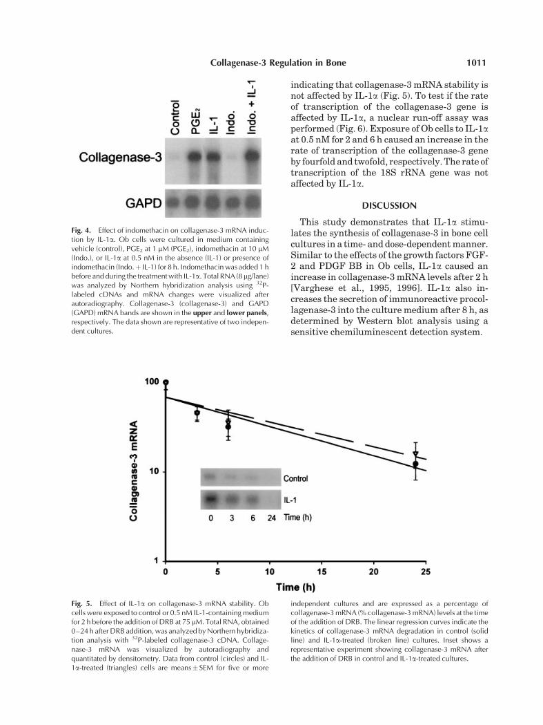
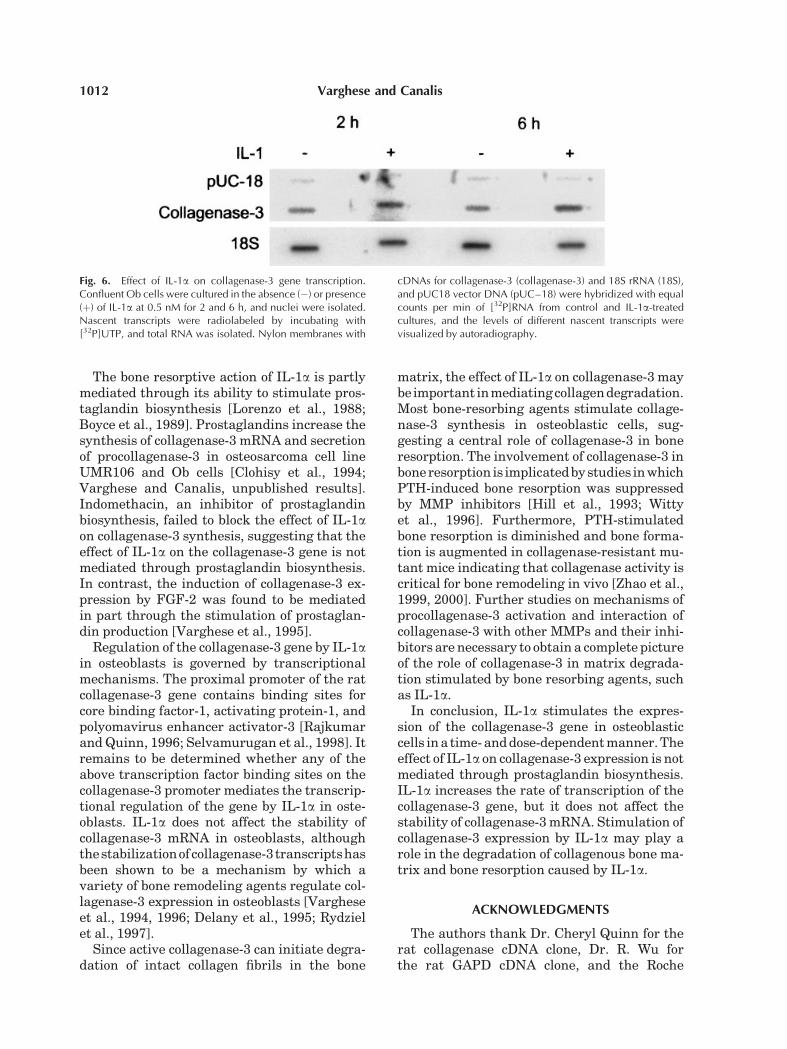
indicating that collagenase-3 mRNA stability isnot affected by IL-1a (Fig. 5). To test if the rateof transcription of the collagenase-3 gene isaffected by IL-1a, a nuclear run-off assay wasperformed (Fig. 6). Exposure of Ob cells to IL-1aat 0.5 nM for 2 and 6 h caused an increase in therate of transcription of the collagenase-3 geneby fourfold and twofold, respectively. The rate oftranscription of the 18S rRNA gene was notaffected by IL-1a.
DISCUSSION
This study demonstrates that IL-1a stimu-lates the synthesis of collagenase-3 in bone cellcultures in a time- and dose-dependentmanner.Similar to the effects of the growth factors FGF-2 and PDGF BB in Ob cells, IL-1a caused anincrease in collagenase-3 mRNA levels after 2 h[Varghese et al., 1995, 1996]. IL-1a also in-creases the secretion of immunoreactive procol-lagenase-3 into the culturemedium after 8 h, asdetermined by Western blot analysis using asensitive chemiluminescent detection system.
Fig. 4. Effect of indomethacin on collagenase-3 mRNA induc-tion by IL-1a. Ob cells were cultured in medium containingvehicle (control), PGE2 at 1 mM (PGE2), indomethacin at 10 mM(Indo.), or IL-1a at 0.5 nM in the absence (IL-1) or presence ofindomethacin (Indo.þ IL-1) for 8 h. Indomethacin was added 1 hbefore and during the treatment with IL-1a. Total RNA (8mg/lane)was analyzed by Northern hybridization analysis using 32P-labeled cDNAs and mRNA changes were visualized afterautoradiography. Collagenase-3 (collagenase-3) and GAPD(GAPD) mRNA bands are shown in the upper and lower panels,respectively. The data shown are representative of two indepen-dent cultures.
Fig. 5. Effect of IL-1a on collagenase-3 mRNA stability. Obcells were exposed to control or 0.5 nM IL-1-containing mediumfor 2 h before the addition of DRB at 75 mM. Total RNA, obtained0–24 h after DRB addition, was analyzed by Northern hybridiza-tion analysis with 32P-labeled collagenase-3 cDNA. Collage-nase-3 mRNA was visualized by autoradiography andquantitated by densitometry. Data from control (circles) and IL-1a-treated (triangles) cells are means� SEM for five or more
independent cultures and are expressed as a percentage ofcollagenase-3 mRNA (% collagenase-3 mRNA) levels at the timeof the addition of DRB. The linear regression curves indicate thekinetics of collagenase-3 mRNA degradation in control (solidline) and IL-1a-treated (broken line) cultures. Inset shows arepresentative experiment showing collagenase-3 mRNA afterthe addition of DRB in control and IL-1a-treated cultures.
Collagenase-3 Regulation in Bone 1011
The bone resorptive action of IL-1a is partlymediated through its ability to stimulate pros-taglandin biosynthesis [Lorenzo et al., 1988;Boyce et al., 1989]. Prostaglandins increase thesynthesis of collagenase-3 mRNA and secretionof procollagenase-3 in osteosarcoma cell lineUMR106 and Ob cells [Clohisy et al., 1994;Varghese and Canalis, unpublished results].Indomethacin, an inhibitor of prostaglandinbiosynthesis, failed to block the effect of IL-1aon collagenase-3 synthesis, suggesting that theeffect of IL-1a on the collagenase-3 gene is notmediated through prostaglandin biosynthesis.In contrast, the induction of collagenase-3 ex-pression by FGF-2 was found to be mediatedin part through the stimulation of prostaglan-din production [Varghese et al., 1995].
Regulation of the collagenase-3 gene by IL-1ain osteoblasts is governed by transcriptionalmechanisms. The proximal promoter of the ratcollagenase-3 gene contains binding sites forcore binding factor-1, activating protein-1, andpolyomavirus enhancer activator-3 [RajkumarandQuinn, 1996; Selvamurugan et al., 1998]. Itremains to be determined whether any of theabove transcription factor binding sites on thecollagenase-3 promoter mediates the transcrip-tional regulation of the gene by IL-1a in oste-oblasts. IL-1a does not affect the stability ofcollagenase-3 mRNA in osteoblasts, althoughthestabilizationofcollagenase-3 transcriptshasbeen shown to be a mechanism by which avariety of bone remodeling agents regulate col-lagenase-3 expression in osteoblasts [Vargheseet al., 1994, 1996; Delany et al., 1995; Rydzielet al., 1997].
Since active collagenase-3 can initiate degra-dation of intact collagen fibrils in the bone
matrix, the effect of IL-1a on collagenase-3 maybeimportant inmediatingcollagendegradation.Most bone-resorbing agents stimulate collage-nase-3 synthesis in osteoblastic cells, sug-gesting a central role of collagenase-3 in boneresorption. The involvement of collagenase-3 inboneresorption is implicatedbystudies inwhichPTH-induced bone resorption was suppressedby MMP inhibitors [Hill et al., 1993; Wittyet al., 1996]. Furthermore, PTH-stimulatedbone resorption is diminished and bone forma-tion is augmented in collagenase-resistant mu-tant mice indicating that collagenase activity iscritical for bone remodeling in vivo [Zhao et al.,1999, 2000]. Further studies on mechanisms ofprocollagenase-3 activation and interaction ofcollagenase-3 with other MMPs and their inhi-bitors are necessary to obtain a complete pictureof the role of collagenase-3 in matrix degrada-tion stimulated by bone resorbing agents, suchas IL-1a.
In conclusion, IL-1a stimulates the expres-sion of the collagenase-3 gene in osteoblasticcells ina time- anddose-dependentmanner.Theeffect of IL-1a on collagenase-3 expression is notmediated through prostaglandin biosynthesis.IL-1a increases the rate of transcription of thecollagenase-3 gene, but it does not affect thestability of collagenase-3mRNA. Stimulation ofcollagenase-3 expression by IL-1a may play arole in the degradation of collagenous bone ma-trix and bone resorption caused by IL-1a.
ACKNOWLEDGMENTS
The authors thank Dr. Cheryl Quinn for therat collagenase cDNA clone, Dr. R. Wu forthe rat GAPD cDNA clone, and the Roche
Fig. 6. Effect of IL-1a on collagenase-3 gene transcription.Confluent Ob cells were cultured in the absence (�) or presence(þ) of IL-1a at 0.5 nM for 2 and 6 h, and nuclei were isolated.Nascent transcripts were radiolabeled by incubating with[32P]UTP, and total RNA was isolated. Nylon membranes with
cDNAs for collagenase-3 (collagenase-3) and 18S rRNA (18S),and pUC18 vector DNA (pUC–18) were hybridized with equalcounts per min of [32P]RNA from control and IL-1a-treatedcultures, and the levels of different nascent transcripts werevisualized by autoradiography.
1012 Varghese and Canalis

Institute of Molecular Biology for the gift ofIL-1a. Authors are grateful to late Dr. JohnJeffrey for the rat collagenase antibody. Theauthors also thank Cathy Boucher, SusanO’Lone, Kristine Sasala, and Kyung Yu fortechnical assistance.
REFERENCES
Ausubel FM, Brent R, Kingsten RE, Moore DD, SeidmanJG, Smith JA, Struhl K. 1992. Current protocols in mole-cular biology. New York: Greene and Wiley Interscience.
Borden P, Solymar D, Sucharczuk A, Lindman B, CannonP, Heller RA. 1996. Cytokine control of interstitial col-lagenase and collagenase-3 gene expression in humanchondrocytes. J Biol Chem 38:23577–23581.
Boyce BF, Aufdemorte TB, Garrett IR, Yates AJ, MundyGR. 1989. Effects on interleukin-1 on bone turnover innormal mice. Endocrinology 125:1142–1150.
Canalis E, Rydziel S, Delany AM, Varghese S, Jeffrey JJ.1995. Insulin-like growth factors inhibit interstitial col-lagenase synthesis in bone cell cultures. Endocrinology136:1348–1354.
Chomczynski P, Sacchi N. 1987. Single-step method ofRNA isolation by acid–guanidium–thiocyanate–phe-nol–chloroform extraction. Anal Biochem 162:156–159.
Clohisy JC, Connolly TJ, Bergman KD, Quinn CO,Partridge NC. 1994. Prostanoid-induced expression ofmatrix metalloproteinase-1 messenger ribonucleic acidin rat osteosarcoma cells. Endocrinology 135:1447–1454.
Conca W, Kaplan PB, Krane SM. 1989. Increases in levelsof procollagenase messenger RNA in cultured fibroblastsinduced by human recombinant interleukin 1 b or serumfollow c-jun expression and are dependent on new proteinsynthesis. J Clin Invest 83:1753–1757.
Dayer J-M, de Rochemonteix B, Burrus B, Demczuk S,Dinarello CA. 1986. Human recombinant interleukin-1stimulates collagenase and prostaglandin E2 productionby synovial fibroblasts. J Clin Invest 77:645–648.
Delaisse J-M, Eeckhout Y, Vaes G. 1988. Bone-resorbingagents affect the production and distribution of procolla-genase as well as the activity of collagenase in bonetissue. Endocrinology 123:264–276.
Delany AM, Jeffrey JJ, Rydziel S, Canalis E. 1995. Cortisolincreases interstitial collagenase expression in osteo-blasts by post-transcriptional mechanisms. J Biol Chem270:26607–26612.
Dinarello CA. 1991. Interleukin-1 and interleukin-1 antag-onism. Blood 777:1627–1652.
Feinberg AP, Vogelstein B. 1983. A technique for radi-olabeling DNA restriction endonuclease fragments tohigh specific activity. Anal Biochem 132:6–13.
Freije JM, Diez-Itza I, Balbin M, Sanchez LM, Blasco R,Tolivia J, Lopez-Otin C. 1994. Molecular cloning and ex-pression of collagenase 3, a novel human matrix metal-loproteinase produced by breast carcinomas. J Biol Chem269:16766–16773.
Gowen M, Wood DD, Ihrie EJ, McGuire MKB, RussellRGG. 1983. An interleukin-1-like factor stimulates boneresorption in vitro. Nature 306:378–380.
Hanazawa S, Ohmori Y, Amano S, Miyoshi T, KumegawaM, Kitano S. 1985. Spontaneous production of inter-leukin-1-like cytokine from a mouse osteoblastic cell line
(MC3T3-E1). Biochem Biophys Res Commun 131:774–779.
Heath JK, Atkinson SJ, Meikle MC, Reynolds JJ. 1984.Mouse osteoblasts synthesize collagenase in response tobone resorbing agents. Biochem Biophys Acta 802:151–154.
Hill PA, Reynolds JJ, Meikle MC. 1993. Inhibition ofstimulated bone resorption in vitro by TIMP-1 and TIMP-2. Biochem Biophys Acta 1177:71–74.
Jeffrey JJ, Roswit WT, Ehlich LS. 1990. Regulation ofcollagenase production by steroids in uterine smoothmuscle cells: An enzymatic and immunologic study. J CellPhysiol 143:396–403.
Keeting PE, Rifas L, Harris SA, Colvard DS, Spelsberg TC,Peck WA, Riggs BL. 1987. Evidence for interleukin-1bproduction by cultured normal human osteoblast-likecells. J Bone Miner Res 6:827–833.
Kusano K, Miyaura C, Inada M, Tamura T, Ito A, NagaseH, Kamoi K, Suda T. 1988. Regulation of matrix metal-loproteinases (MMP-2, -3, -9, and -13) by interleukin-1and interleukin-6 in mouse calvaria: Association of MMPinduction with bone resorption. Endocrinology 139:1338–1345.
Lorenzo JA, Sousa SL, Alander C, Raisz LG, Dinarello CA.1987. Comparison of the bone-resorbing activity inthe supernatants from phytohemaglutinin-stimulatedhuman peripheral blood mononuclear cells with that ofcytokines through the use of an antiserum to interleukin1. Endocrinology 121:1164–1170.
Lorenzo JA, Sousa SL, Centrella M. 1988. Interleukin-1 incombination with transforming growth factor-alpha pro-duces enhanced bone resorption in vitro. Endocrinology123:2194–2200.
Lorenzo JA, Sousa SL, Van den Brink-Webb SE, Korn JH.1990. Production of both interleukin-1a and b by new-born mouse calvarial cultures. J Bone Miner Res 5:77–83.
McCarthy TL, Centrella M, Canalis E. 1988. Further bio-chemical and molecular characterization of primary ratparietal bone cell cultures. J Bone Miner Res 3:401–408.
Meikle MC, Bord S, Hembry RM, Compston J, Coucher PI,Reynolds JJ. 1992. Human osteoblast in culture synthe-size collagenase and other metalloproteinases in re-sponse to osteotropic hormones and cytokines. J CellSci 103:1093–1099.
Mitchel PG, Magna HA, Reeves LM, Lopresti-Morrow LL,Yocum SA, Rosener PJ, Geoghegan KF, Hambor JE.1996. Clonning, expression, and type II collagenolyticactivity of matrix metalloproteinase-13 from humanosteoarthritic cartilage. J Clin Invest 97:761–768.
Murphy G. 1995. Matrix metalloproteinase and their in-hibitors. Acta Orthop Scand Suppl 66:55–60.
Nguyen L, Dewhirst FE, Hauschka PV, Stashenko P. 1991.Interleukin-1b stimulates bone resorption and inhibitsbone formation in vivo. Lymphokine Cytokine Res 10:15–21.
Pacifici R. 1996. Estrogen, cytokines, and pathogenesisof postmenopausal osteoporosis. J Bone Miner Res 11:1043–1051.
Pacifici R, Rifas L, Teitelbaum S, Slatopolsky E,McCracken R, Bergfeld M, Lee W, Avioli LV, Peck WA.1987. Spontaneous release of interleukin 1 from humanblood monocytes reflects bone formation in idiopathicosteoporosis. Proc Natl Acad Sci USA 84:4616–4620.
Collagenase-3 Regulation in Bone 1013
Pacifici R, Rifas L, McCracken R, Vered I, McMurtry C,Avioli LV, Peck WA. 1989. Ovarian steroid treatmentblocks a postmenopausal increase in blood monocyteinterleukin 1 release. Proc Natl Acad Sci USA 86:2398–2402.
Partridge NC, Jeffrey JJ, Ehlich LS, Titelbaum SL, FliszarC, Welgus SG, Kahn AJ. 1987. Hormonal regulation ofthe production of collagenase and collagenase inhibitoractivity by rat osteogenic sarcoma cells. Endocrinology120:1956–1962.
Quinn CO, Scott DK, Brinckerhoff CE, Matrisian LM,Jeffrey JJ, Partridge NC. 1990. Rat collagenase: Cloning,amino acid sequence comparison, and parathyroid hor-mone regulation in osteoblastic cells. J Biol Chem 265:22342–22347.
Rajkumar RA, Quinn CO. 1996. Parathyroid hormoneinduction of rat interstitial collagenase mRNA in osteo-sarcoma cells is mediated through an AP-1 binding site.Mol Endocrinol 10:867–878.
Ralston SH. 1994. Analysis of gene expression in humanbone biopsies by polymerase chain reaction: Evidence forenhanced cytokine expression in postmenopausal osteo-porosis. J Bone Miner Res 9:883–890.
Rifas L, Fausto A, Scot MJ, Avioli LV, Welgus HG.1994. Expression of metalloproteinases and tissue inhi-bitors of metalloproteinases in human osteoblast-likecells: Differentiation is associated with repression ofmetalloproteinase biosynthesis. Endocrinology 134:213–221.
Rydziel S, Varghese S, Canalis E. 1997. Transforminggrowth factor b1 inhibits collagenase 3 expression bytranscriptional and post-transcriptional mechanisms inosteoblast cultures. J Cell Physiol 170:145–152.
Sakamoto M, Sakamoto S. 1984. Immunocytochemicallocalization of collagenase in isolated mouse bone cells.Biomed Res 5:29–38.
Sambrook J, Fritsch EF, Maniatis T. 1989. Molecularcloning—a laboratory manual. 2nd edition. New York:Cold Spring Harbor Laboratory Press.
Sato K, Fujii Y, Asano S, Ohtsaki T, Kawakami M, KasonoK, Tsushima T, Shizume K. 1986. Recombinant humaninterleukin-1 alpha and beta stimulate mouse osteoblast-like cells (MC3T3-E1) to produce macrophage-colonystimulatory activity and prostaglandin E2. Biochem Bio-phys Res Commun 141:285–291.
Selvamurugan N, Chou W, Pearman AT, Pulumati MR,Partridge NC. 1998. Parathyroid hormone regulates therat collagenase-3 promoter in osteoblastic cells throughthe co-operative interaction of the activator protein-1 siteand runt domain binding sequence. J Biol Chem 273:10647–10657.
Shen V, Kohler G, Jeffrey JJ, Peck WA. 1988. Bone-resorbing agents promote and interferon-g inhibits bonecell collagenase production. J BoneMiner Res 3:657–666.
Sokal RR, Rohlf FJ. 1981. Biometry. 2nd edition. SanFrancisco: WH Freeman and Co.
Stashenko P, Dewhirst FE, Rooney ML, Desjardins LA,Heeley JD. 1987. Interleukin-1b is a potent inhibitor ofbone formation in vitro. J Bone Miner Res 2:559–565.
Tatakis DN. 1993. Interleukin-1 and bone metabolism: Areview. J Periodontol 64:416–431.
Tso JY, Sun XH, Kao T-H, Reece KS, Wu R. 1985. Isolationand characterization of rat and human glyceraldehyde-3-phosphate dehydrogenase cDNAs: Genomic complexityand molecular evolution of the gene. Nucleic Acids Res13:2485–2502.
Varghese S, Canalis E. 1997. Regulation of collagenase-3 bybone morphogenetic protein-2 in bone cell cultures.Endocrinology 138:1035–1040.
Varghese S, Rydziel S, Jeffrey JJ, Canalis E. 1994. Regula-tion of interstitial collagenase expression and collagendegradation by retinoic acid in bone cells. Endocrinology134:2438–2444.
Varghese S, Ramsby ML, Jeffrey JJ, Canalis E. 1995. Basicfibroblast growth factor stimulates expression of inter-stitial collagenase and inhibitors of metalloproteinases inrat bone cells. Endocrinology 136:2156–2162.
Varghese S, Delany AM, Liang L, Gabbitas B, Jeffrey JJ,Canalis E. 1996. Transcriptional and posttranscriptionalregulation of interstitial collagenase by platelet-derivedgrowth factor BB in bone cell cultures. Endocrinology137:431–437.
Witty JP, Foster SA, Stricklin GP, Matrisian LM, SternPH. 1996. Parathyroid hormone-induced resorption infetal rat limb bones is associated with production ofthe metalloproteinases, collagenases, and gelatinase B.J Bone Miner Res 11:72–78.
Woessner JR. 1991. Matrix metalloproteinases and theirinhibitors in connective tissue remodeling. FASEB J5:2145–2154.
Zandomeni R, Burnick D, Ackerman S, Mittleman B,Weinmann R. 1983. Mechanism of action of DRB. Effecton specific in vitro initiation of transcription. J Mol Biol167:561–574.
Zhao W, Byrne MH, Boyce BF, Krane SM. 1999. Boneresorption induced by parathyroid hormone is strikinglydiminished in collagenase-resistant mutant mice. J ClinInvest 103:517–524.
Zhao W, Byrne MH, Wang Y, Krane SM. 2000. Osteocyteand osteoblast apoptosis and excessive bone depositionaccompany failure of collagenase cleavage of collagen.J Clin Invest 106:941–949.
1014 Varghese and Canalis