TRANSANAL ACCESS PLATFORM - PrimeWebcdn.primeweb.fi/ · TRANSANAL ACCESS PLATFORM PROCEDURAL GUIDE...
18
TRANSANAL ACCESS PLATFORM PROCEDURAL GUIDE FOR TRANSANAL MINIMALLY INVASIVE SURGERY (TAMIS) Featuring Tips & Tricks from Dr. Matthew Albert, Florida Hospital
Transcript of TRANSANAL ACCESS PLATFORM - PrimeWebcdn.primeweb.fi/ · TRANSANAL ACCESS PLATFORM PROCEDURAL GUIDE...

T R A N S A N A L ACC E S S P L AT F O R M
P R O C E D U R A L G U I D E F O R T R A N S A N A L M I N I M A L LY I N VA S I V E S U R G E R Y
( TA M I S )
F e a t u r i n g T i p s & Tr i c k s f r o m D r. M a t t h e w A l b e r t , F l o r i d a H o s p i t a l

TAMIS is designed to resect benign polyps and well selected malignancies in the distal and mid-rectum using new advanced access platforms combined with standard laparoscopic instrumentation.
In collaboration with Dr. Matthew Albert and other leading surgeons, Applied Medical is pleased to offer TAMIS workshops using GelPOINT path transanal access platform. Based on materials from these workshops, this quick reference guide was created to give you an overview of the surgical techniques using TAMIS. As this guide is just a summary of some key points from the workshops, Applied Medical recommends all surgeons be adequately trained at a TAMIS workshop before performing this technique.
Any content and views expressed therein are those of Dr. Albert and not of Applied Medical.
TA M I S O V E R V I E W
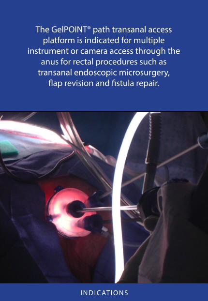
The GelPOINT® path transanal access platform is indicated for multiple
instrument or camera access through the anus for rectal procedures such as
transanal endoscopic microsurgery, flap revision and fistula repair.
I N D I C AT I O N S

Model Number Description Size QuantityCNO11 GelPOINT Path 4cm x 5.5cm 1/BOX
GelPOINT Path ComponentsGelSeal Cap 7.5cm diameter 1Access Channel with Introducer 4cm x 5.5cm 1Self-Retaining Sleeves 10mm 3Obturator 10mm 1
Suture tabs for added security
4x5.5cm access channel provides maximum working space and dilation
Self-retaining sleeves allow the exchange of 5-10mm instruments
Interchangeable insufflation/smoke evacuation ports
Removable cap provides simple specimen removal, versatility in sleeve placement and maintenance of pneumorectum using renowned GelSeal® technology
GelPOINT® Path TRANSANAL ACCESS PLATFORM
G E L P O I N T PAT H S P E C I F I C AT I O N S

LAPAROSCOPE
n 5 or 10 mm/30° or 45° angled laparoscope
n Angled light cord adaptor
n Angled/reticulating instruments
- Rat tooth graspers, scissors, needle driver
ENERGY DEVICES
n Suction irrigator with cautery tip
- Evacuates smoke in short burst during cauterization
or
n Monopolar cautery or other energy devices
- Needle tip accumulates less char on tip than spatula
I N S T R U M E N TAT I O N

SUTURE
n Absorbable suture n Ethicon 3-0 PDS™, VICRYL™ n Covidien V-Loc™ or Angiotech Quill™ suture
SUTURING DEVICES
n Up to 10mm suturing device
Extracorporeal
n Knotpusher
Intracorporeal
n Covidien Endo Stitch™ suturing device n LSI RD180™ Running Device
Suture clips
n Ethicon LAPRA-TY™ n LSI TK Ti-KNOT™ device n Richard Wolf TEM suture clips
S U T U R E / S U T U R I N G D E V I C E S

PREOPERATIVE PREPARATIONS
n Complete mechanical bowel preparation
n Preoperative antibiotics per Surgical Care Improvement Project (SCIP)
- Cefotan 2 g, Metronidazole 500 mg
n Perianal skin preparation and sterile draping per standard proctologic surgery protocol
ANESTHESIA
n General endotracheal anesthesia per standard OR protocols
- Patient must be FULLY PARALYZED to maintain rectal distention
S T E P S : P R E O P E R AT I V E P R E PA R AT I O N S

Unlike traditional TEM procedures, TAMIS procedures do not require positioning based on tumor location, however following are some recommendations on
patient positioning and their potential benefits.
TRADITIONAL LITHOTOMY POSITION
n Facilitates comfortable sitting position for the surgeon
n Most advantageous for anesthesiologist
n Legs should be abducted and flexed past 90 degrees at the hips to provide optimal exposure of the perianal region and create sufficient space for instrument manipulation
MODIFIED PRONE POSITION
n May be helpful for anterior lesions
n Degree of upper-body downward tilt depends on patient’s body habitus and circulatory status
n Legs should be abducted and flexed at the hips
RIGHT OR LEFT LATERAL DECUBITUS POSITION
n May be advantageous for obese patients to facilitate pneumorectum
n Legs should be abducted and flexed at the hips, upper leg is secured to contoured rest on anterior side of table while lower leg is placed on leg rest of table and angled forward beneath the hip
S T E P S : PAT I E N T P O S I T I O N I N G

1. Apply generous lubrication to access channel and introducer. Pre-dilate the anus using standard transanal surgery techniques.
2. Manually (A) or using forceps (B), compress access channel in a folded form and place into anus until flange is securely seated behind levator sling (C).
3. Introducer may be inserted to aid in placing the access channel into position (D).
4. Hold access channel in place while suturing through suture tabs to secure (E).
5. Access channel is now fully placed (F).
S T E P S : I N S E R T I N G T H E ACC E S S C H A N N E L
A B
D
E F
C

CAUTION:
TO AVOID POSSIBLE INJURY TO RECTAL WALL,INSERT TROCARS INTO GELSEAL CAP PRIOR TO
PLACING GELSEAL CAP ONTO ACCESS CHANNEL.
1. Using the 10mm obturator, place the (3) 10mm sleeves, in a triangular fashion (A), see red X for position, through the gel in the GelSeal cap. Ensure sleeves are at least 1cm from the cap’s plastic perimeter and the insufflation and smoke evacuation ports (B).
2. Apply downward force to the sleeve until the sleeve tip and flange have passed through the GelSeal cap (C). Remove obturator.
3. Attach the GelSeal cap to the access channel by sliding the blue tab located on the bottom of the GelSeal cap under the access channel’s upper ring (D).
4. Push the upper ring against the inner portion of the GelSeal cap. Secure the opposite side of the access channel by closing the lever and locking the GelSeal cap in place (E).
5. Cap is now fully attached and ready for use (F).
S T E P S : I N S E R T I N G T R O C A R S & AT TAC H I N G C A P
B C
D E F
A

NOTE:
ATTACH INSUFFLATION TUBING ON EITHER STOPCOCK PORT. REMAINING PORT IS DESIGNATED FOR SMOKE EVACUATION.
1. Attach insufflation tubing to either one of the stopcock ports located on the GelSeal cap (A).
2. With high flow setting, start pressure at 8mm mercury and increase to 15-20 as needed for desired rectal distention.
3. If rectum pulsates or collapses:
n First check with anesthesiologist to ensure patient is fully paralyzed!
n If not resolved, check smoke evacuation port is in closed position
n Check seal on trocar ports
4. To evacuate smoke during the procedure, move the stopcock valve on the designated smoke evacuation port to the open position. After smoke evacuation, move the stopcock valve to the closed position (B).
INTERCHANGEABLE INSUFFLATION/SMOKE EVACUATION PORTS
S T E P S : I N S U F F L AT I O N / S M O K E E VAC UAT I O N
BA

n Posterior lesion
- Camera at top of triangle (A)
n Anterior lesion
- Camera at bottom of triangle (B)
n Angle the scope to be out of your way!
n Bariatric length scopes and port changes may be helpful
S T E P S : C A M E R A D R I V I N G
A
B
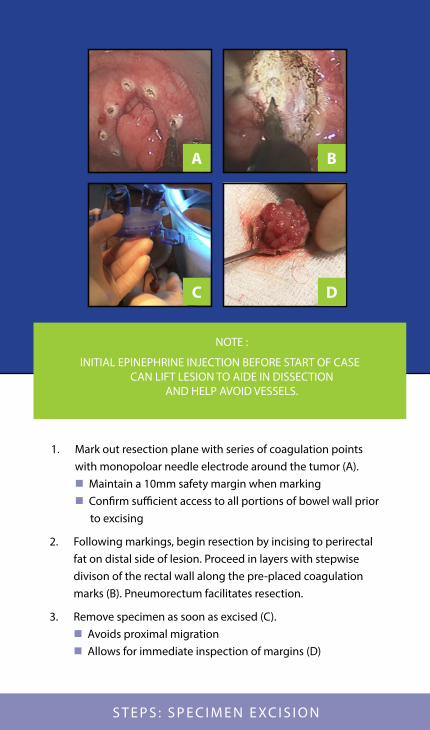
1. Mark out resection plane with series of coagulation points with monopoloar needle electrode around the tumor (A). n Maintain a 10mm safety margin when marking n Confirm sufficient access to all portions of bowel wall prior to excising
2. Following markings, begin resection by incising to perirectal fat on distal side of lesion. Proceed in layers with stepwise divison of the rectal wall along the pre-placed coagulation marks (B). Pneumorectum facilitates resection.
3. Remove specimen as soon as excised (C). n Avoids proximal migration n Allows for immediate inspection of margins (D)
NOTE :
INITIAL EPINEPHRINE INJECTION BEFORE START OF CASE CAN LIFT LESION TO AIDE IN DISSECTION
AND HELP AVOID VESSELS.
S T E P S : S P E C I M E N E XC I S I O N
A B
C D

n Lowering insufflation pressure will help reapproximate a larger defect
n Monofilament absorbable suture
- 3-0 PDS, VICRYL
- V-Loc or Quill barbed suture may facilitate suture retention
- Knots can be used, however, LAPRA-TY or similar suture clips on ends may enhance progression
n Rectal wall defect may be closed with standard suturing technique, using preferred suturing device up to 10mm, running continuous
n Large rectal wall defects may be closed beginning from the center to transform large round defect into two smaller, more manageable gaps (A)
n Fully closed defect (B)
NOTES :
THE GOAL IS TO FULLY REAPPROXIMATE THE BOWEL WALL WITHOUT NARROWING THE LUMEN. IF DEFECT IS VERY DISTAL MAY
REMOVE GELPOINT PATH AND CLOSE TRANSANALLY.
ENTRY INTO PERITONEAL CAVITY Resection of anterior lesion located approximately 9cm or greater from
anal verge may result in inadvertent entry into peritoneal cavity during full thickness excision, requiring
suture closure of the peritoneum and rectal wall.
S T E P S : S U T U R I N G / D E F E C T C LO S U R E
BA

n Don’t panic!
n Maintain visualization (use short bursts of suction with minimal irrigation)
n Attempt compression with grasper prior to blind electrocautery
n Utilization of a bipolar energy device may quickly resolve the problem (Covidien LigaSure™, Ethicon ENSEAL™, Ethicon HARMONIC™)
Tip: Initial epinephrine injection at start of case can lift lesion to aid in dissection and minimize major vessel bleeding.
NOTES :
OCCASIONAL VESSELS MAY BE ENCOUNTERED IN THE DISSECTION THROUGH THE RECTAL WALL AND
MESORECTUM CAUSING SUDDEN BLEEDING.
WHEN BLEEDING DOES OCCUR REMEMBER THE FOLLOWING:
S T E P S : H E M O S TA S I S

EARLY POSTOPERATIVE CARE
n Resumption of regular diet per standard protocol
S T E P S : P O S TO P E R AT I V E C A R E
EARLY POSTOPERATIVE CARE
n Resumption of regular diet per standard protocol
n Discharge may be within 24 hours
n Contrast enema on postoperative day ONE if intraperitoneal entry was made during procedure
ONCOLOGIC FOLLOW-UP
n Follow standard recommendations of the professional societies and NCCN guidelines
n If postoperative histology shows positive margins, poor pathologic features or more advanced tumor stage, standard oncologic resection or alternative treatment options should be considered
GelPOINT Path Transanal Access Platform
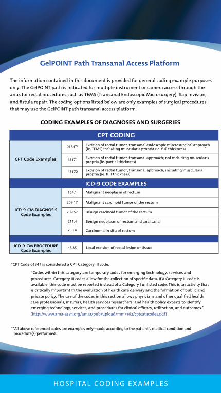
CODING EXAMPLES OF DIAGNOSES AND SURGERIES
-
*CPT Code 0184T is considered a CPT Category III code.
(http://www.ama-assn.org/ama1/pub/upload/mm/362/cptcat3codes.pdf)
**All above referenced codes are examples only – code according to the patient’s medical condition and procedure(s) performed.
CPT CODING
ICD-9 CODE EXAMPLES
CPT Code Examples
0184T* Excision of rectal tumor, transanal endoscopic mircrosurgical approach (ie. TEMS) including muscularis propria (ie. full thickness)
ICD-9-CM DIAGNOSIS Code Examples
ICD-9-CM PROCEDURECode Examples
154.1
209.17
209.57
211.4
230.4
Malignant neoplasm of rectum
Malignant carcinoid tumor of the rectum
Benign carcinoid tumor of the rectum
Benign neoplasm of rectum and anal canal
Carcinoma in situ of rectum
48.35 Local excision of rectal lesion or tissue
45171 Excision of rectal tumor, transanal approach; not including muscularis propria (ie. partial thickness)
45172 Excision of rectal tumor, transanal approach; including muscularis propria (ie. full thickness)
“Codes within this category are temporary codes for emerging technology, services and
procedures. Category III codes allow for the collection of speci�c data. If a Category III code is
available, this code must be reported instead of a Category I unlisted code. This is an activity that
is critically important in the evaluation of health care delivery and the formation of public and
private policy. The use of the codes in this section allows physicians and other quali�ed health
care professionals, insurers, health services researchers, and health policy experts to identify
emerging technology, services, and procedures for clinical e�cacy, utilization, and outcomes.”
The information contained in this document is provided for general coding example purposes
only. The GelPOINT path is indicated for multiple instrument or camera access through the
anus for rectal procedures such as TEMS (Transanal Endoscopic Microsurgery), �ap revision,
and �stula repair. The coding options listed below are only examples of surgical procedures
that may use the GelPOINT path transanal access platform.
H O S P I TA L CO D I N G E X A M P L E S

©2014 Applied Medical Resources Corporation. All rights reserved. Applied Medical, the Applied Medical logo design and marks designated with a ® are trademarks of
Applied Medical Resources Corporation, registered in one or more of the following countries: Australia, Canada, Japan, the United States and/or the European Union. SC01536F
V-Loc, Endo Stitch, and LigaSure are trademarks of Covidien. Quill is a trademark of Angiotech.PDS, VICRYL, LAPRA-TY, ENSEAL, and HARMONIC are trademarks of Ethicon.
RD180 and Ti-Knot are trademarks of LSI Solutions.
For more information www.appliedmedical.com/gelpoint
http://contact.appliedmedical.com/

![Transanal endoscopic microsurgery for radical resection of ...Transanal endoscopic microsurgery in sigmoid cancer 1451 JBUON 2019; 24(4): 1451 large rectal polyps by TEM [11]. However,](https://static.fdocuments.us/doc/165x107/60f7e35c60455642d5494ef7/transanal-endoscopic-microsurgery-for-radical-resection-of-transanal-endoscopic.jpg)