TRAIL-coated leukocytes that kill cancer cells in the circulation · TRAIL-coated leukocytes that...
6
TRAIL-coated leukocytes that kill cancer cells in the circulation Michael J. Mitchell, Elizabeth Wayne, Kuldeepsinh Rana, Chris B. Schaffer, and Michael R. King 1 Department of Biomedical Engineering, Cornell University, Ithaca, NY 14853 Edited by Robert Langer, Massachusetts Institute of Technology, Cambridge, MA, and approved December 13, 2013 (received for review August 29, 2013) Metastasis through the bloodstream contributes to poor prognosis in many types of cancer. Mounting evidence implicates selectin- based adhesive interactions between cancer cells and the blood vessel wall as facilitating this process, in a manner similar to leukocyte trafficking during inflammation. Here, we describe a unique approach to target and kill colon and prostate cancer cells in the blood that causes circulating leukocytes to present the cancer-specific TNF-related apoptosis inducing ligand (TRAIL) on their surface along with E-selectin adhesion receptor. This approach, demonstrated in vitro with human blood and also in mice, mimics the cytotoxic activity of natural killer cells and increases the sur- face area available for delivery of the receptor-mediated signal. The resulting “unnatural killer cells” hold promise as an effective means to neutralize circulating tumor cells that enter blood with the potential to form new metastases. drug delivery | nanomedicine O ver 90% of cancer-related deaths are due to cancer me- tastasis, the spread of cancer cells from a primary tumor to anatomically distant organs (1). In many types of cancer, cancer cells from the primary tumor can intravasate into the peripheral circulation as circulating tumor cells (CTCs) (2, 3). These CTCs can then interact with the receptor-bearing endothelial cell wall under flow in other organs, in a manner similar to leukocyte extravasation during inflammation and lymphocyte homing to lymphatic tissues (4). Recent studies have shown that CTCs from many types of primary tumors express sialylated carbohydrate ligands similar to leukocytes, which mediate interactions with selectins on the endothelium (5, 6). Selectins possess rapid, force-dependent binding kinetics, which can trigger the rolling adhesion of CTCs along the blood vessel wall (7, 8). CTCs can subsequently transition from rolling to firm adhesion, allowing for transendothelial migration into tissues and eventual forma- tion of micrometastases (9). Surgery and radiation have proven effective at treating primary tumors that do not invade the basement membrane; however, the difficulty of detecting distant micrometastases has made the majority of metastatic cancer treatments unsuccessful. The development of technologies to directly target CTCs in vivo holds promise in reducing both the metastatic load and the formation of new tumors. Although studies have reported that as many as 1 × 10 6 cancer cells detach from the primary tumor (per gram) of patients per day (10, 11), CTCs are difficult to detect due to their sparse concentrations in the bloodstream, as low as 1–100 cells per mL (3, 12). Additionally, there are ∼1 × 10 6 leukocytes or 1 × 10 9 erythrocytes per single CTC in blood (13). Despite the difference in numbers, both leukocytes and CTCs share similar characteristics in terms of their migration within the bloodstream. Highly deformable erythrocytes experience a drift velocity away from the vessel wall and collect in the center region, displacing less deformable leukocytes and CTCs to the near-wall region in a mechanism termed margination (14). Such margination phenomena can effectively surround CTCs within the circulating leukocyte population, thus making leukocytes a potentially attractive carrier of treatments to CTCs by exploiting their numerous adhesion receptors. The utilization of leukocytes to treat CTCs directly within the bloodstream has not previously been explored. Here, we describe a therapeutic approach to target and kill circulating cancer cells in the bloodstream by functionalizing leukocytes with the apopto- sis-inducing ligand TRAIL, and the adhesion receptor E-selectin directly within blood under shear flow. The functionalization of leukocytes under flow, effectively creating a form of “unnatural killer cells” within the bloodstream, is shown to be highly ef- fective at treating circulating cancer cells in flowing human blood in vitro, and in the peripheral circulation of mice in vivo. Results E-Selectin /TRAIL Liposomes Adhesively Interact and Induce Apoptotic Cancer-Cell Death Under Shear Flow. Many types of circulating tu- mor cells (CTCs) and cancer cell lines derived from colon, breast, prostate, and pancreas are known to display glycosylated ligands that allow them to adhesively interact with E-selectin (ES) under physiological shear flow (15). This interaction has been proposed to explain why some cancers home to tissue- specific capillary beds such as the bone marrow and liver (16, 17). In an attempt to target and kill cancer cells of this form, nanoscale liposomes conjugated with a mixture of recombinant human ES protein and tumor necrosis factor (TNF)-related apoptosis-inducing ligand (TRAIL) were developed (Fig. 1A, Fig. S1, and Table S1). TRAIL binds to death receptors 4 and 5 on the surface of cancer cells to induce apoptosis through the intrinsic and extrinsic pathways (18, 19). ES/TRAIL liposomes consisting of a 10% weight ratio of (1,2-dioleoyl-sn-glycero-3-{[N-(5-amino- 1-carboxy- pentyl) iminodiacetic acid] succinyl}(nickel salt) (DOGS-Ni-NTA), used to conjugate ES and TRAIL to the liposome surface, were found to be most effective at inducing apoptotic cell death in a colorectal adenocarcinoma (COLO 205) cell line under static conditions (Fig. S2), as determined using an Annexin-V apoptosis assay. Significance This paper describes a unique approach to target and kill cancer cells in the bloodstream, in which the extensive surface area of circulating leukocytes is used to display the cancer-specific TNF-related apoptosis inducing ligand (TRAIL) and E-selectin adhesion receptor to the surrounding fluid. The approach is inspired by the cytotoxic activity of natural killer cells and is quite effective at killing cancer cells both in vitro with human blood samples and in mouse blood circulation. The mecha- nism is surprising and unexpected in that this repurposing of leukocytes in flowing blood is more effective than directly targeting the cancer cells with liposomes or soluble protein. Author contributions: M.R.K. conceived of research; M.J.M., E.W., K.R., C.B.S., and M.R.K. designed research; M.J.M., E.W., and K.R. performed research; C.B.S. contributed new reagents/analytic tools; M.J.M., E.W., and K.R. analyzed data; M.J.M., E.W., C.B.S., and M.R.K. wrote the paper. The authors declare no conflict of interest. This article is a PNAS Direct Submission. Freely available online through the PNAS open access option. 1 To whom correspondence should be addressed. E-mail: [email protected]. This article contains supporting information online at www.pnas.org/lookup/suppl/doi:10. 1073/pnas.1316312111/-/DCSupplemental. www.pnas.org/cgi/doi/10.1073/pnas.1316312111 PNAS Early Edition | 1 of 6 ENGINEERING APPLIED BIOLOGICAL SCIENCES
Transcript of TRAIL-coated leukocytes that kill cancer cells in the circulation · TRAIL-coated leukocytes that...

TRAIL-coated leukocytes that kill cancer cells inthe circulationMichael J. Mitchell, Elizabeth Wayne, Kuldeepsinh Rana, Chris B. Schaffer, and Michael R. King1
Department of Biomedical Engineering, Cornell University, Ithaca, NY 14853
Edited by Robert Langer, Massachusetts Institute of Technology, Cambridge, MA, and approved December 13, 2013 (received for review August 29, 2013)
Metastasis through the bloodstream contributes to poor prognosisin many types of cancer. Mounting evidence implicates selectin-based adhesive interactions between cancer cells and the bloodvessel wall as facilitating this process, in a manner similar toleukocyte trafficking during inflammation. Here, we describe aunique approach to target and kill colon and prostate cancer cellsin the blood that causes circulating leukocytes to present thecancer-specific TNF-related apoptosis inducing ligand (TRAIL) ontheir surface along with E-selectin adhesion receptor. This approach,demonstrated in vitro with human blood and also in mice, mimicsthe cytotoxic activity of natural killer cells and increases the sur-face area available for delivery of the receptor-mediated signal.The resulting “unnatural killer cells” hold promise as an effectivemeans to neutralize circulating tumor cells that enter blood withthe potential to form new metastases.
drug delivery | nanomedicine
Over 90% of cancer-related deaths are due to cancer me-tastasis, the spread of cancer cells from a primary tumor to
anatomically distant organs (1). In many types of cancer, cancercells from the primary tumor can intravasate into the peripheralcirculation as circulating tumor cells (CTCs) (2, 3). These CTCscan then interact with the receptor-bearing endothelial cell wallunder flow in other organs, in a manner similar to leukocyteextravasation during inflammation and lymphocyte homing tolymphatic tissues (4). Recent studies have shown that CTCs frommany types of primary tumors express sialylated carbohydrateligands similar to leukocytes, which mediate interactions withselectins on the endothelium (5, 6). Selectins possess rapid,force-dependent binding kinetics, which can trigger the rollingadhesion of CTCs along the blood vessel wall (7, 8). CTCs cansubsequently transition from rolling to firm adhesion, allowingfor transendothelial migration into tissues and eventual forma-tion of micrometastases (9). Surgery and radiation have proveneffective at treating primary tumors that do not invade thebasement membrane; however, the difficulty of detecting distantmicrometastases has made the majority of metastatic cancertreatments unsuccessful.The development of technologies to directly target CTCs in
vivo holds promise in reducing both the metastatic load and theformation of new tumors. Although studies have reported that asmany as 1 × 10⁶ cancer cells detach from the primary tumor (pergram) of patients per day (10, 11), CTCs are difficult to detectdue to their sparse concentrations in the bloodstream, as low as1–100 cells per mL (3, 12). Additionally, there are ∼1 × 10⁶
leukocytes or 1 × 10⁹ erythrocytes per single CTC in blood (13).Despite the difference in numbers, both leukocytes and CTCsshare similar characteristics in terms of their migration withinthe bloodstream. Highly deformable erythrocytes experiencea drift velocity away from the vessel wall and collect in the centerregion, displacing less deformable leukocytes and CTCs to thenear-wall region in a mechanism termed margination (14). Suchmargination phenomena can effectively surround CTCs withinthe circulating leukocyte population, thus making leukocytesa potentially attractive carrier of treatments to CTCs by exploitingtheir numerous adhesion receptors.
The utilization of leukocytes to treat CTCs directly within thebloodstream has not previously been explored. Here, we describea therapeutic approach to target and kill circulating cancer cellsin the bloodstream by functionalizing leukocytes with the apopto-sis-inducing ligand TRAIL, and the adhesion receptor E-selectindirectly within blood under shear flow. The functionalization ofleukocytes under flow, effectively creating a form of “unnaturalkiller cells” within the bloodstream, is shown to be highly ef-fective at treating circulating cancer cells in flowing human bloodin vitro, and in the peripheral circulation of mice in vivo.
ResultsE-Selectin /TRAIL Liposomes Adhesively Interact and Induce ApoptoticCancer-Cell Death Under Shear Flow. Many types of circulating tu-mor cells (CTCs) and cancer cell lines derived from colon,breast, prostate, and pancreas are known to display glycosylatedligands that allow them to adhesively interact with E-selectin(ES) under physiological shear flow (15). This interaction hasbeen proposed to explain why some cancers home to tissue-specific capillary beds such as the bone marrow and liver (16, 17).In an attempt to target and kill cancer cells of this form, nanoscaleliposomes conjugated with a mixture of recombinant human ESprotein and tumor necrosis factor (TNF)-related apoptosis-inducingligand (TRAIL) were developed (Fig. 1A, Fig. S1, and Table S1).TRAIL binds to death receptors 4 and 5 on the surface of cancercells to induce apoptosis through the intrinsic and extrinsicpathways (18, 19). ES/TRAIL liposomes consisting of a 10%weight ratio of (1,2-dioleoyl-sn-glycero-3-{[N-(5-amino- 1-carboxy-pentyl) iminodiacetic acid] succinyl}(nickel salt) (DOGS-Ni-NTA),used to conjugate ES and TRAIL to the liposome surface,were found to be most effective at inducing apoptotic celldeath in a colorectal adenocarcinoma (COLO 205) cell line understatic conditions (Fig. S2), as determined using an Annexin-Vapoptosis assay.
Significance
This paper describes a unique approach to target and kill cancercells in the bloodstream, in which the extensive surface area ofcirculating leukocytes is used to display the cancer-specificTNF-related apoptosis inducing ligand (TRAIL) and E-selectinadhesion receptor to the surrounding fluid. The approach isinspired by the cytotoxic activity of natural killer cells and isquite effective at killing cancer cells both in vitro with humanblood samples and in mouse blood circulation. The mecha-nism is surprising and unexpected in that this repurposing ofleukocytes in flowing blood is more effective than directlytargeting the cancer cells with liposomes or soluble protein.
Author contributions: M.R.K. conceived of research; M.J.M., E.W., K.R., C.B.S., and M.R.K.designed research; M.J.M., E.W., and K.R. performed research; C.B.S. contributed newreagents/analytic tools; M.J.M., E.W., and K.R. analyzed data; M.J.M., E.W., C.B.S., andM.R.K. wrote the paper.
The authors declare no conflict of interest.
This article is a PNAS Direct Submission.
Freely available online through the PNAS open access option.1To whom correspondence should be addressed. E-mail: [email protected].
This article contains supporting information online at www.pnas.org/lookup/suppl/doi:10.1073/pnas.1316312111/-/DCSupplemental.
www.pnas.org/cgi/doi/10.1073/pnas.1316312111 PNAS Early Edition | 1 of 6
ENGINEE
RING
APP
LIED
BIOLO
GICAL
SCIENCE
S

In the postcapillary venules where selectin-mediated adhesionand cell extravasation into tissues typically occur, moderate shearrates can initiate flowing cell interactions with the endothelialcell wall (20, 21). To recreate these physical forces in vitro,a cone-and-plate shear assay was developed to probe the inter-actions of cancer cells and ES/TRAIL liposomes under venularshear rates. After exposure to shear flow (shear rate: 188 s−1) for2 h, COLO 205 cells exposed to ES liposomes displayed theirnormal morphology whereas substantial membrane blebbing wasobserved in samples exposed to ES/TRAIL liposomes, character-istic of cells undergoing apoptosis (Fig. 1B). Annexin-V assayrevealed that exposure to shear flow for 2 h induced minimalCOLO 205 cell apoptosis in untreated controls (Fig. 1C), in ad-dition to treatment with liposomes in the absence of conjugatedprotein (Fig. 1D) or conjugated solely with ES (Fig. 1E) or TRAIL(Fig. 1F). However, a combination of ES/TRAIL conjugated to theliposome surface induced a significant decrease in COLO 205 vi-ability following exposure to shear flow (Fig. 1 G and H).
To investigate the adhesive characteristics of ES/TRAILliposomes to cancer cells under flow, COLO 205 cells were ex-posed to ES/TRAIL liposomes consisting of fluorescent choles-terol and exposed to shear flow as in previous in vitro shear assays.Flow cytometry revealed that >99.9% of the COLO 205 cellpopulation was adhered to ES/TRAIL liposomes after exposure toshear flow (Fig. S3 A and B). Fluorescent micrographs andbrightfield overlay images clearly displayed ES/TRAIL lip-osomes adhered to the surface of COLO 205 cells (Fig. S3 C andD). These data suggest that the presence of the ES adhesionreceptor enhances the effect of TRAIL by promoting tightercontacts with the cancer-cell membrane.
ES/TRAIL Liposomes Functionalize Leukocytes in Whole Blood UnderShear Flow in Vitro. In addition to CTCs, circulating leukocytesalso possess ligands for ES, which are necessary in the in-flammatory response and lymphocyte homing to lymphatic tis-sues (1, 22). To assess the potential to functionalize leukocyteswith ES/TRAIL to target and kill CTCs, we treated human bloodwith fluorescent ES/TRAIL liposomes under shear flow in a cone-and-plate viscometer. Upon exposure to shear (shear rate: 188 s−1),ES/TRAIL liposomes readily bind to leukocytes via selectinligands on the leukocyte surface (Fig. 2A).To quantify leukocyte subpopulations that adhere to ES/TRAIL
liposomes under flow, leukocytes were separated from wholeblood and analyzed for both leukocyte marker expression andadherent ES/TRAIL liposomes using flow cytometry. Function-alized leukocytes were labeled with CD3, CD14, CD16, CD19,and CD56 antibodies, as such markers are commonly expressedon most T lymphocytes, monocytes, neutrophils, B-lymphocytes,and natural killer (NK) cells, respectively (23). Minimal adhesionof ES/TRAIL liposomes to leukocytes in blood was observed inthe presence of a functional blocking ES antibody (Fig. 2 B–G).Minimal ES/TRAIL liposome adhesion was also observed im-mediately after treatment with whole blood. However, afterexposure to shear flow, flow cytometry analysis revealed thatleukocyte subpopulations positive for CD3 (Fig. 2B), CD14 (Fig.2C), CD16 (Fig. 2D), CD19 (Fig. 2E), and CD56 (Fig. 2F) ad-hered to ES/TRAIL liposomes to varying degrees (Fig. 2G). Ad-hesion was also observed on populations of lymphocytes, whichsuggests that some cytotoxic patrolling of the lymphatic system mayalso occur in vivo. Leukocyte subpopulations can vary in their E-selectin ligand expression (24) and thus could explain the varia-tions in the number of bound ES/TRAIL liposomes.
ES/TRAIL Functionalization Does Not Induce Significant Leukocyte orEndothelial Cell Death. To assess the effects of ES/TRAIL func-tionalization on leukocyte viability, mononuclear leukocytes iso-lated from human blood were treated with ES/TRAIL liposomesunder both static and shear-flow conditions. Annexin-V assaysrevealed no significant differences in leukocyte viability whenleukocytes were incubated with ES (Fig. S4B) or ES/TRAIL(Fig. S4C) liposomes under static conditions for 24 h, comparedwith untreated leukocyte controls (Fig. S4 A and D). Uponfunctionalization with liposomes under shear flow for 2 h (shearrate, 188 s−1), no significant decreases were found in ES (Fig.S4F) or ES/TRAIL (Fig. S4G) functionalized leukocytes, com-pared with untreated leukocyte controls (Fig. S4 E andH). Thesedata suggest that ES/TRAIL liposomes can functionalize leu-kocytes under flow to target and kill cancer cells, while exertingnegligible cytotoxic effects on leukocytes. It is important to notethat individual subpopulations of leukocytes have shown apo-ptotic effects in the presence of TRAIL (25); however, this studyfocused on the overall effects of ES/TRAIL liposomes on blood-borne leukocytes.To assess the effects of ES/TRAIL functionalization on en-
dothelial cell viability, human umbilical vein endothelial cells(HUVECs) were treated with ES/TRAIL liposomes in humanblood under shear-flow conditions in vitro. Treatment with ES/TRAIL liposomes or an equivalent concentration of solubleTRAIL in human blood under shear flow for 4 h induced no
10.31%82.53%
5.57%1.59%
9.39%81.73%
6.49%2.39%
1 2 3 4 5 6 7
101 102 103 104 105 106 107101102103104105106107
101 102 103 104 105 106 107101102103104105106107
101 102 103 104 105 106 107101102103104105106107
101 102 103 104 105 106 107101102103104105106107
101 102 103 104 105 106 107101102103104105106107
Untreated Naked TRAIL
ES ES/TRAIL
Annexin-V
Pro
pidi
um Io
dide
C D
F G
E
H
BA
***
Unt
reat
ed
Nak
ed
TRA
IL ES
ES
/TR
AIL
100
80
60
40
20
0% V
iabl
e C
OLO
205
ES
ES/TRAIL
Hydration
ExtrusionES/TRAILLiposome
ES TRAIL
ES
TRAIL
Thin Lipid Film
MultilamellarLiposome
UnilamellarLiposome
LiposomeLiposome
13.22% 36.34%
36.13% 14.31%
2.11% 6.70%
81.17% 10.02%
1.98% 7.31%
77.25% 13.46%
Fig. 1. ES/TRAIL liposomes adhesively interact with and kill cancer cellsunder uniform shear flow. (A) Synthesis of ES, TRAIL, and ES/TRAIL uni-lamellar liposomes using a thin film hydration method. Briefly, lipids inchloroform were dried overnight to form a thin lipid film. Lipids were thenhydrated and subjected to freeze–thaw cycles to formmultilamellar liposomes,which were extruded through membranes to form unilamellar liposomes. ES,TRAIL, or a combination of ES and TRAIL was then conjugated to Ni-NTA on theliposome surface. To assess the ability of ES/TRAIL liposomes to target and killcancer cells under flow, ES/TRAIL liposomes were added to a suspension ofCOLO 205 cancer cells and exposed to shear flow in a cone-and-plate viscometerat a shear rate of 188 s−1 for 2 h. Cells were then removed, washed, placed intoculture for 24 h, and assessed for cell viability. (B) COLO 205 morphology aftertreatment with ES (Upper) and ES/TRAIL (Lower) liposomes under shear flow.(Scale bar, 20 μm.) (C–G) Representative propidium iodide/Annexin-V flowcytometry plots of unsheared cancer cells (C) and cells sheared with naked (D),TRAIL-bound (E), ES-bound (F), and ES/TRAIL-bound liposomes (G) under shearflow. Cells were classified into four categories based on dye uptake: viable cells[negative for Annexin-V and propidium iodide (PI)], early apoptotic cells (posi-tive for Annexin-V only), late apoptotic cells (positive for Annexin-V and PI), andnecrotic cells (positive for PI only). (H) Percent of viable cells after treatmentfor each group. n = 3 for all samples. Bars represent the mean ± SD in eachtreatment group. ***P < 0.0001 (one-way ANOVA with Tukey posttest).
2 of 6 | www.pnas.org/cgi/doi/10.1073/pnas.1316312111 Mitchell et al.

significant differences in HUVEC viability, compared with un-treated HUVECs exposed to shear in human blood (Fig. S5).These data suggest that ES/TRAIL-functionalized leukocytesexert negligible toxic effects on human endothelial cells underblood-flow conditions.
Apoptotic Effects of ES/TRAIL Therapy Are Enhanced in Human BloodUnder Flow. Clinically, CTCs are sparsely distributed in thecomplex milieu of whole blood, at concentrations as low as 1–100cells per mL (3, 12). To examine whether ES/TRAIL liposomeswould effectively target cancer cells in the presence of blood cellsand serum under flow conditions, we fluorescently labeled co-lorectal COLO 205 and prostate PC-3 cancer-cell lines andspiked them into human peripheral blood. Surprisingly, underidentical shear-flow conditions, ES/TRAIL therapy was evenmore effective at killing cancer cells in the presence of humanblood (Fig. 3 A–D), compared with shearing COLO 205 or PC-3cells alone in buffer (Fig. 3E), with <5% of the fluorescent, viable
cancer-cell populations remaining after ES/TRAIL treatment (Fig.3E). These results suggest that ES/TRAIL therapy is effective attargeting circulating cancer cells derived from multiple organs inhuman blood.To evaluate the impact of blood cells on the efficacy of ES/
TRAIL treatment, fluorescent COLO 205 and PC-3 cells werespiked in human blood of varying hematocrit percentages. Alladditional blood-cell components were maintained whereas thevolume of removed erythrocytes was replaced with plasma fromthe same blood donor. Interestingly, the apoptotic effects werehematocrit-dependent, as higher hematocrit significantly de-creased the number of viable COLO 205 and PC-3 cells after ES/TRAIL treatment (Fig. 3F). The enhanced apoptotic effectsuggests that blood-cell collisions under flow can promote theapoptotic effects of ES/TRAIL liposomes and agrees with ourintuitive understanding of blood rheology, as the presence oferythrocytes is known to create erratic cell paths with frequentcross-streamline displacements (26) and increase the lifetime ofcolliding doublets to promote aggregate formation (27).To assess the mechanism by which leukocytes act as a pervading
carrier surface for functional TRAIL, blood was pretreated withES/TRAIL liposomes under shear flow, with blood cells sub-sequently separated from unbound ES/TRAIL via centrifugationand replaced with fresh blood plasma. ES/TRAIL therapeutic
A
B
F
C D
E
0
20
40
60
80
100ES Block
GPost-Mix Shear
% E
S/T
RA
IL+
cells
CD
3+
CD
14+
CD
16+
CD
19+
CD
56+
***
******
***
***
CD3+ CD14+ CD16+
CD19+ CD56+
101010 10 100
20
40
60
80
100
1 2 3 40
- No Label- ES Block- Post-Mix- Shear
0
20
40
60
80
100
0
20
40
60
80
100
101010 10 101 2 3 40 101010 10 101 2 3 40
0
20
40
60
80
100
0
20
40
60
80
100
101010 10 101 2 3 40 101010 10 101 2 3 40
FL ES/TRAIL
% o
f Max
Fig. 2. ES/TRAIL liposomes adhere to multiple leukocyte subpopulationsafter exposure to shear flow in whole blood. (A) Confocal images of ES/TRAILliposomes (green) bound to human leukocytes (blue, cell nuclei) after ex-posure to shear flow in whole blood in a cone-and-plate viscometer at188 s−1 for 30 min. Leukocytes have nuclear morphology characteristic ofmonocytes (Left), lymphocytes (Center), and neutrophils (Right). (Scale bar,5 μm.) (B–G) To assess the adhesion of ES/TRAIL liposomes to leukocytesubpopulations, fluorescent ES/TRAIL liposomes were added to human bloodand exposed to shear flow in a cone-and-plate viscometer at a shear rate of188 s−1 for 30 min. Leukocytes were isolated from blood using a Polymorphsdensity gradient and labeled with CD3, CD14, CD16, CD19, and CD56, whichis typically expressed on T lymphocytes, monocytes, neutrophils, B-lympho-cytes, and natural killer cells, respectively. Expression of fluorescent ES/TRAIL(FL ES/TRAIL) liposomes on the surface of leukocytes that are CD3+ (B),CD14+ (C), CD16+ (D), CD19+ (E ), and CD56+ (F ), determined using flowcytometry. Expression of CD3, CD14, CD16, CD19, and CD56 on the leu-kocyte surface was determined using isotype controls. No label, unshearedcells that were not treated with fluorescent ES/TRAIL liposomes; ES Block,cells treated with fluorescent ES/TRAIL liposomes that were pretreatedwith an ES functional blocking antibody; Post-Mix, cells labeled withfluorescent ES/TRAIL liposomes immediately after mixing liposomes inwhole blood. (G) Percent of CD3+, CD14+, CD16+, CD19+, and CD56+leukocytes adhered to ES/TRAIL liposomes. n = 3 for all samples. Barsrepresent the mean ± SD in each treatment group. ***P < 0.0001 (one-wayANOVA with Tukey posttest).
P3
Viable unsheared !" Viable
E-selectin
ES + TRAIL
P3
ES +TRAIL
P3
E-selectin
SS
C (x
105
)
FSC (x 106)
(x 1
02)
ES ES/TRAIL
FSC (x 106)
Unsheared ES/TRAIL ES ES/TRAILUnsheared
5.0 2.5 0
1.0
2.0
0
2.5
10
5.0
7.5
6.0 3.0
ES
Via
ble
cells
per
5 ) 10
8
6 4
2 0 ES/TRAIL ES Unsheared
COLO 205 PC-3**********
V
iabl
e C
OLO
205
mL
bloo
d (x
10
6.0 3.0 6.0 3.0
Buffer Blood
40
30
20
10
0COLO 205 PC-3
% V
iabl
e ce
lls
Pla
sma
Pla
sma00
11.3
11.3
22.5
22.545 45
COLO 205 PC-3
Hematocrit (%) Hematocrit (%)
% V
iabl
e ce
lls
0
10
20
30*** ***
*
*
**
*
**
***
A B
C D
E F
Fig. 3. ES/TRAIL liposome therapeutic effects are enhanced in human bloodunder flow in vitro. (A) Flow cytometry of COLO 205 cancer cells aftertreatment with ES/TRAIL or ES liposomes in blood under shear flow in acone-and-plate viscometer at 188 s−1 for 2 h. Unsheared, viable untreatedcancer cell control. (B) Representative flow cytometry histogram showingthe number of viable cancer cells collected. (C) Representative micrographsof COLO 205 cells (white) in blood when treated with ES/TRAIL (Left) and ESonly (Right) liposomes in blood under shear flow. (Scale bar, 50 μm.) (D)Number of viable COLO 205 and PC-3 cells per volume of blood aftertreatment with ES/TRAIL or ES liposomes in blood under shear flow. n = 3 forall samples. Bars represent the mean ± SD in each treatment group. **P <0.001, ***P < 0.0001 (unpaired t test). (E) Comparison of fraction of COLO 205and PC-3 cells that remained viable after treatment with ES/TRAIL liposomesin buffer versus blood. n = 3 for all samples. Bars represent the mean ± SD ineach treatment group. ***P < 0.0001 (unpaired t test). (F) Fraction of COLO205 and PC-3 cells that remained viable after treatment with ES/TRAIL lip-osomes in blood with varying percentages of normal hematocrit. Hematocritwas varied whereas other blood components remained constant, based ona normal hematocrit of 45%. Plasma indicates removal of all blood cells. n = 3for all samples. Bars represent the mean ± SD in each treatment group. *P <0.05 (one-way ANOVA with Tukey posttest).
Mitchell et al. PNAS Early Edition | 3 of 6
ENGINEE
RING
APP
LIED
BIOLO
GICAL
SCIENCE
S
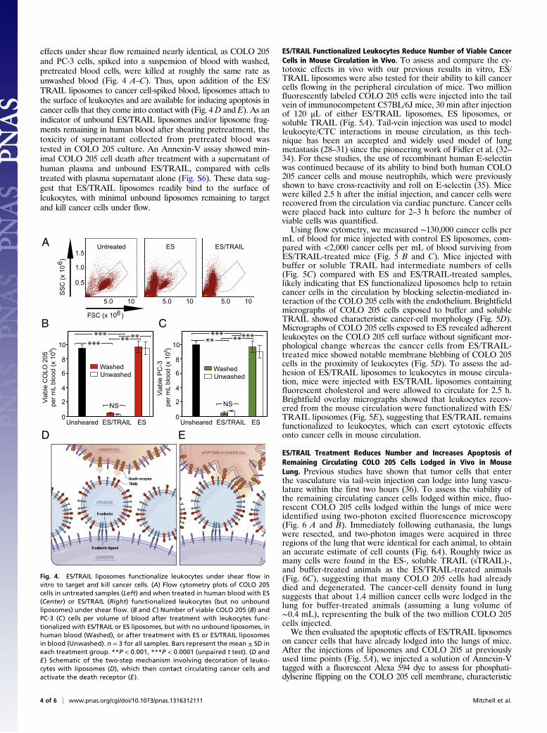
effects under shear flow remained nearly identical, as COLO 205and PC-3 cells, spiked into a suspension of blood with washed,pretreated blood cells, were killed at roughly the same rate asunwashed blood (Fig. 4 A–C). Thus, upon addition of the ES/TRAIL liposomes to cancer cell-spiked blood, liposomes attach tothe surface of leukocytes and are available for inducing apoptosis incancer cells that they come into contact with (Fig. 4D and E). As anindicator of unbound ES/TRAIL liposomes and/or liposome frag-ments remaining in human blood after shearing pretreatment, thetoxicity of supernatant collected from pretreated blood wastested in COLO 205 culture. An Annexin-V assay showed min-imal COLO 205 cell death after treatment with a supernatant ofhuman plasma and unbound ES/TRAIL, compared with cellstreated with plasma supernatant alone (Fig. S6). These data sug-gest that ES/TRAIL liposomes readily bind to the surface ofleukocytes, with minimal unbound liposomes remaining to targetand kill cancer cells under flow.
ES/TRAIL Functionalized Leukocytes Reduce Number of Viable CancerCells in Mouse Circulation in Vivo. To assess and compare the cy-totoxic effects in vivo with our previous results in vitro, ES/TRAIL liposomes were also tested for their ability to kill cancercells flowing in the peripheral circulation of mice. Two millionfluorescently labeled COLO 205 cells were injected into the tailvein of immunocompetent C57BL/6J mice, 30 min after injectionof 120 μL of either ES/TRAIL liposomes, ES liposomes, orsoluble TRAIL (Fig. 5A). Tail-vein injection was used to modelleukocyte/CTC interactions in mouse circulation, as this tech-nique has been an accepted and widely used model of lungmetastasis (28–31) since the pioneering work of Fidler et al. (32–34). For these studies, the use of recombinant human E-selectinwas continued because of its ability to bind both human COLO205 cancer cells and mouse neutrophils, which were previouslyshown to have cross-reactivity and roll on E-selectin (35). Micewere killed 2.5 h after the initial injection, and cancer cells wererecovered from the circulation via cardiac puncture. Cancer cellswere placed back into culture for 2–3 h before the number ofviable cells was quantified.Using flow cytometry, we measured ∼130,000 cancer cells per
mL of blood for mice injected with control ES liposomes, com-pared with <2,000 cancer cells per mL of blood surviving fromES/TRAIL-treated mice (Fig. 5 B and C). Mice injected withbuffer or soluble TRAIL had intermediate numbers of cells(Fig. 5C) compared with ES and ES/TRAIL-treated samples,likely indicating that ES functionalized liposomes help to retaincancer cells in the circulation by blocking selectin-mediated in-teraction of the COLO 205 cells with the endothelium. Brightfieldmicrographs of COLO 205 cells exposed to buffer and solubleTRAIL showed characteristic cancer-cell morphology (Fig. 5D).Micrographs of COLO 205 cells exposed to ES revealed adherentleukocytes on the COLO 205 cell surface without significant mor-phological change whereas the cancer cells from ES/TRAIL-treated mice showed notable membrane blebbing of COLO 205cells in the proximity of leukocytes (Fig. 5D). To assess the ad-hesion of ES/TRAIL liposomes to leukocytes in mouse circula-tion, mice were injected with ES/TRAIL liposomes containingfluorescent cholesterol and were allowed to circulate for 2.5 h.Brightfield overlay micrographs showed that leukocytes recov-ered from the mouse circulation were functionalized with ES/TRAIL liposomes (Fig. 5E), suggesting that ES/TRAIL remainsfunctionalized to leukocytes, which can exert cytotoxic effectsonto cancer cells in mouse circulation.
ES/TRAIL Treatment Reduces Number and Increases Apoptosis ofRemaining Circulating COLO 205 Cells Lodged in Vivo in MouseLung. Previous studies have shown that tumor cells that enterthe vasculature via tail-vein injection can lodge into lung vascu-lature within the first two hours (36). To assess the viability ofthe remaining circulating cancer cells lodged within mice, fluo-rescent COLO 205 cells lodged within the lungs of mice wereidentified using two-photon excited fluorescence microscopy(Fig. 6 A and B). Immediately following euthanasia, the lungswere resected, and two-photon images were acquired in threeregions of the lung that were identical for each animal, to obtainan accurate estimate of cell counts (Fig. 6A). Roughly twice asmany cells were found in the ES-, soluble TRAIL (sTRAIL)-,and buffer-treated animals as the ES/TRAIL-treated animals(Fig. 6C), suggesting that many COLO 205 cells had alreadydied and degenerated. The cancer-cell density found in lungsuggests that about 1.4 million cancer cells were lodged in thelung for buffer-treated animals (assuming a lung volume of∼0.4 mL), representing the bulk of the two million COLO 205cells injected.We then evaluated the apoptotic effects of ES/TRAIL liposomes
on cancer cells that have already lodged into the lungs of mice.After the injections of liposomes and COLO 205 at previouslyused time points (Fig. 5A), we injected a solution of Annexin-Vtagged with a fluorescent Alexa 594 dye to assess for phosphati-dylserine flipping on the COLO 205 cell membrane, characteristic
A
B C
D E
Fig. 4. ES/TRAIL liposomes functionalize leukocytes under shear flow invitro to target and kill cancer cells. (A) Flow cytometry plots of COLO 205cells in untreated samples (Left) and when treated in human blood with ES(Center) or ES/TRAIL (Right) functionalized leukocytes (but no unboundliposomes) under shear flow. (B and C) Number of viable COLO 205 (B) andPC-3 (C) cells per volume of blood after treatment with leukocytes func-tionalized with ES/TRAIL or ES liposomes, but with no unbound liposomes, inhuman blood (Washed), or after treatment with ES or ES/TRAIL liposomesin blood (Unwashed). n = 3 for all samples. Bars represent the mean ± SD ineach treatment group. **P < 0.001, ***P < 0.0001 (unpaired t test). (D andE ) Schematic of the two-step mechanism involving decoration of leuko-cytes with liposomes (D), which then contact circulating cancer cells andactivate the death receptor (E ).
4 of 6 | www.pnas.org/cgi/doi/10.1073/pnas.1316312111 Mitchell et al.

of apoptosis. Mouse lungs were imaged using two-photon mi-croscopy to determine whether Hoescht-labeled COLO 205 cellswere also positive for Annexin-V labeling. In addition to the de-creased density of cancer cells lodged in mouse lung (Fig. 6C), wealso found a dramatic increase in apoptosis of the cancer cells(Fig. 6 B and D) in the ES/TRAIL liposome-treated mice com-pared with other groups. Soluble TRAIL protein injected intomice following the same protocol displayed minimal cytotoxic ac-tivity comparable with control, as expected due to its short circu-lation half-life (37). Mice injected with ES/TRAIL liposomessurvived for over 2 wk with no loss in body weight (n = 3). Thesedata suggest that ES/TRAIL treatment serves to decrease thenumber of remaining circulating COLO 205 cells lodged in mouselung, while increasing the fraction of them that are apoptotic.
DiscussionNatural killer cells, activated by interleukin-2 or other factors,are induced to present TRAIL protein on their surface. Thesecells participate in immunosurveillance against micrometastasesin the body and comprise 10–20% of peripheral blood mono-nuclear cells (38, 39). Although the liposome-coated leukocytesdescribed here are not specifically programmed to actively in-vade tissues and seek out solid tumors, they do have frequentopportunities for incidental contact with CTCs in the blood-stream. Interestingly, infiltration of neutrophils and macro-phages throughout the interior of solid tumor masses has beenfound in dynamic, self-seeding tumors, suggesting that somedegree of homing of normally functioning leukocytes to solidtumors could be expected (40, 41). We find that TRAIL is mostpotent when in its natural state—tethered to the surface of leu-kocytes in shear flow—rather than freely soluble or on untetheredliposomes in the absence of blood. Tethering nanoscale liposomesto the surface of peripheral blood leukocytes is also beneficial
for increasing liposome circulation time, by avoiding renalclearance mechanisms.So why do leukocytes coated with ES/TRAIL liposomes have
much higher cytotoxic activity in shear flow, compared withisolated ES/TRAIL liposomes or soluble TRAIL protein? Theanswer may lie in the compressive force between surfaces. Twospherical particles colliding in linear shear flow will experiencea compressive force between them, which scales as Fc ∼ μ*G*a*b,where μ is the fluid viscosity, G is the shear rate, and a and b arethe radii of the smaller and larger sphere, respectively (42). Thus,a 10-μm-diameter leukocyte colliding with a cancer cell will ex-perience 100 times the compressive force of a 100-nm liposomecolliding with a cancer cell. Compressive forces act to flatten downany cell-surface glycocalyx composed of biologically inert macro-molecules, thus allowing TRAIL to come within a reactive dis-tance to the cancer cell death receptors and form bonds. Thephysics of force-induced flattening and penetration of cell glyco-calyx to facilitate surface receptor binding to ligands on an op-posing cell surface has been analyzed in the context of leukocyteadhesion to the vascular endothelium (43, 44).Recombinant human TRAIL/Apo2L, also known as PRO1762
developed by Amgen/Genentech, has been the subject of nu-merous Phase 1, 1a, 2, and 3 clinical trials over the past decade,with minimal adverse effects reported (45, 46). There are manyintracellular proteins, such as the inhibitors of apoptosis protein(IAPs) family members, that also confer TRAIL resistance tonormal cells (47). Additionally, the dosages of TRAIL used inthis current study ranged from 0.06–0.08 mg/kg, two orders of
b
ES/TRAIL sTRAIL
ES Buffer
D
0
0.5
1.0
1.5
2.0
ES/TRAIL sTRAIL ES Buffer
(cel
ls/m
m C
OLO
205
cel
ls in
blo
od3
x 10
0,00
0)
C
E
Blood collection
COLO 205 cells
Liposomeinjectiont = 0 hrs t = 0.5 hrs t = 2.5 hrs
Cancer cellinjection
ViabilitymeasurementsA
******
FSC (x 10 )
SS
C (x
10
)
6
6
B
Fig. 5. ES/TRAIL functionalized leukocytes target and kill cancer cells in thecirculation of mice in vivo. (A) Schematic of in vivo mouse experiment. (B)Flow cytometry of untreated COLO 205 cancer cells (Left) and those re-covered from cardiac puncture from mice treated with ES (Center) and ES/TRAIL liposomes (Right). (C) Number of viable cancer cells recovered pervolume of mouse blood for mice treated with ES/TRAIL liposomes, solubleTRAIL (sTRAIL), ES liposomes, and buffer injections. n = 3 for all samples. Barsrepresent the mean ± SD in each treatment group. *P < 0.01, **P < 0.001,***P < 0.0001 (one-way ANOVA with Tukey posttest). (D) Representativemicrographs of COLO 205 cells removed from circulation in mice treatedwith ES/TRAIL liposomes (Upper Left), sTRAIL (Upper Right), ES liposomes(Lower Left), and buffer (Lower Right) injections. (Scale bar, 20 μm.) (E)Leukocytes functionalized with fluorescent ES/TRAIL liposomes (green) uponremoval from mouse circulation 2.5 h after injection. (Scale bar, 50 μm.)
ES/TRAIL sTRAIL
ES Buffer
A B
C D
(Cel
ls/m
mD
ensi
ty o
f CO
LO 2
05 C
ells
in
Lun
g3
x 10
00)
2
4
6
8
10
ES
20
40
60
80
100
(Per
cent
age
of T
otal
) Ap
opto
tic C
OLO
205
Cel
ls in
Lung
ES/TRAIL0
sTRAIL Buffer ESES/TRAIL sTRAIL Buffer
*****
** ** **
0
Fig. 6. Decreased number and increased apoptosis in COLO 205 cells lodgedin mouse lung after treatment with ES/TRAIL liposomes. (A) Schematic ofmouse lung and example two-photon excited fluorescence (2PEF) image stackfrom mouse lung where Hoechst-labeled COLO 205 cells (green) are arrestedin lung tissue (visible by autofluorescence, yellow). (Scale bar, 80 μm.) (B) The2PEF images of Hoescht-labeled COLO 205 cells (green) with Alexa Flour 568-labeled Annexin-V apoptosis probe (red) for each experimental group. Redarrows point to apoptotic COLO 205 cells (red and green colocalized), andblue arrows indicate nonapoptotic COLO 205 cells (green only). White circlesindicate regions of autofluorescence from lung tissue. (Scale bar, 30 μm.) (C)Density of COLO 205 cells lodged in the lung for each experimental group. (D)Percentage of lodged COLO 205 cells positive for Annexin-V probe for eachexperimental group. Individual data points represent data from one imagestack, with points shown in the same color representing image stacks fromthe same animal. Superimposed box plots bound the 25th to 75th percentageof all data points and the whiskers extend 1.5 times the interquartile rangebeyond the boxes. The horizontal lines within the boxplot represent themedian. n = 3 animals for each experimental group. *P < 0.01, **P < 0.0001(one-way ANOVA with Tukey posttest).
Mitchell et al. PNAS Early Edition | 5 of 6
ENGINEE
RING
APP
LIED
BIOLO
GICAL
SCIENCE
S

magnitude lower than the clinical dosages of 1–30 mg/kg used inhuman clinical trials. Although different types of cancer cellsshow different levels of sensitivity to TRAIL-induced apoptosis,it has been well documented that there is a wide range of agentsknown to sensitize cancer cells to TRAIL-mediated apoptosis,including conventional chemotherapeutics (camptothecin, cis-platin, doxorubicin, 5-fluorouracil, irinotecan, paclitaxel, gemci-tabine), proteasome inhibitors, Bcl-2 inhibitors, IAP antagonists,histone deacetylase inhibitors, CD20 antibodies, irradiation, syn-thetic triterpenolds, Sorafenib, aspirin, and natural products such ascurcumin and piperlongumine (48).What remains to be seen is whether ES/TRAIL liposomes can
successfully prevent the formation of metastatic tumors; futurework should focus on addressing this question. Additionally,human hepatocytes have shown sensitivity to TRAIL (49) al-though ES/TRAIL liposome adhesion to the leukocyte surfacecould reduce TRAIL uptake by the reticulo-endothelial systemin the liver. The present study, however, represents an importantfirst step toward the targeting of CTCs in the bloodstream as
a means to prevent cancer metastasis. Clinically, for instance,one could envision using these liposomes as a preventive mea-sure upon diagnosis of highly metastatic hematogenous cancerssuch as those originating in breast, prostate, and lung.
Materials and MethodsAll reagents and additional procedures used in this study, including cellculture, liposome synthesis, static and shear treatment assays in buffer andhuman blood, leukocyte isolation and functionalization with ES/TRAIL inhuman blood, mouse studies, circulating cancer cell analysis, two-photonimaging of mouse tissue, flow cytometry, and statistical analyses are de-scribed in SI Materials and Methods. All human subject protocols were ap-proved by the Institutional Review Board for Human Participants of CornellUniversity. All animal procedures were approved by the Cornell UniversityInstitutional Animal Care and Use Committee.
ACKNOWLEDGMENTS. The authors thank Dr. Razelle Kurzrock for discussionon TRAIL toxicity, Julie Kohn and Brooke Mason of the Reinhart-King Lab forendothelial cell protocols, and Jeff Mattison for blood work. The workdescribed was supported by the Cornell Center on the Microenvironment andMetastasis through Award U54CA143876 from the National Cancer Institute.
1. Chaffer CL, Weinberg RA (2011) A perspective on cancer cell metastasis. Science331(6024):1559–1564.
2. Riethdorf S, Wikman H, Pantel K (2008) Review: Biological relevance of disseminatedtumor cells in cancer patients. Int J Cancer 123(9):1991–2006.
3. Maheswaran S, Haber DA (2010) Circulating tumor cells: A window into cancer bi-ology and metastasis. Curr Opin Genet Dev 20(1):96–99.
4. Coussens LM, Werb Z (2002) Inflammation and cancer. Nature 420(6917):860–867.5. McDonald B, et al. (2009) Systemic inflammation increases cancer cell adhesion to
hepatic sinusoids by neutrophil mediated mechanisms. Int J Cancer 125(6):1298–1305.6. van Ginhoven TM, van den Berg JW, Dik WA, Ijzermans JNM, de Bruin RWF (2010)
Preoperative dietary restriction reduces hepatic tumor load by reduced E-selectin-mediated adhesion in mice. J Surg Oncol 102(4):348–353.
7. Gassmann P, Kang ML, Mees ST, Haier J (2010) In vivo tumor cell adhesion in thepulmonary microvasculature is exclusively mediated by tumor cell—endothelial cellinteraction. BMC Cancer 10:177.
8. Köhler S, Ullrich S, Richter U, Schumacher U (2010) E-/P-selectins and colon carcinomametastasis: First in vivo evidence for their crucial role in a clinically relevant model ofspontaneous metastasis formation in the lung. Br J Cancer 102(3):602–609.
9. Rahn JJ, et al. (2005) MUC1 mediates transendothelial migration in vitro by ligatingendothelial cell ICAM-1. Clin Exp Metastasis 22(6):475–483.
10. Chang YS, et al. (2000) Mosaic blood vessels in tumors: Frequency of cancer cells incontact with flowing blood. Proc Natl Acad Sci USA 97(26):14608–14613.
11. Butler TP, Gullino PM (1975) Quantitation of cell shedding into efferent blood ofmammary adenocarcinoma. Cancer Res 35(3):512–516.
12. Allard WJ, et al. (2004) Tumor cells circulate in the peripheral blood of all majorcarcinomas but not in healthy subjects or patients with nonmalignant diseases. ClinCancer Res 10(20):6897–6904.
13. Yu M, Stott S, Toner M, Maheswaran S, Haber DA (2011) Circulating tumor cells:Approaches to isolation and characterization. J Cell Biol 192(3):373–382.
14. Firrell JC, Lipowsky HH (1989) Leukocyte margination and deformation in mesentericvenules of rat. Am J Physiol 256(6 Pt 2):H1667–H1674.
15. Läubli H, Borsig L (2010) Selectins promote tumor metastasis. Semin Cancer Biol 20(3):169–177.
16. Gout S, Tremblay PL, Huot J (2008) Selectins and selectin ligands in extravasation ofcancer cells and organ selectivity of metastasis. Clin Exp Metastasis 25(4):335–344.
17. Barthel SR, et al. (2009) Alpha 1,3 fucosyltransferases are master regulators of pros-tate cancer cell trafficking. Proc Natl Acad Sci USA 106(46):19491–19496.
18. Ashkenazi A, Holland P, Eckhardt SG (2008) Ligand-based targeting of apoptosis incancer: the potential of recombinant human apoptosis ligand 2/Tumor necrosis fac-tor-related apoptosis-inducing ligand (rhApo2L/TRAIL). J Clin Oncol 26(21):3621–3630.
19. Walczak H, et al. (1999) Tumoricidal activity of tumor necrosis factor-related apo-ptosis-inducing ligand in vivo. Nat Med 5(2):157–163.
20. Kim MB, Sarelius IH (2003) Distributions of wall shear stress in venular convergencesof mouse cremaster muscle. Microcirculation 10(2):167–178.
21. Mitchell MJ, King MR (2013) Computational and experimental models of cancer cellresponse to fluid shear stress. Front Oncol 3:44.
22. Springer TA (1994) Traffic signals for lymphocyte recirculation and leukocyte emi-gration: the multistep paradigm. Cell 76(2):301–314.
23. Doherty TA, et al. (2012) STAT6 regulates natural helper cell proliferation during lunginflammation initiated by Alternaria.Am J Physiol Lung Cell Mol Physiol 303(7):L577–L588.
24. Malý P, et al. (1996) The alpha(1,3)fucosyltransferase Fuc-TVII controls leukocytetrafficking through an essential role in L-, E-, and P-selectin ligand biosynthesis. Cell86(4):643–653.
25. Janssen EM, et al. (2005) CD4+ T-cell help controls CD8+ T-cell memory via TRAIL-mediated activation-induced cell death. Nature 434(7029):88–93.
26. Goldsmith HL (1968) The microrheology of red blood cell suspensions. J Gen Physiol52(1):5–28.
27. Goldsmith HL, Bell DN, Braovac S, Steinberg A, McIntosh F (1995) Physical andchemical effects of red cells in the shear-induced aggregation of human platelets.Biophys J 69(4):1584–1595.
28. Yan L, Cai Q, Xu Y (2013) The ubiquitin-CXCR4 axis plays an important role in acutelung infection-enhanced lung tumor metastasis. Clin Cancer Res 19(17):4706–4716.
29. Tucci P, et al. (2012) Loss of p63 and its microRNA-205 target results in enhanced cellmigration andmetastasis in prostate cancer. Proc Natl Acad Sci USA 109(38):15312–15317.
30. Chang C-Y, Lin S-C, Su W-H, Ho C-M, Jou Y-S (2012) Somatic LMCD1 mutations pro-moted cell migration and tumor metastasis in hepatocellular carcinoma. Oncogene31(21):2640–2652.
31. Kim S, et al. (2009) Carcinoma-produced factors activate myeloid cells through TLR2to stimulate metastasis. Nature 457(7225):102–106.
32. Fidler IJ (1975) Biological behavior of malignant melanoma cells correlated to theirsurvival in vivo. Cancer Res 35(1):218–224.
33. Fidler IJ (1974) Inhibition of pulmonary metastasis by intravenous injection of spe-cifically activated macrophages. Cancer Res 34(5):1074–1078.
34. Fidler IJ, Nicolson GL (1976) Organ selectivity for implantation survival and growth ofB16 melanoma variant tumor lines. J Natl Cancer Inst 57(5):1199–1202.
35. Kato N, et al. (2009) The E-selectin ligand basigin/CD147 is responsible for neutrophilrecruitment in renal ischemia/reperfusion. J Am Soc Nephrol 20(7):1565–1576.
36. Fidler IJ, Nicolson GL (1977) Fate of recirculating B16 melanoma metastatic variantcells in parabiotic syngeneic recipients. J Natl Cancer Inst 58(6):1867–1872.
37. Xiang H, Nguyen CB, Kelley SK, Dybdal N, Escandón E (2004) Tissue distribution,stability, and pharmacokinetics of Apo2 ligand/tumor necrosis factor-related apo-ptosis-inducing ligand in human colon carcinoma COLO205 tumor-bearing nude mice.Drug Metab Dispos 32(11):1230–1238.
38. Takeda K, et al. (2005) TRAIL identifies immature natural killer cells in newborn miceand adult mouse liver. Blood 105(5):2082–2089.
39. Waldhauer I, Steinle A (2008) NK cells and cancer immunosurveillance. Oncogene27(45):5932–5943.
40. KimMY, et al. (2009) Tumor self-seeding by circulating cancer cells. Cell 139(7):1315–1326.41. Bernal M, et al. (2011) Leukocyte infiltrate in gastrointestinal adenocarcinomas is
strongly associated with tumor microsatellite instability but not with tumor immu-nogenicity. Cancer Immunol Immunother 60(6):869–882.
42. Shankaran H, Neelamegham S (2004) Hydrodynamic forces applied on intercellularbonds, soluble molecules, and cell-surface receptors. Biophys J 86(1 Pt 1):576–588.
43. Zhao Y, Chien S, Weinbaum S (2001) Dynamic contact forces on leukocyte microvilliand their penetration of the endothelial glycocalyx. Biophys J 80(3):1124–1140.
44. Sabri S, et al. (2000) Glycocalyx modulation is a physiological means of regulating celladhesion. J Cell Sci 113(Pt 9):1589–1600.
45. Subbiah V, et al. (2012) Targeting the apoptotic pathway in chondrosarcoma usingrecombinant human Apo2L/TRAIL (dulanermin), a dual proapoptotic receptor (DR4/DR5) agonist. Mol Cancer Ther 11(11):2541–2546.
46. Herbst RS, et al. (2010) Phase I dose-escalation study of recombinant human Apo2L/TRAIL, a dual proapoptotic receptor agonist, in patients with advanced cancer. J ClinOncol 28(17):2839–2846.
47. Zhang XD, Nguyen T, Thomas WD, Sanders JE, Hersey P (2000) Mechanisms of re-sistance of normal cells to TRAIL induced apoptosis vary between different cell types.FEBS Lett 482(3):193–199.
48. Ashkenazi A, Herbst RS (2008) To kill a tumor cell: The potential of proapoptoticreceptor agonists. J Clin Invest 118(6):1979–1990.
49. Jo M, et al. (2000) Apoptosis induced in normal human hepatocytes by tumor necrosisfactor-related apoptosis-inducing ligand. Nat Med 6(5):564–567.
6 of 6 | www.pnas.org/cgi/doi/10.1073/pnas.1316312111 Mitchell et al.