Trafficking Macrophage Migration Using Reporter Gene Imaging...
8
Trafficking Macrophage Migration Using Reporter Gene Imaging with Human Sodium Iodide Symporter in Animal Models of Inflammation Ji Hyoung Seo *1,2 , Yong Hyun Jeon *1 , Yong Jin Lee 1 , Gil Sook Yoon 3 , Dong-Il Won 4 , Jeoung-Hee Ha 5 , Shin Young Jeong 1 , Sang Woo Lee 1 , Byeong Cheol Ahn 1 , and Jaetae Lee 1 1 Department of Nuclear Medicine, Kyungpook National University, Daegu, Korea; 2 Department of Nuclear Medicine, Haeundae Paik Hospital, Inje University College of Medicine, Busan, Korea; 3 Department of Pathology, Kyungpook National University, Daegu, Korea; 4 Department of Clinical Pathology, Kyungpook National University, Daegu, Korea; and 5 Department of Pharmacology, Kyungpook National University, Daegu, Korea The aim of this study was to investigate the feasibility of nuclear molecular imaging using the human sodium iodide symporter (hNIS) as a reporter gene to monitor macrophage migration toward the inflammatory foci. Methods: A stable macrophage cell line coexpressing hNIS and green fluores- cent protein (GFP) genes (RAW264.7/hNIS-GFP and R NIS cell) was established from an immortalized macrophage cell line (RAW264.7 cells). 125 I uptake was determined (for hNIS pro- tein functional activity), and flow cytometry analysis (to exam- ine GFP gene expression), a cell proliferation assay, a cytokine assay, and a phagocytic activity assay were performed. 99m Tc-pertechnetate images were acquired at 1 d after sub- cutaneous inoculation of R NIS cells in nude mice. Chemical inflammation was induced for in vivo imaging in the thigh of nude mice by turpentine oil injection. Small-animal PET with 18 F-FDG and 124 I was performed with an intravenous admin- istration of RAW264.7 or R NIS cells in inflammation-induced animals. Results: The expression of hNIS and GFP genes was confirmed in R NIS cells by flow cytometry and immuno- fluorescent staining. 125 I uptake was about 67 times higher in R NIS cells than in RAW264.7 cells. No significant difference was observed in cell proliferation, cytokine production, and phagocytic activity between RAW264.7 and R NIS cells. 99m Tc-pertechnetate imaging revealed increased tracer uptake at the inoculation site. PET with 124 I demonstrated a donut-shaped uptake, correlating with uptake shown by the 18 F-FDG PET images, at the inflammation site of mice admin- istered R NIS cells. 124 I uptake (percentage injected dose per gram) was about 2.12 times higher at the inflammation site in the R NIS mice than in RAW264.7 mice. By immunohistochemis- try, the migration of macrophages was further confirmed by positive staining for GFP and hNIS at the inflammation site of R NIS mice. Conclusion: These data support the feasibility of hNIS reporter gene imaging to monitor the macrophage migra- tion toward an inflammatory lesion. Macrophages expressing hNIS may provide a new strategy to investigate the cellular behavior seen with inflammatory response in a preclinical model. Key Words: reporter gene imaging; sodium iodide symporter; macrophage; 124 I PET J Nucl Med 2010; 51:1637–1643 DOI: 10.2967/jnumed.110.077891 Macrophages are important immune cells implicated in onset, progression, and manifestation of various patho- logic processes, such as growth and spread of malignant tumors, and chronic inflammatory diseases (1). Macro- phages act as hosts and reservoirs for pathogens and pro- duce proinflammatory or proangiogenic mediators (2–4). Various attempts have been made to visualize the macro- phage-mediated inflammatory process in vivo, but specific visualization of these cells in the inflammatory process remains a considerable task with the currently available imaging modalities, such as radionuclide imaging or MRI, which are considered appropriate to visualize the in- flammatory changes in tissue (5). Nuclear methods such as PET and SPECT exhibit relatively poor spatial resolution, but their good sensitivity is considered a powerful feature in molecular imaging (6). Development of molecular imaging with PET has been made much easier as assays in cell culture and small-animal models are first validated and then the same tracer used in those procedures is used in established clinical PET sites (7). Therefore PET-based molecular–genetic imag- ing has developed rapidly in the past decade. Most current molecular–genetic imaging strategies are based on a reporter gene and a complementary reporter probe. The reporter gene is constitutively expressed, and reporter imaging is used to track gene therapy vectors or transduced cells in the body (8). One such example of a reporter gene system is the sodium iodide symporter (NIS). NIS reporter gene imaging has the advantages of using known radiotracers such as 99m Tc-pertechnetate and radio- iodine. Additional benefits are that NIS is nonimmuno- genic, with no observed adverse effects on cell viability and function (9), and this technique does not require a complex probe synthesis. Received Apr. 7, 2010; revision accepted Jul. 14, 2010. For correspondence contact: Jaetae Lee, Department of Nuclear Medicine, Kyungpook National University Hospital, 50 Samduk 2-ga, Daegu, 700-721, Republic of Korea. E-mail: [email protected] *Contributed equally to this work. COPYRIGHT ª 2010 by the Society of Nuclear Medicine, Inc. MACROPHAGE TRAFFICKING USING hNIS GENE • Seo et al. 1637 by on July 30, 2020. For personal use only. jnm.snmjournals.org Downloaded from
Transcript of Trafficking Macrophage Migration Using Reporter Gene Imaging...

Trafficking Macrophage Migration Using ReporterGene Imaging with Human Sodium Iodide Symporterin Animal Models of Inflammation
Ji Hyoung Seo*1,2, Yong Hyun Jeon*1, Yong Jin Lee1, Gil Sook Yoon3, Dong-Il Won4, Jeoung-Hee Ha5,Shin Young Jeong1, Sang Woo Lee1, Byeong Cheol Ahn1, and Jaetae Lee1
1Department of Nuclear Medicine, Kyungpook National University, Daegu, Korea; 2Department of Nuclear Medicine, HaeundaePaik Hospital, Inje University College of Medicine, Busan, Korea; 3Department of Pathology, Kyungpook National University,Daegu, Korea; 4Department of Clinical Pathology, Kyungpook National University, Daegu, Korea; and 5Department ofPharmacology, Kyungpook National University, Daegu, Korea
The aim of this study was to investigate the feasibility ofnuclear molecular imaging using the human sodium iodidesymporter (hNIS) as a reporter gene to monitor macrophagemigration toward the inflammatory foci. Methods: A stablemacrophage cell line coexpressing hNIS and green fluores-cent protein (GFP) genes (RAW264.7/hNIS-GFP and RNIS cell)was established from an immortalized macrophage cell line(RAW264.7 cells). 125I uptake was determined (for hNIS pro-tein functional activity), and flow cytometry analysis (to exam-ine GFP gene expression), a cell proliferation assay, a cytokineassay, and a phagocytic activity assay were performed.99mTc-pertechnetate images were acquired at 1 d after sub-cutaneous inoculation of RNIS cells in nude mice. Chemicalinflammation was induced for in vivo imaging in the thigh ofnude mice by turpentine oil injection. Small-animal PET with18F-FDG and 124I was performed with an intravenous admin-istration of RAW264.7 or RNIS cells in inflammation-inducedanimals. Results: The expression of hNIS and GFP geneswas confirmed in RNIS cells by flow cytometry and immuno-fluorescent staining. 125I uptake was about 67 times higher inRNIS cells than in RAW264.7 cells. No significant differencewas observed in cell proliferation, cytokine production, andphagocytic activity between RAW264.7 and RNIS cells.99mTc-pertechnetate imaging revealed increased traceruptake at the inoculation site. PET with 124I demonstrated adonut-shaped uptake, correlating with uptake shown by the18F-FDG PET images, at the inflammation site of mice admin-istered RNIS cells. 124I uptake (percentage injected dose pergram) was about 2.12 times higher at the inflammation site inthe RNIS mice than in RAW264.7 mice. By immunohistochemis-try, the migration of macrophages was further confirmed bypositive staining for GFP and hNIS at the inflammation site ofRNIS mice. Conclusion: These data support the feasibility ofhNIS reporter gene imaging to monitor the macrophage migra-tion toward an inflammatory lesion. Macrophages expressinghNIS may provide a new strategy to investigate the cellularbehavior seen with inflammatory response in a preclinical model.
Key Words: reporter gene imaging; sodium iodide symporter;macrophage; 124I PET
J Nucl Med 2010; 51:1637–1643DOI: 10.2967/jnumed.110.077891
Macrophages are important immune cells implicatedin onset, progression, and manifestation of various patho-logic processes, such as growth and spread of malignanttumors, and chronic inflammatory diseases (1). Macro-phages act as hosts and reservoirs for pathogens and pro-duce proinflammatory or proangiogenic mediators (2–4).Various attempts have been made to visualize the macro-phage-mediated inflammatory process in vivo, but specificvisualization of these cells in the inflammatory processremains a considerable task with the currently availableimaging modalities, such as radionuclide imaging orMRI, which are considered appropriate to visualize the in-flammatory changes in tissue (5). Nuclear methods such asPET and SPECT exhibit relatively poor spatial resolution,but their good sensitivity is considered a powerful featurein molecular imaging (6). Development of molecular imagingwith PET has been made much easier as assays in cell cultureand small-animal models are first validated and then the sametracer used in those procedures is used in established clinicalPET sites (7). Therefore PET-based molecular–genetic imag-ing has developed rapidly in the past decade.
Most current molecular–genetic imaging strategies arebased on a reporter gene and a complementary reporterprobe. The reporter gene is constitutively expressed, andreporter imaging is used to track gene therapy vectors ortransduced cells in the body (8). One such example of areporter gene system is the sodium iodide symporter (NIS).NIS reporter gene imaging has the advantages of usingknown radiotracers such as 99mTc-pertechnetate and radio-iodine. Additional benefits are that NIS is nonimmuno-genic, with no observed adverse effects on cell viabilityand function (9), and this technique does not require acomplex probe synthesis.
Received Apr. 7, 2010; revision accepted Jul. 14, 2010.For correspondence contact: Jaetae Lee, Department of Nuclear
Medicine, Kyungpook National University Hospital, 50 Samduk 2-ga,Daegu, 700-721, Republic of Korea.E-mail: [email protected]*Contributed equally to this work.COPYRIGHT ª 2010 by the Society of Nuclear Medicine, Inc.
MACROPHAGE TRAFFICKING USING hNIS GENE • Seo et al. 1637
by on July 30, 2020. For personal use only. jnm.snmjournals.org Downloaded from

The aim of this study was to investigate the feasibility ofnuclear molecular imaging using the human sodium iodidesymporter (hNIS) as a reporter gene that functions tomonitor macrophage migration toward a chemicallyinduced inflammatory lesion. An immortalized macrophagecell line coexpressing hNIS and green fluorescent protein(GFP) genes (RAW264.7/hNIS-GFP and RNIS cells) wasestablished. 125I uptake was determined, and flow cytome-try analysis, a cell proliferation assay, a cytokine assay, anda phagocytic activity assay were performed. Small-animalPET with 18F-FDG and 124I was performed with an intra-venous administration of RAW264.7 or RNIS cells ininflammation-induced animals.
MATERIALS AND METHODS
Cells and AnimalsSpecific pathogen-free 6-wk-old female BALB/c nude mice
(n 5 20) were obtained from SLC Inc. All animal experimentprotocols were approved by the Committee for the Handling andUse of Animals, Kyungpook National University. The murine mac-rophage RAW264.7 cell line was kindly provided by Dr. Chul WooKim (Seoul National University).
Establishment of Stable Macrophage Cell LineCoexpressing hNIS and GFP Genes
To produce the lentivirus coexpressing hNIS and GFP genes,human embryonic kidney 293 cells were plated at 5 · 106 cells ina 100-cm tissue culture plate, and cells were cotransfected withreplication-incompetent lentiviral vector (pLenti/PGK-hNIS-IRES-GFP), vesicular stomatitis virus glycoprotein, and dR8.74using a calcium phosphate transfection kit and reagents asdescribed by the manufacturer (Invitrogen). Growth mediumwas changed at 24 h, and lentivirus-containing supernatant washarvested at 48 h. Harvested supernatant was centrifuged at 4�C,3,000 rpm, for 15 min and stored at 280�C.
To establish a stable macrophage cell line coexpressing hNISand GFP, RAW264.7 cells were transduced by adding 1 mL oflentivirus-containing supernatant and 10 mg of polybrene permilliliter. After 48 h, GFP-positive cells were enriched using FAC-Saria (BD Biosciences). The selection procedure was performedtwice for 6 wk.
Immunofluorescent StainingRAW264.7 or RNIS cells were seeded at a density of 4 · 104
cells per well on collagen-precoated chamber slides (Nunc) andgrown for 24 h. The hNIS-specific immunostaining was performedwith a Cytofix/cytoperm kit (BD Pharmingen). The cells werewashed twice with phosphate-buffered saline (PBS). The washedcells were fixed and permeabilized with buffer (eBioscience) for20 min at 4�C. After that, the cells were washed with 1X BDPerm/Wash buffer (BD Bioscience) and stained with anti-hNISantibody (Milipore) diluted with 1X BD Perm/Wash buffer for 1h at room temperature. After the cells were washed 3 times with1X BD Perm/Wash buffer, Alexa Fluor 568–conjugated secondaryantibody (Molecular Probes, Inc.) was applied for 40 min at roomtemperature. The cells were washed 3 times with 1X BD Perm/Wash buffer. The slides were mounted with Vectashield mountingmedium (Vector Laboratories), covered with glass cover slips, andexamined using a laser confocal scanning system (TCS SP2;Leica).
Radioiodide Uptake AssaysParental RAW264.7 cells and RNIS cells (5 · 104, 1 · 105, 3 ·
105, and 5 · 105) were plated in 24-well plates and cultured withDulbecco’s modified Eagle’s medium containing 10% fetal bovineserum for 24 h. 125I uptake was determined by incubating the cellswith 500 mL of Hank’s balanced salt solution (bHBSS) (GibcoBRLCo.) containing 3.7 kBq (0.1 mCi) of carrier-free 125I-NaI and10 mM NaI at 37�C for 30 min. After incubation, the cells werequickly washed twice with bHBSS and detached using 500 mL oftrypsin. Radioactivity was measured using a g-counter.
Iodide uptake was measured at various times points. Cells(200,000) were plated and incubated with 500 mL of bHBSScontaining 3.7 kBq (0.1 mCi) of carrier-free 125I-NaI and 10 mMNaI at different times (1, 5, 15, 30, 45, and 60 min). For theblocking study, cells were incubated in 125I-NaI medium eitherwith or without 100 mM of potassium perchlorate.
For the iodide efflux study, cells were incubated with 10 mMNaI and 3.7 kBq (0.1 mCi) of carrier-free 125I-NaI in 500 mL ofbHBSS incubation buffer at 37�C for 30 min. Cells were thenwashed twice and incubated with bHBSS at 37�C. bHBSS wasremoved at different times (0, 1, 3, 6, 9, 12, 15, 18, 21, 24, 27, and30 min), and its radioactivity was measured. Cells were collectedafter the last time, and residual radioactivity was determined ascounts per minute. All data are expressed as the percentageremaining for the total activity.
Cell Proliferation AssayParental RAW264.7 cells and RNIS cell cells were plated at 2 ·
104 per well in 96-well plates. Two days later, a 10-mL cell countsolution (CCK-8; Dojindo) was added to the culture medium ofcells, and plates were incubated at 37�C for 4 h. Absorbance wasmeasured at a wavelength of 450 nm using a microplate reader(Bio-Rad Laboratories).
Cytokine AssayTo assess the functionality of macrophages, cytokine produc-
tion was measured. Parental RAW264.7 and RNIS cells werestimulated with 1 mg of lipopolysaccharide per milliliter. After24 h, supernatants were harvested and analyzed for IL-12p70and tumor necrosis factor (TNF)-a by enzyme-linked immuno-sorbent assay using commercially available kits (BD Pharmingen).Cytokine concentrations were quantified using a microplatereader.
Phagocytic Activity AssayTo determine the effects of virus infection on phagocytosis of
macrophage cells, we determined phagocytic activity ofRAW264.7 and RNIS cells using 7-aminoactinomycin D (7-AAD; BD Biosciences Pharmingen)–stained Escherichia coliand fluorescent-activated cell sorter (FACS) analysis (10–13).E. coli (1 · 108) in 1 mL of PBS was incubated at 60�C for 1 hfor sterilization, and 1 mL of sterilized E. coli was further coin-cubated with 50 mL of 7-AAD for 2 h at room temperature. Foropsonization of E. coli to allow phagocytosis by macrophages,5 mL of 7-AAD–stainined E. coli were incubated with 1 mL ofhuman serum for 20 min at room temperature. Opsonized E. colistained with 7-AAD was cocultured with 2.5 · 105 RAW264.7 andRNIS cells, which were incubated in 50 mL of RPMI 1640 culturemedium (Hyclone) containing 10% fetal bovine serum for 1 h ateither 4�C or 37�C. At 1 h after incubation, the samples werewashed with cold PBS twice, and fluorescence activities wereanalyzed using a FACS Calibur (BD Bioscience). The phagocytic
1638 THE JOURNAL OF NUCLEAR MEDICINE • Vol. 51 • No. 10 • October 2010
by on July 30, 2020. For personal use only. jnm.snmjournals.org Downloaded from
activity of each cell was expressed as increases in the percentageof 7-AAD–positive RAW264.7 or RNIS cells, compared with thosetreated with nonstained E. coli.
Nuclear Imaging with 99mTc-PertechnetateRNIS cells, 8 · 106 and 2 · 107, were subcutaneously implanted
in a volume of 0.1 mL into the right and left thighs, respectively,of specific pathogen-free BALB/c nude mice (6-wk-old female,n 5 3). Twenty-four hours after inoculation, 18.5 MBq (500 mCi)of 99mTc-pertechnetate in 100 mL of PBS was injected intrave-nously, and a whole-body scan was obtained at 30 min after in-jection using a g-camera equipped with a pinhole collimator(Infinia II; GE Healthcare).
Small-Animal PET with 18F-FDG and 124I inInflammation Animal Model
The animal experimental procedure is demonstrated sche-matically in Supplemental Figure 1 (supplemental materials areavailable online only at http://jnm.snmjournals.org). Turpentineoil (50 mL/mouse) was intramuscularly injected into the rightthigh of nude mice (5 mice/group). Lipopolysaccharide (1 mg/mL) was injected to promote the inflammatory process. Inflam-mation was confirmed 7 d later, and parental RAW264.7 cells orRNIS cells (3 · 106/mouse) were intravenously injected intoinflammation-induced mice. Seven days after macrophage injec-tion, 3.7 MBq (100 mCi) of 124I was administered into mice.After 1 h, mice were placed prone on the bed of the small-animal PET scanner. 18F-FDG PET was performed after in-travenous injection of 11.1 MBq (300 mCi) of 18F-FDG inmice under general anesthesia with 1%–2% isoflurane in100% O2 during injection and imaging. The images were recon-
structed with a 2-dimensional ordered-subsets expectation max-imization algorithm. Corrections were not done for attenuationor scattering. Accumulation of 124I in the inflamed tissue wasquantitatively determined from small-animal PET images byplacing a region of interest on the inflamed tissue visualizedin coronal 124I images and expressed as percentage injecteddose per gram.
Histopathologic AnalysisTissues that showed inflammation were removed from mice and
preserved in 10% formalin until required. All inflammatory tissueswere embedded in paraffin and sectioned at 5 mm. For immuno-histologic analysis of hNIS and GFP expression, the paraffin sec-tions were incubated for 1 h with anti-hNIS or anti-GFP antibodiesat 1:100, respectively. Sections were then incubated for 30 minwith a goat antirabbit secondary antibody conjugated to peroxi-dase (Immunotech).
RESULTS
Assessment of hNIS and GFP Gene Expression inRAW264.7 and RNIS Cells
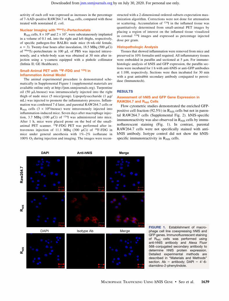
Flow cytometric studies demonstrated the enriched GFP-positive cell fraction (92.5%) in RNIS cells but not in paren-tal RAW264.7 cells (Supplemental Fig. 2). hNIS-specificimmunoreactivity was also observed in RNIS cells by immu-nofluorescent staining (Fig. 1). In contrast, parentalRAW264.7 cells were not specifically stained with anti-hNIS antibody. Isotype control did not show the hNIS-specific immunoreactivity in RNIS cells.
FIGURE 1. Establishment of macro-phage cell line coexpressing hNIS andGFP genes. Immunofluorescent stainingof RNIS cells was performed usinganti-hNIS antibody and Alexa Fluor568–conjugated secondary antibody todetermine hNIS protein expression.Detailed experimental methods aredescribed in “Materials and Methods”section. Ab 5 antibody; DAPI 5 49-6-diamidino-2-phenylindole.
MACROPHAGE TRAFFICKING USING hNIS GENE • Seo et al. 1639
by on July 30, 2020. For personal use only. jnm.snmjournals.org Downloaded from
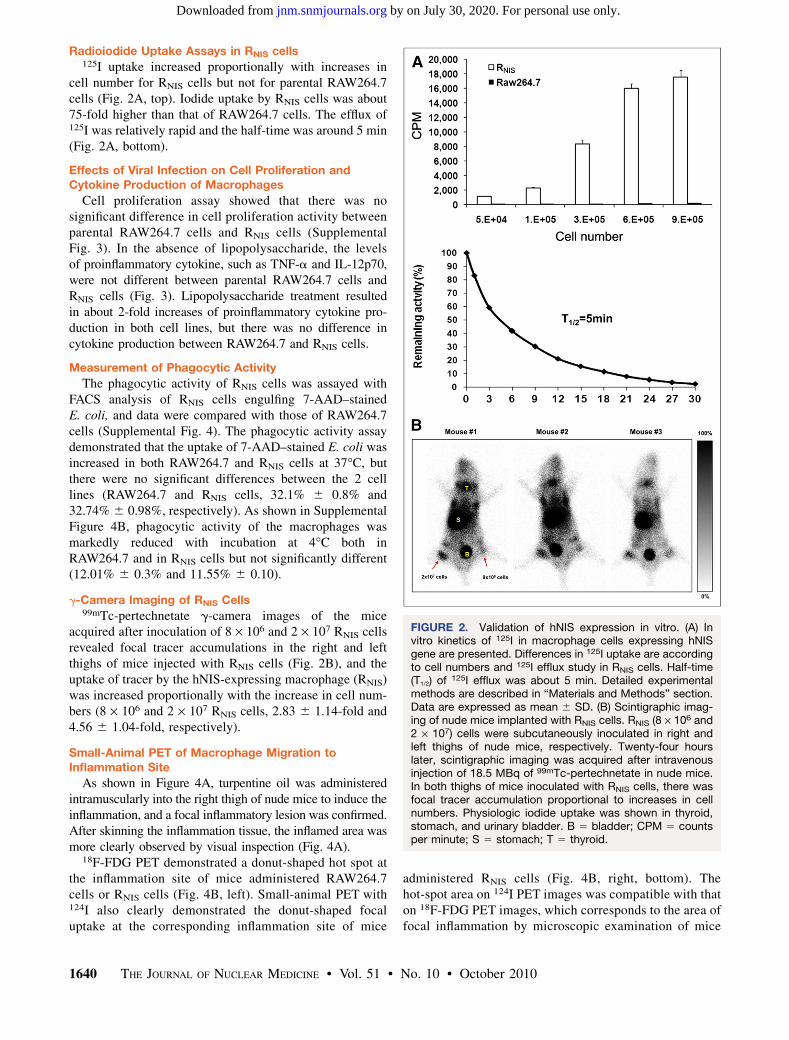
Radioiodide Uptake Assays in RNIS cells125I uptake increased proportionally with increases in
cell number for RNIS cells but not for parental RAW264.7cells (Fig. 2A, top). Iodide uptake by RNIS cells was about75-fold higher than that of RAW264.7 cells. The efflux of125I was relatively rapid and the half-time was around 5 min(Fig. 2A, bottom).
Effects of Viral Infection on Cell Proliferation andCytokine Production of Macrophages
Cell proliferation assay showed that there was nosignificant difference in cell proliferation activity betweenparental RAW264.7 cells and RNIS cells (SupplementalFig. 3). In the absence of lipopolysaccharide, the levelsof proinflammatory cytokine, such as TNF-a and IL-12p70,were not different between parental RAW264.7 cells andRNIS cells (Fig. 3). Lipopolysaccharide treatment resultedin about 2-fold increases of proinflammatory cytokine pro-duction in both cell lines, but there was no difference incytokine production between RAW264.7 and RNIS cells.
Measurement of Phagocytic Activity
The phagocytic activity of RNIS cells was assayed withFACS analysis of RNIS cells engulfing 7-AAD–stainedE. coli, and data were compared with those of RAW264.7cells (Supplemental Fig. 4). The phagocytic activity assaydemonstrated that the uptake of 7-AAD–stained E. coli wasincreased in both RAW264.7 and RNIS cells at 37�C, butthere were no significant differences between the 2 celllines (RAW264.7 and RNIS cells, 32.1% 6 0.8% and32.74% 6 0.98%, respectively). As shown in SupplementalFigure 4B, phagocytic activity of the macrophages wasmarkedly reduced with incubation at 4�C both inRAW264.7 and in RNIS cells but not significantly different(12.01% 6 0.3% and 11.55% 6 0.10).
g-Camera Imaging of RNIS Cells99mTc-pertechnetate g-camera images of the mice
acquired after inoculation of 8 · 106 and 2 · 107 RNIS cellsrevealed focal tracer accumulations in the right and leftthighs of mice injected with RNIS cells (Fig. 2B), and theuptake of tracer by the hNIS-expressing macrophage (RNIS)was increased proportionally with the increase in cell num-bers (8 · 106 and 2 · 107 RNIS cells, 2.83 6 1.14-fold and4.56 6 1.04-fold, respectively).
Small-Animal PET of Macrophage Migration toInflammation Site
As shown in Figure 4A, turpentine oil was administeredintramuscularly into the right thigh of nude mice to induce theinflammation, and a focal inflammatory lesion was confirmed.After skinning the inflammation tissue, the inflamed area wasmore clearly observed by visual inspection (Fig. 4A).
18F-FDG PET demonstrated a donut-shaped hot spot atthe inflammation site of mice administered RAW264.7cells or RNIS cells (Fig. 4B, left). Small-animal PET with124I also clearly demonstrated the donut-shaped focaluptake at the corresponding inflammation site of mice
administered RNIS cells (Fig. 4B, right, bottom). Thehot-spot area on 124I PET images was compatible with thaton 18F-FDG PET images, which corresponds to the area offocal inflammation by microscopic examination of mice
FIGURE 2. Validation of hNIS expression in vitro. (A) Invitro kinetics of 125I in macrophage cells expressing hNISgene are presented. Differences in 125I uptake are accordingto cell numbers and 125I efflux study in RNIS cells. Half-time(T1/2) of 125I efflux was about 5 min. Detailed experimentalmethods are described in “Materials and Methods” section.Data are expressed as mean 6 SD. (B) Scintigraphic imag-ing of nude mice implanted with RNIS cells. RNIS (8 · 106 and2 · 107) cells were subcutaneously inoculated in right andleft thighs of nude mice, respectively. Twenty-four hourslater, scintigraphic imaging was acquired after intravenousinjection of 18.5 MBq of 99mTc-pertechnetate in nude mice.In both thighs of mice inoculated with RNIS cells, there wasfocal tracer accumulation proportional to increases in cellnumbers. Physiologic iodide uptake was shown in thyroid,stomach, and urinary bladder. B 5 bladder; CPM 5 countsper minute; S 5 stomach; T 5 thyroid.
1640 THE JOURNAL OF NUCLEAR MEDICINE • Vol. 51 • No. 10 • October 2010
by on July 30, 2020. For personal use only. jnm.snmjournals.org Downloaded from

administered RNIS cells. But the 124I uptake was notdetected at the inflammation site of mice injected withparental RAW264.7 cells (Fig. 4B, right, upper). The per-centage injected dose per gram in the region of interestdrawn on the inflammation site was 2.12 times higher inmice injected with RNIS cells than in mice injected withparental RAW264.7 cells (5.32 6 0.58 vs. 2.51 6 0.35,P 5 0.027; Fig. 4C).
Immunohistochemistry
Histopathologic examinations revealed extensive coagu-lation necrosis at the site of turpentine oil injection andviable foamy macrophages in surrounding necrotic foci ofinflamed skeletal muscle administered RAW264.7 cells. Asshown in Figure 5, immunohistochemical staining did notshow any reactivity for GFP and hNIS at the inflammationsite of the mice injected with parental RAW264.7 cells. Onthe other hand, at the inflammation site of mice adminis-tered RNIS cells, the cells showed diffuse and strong positivestaining for GFP with cytoplasmic pattern. The macro-phages accumulated in the lesion showed diffuse and strongpositivity for hNIS with cytoplasmic membrane pattern.
DISCUSSION
We have successfully demonstrated the migration ofmacrophages to inflamed tissue in an animal model byhNIS gene transfection and 124I small-animal PET.
Early attempts were made to visualize the macrophagesin vivo, using radionuclides such as 111In and 125I (14–16).In the era of molecular imaging, optical imaging usingenhanced GFP and Fluc genes (17,18) and 1,1-diocta-decyl-3,3,3,3-tetramethylindotricarbocyanine iodide celltracker dyes (19), MRI using superparamagnetic iron oxide(20,21), and CT using a nanoparticulate contrast agent (22)also have been applied for macrophage trafficking.
Although nuclear medicine imaging with reporter genetechnology has been widely accepted in various fields, ithas not yet been successfully applied for trafficking
FIGURE 3. Effect of reporter gene transduction on prolif-eration and cytokine production (by lipopolysaccharidetreatment) of macrophage cells. Cells were stimulated withlipopolysaccharide (1 mg/mL), and then 24 h later, culturesupernatant was analyzed to determine amount of IL-12p70 and TNF-a using enzyme-linked immunosorbentassay. Data are expressed as mean 6 SD. LPS 5 lipopoly-saccharide.
FIGURE 4. Small-animal PET in focal inflammation model. (A) Representative photograph of inflammation model induced byturpentine oil. Yellow arrows indicate stripped inflammatory tissue. (B) Small-animal PET images of each mouse group admin-istered parental RAW264.7 or RNIS cells. (Left) 18F-FDG PET scan demonstrates donut-shaped hot spot at inflammation sites ofmice administered RAW264.7 and RNIS cells. (Right) 124I PET scan also clearly demonstrates donut-shaped focal uptake atinflammation site of mice administered RNIS cells. Hot spot of PET images with 124I correlated strongly with that of 18F-FDG PETimages at inflammation site of mice administered RNIS cells, but tracer uptake was not observed at inflammation site of miceinjected with parental RAW264.7 cells. (C) Iodine accumulation was 2.17 times higher in region of interest drawn over inflam-mation site of mice injected with RNIS cells. %ID/g 5 percentage injected dose per gram.
MACROPHAGE TRAFFICKING USING hNIS GENE • Seo et al. 1641
by on July 30, 2020. For personal use only. jnm.snmjournals.org Downloaded from

macrophage migration in vivo. In a recent study, Ren et al.have successfully transfected a murine macrophage cellline RAW264.7 with a bioluminescent reporter gene anddemonstrated sequential noninvasive imaging in vivousing bioluminescence (18). However, the delivery ofrecombinant genetic constructs into macrophages is gen-erally known to be difficult, which limits the use of power-ful molecular approaches to the studies of the macrophagebiology. It is known that the widely accepted transfectionmethods using synthetic carriers to deliver naked plasmidDNA are inefficient in primary macrophages (23). Only afew monocyte or macrophage cell lines, such as THP-1 orRAW264.7, can be efficiently transfected by plasmidDNA. Recombinant vectors based on adenovirus and len-tivirus had been used to deliver genetic constructs intomacrophages much more efficiently (24). Ex vivo manip-ulation, genetically (e.g., using gene coding for activatingcytokines such as interferon-a) or by treatment with acti-vating agents such as cytokines or lipopolysaccharide, canenhance the ability of adoptively transferred macrophagesto migrate into diseased tissues and enables them to carryDNA constructs into such sites (1). Previous studies thatfocused on macrophage homing have found the presenceof labeled macrophages at tumor or wound sites to persistfor only about 6–7 d after systemic administration in casesof primary macrophages (15,25). Meanwhile, if stabletransduction of the gene has been achieved, reportergene–encoding products will be continuously expressedas long as the cell is viable, even after cell division (26).
Stable transduction is obtained by ex vivo transfer usingretroviral or lentiviral vectors, which result in incorpora-tion of the reporter gene into the cell genome (9). Thus,the immortalized macrophage cell line RAW264.7 andlentiviral vectors were chosen as an integrating systemto obtain stable expression of the hNIS protein in thisstudy.
In the current study, both hNIS and GFP genes weretransduced into RAW264.7 cells via the lentiviral vectorsystem. GFP is a widely used, convenient marker forassessing in vitro and in vivo gene transduction, becauseits expression can be readily detected in live cells andtissues by flow cytometry or fluorescent microscopy. Inaddition, hNIS has the benefit of being able to image witheasily available radiotracers such as 99mTc-pertechnateand radioiodine and nuclear imaging instruments, thuspotentiating its clinical application. Coexpression ofhNIS and GFP genes was confirmed by flow cytometricstudy and immunofluorescent staining. The blue fluores-cent nuclei and red fluorescent membranes were clearlydemonstrated with the confocal microscopic analysisusing 49-6-diamidino-2-phenylindole and anti-hNIS stain.Also, significant increases in iodine uptake in RNIS cells,compared with parent cells, were observed, and thedegree of uptake was proportional to the cell numbers.Functional expression of the hNIS gene in RNIS cellswas also verified in vivo by nuclear imaging with99mTc-pertechnetate of mice with subcutaneously im-planted RNIS cells. The cell proliferation and proinflam-matory cytokine production activity were not altered bythe transduction of the dual reporter gene system, sug-gesting that the cell growth and cytokine productionneeded in the inflammatory process are preserved as inthe parent cells. In addition, the phagocytic activity,which is the most important function of the macrophage,was not affected, when assessed with incorporated 7-AADmeasurement, using a method for phagocytic activity meas-urement reported previously (10–13).
For in vivo imaging to assess macrophage migration,soft-tissue inflammation was induced by intramuscularinoculation of turpentine oil into the right thigh of thenude mice because this local inflammation model had beenvalidated in previous studies (27,28). RNIS cells wereinjected at 7 d after the induction of local inflammation,which minimizes the acute inflammatory reaction andreflects mainly the effect of chronic inflammation charac-terized by mononuclear cell infiltration. Imaging of theinflammation animal model was performed on the seventhday after RNIS cell injection; day 7 was chosen for imagingin this study on the basis of the results of an optical imagingstudy that used reporter RAW264.7 stable cell lines, inwhich the authors reported that significantly higher biolu-minescent signals were emitted from day 6 onward (18).Injected macrophages were seen to be distributed in thelung for the first day, then circulated and migrated to theliver, spleen, and finally to the site of inflamed tissue for
FIGURE 5. Immunohistochemical analysis of hNIS andGFP genes. In inflammation site of mice administeredRAW264.7 cells, skeletal muscle showed extensive coagu-lation necrosis with viable foamy macrophages. By immuno-histochemistry, RAW264.7 cells revealed negative reactivityfor GFP and NIS (·400). In inflammation site of mice admin-istered RNIS cells, cells showed diffuse and strong positivestaining for GFP with cytoplasmic patterns. Also, macro-phages revealed diffuse and strong positivity for NIS withcytoplasmic membrane pattern (·400).
1642 THE JOURNAL OF NUCLEAR MEDICINE • Vol. 51 • No. 10 • October 2010
by on July 30, 2020. For personal use only. jnm.snmjournals.org Downloaded from

the next few days. Likewise, 7 d after macrophage injec-tion, 124I PET demonstrated a donut-shaped pattern oftracer accumulations along the rim of the inflammatorytissue in mice injected with RNIS cells. Similarly, intense18F-FDG uptake reflected the active inflammation that wasobserved along the rim of the inflammatory tissue, with acentral photon-deficient area corresponding to coagulationnecrosis along with core turpentine oil. On the other hand,no 124I uptake was shown at the inflammation site of the miceinjected with parental RAW264.7 cells. The findings of his-tologic examination confirmed the migration of RNIS cells tothe inflammation site. The macrophages showing strong pos-itive staining for GFP and hNIS were recruited around theinflammation site in the skeletal muscle of the mice.After we successfully localized the NIS-transfected
macrophages in inflammation lesions with nuclear imaging,animals were sacrificed for further verification of thelocalized macrophage cells with immunohistochemicalstaining. Thus, an image of the long-term trafficking ofmicrophage migration from early (2 and 4 d after cellinjection) to late times was not provided in this study. Toextend the macrophage migration study, visualization of themacrophage migration using a dual-reporter gene, such asfirefly luciferase and hNIS gene, would be helpful. Wepresume that dual-reporter genes expressing macrophagesmay show the macrophage migration and proliferation insuperficial or deep tissue from early to late times.This could be the first study to implement nuclear
reporter gene imaging using hNIS genes to monitor macro-phage migration toward inflamed tissue. The current studyoffers the possibility of developing immune cell traffickingimaging using reporter genes. Immune cells such asmacrophages and dendritic cells may be used in extensiveresearch on inflammatory processes, immune systems, ortumor pathology through nuclear molecular imaging.
CONCLUSION
Immortalized macrophage RAW264.7 cells were suc-cessfully transduced with hNIS genes for trafficking.Macrophage migration to the local inflammation site wasdemonstrated by in vivo imaging using 124I and also byhistopathologic examination. These data support the feasi-bility of reporter gene imaging using hNIS to monitor mac-rophage migration toward inflamed tissue. Macrophagesexpressing hNIS may provide a new strategy to investigatethe inflammatory response in preclinical models.
ACKNOWLEDGMENTS
This work was supported by the Basic Atomic EnergyResearch Institute (BAERI, 2010-0017515) and BK21(2009) and the Korean Ministry of Education, Scienceand Technology (The Regional Core Research Program/Medical Convergence Technology Development Consor-tium for Anti-aging and Well-being).
REFERENCES
1. Burke B, Sumner S, Maitland N, Lewis CE. Macrophages in gene therapy:
cellular delivery vehicles and in vivo targets. J Leukoc Biol. 2002;72:417–428.
2. Feldmann M, Steinman L. Design of effective immunotherapy for human
autoimmunity. Nature. 2005;435:612–619.
3. Orlic D, Kajstura J, Chimenti S, et al. Bone marrow cells regenerate infarcted
myocardium. Nature. 2001;410:701–705.
4. Ross JAAM. The biology of the macrophage. In: Burke B, Lewis CE, eds. The
Macrophage. 2nd ed. Oxford, U.K.: Oxford University Press; 2002.
5. Boerman OC, Rennen H, Oyen WJ, Corstens FH. Radiopharmaceuticals to
image infection and inflammation. Semin Nucl Med. 2001;31:286–295.
6. Mulder WJ, Strijkers GJ, van Tilborg GA, Griffioen AW, Nicolay K. Lipid-based
nanoparticles for contrast-enhanced MRI and molecular imaging. NMR Biomed.
2006;19:142–164.
7. Gambhir SS. Molecular imaging of cancer with positron emission tomography.
Nat Rev Cancer. 2002;2:683–693.
8. Serganova I, Blasberg R. Reporter gene imaging: potential impact on therapy.
Nucl Med Biol. 2005;32:763–780.
9. Bengel FM, Schachinger V, Dimmeler S. Cell-based therapies and imaging in
cardiology. Eur J Nucl Med Mol Imaging. 2005;32(suppl 2):S404–S416.
10. Gaforio JJ, Serrano MJ, Algarra I, Ortega E, Alvarez de Cienfuegos G. Phagocytosis
of apoptotic cells assessed by flow cytometry using 7-aminoactinomycin D.
Cytometry. 2002;49:8–11.
11. Grunwald U, Fan X, Jack RS, et al. Monocytes can phagocytose gram-negative
bacteria by a CD14-dependent mechanism. J Immunol. 1996;157:4119–4125.
12. Brulez HF, ter Wee PM, Snijders SV, Donker AJ, Verbrugh HA. Mononuclear
leucocyte function tests in the assessment of the biocompatibility of peritoneal
dialysis fluids. J Clin Pathol. 1999;52:901–909.
13. Won DI, Heo WB, Suh JS. Age-related decrease and a simple flow cytometric
assay of neutrophil function. Korean J Lab Med. 2005;25:273–279.
14. Abreo K, Lieberman LM, Moorthy AV. Distribution studies of 111In-oxine-
labeled peritoneal mononuclear cells in tumor-bearing rats. Int J Nucl Med
Biol. 1985;12:53–55.
15. Audran R, Collet B, Moisan A, Toujas L. Fate of mouse macrophages
radiolabelled with PKH-95 and injected intravenously. Nucl Med Biol.
1995;22:817–821.
16. Faradji A, Bohbot A, Frost H, et al. Phase I study of liposomal MTP-PE-
activated autologous monocytes administered intraperitoneally to patients with
peritoneal carcinomatosis. J Clin Oncol. 1991;9:1251–1260.
17. Pastorino S, Massazza S, Cilli M, Varesio L, Bosco MC. Generation of high-titer
retroviral vector-producing macrophages as vehicles for in vivo gene transfer.
Gene Ther. 2001;8:431–441.
18. Ren PG, Lee SW, Biswal S, Goodman SB. Systemic trafficking of macrophages
induced by bone cement particles in nude mice. Biomaterials. 2008;29:4760–4765.
19. Eisenblatter M, Ehrchen J, Varga G, et al. In vivo optical imaging of cellular
inflammatory response in granuloma formation using fluorescence-labeled
macrophages. J Nucl Med. 2009;50:1676–1682.
20. Kaim AH, Jundt G, Wischer T, et al. Functional-morphologic MR imaging with
ultrasmall superparamagnetic particles of iron oxide in acute and chronic soft-
tissue infection: study in rats. Radiology. 2003;227:169–174.
21. Ruehm SG, Corot C, Vogt P, Kolb S, Debatin JF. Magnetic resonance imaging of
atherosclerotic plaque with ultrasmall superparamagnetic particles of iron oxide
in hyperlipidemic rabbits. Circulation. 2001;103:415–422.
22. Hyafil F, Cornily JC, Feig JE, et al. Noninvasive detection of macrophages using a
nanoparticulate contrast agent for computed tomography. Nat Med. 2007;13:636–641.
23. Heider H, Verca SB, Rusconi S, Asmis R. Comparison of lipid-mediated and
adenoviral gene transfer in human monocyte-derived macrophages and COS-7
cells. Biotechniques. 2000;28:260–265, 268–270.
24. Pan H, Mostoslavsky G, Eruslanov E, Kotton DN, Kramnik I. Dual-promoter
lentiviral system allows inducible expression of noxious proteins in
macrophages. J Immunol Methods. 2008;329:31–44.
25. Chokri M, Lallot C, Ebert M, Poindron P, Batholeyns J. Biodistribution of
indium-labelled macrophages in mice bearing solid tumors. Int J
Immunotherapy. 1990;6:79–84.
26. Higuchi T, Anton M, Dumler K, et al. Combined reporter gene PET and iron
oxide MRI for monitoring survival and localization of transplanted cells in the
rat heart. J Nucl Med. 2009;50:1088–1094.
27. Tron K, Novosyadlyy R, Dudas J, Samoylenko A, Kietzmann T, Ramadori G.
Upregulation of heme oxygenase-1 gene by turpentine oil-induced localized
inflammation: involvement of interleukin-6. Lab Invest. 2005;85:376–387.
28. Yamada S, Kubota K, Kubota R, Ido T, Tamahashi N. High accumulation of
fluorine-18-fluorodeoxyglucose in turpentine-induced inflammatory tissue. J
Nucl Med. 1995;36:1301–1306.
MACROPHAGE TRAFFICKING USING hNIS GENE • Seo et al. 1643
by on July 30, 2020. For personal use only. jnm.snmjournals.org Downloaded from

Doi: 10.2967/jnumed.110.077891Published online: September 16, 2010.
2010;51:1637-1643.J Nucl Med. Sang Woo Lee, Byeong Cheol Ahn and Jaetae LeeJi Hyoung Seo, Yong Hyun Jeon, Yong Jin Lee, Gil Sook Yoon, Dong-Il Won, Jeoung-Hee Ha, Shin Young Jeong, Sodium Iodide Symporter in Animal Models of InflammationTrafficking Macrophage Migration Using Reporter Gene Imaging with Human
http://jnm.snmjournals.org/content/51/10/1637This article and updated information are available at:
http://jnm.snmjournals.org/site/subscriptions/online.xhtml
Information about subscriptions to JNM can be found at:
http://jnm.snmjournals.org/site/misc/permission.xhtmlInformation about reproducing figures, tables, or other portions of this article can be found online at:
(Print ISSN: 0161-5505, Online ISSN: 2159-662X)1850 Samuel Morse Drive, Reston, VA 20190.SNMMI | Society of Nuclear Medicine and Molecular Imaging
is published monthly.The Journal of Nuclear Medicine
© Copyright 2010 SNMMI; all rights reserved.
by on July 30, 2020. For personal use only. jnm.snmjournals.org Downloaded from