Tracking In situ Biofilm formation in Response to Iron ... · Microbial Diversity 2008 MM Ohm 1...
22
Project Report Microbial Diversity 2008 MM 1 Tracking In situ Biofilm formation in Response to Iron – Using Culture Independent Approaches Misha Mehta University of Minnesota Twin Cities Microbial Diversity 2008
-
Upload
hoangthuan -
Category
Documents
-
view
221 -
download
0
Transcript of Tracking In situ Biofilm formation in Response to Iron ... · Microbial Diversity 2008 MM Ohm 1...

… Project Report Microbial Diversity 2008 MM
Ohm
1
Tracking In situ Biofilm formation in Response to Iron –
Using Culture Independent Approaches
Misha Mehta University of Minnesota Twin Cities
Microbial Diversity 2008

… Project Report Microbial Diversity 2008 MM
Ohm
2
Abstract Various different biofilm attachment surfaces play a role indicating the type of organisms
capable of initial attachment and colonization to form a mature biofilm. Inert surfaces
like glass are less selective compared to specific surfaces like metals, however
microorganisms are known to colonized various surfaces like metals, medical supplies
etc. However, it is unknown how microorganisms preferentially attach to one surface
over another in a given steady ecosystem. The main question of this independent
hypothesis is do diverse microbial communities respond and change in presence of a
desired attachment surface/substrate. This can be answered by tracking the dynamic
development of a biofilm community formed in response to a particular substrate using
molecular and advance imaging tools.
Introduction Bacteria present in any given environment are often found adhered to various surfaces,
forming biofilms. These surfaces can vary depending on the surrounding environment,
for example in the aquatic oligotrophic environment bacteria are transiently attached to
insoluble particulate matter whereas in nutrient rich environments like activated sludge,
bacteria are attached to flocs. It is the attachment substrate surface, which is of
importance in natural as well as the artificial environment that dictates the type and
structure of the biofilm. The biofilm structures differ in degree of complexity, ranging
from single-cell layered constructions, to well developed multilayered complex biofilm.
A free floating bacterial cell initiates the attachment process by adhesion molecules, that
allow binding to the selective surface, this attachment can be reversible or irreversible
depending on the adhesion molecules produced. Next the attached cell either divides or
recruits other bacterial cells via cell-cell communication at a critical cell density,
resulting in formation of a microcolony, and eventually as the number of cells increase
they produce exopolysaccharides that form a three dimensional biofilm. (Fig.1)
The main goal of this independent project was to study the changes in microbial
communities in presences of a given substrate in the natural environment. To study this
School Street Marsh was selected as the environmental site and different forms of iron
were tested as a substrate for attachment and community analysis.

… Project Report Microbial Diversity 2008 MM
Ohm
3
Material and Methods
To study the effect of biofilm formation on different attachment surfaces a variety of
different substrates were introduced in School Street Marsh for two weeks. Four
different types of attachment surfaces were used, etched glass slides coated with
ferrihydrite slurry, iron and copper foils. These were compared to the control plain etched
glass slides. In order to hold test and retrieve samples back for analysis, each individual
glass slide or foil was attached to a string. Samples were collected at regular intervals
(1day, 1 week and 2 weeks) and prepared either for microscopy and different molecular
techniques like in-situ hybridization or RFLP analysis.
A] Experiment set up and collection Material-
Plain etched glass slides, ferrihydrite coated slides, iron and copper foils, binder clips,
epoxy glue, string and tape.
Glass slides were first washed with milli-Q water, next with ethanol and etched using
Armour etch (glass etching cream).
Ferrihydrite coated glass slides were made by making a ferrihydrite slurry by dissolving
300mM FeCl3 into one liter of milli-Q water, pH was adjusted to circumneutral with
NaOH pellets and slurry was washed with milli-Q water several times before using.
Approximately 8ml of slurry was pippetted onto an etched glass slide and placed in the
50°C oven for 30 minutes to help coat the glass slide.
Assembly-
Plain etched glass slides, ferrihydrite coated slides, iron and copper foils were attached to
a string either by using epoxy glue or a binder clip which was then attached to a string.
Slides and foil were placed in between the oxic/anoxic zone of the marsh and held steady
by tying the string to the trees above.
Collection-
Slides were collected at the set time interval, and fixed direct either in 3% glutaraldehyde
for 2 hours for SEM imaging or in 4%formaldehyde overnight for in situ hybridization or
in 1X PBS for enrichments or other molecular analysis.

… Project Report Microbial Diversity 2008 MM
Ohm
4
B] Molecular Analysis DNA Extraction-
DNA was extracted from freshly collected different slides and foils, by washing and
scraping the biofilm off using 1XPBS and this solution was used for DNA extraction
using the MOBIO Power Soil DNA extraction kit, following the manufactures
instructions. Eluted DNA was stored at -20°C for further analysis.
16S PCR Amplification-
Primers:
Archaeal:
Forward- 4F 5’-TCCGGTTGATCCTGCCTG- 3’
Reverse- 1392R 5’-AGGGGCGGTGTCTACA- 3’
Bacterial:
Forward- 8F 5’ – AGAGTTTGATCCTGGCTCAG – 3’
Reverse- 1492R 5’ – GGTTACCTTGTTACGACTT – 3’
PCR Cycle:
Archaeal Bacterial 1. 95°C for 5 minutes 1. 95°C for 5 minutes 2. 94°C for 30 second 2. 95°C for 30 second 3. 56°C for 30 second 3. 46°C for 30 second 4. 72°C for 1.5 minutes 4. 72°C for 1.5 minutes 5. Repeat steps 2-4 for 35 cycles 5. Repeat steps 2-4 for 30 cycles 6. 72°C for 6 minutes 6. 72°C for 5 minutes 7. Hold @ 4°C 7. Hold @ 4°C

… Project Report Microbial Diversity 2008 MM
Ohm
5
Fluorescence In Situ Hybridization (FISH)/ Catalyzed Reporter Desposition-Fluorescence In Situ Hybridization (CARD-FISH)- Materials- Probes Used for FISH and CARD FISH: Probe Sequence Geo3-A Geo3-B Geo3-C EUB I,II,III NON 338 ARCH915 Gam42a & comp Alf968 Beta42a & comp Delta495a & comp Delta495b& comp Delta495c& comp
CCGCAACACCTAGTACTCATC CCGCAACACCTAGTTCTCATC CCGCAACACCTGGTTCTCATC GCTGCCTCCCGTAGGAGT GCAGCCACCCGTAGGTGT GCTGCCACCCGTAGGTGT ACTCCTACGGGAGGCAGC GTGCTCCCCCGCCAATTCCT GCCTTCCCACATCGTTT GCCTTCCCACTTCGTTT GGTAAGGTTCTGCGCGTT GCCTTCCCACTTCGTTT GCCTTCCCACATCGTTT AGTTAGCCGGTGCTTCCT AGTTAGCCGGTGCTTCTT AGTTAGCCGGCGCTTCCT AGTTAGCCGGCGCTTCKT AATTAGCCGGTGCTTCCT AATTAGCCGGTGCTTCTT
Sequences in bold are cy3 mono-labeled probes General FISH/CARD-FISH protocol- FISH, CARD-FISH and DAPI staining was first optimized on high iron containing
sediment and water samples collected from school street marsh, in order to modify
Pernthaler et.al., 2002, FISH protocol for performing in situ hybridization directly on
biofilms developed on glass slides with/without iron oxide. Samples and glass slides were
fixed as described above. The sediment samples were centrifuged at 16,000 rpm for 5
minutes, decanted the supernatant and resuspend the pellet in 1X PBS (pH7.6), and
repeated this step twice. Resuspend the pellet in a mixture of 1X PBS to ethanol (40:60);
this can be stored in -20°C, till further processing. To disperse cells attached to sediment
/iron oxide particles, 50µl of the sediment pellet was suspended in 1:1 ratio of 1X PB to
ethanol and sonicated. The sonication probe was set at about 4 watts (instrument in
Rowe) and the suspended pellet was kept on ice and pulsed 12 times for 30 seconds each
(Ishii et. al., 2004 and Dagmar’s protocol). 50µl of the sonicated sediment sample was
diluted with 2ml of 1X PBS and filter through 0.2µm GTTP filter. Filters were air-dried
and then were stained with DAPI to see if these were suitable for CARD-FISH or stored

… Project Report Microbial Diversity 2008 MM
Ohm
6
at -20C till further processing. The following CARD-FISH steps were performed as
described in the lab manual.
Fixed glass slides containing biofilms were washed twice in 1X PBS and then stored in
1:1 mixture of 1X PBS to ethanol at -20°C till further processing either with
FISH/CARD-FISH. Each fixed slide was cut into seven-eight pieces for hybridization
with specific probes.
Sequential FISH/CARD-FISH-
To study dual hybridization on one biofilm glass slide, sequential hybridization was done.
First a CARD-FISH was done using general eubacterial (EUB I, II, III) or delta probes
and then specific probes were further used to identify individual groups, or a mono
labeled Geobacter specific probes were used.
C] Imaging Scanning electron microscopy-
Specimen Processing:
SEM was used to visualize the three-dimensional surface structure of the biofilm. Slides
were fixed as described above, after fixing three serial 20 minutes washes in 1X PBS
were done. Next, were series of ethanol dehydration steps in 50%, 70%, 85% and 95%
for 10 minutes on ice and then three 15 minute washes in 100% ethanol were done.
Fixed specimen slides were dried using the critical point dryer and spatter coated.
Laser Scanning Confocal Microscopy-
Zeiss inverted LSM-510 laser scanning confocal system was used to image FISH/CARD-
FISH slides, using 2 photon confocal at 790nm for DAPI and 488nm for FITC labeled
probes and 560 for cy3 labeled.
Epifluoresences Microscopy-
CARD-FISH/FISH prepared slides were observed under the GFP filter for FITC and
Alexia 488 labeled probes and dsRED was used for cy3 and Alexia 594 labeled probes
were used.

… Project Report Microbial Diversity 2008 MM
Ohm
7
Results- One-day-old biofilms analysis-
Both plain and iron oxide coated glass slides were observed under the SEM, and saw
initial attachment by variety of different organisms, also some eukaryotic cells were
observed along with a lot of extracellular polysaccharide matrix (Fig. 2 a, b, c, d).
DAPI staining was done on these one day slides and approximate thickness was
determined by using z-stack thickness, biofilm formed on the plain slide was about 64µm
and iron coated slide was about 30µm thick (Fig. 3 a, b).
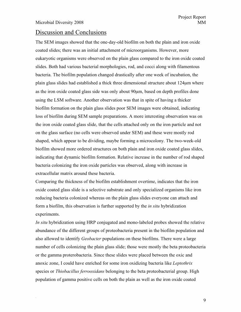
One-week-old biofilm analysis-
One week old slides under SEM look remarkably different compared to one day old
slides, after the initial attachment, it appears that there are microcolony formation on the
iron oxide coated glass slide, however very few cells were observed for the plain glass
control slides (Fig. 4a, b, c, d). Figure 4e shows an SEM image of a one-week-old biofilm
developed on an iron foil; small rod shaped cells can be seen. It was very difficult to
image biofilm on the iron foil as it was negatively charged and the particles moved due to
the electron bombardment.
An unusual observation was seen when the DAPI stained one-week plain glass slides
showed a thick biofilm formation compared to SEM images, indicating loss of attached
cells during SEM sample preparation (Fig. 5a, b, c, d). On iron-coated glass slides very
distinct rod shaped cells were observed attached only to the iron oxide particles. Biofilm
slides were hybridized with general eubacterial, non338 and proteobacterial specific
probes, along with an archaeal probes, however no archaeal signals were detected.
Thickness of these biofilms was detected by doing a z stacking and then depth profiles
were generated using LSM software. The plain slide was about~124µm and the iron
coated was ~90µm thick. Example of a CARD-FISH with delta probes was done on one-
week-old plain and another one done with delta and Geobacter on a one-week iron oxide
glass slide which is shown in figure 6 and 7.

… Project Report Microbial Diversity 2008 MM
Ohm
8
Two-week-old biofilm-
Slides collected after two weeks had a very thick biofilm development, as seen with
depth profiling. SEM image of plain glass slide showed excess extracellular matrix as
seen in fig.8a. Iron oxide coated glass slide showed an increase in bacterial cells attached
to the iron oxide particles along with extracellular matrix production. FISH and
CARD/FISH analysis done on two-week samples showed relative abundance of beta,
gamma and delta proteobacteria, also showed 164µm thick shown in figure 9. Most of the
delta positive cells were also Geobacter positive, indicating colonization of the iron oxide
particles essentially by Geobacter species (Fig 10).
Copper foil as s substrate could not be carried out till end as, the one of the foil was lost
to the swamp during sample collection and the other was used for DNA extraction and
RFLP analysis.
Table 1 shows the approximate numbers of probe specific hybridized cells that were
enumerated by counting several fields through the biofilm. This result shows that the iron
coated glass slide had a dominant population of Geobacter compared to the plain glass
slide, indicating substrate specific attachment preference.
Table1 FISH and CARD/FISH enumerations
Alfa
Beta
Gamma
Delta
Geobacter
- Iron
++
+++
++++
++
+
+ Iron
+
+/-
+++
+++
++
Molecular Results
DNA was extracted from samples collect after two week analysis using the MOBIO
power soil kit, but very low yield of DNA was obtained. Also, 16S PCR amplification
was tried to generate clone libraries and RFLP patterns, but due to low DNA yield and
presence of iron, no amplified product was obtained.
… Project Report Microbial Diversity 2008 MM
Ohm
9
Discussion and Conclusions The SEM images showed that the one-day-old biofilm on both the plain and iron oxide
coated slides; there was an initial attachment of microorganisms. However, more
eukaryotic organisms were observed on the plain glass compared to the iron oxide coated
slides. Both had various bacterial morphologies, rod, and cocci along with filamentous
bacteria. The biofilm population changed drastically after one week of incubation, the
plain glass slides had established a thick three dimensional structure about 124µm where
as the iron oxide coated glass side was only about 90µm, based on depth profiles done
using the LSM software. Another observation was that in spite of having a thicker
biofilm formation on the plain glass slides poor SEM images were obtained, indicating
loss of biofilm during SEM sample preparations. A more interesting observation was on
the iron oxide coated glass slide, that the cells attached only on the iron particle and not
on the glass surface (no cells were observed under SEM) and these were mostly rod
shaped, which appear to be dividing, maybe forming a microcolony. The two-week-old
biofilm showed more ordered structures on both plain and iron oxide coated glass slides,
indicating that dynamic biofilm formation. Relative increase in the number of rod shaped
bacteria colonizing the iron oxide particles was observed, along with increase in
extracellular matrix around these bacteria.
Comparing the thickness of the biofilm establishment overtime, indicates that the iron
oxide coated glass slide is a selective substrate and only specialized organisms like iron
reducing bacteria colonized whereas on the plain glass slides everyone can attach and
form a biofilm, this observation is further supported by the in situ hybridization
experiments.
In situ hybridization using HRP conjugated and mono-labeled probes showed the relative
abundance of the different groups of proteobacteria present in the biofilm population and
also allowed to identify Geobacter populations on these biofilms. There were a large
number of cells colonizing the plain glass slide; those were mostly the beta proteobacteria
or the gamma proterobacteria. Since these slides were placed between the oxic and
anoxic zone, I could have enriched for some iron oxidizing bacteria like Leptothrix
species or Thiobacillus ferrooxidans belonging to the beta proteobacterial group. High
population of gamma positive cells on both the plain as well as the iron oxide coated

… Project Report Microbial Diversity 2008 MM
Ohm
10
slides could possibly indicate presence of Shewanella species associated with iron
reduction. Based on in situ hybridization using mono-labeled Geobacter specific probes,
confirmed a relative high abundance of Geobacter species on the iron oxide coated slides,
also a low signal was detected on the plain glass slides.
Future Experiments This project has shown some very preliminary results that certain bacterial cells
selectively attach to specific substrates like iron oxide in high numbers, but non
selectively attach to inert surfaces like glass. One very interesting follow up hypothesis is
that what makes iron reducing bacteria like Geobacter selectively attach to iron oxide
coated glass surface in high numbers compared to plain glass slide in iron rich ecosystem.
One interesting question is how does Geobacter sense iron, ferrotaxsis? Is it the
availability of the iron (form of iron) or requirement of a solid surface to perform co-
ordinated functions in a density dependent manner? Other follow-up experiments could
be routine questions like who are these other bacteria besides Geobacter that are growing
on iron oxide? This can be answered by performing 16S identification analysis and
setting up enrichments from these biofilms.
Acknowledgements I would like to thank Bill and Tom for their continuous inputs on this project. Bill for the
long discussions about logistics of this project and Tom for helping me figure out how to
make iron oxide coated slides. All the TA’s for their support and helpful discussions
specially, Amy for helping me with the confocal and Dagmar for teaching me FISH and
CARD-FISH and constantly helping me to optimize it for my biofilms, also for blessing
all my FISH experiments! Of course Stephanie for always telling us not to forget to have
fun, for making coffee and spending hours trying to install ARB on my laptop. Dr. Derek
Lovely for sending me Geobacter specific probes. This course had been a wonderful
experience, and has helped stimulate lots of ideas and new directions for my thesis. I
would like to thank the class of 2008 for making this an excellent summer. Especially
Lee and Renate for going to the marsh with me everyday for sampling. Lastly, I would

… Project Report Microbial Diversity 2008 MM
Ohm
11
like to thank my advisor Dr. Daniel Bond for encouraging me to apply for this course and
also for sponsoring me.
References 1) Ishii K., MuBmann M., MacGregor B.J., and Amann R. (2004). An improved
fluorescence in situ hybridization protocol for the identification of bacteria and archaea in
the marine sediments. FEMS Microbiol. Ecology 50, 203-212
2) Pernthaler A., Pernthaler J., and Amann R. (2002). Fluorescence in-situ hybridization
and Catalyzed Reporter Desposition for the Identification of Marine Bacteria. Appl.
Environ. Microbiol. 68, No.6, 3094-3101.
3) Siering P.L., and Ghiorse W.C (1997). Development and Application of 16S rRNA-
Targeted Probes for Detection of Iron and Manganese Oxidizing Sheathed Bacteria in
Environmental Samples. Appl. Environ. Microbiol. 63 No. 2, 644-651.
4) Ritcher H., Lanthier M., Nevin K. P., and Lovely D.R. (2007). Lack of Electricity
Production by Pelobacter carbinolicus Indicates that the Capacity for Fe (III) Oxide
Reduction Does Not Necessary Confer Electron Transfer Ability to Fuel Cell Anodes.
Appl. Environ. Microbiol. 73 No. 16, 5347-5353.

Free floating planktonic cells
Surface
Initial attachment
Formation of a microcolony
Three dimensional biofilmFig.1
Biofilm formation

Figure 2
a b
c d
Plain glass slide, one day old
Iron coated one day old slides

Figure 3
One day DAPI Stain
Plaina
Iron coatedb
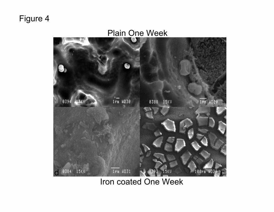
Figure 4
Plain One Week
Iron coated One Week
a b
cd

Figure 4e
Iron Foil One week
Figure 5
Plain one week
DAPI Stain for Depth Profiles
Iron one weeka
b
c
d

Figure 6
One week iron oxide slide
-DAPI-Delta specific

Figure 7 One week iron oxide slide
-DAPI-Delta-Geobacter
Overlay
CARD-FISH and Mono-labeled
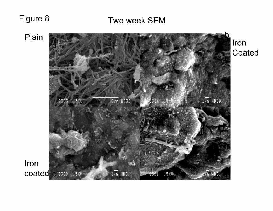
Figure 8 Two week SEM
Plaina
bIron Coated
Iron coated c d

Figure 9 Two week iron oxide biofilm
Depth Profile

Figure 10Mono-labeled FISH on two week iron oxide
-DAPI-GeobacterOverlay


![The Effect of Gold and Iron-Oxide Nanoparticles on Biofilm …€¦ · costs related to healthcare-associated infection are greater thanC7billioninEuropeannually[2].Amajorproportion](https://static.fdocuments.us/doc/165x107/5f56e2e1ed1edc54407308d6/the-effect-of-gold-and-iron-oxide-nanoparticles-on-biofilm-costs-related-to-healthcare-associated.jpg)