Trabeculectomy, trabeculotomy, goniotomy and their complications
99
Trabeculectomy, Trabeculotomy, Goniotomy and their complications DR. NAMRATA GUPTA
-
Upload
namrata-gupta -
Category
Healthcare
-
view
651 -
download
11
Transcript of Trabeculectomy, trabeculotomy, goniotomy and their complications

Trabeculectomy, Trabeculotomy,
Goniotomy and their complications
DR. NAMRATA GUPTA

Incisional Surgery
Frequently used for chronic forms of glaucoma in adults –Filtering procedure
External filtration:
• Full thickness(Scheie) procedures: Thermal sclerostomy, sclerectomy, Elliott’s trephination
• Guarded procedures: Trabeculectomy
Internal filtration:
• Cyclodialysis
Trabecular meshwork disruption:
• Trabeculotomy ab externo
• Goniotomy

Trabeculectomy

Introduction
• Trabeculectomy, a guarded filteration procedure remains the ‘gold standard’ for long lasting intraocular pressure reduction in uncontrolled glaucoma
• Popularized by Cairns (1968)

Mechanism of action
• Creation of a fistula at the limbus which allows a direct communication between anterior chamber and subconjunctival space bypassing the trabecular meshwork, schlemm canal and collecting channels

Theories of mechanism
1543
2

Pre-operative evaluation

Indications
• Intraocular pressure too high to prevent future glaucoma damage and functional visual loss
• Documented progression of glaucoma damage at current level of intraocular pressure with treatment
• Presumed rapid rate of progression of glaucoma damage without intervention
• Poor compliance with medical therapy : cost , inconvenience, understanding of disease
• Intolerance to medical therapy due to side effects

Assessment of filtration risk factors
• Thorough slit lamp evaluation, gonioscopy, record review of past surgery
• Best site for filtration determined: PAS, IOL and haptic orientation, aberrant vessels, wound dehiscence, limbalscarring, vitreous prolapse
• Risk factors for filtration failure: African race, uveitis, aphakia, neovascular glaucoma, prior failed filtration, prolonged anti-glaucoma medication
• Ocular surface disease: ocular rosacea, blepheritis

Surgical Technique
Perioperative preparations:
• Intravenous sedation : pediatric, adults unable to co-operate
• Local anesthesia: Retro-bulbar injection, peribulbarinjection, subtenon, subconjunctival or topical anesthesia
• Positioning to maximize exposure to superior globe: protection by lid, no diplopia after PI
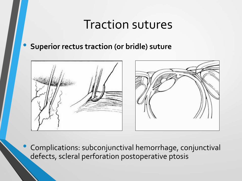
Traction sutures
• Superior rectus traction (or bridle) suture
• Complications: subconjunctival hemorrhage, conjunctivaldefects, scleral perforation postoperative ptosis

Traction sutures
• Clear Corneal traction sutures: A 7-0 polyglactin (vicryl) suture is passed through approx. ¾ th thickness of superior peripheral cornea(4-5 mm width) 1mm form limbus
• May distort the cornea and anterior chamber during surgery

Conjunctival flap
General principles:
• Gentle handling- buttonholing (antifibrotics)
• Removal of portion of Tenon capsule : source of fibroblast (controversial)
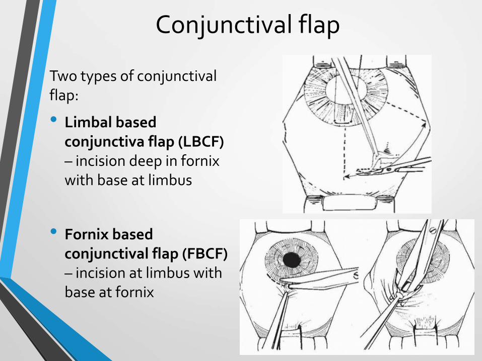
Conjunctival flap
Two types of conjunctivalflap:
• Limbal based conjunctiva flap (LBCF) – incision deep in fornix with base at limbus
• Fornix based conjunctival flap (FBCF) – incision at limbus with base at fornix
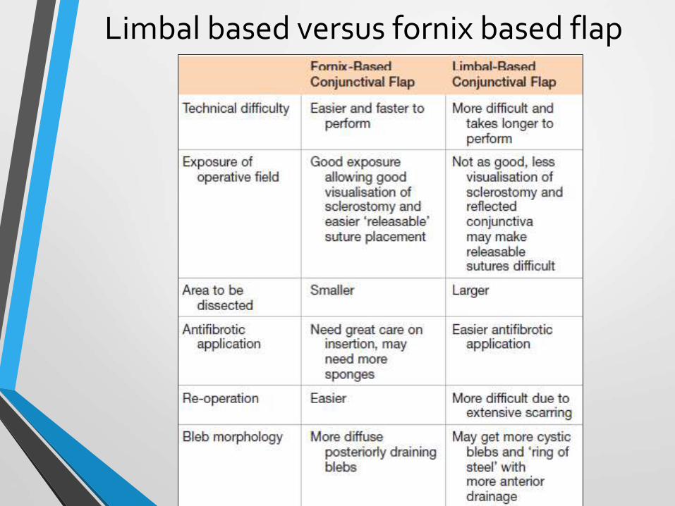
Limbal based versus fornix based flap
Anti-metabolite decision
• Adjunctive antimetabolites inhibit the natural healing response that may preclude successful filtration surgery
• Stratified according to patient risk factors

5-Fluorouracil
• Pyrimidine analogue antimetabolite
• Inhibition of thymidylate synthesis, blocks DNA synthesis
• Inhibit fibroblastic proliferation
• Concentration: Cellulose sponge soaked in 50mg/ml for 5 mins
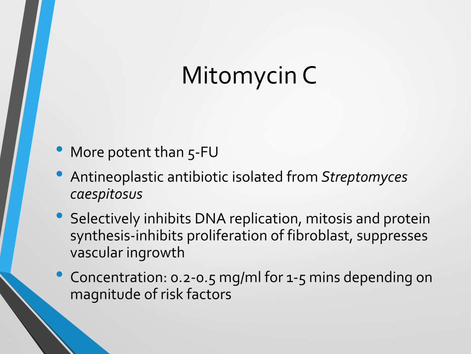
Mitomycin C
• More potent than 5-FU
• Antineoplastic antibiotic isolated from Streptomyces caespitosus
• Selectively inhibits DNA replication, mitosis and protein synthesis-inhibits proliferation of fibroblast, suppresses vascular ingrowth
• Concentration: 0.2-0.5 mg/ml for 1-5 mins depending on magnitude of risk factors

Delivering the anti-fibrotic agent
• Cellulose sponge ̴5 × 3 mm soaked in antimetabolite is placed under dissected tenon’s capsule for 5 mins before paracentesis of AC followed by thorough irrigation with BSS
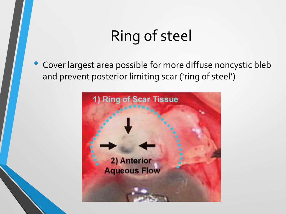
Ring of steel
• Cover largest area possible for more diffuse noncystic bleb and prevent posterior limiting scar (‘ring of steel’)

Moorfields Eye Hospital (More Flow) intra-operative Single Dose Anti- Scarring
Regimen 2006
Low Risk Patients (Nothing or intra-operative 5-FU 50mg/ml)
• No risk factors
• Topical medications (beta-blockers/pilocarpine)
• Afro-Caribean
• Youth <40 with no other risk factors

Intermediate risk patients (intraoperative 5-FU 50mg/ml or MMC 0.2 mg/ml)
• Topical medications (adrenaline)
• Previous cataract surgery without conjunctival incision
• Combined glaucoma filtration surgery/cataract extraction
• Previous conjunctival surgery eg. Squint surgery/ detachment surgery/ trabeculotomy
More flow contd.
High risk patients (Intra operative MMC 0.5 mg/ml)
• Neovascular glaucoma
• Chronic persistent Uveitis
• Previous failed trabeculectomy/tubes
• Chronic conjunctival inflammation
• Secondary glaucomas: inflammatory, post-traumatic angle recession, iridocorneal endothelial syndrome
• Aphakic glaucoma
More flow contd.

Complication of antimetabolites
• Corneal epithelial defects
• Post-operative wound leaks
• Cystic thin walled bleb: Chronic hypotony, late-onset bleb leak, endophthalmitis

Scleral flap dissection
• Provide resistance to aqueous outflow and prevent hypotony
• Act as a safety valve to minimize IOP fluctuations
• Technique:
• Rectangular(3.5 x 4.5 mm) or triangular partial thickness ( ̴50%)
• Lamellar dissection anteriorly just into clear cornea

Difficulties
• Thin scleral flaps– reduced flap resistence and hypotony
• Flap dehiscence, full thinkness button holes, cheese wiring
• Important in eyes with low scleral rigidity: buphthalmos, myopia, antifibrotic use

Paracentesis
An oblique paracentesis in inferior cornea allows fine control of the AC:
• IOP titration after tying the scleral flap sutures
• Reformation (or decompression ) of AC intra or post operatively- BSS or viscoelastics
• Infusion for continuous IOP maintainence in high risk
• Control and washout of AC hemorrhage
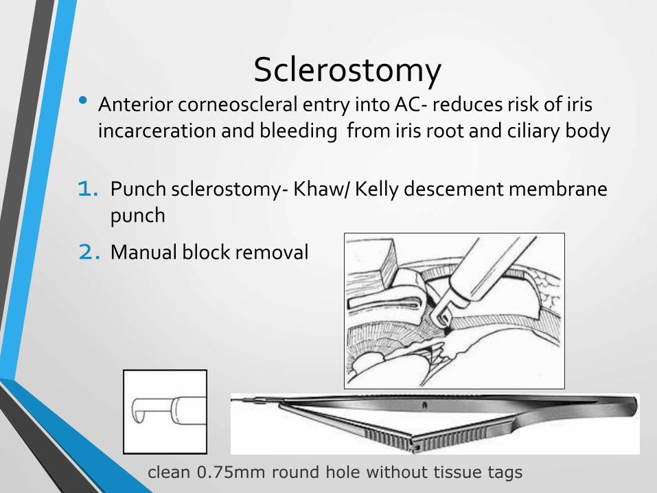
Sclerostomy• Anterior corneoscleral entry into AC- reduces risk of iris
incarceration and bleeding from iris root and ciliary body
1. Punch sclerostomy- Khaw/ Kelly descement membrane punch
2. Manual block removal
clean 0.75mm round hole without tissue tags

Peripheral iridectomy
• Routine part of all standard filtering procedures
• Performed from sclerostomy site with extent beyond sclerostomy margins to avoid obstruction of sclerostomy by peripheral iris
Complications:
• Hyphaema, inflammations, iridodialysis

Closure of the wound
• Approximation of scleral flap with nylon 10-0 that achieves mild to moderate resistance to aqueous flow maintaining AC depth is optimal
• Adequate flow resistance can be tested by injecting BSS into AC via paracentesis


Scleral flap sutures- Fixed, Releasable , Adjustable
Fixed sutures and laser suture lysis
• Laser suture lysis introduced by Lieberman (1983) using argon laser
• Facilitated by compressing overlying conjunctiva to visualize scleral suture or high magnification suture lysis contact lens (Hoskins or Blumenthal lens)
• Argon laser: 50µm, 0.1 sec duration, 250-1000 mW power
• Within first 3 weeks: enhance filtration before scarring occurs
• Delayed (upto 8 weeks) if intraoperative antimetabolite used

Laser suture lysis

Releasable sutures
• Preferred: Scleral flap sutures obscured by subconjunctivalhemorrhage, thickened tenon’s capsule or fibrosis
• First originated from Shaffer et al (1971)
• Simple, low-cost and efficacious


Adjustable sutures
• Allows trans-conjunctival adjustment of tension post-operatively for gradual titration of IOP using specially designed forcep with blunt tip
• Khaw adjustable suture forcep

Adjustable suture forcep

Post-operative managementMedications:
• Topical steroids: early restoration of blood aqueous barrier and suppression of wound healing
• Prednisolone acetate (1%) 2 hourly for 2 weeks and tapered over 8 weeks
• Topical antibiotics: 4 weeks post operatively
• Topical mydriatic/cycloplegic agent : Atropine 1% prevents AC shallowing and risk of malignant glaucoma
• Oral or IV steroids: not routinely used , in severe uveiticglaucoma

Scleral flap suture manipulation
• Removal of one or more releasable scleral flap sutures
• Laser suture lysis
• Adjustable suture loosening
Incorrect timing may result in hypotony or permanent subconjunctival fibrosis and GFS failure
Adjuvant subconjunctival 5-Fluorouracil
• Inhibits fibrosis in Tenon’s layer
• After first postoperative week for up to several months to modulate wound healing (useful in presence of subtenonlake)
• 5mg (0.1 ml of 50mg/ml) 5-FU deep in superior fornix of 90° from the bleb
Adjuvant subconjunctival 5-Fluorouracil
• Signs of impending bleb failure
• Increased bleb vascularity
• Thickening of conjunctiva and tenon’s capsule
• Reduction in bleb size and height
• Reduction of conjunctival microcyst
• Progressive elevation of IOP

Bleb Needling Revision
• Failure of previous methods
• Puncturing and loosening the scar tissue of filtration bleb to increase sub-tenon’s aqueous lake
Two types of BNR:
1. Sub-Tenon’s Needling
2. Subscleral flap Needling
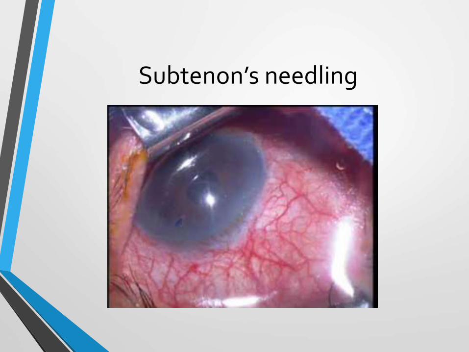
Subtenon’s needling

Subscleral Flap Needling
• Early post operative subscleral flap needling if sub tenon’sNeedling fails

Complications
Intra-operative complications
1. Conjunctival tear
2. Hemorrhage
3. Scleral flap damage
4. Supra-choroidal Hemorrhage
5. Decompression Retinopathy

Intra operative Complications
• Tearing or buttonholing of cinjunctival flap:
Prevention :
• Minimal handling
• Topical adrenaline to reduce vascularity and bleeding
• Stromal hydration- BSS injected under cinunctiva/tenon to make them thicker
• Blunt dissection

Tearing or buttonholing of conjunctival flap
Management:
• Small holes- Tissue adhesives, light bipolar cautery
Figure of 8 or mattress suture
• Large holes- Purse string vicryl suture

Hemorrhage:
Should be minimized as blood is potent stimulus for fibrosis
Risk factors:
• Long term anti-glaucoma medication
• Aspirin, anti-coagulants
Management:
• Wet field diathermy
• Gentle sustained pressure over fistula
• Large air bubble or viscoelastic in AC

Scleral flap damage: tearing and buttonholing
• Avoid thin flap and excessive manipulation
Management:
• Minor damage- repair
• Severe damage- autologous or donor sclera patch

Suprachoroidal hemorrhage: Sudden reduction in IOP with rupture of a large choroidal vessel
• Risk factors:
• High pre-operative pressure
• Generalized atherosclerosis
• Elevated intraoperative pulse
• Young patient
• Large eyes, nanophthalmos
• Sturge –weber syndrome
• Management:
• All wounds closed rapidly
• Peripheral SCH: conservative
• Extensive SCH- rainage

Decompression retinopathy
• Retinal hemorrhage following rapid IOP reduction
Vitreous loss
• Inadvertent damage to lens/zonule complex during PI
• Prevention:
• Anterior sclerostomy
• Avoid basal PI’s
• Tube surgery in iridolenticluar instability

Post operative complications
Early post-operative complications:
1) Hypotony and shallow anterior chamber
2) Hypotony and deep anterior chamber
3) Elevated intraocular pressure and flat anterior chamber
4) Elevated intraocular pressure and deep anterior chamber

Spaeth classification of post operative shallow anterior chamber:
• Grade 1: peripheral iris- cornea apposition but preserved AC in front of pupillary space
• Grade 2: Greater apposition between mid iris and cornea but space between lens(or vitreous) and cornea in pupillary space is retained
• Grade 3: Flat AC with complete contact of iris and pupillary space with posterior surface of cornea

Hypotony with flat anterior chamber:
A. Negative seidel test with grade I or II flat AC with hypotony with a formed bleb- Scleral flap leak
Management:
• Conservative : Topical steroids and long acting cycloplegic
• Restricted activity and avoid Valsalva
• Pressure patch of filtration site
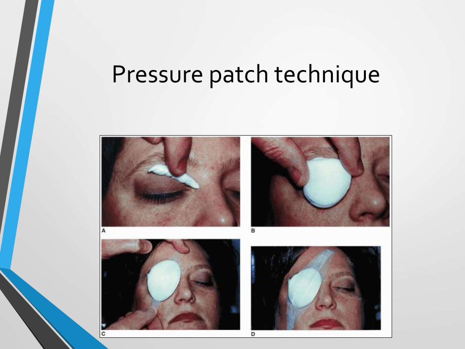
Pressure patch technique

Hypotony with flat anterior chamber:
B. Positive Seidel test with grade I flat anterior chamber and low intraocular pressure- small leaks around sutures
• Increased frequency of topical antibiotics
• Pressure patching
• Therapeutic contact lens- Fibrin tissue glue or cyanoacrylate glue
• Simmon shell

C. Positive seidel’s test with low bleb height with grade II or III flat AC with hypotony: Conjunctival button hole
Management:
• Conjunctival tear:
Healthy conjunctiva: Purse string suture
Fragile conjunctiva: Pressure patching, 20-22 mm BCL, Tenoncapsule sutured a tear site
• AC reformation: Air, BSS, viscoelastics

Hypotony with flat anterior chamber:
B. Excessive filtration:
• Loose scleral flap closure or large filtering bleb (anti-fibrotic)
• Large soft contact lens
• Symblepheron ring
• Simmons shell
• Surgical: Viscoelastics, BSS, 15% perfluoropropane(C3F8), 40% sulfur hexa fluoride(SF6)
Symblepheron ring
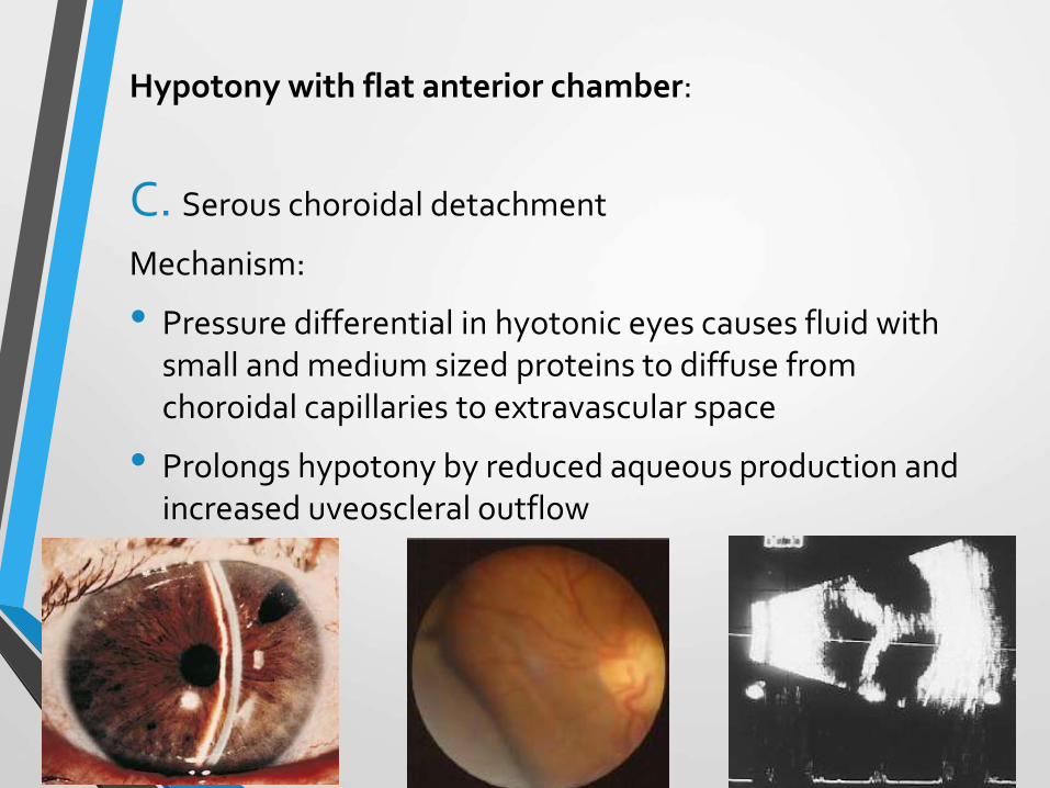
Hypotony with flat anterior chamber:
C. Serous choroidal detachment
Mechanism:
• Pressure differential in hyotonic eyes causes fluid with small and medium sized proteins to diffuse from choroidal capillaries to extravascular space
• Prolongs hypotony by reduced aqueous production and increased uveoscleral outflow

Management:
• Resolve spontaneously after IOP rises above 7-9 mmHg: Atropine and oral and topical steroids
• Surgery: Persistence of grade 3 flat AC more than 1 week with corneal endothelial compromise or persistence of ‘kissing choroidals’ or suspected SCH:
• One or more sclerotomies 4mmbehind the limbus over pars plana in inferior quadrants and deepening AC with BSS

Hypotony with deep anterior chamber:
• A lower-than-normal IOP in first 2 weeks with no associated complication resolve spontaneously
• Persistent hypotony(<6mmg): Hypotony Maculopathy
• Fine macular striae radiating from fovea
• Extensive choroidal folds
• Tortuous retinal vessels
• Disc edema
• No evidence of vascular leak
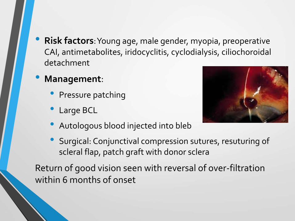
• Risk factors: Young age, male gender, myopia, preoperative CAI, antimetabolites, iridocyclitis, cyclodialysis, ciliochoroidaldetachment
• Management:
• Pressure patching
• Large BCL
• Autologous blood injected into bleb
• Surgical: Conjunctival compression sutures, resuturing of scleral flap, patch graft with donor sclera
Return of good vision seen with reversal of over-filtration within 6 months of onset

3. Elevated IOP with flat anterior chamber
a) Aqueous misdirection syndrome(malignant glaucoma)
b) Delayed suprachoroidal hemorrhage
c) An incomplete iridectomy with pupillary block glaucoma-
Patentcy should be established immediately on diagnosis by laser or, if necessary, incisional surgery

Aqueous misdirection(ciliary block /malignant glaucoma)
• Misidrection of aqueous to circulate into vitreous
• Grade II or III shallow AC
• Higher than expected IOP in early post-operative period
• Patent iridotomy
• Rarity of spontaneous resolution- ‘malignant’

Malignant glaucoma
Management:
• Atropine and topical steroids
• Aqueous suppressants: Brimonidine, Timolol, CAI’s, IV mannitol
• Surgical intervention: waiting period of 5 days advised
• Nd-Yag laser- pupillary block, retrocapsular block or hyaloidblock
• Pars plana vitreous aspiration or Pars plana vitrectomy
• Cyclodiode photocoagulation (refractory cases)

Suprachoroidal hemorrhage
Risk factors:
• Aphakic
• Post-traumatic
• Vitrectomised eyes
• Large eyes- Pathological myopia/Congenital glaucoma
• Systemic anticoagulants
• Significant post-operative hypotony

Suprachoroidal hemorrhage
• Sudden severe pain with loss of vision during first 4-5 post-operative days
• High IOP with nausea and vomiting
Management:
• Aqueous suppressants/ hyperosmotic agents
• Surgical drainage: Posterior sclerostomy over area of elevated choroid after 7-10 days with simultaneous AC infusion
Poor prognosis with concomitant RD or 360° SCH

4. Elevated IOP with deep anterior chamber:
Indicates inadequate filtration
• Tight scleral flap
• Obstruction of fistula by iris, ciliary process, lens, blood or vitreous
Management:
• Tight flap: Laser suture lysis
• Obstruction of fistula: Retraction of obstructing tissue with argon laser of Nd-Yag disruption under gonioscopy
• Pressure on posterior lip: clot or vitreous at sclerostomy site
• Internal bleb revision: Cyclodialysis spatula inserted 90-180° away through clear corneal incision into the fistula to elevate scleral flap

Failing blebCharacteristics:
• Typically low-flat
• Heavily vascularized
• No microcysts
Management:
• Increased frequency of topical steroid ± subconjunctival steroid/5-FU
• Intermittent digital pressure
• Laser suture lysis/ removal of adjustable suture
• Failure of above: Anti-glaucoma resumed and revision of surgery
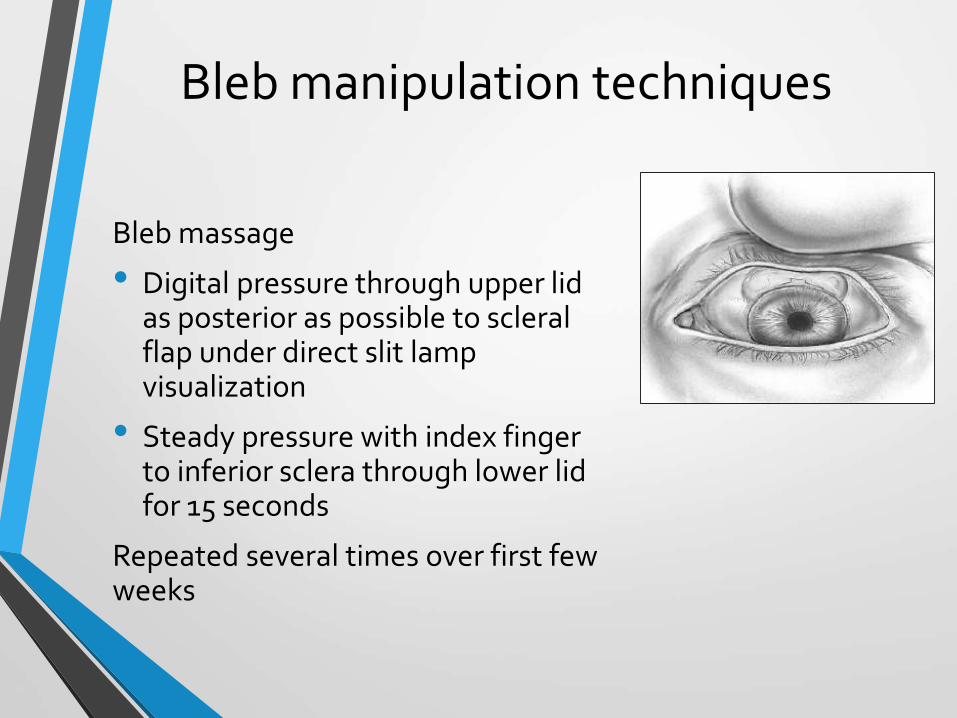
Bleb manipulation techniques
Bleb massage
• Digital pressure through upper lid as posterior as possible to scleral flap under direct slit lamp visualization
• Steady pressure with index finger to inferior sclera through lower lid for 15 seconds
Repeated several times over first few weeks

Encapsulated filtering bleb
• In 3.6% - 28% within first 2 months after surgery
• Tenon capsule cysts: Highly elevated, smooth doomed bleb with intervening avascular spaces and no microcysts
• Patent sclerostomy on gonioscopy
• Management:
• Resume anti-glaucoma medication until improvement occurs
• Subconjunctival needling with 5mg of 5-FU subconjunctivally

Other early post-operative complications
• Uveitis
• Hyphaema
• Dellen: Adjacent to large filtering bleb
• Loss of central vision:
“Snuff-out syndrome”- common in old age, hypotony, macular splitting(visual field loss within 10° of fixation)

Late post operative complication
a) Late failure of filtration :
• Fibrosis of scleral flap
• Scarring of conjunctiva
• Poor response to drugs or digital pressure
• Management:
• Ab externo or ab interno incision of membranous tissue over fistula
• Argon laser for pigmented membrane
• Nd-Yag laser for non pigmented membrane
• Incisional surgery
Late post operative complication
b) Leaking filtering bleb
• Thin walled large avascular blebs are at risk
• Use of adjunctive anti-metabolite
• Management:
• Cyanoacrylate glue, autologous fibrin, large BCL
• Bleb revision: New conjuntival flap or rotational conunctivalflap

c) Bleb related infections
• Blebitis and endophthalmitis are potentially blinding emergencies
• Tends to occur months or years after surgery
• Prevention of late infection
• Avoid excessive antifibrotic treatment
• Avoid thin scleral flaps
• Bleb under upper lid
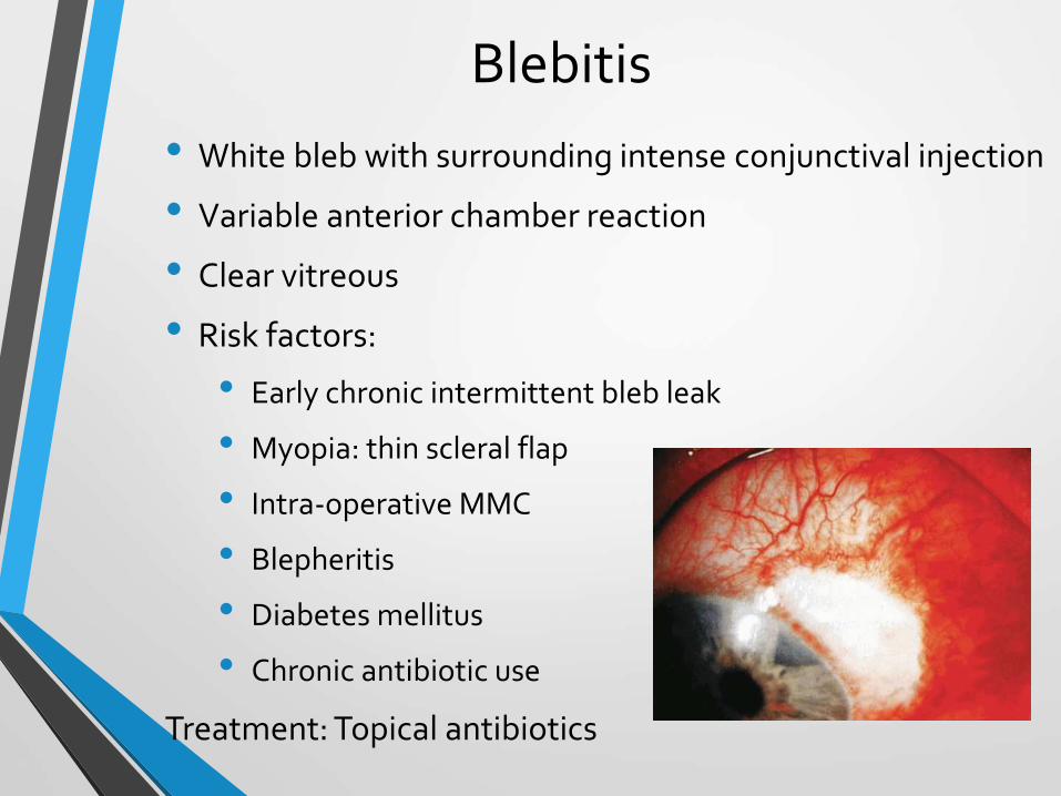
Blebitis
• White bleb with surrounding intense conjunctival injection
• Variable anterior chamber reaction
• Clear vitreous
• Risk factors:
• Early chronic intermittent bleb leak
• Myopia: thin scleral flap
• Intra-operative MMC
• Blepheritis
• Diabetes mellitus
• Chronic antibiotic use
Treatment: Topical antibiotics

Bleb-related Endopthalmitis
1. Early postoperative Endophthalmitis:
• Onset within first 3 months
• Staphylococcus epidermidis
2. Delayed- onset Endophthalmitis
• Onset after 3 months
• Streptococcus, staphylococcus, H. influenzae
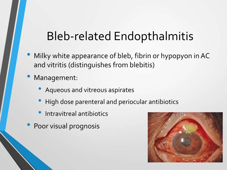
Bleb-related Endopthalmitis
• Milky white appearance of bleb, fibrin or hypopyon in AC and vitritis (distinguishes from blebitis)
• Management:
• Aqueous and vitreous aspirates
• High dose parenteral and periocular antibiotics
• Intravitreal antibiotics
• Poor visual prognosis
Other late post operative complications
• Development and progression of cataracts
• Spontaneous hyphaema
• Hypotony and ciliochoroidal detachment: inflammation, aqueous suppressants
• Rare: Upperlid retraction, sympathetic ophthalmia

Goniotomy and Trabeculotomy

Definition
Goniosurgery
• First introduced by Barkan (1938)
• Specific surgical techniques applied to the anterior segment of the eye for the treatment of childhood glaucoma
• Principle:
Incision of the obstructing trabecular meshwork tissue allows direct conduit between anterior chamber and schlemm’s canal
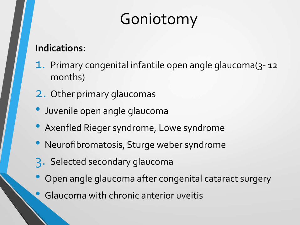
Goniotomy
Indications:
1. Primary congenital infantile open angle glaucoma(3- 12 months)
2. Other primary glaucomas
• Juvenile open angle glaucoma
• Axenfled Rieger syndrome, Lowe syndrome
• Neurofibromatosis, Sturge weber syndrome
3. Selected secondary glaucoma
• Open angle glaucoma after congenital cataract surgery
• Glaucoma with chronic anterior uveitis

• Goniotomy is advisable as soon as possible after diagnosis as early as second or third day of life in glaucoma present at birth
• It is recommended once or twice in children younger than 2-3 years
• Shaffer reported that one to two goniotomies cured 94% of cases between 1 and 24 months
• Advantages: Does not disturb conjunctiva, direct visualization of trabecular meshwork

GoniotomyPreoperative care:
• Preoperative glaucoma medications to reduce IOP and clear cornea
• Beta-blockers (0.25%), Oral acetazolamide (10-15 mg/day) or topical dorzolamide with apraclonidine 0.5%
• Pilocarpine 1-2% just before surgery to promote miosis
• Rule out congenital NLD block or URTI
• Isopropyl alcohol 7-% to remove corneal epithelium if edema persists(intraoperatively)

• Procedure:
• Eye positioned with plane of iris tilted away from surgeon at 45°
• Locking fixation forceps on vertical recti, if nasal or temporal goniotomy is to be performed
• Operating microscope tilted towards the surgeon for comfortable view of angle through goniolens

• Swan goniotomy knife
• Swan Jacob goniolens


Complications:
• Risk of general anesthesia in neonates and infants
• Hemorrhage: Incision into anterior ciliary body, sclera
• Cataract: lens injury
• Infection
• Epithelial ingrowth
• Failure: Incision anterior to schwalbe line

Post-operative care:
• Patient’s head turned towards side of puncture wound for first hour that facilitates blood from goniotomy incision to flow away
• Routine F/U: Infection, cornea, AC, IOP
• EUA after 4-6 weeks
• Reoperation may be performed after 3 weeks
• Trabeculotomy: If two goniotomies fail

Trabeculotomy
• Performed by cannulating the schlemm canal from an external approach with subsequent centripetal rupture through the trabecular meshwork into the anterior chamber
Indications:
• Same as that of goniotomy in presence of corneal edema or opacification
• After failure of two previous goniotomies
• Combined with trabeculectomy (failure to cannulateschlemm canal)
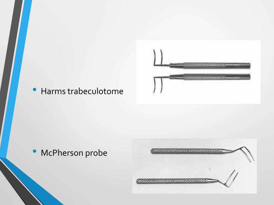
• Harms trabeculotome
• McPherson probe

Procedure • Thick scleral flap created(deeper than trabeculectomy)
• Radial incision made in bed of flap at sclero-corneal junction and deepened until schlemm canal identified anterior to scleral spur(near posterior limbal gray zone)
• A 6-0 blunt tip prolene is threaded on either side of radial incision to confirm patency
• Internal arm of trabeculotome passed gently into canal and rotated into AC through intervening TM

Purse- string 360° Trabeculotomy
• After unroofing and identifying schlemm’s canal, a 6-0 prolene suture is threaded 360°around
• After reappearing form the opposite direction at initial surgical site, suture is drawn like a purse string rupturing the entire canal in centripetal fashion

Complications
• Intra or Post operative hyphaema
• Descement’s detachment
• Iridodialysis
• Iris prolapse

Summary
• Trabeculectomy is a surgical procedure featuring a partial thickness scleral flap that creates fistula between anterior chamber and subconjunctival space for filtration of aqueous fluid and formation of bleb ± antimetabolites
• Gold standard for glaucoma surgery
• Associated features- Hypotony, choroidal detachment, bleb leak and bleb associated endophthalmitis
• Goniotomy and trabeculotomy are specific surgical techniques applied to anterior segment for treatment of childhood glaucoma

Bibliography
• R.Rand Allingham. Shields Text Book of Glaucoma, 6th
edition , 2011
• Robert L Stamper, Marc F Lieberman, Michael V Drake. Becker- Shaffer’s Diagnosis & Therapy of the Glaucomas, 8th edition, 2010.
• Daniel M Albert, Joan W Miller, Dimitri T Azar. Albert & Jakobiec’s Principle and Practice of Ophthalmology, 3rd
edition, 2008.
• American Academy of Ophthalmology. The Glaucoma, Section -7, 2011-12.
• Myron Yanoff and Jay S. Duker – Ophthalmology, 3rd
edition, 2009.

THANK YOU