Towards a Statistical Atlas of Cardiac Fiber Structure · Towards a Statistical Atlas of Cardiac...
9
Towards a Statistical Atlas of Cardiac Fiber Structure Jean-Marc Peyrat 1 , Maxime Sermesant 1 , Xavier Pennec 1 , Herv´ e Delingette 1 , Chenyang Xu 2 , Elliot McVeigh 3 , and Nicholas Ayache 1 1 INRIA - Asclepios Research Project, Sophia Antipolis, France [email protected] 2 Siemens Corporate Research, Princeton, New Jersey, USA 3 Laboratory of Cardiac Energetics, National Heart Lung and Blood Institute, National Institute of Health, Bethesda, Maryland, USA Abstract. We propose here a framework to build a statistical atlas of diffusion tensors of canine hearts. The anatomical images of seven hearts are first non-rigidly registered in the same reference frame and their as- sociated diffusion tensors are then transformed with a method that pre- serves the cardiac laminar sheets. In this referential frame, the mean ten- sor and its covariance matrix are computed based on the Log-Euclidean framework. With this method, we can produce a smooth mean tensor field that is suited for fiber tracking algorithms or the electromechanical modeling of the heart. In addition, by examining the covariance matrix at each voxel it is possible to assess the variability of the cardiac fiber directions and of the orientations of laminar sheets. The results show a strong coherence of the diffusion tensors and the fiber orientations among a population of seven normal canine hearts. 1 Introduction While the main geometrical arrangement of myofibers has been known for decades, its variability between subjects and species still remains largely unknown. Un- derstanding this variability is not only important for a better description of physiological principles but also for the planning of patient-specific cardiac ther- apies [15]. Furthermore, the knowledge of the relation between the myocardium shape and its myofiber architecture is an important and required stage towards the construction of computational models of the heart [4, 13] since the fiber ori- entation plays a key role when simulating the electrical and mechanical function of the heart. High resolution measurements of fiber orientation has been recently eased with the use of Diffusion Tensor Imaging (DTI) since there is a correlation between the myocardium fiber structure and diffusion tensors [14]. DTI also has the advantage to provide directly this information in 3D with high resolution but unfortunately it is still not available in vivo due to the cardiac motion. There has been several studies [6, 7, 17] in the past decade that have measured the variability of fiber orientation from DTI (similar studies has been done for the
Transcript of Towards a Statistical Atlas of Cardiac Fiber Structure · Towards a Statistical Atlas of Cardiac...
Towards a Statistical Atlas of Cardiac FiberStructure
Jean-Marc Peyrat1, Maxime Sermesant1, Xavier Pennec1, Herve Delingette1,Chenyang Xu2, Elliot McVeigh3, and Nicholas Ayache1
1 INRIA - Asclepios Research Project, Sophia Antipolis, [email protected]
2 Siemens Corporate Research, Princeton, New Jersey, USA3 Laboratory of Cardiac Energetics, National Heart Lung and Blood Institute,
National Institute of Health, Bethesda, Maryland, USA
Abstract. We propose here a framework to build a statistical atlas ofdiffusion tensors of canine hearts. The anatomical images of seven heartsare first non-rigidly registered in the same reference frame and their as-sociated diffusion tensors are then transformed with a method that pre-serves the cardiac laminar sheets. In this referential frame, the mean ten-sor and its covariance matrix are computed based on the Log-Euclideanframework. With this method, we can produce a smooth mean tensorfield that is suited for fiber tracking algorithms or the electromechanicalmodeling of the heart. In addition, by examining the covariance matrixat each voxel it is possible to assess the variability of the cardiac fiberdirections and of the orientations of laminar sheets. The results show astrong coherence of the diffusion tensors and the fiber orientations amonga population of seven normal canine hearts.
1 Introduction
While the main geometrical arrangement of myofibers has been known for decades,its variability between subjects and species still remains largely unknown. Un-derstanding this variability is not only important for a better description ofphysiological principles but also for the planning of patient-specific cardiac ther-apies [15]. Furthermore, the knowledge of the relation between the myocardiumshape and its myofiber architecture is an important and required stage towardsthe construction of computational models of the heart [4, 13] since the fiber ori-entation plays a key role when simulating the electrical and mechanical functionof the heart. High resolution measurements of fiber orientation has been recentlyeased with the use of Diffusion Tensor Imaging (DTI) since there is a correlationbetween the myocardium fiber structure and diffusion tensors [14]. DTI also hasthe advantage to provide directly this information in 3D with high resolution butunfortunately it is still not available in vivo due to the cardiac motion. Therehas been several studies [6, 7, 17] in the past decade that have measured thevariability of fiber orientation from DTI (similar studies has been done for the
brain [9]). These studies estimated the fiber direction as the primary eigenvec-tor of each tensor and for instance compared its transmural variation with thatobserved from dissection experiments.
We propose here to extend these studies by building a statistical model ofthe whole diffusion tensor and not only its first eigenvector. This tensor analysisallows us to study the variability of laminar sheets which are associated with thetertiary eigenvector. Performing this analysis based on vector analysis (insteadof tensor analysis) would have been difficult because the secondary and tertiaryeigenvalues have often very similar values and may lead to interpretation errors.To the best of our knowledge, this is the first attempt to perform a first andsecond order statistical analysis of DT images of canine hearts.
Our statistical analysis proceeds as follows. We first register the canine heartimages in a common reference frame using anatomical MRIs. For each heart, weget a deformation field we use to register and transform properly the diffusiontensors considering properties of the cardiac fiber microstructure. Finally weuse coherent statistical tools on tensors to study the variability of individualhearts from this average model and to evaluate the relevance of such a model.An application of this framework is carried out using a dataset of seven normalcanine hearts.
2 Material and Method
2.1 Data Acquisition
We used a dataset of seven ex vivo fixed normal canine hearts acquired [8]and provided by the Center of Cardiovascular Bioinformatics and Modeling(CCBM)4 at the Johns Hopkins University. Each heart was placed in an acryliccontainer filled with Fomblin, a perfluoropolyether (Ausimon, Thorofare, NJ).Fomblin has a low dielectric effect and minimal MR signal thereby increasingcontrast and eliminating unwanted susceptibility artifacts near the boundaries ofthe heart. The long axis of the hearts was aligned with the z-axis of the scanner.Images were acquired with a 4-element knee phased array coil on a 1.5 T GECV/i MRI Scanner (GE, Medical System, Wausheka, WI) using a gradient sys-tem with 40 mT/m maximum gradient amplitude and a 150 T/m/s slew rate.Different resolutions have been used around 0.3 × 0.3 × 0.9 mm3 and from 14to 28 gradient directions. The images have been subsampled into 128× 128× 64images with a resolution around 0.6 × 0.6 × 1.8 mm3. The temperature duringacquisition varied between 18− 25 ◦C from one heart to another.
2.2 Myocardium Registration
In order to analyze the statistical variability of the tensors without introduc-ing a bias, we choose to register the images based on independent informa-tion: the anatomical MRIs. Before the registration stage, we pre-process semi-automatically the anatomical MRIs by extracting the image background, and by4 http://www.ccbm.jhu.edu/research/DTMRIDS.php

cropping each image above the valve plane. We register each heart on a templategiven by a Procrustes iterative mean estimation. The registration is initializedwith an affine global transformation of the hearts defined from three significantlandmarks: the apex point and the two corner points of the right ventricle onthe valve plane. The apex point is used to define a scaling along the axis of theheart. The matching of the corner points defines a 3D translation, a 2D rota-tion on the valve plane, and a 2D scaling along a line defined by the 2 cornerpoints. The second registration step is based on a hybrid non-rigid intensity- andlandmark-based registration algorithm [5]. This algorithm gives us the ability tointeractively refine the registration of a local region. The output of this processis a dense deformation field for each anatomically registered cardiac image.
2.3 Transformation of Cardiac Diffusion Tensors
The next stage is to transform the DT image based on the estimated deformationfield. For each voxel, the global deformation field is approximated at the firstorder by an affine transformation [16]. This underlying affine transformation A iscomputed from the identity matrix Id and the Jacobian ∇F of the deformationfield F :
A = Id +∇F .
Now we have the well known problem of applying an affine transformationto a DT image. Since it was shown that there is a correlation between thecardiac tissue microstructure and the eigensystem of the diffusion tensors [7,8, 14], we can transform the underlying tissue microstructure with the affinetransformation. Then we reconstitute the diffusion tensor from this transformedmicrostructure knowing the relationship between the two of them. The solutionwe propose here to handle this problem proved to be similar to the Preservationof the Principal Direction (PPD) reorientation strategy proposed in [1]:
V ′1 = AV1
||AV1||V ′
3 = (A−1)T V3
||(A−1)T V3||V ′
2 = V3 ⊗ V1
where V1, V2, V3 are the primary, secondary and tertiary eigenvectors of theoriginal diffusion tensor and where V ′
1 , V ′2 , V ′
3 are the ones of the transformeddiffusion tensor.
The PPD has been already justified and validated in the case of brain DTI [1]but only partially in the case of cardiac DTI. We propose here a more completejustification of the PPD strategy for cardiac DTI transformation.
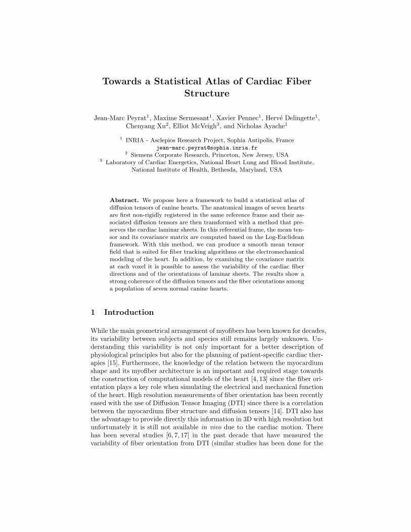
As described in Figure 1, the myocardium microstructure is made of laminarsheets of muscle fibers [10]. The space between the laminar sheets is composedof extracellular water and collagen network linking these sheets together. Thisextracellular space has a mostly unrestricted diffusion in the direction of the twofirst eigenvectors of the diffusion tensor. The interface plane between the laminarsheets and the extracellular space is an important barrier for water moleculesand its normal defines the third eigenvector. The extracellular water between
fibers in the laminar planes can explain the difference between the primaryeigenvector in the fiber direction and the secondary eigenvector orthogonal tothe fiber direction. But the cardiomyocyte geometry is also proposed to explainthe privileged diffusion direction in the fiber orientation. A cardiomyocyte ismuch longer (50-120 µm) than wider (5-25 µm) and much longer than the meanfree path (roughly 10 µm considering the diffusion time of the acquisitions). Itmeans that the cell membrane does not have as much influence on the waterdiffusion in the fiber direction as in the other directions where the width of thecells is close to the mean free path.
Let us now analyze the effect of the basic transformations (translations, rota-tions, shears and scaling) describing an affine transformation. The way to trans-form the fiber structure and thus the diffusion tensors through translation androtation is obvious. The scaling only changes the density of fibers microstructureinside a voxel considering the acquired voxel resolution and finally not the dif-fusion rate. The shearing induced by the affine transformation is not so simpleto apply to the diffusion tensors. We illustrate in Figure 1 the shearing appliedto the basic microstructure of cardiac fibers. The direct transformation of theoriginal eigenvectors Vi leads to the vector AVi and the transformation deducedfrom the fiber structure deformation leads to the vector V ′
i . As we determine thefiber structure deformation through the fiber direction deformation, the trans-formed primary eigenvector V ′
1 is the same as the direct transformation of theoriginal primary eigenvector V1:
V ′1 = AV1
||AV1||.
The tertiary eigenvector is defined by n, the vector normal to the laminarsheets which are considered locally plane. The image of a plane through an affinetransformation is a plane. It means that these laminar sheets are stable. We justhave to determine n′ the unit vector normal to the plane defined by the imageV ′
1 of V1 and the image AV2 of V2 [18]:
n′ = (A−1)T n
||(A−1)T n||.
The transformed secondary eigenvector obtained from the correlation be-tween the structure of the fibers happens to be the one that builds an orthonor-mal basis with the two others. It means that constructing first the secondary (asdone for the PPD) or the tertiary (as we do) and then determine the other oneto obtain an orthonormal basis leads to the same results. This is the key pointthat justifies the use of the PPD in our specific case of cardiac DTI.
2.4 Tensor Statistics
The Log-Euclidean framework [2] provides a consistent and rigorous frameworkto study the statistical variability of DTI for each voxel of the heart. In thisframework the space of diffusion tensors is a vectorial space which means itinherits from all the statistical properties and tools we can get from a vectorial
Fig. 1. [Left] Cardiac Fiber Structure (from LeGrice et al. [10]). - [Middle Left] Originalbasic fiber microstructure with eigenvectors Vi. [Middle Right and Right] Shearingapplied to the basic fiber microstructure : continuous arrows AVi are the transformedeigenvectors through the shearing and dashed arrows V ′
i are the eigenvectors relatedto the correlation between the fiber microstructure and the diffusion tensor.
space. We thus compute the mean of all the registered DTI and the correspondingcovariance [11] at each voxel:
Dlog = exp( 1N
N∑i=1
log(Di))
Cov = 1N − 1
N∑i=1
vect(∆Di) · vect(∆Di)T
where vect(∆Di) is the vectorial representation [11] of ∆Di = log(Di)−log(Dlog).The difficulty to visualize the 6×6 covariance matrix of diffusion tensors leads
us to study first its norm√
Trace(Cov), which allows us to identify the variableand stable regions of the heart. To help us in translating the DTI variability intothe fiber structure variability, we can analyze this covariance matrix extracting 6specific variance parameters at each voxel in the coordinate system of the meantensor: 3 for each eigenvalue variability and the 3 for each pair of orthonormaleigenvectors orientation variability around the third one [12].
3 Results
We applied the proposed framework to the dataset of seven canine hearts pre-sented previously. We obtain a smooth cardiac DTI atlas catching the sharedtransmural variation of the fibers directions (see Figure 2) that is similar to theone generally observed [14]. The norm of the covariance matrix in Figure 3 shows

a global stability of the compact myocardium and several variable regions espe-cially at the RV and LV endocardial apices where the fiber structure is probablyless organized. Some other variabilities at the surface of the heart might also bedue to acquisition or registration artifacts.
In order to have a better interpretation of this covariance matrix and to un-derstand the origin of the variabilities, we decompose it into specific modes asdescribed previously. We can see in Figures 3 that the eigenvalues variabilitiesare homogeneous in the compact myocardium. The mean standard deviationsnormalized by their mean value σi
λiare 0.16, 0.24 and 0.22 for the 1st, 2nd and
3rd eigenvalues. Note that these variabilities and their difference are artificiallyincreased by the difference of the temperature during the different image acqui-sitions (temperature ranging from 18 to 25 ◦C). Anyway mostly the order of theeigenvalues is really meaningful in terms of fiber architecture and thus in termsof electrical conductivity that are introduced in electromechanical modeling (itdoes not seem to be any direct correlation between the diffusion and electri-cal conductivity rates). At least these statistical results give us an idea of theaverage diffusion rates that could help in detecting locally defined pathologies.
The separation of the eigenvalues variability and the eigenvectors orientationvariability is important to evaluate the variability of the myocardial fibers ar-chitecture. As seen in Figures 3, the orientation of the fibers is stable among apopulation (mean standard deviation of 8.8 and 9.4 degrees around the secondaryand the tertiary eigenvectors) for the two rotations in the planes containing theprimary eigenvector. It means that the fiber orientation of the average cardiacDTI is shared by the dataset. The orientation of the laminar sheets describedby the rotation of the plane Span(V2, V3) around V1 shows a much higher meanstandard deviation of 23 degrees. First of all this variability is underevaluatedconsidering that the statistical study we proposed is a simplification at the firstorder [12]. Secondly this variability is due to the difficulty to differentiate thesecondary and tertiary eigenvectors when they have similar eigenvalues. Whenwe study the fiber organization (i.e. the 3 rotation eigenmodes) we are not any-more in the diffusion tensor space and an isotropic plane of diffusion leads to alow accuracy in translating it into structural information. Another explanationmight also be the existence of two populations of symmetric laminar sheets or-ganization in canine hearts [8] corresponding to the optimal configurations tomaximize the systolic shear [3]. Mixing these two populations leads to the com-putation of an average cardiac fiber architecture model that does not representa real case of laminar sheets organization. A computation of the normal of thelaminar sheets and a further study of their orientation variability is essential todetermine its origin and of course to translate an atlas of cardiac DTI into anatlas of the cardiac fiber architecture.
4 Discussion
We proposed a theoretically grounded, simple and powerful framework work-ing directly on diffusion tensors to build an average model and to study their

Fig. 2. [Left and Upper Right] Average canine cardiac DTI. [Lower Right] Fiber trackingin the left ventricle wall with high stiffness parameters for a better visualization of thetransmural variation of fibers directions. The RGB colors represent the components ofthe primary eigenvector Red = |Vx|, Green = |Vy|, Blue = |Vz| with x, y in the axialplane and z orthogonal to the axial plane as described with the colored sphere.
Fig. 3. [upper] Norm of the covariance matrix in 3 orthogonal views. [from middleleft to middle right] Decomposition of the covariance in 6 eigenmodes describing thevariability of the 1st, 2nd, 3rd eigenvalues. [from lower left to lower right] Rotationvariability of the plane orthogonal to the 1st, 2nd and 3rd eigenvectors.
variability over a population of cardiac DTIs. The use of these average modelsinstead of analytical models is an important stage to refine the electromechanicalmodeling with more accurate and reliable data and also with additional informa-tions concerning the anisotropy in the plane orthogonal to the fiber directions.
This framework is a first step towards a statistical atlas of the cardiac fiberarchitecture that will probably lead to a better understanding of the cardiac fiberarchitecture shared by a population of healthy or failing hearts, or to compareand to differentiate populations of hearts (canine-human or normal-failing).
In the next step of our research, a larger dataset of hearts would be helpful toget a statistically more reliable atlas. Also, we would like to apply this frameworkto human hearts in the context of specific clinical applications.
Acknowledgments
This research was funded by Siemens Corporate Research, Princeton, NJ. Ac-quisition of the DTI data was funded by the Intramural Research Program ofthe National Heart Lung and Blood Institute (E.R. McVeigh Z01-HL4004609).We thank Drs. Patrick A. Helm and Raimond L. Winslow at the Center forCardiovascular Bioinformatics and Modeling for provision of data, A. Azar forhis technical support on the registration tools, P. Fillard for provision of diffu-sion tensors and fiber tracking computation and visualization tools 5, and I.J.LeGrice for provision of an illustration in Figure 1.
References
1. D.C. Alexander et al. Spatial Transformations of Diffusion Tensor Magnetic Res-onance Images. IEEE TMI, 20(11):1131–1139, 2001.
2. V. Arsigny et al. Fast and Simple Calculus on Tensors in the Log-EuclideanFramework. In Proc. of MICCAI’05, volume LNCS 3749, pages 115–122, 2005.
3. T. Arts et al. Relating Myocardial Laminar Architecture to Shear Strain andMuscle Fiber Orientation. Am J Physiol, 280:H2222–H2229, 2001.
4. N. Ayache, editor. Computational Models for the Human Body. Handbook ofNumerical Analysis. Elsevier, 2004.
5. A. Azar et al. An interactive Intensity- and Feature-Based Non-Rigid RegistrationFramework for 3D Medical Images. In Proc. of ISBI’06, Arlington, VI, USA, 2006.
6. Y. Cao et al. Large Deformation Diffeomorphic Metric Mapping of Fiber Orienta-tions. In Proc. of ICCV’05, pages 1379–1386, 2005.
7. P. Helm. A Novel Technique for Quantifying Variability of Cardiac Anatomy:Application to the Dyssynchronous Failing Heart. PhD thesis, Johns Hopkins Uni-versity, 2005.
8. P. Helm et al. Ex Vivo 3D Diffusion Tensor Imaging and Quantification of CardiacLaminar Structure. Magn. Reson. Med., 54(4):850–859, 2005.
9. D.K. Jones et al. Spatial Normalization and Averaging of Diffusion Tensor MRIData Sets. NeuroImage, 17:592–617, 2002.
5 http://www-sop.inria.fr/asclepios/personnel/Pierre.Fillard/softwares/

10. I.J. LeGrice et al. Laminar structure of the heart: ventricular myocyte arrangementand connective tissue architecture in the dog. Am J Physiol, 1995.
11. X. Pennec et al. A Riemannian Framework for Tensor Computing. IJCV, 2006.12. J.M. Peyrat et al. Towards a Statistical Atlas of Cardiac Fiber Architecture.
Research Report 5906, INRIA, May 2006.13. F. Sachse, editor. Computational Cardiology - Modeling of Anatomy, Electrophys-
iology, and Mechanics. LNCS. Springer, 2004.14. D.F. Scollan. Reconstructing the Heart: Development and Application of
Biophysically-based Electrical Models of Propagation in Ventricular MyocardiumReconstructed from Diffusion Tensor MRI. PhD thesis, Johns Hopkins University,2002.
15. M. Sermesant et al. Simulation of cardiac pathologies using an electromechanicalbiventricular model and XMR interventional imaging. Med. Image Anal., 5(9):467–80, October 2005.
16. R. Sierra. Nonrigid Registration of Diffusion Tensor Images. Master’s thesis, ETHZurich, March 2001.
17. H. Sundar et al. Estimating Myocardial Fiber Orientation by Template Warping.In Proc. of ISBI’06, Arlington, VI, USA, 2006.
18. K. Turkowski. Graphics Gems, chapter Properties of Surface-Normal Transforma-tions. Academic Press, Inc., 1990.