Topographical Enhancement of Cell Adhesion on Poorly ...
96
Masthead Logo University of South Florida Scholar Commons Graduate eses and Dissertations Graduate School September 2015 Topographical Enhancement of Cell Adhesion on Poorly Adhesive Materials Maritza Muniz Maisonet University of South Florida, [email protected] Follow this and additional works at: hps://scholarcommons.usf.edu/etd Part of the Chemical Engineering Commons is Dissertation is brought to you for free and open access by the Graduate School at Scholar Commons. It has been accepted for inclusion in Graduate eses and Dissertations by an authorized administrator of Scholar Commons. For more information, please contact [email protected]. Scholar Commons Citation Muniz Maisonet, Maritza, "Topographical Enhancement of Cell Adhesion on Poorly Adhesive Materials" (2015). Graduate eses and Dissertations. hps://scholarcommons.usf.edu/etd/5748
Transcript of Topographical Enhancement of Cell Adhesion on Poorly ...

Masthead LogoUniversity of South Florida
Scholar Commons
Graduate Theses and Dissertations Graduate School
September 2015
Topographical Enhancement of Cell Adhesion onPoorly Adhesive MaterialsMaritza Muniz MaisonetUniversity of South Florida, [email protected]
Follow this and additional works at: https://scholarcommons.usf.edu/etd
Part of the Chemical Engineering Commons
This Dissertation is brought to you for free and open access by the Graduate School at Scholar Commons. It has been accepted for inclusion inGraduate Theses and Dissertations by an authorized administrator of Scholar Commons. For more information, please [email protected].
Scholar Commons CitationMuniz Maisonet, Maritza, "Topographical Enhancement of Cell Adhesion on Poorly Adhesive Materials" (2015). Graduate Theses andDissertations.https://scholarcommons.usf.edu/etd/5748

Topographical Enhancement of Cell Adhesion on Poorly Adhesive Materials
by
Maritza Muñiz Maisonet
A dissertation submitted in partial fulfillment of the requirements for the degree of
Doctor of Philosophy in Chemical Engineering Department of Chemical and Biomedical Engineering
College of Engineering University of South Florida
Co-Major Professor: Ryan G. Toomey, Ph.D. Co-Major Professor: Nathan D. Gallant, Ph.D.
John M. Wiencek, Ph.D. Mark Jaroszeski, Ph.D. Garrett Matthews, Ph.D.
Date of Approval: July 23, 2015
Keywords: Topography, Electrospun Fibers, PNIPAAm, Biomaterials, Cell Attachment
Copyright © 2015, Maritza Muñiz Maisonet

DEDICATION
I dedicate this dissertation to my family for their dedicated love and support
throughout this journey, without them this would not have been a possibility, in special,
to my mom for always believing in me and for not letting me give up. She is my rock. I
Love you!

ACKNOWLEDGMENTS
I would like to express my deepest gratitude to my advisors Dr. Ryan G. Toomey
and Dr. Nathan D. Gallant for their constant support, guidance and encouragement
throughout the duration of my PhD. Thank you for always being there for me not only for
your support in the academic part of this journey but also for your moral support,
understanding, and kind words of encouragement when I needed a boost. To my
committee members Dr. John M. Wiencek, Dr. Mark Jaroszeski, and Dr. Garrett
Matthews, for your guidance and contributions to my research.
I like to thank all the members from the Smart Materials Lab and Cellular
Mechanotransduction and Biomaterials Lab for their help, guidance, friendship, for
listening, and all the fun times that we spent in the lab as well outside the academic
environment. In particular I would like to thank Kranthi Kumar Elineni for taking the time
to serve as a mentor and pass on some of his knowledge and valuable skills that helped
me progress in my research. I also want to thank him for his constant feedback,
encouragement, friendship, and laughs.
Lastly I would like to thank my family for their unconditional love and for being my
biggest cheerleaders. They have always been there for me lifting me up and giving me
the strength to keep pushing myself.
Thank you everybody for being a great support group. I will always be grateful!

i
TABLE OF CONTENTS
LIST OF TABLES ............................................................................................................ iv
LIST OF FIGURES .......................................................................................................... v
ABSTRACT ..................................................................................................................... vi
CHAPTER 1: INTRODUCTION ....................................................................................... 1 1.1 Motivation and Significance ............................................................................ 2 1.2 Objectives and Hypothesis ............................................................................. 4
1.2.1 Objective 1 ........................................................................................ 4 1.2.2 Objective 2 ........................................................................................ 4 1.2.3 Objective 3 ........................................................................................ 5
1.3 Summary of Chapters ..................................................................................... 5
CHAPTER 2: BACKGROUND ....................................................................................... 8 2.1 Biomaterials .................................................................................................... 8
2.1.1 Biomaterials Overview ...................................................................... 8 2.1.2 Biomaterials in Tissue Engineering ................................................... 9
2.2 Cell Adhesion ............................................................................................... 10 2.3 Cells-Material Adhesion Interactions ............................................................ 11 2.4 Topography .................................................................................................. 13 2.5 Surface Chemistry ........................................................................................ 15 2.6 Influence of Surface Chemistry on Cell Response ....................................... 15
CHAPTER 3: EXPERIMENTAL MATERIALS AND METHODS .................................... 18 3.1 PNIPAAm Synthesis ..................................................................................... 18 3.2 PNIPAAm Solution Preparation .................................................................... 19 3.3 Electrospinning ............................................................................................ 20
3.3.1 Theory of Electrospinning ............................................................... 20 3.3.2 Experimental Protocol ..................................................................... 23
3.4 Spin Coating ................................................................................................. 24 3.4.1 Spin Coating Principles ................................................................... 24 3.4.2 Experimental Protocol ..................................................................... 25 3.4.3 Surface Preparation (Deposition Techniques) ................................ 26
3.4.3.1 APTES .............................................................................. 26 3.4.3.2 PEGSAM ........................................................................... 26
CHAPTER 4: FABRICATION AND CHARACTERIZATION OF PLATFORMS ............. 28 4.1 Introduction ................................................................................................... 28

ii
4.2 Materials and Methods ................................................................................. 29 4.2.1 Substrate Preparation ..................................................................... 29 4.2.2 Electrospinning Setup and Parameters ........................................... 30 4.2.3 Imaging and Analysis ...................................................................... 31 4.2.4 Statistical Analysis .......................................................................... 31
4.3 Results and Discussion ................................................................................ 31 4.3.1 Variation of Projected Fiber Density by Electrospinning ................. 31
4.4 Conclusions .................................................................................................. 36
CHAPTER 5: CELL ADHESION AND CELL SPREADING ON NON-ADHESIVE SURFACES ................................................................................. 38
5.1 Introduction ................................................................................................... 38 5.2 Materials and Methods ................................................................................. 41
5.2.1 Cell Culture and Reagents .............................................................. 41 5.2.2 Imaging and Analysis ...................................................................... 41 5.2.3 Statistical Analysis .......................................................................... 42
5.3 Results.......................................................................................................... 42 5.3.1 Topographic Enhancement of Cell Adhesion to PNIPAAm ............. 42 5.3.2 Cell Adhesion on PNIPAAm, APTES and PEGSAM Surfaces
with Varied Fiber Densities ................................................................. 44 5.3.2.1 PNIPAAm .......................................................................... 44 5.3.2.2 APTES .............................................................................. 45 5.3.2.3 PEGSAM ........................................................................... 45 5.3.2.4 Comparison of PNIPAAm, PEGSAM and APTES for
Each Fiber Density .................................................................. 45 5.3.3 Cell Spreading on PNIPAAm, APTES and PEG Surfaces with
Varied Fiber Densities ........................................................................ 50 5.3.3.1 PNIPAAm .......................................................................... 50 5.3.3.2 APTES .............................................................................. 50 5.3.3.3 PEG .................................................................................. 51 5.3.3.4 Comparison of PNIPAAm, PEG and APTES for Each
Fiber Density ............................................................................ 51 5.4 Discussion .................................................................................................... 52 5.5 Conclusions .................................................................................................. 55
CHAPTER 6: CELL ADHESION AND SPREADING RESPONSE ON PHOTO-CROSSLINKED PNIPAAm THIN FILM COATINGS .................................. 57
6.1 Introduction ................................................................................................... 57 6.2 Materials and Methods ................................................................................. 59
6.2.1 Substrate Preparation ..................................................................... 59 6.2.2 Cell Culture and Reagents .............................................................. 59 6.2.3 Imaging and Analysis ...................................................................... 60 6.2.4 Statistical Analysis .......................................................................... 60
6.3 Results.......................................................................................................... 61 6.3.1 Topographic Enhancement of Cell Adhesion to PNIPAAm
Thin Films ........................................................................................... 61

iii
6.3.1.1 Polystyrene Petri Dish Film Supports ................................ 61 6.3.1.2 Glass Cover Slip Film Supports ........................................ 62 6.3.1.3 Comparison of Petri Dish with Glass Cover Slip
Underlying Film Supports ........................................................ 62 6.4 Discussion .................................................................................................... 65 6.5 Conclusions .................................................................................................. 65
CHAPTER 7: CONCLUSIONS AND FUTURE WORK .................................................. 67 7.1 Conclusions .................................................................................................. 67 7.2 Future Directions .......................................................................................... 69
REFERENCES .............................................................................................................. 71

iv
LIST OF TABLES
Table 1: Spaces between fibers .................................................................................... 33 Table 2: PNIPAAm thickness (nm) ................................................................................ 63

v
LIST OF FIGURES
Figure 1: Interactions of cells with the surface of a material .......................................... 12 Figure 2: Schematic of PNIPAAm synthesis ................................................................. 19 Figure 3: Schematic of electrospinning setup ................................................................ 22 Figure 4: Taylor cone .................................................................................................... 22 Figure 5: Spin coater ..................................................................................................... 25 Figure 6: Platform fabrication approach ........................................................................ 29 Figure 7: Projected fiber density as a function of electrospinning time .......................... 32 Figure 8: Fiber mat morphology and density varied with electrospinning time .............. 34 Figure 9: Average fiber diameter as a function of projected fiber density ...................... 36 Figure 10: Non-adhesive surface with non-adhesive surface topography ..................... 40 Figure 11: Cell adhesion on (A) glass, (B) PNIPAAm film and (C) PNIPAAm film
covered with electrospun PNIPAAm Fibers (~30% fiber density) substrates ...................................................................................................... 43
Figure 12: Fluorescence images of attached cells for all the underlying surface
chemistries and fiber densities ...................................................................... 47 Figure 13: (A) Cell adhesion on low (<15%), medium (15-60%) and high
PNIPAAm fiber density (>60%) with PNIPAAm, APTES and PEGSAM underlying surface chemistry ......................................................................... 48
Figure 14: (A) Cell spreading on low (<15%), medium (15-60%) and high
PNIPAAm fiber density (>60%) with PNIPAAm, APTES and PEGSAM underlying surface chemistry ......................................................................... 49
Figure 15: Cell adhesion on PNIPAAm films ................................................................. 63 Figure 16: Fluorescence images of attached cells on PNIPAAm thin films with
petri dish and glass cover slip underlying film supports ................................ 64

vi
ABSTRACT
The overall thrust of this dissertation is to gain a fundamental understanding of
the synergistic effects between surface topography and chemical functionality of poorly
adhesive materials on enhancing the adhesion of mouse embryonic fibroblasts. Cellular
response to surface topography and chemical functionality have been extensively
studied on their own providing valuable information that helps in the design of new and
improved biomaterials for tissue engineering applications. However, there is a lack of
understanding of the synergistic effect of microscale and nanoscale topography with
chemical functionality and the relative impact and contribution of each in modulating
cellular behavior. By understanding the relationship between these cues, in particular
using materials that are poorly adhesive, this study will provide new clues as to how
cells adapt to their environment and also suggest new dimensions of biomaterial design
for fine-tuning cellular control.
A microstructure that combined non adhesive materials with defined surface
topography and surface chemistry is presented, to assess and correlate the
enhancement of mouse embryonic fibroblasts cell adhesion and spreading. Poly (N-
isopropylacrylamide) or PNIPAAm electrospun fibers were overlaid on PNIPAAm thin
films (100 nm) at various time points to investigate the role of topography on such
coatings by keeping the chemical functionality the same. After doing this, several
topographical patterns were developed, spanning from sparse to dense fiber mats, and

vii
cell adhesion strongly depended on the relative available areas for attachment on either
the fibers or the supporting surface. To gain a better understanding of this finding, two
surface chemistries, non-adhesive (self-assembled monolayer of polyethylene glycol
(PEGSAM) alkanethiol on gold) or an adhesive coating (3-aminopropyltriethoxysilane
(APTES) on glass) with well characterized adhesive properties were included in this
study to assess the effect of topographical cues provided by the PNIPAAm electrospun
fibers on cellular responses. With the deposition of the PNIPAAm fibers onto a
PEGSAM surface, cell adhesion increased to almost 100%, and unlike the PNIPAAm
surface, cell spreading was significantly enhanced. With the deposition of PNIPAAm
fibers onto APTES, both cell adhesion and spreading were unaffected up to 60% fiber
coverage. For both surfaces, PNIPAAm fiber densities above 60% coverage lead to
adhesion and spreading independent of the underlying surface. These findings indicate
the presence of a sparse topographical feature can stimulate cell adhesion on a
typically non-adhesive material, and that a chemical dissimilarity between the
topographic features and the background enhances this effect through greater cell-
surface interaction.
In addition to the aforementioned studies, cell response was also assessed on
PNIPAAm thin films coatings with thicknesses ranging from 100 nm to 7 nm. Cell
adhesion and spreading was enhanced as the thickness of the thin film decreased. This
change was more noticeable below 30 nm, wherein 7 nm shows the highest cell
adhesion and spreading enhancement. The results reported are preliminary results and
further experiments will be conducted, to support the data. It is believed that cellular
response was enhanced due to a change in surface topography at the nanoscale level.

1
CHAPTER 1: INTRODUCTION
This dissertation contains the study of the multiscale effects of topography and
surface chemistry on cell adhesion to non-adhesive materials. Cell adhesion to
extracellular matrix (ECM) is essential for cellular organization, survival and regulation
of various functions: including cell spreading, migration and proliferation. It also plays an
important role in tissue function, repair and regeneration [1]. Cells, when adhered to the
extracellular matrix (ECM), can sense and respond to a variety of physicochemical
properties of the surface that they come in contact with. The physicochemical properties
of the surface include the local density and molecular nature of adhesive ligands; in
addition to the surface chemistry, topography and stiffness. The nature of the adsorption
of cell adhesion-mediating ECM molecules to the surface for integrin receptors
recognition is significantly influenced by the physicochemical properties of the surface
layer material [2]. It is important to indicate that when cells interact with the surface layer
of a synthetic material; cells see a surface with an adsorbed layer of water and proteins
from biological fluids, instead of a bare surface.
In the case that cells are not able to synthesize and deposit their own ECM due
to not having the suitable or appropriate physicochemical environment they may
undergo apoptosis giving rise to some pathological conditions with lethal consequences
in some cases [3]. It is important to gain an understanding of the physical and chemical
properties effects of a material on cell adhesion, to appropriately control cell-material

2
interactions to prevent non desired events leading to the rejection of an implant by the
body. Also, the understanding of the synergistic effects of the material properties helps
in the design of new and improved materials to perform certain biological functions by
substituting or repairing different tissues such as bone, cartilage or ligaments and
tendons, and even by guiding bone repair when necessary [4].
1.1 Motivation and Significance
The physical properties of the surface of a poorly adhesive material, including
topographical features (or structures) in the micron to the nano size range, can
modulate cell adhesion. The adhesive properties of a poorly adhesive material such as,
PNIPAAm have been reported under different conditions and deposition techniques, but
several conflicting findings have been reported. It has not been reported as to how the
structure of the coating layer of PNIPAAm modulates cell adhesion. Protein adsorption
on PNIPAAm surface occurs and yet this material is non- adhesive but in some cases
has been shown to be adhesive.
PNIPAAm is a thermoresponsive polymer that undergoes a volume phase
transition at a lower critical solution temperature of 32ºC in aqueous solutions. The
polymer undergoes a volume phase change when increasing the temperature above its
LCST, going from a single phase with swollen hydrophilic state to a collapsed
hydrophobic state. The collapsed chains apparently adsorb protein, whereas the water-
swollen state below the LCST is assumed to repel protein. However, several groups
have shown dependence in PNIPAAm thickness, attributing it to a change in
hydrophobicity and the method of preparation. Usually the change in hydrophobicity is

3
small. For example, PNIPAAm spin coated on a preconditioned silicon wafer did not
adsorb human serum albumin either above or below the LCST, however it was
observed on PNIPAAm hydrogels polymerized by electron beam irradiation on tissue
culture polystyrene or on glass, that fibronectin adsorbed on thin (15-20 nm) PNIPAAm
gels but not on thick (>30 nm) coatings [5-8]. Cells did not adhere on thicker gels
repelling cells above and below the LCST, even though there was a 10º increase in the
water contact angle on the dry, thick gels above the LCST [6, 8]. Plasma-deposited
PNIPAAM coatings reversibly adsorb protein above 32ºC, regardless of the film
thickness [8-10]. One group looked at the protein resistance of PNIPAAm brushes
grafted from silicon wafers as a function of the chain molecular weight, grafting density,
and temperature. Above LCST, very low levels of protein adsorb on densely grafted
brushes, and the amounts of adsorbed protein increase with decreasing brush-grafting-
densities. However, another group showed that protein adsorption on thick films
(brushes) increased when compared to thin films [11].
Even though these findings are conflicting, it seems that there is a structure or
thickness adhesion dependence that modulates PNIPAAm adhesive properties. This
work was motivated by this premise. In this work we wanted to explore if topographic
features in the form of overlaid micron scale fiber mats on PNIPAAM thin films (100 nm)
or nanoscale textures supporting thin films (with various thickness) modulate PNIPAAm
adhesive properties by assessing cell adhesion and spreading. In both cases, we can
assess whether a topographical structure that was either introduced by the overlaid fiber
mats or perhaps it was already present on PNIPAAm thin films when the film thickness
is varied, can alter PNIPAAm adhesive properties while keeping the chemistry of the

4
surface the same. To help support this study, two well-studied surface chemistries with
adhesive and non-adhesive properties are incorporated to help elucidate the role of
surface topography in altering the material adhesive properties.
A fundamental understanding of the relationship of topographical and surface
chemistry cues, when poorly adhesive materials are used, will provide valuable
information as to how a biomaterial surface chemistry and topography can be tuned to
control and achieve a desired cellular response.
1.2 Objectives and Hypothesis
The overall objective of this research is to gain an insight on how micro and nano
environmental cues such as topography and surface chemistry modulate key cellular
responses during initial interactions, including cell attachment, and spreading.
1.2.1 Objective 1
Our first objective is to develop a platform of fiber- based topographical surfaces
that enable independent variation of surface chemistry and topography. The working
hypothesis is that a stable structure with combined surface topography and surface
chemistry it is suitable for mouse embryonic fibroblasts cell adhesion and spreading. To
test this, electrospinning parameters as well as PNIPAAm solution conditions are
optimized to allow the successful formation of fibers.
1.2.2 Objective 2
Our second objective is to investigate cell adhesion and spreading as functions
of underlying surface chemistry and topography of poorly adhesive materials. The

5
working hypothesis is that cell adhesion, and spreading is influenced and enhanced by
the interplay of topographical features of a fiber based network and its underlying
surface chemistry, even though the materials have poor adhesive properties. To test the
hypothesis that topography can enhance cell adhesion on non-adhesive surfaces, we
employed surfaces with well-defined chemistries and varied the surface coverage with
electrospun PNIPAAm fiber mats.
1.2.3 Objective 3
Our third objective is to investigate cell adhesion and spreading on PNIPAAm
thin films with thicknesses ranging from 100 nm to 7 nm. The working hypothesis is that
cell adhesion and spreading is enhanced below 30 nm due to the presence of
nanoscale topography. To test the hypothesis PNIPAAm thin film coatings were
spuncast on two types of substrates with distinctive surface rougness. By controlling the
concentration of PNIPAAm in solution, PNIPAAm thin films with various thicknesses
were created.
1.3 Summary of Chapters
Chapters 1 and 2 provide an overview of the motivation, significance, goal and
objectives to conduct this research study as well as some background to the topics that
are relevant to this subject.
Chapter 3 lists and describes the experimental methods and materials used to
execute the research objectives.

6
Chapter 4 reports the electrospinning setup parameters and PNIPAAm solution
properties used to create the microscale topography as well as the procedure for the
surface chemistry deposition of PNIPAAm thin film, APTES and PEGSAM. In this
chapter we also report results including the variation of projected fiber density by
electrospinning showing an exponential rise with respect to collection time; the
explanation as to why the data was separated into low, medium and high projected fiber
density bins with respect to the fraction of spaces between fibers, and the average fiber
diameter for each fiber density.
Chapter 5 presents results of the initial observation that fiber-based
topographical features enhanced cell adhesion to PNIPAAm which motivated the study
of the interplay between surface topography and surface chemistry on cell-material
adhesion properties. Cell adhesion and spreading results on PNIPAAm, APTES and
PEGSAM surfaces with low, medium and high projected fiber densities are presented.
These results revealed that cell adhesion was enhanced in the presence of poorly
adhesive surfaces before decreasing to approximately 50% attachment on the highest
fiber density. Cell spreading area was influenced differently with respect to each
projected fiber density and surface chemistries groups. Adherent cell spreading area on
PEGSAM was enhanced before decreasing on the highest fiber density. A similar trend
was observed for APTES. Cell spreading was minimal on PNIPAAm and unchanged by
fiber density.
Chapter 6 reports preliminary results of cell adhesion on PNIPAAm thin films
spuncast on two type of substrates; petri dish and glass covered slip. Preliminary results
suggest that cell adhesion was enhanced on both types of substrates as the thickness

7
of the film got thinner, due to the presence of nanoscale topography rather than just a
thickness reduction effect.
Finally, Chapter 7 presents overall research conclusions and provides directions
for future studies in an attempt to complete the experiments necessary to complete
studies from Chapter 6.

8
CHAPTER 2: BACKGROUND
2.1 Biomaterials
2.1.1 Biomaterials Overview
A biomaterial is referred to as “any material, natural or man-made, that
comprises whole or part of a living structure or biomedical device which performs,
augments, or replaces a natural function” [12]. The surface characteristics of a
biomaterial both chemically and physically, determine the interaction between the living
host tissue with the implant [13, 14]. These material properties remain throughout the
lifetime of the implant. The physical and chemical properties of the biomaterial can be
modified to mimic the properties of tissues which they are meant to enhance or
substitute [15, 16]. An understanding of the interactions of cells with materials helps in
the development of new materials for biological applications.
Advancements in the field of biomaterials and tissue engineering have
represented the development of biomaterials, with well-defined templates that emulate
native properties of the extracellular matrix (ECM). Throughout the years, the design of
biomaterials has evolved, allowing the use of more materials, as well as the integration
and control over the surface properties desired for a specific application.
There are two main strategies for modulating cell–material interactions. The first
strategy involves the construction of an inert surface that resists protein adsorption to

9
ensure that cell adhesion does not occur [17]. If cell adhesion does not occur, no
activation of the immune system, blood coagulation, thrombosis, extracellular matrix
deposition and other interactions between material and surrounding environments takes
place. This type of biomaterial has been used to fabricate implants in which protein
adsorption is not desired such as, parts of joint prostheses [18], and blood-contacting
medical devices [18, 19]. The other strategy to modulate cell–material interactions is to
construct biomaterials that support cell–material interactions in a well-regulated manner.
These interactions will likely promote protein adsorption to the biomaterial surface
leading to cellular response such as: cell adhesion, migration, proliferation,
differentiation, long-term viability and cell functioning (contraction or secretion of
extracellular matrix) [17].
The design of these two types of surfaces requires special attention to the physical
and chemical properties of the material to lead to the optimal control over the desired
interaction between the cell and its surroundings.
2.1.2 Biomaterials in Tissue Engineering
One of the primary uses of biomaterials is to replace hard or soft tissue that has
been damaged or destroyed [20]. Tissues and organs in the body may experience some
type of destructive process such as: fracture, infection, deformity, failure, and loss of
function or damage by a disease. The tissue and structures that have been damaged
may be removed and replaced with a synthetic biomaterial [21]. Physicians primarily
treat organ failure or tissue loss by performing organ transplantation; from a donor to a
recipient or from the patient’s own tissue from one site to another; or reconstructive

10
surgery [22]. Even though these surgical procedures have benefited a lot of lives, they
have their challenges. The need of organ donors is much higher compared to the
number of people that are organ donors [23].
Every day in the United States 18 people die waiting for an organ and more than
117,000 men, women, and children await life-saving organ transplants [23, 24]. Another
challenge with organ transplants is the rejection of the transplanted organ by the body
due to the reaction of the antibodies in the blood stream to the new organ resulting in
organ failure.
2.2 Cell Adhesion
Mammalian cells have the fundamental property of adhering to surfaces or to
other cells. The cell adhesion process can be divided into major phases; including cell
attachment, cell spreading, actin filaments assembly and the formation of focal
adhesion complexes. This process is mediated by transmembrane proteins known as
cell adhesion molecules (CAMs). These proteins can be classified into four groups,
which include integrins, cadherins, selectins, and the Immunoglobulin superfamily [25].
These transmembrane adhesion proteins link the cytoskeleton to extracellular ligands.
Cell to cell adhesion is usually mediated by cadherins. Cadherins play an important role
in cell adhesion because they form adherens junctions to bind cells together to form
tissues[26].
Cell adhesion to the ECM is primarily mediated by the integrin family of receptors
[27]. Integrins are heterodimers that bind to specific sites on ligands such as fibronectin,
collagen and laminin. Integrins couple the ECM outside a cell to the cytoskeleton inside

11
the cell. They function as transmembrane linkers allowing the cell to grip the matrix and
apply tension via the cytoskeleton contractility. Integrin mediated adhesion begins with a
conformational change in the receptor resulting in mechanical coupling to the ligand
[28]. It is then followed by the clustering of bound receptors to form focal adhesions,
supramolecular complexes that strengthen adhesion and transmit signals [29]. Cell
adhesion to the ECM is a well regulated and critical process because it can direct
proper cellular response and determine cell fate.
2.3 Cell-Material Adhesion Interactions
The adhesion interactions that take place between cells and synthetic materials
are primarily mediated by the proteins adsorbed from biological fluids onto the surface
layer of the material [2]. Cells never see a bare surface; they see a surface that has
been previously coated with water and proteins. As soon as a material has been
implanted the protein adsorption takes place and cells recognize this foreign surface
through the adsorbed layer [3]. Initially cells respond to the adsorbed proteins, rather to
the surface itself [2, 30]. (See Figure 1).
When cells are incubated on a material substrate or come in contact with an
implant in the body, the proteins from culture media or from the biological fluid adsorb
on the material surface layer. When a cell encounters the adsorbed protein, integrin
receptors bind, bound receptors cluster, the cytoskeleton is reorganized and the cell
actively spreads onto the material surface. Mechanical forces are generated by the
contraction of the actomyosin cytoskeleton spanning between adhesion sites. The
cytoskeleton of all anchorage dependent cells is maintained in a state of mechanical

12
tension generated by myosin motors and transmitted by the actin fibers. This
mechanical tension is balanced by microtubules, which are involved in maintaining the
structure of the cell, but mostly by cell-ECM adhesion or cell-cell adhesion. Microtubules
together with microfilaments and intermediate filaments form the cytoskeleton. Lastly,
cells synthesize ECM proteins at interface between the cell and the substrate [31].
Figure 1: Interactions of cells with the surface of a material

13
2.4 Topography
It has been of knowledge for years that cells interact and react to the topography
of the environment they are attached to [31]. The influence of surface topographic
structures on cell response has been extensively studied since then, providing valuable
information about the control exerted over shape, orientation and adhesion of cells [32].
Throughout the years the advance in micro-technology has allowed the fabrication of
more accurate and diverse microscale and nanoscale topographical features, and the
use of a wide range of biocompatible materials. Methods such as photolithography (PL),
electro-beam lithography, microcontact printing, and electrospinning have been reported
for the fabrication of these substrates and allowing for the engineering of its properties
and patterning of a wide range of biomaterials [33].
When a biomaterial is fabricated, whether it is for in vitro studies, a prosthetic
device or an implant, it is likely that some type of topography will be created intentionally
or by accident [34]. In the case that the topography is created by accident, it might not
be noticeable at the macroscale level. When topography has been created intentionally
on the surface of a material, the scale and the type of topography dictates the effects
that topography has on cellular response [35, 36].
In 1911, Harrison studied the interactions of cells grown on spider web fibers with
the fibers surface topography [37]. Another early work in topography, specifically with
contact guidance on fibers and grooves showed that cells aligned and migrated along
these features [38]. It was shown that cells most likely reacted to the properties of the
features, such as curvature and not to the molecular orientation of the substrate.

14
Surfaces with well-defined features, including islands, pillars, grooves and ridges, have
been used for cell response to topography studies [39]. Cellular response has been
compared for a smooth surface with a groove like pattern surface while keeping the
chemistry of the surface the same. It was observed that cells on the grooves were
elongated and oriented along the grooves and cell height was ~1.5-fold greater than
that of cells on the smooth surface. Fibroblasts have exhibited a similar elongated
structure, and branched shapes on substrates with less actin stress fibers compared to
smooth surfaces [40]. In this same study, migration was assessed showing that, the
transmigration between micropillars depends on the spacing between the micropillars.
The effect of contact guidance has been also observed when fibers within an
electrospun scaffold are aligned. Leong et al. observed that aligned electrospun fibers
enhanced human Schwann cells maturation more than randomly oriented fibers [41].
Schwann cells aligned and elongated unidirectionally along the fiber axis when cultured
on aligned fibers. When cultured on random fibers, they were randomly oriented.
Aspects of the fibrous scaffolds such as pore size, porosity and fiber diameter have
been shown to affect cell response [42]. Scaffold design can be tailored to control cell
migration through the scaffold.
Surface roughness can also influence cell behavior by modulating cell
morphology, proliferation and phenotype expression. It has been reported that cells that
are in contact with microrough surfaces, are stimulated towards differentiation in
comparison with cells on smooth surfaces. One example of this behavior is that
implants with microtextured titanium surfaces enhance bone formation in vivo and
osteoblast phenotypic expression in vitro. It has been observed that cells cultured on Ti

15
surfaces with microrough features exhibit reduced proliferation but differentiation is
enhanced when compared to cells grown on tissue culture plastic or smooth Ti
substrates [43].
2.5 Surface Chemistry
Protein adsorption to the surface plays an important role in cell adhesion, since it
mediates cell adhesion and also provides signals to the cell through the cell adhesion
receptors. The activity of protein-coated substrates can show a dependence on the
choice of substrate and its characteristics. Adsorbed proteins including immunoglobins,
vitronectin, fibrinogen, and Fibronectin (FN) mediate the attachment of cells to the
substrate. It has been shown that hydrophobic surfaces tend to absorb more proteins,
while hydrophilic surfaces tend to resist protein adsorption [44].
2.6 Influence of Surface Chemistry on Cell Response
The surface chemistry of biomaterials can modulate in vitro and in vivo cellular
responses including adhesion, survival, cell cycle progression, and expression of
differentiated phenotypes [45, 46]. Difference in cellular responses to biomaterial
surface properties can be attributed to the difference in adsorbed proteins species,
concentration and/or biological activity [47]. Cell-material interactions are mediated by
the proteins adsorbed onto the surface of the material and provide signals to the cell
through the cell adhesion receptors [48]. The adsorption of the proteins occurs right
after the biomaterial is implanted into the organism or comes into contact with cell
culture environment. These cell-material interactions regulate cell and host responses to
implanted devices, biological integration of biomaterials and tissue-engineered

16
constructs, and the performance of cell arrays and biotechnological cell culture supports
[45, 46]. The design of biomaterials requires an understanding on how a material
property such as surface chemistry affects cellular behavior, to be able to incorporate or
modify biologically relevant properties into the biomaterial. Material surface properties
can consist of the hydrophobicity, charge, roughness, elasticity, and chemical
composition of the material.
The surface hydrophobicity can govern cell response and it is measured by
contact angle. The lower the contact angle the more hydrophilic the surface is. Some
studies have shown that the more hydrophilic the film is, the higher the cell adhesion is
to the surface [44, 49, 50]. It has been shown that fibroblasts have maximum adhesion
when contact angles are between 60º to 80º [51]. In the case of osteoblasts, adhesion
has been reported to decrease as the contact angle on the surface is increased from 0º
to 106º. Anionic and neutral hydrophilic surfaces increase macrophage monocyte
apoptosis and reduce macrophage fusion to modulate inflammatory responses to
implanted materials [52].
Protein adsorption to the surface plays an important role in cell adhesion, since it
mediates cell adhesion and also provides signals to the cell through the cell adhesion
receptors. The activity of protein-coated substrates can show a dependence on the
choice of substrate and its characteristics. Adsorbed proteins including immunoglobins,
vitronectin, fibrinogen, and Fibronectin (FN) mediate the attachment of cells to the
substrate. It has been shown that hydrophobic surfaces tend to absorb more proteins,
while hydrophilic surfaces tend to resist protein adsorption [44] ). García and coworkers,
for example, cultured myoblast cells on two different types of polystyrene [53]. Although

17
both surfaces were coated with similar densities of fibronectin, cells proliferated on one
substrate but differentiated on the other. The patterning of surface chemistry has shown
to have an influence on cell motility, an effect similar to contact guidance [54].

18
CHAPTER 3: EXPERIMENTAL MATERIALS AND METHODS
3.1 PNIPAAm Synthesis
The monomer methacroylbenzophenone (MaBP) was synthesized following the
protocol previously reported by our group [55]. The copolymer PNIPAAm was
synthesized using N-Isopropylacrylamide (NIPAAm), 3 mol % of MaBP and 0.1mol%
AIBN as the initiator via free-radical polymerization. Figure 2 shows both reactions. The
reaction was carried out in dioxane for 18 hours at 65 ºC. The sample was degassed
with nitrogen by freeze-thaw cycles prior to reaction. The polymer was precipitated in
diethyl ether and dried under high vacuum. The structure was confirmed with NMR. All
reagents were purchased from Sigma. Acetone and dioxane were distilled from calcium
hydride before use, and NIPAAm was recrystallized from hexanes. All other reagents
were used as received.
This copolymer is a photocrosslinkable thermoresponsive polymer that
undergoes a reversible volume phase transition; going from a swollen hydrophilic state
to a collapsed hydrophobic state when cued by changes in temperature. Since this
copolymer has the characteristic of being photocrosslinkable it allows the fabrication of
stable fiber based platforms by preventing them from disintegrating. It also offers the
opportunity of building surfaces with different elastic modulus and uniformity. This
copolymer was selected for these studies because it has shown low protein adsorption
and cell adhesion when it is present in it is uniform state making it desirable for these

19
studies to show that in fact a topographical change can enhance cell attachment and
spreading [56-58].
Figure 2: Schematic of PNIPAAm synthesis
3.2 PNIPAAm Solution Preparation
1. PNIPAAm Solution for Electrospinning
a. A 15 wt % PNIPAAm solution in isopropanol was dissolved overnight.
b. Filtered with a 0.45 µ filter prior to use.
2. PNIPAAm solution used for the preparation of 100 nm thin film

20
a. A 3 wt % PNIPAAm solution in cyclohexanone was dissolved overnight.
b. Filtered with a 0.45 µ filter prior to use.
3. PNIPAAm solution used for the preparation of thin films with different
thicknesses
a. A series of PNIPAAm solutions in ethanol were prepared (3 wt %, 2 wt
%, 1 wt %, 0.5 wt %, 0.25 wt %, 0.125 wt %) and dissolved overnight.
b. Filtered with a 0.45 µ filter prior to use.
3.3 Electrospinning
3.3.1 Theory of Electrospinning
Electrospinning is a technique that involves the use of high voltage on an
electrically charged polymer solution or melts to draw out a jet between two electrodes,
which dries creating a polymer fiber. The equipment necessary for this technique
consists of four major components: a high voltage power supply, a syringe pump, a
metal syringe needle, and a stationary grounded target to collect the produced fibers,
(See Figure 3) [59].
In a typical electrospinning set-up, a polymer solution or melt is dispensed
through a metal needle that is attached to a syringe [60]. The flow rate at which the
solution is dispensed is controlled with a positive displacement syringe pump. High
voltage is applied to the needle, which contains the polymer fluid held by its surface
tension, inducing a charge on the surface of the liquid. The electrostatic repulsion
causes a force directly opposite to the surface tension. As the intensity of the electric

21
field is increased, the pendant drop formed at the tip of the needle is elongated, forming
a conical shape known as Taylor cone [61, 62]. At this point electrostatic repulsion and
surface tension are balanced reaching an equilibrium point. When the electric field
supplied surpasses a critical value, the electrostatic force within the charged solution
overcomes its surface tension, and a charged jet is ejected from the Taylor cone
accelerating towards the grounded target (Figure 4). As the jet travels, the charge
moves to the surface of the fibers due to the evaporation of the solvent changing the
current flow mode from ohmic to convective. Even though the jet ejected from the Taylor
cone is stable flowing away in a nearly straight line, it becomes unstable entering a
bending instability also referred to as whipping instability region, in which the jet is bent
back and forth achieving a spiral path. This process is triggered by electrostatic
repulsion initiated at small bends in the jet. During this process the diameter of the jet is
being reduced causing thinning and stretching. As the jet travels, the remaining solvent
evaporates, leaving a charged fiber. The fiber proceeds to the grounded collector,
wherein fibers are randomly deposited in the form of a non-woven scaffold. Typically a
flat electrode is used to collect the fibers that are being randomly deposited.

22
Figure 3: Schematic of electrospinning setup
Figure 4: Taylor cone

23
3.3.2 Experimental Protocol
1. Electrospinning setup parameters were established
a. Needle inside diameter - 22 gauge stainless steel blunt needle
b. Needle size – 1 mL
c. Flow rate - 1 mL/h
d. Voltage - 15 kV
e. Distance from needle location to conductive material at ground - 15 cm
f. Conductive material at ground (0V)
2. Solution Properties
A 15 wt % PNIPAAm solution in isopropanol was dissolved overnight.
3. Electrospinning procedure
a. A 25 mm glass cover slip previously treated with the desired surface
chemistry is glued onto the grounded target.
b. The high voltage generator in turn on and once it reaches 15 kV the
electrospinning deposition is timed. The times of deposition used were
10s, 30s, 50, 90s, and 300s.
c. The substrate is removed from the grounded target and The PNIPAAm
fiber mats were then cross-linked by UV light (365 nm) for 30 min.

24
d. Steps a through c are repeated for each surface chemistry, including
PNIPAAm, APTES and PEGSAM.
3.4 Spin Coating
3.4.1 Spin Coating Principles
A spin coater is a common method used to apply a thin film of uniform thickness
to flat substrates (Figure 5). It is often used in micro fabrication for the production of
photoresists. In a typical spin coating process the first step consists of placing the
substrate into the spin coater which is held in place by applying vacuum [63, 64]. The
deposition of the thin film is usually done in three steps. The first step is the dispense
step and it can be done two ways; in the static dispense the solution is dispensed to the
center of the substrate surface, and in the dynamic dispense the solution is dispensed
while the substrate is rotating at a slow speed allowing the solution to spread. It is then
followed by a high speed step, to spread the fluid (in the case of the static dispense)
and thin the fluid. The typical spin speeds range from 1500 – 6000 rpm which depends
on the solution and substrate properties and it can take from 10 s to several minutes.
The high speed and time generally defines the thickness of the film. In this step the
solution flows radially due to the centrifugal force and the excess solution is ejected off
the edge of the substrate. The film continues to thin slowly as it dries up to a point that
disjoining pressure causes the film to reach an equilibrium thickness or until the
viscosity increases due to the solvent evaporation. The last step consists of a drying
step which is sometimes included to remove excess solvents from the film without
25
considerably thinning the film. This process is done under a fume hood because the
coating material is usually volatile.
Figure 5: Spin coater
3.4.2 Experimental Protocol
1. PNIPAAm solution in cyclohexanone
PNIPAAm solution was dispensed on a 25 mm glass cover slip that was
previously coated with APTES, by static dispense [65]. The solution was accelerated to
a high speed of 2000 rpm for 45 s. The substrate was then removed and cross-linked by
UV light (365 nm) for 30 min.
2. PNIPAAm in ethanol solution
PNIPAAm solution was dispensed on a 25 mm glass cover slip that was
previously coated with APTES or polystyrene petri dish, by dynamic dispense at 150

26
rpm for 15 s [66]. The solution was then accelerated to a high speed of 6000 rpm for 30
s. The glass cover slip was attached to a silicon wafer to avoid deformation of the
substrate due to the vacuum that holds the substrate in place, inducing the production
of patterns on the surface of the substrate.
3.4.3 Surface Preparation (Deposition Techniques)
3.4.3.1 APTES
1. The substrate, a 25 mm round cover slip was first sonicated in ethanol for 15
min.
2. Blown dry with nitrogen.
3. Plasma cleaned for 5 min.
4. The substrate was placed in a glass container
5. A 1 v/v% solution of APTES in acetone was dispensed into the glass container
6. The substrate was soaked in the solution for 15 min.
7. Rinsed with acetone.
8. Dried in an oven for 10 min. at 110ºC.
3.4.3.2 PEGSAM
1. The substrate, a 25 mm round cover slip was first sonicated in ethanol for 15
min.
2. Blown dry with nitrogen.

27
3. Plasma cleaned for 5 min.
4. Thin films of titanium and gold (10 nm and 20 nm, respectively) were
sequentially deposited on the substrate by electron-beam deposition.
5. The substrates were submerged in 2 mM triethylene glycol mono-11-
mercaptoundecyl ether (Aldrich) in ethanol (200 proof) for 2 hours and blown dry
with a stream of nitrogen gas.

28
CHAPTER 4: FABRICATION AND CHARACTERIZATION OF PLATFORMS
4.1 Introduction
Composite surfaces have been fabricated to study the combined effect of surface
topography and chemical functionality in cellular response. The combined effect of
these properties has shown that cells respond more to one property over the other, but
the contribution of each property is not well understood. Due to the lack of
understanding of the interplay between surface topography and chemical functionality in
modulating cellular behavior, emerged the idea of studying how the adhesivity of a
surface that typically does not support significant cell adhesion may be altered by the
presence of topographical features, even if those features are poorly adhesive as well.
The fabrication of a structure that combined surface topography and chemical
functionality with materials that are typically non-adhesive to cells was needed in order
to conduct this study.
The structure or platform fabrication approach is shown in Figure 6. The surface
topography consists of PNIPAAm fibers, which is a poorly adhesive material, laid on a
substrate at different time points creating different distinctive topographies In order to
create the fibers, electrospinning was the method of preference due to the simple setup
and easy way of creating fibers and extensive use in tissue engineering [67]. Prior to the
deposition of the electrospun fibers the surface chemistry is incorporated to the
substrate. Two of the chemistries have well-characterized, disparate adhesive

29
properties, APTES (adhesive coating) and PEGSAM (non-adhesive), and the third one
is a smooth PNIPAAm thin film [68-70].
Figure 6: Platform fabrication approach
4.2 Materials and Methods
4.2.1 Substrate Preparation
The substrate used was a 25 mm round glass cover slip and it was treated either
with APTES or PEGSAM. The APTES substrates were submerged in a solution of 1.0
v/v% 3-aminopropyltriethoxysilane for 15 minutes, rinsed with acetone and dried at
110ºC for 10 minutes [55, 68, 69]. For the preparation of the PEGSAM surfaces, the

30
glass cover slips were sonicated in ethanol for 15 minutes, blown dry with a stream of
nitrogen gas and oxygen plasma cleaned [70]. Thin films of titanium and gold (10 nm
and 20 nm, respectively) were sequentially deposited on the substrate by electron-beam
deposition. The substrates were then submerged in 2 mM triethylene glycol mono-11-
mercaptoundecyl ether (Aldrich) in ethanol (200 proof) for 2 hours and blown dry with a
stream of nitrogen gas.
For the preparation of PNIPAAm thin films, the glass substrates were treated with
APTES, following the protocol already described. A solution of PNIPAAm in
cyclohexanone (3 wt %) was spuncast at 2000 rpm for 45s onto the APTES treated
glass substrate. The substrate with the polymer film was cross-linked by UV light (365
nm) for 30 min. Film thickness was assessed by ellipsometry [65].
4.2.2 Electrospinning Setup and Parameters
The electrospinning set up consisted of a high voltage power supply (Gamma
High Voltage), and variable syringe pump (KD Scientific). A concentration of 15 wt % of
PNIPAAm was prepared in isopropanol (Sigma). The PNIPAAm solution was dispensed
via a 22 gauge stainless steel blunt needle (Small Parts) attached to a 1mL syringe
(Becton Dickinson) at a constant flow rate of 1 mL/h and a voltage of 15 kV was
supplied. The distance between the tip of the needle and the grounded collecting plate
was 15 cm. The fibers were collected on a 25 mm glass cover slip placed on the
grounded collecting plate. For the preparation of platforms with different surface fiber
density the time of electrospinning was controlled; thereby controlling the amount of
fibers deposited onto the grounded plate. The times of deposition used were 10s, 30s,

31
50, 90s, and 300s. The PNIPAAm fiber mats were then cross-linked by UV light (365
nm) for 30 min.
4.2.3 Imaging and Analysis
Fiber density was extracted from phase contrast images. The projected surface
fiber density was estimated by using an image analysis thresholding algorithm. The
same binary mask was used to measure the size of each area not covered by fibers.
Images (phase contrast, red and green fluorescence) at 10 locations were acquired for
each sample and the data were reported as the mean ± SD of at least 3 experiments.
4.2.4 Statistical Analysis
Experiments were performed in triplicate in at least three independent
experiments. Data are reported as mean ± SD, and statistical comparisons using
SigmaPlot 11 (Systat Software, San Jose, CA) were based on analysis of variance and
the Holm-Sidak test for pairwise comparisons, with a p-value < 0.05 considered
significant.
4.3 Results and Discussion
4.3.1 Variation of Projected Fiber Density by Electrospinning
To understand the role of topography in enhancing cell adhesion despite the poor
adhesivity of the material, we used collection time to vary the surface density of
electrospun fibers upon PNIPAAm and well-defined adhesive and non-adhesive
chemistries (PEGSAM and APTES, respectively). Prior to the deposition of electrospun
fibers, the target substrates were coated with APTES, PEGSAM or a cross-linked

32
PNIPAAm film. Randomly oriented electrospun fibers were collected for 10s, 30s, 50s,
90, or 300s on batches of substrates placed on a grounded collecting plate (Figure 7).
The projected fiber area coverage followed an exponential rise with respect to collection
time [projected coverage =( )( )btea
−−1 ]. This was attributed to the decreasing probability
of surface area coverage for each additional fiber; i.e. the first fiber has a 100%
probability of directly covering the surface, the second fiber has a slightly lower
probability due to fiber overlap, and so on.
Figure 7: Projected fiber density as a function of electrospinning time. Fiber coverage exhibited exponential rise to saturation behavior with respect to time [projected
coverage =( )( )btea
−−1 ].
Although we varied the projected surface fiber coverage with time of
electrospinning on a global scale, local differences in fiber density were also present

33
due to the instability of the polymer jet during electrospinning. This instability generated
random patterns of fibers independent of the underlying substrate (Figure 8).
Therefore, in order to account for these slight variations, the data were separated into
low, medium and high projected fiber density bins which corresponded to less than 15%
global fiber coverage, 15% - 60% global coverage, and greater than 60% global
coverage, respectively (Table 1).
Table 1: Spaces between fibers
Mean area (μm2)
S.D. area (μm2)
Fraction >250 μm2
Fraction >700 μm2
Fraction >7000 μm2
Low fiber density 1120 482 0.51 0.33 0.03
Medium fiber density 267 80 0.36 0.09 0.00
High fiber density 66 16 0.02 0.00 0.00
34
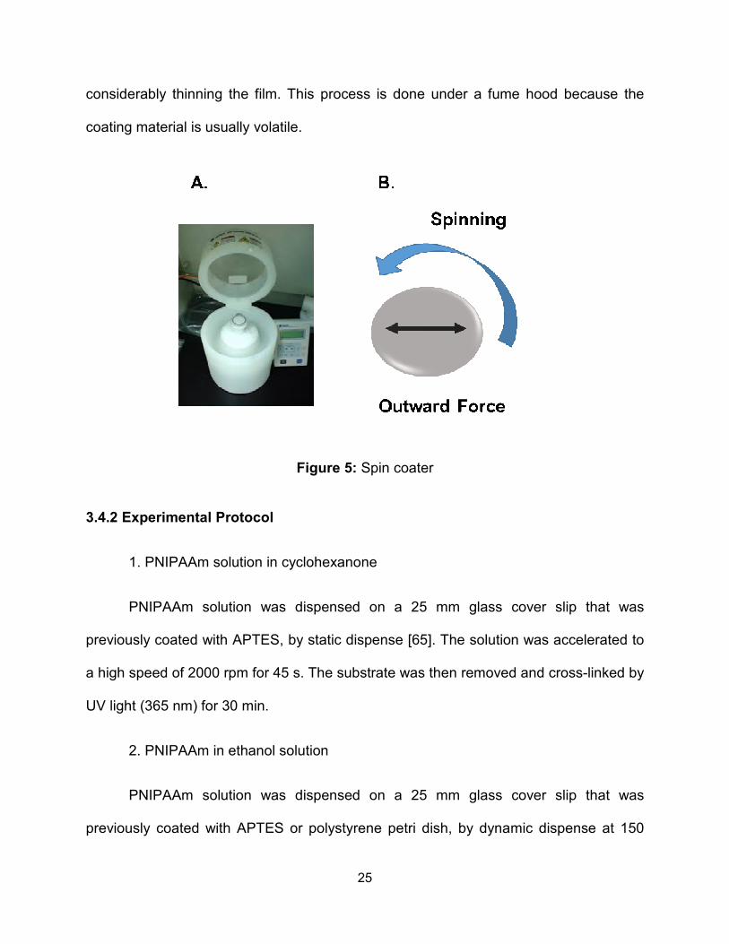
Figure 8: Fiber mat morphology and density varied with electrospinning time. The overlap of fibers resulted in contrasting surfaces: low fiber density (2D topography), medium fiber density (transition from 2D to 3D) and high fiber density (3D topography) with minimally exposed support surface. (bars = 100 μm)
PNIPAAm
APTES
PEGSAM
10 s 30 s 50 s 90 s 300 s

35
These ranges were chosen so that at low fiber coverage (<15%), the average
spacing between fibers was greater than the maximum cell size with 3% at least 10
times the area of a fully spread cell; at medium fiber coverage (15-60%), the average
spacing between fibers was greater than minimally spread cells with 9% of spaces
exceeding the maximum cell size; and at high fiber coverage (> 60%), the average
spacing was less than the cell size. The fiber densities at low and medium coverage
were utilized to study how two-dimensional spacing between physical features affects
cell adhesion and spreading. Fiber coverage above 60% forms a prominent 3D mesh-
like surface and was expected to prevent most cells from accessing the underlying
substrate.
Well characterized surface chemistries were also employed to provide a better
understanding on how topography and the underlying surface collectively regulate cell
adhesion and spreading. Statistical fiber diameter distributions in the three projected
fiber density bins were obtained in order to evaluate if the instability of the polymer
solution jet, time of electrospinning, or target surface had any effect on the fiber
diameters (Figure 9)
All fibers fell within a similar range of approximately 2-5 μm in diameter. Only the
low and medium fiber densities for APTES were found to be significantly different with p
= 0.029. This difference did not have any effect on cell adhesion or spreading (See
Chapter 5).

36
Figure 9: Average fiber diameter as a function of projected fiber density. The only statistically significant difference was between low and medium fiber densities on APTES. 4.4 Conclusions
Microstructure composite surfaces with combined non-adhesive materials were
fabricated and characterized. It was possible to incorporate two well characterized
surface chemistries, APTES and PEGSAM alongside with PNIPAAm thin film (100 nm)
with three different surface topographies provided by electrospun PNIPAAm fibers. The
fiber density was classified into low, medium and high projected fiber density bins which
corresponded to less than 15% global fiber coverage, 15% - 60% global coverage, and
greater than 60% global coverage, respectively. This classification was designated to
Projected fiber density
<15% 15-60% >60%
Ave
rag
e F
Iber D
iam
ete
r (µµ µµ m)
0
2
4
6
PNIPAAm
APTES
PEGSAM
p = 0.029

37
provide three scenarios in which the average spacing between fibers was greater than
the maximum cell size, greater than the minimally spread cells and less than the cell
size. Also, the surface topography incorporated into the microstructure composite
surface displayed micron size topography with approximately 2-5 μm in diameter range.
The microstructure fabricated will provide a variety of distinctive surface topography and
surface chemistries characteristics that will help in conducting cell adhesion studies and
elucidate how the adhesivity of surfaces that typically do not support significant cell
adhesion may be altered by the presence of topographical features, even if those
features are poorly adhesive as well.

38
CHAPTER 5: CELL ADHESION AND CELL SPREADING ON NON ADHESIVE
SURFACES
5.1 Introduction
The manner in which a cell interacts with a biomaterial is largely determined by
the physical and chemical nature of the biomaterial surface [14]. It is known, for
example, that topography and surface chemistry both affect cellular response in very
significant ways [71, 72]. Such cues on their own have provided valuable information
such as how to design surfaces that exert control over cell shape [73-76], spreading [77,
78] and adhesion [48, 78-82]. There remains, however, a lack of understanding of the
interplay between topography and surface chemistry and the relative impact and
contribution of each in modulating cellular behavior [83, 84]. A fundamental
understanding of this interplay is necessary to advance biomaterials applications
through the design of surfaces that more effectively direct cell function.
The few studies that have looked at the combined effect of surface topography
and chemical functionality indicate that cells preferentially respond to one property over
the other, but the contribution of each property is not well defined or understood [85-87].
Often the response to combinations cannot be predicted from the individual
contributions of each property [88]. For instance, several studies have investigated the
relative contribution of mechanical guidance cues with chemical guidance cues (i.e., the
effect of topographical patterns versus chemical patterns), and have found that

39
topographically induced cell alignment often dominates over alignment with chemical
patterns, when both are present on the same surface [54, 83, 84, 86, 87, 89-93]. Others
have looked at the effect of topographical cues on surfaces of uniform chemical
presentation. These reports suggest that while topography can influence cell behavior,
the underlying surface chemistry determines the relative influence of the topography
[43, 85, 94-103]. Still, no unified principles have emerged that explain the relationship
between the physical and chemical features of a surface and cell guidance.
Herein, we investigated a curious phenomenon whereby surfaces that typically
do not support significant cell adhesion may have their adhesivity altered by the
presence of topographical features, even if those features are poorly adhesive as well
(See Figure 10). Specifically, smooth poly(N-isopropylacrylamide) (PNIPAAm) thin films
or coatings (greater than 30 nm in thickness) have been shown to display poor intrinsic
cell adhesion [66], despite the presence of serum proteins [56, 58, 104, 105]. To
investigate the role of topography on such coatings, electrospun PNIPAAm fibers (1-5
um diameter) were overlaid on PNIPAAm thin films. Several topographical patterns
were developed, spanning from sparse to dense fiber mats, and cell adhesion strongly
depended on the relative available areas for attachment on either the fibers or the
supporting surface.
To understand the uniqueness of this observation, PNIPAAm fibers were also
electrospun onto a surface known to be non-adhesive (self-assembled monolayer of
polyethylene glycol (PEGSAM) alkanethiol on gold) or an adhesive coating (3-
aminopropyltriethoxysilane (APTES) on glass) to assess the effect of topographical
cues on cellular responses to chemistries with well-characterized, disparate adhesive

40
properties. Cell adhesion and spreading were enhanced on PEGSAM surfaces by the
presence of fibers up to a threshold density; and the highest fiber densities reduced
adhesion on APTES and PEGSAM coatings. Most significantly, the combination of
PNIPAAm fibers on PEGSAM surfaces, despite both materials being non-adhesive
alone, was able to produce cell attachment and spreading similar to the strongly
adhesive APTES surfaces. Together these findings point to the complex synergy
between surface chemistry and physical structure. This unique experimental design
provides new clues as to how cells adapt to their environment and also suggests new
dimensions of biomaterial design for fine-tuning cellular control.
Figure 10: Non-adhesive surface with non-adhesive surface topography

41
5.2 Materials and Methods
5.2.1 Cell Culture and Reagents
Dulbecco’s modified Eagle’s medium (Invitrogen, Carlsbad, CA) supplemented
with 10% new born calf serum (Invitrogen) and 1% penicillin-streptomycin (Invitrogen)
was used as complete growth media (CGM). Cell culture reagents, including human
plasma fibronectin and Dulbecco’s phosphate-buffered saline (DPBS), Hoechst-33242
and rhodamine-conjugated phalloidin were purchased from Invitrogen. NIH3T3 mouse
embryonic fibroblasts (American Type Culture Collection, Manassas, VA) were cultured
in CGM on tissue culture polystyrene. Cells were passaged every other day and used
between passages 5 and 20. For experiments, cells were enzymatically lifted from the
culture dish using trypsin-EDTA (Invitrogen) and then seeded onto the substrates at a
density of 100 cell/mm2 in CGM.
5.2.2 Imaging and Analysis
After incubating the cells on the substrates for 4h, the cells were rinsed in DPBS
(Dulbecco’s phosphate buffered saline) and the adherent cells were fixed in 3.7%
formaldehyde, permeabilized with 0.1% Triton X-100, and stained with Hoechst dye to
identify the nucleus and rhodamine-phalloidin to identify actin filaments. The number of
adherent cells was counted at specific positions using a Nikon eclipse Ti-U fluorescent
microscope (Nikon Instruments, Melville, N.Y.) fitted with a motorized stage and NIS-
Elements Advanced Research software (Nikon Instruments) to obtain cell attachment
quantification. A thresholding algorithm was utilized to create binary masks of cell
boundaries to quantify cell spreading area.

42
5.2.3 Statistical Analysis
Experiments were performed in triplicate in at least three independent
experiments. Data were reported as mean ± SD, and statistical comparisons using
SigmaPlot 11 (Systat Software, San Jose, CA) were based on analysis of variance and
the Holm-Sidak test for pairwise comparisons, with a p-value < 0.05 considered
significant.
5.3 Results
5.3.1 Topographic Enhancement of Cell Adhesion to PNIPAAm
This study arose from the desire to assess the interplay between surface
topography and surface chemistry on cell-material adhesion properties. We investigated
surfaces with topographical features that were generated from materials that were either
the same or different from the underlying surface. In all cases, mouse embryonic
fibroblast cells were seeded onto the substrates at a density of 100 cell/mm2 in CGM
and incubated for 4 h in culture medium prior to assessing adhesion. We were
motivated to investigate this by the initial observation that fiber-based topographical
features enhanced cell adhesion to PNIPAAm (Figure 11).
Cell attachment on smooth 100 nm thick cross-linked PNIPAAm thin films was
minimal compared to plain glass controls, demonstrating that these uniform films are not
suitable for cell adhesion. However, when the PNIPAAm thin films were overlaid with
PNIPAAm fibers of similar composition, cell adhesion was enhanced approximately 20-
fold. There was a statistical difference between the smooth 100 nm thick cross-linked
PNIPAAm thin films and both the control samples and smooth 100 nm thick cross-linked

43
PNIPAAm thin films with 30% fiber coverage. No statistical difference was observed
between the control samples and the smooth 100 nm thick cross-linked PNIPAAm thin
film with 30% fiber coverage samples. This result suggests topography alone may be
able to induce adhesion on poorly adhesive materials.
Figure 11: Cell adhesion on (A) glass, (B) PNIPAAm film and (C) PNIPAAm film covered with electrospun PNIPAAm fibers (~30% fiber density) substrates. Approximately 100 cell/mm2 were seeded on the substrates and incubated for 4h before (D) quantification with fluorescence microscopy. * indicates p < 0.05 compared to the smooth PNIPAAm film. (bars = 100 μm)

44
5.3.2 Cell Adhesion on PNIPAAm, APTES and PEGSAM Surfaces with Varied
Fiber Densities
The number of adherent cells following 4 hours in CGM was quantified at specific
positions on each substrate (Figures 12 and 13). We first analyzed the trends of cell
attachment dependence on fiber density for each underlying surface chemistry. Then
we considered the differences between the three surface chemistries for each fiber
density.
5.3.2.1 PNIPAAm
Cell adhesion on a bare PNIPAAm film with 0% fiber density was not supported
with a low number of cells adhered (<10 cells/mm²). This result is in agreement with
other studies done on a bare PNIPAAm surface [56, 58] and consistent with our initial
observations (Figure 11). However, cell adhesion was enhanced by the incorporation of
PNIPAAm fibers onto the PNIPAAm surface, and cell adhesion was maximum on the
medium fiber density (approximately 60 cells/mm2). No statistical differences were
observed among low, medium and high fiber densities; on the other hand, there was a
significant difference between bare PNIPAAm films and medium PNIPAAm fiber density
on PNIPAAm films.
5.3.2.2 APTES
As expected, cell adhesion was highly supported on the APTES control sample
with 0% fiber density, comparable to or greater than on plain glass controls. Cell
adhesion was similarly supported on low (<15%) and medium (15%-60%) fiber
coverages, indicating that cell adhesion was not affected by the non-adhesive

45
topographical features. However, by increasing fiber density above 60%, cell adhesion
was reduced by about 50%.
5.3.2.3 PEGSAM
Negligible cell adhesion was observed on the uniform PEGSAM surface (0%
fiber density). The few cells that adhered to this surface can likely be attributed to
defects in the gold or SAM layers. Similar to the PNIPAAm substrates, the incorporation
of PNIPAAm fibers on the PEG surfaces significantly enhanced cell adhesion with
maximum cell attachment occurring on the medium fiber density (approximately 85
cells/mm2). Cell adhesion was significantly greater on all fiber densities compared to
PEGSAM with no fibers.
5.3.2.4 Comparison of PNIPAAm, PEGSAM and APTES for Each Fiber Density
Cell adhesion on the topographically featureless control samples, denoted as 0%
fiber density, was markedly different between the APTES surface and the two other
surfaces. This amine-functional surface exhibited complete cell attachment compared to
the cell seeding density of approximately 100 cells/mm². However, PNIPAAm and
PEGSAM supported minimal cell adhesion (<10 cells/mm²). These adhesive properties
were consistent with our expectations based on previous reports. For the low projected
fiber density (<15%), cell adhesion on APTES was maximal, while improved but
intermediate levels of cell attachment were observed for PNIPAAm and PEGSAM. On
medium fiber density (15%-60%), cell adhesion was comparable for APTES and
PEGSAM with an average of 80-90 cells/ mm² while PNIPAAm was slightly less
(approximately 60 cells/mm²). There were no significant differences among the three

46
surfaces. For high fiber density (>60%), cell adhesion was reduced overall to
approximately 45 cells/mm² for all surface chemistries.

47
Figure 12: Fluorescence images of attached cells for all the underlying surface chemistries and fiber densities. (bars=100 μm)
.

48
Projected fiber density
0 % <15 % 15-60 % >60 %
Att
ac
he
d c
ells / m
m2
0
20
40
60
80
100
120
140 PNIPAAm
APTES
PEGSAM
B
A
C
Figure 13: (A) Cell adhesion on low (<15%), medium (15-60%) and high PNIPAAm fiber density (>60%) with PNIPAAm, APTES and PEGSAM underlying surface chemistry. Cells were seeded on the substrates with a cell density of 100 cells/mm2 and incubated for 4h. (B) Statistical comparisons for cell adhesion for the three binned fiber densities on each individual underlying surface chemistry. (C) Statistical comparisons of cell attachment among surface chemistries for each fiber density. Significant differences were identified by using ANOVA and the Holm-Sidak test for pairwise comparisons, with a p-value < 0.05 considered significant (indicated by the shaded boxes).

49
Projected fiber density
0 % <15 % 15-60 % >60 %
Cell s
pre
ad
ing
are
a (
µµ µµm2)
0
200
400
600
800
1000PNIPAAm
APTES
PEGSAM
B
A
C
Figure 14: (A) Cell spreading on low (<15%), medium (15-60%) and high PNIPAAm fiber density (>60%) with PNIPAAm, APTES and PEGSAM underlying surface chemistry. Cells were seeded on the substrates with a cell density of 100 cells/mm2 and incubated for 4h. (B) Statistical comparisons for cell spreading for the three binned fiber densities on each individual underlying surface chemistry. (C) Statistical comparisons of cell spreading among surface chemistries for each fiber density. Significant differences were identified by using ANOVA and the Holm-Sidak test for pairwise comparisons, with a p value < 0.05 considered significant (indicated by the shaded boxes). Crossed out boxes indicate low cell density (<10 cell/mm2).

50
5.3.3 Cell Spreading on PNIPAAm, APTES and PEG Surfaces with Varied Fiber
Densities
The extent of cell spreading was also analyzed for the attached cells by
quantifying the projected cell areas for each substrate and fiber density combination.
The results were analyzed with similar comparisons among the fiber densities and
surface chemistries as described above (Figure 14).
5.3.3.1 PNIPAAm
Despite the previously described enhancement to cell attachment, cell spreading
on low, medium and high PNIPAAm fiber densities upon an underlying PNIPAAm film
was not enhanced. Although a statistical difference was observed when the uniform
PNIPAAm control substrate was compared to low, medium and high fiber densities it
needs to be specified that these comparisons were to a PNIPAAm surface with a very
small number of adherent cells (<10 cells/mm²). Thus it is an unreliable comparison.
The spreading on fiber-laden PNIPAAm films was minimal with attached cells remaining
highly rounded.
5.3.3.2 APTES
Cells spread to a robust 700-750 μm² average area on low and medium fiber
density samples coated with APTES. This behavior was comparable to the uniform
APTES controls with fibers. Interestingly, for high fiber surface coverage, cell spreading
area was reduced to an average of approximately 300 μm².

51
5.3.3.3 PEG
In contrast to the PNIPAAm surfaces, the incorporation of fibers on the PEGSAM
treated surfaces showed that cell spreading could also be supported and enhanced on
a non-adhesive surface. On low and medium fiber densities, cell spreading was
enhanced reaching a maximum of nearly 700 µm² on the medium fiber coverage,
showing that a topographical change supports cell adhesion and spreading even though
the underlying substrate had a non-fouling surface that does not typically support cell
attachment or spreading. For a high projected fiber density surface (>60%) cell
spreading area was again reduced to about 300µm2. Statistical differences were
observed between the uniform PEG control substrates and the low and medium fiber
densities (no fiber controls have few adhered cells), and between the medium and high
fiber densities.
5.3.3.4 Comparison of PNIPAAm, PEG and APTES for Each Fiber Density
Uniform control samples, denoted as 0% fiber coverage samples, were included
in this study along with a range of fiber densities to investigate the hypothesis that the
addition of surface topography indeed had an effect in enhancing cell adhesion and
spreading regardless of the underlying surface chemistry. It was observed that for
uniform surfaces, cells on the APTES surface spread significantly more than on
PNIPAAm and PEGSAM surfaces, though the PNIPAAm and PEGSAM surfaces had
few attached cells (<10 cells/mm2) to analyze for spreading.
More importantly, the addition of PNIPAAm fibers to the three surface
chemistries influenced cell spreading area differently within each projected fiber density

52
group. For the low projected fiber density (<15%) group, the APTES surface showed the
highest spreading area. Surprisingly, cell spreading area was enhanced for PEGSAM
compared to the control while on the PNIPAAm it was not. All three samples were
statistically different. On the medium fiber densities (15%-60%), cell spreading
remained high on the APTES and low on the PNIPAAm, similar to the low fiber
coverage. However the cell spreading area on PEGSAM was enhanced to the level of
the APTES (approximately 700 μm2). For the highest projected fiber density fiber mats
(>60%), cell spreading area was minimal for all three surface chemistries. In other
words, the high fiber density reduced cell spreading to an area equivalent to cells on the
PNIPAAm surface, indicating that cell spreading is independent of underlying surface
chemistry for the highest density fiber mats.
5.4 Discussion
The complexity of multiple chemical or physical cues can trigger biological
responses that are distinct compared to the individual stimuli. This study focused on the
interplay of surface chemistry and physical topography as biomaterial regulators of cell
behavior in composite materials.
Platforms created by using electrospun nano- or micro-fibers may mimic
structurally the extracellular matrix of native tissues and provide cues that regulate
cellular survival or tissue regeneration [106]. We used electrospinning to vary the
projected surface coverage of the fibers to understand the cellular response to
biomaterial surface topographies ranging from two dimensional surfaces (null or low
fiber density) to three dimensional fibrous polymer scaffolds that mimic tissue

53
architecture (high fiber density). Fiber diameter varied over a small range
(approximately 2-5 µm); thus, is possible that the observed responses are unique to the
fiber size used in this study. Fiber orientation and curvature may play a role in the
enhancement of cell adhesion though these parameters were random and
unquantifiable in this design. By binning the fiber density into low, medium and high
density regions, we focused specifically on the effects of cell-fiber and cell-substrate
interactions.
Our findings showed that on non-adhesive surfaces (PNIPAAm, PEGSAM), cell
adhesion increased with increasing PNIPAAm fiber density as long as the underlying
surface was available (up to 60% fiber coverage). For both the PNIPAAm and the
PEGSAM surfaces, cell adhesion occurred primarily at the fiber/surface interface;
however, only in the case of the PEGSAM surface did the cells spread significantly
along the fiber. The same spreading was not observed for the PNIPAAm surface. At
fiber coverages above 60%, cell adhesion on both surfaces decreased and approached
approximately only 50% of the theoretical value.
The reason for the localization of the cells at the fiber/surface interface most
likely results from the ability of cells to sense and respond to curvature [107, 108].
Curvature in the cell membrane can localize integral membrane proteins, and it has
been shown that membrane scaffolding proteins, such as the Bin–amphiphysin–Rvs
(BAR) and epsin N- terminal homology (ENTH) domain superfamilies, preferentially bind
to highly curved membranes [109-112]. For instance, Fioretta et al. observed that
endothelial cells (colony forming cells and mature endothelial cells) were able to sense
the changes in the curvature of electrospun fibers due to the change in the size of the

54
fiber diameter [113]. The endothelial cells responded to the radius by orienting
themselves around the fiber, by either developing a cytoskeleton organized
circumferentially or aligning the cytoskeleton along the scaffold fiber axis with collagen
deposition aligned in the corresponding direction. Similarly, fibers could enhance
adhesion at the sites of curvature on poorly adhesive surfaces via bending the
membrane in such way that adhesion proteins are aggregated to these sites.
While fibers enhanced adhesion to the poorly adhesive surfaces, the trend was
reversed at the highest fiber densities. Furthermore, at the highest fiber densities, cell
attachment and spreading on APTES, which strongly supports cell adhesion and
spreading [68, 69], decreased to levels similar to the non-adhesive materials. We
hypothesize that because the area between the fibers for low and medium fiber
densities was comparable to or greater than a cell’s projected area, cells were provided
sufficient access to the underlying APTES surface chemistry; however, when the fiber
surface coverage was increased further, the cells primarily interacted only with
PNIPAAm fibers. In other words, the underlying surface chemistry became irrelevant as
the area between the fibers shrunk to average sizes smaller than the cells’ preferred
projected area.
Explaining the significant difference in the cell spreading on fibers between the
PEGSAM and PNIPAm surfaces is more challenging because the smooth surfaces
similarly repelled cell adhesion. Cell adhesion to synthetic biomaterials is typically
mediated by adsorbed proteins [45], and PEGSAM is known to be non-fouling (resistant
to protein adsorption) and therefore resistant to cell adhesion except where defects in
the SAM occur [114, 115]. On the other hand, proteins readily adsorb to PNIPAAm, and

55
if virgin PNIPAAm is initially incubated with adhesion molecules such as fibronectin or
poly-lysine, cell attachment improves significantly [116]. Perhaps the adsorption of
adhesive serum components onto PNIPAAm fibers on the non-fouling PEGSAM creates
a chemical heterogeneity that encourages cell spreading. This of course brings up the
interesting postulation that while topography can enhance adhesion on weakly adhesive
surfaces, chemical heterogeneity is necessary for cell spreading. The mechanism of
enhanced spreading on topographical composites may also result from the synergy of
the chemical heterogeneity and the aggregation of cell surface proteins due to the
membrane bending on the fibers or at the fiber-surface interface.
In summary, these results indicate that smooth non-adhesive materials can be
converted to cell adhesive by the addition of topographic features. Moreover, the
combination of two different non-adhesive materials in fibrous and planar forms resulted
in maximal cell adhesion and spreading. These findings have implications for the
fabrication of instructive biomaterials with greater control over cell functions using
combinations of surface chemistry, topography and composite materials with
contrasting properties. This may also be a significant step toward explaining why cell
behavior in 3-dimensional cultures is often different than on simple planar cell supports.
5.5 Conclusions
Micron diameter PNIPAAm micron fibrous scaffolds of low (<15%), medium (15-
60%) and high (>60%) density were superimposed on uniform coatings of PNIPAAm,
PEGSAM, and APTES in order to investigate the effect of topography on the adhesion
and spreading on embryonic fibroblast cells. The investigation revealed a complex

56
interplay between chemical and physical clues, especially an unexpected synergism
between two disparate and non-adhesive material surfaces. Surprisingly, the PEGSAM
surface with a medium density mat of PNIPAAm fibers was statistically equivalent to the
APTES surface with regards to both the fraction of adhered cells and the area of cell
spreading. The disparate material phases are critical. The PNIPAAm surface with a
medium density PNIPAAm had no statistical effect on spreading although it did enhance
cell attachment density compared to the smooth PNIPAAm surface. These results point
to not only the effect of topography of non-adhesive surfaces (which enhances cell
adhesion in comparison to smooth surfaces), but how chemical dissimilarity between
two non-adhesive materials can stimulate cell spreading. Taken in aggregate, these
findings could motivate the design of new composite biomaterials that regulate cell
adhesion and other functions.

57
CHAPTER 6: CELL ADHESION AND SPREADING RESPONSE ON PHOTO-
CROSSLINKED PNIPAAm THIN FILM COATINGS
6.1 Introduction
As already stated in the background section (Chapter 2) surface topography
plays a major role in modulating cellular behavior. Topography can be intentionally
created or by accident and in both cases topography can vary from a macro to a
nanometer scale. At the macromolecular level a surface may appear to be smooth, but
in fact if the surface is looked at a microscale or nanoscale level the surface may not
appear to be smooth anymore, displaying some type of topography. The scale of the
topography present on a biomaterial can have a strong effect in modulating cellular
response influencing the physicochemical interactions central to biological processes
that involve proteins and cells. Macro and micron scale topography have been
extensively studied showing that chemical functionality and topography have a strong
effect over cell shape, spreading and adhesion [117]. However, there is less knowledge
about how cells react to nanoscale topography. However, cells are prone to react to
nanostructures due to the in vivo characteristics of their surrounding
microenvironments. For example, the extracellular matrix (ECM), the protein matrix
surrounding cells in tissues, contains collagen fibrils that are of nanometer scale and the
cell’s own surface is organized on the nanoscale level, including receptors and filopodia
[118, 119]. For instance, bone tissue contains an array of collagen type I fibrils with a
spacing of 68 nm between fibers and 35 nm in depth [120].

58
This study was motivated by the work that we presented in Chapter 5 as well as
work published by other groups that showed that cell adhesion was enhanced on
PNIPAAm film with a thickness < 30 nm [66]. They believe that cell enhancement is due
to the reduction in thickness of PNIPAAm film layer. We hypothesized that this cell
adhesion enhancement is actually a result of nanoscale topography on the surface of
the PNIPAAm film. That surface topography is likely a function of the topography of the
underlying substrate. We report preliminary results on cell adhesion on various
thickness films of PNIPAAm coated on polystyrene petri dishes and glass cover slips
which have different topographies (roughness). Polystyrene petri dishes and glass
cover slips (borosilicate) were chosen for this study because they have different
roughness properties. It has been reported that the roughness of polystyrene petri
dishes varies depending on the brand between 1.5 nm to 6.5 nm. In this study we are
using Corning and the roughness corresponding to this brand is ~3.5 nm. For
borosilicate glass cover slips the roughness has been reported to be between
approximately 0.2 nm – 1.0 nm. We hypothesized that the surface roughness of the
support and the thickness of the PNIPAAm film can modulate the adhesive properties of
the film. The preliminary results suggest that the adhesive properties of this material are
enhanced and it showed a dependence on the roughness of the support and the
thickness of the film. To complete this study, we need to do further experiments (n=3)
and use AFM imaging to characterize the PNIPAAm films for both types of surfaces.

59
6.2 Materials and Methods
6.2.1 Substrate Preparation
Two types of substrates were used; round glass cover slips and polystyrene petri
dishes (25 mm). The glass cover slips were sonicated in ethanol for 15 minutes, blown
dry with a stream of nitrogen gas and oxygen plasma cleaned. No cleaning method was
used for the polystyrene petri dishes. The glass cover slip substrates were submerged
in a solution of 1.0 v/v% 3-aminopropyltriethoxysilane for 15 minutes, rinsed with
acetone and dried at 110ºC for 10 minutes [55, 68, 69].
PNIPAAm thin films were prepared following the protocol reported by Maria E.
Nash et al. [66]. A 300 mL aliquot of PNIPAAm in ethanol solution (0.125 wt %, 0.25 wt
%, 0.5 wt % and 3 wt %) was deposited onto the petri dishes and the APTES treated
glass substrate to a slowly spinning substrate. The PNIPAAm solution was initially
deposited onto each substrate at a speed of 150 rpm, followed by a high speed of 6000
rpm for 30s. The substrate with the polymer film was cross-linked by UV light (365 nm)
for 30 min. Film thickness was assessed by ellipsometry [65].
6.2.2 Cell Culture and Reagents
Dulbecco’s modified Eagle’s medium (Invitrogen, Carlsbad, CA) supplemented
with 10% new born calf serum (Invitrogen) and 1% penicillin-streptomycin (Invitrogen)
was used as complete growth media (CGM). Cell culture reagents, including human
plasma fibronectin and Dulbecco’s phosphate-buffered saline (DPBS), Hoechst-33242
and rhodamine-conjugated phalloidin were purchased from Invitrogen. NIH3T3 mouse
embryonic fibroblasts (American Type Culture Collection, Manassas, VA) were cultured
60
in CGM on tissue culture polystyrene. Cells were passaged every other day and used
between passages 5 and 20. For experiments, cells were enzymatically lifted from the
culture dish using trypsin-EDTA (Invitrogen) and then seeded onto the substrates at a
density of 100 cell/mm2 in CGM.
6.2.3 Imaging and Analysis
After incubating the cells on the substrates for 4h, the cells were rinsed in DPBS
(Dulbecco’s phosphate buffered saline) and the adherent cells were fixed in 3.7%
formaldehyde, permeabilized with 0.1% Triton X-100, and stained with Hoechst dye to
identify the nucleus and rhodamine-phalloidin to identify actin filaments. The number of
adherent cells was counted at specific positions using a Nikon eclipse Ti-U fluorescent
microscope (Nikon Instruments, Melville, N.Y.) fitted with a motorized stage and NIS-
Elements Advanced Research software (Nikon Instruments) to obtain cell attachment
quantification.
6.2.4 Statistical Analysis
The preliminary results shown in this section correspond to one experiment for
each substrate type. Once the experiments are completed, (n=3) which means that
experiments will be performed in triplicate in at least three independent experiments,
data will be reported as mean ± SD, and statistical comparisons using SigmaPlot 11
(Systat Software, San Jose, CA) will be based on analysis of variance and the Holm-
Sidak test for pairwise comparisons, with a p-value < 0.05 considered significant.

61
6.3 Results
6.3.1 Topographic Enhancement of Cell Adhesion to PNIPAAm Thin Films
A range of PNIPAAm film thicknesses was created by varying the polymer
concentration while keeping the spin coating recipe constant. The thickness for films
formed from each PNIPAAm solution concentration was measured by ellipsometry and
the results are reported in Table 2. The number of adherent cells following 4 hours in
CGM was quantified at specific positions on each substrate (Figures 15 and 16). We
first analyzed the trends of cell attachment dependence on PNIPAAm film thickness for
each type of substrate and compared them to cell attachment on the controls
substrates, bare glass and polystyrene petri dish. Then we considered the differences
between the petri dishes and glass cover slips. The results reported in this section are
preliminary results and further experiments will be conducted to support data and a
statistical analysis will be executed.
6.3.1.1 Polystyrene Petri Dish Film Supports
Preliminary results suggest that cell adhesion and spreading is enhanced as
PNIPAAm film thickness is reduced. PNIPAAm film with a thickness of 158 nm exhibits
a minimal cell adhesion (<10 cells/mm²). The non-adhesive property for this thickness
was consistent with our expectations based on previous reports. Cell adhesion on the
PNIPAAm films with thicknesses 7 nm, 10 nm and 20 nm was enhanced, and it is
equivalent to the control sample.

62
6.3.1.2 Glass Cover Slip Film Supports
Similarly, PNIPAAm films with a thickness of 158 nm exhibits minimal cell
adhesion (<10 cells/mm²) upon a glass support and this is consistent with previous
reports. Preliminary results suggest that cell adhesion is only slightly enhanced for
PNIPAAm films in the 10-20nm range, and reached maximum attachment on 7 nm thick
films.
6.3.1.3 Comparison of Petri Dish with Glass Cover Slip Underlying Film Supports
Cell adhesion on both petri dish and glass cover slip control samples showed
similar cell adhesion, and it compared to the cell seeding density of approximately 100
cells/mm². For the samples with high thickness (158 nm), both surfaces showed minimal
cell adhesion (<10 cells/mm2) and the values were similar to each other. There is a
marked contrast in cell adhesion between the petri dish and the glass cover slip
samples with PNIPAAm film thickness of 10 nm and 20 nm. In both cases cell adhesion
levels on glass cover slip, were lower (intermediate levels) when compared to the petri
dish values. Cell adhesion on the thinnest PNIPAAm film (7nm) was greatly enhanced
for the glass cover slip sample and the value was comparable to the number of cells
attached on the petri dish sample.

63
Table 2: PNIPAAm thickness (nm)
Figure 15: Cell adhesion on PNIPAAm films. Cells were seeded on the substrates with a cell density of 100 cells/mm2 and incubated for 4h.
PNIPAAm Solution Concentration (wt %) Thickness (nm)
0.125 7
0.25 10
0.5 20
3 158

64
Figure 16: Fluorescence images of attached cells on PNIPAAm thin films with petri dish and glass cover slip underlying film supports (bars=100 μm)

65
6.4 Discussion
To test the hypothesis that nanoscale topography can enhance cell adhesion on
PNIPAAm thin films, we prepared PNIPAAm thin films with controlled thickness by
varying the concentration of the solution deposited during the spin coating process. Our
preliminary results suggest that cell adhesion increased as the PNIPAAm film thickness
decreased, with a minimal cell attachment on PNIPAAm films with thickness of 158 nm.
Cell adhesion was markedly enhanced on both petri dish and glass cover slip on the
7nm PNIPAAM thin film samples. Even though we see a contrast on cell attachment
between petri dishes and glass cover slips, within the same thickness group we can say
that cell adhesion was enhanced and there is a threshold in PNIPAAm thickness in
which PNIPAAm adhesive properties are modulated.
To further understand and support our hypothesis we need to complete at least
three experiments for each condition (n=3). Also, Atomic Force Microscopy Imaging and
surface roughness measurements would be a suitable way to obtain information about
the surface topography of the films to support our hypothesis.
6.5 Conclusions
We believe that the adhesivity of PNIPAAm thin films is modulated by nanoscale
topography present on the surface of the PNIPAAm layer. Thick films will be smooth
while the surface topography becomes more similar to the underlying support as the
thickness of the PNIPAAm film decreases. Further experiments are expected to support
this hypothesis that the transition from non-adhesive to adhesive for PNIPAAm is
triggered by the presence of nanoscale topography. Cells may sense this topography by

66
changes in protein adsorption or local curvature in their membranes [109-112, 121,
122]. Whatever the mechanism, this topographic effect explains the dependence of cell
adhesion to PNIPAAm on film thickness which cells cannot directly sense.
67
CHAPTER 7: CONCLUSIONS AND FUTURE WORK
7.1 Conclusions
The key findings in this dissertation are summarized as follows:
1. A stable and reproducible microstructure with combined topography and
surface chemistry of non-adhesive materials was successfully fabricated for
cellular response studies.
2. Adhesive properties of non-adhesive materials were enhanced by the
presence of microscale topography, even when the material used to create the
topography was poorly adhesive as well.
3. The electrospun fiber density that created the microscale topography showed
a thresholding factor (>60%) independent of the underlying surface chemistry, in
which cell adhesion and spreading was no longer supported.
4. Medium fiber density (15% - 60%) with PEGSAM underlying surface chemistry
showed cell adhesion enhancement similar to the APTES adhesive coating and
cell seeding density (100 cells/mm2).
5. Low and medium fiber density on PNIPAAm thin film layer (100nm) enhanced
cell adhesion but cell spreading response was minimal; and unchanged.

68
6. Preliminary results of cell adhesion on PNIPAAm thin film layers, spin coated
on petri dishes and glass cover slip is enhanced as the thickness of the film is
reduced.
7. Cell adhesion on the thickest PNIPAAm film for both petri dish and glass cover
slip was minimal and in agreement with previously reported results
8. PNIPAAm films spin coated on petri dishes showed higher cellular response
when compared to the same PNIPAAm film thicknesses spin coated on glass
cover slips.
9. Further experiments will support preliminary results showing that nanoscale
topography enhances cell adhesion on PNIPAAm thin film and not the film
thickness.
Physical properties of the surface of PNIPAAm, a poorly adhesive material under
many conditions, can modulate cell adhesion. We observed that topographic features in
the form of overlaid micron scale fiber mats or nanoscale textures supporting thin films
were able to support cell adhesion similar to highly adhesive materials. In contrast,
smooth films repelled cell adhesion. It was shown that there is a dependence on the
fiber density of the overlaid micron fiber PNIPAAm mats and the PNIPAAm film
thickness (higher the thickness the smother the film becomes) in the adhesive
properties of PNIPAAm. A fiber density below 60% of overlaid micron scale fiber mats
on a non-adhesive material enhanced the adhesive properties of the material, showing
maximum enhancement when the materials are of different surface chemistries. As for
the PNIPAAm films, it was shown that the underlying roughness of the supporting

69
material affect the adhesive properties of PNIPAAm film differently depending on the
thickness of the film. The roughness of the substrate used, can modulate and enhance
the adhesive properties of a poorly adhesive material to some extent. A combination of
the substrate roughness and the film thickness can exert control over cell adhesion.
It can be concluded that surface topography matters, and it can enhance the
adhesive properties of a poorly adhesive material comparable to an adhesive surface.
By controlling the substrate used, the PNIPAAm film thickness and the fiber density of
the overlaid fiber PNIPAAm mats cell adhesion can be modulated. These results will
help in the design of new biomaterials that required control over cell adhesion
properties.
7.2 Future Directions
The proposed work will help elucidate some of the findings within this work.
1. Evaluate the mechanism that promotes cell adhesion on non-adhesive
materials
a. Quantification of protein adsorption on PNIPAAm electrospun fibers.
2. Evaluate cell adhesion enhancement on PNIPAAm thin films
a. Further experiments on both type of surfaces, petri dish and glass cover
slip will support the preliminary results.
b. Statistical analysis will be done to show any difference between the two
types of surfaces and within each group.

70
3. Atomic force microscopy (AFM) will provide qualitative and quantitative
information regarding PNIPAAm surface topography (roughness).

71
REFERENCES
1. Gattazzo, F., A. Urciuolo, and P. Bonaldo, Extracellular matrix: A dynamic
microenvironment for stem cell niche(). Biochimica et Biophysica Acta, 2014.
1840(8): p. 2506-2519.
2. Anselme, K., L. Ploux, and A. Ponche, Cell/Material Interfaces: Influence of
Surface Chemistry and Surface Topography on Cell Adhesion. Journal of
Adhesion Science and Technology, 2010. 24(5): p. 831-852.
3. Anderson, J.M., A. Rodriguez, and D.T. Chang, FOREIGN BODY REACTION
TO BIOMATERIALS. Seminars in immunology, 2008. 20(2): p. 86-100.
4. Navarro, M., et al., Biomaterials in orthopaedics. Vol. 5. 2008. 1137-1158.
5. Burkert, S., et al., Protein Resistance of PNIPAAm Brushes: Application to
Switchable Protein Adsorption. Langmuir, 2010. 26(3): p. 1786-1795.
6. Akiyama, Y., et al., Ultrathin poly(N-isopropylacrylamide) grafted layer on
polystyrene surfaces for cell adhesion/detachment control. Langmuir, 2004.
20(13): p. 5506-11.
7. Fukumori, K., et al., Temperature-responsive glass coverslips with an ultrathin
poly(N-isopropylacrylamide) layer. Acta Biomater, 2009. 5(1): p. 470-6.
8. Fukumori, K., et al., Characterization of Ultra-Thin Temperature-Responsive
Polymer Layer and Its Polymer Thickness Dependency on Cell
Attachment/Detachment Properties. Macromolecular Bioscience, 2010. 10(10): p.
1117-1129.

72
9. Canavan, H.E., et al., Surface characterization of the extracellular matrix
remaining after cell detachment from a thermoresponsive polymer. Langmuir,
2005. 21(5): p. 1949-55.
10. Canavan, H.E., et al., Comparison of Native Extracellular Matrix with Adsorbed
Protein Films Using Secondary Ion Mass Spectrometry. Langmuir, 2007. 23(1):
p. 50-56.
11. Xue, C., et al., Protein Adsorption on Poly(N-isopropylacrylamide) Brushes:
Dependence on Grafting Density and Chain Collapse. Langmuir, 2011. 27(14): p.
8810-8818.
12. Williams, D.F., The Williams Dictionary of Biomaterials. 1999, Liverpool:
Liverpool University Press.
13. Roach, P., et al., Modern biomaterials: a review—bulk properties and
implications of surface modifications. 2007, Springer Netherlands. p. 1263-1277.
14. Hubbell, J.A., Biomaterials in Tissue Engineering. Nat Biotech, 1995. 13(6): p.
565-576.
15. Chan, B.P. and K.W. Leong, Scaffolding in tissue engineering: general
approaches and tissue-specific considerations. European Spine Journal, 2008.
17(Suppl 4): p. 467-479.
16. Kirchhof, K. and T. Groth, Surface modification of biomaterials to control
adhesion of cells. Clin Hemorheol Microcirc, 2008. 39(1-4): p. 247-51.
17. Bacakova, L., et al., Cell adhesion on artificial materials for tissue engineering.
Physiol Res, 2004. 53 Suppl 1: p. S35-45.

73
18. Cook, A.D., et al., Characterization and development of RGD-peptide-modified
poly(lactic acid-co-lysine) as an interactive, resorbable biomaterial. J Biomed
Mater Res, 1997. 35(4): p. 513-23.
19. Kim, J.H. and S.C. Kim, PEO-grafting on PU/PS IPNs for enhanced blood
compatibility--effect of pendant length and grafting density. Biomaterials, 2002.
23(9): p. 2015-25.
20. O'Brien, F.J., Biomaterials & scaffolds for tissue engineering. Materials Today,
2011. 14(3): p. 88-95.
21. Lam, M.T. and J.C. Wu, Biomaterial applications in cardiovascular tissue repair
and regeneration. Expert review of cardiovascular therapy, 2012. 10(8): p. 1039-
1049.
22. Eun, S.C., Composite Tissue Allotransplantation Immunology. Archives of Plastic
Surgery, 2013. 40(2): p. 141-153.
23. Langer, R. and J.P. Vacanti, Tissue engineering. Science, 1993. 260(5110): p.
920-6.
24. Cleveland Clinic, Treatments and Procedures. 1995 -2013.
25. Lundbæk, J.A., et al., Lipid bilayer regulation of membrane protein function:
gramicidin channels as molecular force probes. Journal of The Royal Society
Interface, 2010. 7(44): p. 373-395.
26. Shapiro, L. and W.I. Weis, Structure and Biochemistry of Cadherins and
Catenins. Cold Spring Harbor Perspectives in Biology, 2009. 1(3): p. a003053.
27. De Arcangelis, A. and E. Georges-Labouesse, Integrin and ECM functions: roles
in vertebrate development. Trends in Genetics, 2000. 16(9): p. 389-395.

74
28. Schwartz, M.A., Integrins and extracellular matrix in mechanotransduction. Cold
Spring Harb Perspect Biol, 2010. 2(12): p. a005066.
29. Gallant, N.D., K.E. Michael, and A.J. García, Cell Adhesion Strengthening:
Contributions of Adhesive Area, Integrin Binding, and Focal Adhesion Assembly.
Molecular Biology of the Cell, 2005. 16(9): p. 4329-4340.
30. Wilson, C.J., et al., Mediation of biomaterial-cell interactions by adsorbed
proteins: a review. Tissue Eng, 2005. 11(1-2): p. 1-18.
31. Berrier, A.L. and K.M. Yamada, Cell-matrix adhesion. J Cell Physiol, 2007.
213(3): p. 565-73.
32. Chou, L., et al., Substratum surface topography alters cell shape and regulates
fibronectin mRNA level, mRNA stability, secretion and assembly in human
fibroblasts. Journal of cell science, 1995. 108(4): p. 1563-1573.
33. Chai, C. and K.W. Leong, Biomaterials Approach to Expand and Direct
Differentiation of Stem Cells. Molecular therapy : the journal of the American
Society of Gene Therapy, 2007. 15(3): p. 467-480.
34. Curtis, A. and C. Wilkinson, Topographical control of cells. Biomaterials, 1997.
18(24): p. 1573-1583.
35. Hoffman-Kim, D., J.A. Mitchel, and R.V. Bellamkonda, Topography, Cell
Response, and Nerve Regeneration. Annual review of biomedical engineering,
2010. 12: p. 203-231.
36. Biggs, M.J.P., R.G. Richards, and M.J. Dalby, Nanotopographical modification: a
regulator of cellular function through focal adhesions. Nanomedicine :
nanotechnology, biology, and medicine, 2010. 6(5): p. 619-633.

75
37. RG., H., The cultivation of tissues in extraneous medium as a method of
morphogenetic study. Anat Rec., 1912. 6: p. 181-193.
38. Weiss, P., The problem of specificity in growth and development. Yale J Biol
Med, 1947. 19(3): p. 235-78.
39. Charest, J.L., et al. Polymer cell culture substrates with combined
nanotopographical patterns and micropatterned chemical domains. AVS.
40. Ghibaudo, M., et al., Substrate Topography Induces a Crossover from 2D to 3D
Behavior in Fibroblast Migration. Biophysical Journal, 2009. 97(1): p. 357-368.
41. Chew, S.Y., et al., The effect of the alignment of electrospun fibrous scaffolds on
Schwann cell maturation. Biomaterials, 2008. 29(6): p. 653-661.
42. Chang, H.-I. and Y. Wang, Cell Responses to Surface and Architecture of Tissue
Engineering Scaffolds. Regenerative Medicine and Tissue Engineering - Cells
and Biomaterials. 2011.
43. Zinger, O., et al., Differential regulation of osteoblasts by substrate
microstructural features. Biomaterials, 2005. 26(14): p. 1837-1847.
44. Xu, L.-C. and C.A. Siedlecki, Effects of surface wettability and contact time on
protein adhesion to biomaterial surfaces. Biomaterials, 2007. 28(22): p. 3273-
3283.
45. Keselowsky, B.G., D.M. Collard, and A.J. Garcıa, Surface chemistry modulates
focal adhesion composition and signaling through changes in integrin binding.
Biomaterials, 2004. 25(28): p. 5947-5954.

76
46. Keselowsky, B.G., D.M. Collard, and A.J. Garci-a, Integrin binding specificity
regulates biomaterial surface chemistry effects on cell differentiation.
Proceedings of the National Academy of Sciences, 2005. 102(17): p. 5953-5957.
47. Zhang, M., et al., Properties and biocompatibility of chitosan films modified by
blending with PEG. Biomaterials, 2002. 23(13): p. 2641-2648.
48. Llopis-Hernández, V., et al., Role of Surface Chemistry in Protein Remodeling at
the Cell-Material Interface. PLoS ONE, 2011. 6(5): p. e19610.
49. Goddard, J.M. and J.H. Hotchkiss, Polymer surface modification for the
attachment of bioactive compounds. Progress in Polymer Science, 2007. 32(7):
p. 698-725.
50. Zhu, X., et al., Electrospun Fibrous Mats with High Porosity as Potential
Scaffolds for Skin Tissue Engineering. Biomacromolecules, 2008. 9(7): p. 1795-
1801.
51. Balcells, M. and E.R. Edelman, Effect of pre-adsorbed proteins on attachment,
proliferation, and function of endothelial cells. Journal of cellular physiology,
2002. 191(2): p. 155-161.
52. Jäger, M., et al., Significance of Nano- and Microtopography for Cell-Surface
Interactions in Orthopaedic Implants. 2007.
53. Garcia, A.J., M.D. Vega, and D. Boettiger, Modulation of Cell Proliferation and
Differentiation through Substrate-dependent Changes in Fibronectin
Conformation. Molecular biology of the cell, 1999. 10(3): p. 785-798.

77
54. Tan, J. and W.M. Saltzman, Topographical control of human neutrophil motility
on micropatterned materials with various surface chemistry. Biomaterials, 2002.
23(15): p. 3215-3225.
55. Vidyasagar, A., J. Majewski, and R. Toomey, Temperature Induced Volume-
Phase Transitions in Surface-Tethered Poly(N-isopropylacrylamide) Networks.
Macromolecules, 2008. 41(3): p. 919-924.
56. Haraguchi, K., T. Takehisa, and M. Ebato, Control of Cell Cultivation and Cell
Sheet Detachment on the Surface of Polymer/Clay Nanocomposite Hydrogels.
Biomacromolecules, 2006. 7(11): p. 3267-3275.
57. Selezneva, I.I., A.V. Gorelov, and Y.A. Rochev, Use of thermosensitive polymer
material on the basis of N-isopropylacrylamide and N-tert-butylacrylamide
copolymer in cell technologies. Bulletin of Experimental Biology and Medicine,
2006. 142(4): p. 538-541.
58. Moran, M.T., et al., Cell growth and detachment from protein-coated PNIPAAm-
based copolymers. Journal of Biomedical Materials Research Part A, 2007.
81A(4): p. 870-876.
59. Teo, W.E. and S. Ramakrishna, A review on electrospinning design and
nanofibre assemblies. Nanotechnology, 2006. 17(14): p. R89.
60. Bhardwaj, N. and S.C. Kundu, Electrospinning: A fascinating fiber fabrication
technique. Biotechnology Advances, 2010. 28(3): p. 325-347.
61. Larsen, G., R. Spretz, and R. Velarde-Ortiz, Use of Coaxial Gas Jackets to
Stabilize Taylor Cones of Volatile Solutions and to Induce Particle-to-Fiber
Transitions. Advanced Materials, 2004. 16(2): p. 166-169.

78
62. Wilm, M.S. and M. Mann, Electrospray and Taylor-Cone theory, Dole's beam of
macromolecules at last? International Journal of Mass Spectrometry and Ion
Processes, 1994. 136(2–3): p. 167-180.
63. Spin Coating Theory. 1997 - 2015 [cited 2015 June 18]; Available from:
http://www.brewerscience.com/spin-coating-theory.
64. Hall, D.B., P. Underhill, and J.M. Torkelson, Spin coating of thin and ultrathin
polymer films. Polymer Engineering & Science, 1998. 38(12): p. 2039-2045.
65. Patra, L. and R. Toomey, Viscoelastic Response of Photo-Cross-Linked Poly(N-
isopropylacrylamide) Coatings by QCM-D. Langmuir, 2010. 26(7): p. 5202-5207.
66. Nash, M.E., et al., Ultra-thin spin coated crosslinkable hydrogels for use in cell
sheet recovery-synthesis, characterisation to application. Soft Matter, 2012.
8(14): p. 3889-3899.
67. Lannutti, J., et al., Electrospinning for tissue engineering scaffolds. Materials
Science and Engineering: C, 2007. 27(3): p. 504-509.
68. Kuddannaya, S., et al., Surface Chemical Modification of Poly(dimethylsiloxane)
for the Enhanced Adhesion and Proliferation of Mesenchymal Stem Cells. ACS
Applied Materials & Interfaces, 2013. 5(19): p. 9777-9784.
69. Patel, N.G., et al., Rapid cell sheet detachment using spin-coated pNIPAAm films
retained on surfaces by an aminopropyltriethoxysilane network. Acta
Biomaterialia, 2012. 8(7): p. 2559-2567.
70. Elineni, Kranthi K. and Nathan D. Gallant, Regulation of Cell Adhesion Strength
by Peripheral Focal Adhesion Distribution. Biophysical Journal, 2011. 101(12): p.
2903-2911.

79
71. Anderson, D.G., et al., Biomaterial microarrays: rapid, microscale screening of
polymer–cell interaction. Biomaterials, 2005. 26(23): p. 4892-4897.
72. Martino, S., et al., Stem cell-biomaterial interactions for regenerative medicine.
Biotechnology Advances, 2012. 30(1): p. 338-351.
73. Gilchrist, C.L., et al., Micro-scale and meso-scale architectural cues cooperate
and compete to direct aligned tissue formation. Biomaterials, 2014. 35(38): p.
10015-24.
74. Chou, L., et al., Substratum surface topography alters cell shape and regulates
fibronectin mRNA level, mRNA stability, secretion and assembly in human
fibroblasts. J Cell Sci, 1995. 108 ( Pt 4): p. 1563-73.
75. van Dongen, S.F.M., et al., Triggering Cell Adhesion, Migration or Shape Change
with a Dynamic Surface Coating. Advanced Materials, 2013. 25(12): p. 1687-
1691.
76. Heath, D.E., J.J. Lannutti, and S.L. Cooper, Electrospun scaffold topography
affects endothelial cell proliferation, metabolic activity, and morphology. Journal
of Biomedical Materials Research Part A, 2010. 94A(4): p. 1195-1204.
77. Cavalcanti-Adam, E.A., et al., Cell spreading and focal adhesion dynamics are
regulated by spacing of integrin ligands. Biophys J, 2007. 92(8): p. 2964-74.
78. Chen, W., et al., Nanotopography Influences Adhesion, Spreading, and Self-
Renewal of Human Embryonic Stem Cells. ACS Nano, 2012. 6(5): p. 4094-4103.
79. Li, S., et al., Protein adsorption and cell adhesion controlled by the surface
chemistry of binary perfluoroalkyl/oligo(ethylene glycol) self-assembled
monolayers. Journal of Colloid and Interface Science, 2013. 402(0): p. 284-290.

80
80. Kim, S.H., et al., Correlation of proliferation, morphology and biological
responses of fibroblasts on LDPE with different surface wettability. J Biomater
Sci Polym Ed, 2007. 18(5): p. 609-22.
81. Yen, B.T., et al., A comparison of the effects of fibre alignment of smooth and
textured fibres in electrospun membranes on fibroblast cell adhesion. Biomedical
Materials, 2010. 5(2): p. 025005.
82. Chen, M., et al., Role of electrospun fibre diameter and corresponding specific
surface area (SSA) on cell attachment. Journal of Tissue Engineering and
Regenerative Medicine, 2009. 3(4): p. 269-279.
83. Charest, J.L., et al., Combined microscale mechanical topography and chemical
patterns on polymer cell culture substrates. Biomaterials, 2006. 27(11): p. 2487-
2494.
84. Charest, J.L., et al., Polymer cell culture substrates with combined
nanotopographical patterns and micropatterned chemical domains. Journal of
Vacuum Science & Technology B, 2005. 23(6): p. 3011-3014.
85. dos Santos, E.A., et al., Chemical and topographical influence of hydroxyapatite
and β-tricalcium phosphate surfaces on human osteoblastic cell behavior.
Journal of Biomedical Materials Research Part A, 2009. 89A(2): p. 510-520.
86. Britland, S., et al., Synergistic and Hierarchical Adhesive and Topographic
Guidance of BHK Cells. Experimental Cell Research, 1996. 228(2): p. 313-325.
87. Britland, S., et al., Morphogenetic guidance cues can interact synergistically and
hierarchically in steering nerve cell growth, in EBO — Experimental Biology
Online Annual 1996/97. 1998, Springer Berlin Heidelberg. p. 15-34.
81
88. Ponche, A., M. Bigerelle, and K. Anselme, Relative influence of surface
topography and surface chemistry on cell response to bone implant materials.
Part 1: Physico-chemical effects. Proceedings of the Institution of Mechanical
Engineers, Part H: Journal of Engineering in Medicine, 2010. 224(12): p. 1471-
1486.
89. Gallant, N.D., et al., Micro- and Nano-Patterned Substrates to Manipulate Cell
Adhesion. Journal of Nanoscience and Nanotechnology, 2007. 7(3): p. 803-807.
90. Wang, Y.C. and C.-C. Ho, Micropatterning of proteins and mammalian cells on
biomaterials. The FASEB Journal, 2004.
91. Den Braber, E.T., et al., Effect of parallel surface microgrooves and surface
energy on cell growth. Journal of Biomedical Materials Research, 1995. 29(4): p.
511-518.
92. Lim, J.Y. and H.J. Donahue, Cell sensing and response to micro- and
nanostructured surfaces produced by chemical and topographic patterning.
Tissue Eng, 2007. 13(8): p. 1879-91.
93. Shi, J., et al., Incorporating Protein Gradient into Electrospun Nanofibers As
Scaffolds for Tissue Engineering. ACS Applied Materials & Interfaces, 2010. 2(4):
p. 1025-1030.
94. Ber, S., G. Torun Köse, and V. Hasırcı, Bone tissue engineering on patterned
collagen films: an in vitro study. Biomaterials, 2005. 26(14): p. 1977-1986.
95. Jin Ho, L., et al., Interaction of fibroblasts on polycarbonate membrane surfaces
with different micropore sizes and hydrophilicity. Journal of Biomaterials Science,
Polymer Edition, 1999. 10(3): p. 283-294.

82
96. Hallab, N.J., et al., Evaluation of metallic and polymeric biomaterial surface
energy and surface roughness characteristics for directed cell adhesion. Tissue
Eng, 2001. 7(1): p. 55-71.
97. Wirth, C., et al., Biomaterial surface properties modulate in vitro rat calvaria
osteoblasts response: Roughness and or chemistry? Materials Science and
Engineering: C, 2008. 28(5–6): p. 990-1001.
98. Ponsonnet, L., et al., Effect of surface topography and chemistry on adhesion,
orientation and growth of fibroblasts on nickel–titanium substrates. Materials
Science and Engineering: C, 2002. 21(1–2): p. 157-165.
99. Im, B.J., et al., Texture direction of combined microgrooves and submicroscale
topographies of titanium substrata influence adhesion, proliferation, and
differentiation in human primary cells. Archives of Oral Biology, 2012. 57(7): p.
898-905.
100. Lee, S.W., et al., Influence of microgroove dimension on cell behavior of human
gingival fibroblasts cultured on titanium substrata. Clin Oral Implants Res, 2009.
20(1): p. 56-66.
101. Andersson, A.-S., et al., Nanoscale features influence epithelial cell morphology
and cytokine production. Biomaterials, 2003. 24(20): p. 3427-3436.
102. Cyster, L.A., et al., The effect of surface chemistry and structure of titanium
nitride (TiN) films on primary hippocampal cells. Biomolecular Engineering, 2002.
19(2–6): p. 171-175.

83
103. Cyster, L.A., et al., The effect of surface chemistry and nanotopography of
titanium nitride (TiN) films on primary hippocampal neurones. Biomaterials, 2004.
25(1): p. 97-107.
104. Akiyama, Y., et al., Ultrathin Poly(N-isopropylacrylamide) Grafted Layer on
Polystyrene Surfaces for Cell Adhesion/Detachment Control. Langmuir, 2004.
20(13): p. 5506-5511.
105. Mizutani, A., et al., Preparation of thermoresponsive polymer brush surfaces and
their interaction with cells. Biomaterials, 2008. 29(13): p. 2073-2081.
106. Lyu, S., et al., Electrospun Fibers as a Scaffolding Platform for Bone Tissue
Repair. Journal of orthopaedic research : official publication of the Orthopaedic
Research Society, 2013. 31(9): p. 1382-1389.
107. Vogel, V. and M. Sheetz, Local force and geometry sensing regulate cell
functions. Nat Rev Mol Cell Biol, 2006. 7(4): p. 265-275.
108. Rumpler, M., et al., The effect of geometry on three-dimensional tissue growth.
Journal of the Royal Society Interface, 2008. 5(27): p. 1173-1180.
109. Rao, Y. and V. Haucke, Membrane shaping by the Bin/amphiphysin/Rvs (BAR)
domain protein superfamily. Cellular and Molecular Life Sciences, 2011. 68(24):
p. 3983-3993.
110. Hsieh, W.-T., et al., Curvature Sorting of Peripheral Proteins on Solid-Supported
Wavy Membranes. Langmuir, 2012. 28(35): p. 12838-12843.
111. Itoh, T. and T. Takenawa, Mechanisms of membrane deformation by lipid-binding
domains. Progress in Lipid Research, 2009. 48(5): p. 298-305.

84
112. McMahon, H.T. and J.L. Gallop, Membrane curvature and mechanisms of
dynamic cell membrane remodelling. Nature, 2005. 438(7068): p. 590-596.
113. Fioretta, E.S., et al., Differential Response of Endothelial and Endothelial Colony
Forming Cells on Electrospun Scaffolds with Distinct Microfiber Diameters.
Biomacromolecules, 2014. 15(3): p. 821-829.
114. Cheng, N. and X. Cao, Photoactive SAM surface for control of cell attachment.
Journal of Colloid and Interface Science, 2010. 348(1): p. 71-79.
115. Gallant, N.D., et al., Micropatterned Surfaces to Engineer Focal Adhesions for
Analysis of Cell Adhesion Strengthening. Langmuir, 2002. 18(14): p. 5579-5584.
116. Akintewe, O.O., et al., Shape-changing hydrogel surfaces trigger rapid release of
patterned tissue modules. Acta Biomaterialia, 2015. 11(0): p. 96-103.
117. Anselme, K., et al., The interaction of cells and bacteria with surfaces structured
at the nanometre scale. Acta Biomaterialia, 2010. 6(10): p. 3824-3846.
118. Brody, S., et al., Characterizing nanoscale topography of the aortic heart valve
basement membrane for tissue engineering heart valve scaffold design. Tissue
Eng, 2006. 12(2): p. 413-21.
119. Karuri, N.W., et al., Biological length scale topography enhances cell-substratum
adhesion of human corneal epithelial cells. Journal of Cell Science, 2004.
117(15): p. 3153-3164.
120. Antipova, O. and J.P.R.O. Orgel, In Situ D-periodic Molecular Structure of Type II
Collagen. Journal of Biological Chemistry, 2010. 285(10): p. 7087-7096.

85
121. Hosseini, V., et al., Fiber-Assisted Molding (FAM) of Surfaces with Tunable
Curvature to Guide Cell Alignment and Complex Tissue Architecture. Small,
2014. 10(23): p. 4851-4857.
122. Ozdemir, T., et al., Substrate curvature sensing through Myosin IIa upregulates
early osteogenesis. Integrative Biology, 2013. 5(11): p. 1407-1416.